Embed Size (px)
Citation preview
GE J Port Gastrenterol. 2013;20(4):172---176
www.elsevier.pt/ge
CLINICAL CASE
Trousseau’s syndrome due to asymptomatic pancreaticadenocarcinoma
António Murinelloa,∗, Pedro Guedesa, Gizela Rochab, Ana Serranoa,António Figueiredoa, Helena Damásioa, João Freireb, Fernando Cunhac, Liliana Alvesa
a Servico de Medicina Interna 1, Hospital de Curry Cabral, Lisboa, Portugalb Servico de Oncologia Médica, Instituto Português de Oncologia de Lisboa de Francisco Gentil, Lisboa, Portugalc Laboratório de Citologia Aspirativa do Servico de Anatomia Patológica, Instituto Português de Oncologia de Lisboa de FranciscoGentil, Lisboa, Portugal
Received 7 August 2012; accepted 11 September 2012Available online 11 January 2013
KEYWORDSTrousseau’ssyndrome;Migratorythrombophlebitis;Heparin;Occult malignancy;Pancreaticadenocarcinoma
Abstract The authors report a case of Trousseau’s syndrome presenting in a previously asymp-tomatic 58-year-old man diagnosed with pancreatic adenocarcinoma and liver metastasesduring a workup prompted by migratory venous thrombosis and pulmonary embolism. It wasfollowed by an ischaemic stroke that occurred while the patient was just one day off antico-agulant therapy with low-molecular-weight heparin to allow for liver and pancreatic biopsies.Trousseau’s syndrome is defined by recurrent or migratory venous thrombosis, arterial embolismcaused by non-bacterial thrombotic endocarditis, or both, in patients with underlying malig-nancy. Treatment relies on the lifelong administration of heparin, and its interruption --- howeverbrief --- may promote new thrombotic events.© 2012 Sociedade Portuguesa de Gastrenterologia Published by Elsevier España, S.L. All rightsreserved.
PALAVRAS CHAVESíndrome deTrousseau;Tromboflebitemigratória;Heparina;Neoplasia malignaoculta;Adenocarcinomapancreático
Síndrome de Trousseau por adenocarcinoma do pâncreas assintomático
Resumo Os autores relatam um caso de Síndrome de Trousseau, manifestado por trom-bose venosa migratória e embolia pulmonar, num doente de 58 anos assintomático até àdata de internamento. O estudo desencadeado revela adenocarcinoma do pâncreas commetástases hepáticas, e a situacão torna-se ainda mais grave após a ocorrência de um aci-dente vascular cerebral isquémico, aparentemente em relacão com a paragem por 24 horasda terapêutica anti-coagulante com heparina de baixo peso molecular para realizacão debiópsia hepática e pancreática guiadas por exame de imagem. A síndrome de Trousseaudefine-se por tromboses venosas recorrentes ou migratórias, embolias arteriais causadaspor endocardite trombótica não-bacteriana, ou ambas, em doentes com neoplasia maligna
∗ Corresponding author.E-mail addresses: [email protected], [email protected], [email protected] (A. Murinello).
0872-8178/$ – see front matter © 2012 Sociedade Portuguesa de Gastrenterologia Published by Elsevier España, S.L. All rights reserved.http://dx.doi.org/10.1016/j.jpg.2012.09.006

Trousseau’s syndrome due to asymptomatic pancreatic adenocarcinoma 173
subjacente. O tratamento implica a administracão permanente de heparina, e qualquerinterrupcão −ainda que breve− pode proporcionar novo episódio de trombose.© 2012 Sociedade Portuguesa de Gastrenterologia. Publicado por Elsevier España, S.L. Todos osdireitos reservados.
Introduction
Trousseau’s syndrome (TS), named after the French physi-cian Armand Trousseau who first described it in 1865,refers to recurrent or migratory spontaneous venous throm-bosis, arterial embolism due to non-bacterial thromboticendocarditis, or both, in a patient with known or occultmalignancy which is usually difficult to diagnose and mayeven remain elusive until it is disclosed in an autopsy.1
Thrombosis can occur from months to years before can-cer is known, and a negative thorough initial work-up doesnot forgo the need for continued evaluation that will ulti-mately allow an earlier diagnosis.2,3 Cryptogenic thrombosisprevented by heparin but not oral anticoagulants shouldprompt doctors to investigate the possibility of underly-ing malignancy. Patients with TS show persistent low-gradeintravascular coagulation, thus accounting for the need totreat them with full large dose low molecular weight heparinon a lifelong basis.4
These patients show thrombotic diathesis that can bedevastating when left untreated, and the most severe casesmay lead to limb amputation in just a few hours, as aresult of severe disseminated intravascular coagulation (DIC)that can happen before an actual thrombosis ensues. Par-ticular forms of this syndrome are phlegmasia alba dolensand phlegmasia cerulea dolens,5 and a variant of classic TShas been identified, combining multiple arterial and venousthrombi with DIC prone to bleeding.6
Malignant neoplasms are pro-thrombotic, and anoma-lies are possible in each point of Virchow’s triad --- bloodflow (stasis), components (hypercoagulability) or vessel wall(endothelial injury). These surely are synergistic forcesbehind this, and many other factors such as concomitantdiseases, medications and decreased motility have a roleas contributing factors.7,8 TS involves marked changes inthe clotting cascade, brought about by the productionand release of procoagulant substances from tumour cells.Although it may be associated with any kind of neoplasm, TSis most often related to pancreatic, lung, prostate, gastric,colorectal, ovarian and breast cancer.9
Clinical report
Present illness
A 58-year-old man, electronics technician, was admitted inour Internal Medicine ward with deep venous thrombosis ofthe right lower limb. He presented to the Emergency Depart-ment with a 3-day course of right calf pain worsened bywalking, followed by swelling and increased temperature in
the same limb. Throughout the whole period he felt increas-ing fatigue and had an episode of fainting. Just four daysbefore the current symptoms started he had arrived froma vacation in Ecuador, during which his right upper limbhad become swollen, red and hot. He was diagnosed withright arm cellulitis and was started on antibiotic and anti-inflammatory therapy, improving subsequently. He deniedfever, sweating, weight loss or coughing, as well as anydigestive, urinary or other musculoskeletal symptoms.
Past medical history
Past medical history was positive for some childhood infec-tious diseases (measles, mumps, chicken pox), grade Iarterial hypertension (known for 21 years and without med-ication), smoking habits (20 pack-year units), mild alcoholintake (20 g daily), chronic lumbar disc disease, left varic-ocele surgery (at the age of 21) and benign prostatichypertrophy.
Family diseases
His father deceased, with a history of chronic renal fail-ure. There were no discernible accounts of cancer in closerelatives.
Physical examination
His physical examination revealed great overall condi-tion and stable vital signs (BP 113/70 mmHg, HR 70 bpm,RR 20 bpm, apyrexia); no skin lesions, lymphadenopathyor thyromegaly; normal cardiac and respiratory sounds;soft, nontender, nondistended abdomen with normal bowelsounds, no masses on abdominal examination, and no hep-atosplenomegaly; no evidence of infection in his right upperlimb; slight swelling and increased temperature in his rightleg, with positive Homans’ sign; normal neurologic exam andfundus observation within normal limits.
Lab tests
Laboratory tests showed the following: haemoglobin14.6 g/dl; WBC 10.9 × 10⁹/l (68.1%N---20.3%L---7.1%M---4%E);platelets 258.0 × 10⁹/l; ESR 13 mm; CRP 3.5 mg/dl (N < 1);transferrin 195 mg/dl (N: 215---365); ferritin 344.9 ng/ml(26.0---388.0); glucose 84 mg/dl; creatinine 0.6 mg/dl; albu-min 3.7 g/dl; normal serum electrophoresis; AST 42 U/l(17---59); ALT 65 U/l (21---72); GGT 168 U/l (N: 15---73);ALP 209 U/l (N: 38---126); total bilirubin 0.4 mg/dl; amy-lase 591 U/l (N: 30---110); lipase 6356 U/l (N: 23---300);
174 A. Murinello et al.
Figure 1 Contrast-enhanced abdominal CT scan, axial view:hypovascular lesions of the liver compatible with metastaticinfiltration.
LDH 704 U/l (N: 303---618); total cholesterol 180 mg/dl;triglycerides 122 mg/dl; total calcium 9.5 mg/dl; INR 1.1;aPTT 38.0′ ′; factor V 130.5%; factor VIII 152.2%; proteinC 97%; protein S 92.8%; antithrombin III 107%; resistanceto activated protein C 3.14 (within normal limits); Lupusanticoagulant 1.94 ratio (1.6---2.0), Silica clotting time 1.26ratio (>1.16: positive); negative antinuclear antibodies andanti �2-glycoprotein; negative VDRL; normal TSH; nega-tive serologies for both hepatitis B and C. Human chorionicgonadotropin 116 (N < 5) and CA 15.3 = 74.4 U/ml (N < 31); allother tumour markers (PSA, �-fetoprotein, CA 19.9 and CEA)within normal range. Normal urinalysis.
Cardiac tests
Cardiac tests showed the following: (1) EKG --- normal; (2)cardiac ultrasound displaying good left ventricle global sys-tolic function; diastolic dysfunction; no valve abnormalities;mild biatrial dilation; dilated right ventricle with preservedsystolic function; IVC within normal limits, preserved inspi-ratory collapse; no intra-chamber thrombi or tumour.
Radiologic exams
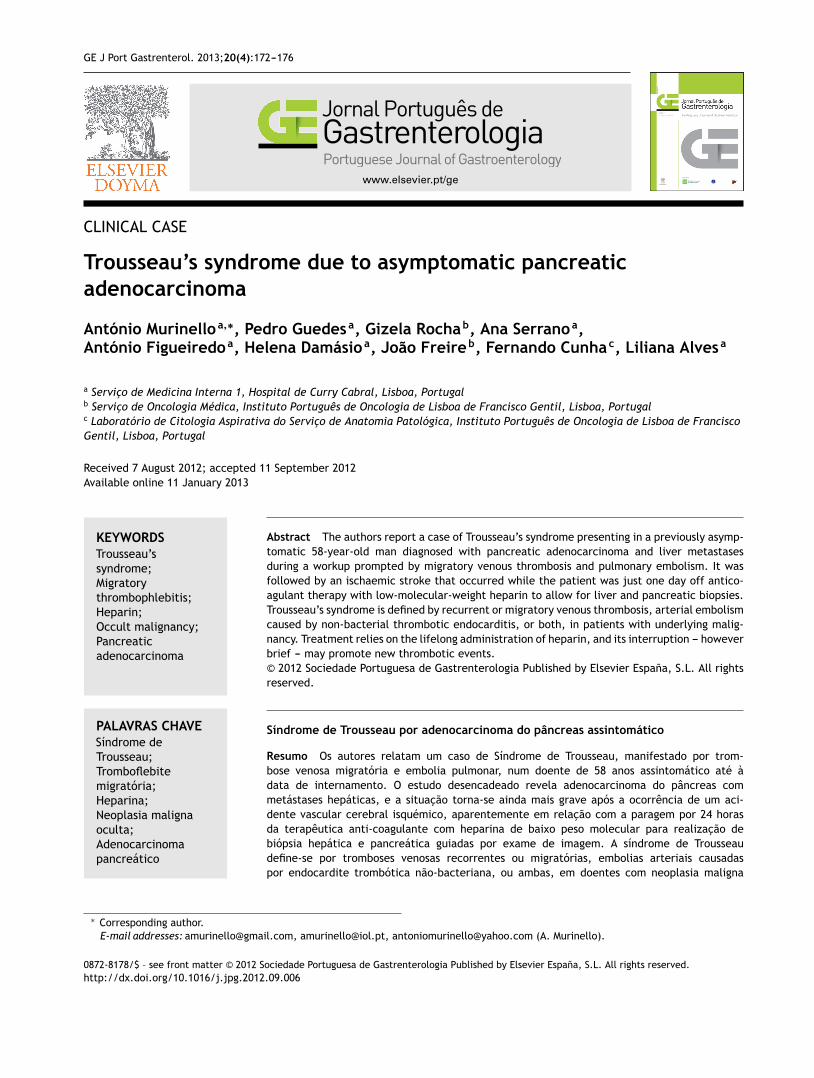
Radiologic exams revealed: (1) chest radiograph --- normal;(2) venous ultrasound and Doppler of the lower limbs; (3)thoracic CT-angiogram and (4) abdominal and pelvic CT scan.The last three exams lead to the following diagnoses: (A)residual superficial venous thrombosis of the right basilicvein, maintaining deep venous (humeral and axillary) systempermeability; (B) deep venous thrombosis of the right pos-terior tibial and calf veins, with normal popliteal, commonfemoral, superficial femoral vein, great saphenous and smallsaphenous vein permeability; left lower limb venous systemwith no lesions; (C) anterior segmental pulmonary embolismin the right upper lobe and the internal segmental branch ofthe ipsilateral inferior lobe; (D) enlarged liver with severalimages compatible with metastases (Fig. 1); and (E) infiltra-tive lesion of the pancreatic uncinate process, involving the
Figure 2 Contrast-enhanced abdominal CT scan, axialview: hypovascular lesion of the pancreatic uncinate process(2.5 cm Ø), with involvement of the superior mesenteric vessels.
superior mesenteric vessels and thus becoming inoperable(Fig. 2).
Treatment
He was treated with subcutaneous enoxaparin 60 mg bid,q12 h, with subsequent improvement.
Outcome
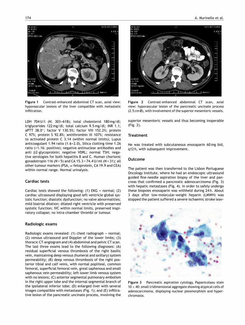
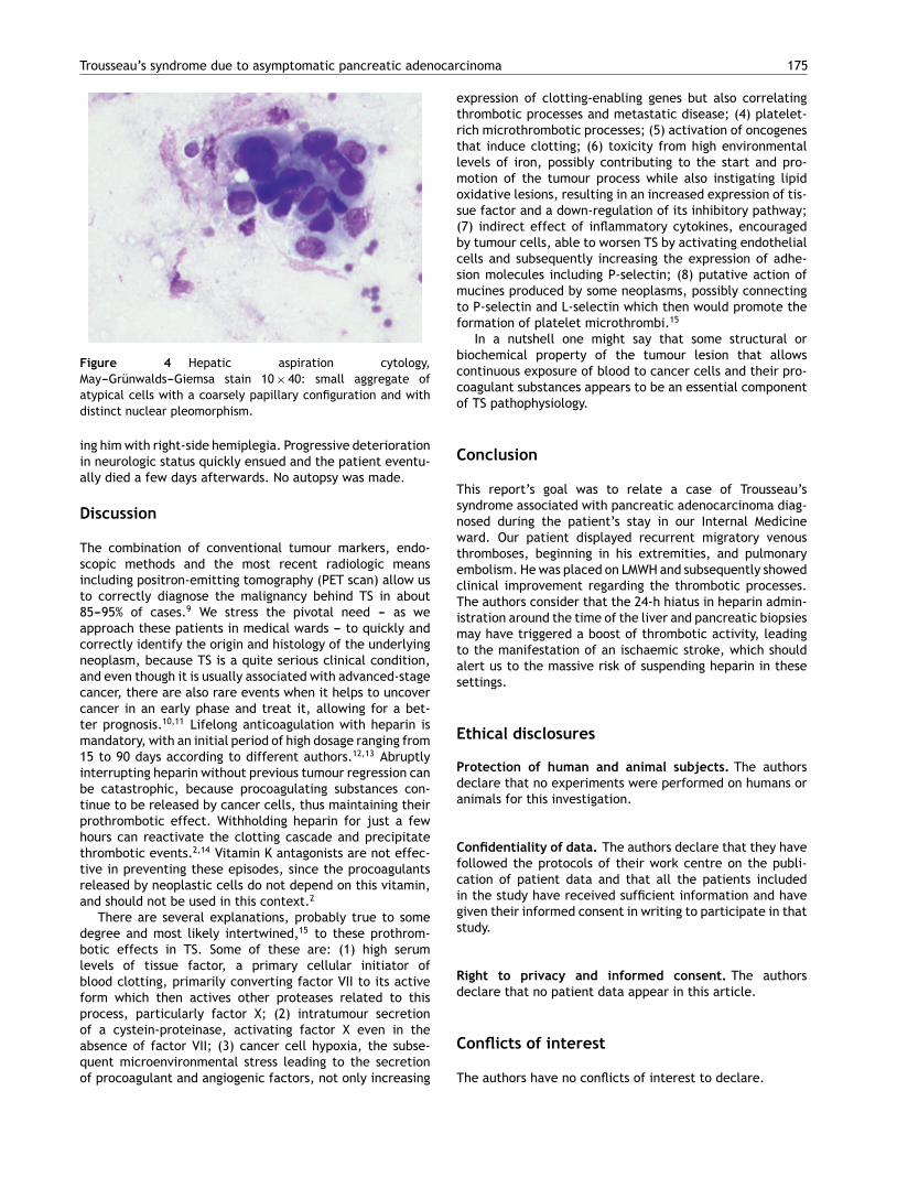
The patient was then transferred to the Lisbon PortugueseOncology Institute, where he had an endoscopic ultrasoundguided fine-needle aspiration biopsy of the liver and pan-creas that confirmed a pancreatic adenocarcinoma (Fig. 3)with hepatic metastases (Fig. 4). In order to safely undergothese biopsies enoxaparin was withheld during 24 h. About3 days after low-molecular-weight heparin (LMWH) wasstopped the patient suffered a severe ischaemic stroke leav-
Figure 3 Pancreatic aspiration cytology, Papanicolaou stain10 × 40: small tridimensional aggregate showing atypical cells ofadenocarcinoma, displaying nuclear pleomorphism and hyper-chromasia.
Trousseau’s syndrome due to asymptomatic pancreatic adenocarcinoma 175
Figure 4 Hepatic aspiration cytology,May---Grünwalds---Giemsa stain 10 × 40: small aggregate ofatypical cells with a coarsely papillary configuration and withdistinct nuclear pleomorphism.
ing him with right-side hemiplegia. Progressive deteriorationin neurologic status quickly ensued and the patient eventu-ally died a few days afterwards. No autopsy was made.
Discussion
The combination of conventional tumour markers, endo-scopic methods and the most recent radiologic meansincluding positron-emitting tomography (PET scan) allow usto correctly diagnose the malignancy behind TS in about85---95% of cases.9 We stress the pivotal need --- as weapproach these patients in medical wards --- to quickly andcorrectly identify the origin and histology of the underlyingneoplasm, because TS is a quite serious clinical condition,and even though it is usually associated with advanced-stagecancer, there are also rare events when it helps to uncovercancer in an early phase and treat it, allowing for a bet-ter prognosis.10,11 Lifelong anticoagulation with heparin ismandatory, with an initial period of high dosage ranging from15 to 90 days according to different authors.12,13 Abruptlyinterrupting heparin without previous tumour regression canbe catastrophic, because procoagulating substances con-tinue to be released by cancer cells, thus maintaining theirprothrombotic effect. Withholding heparin for just a fewhours can reactivate the clotting cascade and precipitatethrombotic events.2,14 Vitamin K antagonists are not effec-tive in preventing these episodes, since the procoagulantsreleased by neoplastic cells do not depend on this vitamin,and should not be used in this context.2
There are several explanations, probably true to somedegree and most likely intertwined,15 to these prothrom-botic effects in TS. Some of these are: (1) high serumlevels of tissue factor, a primary cellular initiator ofblood clotting, primarily converting factor VII to its activeform which then actives other proteases related to thisprocess, particularly factor X; (2) intratumour secretionof a cystein-proteinase, activating factor X even in theabsence of factor VII; (3) cancer cell hypoxia, the subse-quent microenvironmental stress leading to the secretionof procoagulant and angiogenic factors, not only increasing
expression of clotting-enabling genes but also correlatingthrombotic processes and metastatic disease; (4) platelet-rich microthrombotic processes; (5) activation of oncogenesthat induce clotting; (6) toxicity from high environmentallevels of iron, possibly contributing to the start and pro-motion of the tumour process while also instigating lipidoxidative lesions, resulting in an increased expression of tis-sue factor and a down-regulation of its inhibitory pathway;(7) indirect effect of inflammatory cytokines, encouragedby tumour cells, able to worsen TS by activating endothelialcells and subsequently increasing the expression of adhe-sion molecules including P-selectin; (8) putative action ofmucines produced by some neoplasms, possibly connectingto P-selectin and L-selectin which then would promote theformation of platelet microthrombi.15
In a nutshell one might say that some structural orbiochemical property of the tumour lesion that allowscontinuous exposure of blood to cancer cells and their pro-coagulant substances appears to be an essential componentof TS pathophysiology.
Conclusion
This report’s goal was to relate a case of Trousseau’ssyndrome associated with pancreatic adenocarcinoma diag-nosed during the patient’s stay in our Internal Medicineward. Our patient displayed recurrent migratory venousthromboses, beginning in his extremities, and pulmonaryembolism. He was placed on LMWH and subsequently showedclinical improvement regarding the thrombotic processes.The authors consider that the 24-h hiatus in heparin admin-istration around the time of the liver and pancreatic biopsiesmay have triggered a boost of thrombotic activity, leadingto the manifestation of an ischaemic stroke, which shouldalert us to the massive risk of suspending heparin in thesesettings.
Ethical disclosures
Protection of human and animal subjects. The authorsdeclare that no experiments were performed on humans oranimals for this investigation.
Confidentiality of data. The authors declare that they havefollowed the protocols of their work centre on the publi-cation of patient data and that all the patients includedin the study have received sufficient information and havegiven their informed consent in writing to participate in thatstudy.
Right to privacy and informed consent. The authorsdeclare that no patient data appear in this article.
Conflicts of interest
The authors have no conflicts of interest to declare.

176 A. Murinello et al.
References
1. Trousseau A. Phlegmasia alba dolens. Clinique Medicale deHotel-Dieu de Paris, vol. 3. London: New Sydenham Society;1868. pp. 695---727.
2. Bell WR, Starksen NF, Tong S, Poterfield JK. Trousseau’s syn-drome. Devastating coagulopathy in the absence of heparin.American Journal of Medicine. 1985;79:423---30.
3. Carrier M, Le Gal G, Wells PS, Fergusson D, Ramsay T, Rodger MA.Systematic review. The Trousseau syndrome revisited. Shouldwe screen extensively for cancer in patients with venous throm-boembolism? Advances in Internal Medicine. 2008;149:323---33.
4. Callander N, Rapaport SI. Trousseau’s syndrome. Western Jour-nal of Medicine. 1993;158:364---71.
5. Hasegawa S, Aoyama T, Kakinoki R, Toguchida J, Nakamura T.Bilateral Phlegmasia Alba Dolens associated with Trousseau’ssyndrome: a case report. Archives of Physical Medicine andRehabilitation. 2008;89:1187---90.
6. Santos VM, Rodrigues DB, Castro EC, Saldanha JC, Soares S,Teixeira VP, et al. Revista do Hospital das Clinicas; Faculdadede Medicina da Universidade de Sao Paulo. 2001;56:91---6 [inPortuguese].
7. Blann AD, Dunmore S. Arterial and venous thrombosis in cancerpatients. Cardiology Research and Practice. 2011:11 [article ID394740].
8. Sanon S, Lenihan DJ, Mouhayar E. Peripheral arterial ischemicevents in cancer patients. Vascular Medicine. 2011;16:119---30.
9. Batsis JA, Morgenthaler TI. Trousseau syndrome and theunknown cancer: use of positron emission tomographic imag-ing in a patient with paraneoplastic syndrome. Mayo ClinicProceedings. 2005:537---40.
10. Thrumurthy SG, Anuruddha AH, De Zoysa MI, Samarasekera DN.Unexpected outcome from Trousseau syndrome. BMC Surgery.2011;11:1---3.
11. Womack WS, Castellano CJ. Migratory thrombophlebitis associ-ated with ovarian carcinoma. American Journal of Obstetricsand Gynecology. 1952;63:467---9.
12. Masuda EM, Kessler DM, Kistner RL, Eklof B, Sato DT. The naturalhistory of calf vein thrombosis: lysis of thrombi and develop-ment of reflux. Journal of Vascular Surgery. 1998;28:67---73.
13. Lautz TB, Abbas F, Walsh SJ, Chow C, Amaranto DJ, Wang E,et al. Isolated gastrocnemius and soleal vein thrombosis: shouldthese patients receive therapeutic anticoagulation? Annals ofSurgery. 2010;251:735---42.
14. Sack Jr GJ, Levin JK, Bell WR. Trousseau’s syndrome andother manifestations of chronic disseminated coagulopathy inpatients with neoplasms: clinical, pathophysiologic, and thera-peutic features. Medicine. 1977;56:1---37.
15. Varki A. Trousseau’s syndrome: multiple definitions and multiplemechanisms. Blood. 2007;110:1723---9.