Embed Size (px)
Citation preview
Received: 15 May, 2011. Accepted: 6 December, 2011. Invited Review
Functional Plant Science and Biotechnology ©2012 Global Science Books
Molecular Basis of Red Rot Resistance in Sugarcane
Rasappa Viswanathan
Plant Pathology Section, Sugarcane Breeding Institute, Indian Council of Agricultural Research, Coimbatore 641007, India
Corresponding author: * [email protected]
ABSTRACT Red rot of sugarcane caused by Colletotrichum falcatum Went is one of the devastating diseases of sugarcane causing significant loss to sugarcane production in India and other Asian countries. Complex polyploidy and lack of information on inheritance to red rot in sugarcane make breeding for red rot resistance more difficult. Hence researchers have studied the mechanism of red rot resistance in sugarcane in detail. Initial studies based on biochemical tools identified oxidative enzymes and red rot pigments in disease resistance. Further studies revealed the role of pathogenesis-related (PR) proteins and 3-deoxyanthocyanidin phytoalexins especially luteolinidin and apigeninidin in red rot resistance. Recent studies using semiquantitative RT-PCR after pathogen inoculation from sugarcane cultivars varying in red rot resistance, revealed differential accumulation of transcripts of the flavanoid biosynthetic pathway like coumarate-4-hydroxylase, chalcone synthase, chalcone reductase, flavanoid 3�-5� hydroxylase and flavanoid glycosyl transferase and this transcript analysis, further confirmed the role of sugarcane phytoalexins in red rot resistance. Similarly the role of PR- proteins like chitinase and �-1,3-glucanase was established at the transcript level. Detailed molecular studies using differential display (DD)-RT-PCR identified expression of more number of differentially expressed transcripts during the host-pathogen interaction. Full length sequences of many potential transcripts were identified and are being characterized. Also to identify specific proteins involved in host resistance, proteomic approach has been attempted by optimizing sample preparation from stalk tissues, 2-D electrophoresis (2-DE), down-stream processing of identified spots and bioinformatics. Several resistance associated proteins spots were identified and they are being analyzed critically. Overall, application of molecular techniques was found to be useful in identifying transcripts/proteins involved in host defence. Further studies are in progress to validate their specific involvement in red rot resistance. _____________________________________________________________________________________________________________ Keywords: 2-D electrophoresis, Colletotrichum falcatum, differential display, PR-proteins, phytoalexins, red rot, resistance mechanism CONTENTS INTRODUCTION........................................................................................................................................................................................ 40
Symptoms................................................................................................................................................................................................ 41 Impact of the disease ............................................................................................................................................................................... 41 Pathogen .................................................................................................................................................................................................. 42 Breeding for red rot resistance................................................................................................................................................................. 42 Mechanical resistance.............................................................................................................................................................................. 42 Biochemical resistance ............................................................................................................................................................................ 43 Molecular basis of red rot resistance ....................................................................................................................................................... 46
CONCLUSION............................................................................................................................................................................................ 49 ACKNOWLEDGEMENTS ......................................................................................................................................................................... 49 REFERENCES............................................................................................................................................................................................. 49 _____________________________________________________________________________________________________________ INTRODUCTION Sugarcane (Saccharum spp. hybrids) is a major cash crop cultivated in more than 23 million ha in tropical and subtropical regions of the world, producing up to 1.6 billion metric tons of crushable stalks in the year 2009 and accounts for nearly 60% of the total sugar produced in the world. Sugarcane meets ~60% of sweetener’s requirement globally. India is the second largest producer of sugarcane in the world and the crop is cultivated in almost all the states occupying an area of 4.42 million ha with an annual production of ~285 million tonnes of canes in the year 2009 (http://faostat.fao.org). More than 488 sugar mills crush about 50-60% of the canes produced to manufacture crystal sugar in the country. About 30-40% of the produced canes are utilized to manufacture gur and khandsari sugar as an alternative sweetener in thousands of village industries in India. Also significant portion of the canes are crushed for extraction of juice for domestic consumption and about
10% canes are used as seed canes for planting (Sundara 1998). Sugarcane production in various regions of the coun-try is affected by different biotic and abiotic stresses. Among the biotic constraints, red rot a fungal disease caused by Colletotrichum falcatum Went (Perfect state: Glomerella tucumanensis (Speg) Arx Muller) is the major constraint affecting cane production in most of the sugarcane growing areas. Similarly sugarcane production is significantly affec-ted by red rot in different countries like Pakistan, Bangla-desh, Thailand, Australia, Fiji, USA etc. The disease occurs in 77 countries representing all the sugarcane growing con-tinents (Singh and Lal 2000; Viswanathan 2010).
The disease was first recorded in Java during 1893 (Went 1893) and in India during 1901 (Barber 1901). Series of disease epidemics in the sub-tropical India had devas-tated cane cultivation in entire Gangetic plains and Punjab till 1960s. Later the disease spread to tropical India and caused severe damage to the crop and the disease was res-ponsible for the elimination of many commercial varieties
®
Functional Plant Science and Biotechnology 6 (Special Issue 2), 40-50 ©2012 Global Science Books
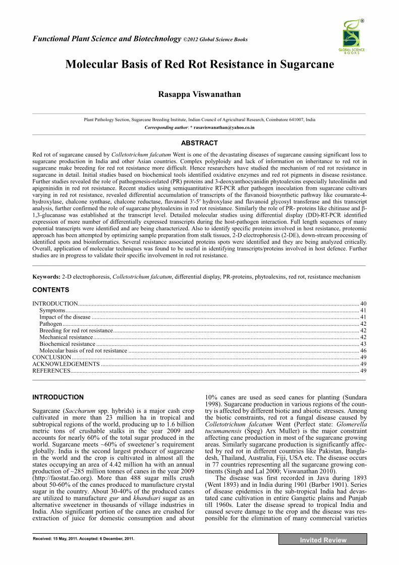
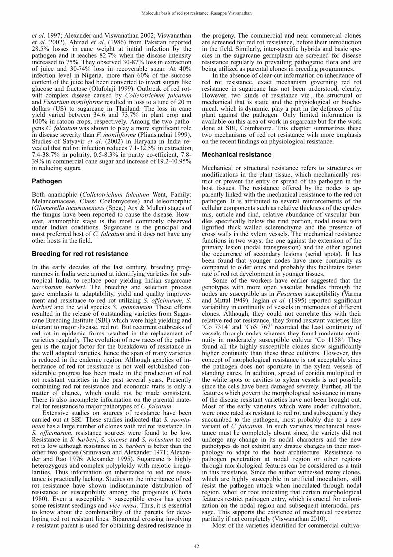
in India through the disease epidemics which occurred in different regions (Viswanathan and Samiyappan 1999; Vis-wanathan 2010). Severe epiphytotics on cv. ‘CoC 671’ the wonder cane of tropical region, during 1980s and 1990s have caused extensive damage to cane plantations in thou-sands of hectares in the Peninsular India. Likewise many important commercial varieties viz., ‘BO 3’, ‘BO 10’, ‘BO 11’, ‘BO 17’, ‘BO 54’, ‘Co 312’, ‘Co 419’, ‘Co 453’, ‘Co 658’, ‘Co 785’, ‘Co 997’, ‘Co 1148’, ‘Co 1158’, ‘Co 6304’, ‘Co 7805’, ‘CoC 671’, ‘CoC 92061’, ‘CoJ 64’, ‘CoS 8436’, ‘CoSe 95422’ etc were lost due to the disease in the last 100 years (Fig. 1). At present the disease is prevalent in most of the sugarcane growing states in the country at varying intensities (Viswanathan 2010). Symptoms Planting of infected setts results in failure of germination or death of germinated settlings. Such settlings exhibit dis-colouration of foliage or drying of whorl (Fig. 2). Charac-teristic symptoms of red rot are noticed during cane forma-tion stage. Infected canes either singly or in clumps show sudden yellowing of leaves and later the leaves gradually dry (Fig. 3). Such isolated clumps or clumps in patches give no external evidence of any diseased condition except vary-ing intensities of rind discolouration (Fig. 4). Splitting of canes longitudinally reveal specific red rot symptoms inside the stalk tissues as reddening of internal tissues with white spots which are usually elongated at right angles to the long axis of the stalk (Fig. 5). Further, infected cane may show the red discolouration throughout the length of the stalk and longitudinal cavities containing either mycelium may deve-lop and later the affected tissues turn muddy, shrink and dry out (Fig. 6). At later stages, the pathogen comes out as pin-kish sporulation at rind tissues, growth rings and leaf scars and fruiting bodies of the fungus (acervuli) on the rind tis-sues. The pathogen does not cause any foliar symptoms except discrete brown or red lesions of varying sizes on the mid ribs (Fig. 7). These lesions may coalesce to form long lesions that extend the entire length of the leaf or they may remain as a series of unconnected lesions. Impact of the disease The pathogen affects the economically valuable stalk tis-sues hence a limited infection can bring about drastic chan-ges in the juice quality. The disease affected cane gives poor sugar recovery because of impaired sucrose metabolism. The red rot infection reduced total carbohydrates in the dis-eased canes and the reduction was more in the highly sus-ceptible varieties (Agnihotri et al. 1989). Moreover, the pathogen produces abundant quantities of acid invertases which break the sucrose into glucose and fructose for the consumption by the pathogen. Higher production of acid invertases in the highly susceptible varieties was recorded upon pathogen infection as compared to resistant varieties (de Silva et al. 1977). Pathogen infection also results in in-creased levels of total soluble salts, acidity, reducing sugars and gum and simultaneously decrease in pH, sucrose and purity of cane juice in affected canes (Singh and Waraitch 1977). Similarly, our studies have revealed that pathogen infection has drastically reduced Brix, sucrose percentage, purity and commercial cane sugar (CCS) per cent in the dis-eased canes. The affected canes recorded 25 to 75% red-uced sucrose content as compared to the healthy canes (Vis-wanathan and Samiyappan 1999). During the milling pro-cess, mixing of juice from healthy and diseased canes results in spoilage of entire juice due to inversion of sucrose.
In subtropical India, severe infection of red rot can cause two third of cane stalks produced (Chona 1980). Severe red rot epidemics in Peninsular India during 1990s caused losses of 30-50% in cane yield in varieties like ‘Co 6304’, ‘CoC 671’, ‘CoC 85061’, ‘CoC 86062’, ‘CoC 92061’, ‘CoSi 86071’, etc. However, yield losses of upto 100% were found in different factory regions (Viswanathan
Fig. 1 Severe red rot infected sugarcane: crop exhibiting extensive drying of foliage and death of canes. Fig. 2 Young sugarcane plant exhibiting yellowing of leaves in the whorl due to red rot infection. Fig. 3 Red rot infection in grown up sugarcane. Affected sugarcane clump shows charac-teristic foliage discoloration and drying. Fig. 4 Red rot infected cane showing discoloured patches on cane rind tissue. Fig. 5 Internal symptoms of red rot. Reddening of internal tissues with white spots inside the canes. Fig. 6 Internal symptoms of red rot. Mycelial growth of the pathogen in the pith cavities. Fig. 7 Red rot symptoms on sugarcane leaves. Coalesced midrib lesions covering entire mid rib of the infected leaf. Fig. 8 Progress of red rot lesion inside the stalks 60 days after pathogen inoculation by plug method in sugarcane genotypes varying in disease resistance. Restric-ted lesion in resistant (R) genotype as compared to the extended lesions covering many internodes in the susceptible (S) genotype. Arrows indicate the site pathogen inoculation. Fig. 9 Progress of red rot lesion on the nodes 10 days after pathogen inoculation by nodal swabbing method in sugarcane genotypes varying in disease resistance. Restricted or no lesion in resistant (R) genotype as compared to the extended lesions on nodes in the susceptible (S) genotype. Fig. 10 Differential accumulation of red rot pigments in sugarcane varieties varying in red rot resistance. Methanolic extract from resistant tissue turns reddish due to release of anthocyanin pigments from the pathogen inoculated tissue (left) whereas, it remains colourless due to non-release of such pigment induction from susceptible tissue after pathogen inoculation (right). All photos unpublished.
1 2
3 4 5
6 7 8
9 10
41
Molecular basis of red rot resistance. Rasappa Viswanathan
et al. 1997; Alexander and Viswanathan 2002; Viswanathan et al. 2002). Ahmad et al. (1986) from Pakistan reported 28.5% losses in cane weight at initial infection by the pathogen and it reaches 82.7% when the disease intensity increased to 75%. They observed 30-87% loss in extraction of juice and 30-74% loss in recoverable sugar. At 40% infection level in Nigeria, more than 60% of the sucrose content of the juice had been converted to invert sugars like glucose and fructose (Olufolaji 1999). Outbreak of red rot-wilt complex disease caused by Colletotrichum falcatum and Fusarium moniliforme resulted in loss to a tune of 20 m dollars (US) to sugarcane in Thailand. The loss in cane yield varied between 34.6 and 73.7% in plant crop and 100% in ratoon crops, respectively. Among the two patho-gens C. falcatum was shown to play a more significant role in disease severity than F. moniliforme (Pliansinchai 1999). Studies of Satyavir et al. (2002) in Haryana in India re-vealed that red rot infection reduces 7.1-32.5% in extraction, 7.4-38.7% in polarity, 0.5-8.3% in purity co-efficient, 7.8-39% in commercial cane sugar and increase of 19.2-40.95% in reducing sugars. Pathogen Both anamophic (Colletotrichum falcatum Went, Family: Melanconiaceae, Class: Coelomycetes) and teleomorphic (Glomerella tucumanenesis (Speg.) Arx & Muller) stages of the fungus have been reported to cause the disease. How-ever, anamorphic stage is the most commonly observed under Indian conditions. Sugarcane is the principal and most preferred host of C. falcatum and it does not have any other hosts in the field.
Breeding for red rot resistance In the early decades of the last century, breeding prog-rammes in India were aimed at identifying varieties for sub-tropical India, to replace poor yielding Indian sugarcane Saccharum barberi. The breeding and selection process gave emphasis to adaptability, yield and quality improve-ment and resistance to red rot utilizing S. officinarum, S. barberi and the wild species S. spontaneum. These efforts resulted in the release of outstanding varieties from Sugar-cane Breeding Institute (SBI) which were high yielding and tolerant to major disease, red rot. But recurrent outbreaks of red rot in epidemic forms resulted in the replacement of varieties regularly. The evolution of new races of the patho-gen is the major factor for the breakdown of resistance in the well adapted varieties, hence the span of many varieties is reduced in the endemic region. Although genetics of in-heritance of red rot resistance is not well established con-siderable progress has been made in the production of red rot resistant varieties in the past several years. Presently combining red rot resistance and economic traits is only a matter of chance, which could not be made consistent. There is also incomplete information on the parental mate-rial for resistance to major pathotypes of C. falcatum.
Extensive studies on sources of resistance have been carried out at SBI. These studies indicated that S. sponta-neun has a large number of clones with red rot resistance. In S. officinarum, resistance sources were found to be low. Resistance in S. barberi, S. sinense and S. robustum to red rot is low although resistance in S. barberi is better than the other two species (Srinivasan and Alexander 1971; Alexan-der and Rao 1976; Alexander 1995). Sugarcane is highly heterozygous and complex polyploidy with meiotic irregu-larities. Thus information on inheritance to red rot resis-tance is practically lacking. Studies on the inheritance of red rot resistance have shown indiscriminate distribution of resistance or susceptibility among the progenies (Chona 1980). Even a susceptible × susceptible cross has given some resistant seedlings and vice versa. Thus, it is essential to know about the combinability of the parents for deve-loping red rot resistant lines. Biparental crossing involving a resistant parent is used for obtaining desired resistance in
the progeny. The commercial and near commercial clones are screened for red rot resistance, before their introduction in the field. Similarly, inter-specific hybrids and basic spe-cies in the sugarcane germplasm are screened for disease resistance regularly to prevailing pathogenic flora and are being utilized as parental clones in breeding programmes.
In the absence of clear-cut information on inheritance of red rot resistance, exact mechanism governing red rot resistance in sugarcane has not been understood, clearly. However, two kinds of resistance viz., the structural or mechanical that is static and the physiological or bioche-mical, which is dynamic, play a part in the defences of the plant against the pathogen. Only limited information is available on this area of work in sugarcane but for the work done at SBI, Coimbatore. This chapter summarizes these two mechanisms of red rot resistance with more emphasis on the recent findings on physiological resistance.
Mechanical resistance Mechanical or structural resistance refers to structures or modifications in the plant tissue, which mechanically res-trict or prevent the entry or spread of the pathogen in the host tissues. The resistance offered by the nodes is ap-parently linked with the mechanical resistance to the red rot pathogen. It is attributed to several reinforcements of the cellular components such as relative thickness of the epider-mis, cuticle and rind, relative abundance of vascular bun-dles specifically below the rind portion, nodal tissue with lignified thick walled sclerenchyma and the presence of cross walls in the xylem vessels. The mechanical resistance functions in two ways: the one against the extension of the primary lesion (nodal transgression) and the other against the occurrence of secondary lesions (serial spots). It has been found that younger nodes have more continuity as compared to older ones and probably this facilitates faster rate of red rot development in younger tissues.
Some of the workers have earlier suggested that the genotypes with more open vascular bundles through the nodes are susceptible as in Fusarium susceptibility (Varma and Mittal 1949). Jaglan et al. (1995) reported significant variability in continuity of vessels in internodes of different clones. Although, they could not correlate this with their relative red rot resistance, they found resistant varieties like ‘Co 7314’ and ‘CoS 767’ recorded the least continuity of vessels through nodes whereas they found moderate conti-nuity in moderately susceptible cultivar ‘Co 1158’. They found all the highly susceptible clones show significantly higher continuity than these three cultivars. However, this concept of morphological resistance is not acceptable since the pathogen does not sporulate in the xylem vessels of standing canes. In addition, spread of conidia multiplied in the white spots or cavities to xylem vessels is not possible since the cells have been damaged severely. Further, all the features which govern the morphological resistance in many of the disease resistant varieties have not been brought out. Most of the early varieties which were under cultivation, were once rated as resistant to red rot and subsequently they succumbed to the pathogen, most probably due to a new variant of C. falcatum. In such varieties mechanical resis-tance must be completely absent since, the variety did not undergo any change in its nodal characters and the new pathotypes do not exhibit any drastic changes in their mor-phology to adapt to the host architecture. Resistance to pathogen penetration at nodal region or other regions through morphological features can be considered as a trait in this resistance. Since the author witnessed many clones, which are highly susceptible in artificial inoculation, still resist the pathogen attack when inoculated through nodal region, whorl or root indicating that certain morphological features restrict pathogen entry, which is crucial for coloni-zation on the nodal region and subsequent internodal pas-sage. This supports the existence of mechanical resistance partially if not completely (Viswanathan 2010).
Most of the varieties identified for commercial cultiva-
42
Functional Plant Science and Biotechnology 6 (Special Issue 2), 40-50 ©2012 Global Science Books
tion possess some levels of resistance. The varieties which possess broad based resistance i.e. resistance against many pathotypes are expected to survive in the field for longer years in the field. In the history of sugarcane cultivation in India almost all the varieties succumbed to the red rot pathogen in the endemic region. Longevity of the varieties is the difference among them. Varieties like ‘CoS 767’ sur-vived for nearly 20 years in the endemic region of Northern India. Although it succumbed to a new pathotype it still remains resistant to many other pathotypes.
An intriguing part of host-pathogen interaction is the differential interaction between the host and different patho-types. Detailed studies on the pathogeneicity of the patho-genic flora at different locations revealed existence of 11 major pathotypes in the country (Viswanathan 2010). The author has found a differential behaviour in many varieties like ‘Co 1148’, ‘Co 7717’, ‘Co 8021’, ‘Co 7805’, ‘Co 86032’, ‘CoS 767’ etc against the pathotypes. This informa-tion clearly suggests that selective adaptation of the patho-genic strains to host genotypes.
Biochemical resistance The mechanical defence mechanism determines the extent of longitudinal expansion of the lesion. On the other hand, physiological resistance mechanism governs lateral (and within the internode, longitudinal) extension of the lesion in the ground tissue. Apart from controlling the tissue destruc-tion, the latter has repercussions on the physiological effects of the tissue. In addition to the sealing-off of fungal hyphae, inversion of sucrose caused by their enzymes is also restric-ted. In this context, Srinivasan and Bhat (1961) suggested while devising new 0-9 scale for rating red rot resistance that such resistance is doubly advantageous and emphasized greater attention on physiologic resistance than mere me-chanical resistance as done previously on the basis of only average lesion length. They stressed that mechanical resis-tance as measured by the number of nodes crossed is highly variable while physiologic resistance as indicated by the nature of the lesion is surely more dependable. In view of the fact that physiological processes as well as structural equipment both of which are inherited, together determine the outcome of the host-parasite interaction. They felt that both types of resistance deserve to be taken into account, while laying greater emphasis on the dynamic aspect.
The pathogenic fungus is a hemibiotroph and the fungal hypha penetrates the host cells in the progressive phase of the disease by forming a minute penetration peg. These pegs can be seen as a constriction in the hypha, which ex-pands to the normal hypha diameter immediately on reach-ing the other side of the cell wall. When the pathogen is spreading through the tissue, the hydrolytic enzymes poly-galacturanase (PG), pectin methyl esterase (PME) and cel-lulases (Cx and Cx) enzymes are not detected. Additionally it was also found that extract of red rot lesion showing re-sistant reaction inhibited PG, PME and Cx enzymes pro-duced by the pathogen, however, extracts from susceptible category had a lower degree of inhibitory effect (Srinivasan 1969). This suggests that the pathogen gains entry through mechanical pressure and enzyme produced by the pathogen also degraded by the host defence machinery during the parasitic phase. Sugars possibly inhibit production of PG, PME, C1 and Cx in the early stages of infection by C. fal-catum in a susceptible variety and it is only when the supply of sugars is exchanged in vivo that the hydrolytic enzymes are elaborated. At a time there is proliferation of hyphal branches in the tissues, there is rapid death of host cells and parasitic hyphae become intercellular and this coincides with the appearance of hydrolytic enzymes. White spots and cavities appear in the ground tissues and this marks the end of the parasitic phase and the beginning of the saprophytic phase of the fungus. Hydrolytic enzymes do not appear to play a part in pathogenesis however they have a role in the saprophytic phase of the fungus which follows the parasitic phase.
Different biochemical features that impart resistance in a particular genotype have been reported. After the patho-gen infection there is a reaction or a change in host cells in advance of the invading pathogen. The protoplasm in the affected area changes its colour and a gummy dark material oozes out of the cells and fills the intercellular spaces. Because of the presence of soluble pigment which is absorbed by the cell walls the infected area turns red. This results in reddening of fibrovascular tissues for several inches from the centre of infection. Pathogen advancement is checked temporarily in this zone of inhibition or stopped. In highly susceptible varieties infected by a compatible virulent strain, white spot account for major area inside the stalk tissues than a less virulent strain. White spots do not appear in resistant varieties or a variety infected by non compatible strain where the affected tissues have various intensities of red colour depending on the intensity of the resistant reaction (Srinivasan and Bhat 1961). Also hydro-lytic enzyme produced by the pathogen was detected only in white spots, regions of a temporary harmony in host-pathogen interaction. Later studies have reported on the role of phenolic compounds in red rot resistance. This may be due to presence of antifungal compounds in the red pigment or by plugging of the pits in the cell walls by the gummy material. This process takes place in advance of infection and seals off further spread of the pathogen in adjoining tissues (Edgerton 1955; Srinivasan and Bhat 1961). Gum formation may also take place in susceptible varieties, but to a lesser extent and usually after the tissue has been in-vaded. Srinivasan and Bhat (1961) termed this as ‘hyper-sensitive gummy reaction’. The nature of resistance depends on how rapidly the counteracting changes occur or how soon the red zone around the infected area develops. In resistant varieties the lesion may remain very small whereas in susceptible varieties they may extend entirely across the stalk (Fig. 8). On the nodal region pathogen makes infec-tion on leaf scar, root primordial, bud and growth ring. Similar to the host behaviour inside the internodal tissues, resistant varieties record dark lesions confined to a few millimeters, where as in susceptible varieties it colonizes entire nodal region and the lesions spread vertically on both directions. Also the lesions are straw colour with limited reddish pigmentation (Fig. 9). This action of the cane tis-sues, especially in resistant varieties may be due to the action of the first invaded cells by taking up the red colour. When this happens the lesion remains very small and often the reaction is so offensive that the advancing mycelium is killed before the reaction it has a chance to spread mate-rially. Mycelium advances slowly into the red zone.
Experience of Srinivasan (1969) revealed that isolations made from restricted dark lesions yielded the pathogen only rarely, while a large majority of transplant from the promi-nent white spots and the mottled ground tissue readily yiel-ded the pathogenic culture. The presence and characters of white spots appear to be related generally to the width of the lesion and prominent white spots are indicative of poor resistance on the part of the host. Histological examination of previous workers showed that in the white spots the pathogen enters temporarily into a harmonious relationship with the host, characteristic of the more evolved type of parasite.
1. Phenolics and oxidative enzymes Some workers (Rao et al. 1968; Wilson and Srivastava 1970) reported higher quantities of phenols in resistant and moderately resistant varieties. Similarly they also found that the resistant varieties maintained a higher level of polyphe-nol oxidase (PPO) activity before and after pathogen inocu-lation as compared to susceptible ones. However, further studies revealed that there was no correlation between total phenolic content and degree of resistance to red rot (Singh et al. 1976; Godshall and Lonergan 1987). These workers have found that in resistant varieties, the level of total phe-nols increased after infection and it maintained, while in
43
Molecular basis of red rot resistance. Rasappa Viswanathan
susceptible varieties, the level of phenolic content dropped after an initial increase. Srinivasan (1969) suggested that phenolics and their oxidation products are involved in this inhibitory process.
Number of host enzymes like peroxidase (PO), poly-phenol oxidase (PPO), phenylalanine ammonia lyase (PAL) and tyrosine ammonia-lyase (TAL) were reported to be associated with resistance in many sugarcane varieties. PPO and other oxidative enzymes have been related to resistance in several host pathogen interactions and are produced as an adaptive enzyme when phenolic compound appear in the tissue. The magnitude and rapidity of the production and release of phenolic compounds is known to be of paramount importance in disease resistance. Studies of Srinivasan (1969) clearly established that PPO activity was two fold in expressed healthy juice of completely resistant variety than the highly susceptible ones. Such juice samples inhibited conidial germination of the pathogen and this might possibly due to higher PPO in the extracted juice. In infected tissues, PPo activity was several times than in a susceptible tissue. PPO activity and resistance appear to be correlated and to be related to the degree of incompatibility between host tis-sue and pathogenic strain. The avirulent strain executed intense PPO activity in each of the host varieties inoculated with it but the others did so only in varieties that are resis-tant to them. PPO has been shown to be present in the red-dened areas. Srivastava and Solomon (1990) found higher levels of PO and PPO in disease resistant mutants.
Higher activities of PAL and TAL involved in phenyl propanoid pathway are reported to be involved in red rot re-sistance (Madan et al. 1991). Studies of Singh et al. (1993) revealed that per cent increase in phenolic concentration was higher in cvs ‘CoS 7918’ and ‘CoS 8315’ which are resistant as compared to other varieties. They also found a significant increase in PPO and peroxidase (PO) activities in the early stage of (4-6 days) stalk infection. Similarly PAL activity was also higher in moderately resistant (MR) varieties as compared to susceptible varieties even before pathogen inoculation. After inoculation, PAL activity in-creased gradually up to 5 days and then declined. Here also the MR varieties showed higher enzyme activities. Detailed studies on PO levels in sugarcane varieties varying in red rot resistance revealed that resistant varieties like ‘BO 91’ and ‘CoS 767’ have multifold of this enzyme as compared to the susceptible varieties like ‘CoC 671’ and ‘CoC 86062’ (Viswanathan et al. 1996a). Comparison of PO isozyme profile after pathogen inoculation at different intervals re-vealed more prominence of PO isozymes on the fourth and the sixth days after pathogen inoculation. Eight days after pathogen inoculation, intensity of PO isozymes reduced. In susceptible and moderately susceptible cultivars the inten-sity of PO isozymes reduced 10 days after pathogen inocu-lation whereas, in resistant cultivars the intensity was main-tained throughout the study period (Thirupathiraja et al. 2004). All these studies have been done with tissues from intact cane tissues. Cellular level enzyme activity in sugar-cane varieties varying in disease reaction showed that calli from a resistant variety ‘BO 91’ recorded very high levels of PO, PAL and TAL than the cells of ‘CoC 671’, which is susceptible. The pathogen toxin treatment elicited signifi-cantly higher levels of PO in the callus tissues of ‘BO 91’ than in ‘CoC 671’. Additionally induction of the defence enzymes was rapid in resistant cells. Among the enzymes, induction of PAL, TAL and PPO was not rapid as that of PO upon pathogen toxin treatment. Further studies on electro-lyte leakage revealed that a more pronounced loss of elec-trolytes occur in the callus cells of ‘CoC 671’ than in ‘BO 91’ (Ramesh Sundar and Viswanathan 1997; Ramesh Sun-dar et al. 1998, 1999). The susceptible variety suffers more damage in their cells due to pathogen toxin activity and that is reflected in electrolyte release to the medium. 2. Phytoalexins The red substance released in cells and intercellular spaces
near the invading hyphae in the infected cane stalks is referred as red rot pigment (RRP). The RRPs formed in res-ponse to C. falcatum infection in sugarcane inhibited coni-dial germination in vitro and slowed mycelial growth (God-shall and Lonergan 1987; Viswanathan et al. 1996b). Frac-tionation of the RRPs showed presence of several com-pounds and some of them are phenolics. Godshall and Lonergan (1987) detected RRPs between 24 and 48 h in the infected susceptible host and found conidial germination in vitro takes place much before the pigment accumulation. Hence, the pigment may not be effective in stopping the initial germination. The RRPs from resistant cane tissues showed seven compounds, while in susceptible cultivar ‘CoC 671’ has shown only four of them in thin layer chro-matography (Viswanathan et al. 1996b). The loss of three pigment fractions in the susceptible cultivar after pathogen infection indicates that the invading pathogen in the stalks may destroy these resistance-contributing fractions.
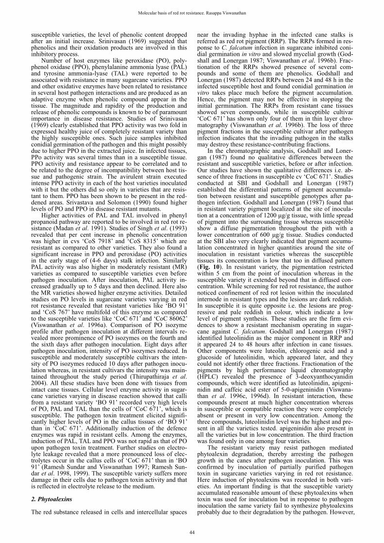
In the chromatographic analysis, Godshall and Loner-gan (1987) found no qualitative differences between the resistant and susceptible varieties, before or after infection. Our studies have shown the qualitative differences i.e. ab-sence of three fractions in susceptible cv ‘CoC 671’. Studies conducted at SBI and Godshall and Lonergan (1987) established the differential patterns of pigment accumula-tion between resistant and susceptible genotypes after pa-thogen infection. Godshall and Lonergan (1987) found that in resistant variety pigment localized at the site of inocula-tion at a concentration of 1200 �g/g tissue, with little spread of pigment into the surrounding tissue whereas susceptible show a diffuse pigmentation throughout the pith with a lower concentration of 600 �g/g tissue. Studies conducted at the SBI also very clearly indicated that pigment accumu-lation concentrated in higher quantities around the site of inoculation in resistant varieties whereas the susceptible tissues its concentration is low that too in diffused pattern (Fig. 10). In resistant variety, the pigmentation restricted within 5 cm from the point of inoculation whereas in the susceptible variety it extended beyond that in diffused con-centration. While screening for red rot resistance, the author noticed confinement of red rot lesion within the inoculated internode in resistant types and the lesions are dark reddish. In susceptible it is quite opposite i.e. the lesions are prog-ressive and pale reddish in colour, which indicate a low level of pigment synthesis. These studies are the firm evi-dences to show a resistant mechanism operating in sugar-cane against C. falcatum. Godshall and Lonergan (1987) identified luteolinidin as the major component in RRP and it appeared 24 to 48 hours after infection in cane tissues. Other components were luteolin, chlorogenic acid and a glucoside of luteolinidin, which appeared later, and they could not identify other three fractions. Fractionation of the pigments by high performance liquid chromatography (HPLC) revealed the presence of 3-deoxyanthocyanidin compounds, which were identified as luteolinidin, apigeni-nidin and caffeic acid ester of 5-0-apigeninidin (Viswana-than et al. 1996c, 1996d). In resistant interaction, these compounds present at much higher concentration whereas in susceptible or compatible reaction they were completely absent or present in very low concentration. Among the three compounds, luteolinidin level was the highest and pre-sent in all the varieties tested. apigeninidin also present in all the varieties but in low concentration. The third fraction was found only in one among four varieties.
The resistant variety may resist pathogen mediated phytoalexin degradation, thereby arresting the pathogen growth in the canes after pathogen inoculation. This was confirmed by inoculation of partially purified pathogen toxin in sugarcane varieties varying in red rot resistance. Here induction of phytoalexins was recorded in both vari-eties. An important finding is that the susceptible variety accumulated reasonable amount of these phytoalexins when toxin was used for inoculation but in response to pathogen inoculation the same variety fail to synthesize phytoalexins probably due to their degradation by the pathogen. However,
44
Functional Plant Science and Biotechnology 6 (Special Issue 2), 40-50 ©2012 Global Science Books
here also resistant variety recorded multifold phytoalexins synthesis than in the susceptible variety (Viswanathan et al. 1996d). These findings clearly demonstrated that the phyto-alexins were accumulated only in incompatible host patho-gen interactions and compatible interactions had no such phytoalexins or with trace quantities. These three com-pounds were identified as phytoalexins in sorghum and C. sublineolum interaction a host pathogen interaction close to sugarcane and C. falcatum (Snyder et al. 1991).
Further studies with a set of host differentials proved that there is accumulation of anthocyanin compounds in in-compatible interactions and no such induction/accumulation in compatible interactions (Viswanathan et al. 2000a; Vis-wanathan 2001). Earlier studies of Brinkler and Seigler (1991) reported a piceatannol compound as sugarcane phytoalexins but no further studies have been done to prove it. Recent studies of Malathi et al. (2008) conclusively proved that 3-deoxyanthocyanidin compounds act as sugar-cane phytoalexins and probably govern resistance in sugar-cane to red rot. The classical incompatible interaction of pathotypes from ‘Co 1148’ and ‘Co 7717’ on the host culti-vars is very much demonstrated earlier at SBI (Padmanaban et al. 1996). This was further elaborated by quantifying phytoalexins in such interactions. The variety ‘Co 1148’ ac-cumulates 3-deoxyanthocyanidins in incompatible interac-tion with Cf7717, which results in resistant reaction. How-ever, the same variety fails to synthesize phytoalexins in case of matching pathotype Cf1148 infection. Similar beha-viour of ‘Co 7717’ to Cf1148 and Cf7717 pathotype inter-actions was proved. Information on phytoalexin induction
in susceptible varieties by phytotoxin also amply demons-trates the role of phytoalexin in red rot resistance in sugar-cane. Though previous studies have been unsuccessful in pinpointing biochemical basis of disease resistance, these studies at SBI have shown the possible role of phytoalexins in red rot resistance. Recent studies conducted at SBI with phytoalexin standards also gave further evidence on the dif-ferential accumulation of luteolinidin and apigeninidin com-pounds (Malathi et al. 2008). Very high level of phyto-alexins was recorded in ‘Co 93009’ a resistant cultivar as compared to traces of phytoalexin fractions in ‘CoC 671’ (Fig. 11).
3. Pathogenesis related (pr) proteins Accumulation of PR-proteins is an important phenomenon of plant defence responses upon infection by pathogens. Many of the PR-proteins have been purified and the puri-fied proteins exhibited strong antifungal activity against many fungal pathogens under in vitro conditions. Induction of high levels of these proteins in host plants reduced dis-ease development in many crop plants. Specific involve-ment of many proteins in disease resistance has resulted in isolation and cloning of these PR-proteins and subsequently, developing transgenic plants expressing PR1a, PR-2 (�-1,3-glucanase), PR3 (chitinases) and PR5 (thaumatin-like pro-teins) in various crop plants. Such transgenic plants expres-sing foreign genes showed enhanced resistance to fungal pathogens. Detailed studies on PR-proteins in red rot resis-tance showed that constitutive activities of chitinase and �-1,3-glucanase were higher in disease resistant varieties as compared to susceptible varieties. Upon pathogen inocula-tion, the resistant variety accumulated higher hydrolytic en-zymes as compared to susceptible cultivars (Viswanathan and Samiyappan 1999). This information suggests a pos-sible role of these enzymes in red rot resistance.
Further studies were conducted to identify the PR-pro-teins using Western blot technique in a set of resistant and susceptible varieties differing in resistance the disease (Vis-wanathan et al. 2005). The red rot resistant cv ‘Co 93009’ showed differential induction of four chitinase proteins with molecular mass ranging from 34-39 kDa after pathogen inoculation in leaf tissues. The intensity of these proteins in-creased with time from 6 to 42 h after inoculation. In sus-ceptible variety ‘CoC 671’ induction of a 35-kDa chitinase protein was recorded. In stalk tissues induction of a 35-kDa chitinase protein was recorded 24 hr after inoculation in resistant variety whereas, in susceptible plants such induc-tion was delayed. Similarly, early induction of 43.0- and 37.5-kDa TLPs by 24 h was observed in response to patho-gen inoculation in the resistant variety whereas in the sus-ceptible variety such induction was less intense and could be seen 8th and 9th days post inoculation. These studies give a clear indication that the PR-proteins may be an important defense mechanism operating in sugarcane against red rot as in other host-pathogen interaction.
4. Mechanism of induced systemic resistance Earlier studies have clearly established the induction of sys-temic resistance in sugarcane against red rot by fluorescent pseudomonads (Viswanathan and Samiyappan 2002a, 2008). These studies gave evidences of enhanced resistance in dis-ease susceptible cultivars like ‘CoC 671’, ‘CoC 90063’, ‘CoC 92061’, etc. under controlled and field conditions. Further studies were conducted in detail on the mechanism of induced resistance by Pseudomonas strains against the disease. Involvement of different PR-proteins such as �-1,3-glucanases, chitinases and thaumatin-like proteins (TLPs) was found to be associated with Pseudomonas-mediated in-duced resistance (Viswanathan and Samiyappan 2001a; Vis-wanathan et al. 2003b). Studies of Viswanathan et al. (2003b) have also shown strong anti-fungal activities of sugarcane chitinases purified from systemically protected stalk tissues against C. falcatum. Their results clearly de-
Fig. 11 HPLC profile showing differential accumulation of 3-deoxy-anthocyanidin. Unpublished figure.
45
Molecular basis of red rot resistance. Rasappa Viswanathan
monstrated that bacterium treated disease susceptible sugar-cane is able to restrict disease development to a level equi-valent to moderately resistant varieties and many PR-pro-teins are involved in that ISR. In addition, enzymes of phenyl-propanoid pathway and oxidative pathway were also found to be involved in ISR (Viswanathan and Samiyappan 2002b). Characterization of Pseudomonas strains revealed that production of different metabolites/antibiotics such as salicylic acid, auxins, siderophores, pyocyanine, pyoluteo-rin and 2,4-diacetyl phloroglucinol and hydrolytic enzyme chitinase contribute to suppression of C. falcatum, induced resistance and growth promotion in sugarcane (Viswana-than and Samiyappan 2001b, 2004, 2006).
Molecular basis of red rot resistance 1. Red rot resistance at the molecular level in sugarcane Plants under are constant threat of infection by pathogens armed with a diverging array of effector molecules to colo-nize their host. Plants have in turn evolved a sophisticated detection and response systems that decipher pathogen signals and induce appropriate defences (Feys and Parker 2000). Recent genetic analysis involving plant mutants defective in resistance response to the invading pathogens has revealed a number of distinct, but interconnecting, sig-naling networks that are under both positive and negative control. These pathways operate atleast partly through the action of small signaling molecules such as salicylate, jasmonate and ethylene. The interplay of signals probably allows the plant to fine-tune defense responses in both local and systemic tissue. At SBI we have taken up both genomic and proteomic studies work to understand the host pathogen interaction of sugarcane-C. falcatum at molecular level. In genomics author’s group has employed reverse transcriptase - polymerase chain reaction (RT-PCR), differential display RT-PCR, reverse northern and rapid amplification of cDNA ends (RACE) and subtraction suppression hybridization (SSH) to identify and characterize the resistance/defense genes involved in rd rot resistance. 2. Pathogen recognition by the host plant Plants are able to recognize fungal pathogens by their sec-reted products, referred as ‘elicitors’. These elicitor mole-cules are signal molecules and elicit synthesis of phyto-alexins, phenolics, lignin, PR-proteins, hydroxyl-proline rich glycoproteins and other defense molecules in host plant and the host recognizes the pathogen from several non-pathogens. Defence responses are induced upon perception of either specific or non-specific elicitors. Specific elicitors are products of pathogen avirulence genes and are hypothe-sized to be recognized by the products of corresponding pathogen race-specific resistant genes. Detailed studies were carried out to purify and characterize elicitors from C. falcatum and to study their role in recognition of C. fal-catum by sugarcane.
A high molecular weight elicitor was isolated from the mycelial walls of C. falcatum and partially purified by gel filtration. The elicitor is characterized as a glycoprotein and the activity of elicitor resides in the carbohydrate moiety. The partially purified elicitor induced the accumulation of phenolics and the activities of phenylalanine ammonia-lyase (PAL) and peroxidase (POX) in sugarcane leaves and suspension- cultured cells. Sugarcane cells in culture res-ponded to C. falcatum elicitor in a manner similar to sugar-cane leaves (Ramesh Sundar et al. 2002a). Similarly, induc-tion of hydrogen peroxide (H2O2), reactive oxygen species (O2
-), lipoxygenase, lipid peroxidation, superoxide dismu-tase (SOD) and catalase was also observed in cell suspen-sion cultures of sugarcane. A rapid outburst and a spurt in the generation of active oxygen species especially H2O2 was observed indicating an early molecular event in recognition of the pathogen by the host cells. However, higher levels of the suppressor enzymes viz., catalase and SOD were found
to be maintained throughout in the cells of sugarcane sus-pension cultures without any elicitor treatment (Ramesh Sundar et al. 2002b). When elicitor isolated from C. fal-catum was compared with C. lindemuthianum a non-patho-gen elicitor differential induction of POX isoforms in sus-pension-cultured cells of sugarcane cv. ‘CoC 671’ was found (Ramesh Sundar and Vidhyasekharan 2003a).
For studying the defense gene activation in sugarcane, the intact plant-pathogen system may not be ideal because the time course of pathogen infection cannot be monitored precisely. Cell cultures instead of whole plants and elicitors instead of live pathogens are found to be suitable models to study the defense gene activation in bean, rice and in sugar-cane because of their high degree of reproducibility, rapid experimental cycles. Further, each cell in the culture is uni-formly exposed to the elicitor preparation and hence the response of cells is relatively uniform. Preliminary results of the study conducted at SBI has provided insight into the mechanisms regulating the pathogen recognition at the interface which facilitates further elaboration of inducible defense response against C. falcatum in sugarcane (Ramesh Sundar and Vidhyasekahran 2003a, 2003b). These studies confirmed that the elicitor molecules from C. falcatum are responsible for specific recognition of the pathogen by the host resistance is determined by the rapidity of the down-ward signaling of defense pathway. Probably variation in initiation of signaling process between the resistant and sus-ceptible genotype determines the pathogen colonization and disease development.
3. Gene expression studies To understand genome complexity of sugarcane, a large scale expressed sequence tag (EST) programme known as ‘SUCEST’ was taken up recently in Brazil. More than 2,60,000 cDNA (complimentary DNA) clones were parti-ally sequenced from 26 standard cDNA Libraries generated from different sugarcane tissues. They annotated 43,141 assembled sequences and found 50% of the putative identi-fied sugarcane genes coding for protein metabolism, cel-lular communication/signal transduction, bioenergetics and stress responses. Vettore et al. (2003) found 80 SUCEST sugarcane assembled sequences (SASs) encoded proteins with clear similarity to the NB-ARC domain, which is cha-racteristic of one of the major classes of disease resistance genes (R genes). The database contained more than 200 Sass encoding WRKY transcription factor domains, which have been implicated in the defense gene regulation in plants. They also found other genes related to defense res-ponses like chitinases, �-1,3-glucanases, chalcone syntheses, chalcone isomerases, isoflavone reductases, hydroxylpro-line-rich glycoproteins, proline-rich proteins, catalases, superoxide dismutases etc. have been putative orthologs in sugarcane, which indicates a high conservation of defense strategies among plants. The wealth of information gene-rated in the SUCEST database promises exciting prospects for the scientists involved in sugarcane improvement and other crops. Many transcripts including the disease resis-tance and defense responses ESTs will be a basic resource for the understanding of the biology of this complex poly-ploidy plant. This information may facilitate in identifying gene(s) involved in disease resistance in sugarcane.
The author’s group has carried out detailed studies on identifying specific transcripts induced during host patho-gen interaction to identify candidate genes involved in red rot resistance. After pathogen inoculation total RNA were isolated from sugarcane stalk tissues and were reverse transcribed to cDNA for expression analysis. Expression of the transcripts, the level of expression and the interval at which a particular gene expression etc, were analyzed using the custom made primers. The following transcripts R30, chitinase, metallothionein, receptor protein kinase (RPK) and reversibly glycosylated protein (RGP) were differenti-ally expressed in resistant and susceptible varieties and their sequences shared sequence similarity with disease resis-
46
Functional Plant Science and Biotechnology 6 (Special Issue 2), 40-50 ©2012 Global Science Books
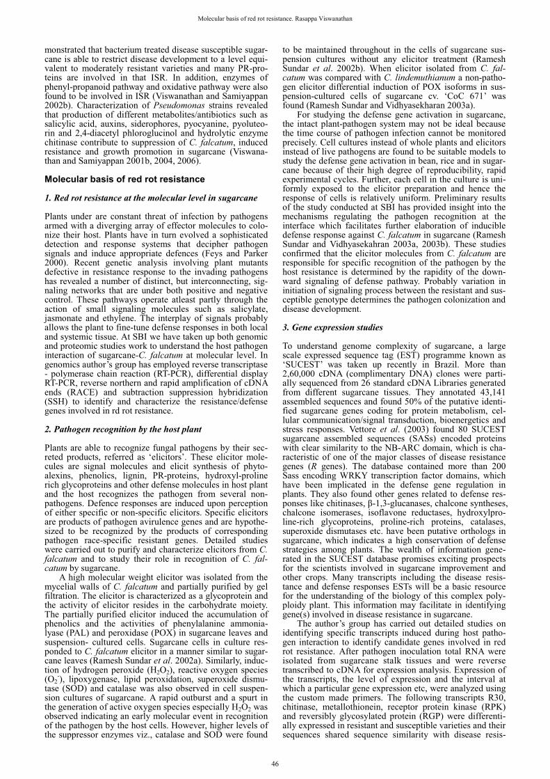
tance genes in other crops. The results also showed an early induction of defense/resistant gene(s) in the resistant variety and in case of susceptible variety, the induction was delayed significantly (Viswanathan et al. 2009a). In further studies, they employed differential display (DD)-RT-PCR a power-ful technique to identify more transcripts involved in patho-gen recognition, signal transduction and plant defense in sugarcane. Differential display technique revealed ~450 transcripts to be differentially expressed upon pathogen ino-culation in sugarcane. About 202 transcripts were selected for their down-regulation and 243 transcripts were selected for their up-regulation upon pathogen inoculation. After homology searches the expressed sequence tags (ESTs) with a match in the databases (both characterized and un-characterized) were categorized into eight groups based primarily on putative function. The total up-regulated and down-regulated transcripts were 63% and 37% respectively, indicating a higher percentage of transcripts that seems to be induced in response to pathogen inoculation. ESTs with a match in the databases were categorized into eight groups based primarily on putative function. Among the known proteins, the signal transduction group included the highest percentage of up-regulated sequences, followed by protein synthesis and storage, general metabolism, transport, defense, cell structure/growth/division, transcription/post transcription and bioenergetics. The important transcripts identified include 14-3-3-like protein, Senescence-associ-ated protein DH, xylanase inhibitor protein 1 precursor, Putative chitinase, Leucine-Rich Repeat family protein, F-box domain containing protein, UMP/CMP kinase-a and Putative hydroxyproline-rich glycoprotein (Viswanathan et al. 2008a) (Fig. 12). Similarly, DD-RT-PCR was conducted with sugarcane cell lines treated with an elicitor molecule identified from red rot pathogen and similar transcripts were found to be differentially regulated upon elicitor treat-ment (Rahul 2010). Further northern and reverse northern blot analyses using radioactive � [32P]dCTP confirmed dif-ferential expression of potential DD transcripts (Viswana-than 2008; Viswanathan et al. 2008b, 2009b). This approach would be more useful to identify the potential transcripts during molecular interaction between the host and the pathogen. Recently Gupta et al. (2009) identified 85 clus-
ters of expressed gene tags (ESTs) that preferentially ex-press upon C. falcatum infection, which were previously unreported. By real time RT-PCR profiling of selected EST clusters they identified several sugarcane clusters that show differential expression in response to biotic and abiotic stress conditions. In addition to six resistant gene analogues, (RGAs) You-Xiong et al. (2007) have isolated a full-length R gene (TIR-NBS-LRR) homologue gene termed SNLR from NCo376, a smut resistant variety in mainland China. They have characterized its expression profile in response to treatments with Sporisorium scitamineum (Syd.) M. Piepenbr., M. Stoll & Oberw 2002 causing smut, salicylic acid and H2O2 by real-time RT-PCR. Further studies in this area of work will be more rewarding and would lead to identifying several candidate genes involved in disease resistance in sugarcane. Using cDNA-AFLP, recently they established differential gene expression in sugarcane during sugarcane-smut pathogen interaction. They found 40 transc-ript-derived fragments (TDFs), 34 newly induced plus six with obvious upregulated expression after infection (Que et al. 2011).
4. Phytoalexin pathway transcripts Since we have established the role of 3-deoxyanthocyanidin phytoalexins in red rot resistance, the genes involved in the flavanoid biosynthetic pathway like coumarate-4-hydro-xylase (C4H), chalcone synthase (CHS), chalcone reductase (CHR), flavanoid 3�-5� hydroxylase (F3’5’H) and flavanoid glycosyl transferase (FGT) were further studied to establish relation between transcript accumulation and differential ac-cumulation of phytoalexins in the resistant and susceptible cultivars (Rahul 2010). The pathogen inoculation enhanced their expression of C4H especially in resistant variety which may have positive correlation with their ability to synthesize various downstream compounds like phenolics and flavonoid compounds as reported earlier from various biochemical studies. Further along the phenypropanoid pathway after the conversion of 4-coumaryl CoA to flavone, the flavone and isoflavonoid pathway is initiated. The CHS and CHR are key enzymes of phenyl-propanoid pathway diverting the substrate, naringenin chalcone to the flavanoid and isoflavanoid branches of the phenyl-propanoid pathway that synthesizes the precursor of a large number of sec-ondary metabolites, including proanthocyanidins, anthocya-nins, flavones, flavonols and isoflavanoid-phytoalexins among others (Zabala et al. 2006). Maximum induction of CHS was found in resistant variety, whereas in susceptible variety this induction was minimal. The F3�5�H transcript was found to be induced upon pathogen inoculation in the leaves of both the varieties and the flavonoid 3�5�-hydro-xylase enzyme functions at an important branch point between flavonol and anthocyanin synthesis, as is evident from studies in petunia (Petunia hybrida), and potato (Sola-num tuberosum). Induction of F3�5�H leads to the synthesis of flavanoids, flavanols, flavanones and phenolics. Reports of plant molecular responses to elicitor or pathogenic infections have pinpointed increase in activity of several genes of the phenyl-propanoid pathway leading to the syn-thesis of phenyl-propanoid metabolites, lignin and flava-noids. In a classical work done by Clive et al. (1999) the ac-cumulation of phytoalexins in sorghum leaves and meso-cotyl tissue after infection with Colletotrichum sublineolum (causing anthracnose) was established. After penetration of the fungus into the host cell it was restricted within infected regions by 72 h and an intense reddish pigmentation was observed by 36 h in the resistant cultivar whereas the susceptible variety the phytoalexin appears only by 48 h with proliferation of the pathogen. The HPLC profile of the phytoalexin from the resistant variety showed presence of luteolinidin, 5-methoxyluteolinidin, apigeninidin and the caffeic acid ester (CAE) of arabinosyl 5-O-apigeninidin, while susceptible variety showed presence of only two fractions apigeninidin and CAE of arabinosyl 5-O-apigeni-nidin. Their temporal studies revealed the accumulation of
Fig. 12 Relative abundance of differential display transcripts iden-tified from C. falcatum inoculated sugarcane (cv ‘Co 93009’), cate-gorized based on their putative functions. Unpublished figure.
47
Molecular basis of red rot resistance. Rasappa Viswanathan
CHS and PR-10 to occur by the 24 h in the resistant variety, while the same occurred only by the 36 h in the susceptible cultivar. Also the level of PR-10 transcript accumulation was lower in the susceptible cultivar than that in the resis-tant cultivar. Timely expression of the specific transcript at the right place in sufficient quantity is a pre-requisite in the development of resistance in a host system. Our studies have clearly shown the differential pattern of spatial ac-cumulation of transcripts of phenylpropanoid pathway in resistant and susceptible varieties after inoculation of C. falcatum in sugarcane.
In a spatial study, transcripts derived from DD-RT PCR and genomic scanning studies were validated for their dif-ferential induction in sugarcane by semi-quantitative RT-PCR. Gene specific forward and reverse primers were de-signed for the selected defense related transcripts viz., xyla-nase inhibitor, glucanase, basal antifungal peptide, POX and isoflavone reductase. After pathogen inoculation in the stalk tissue, spatial expression was carried out in different parts viz., inoculated and adjacent internodes, root, apical meri-stem and third leaf from the top in resistant and susceptible varieties and it established differential regulation of 9 of the 11 transcripts.
5. Rapid amplification of cDNA ends (RACE) The potential defense related transcripts were selected for RACE analysis to clone the full length cDNAs. Compatible primers were designed at the 3� side using the available partial sequence information and used in the RACE proto-col. Using RNA ligase-mediated RACE we have isolated full length sequences of the following four genes viz. 14-3-3 like protein, chitinase, xylanase inhibitor and basal anti-fungal peptide (Viswanathan et al. 2009b). After full length isolation of 14-3-3 like protein by RACE-PCR it has been characterized using bioinformatics tools. The CDD search in the NCBI database revealed the presence of conserved 14-3-3 super family domain, 5�UTR (1-86bp), ORF (87-857 bp) and 3'UTR (858-1094 bp) in the full length sequence of 1094 bp. The 14-3-3 homologues are known to mediate sig-nal transduction by binding to phosphoserine-containing proteins. They are involved in growth factor signalling and also interact with MEK kinases.
6. Characterization of sugarcane chitinase Differential expression of sugarcane chitinase in stalk tis-sues during pathogenesis of C. falcatum was established by RT-PCR studies in a set of resistant and susceptible culti-vars and the result was confirmed by reverse northern anal-ysis. In further studies full length of the gene was isolated and bioinformatic analysis was done to identify its func-tional domains and to predict the three dimensional struc-ture of full length chitinase sequence. Translated sequences revealed the typical characteristics of family 19 glycosyl hydrolase, class I/IV chitinase starting with a signal peptide and ending with a signature domain. Phylogenic study grouped sugarcane chitinase in class IV, based on major deletions in catalytic domain. The close structural template 2DKV (Rice class-I chitinase) was successfully used for the prediction of sugarcane chitinase 3D model (Rahul 2010). The chitinase gene was cloned in an expression vector and studies are in progress to purify the expressed protein and to assess its biological activities.
7. Proteomics approach The term proteome refers to the complete set of proteins that are specified by the genome, and analogous to geno-mics, proteomics describes the study and characterization of this complete set of proteins present in a cell, organ or orga-nism at a given time. Genome-level studies (genomics) reveal or suggest what could theoretically happen, whereas the proteome-level investigations (proteomics) provides in-sights into the actual players involved in mediating specific
cellular processes. In addition, the study of proteins intro-duces another level of complexity at the level of the post-translational modification (PTM) and the biological rele-vance of such modifications. These changes in PTM during the growth and development of organisms (including plants) or in response to stress (including disease) cannot be de-duced from studies investigating genome sequences and/or transcript abundance. Such changes can only be deciphered through proteomics and it is a powerful tool in understan-ding which proteins are present in particular tissue under given conditions and it is one of the fastest growing areas of biological research. In addition to the enzymes, transport, and regulatory proteins many proteins contribute in cas-cades of reactions leading to the metabolites involved in disease resistance. This makes the proteome an essential tar-get for studying metabolic pathways.
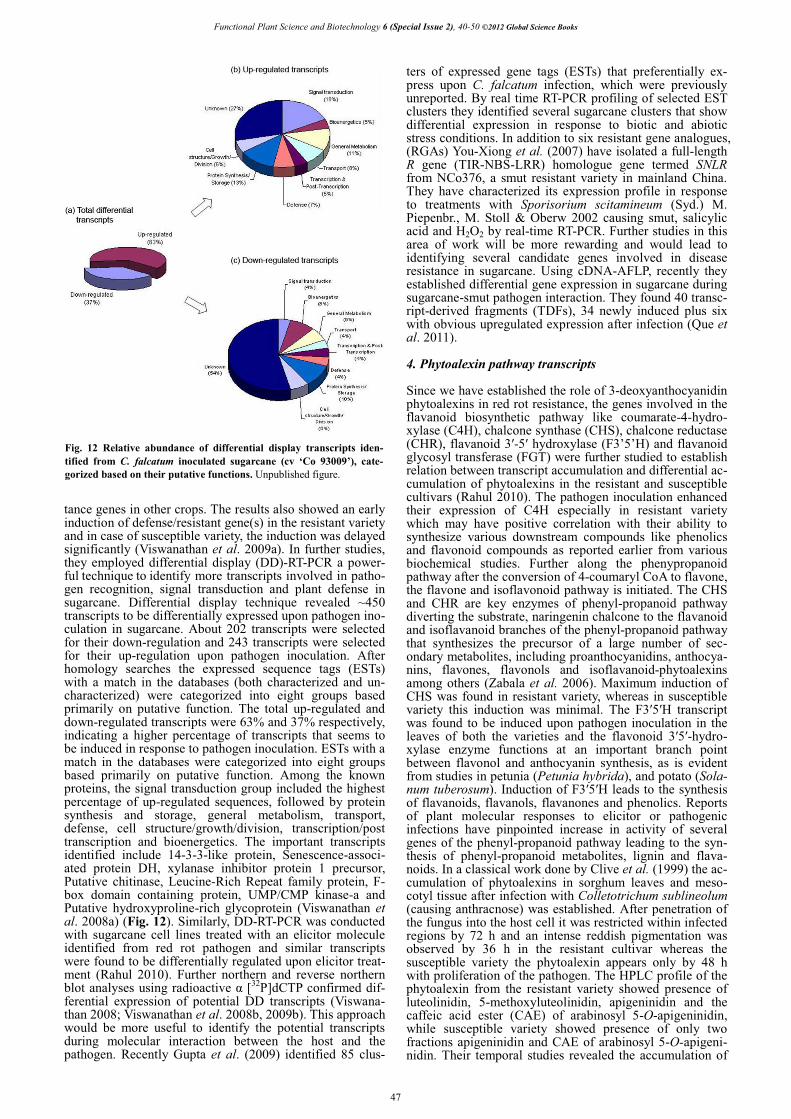
Information on the clearly identified proteins during host-pathogen interactions and defense signaling in sugar-cane is lacking to understand disease resistance mecha-nism(s). Hence we have standardized a protocol for extrac-tion of proteins for 2-DE from the rigid, fibrous sugarcane stalk tissues to identify specific proteins involved in resis-tance to red rot in sugarcane tissues for the proteome anal-ysis. Among the five methods tested, 2D cleanup-phenol method was found to be the most suitable for producing high number of good quality spots and reproducibility. Thirty protein spots commonly present in three methods were selected and subjected to eLD-IT-TOF-MS/MS analy-sis and a reference map has been established for sugarcane stalk tissue proteome. This is the first study on sugarcane stalk proteome analysis which possibly will show a new light on unexplored areas of sugarcane proteome research (Ramesh Sundar et al. 2010). In host pathogen interaction studies the number of protein spots was found to be higher (335 ± 7) in the resistant cultivar after 12 h of pathogen challenge whereas the inoculated susceptible cultivar had the lowest number of protein spots (280 ± 3). More than 250 protein spots that were detected in stalk tissues by pro-
Fig. 13 Identification of proteins (rings) involved in red rot resistance in sugarcane through 2-dimentional gel electrophoresis. Left: Differen-tially induced proteins in the gel; Right: Graphical view of the expressed proteins. Unpublished figure.
48
Functional Plant Science and Biotechnology 6 (Special Issue 2), 40-50 ©2012 Global Science Books
teomic analysis showed reproducible abundance within rep-lications. Approximately 50 protein spots were additionally induced in the resistant cultivar upon pathogen inoculation whereas ~ 24 proteins have disappeared in susceptible cul-tivar (Fig. 13). These studies have established that proteome analysis has the potential to provide significant insights into the molecular events that occur during sugarcane–C. fal-catum interactions and this is the first attempt to standardize proteome analysis and to identify proteins involved in red rot resistance in sugarcane (Viswanathan et al. 2008a). Fur-ther studies using peptide mass finger printing would cha-racterize the up/down regulated proteins and their role(s) in red rot resistance will be established.
CONCLUSION Overall, the studies conducted at SBI revealed definite role of 3-deoxyanthocyanidin phytoalexins and certain PR-pro-teins in red rot resistance. Although they are not the primary determinants of disease resistance they have been well es-tablished as potential antifungal weapons used by the plants to arrest the invading pathogen. Ongoing studies in our lab on identifying molecular basis of red rot resistance would reveal clear understanding of the molecular communication between the host and the pathogen that ultimately decides disease resistance. The newly identified transcripts will be good candidates for in-depth analysis to elucidate C. fal-catum-responsive pathways in sugarcane and facilitate genetic manipulation to tailor this crop for tolerance to red rot and other diseases. Also disease resistance genes identi-fied in our studies that showed differential regulation can provide preferred targets for breeding or to engineer durable disease resistance in sugarcane. Our studies on creating sub-tractive libraries to identify more resistance associated genes are in progress and detailed analyses of gene func-tions of the newly identified genes would help better under-standing of the host resistance mechanisms underlying the response of sugarcane plant to different biotic and abiotic stresses. ACKNOWLEDGEMENTS The author is grateful to Dr N. Vijayan Nair, The Director of the Institute for the constant encouragement and suggestions. Also thankful to the Department of Biotechnology, New Delhi and ICAR, New Delhi for financial support. REFERENCES Agnihotri VP, Madan VK, Lal R (1989) Changes in carbohydrates and nucleic
acids in sugarcane genotypes affected by Colletotrichum falcatum. Interna-tional Sugar Journal 4, 7-8
Ahmad M, Ali R, Fasihi S (1986) Effect of different infection levels of red rot of sugarcane on cane weight and juice quality. Journal of Agricultural Re-search (Pakistan) 24, 129-131
Alexander KC (1995) Constraints in s protection strategy – An overview of constraints in sugarcane disease management. In: Singh GB, Shukla US, Agnihotri VP, Sinha OK, Singh RP (Eds) Sugarcane Production Constraints and Strategies for Research Management of Red Rot, Indian Institute for Sugarcane Research, Lucknow, pp 121-129
Alexander KC, Rao MM (1976) Identification of genetic stocks possessing high resistance to red rot and smut. Sugarcane Pathologist's Newsletter 37, 6-10
Alexander KC, Viswanathan R (2002) Diseases of sugarcane in India and its rapid diagnosis. In: Singh SB, Rao GP, Eswaramoorthy S (Eds) Sugarcane Crop Management, SCI TECH Publishing LLC, Houston, Texas, USA, pp 10-51
Barber CA (1901) Sugarcane diseases in Godawari and Ganjam districts. Mad-ras Department Land Records and Agriculture Bulletin 512 (43), 181-194
Brinkler AM, Siegler DS (1991) Isolation and characterization of piceatannol as a phytoalexin from sugarcane. Phytochemistry 30, 3229-3232
Chona BL (1980) Red rot of sugarcane and sugar industry - a review. Indian Phytopathology 23, 191-207
Clive LS, De verdier K, Nicholson NI (1999) Accumulation of 3-deoxyantho-cyanidin phytoalexins and resistance to Colletotrichum sublineolum in Sorg-hum. Physiological and Molecular Plant Pathology 55, 263-273
de Silva GA, Silva MAAA, de Carvalho PCT (1977) Preliminary studies on
invertases of sugarcane stalks inoculated with C. falcatum Went. Proceedings of International Society Sugar Cane Technologists 16, 407-414
Edgerton CW (1955) Sugarcane and its Diseases, Louisiana State University Press, Louisiana, USA, 290 pp
Feys BJ, Parker JE (2000) Interplay of signaling pathways in plant disease resistance. Trends in Genetics 16, 449-455
Godshall MA, Lonergan TA (1987) The effect of sugarcane extracts on the growth of the pathogenic fungus Colletotrichum falcatum. Physiological and Molecular Plant Pathology 30, 19-42
Gupta V, Raghuvanshi S, Gupta A, Saini N, Gaur A, Khan MS, Gupta RS, Singh J, Duttamajumder SK, Srivastava S, Suman A, Khurana JP, Kapoor R, Tyagi AK (2009) The water-deficit stress- and red-rot-related genes in sugarcane. Functional and Integrative Genomics 10, 207-214
Jaglan BS, Virk KS, Satyavir (1995) Studies on the nature of red rot resistance in sugarcane. In: Singh GB, Shukla US, Agnihotri VP, Sinha OK, Singh RP (Eds) Sugarcane Production Constraints and Strategies for Research Manage-ment of Red Rot, Indian Institute for Sugarcane Research, Lucknow, pp 357-363
Madan KV, Soni N, Nigam M, Solomon S, Agnihotri VP (1991) Enzyme activity and cane genotype resistant to red rot. Sugar Cane 2, 6-8
Malathi P, Viswanathan R, Padmanaban P, Mohanraj D, Ganesh Kumar V, Salin KP (2008) Differential accumulation of 3-deoxy anthocyanidin phyto-alexins in sugarcane varieties varying in red rot resistance in response to Col-letotrichum falcatum infection. Sugar Tech 10, 154-157
Olufolaji DB (1999) Effects of Colletotrichum falcatum (red rot fungus) on the morphology and yield attributes of sugarcane in Nigeria. In: Rao GP, Filho AB, Magarey RC, Autrey LJC (Eds) Sugarcane Pathology (Vol I) Fungal Diseases, Oxford & IBH Publishing Co, New Delhi, pp 155-163
Padmanaban P, Mohanraj D, Viswanathan R, Rao MM, Prakasam N, Jothi R, Alexander KC (1996) Differential interaction of sugarcane clones to pathotypes of Colletotrichum falcatum Went. Sugar Cane 4, 16-20
Pliansinchai U (1999) Trends in sugarcane fungal disease control in Thailand. In: Rao GP, Filho AB, Magarey RC, Autrey LJC (Eds) Sugarcane Pathology (Vol I) Fungal Diseases, Oxford & IBH Publishing Co, New Delhi, pp 209-237
Que Y-X, Lin J-W, Song X-X, Xu L-P, Chen R-K (2011) Differential gene expression in sugarcane in response to challenge by fungal pathogen Ustilago scitaminea revealed by cDNA-AFLP. Journal of Biomedicine and Biotech-nology 2011, Article ID 160934
Rahul PR (2010) Transcriptomic analysis of defense-related gene expression in sugarcane-Colletotrichum falcatum interaction. PhD thesis, Bharathiar Uni-versity, Coimbatore, 198 pp
Ramesh Sundar A, Nagarathinam S, Ganesh Kumar V, Rahul PR, Raveen-dran M, Malathi P, Agrawal GK, Rakwal R, Viswanathan R (2010) Sugarcane proteomics: Establishment of a protein extraction method for 2-DE in stalk tissues and initiation of sugarcane proteome reference map. Elec-trophoresis 31, 1959-1974
Ramesh Sundar A, Velazhahan R, Vidhyasekaran P (2002a) A glycoprotein elicitor isolated from Colletotrichum falcatum induces defense mechanisms in sugarcane leaves and suspension-cultured cells. Journal of Plant Disease and Protection 109, 601-611
Ramesh Sundar A, Velazhahan R, Viswanathan R, Vidhyasekharan P (2002b) Induction of active oxygen species (AOS), lipoxygenase and lipid peroxidation in suspension-cultured sugarcane cells by a glycoprotein elicitor from Colletotrichum falcatum. Journal of Plant Disease and Protection 109, 441-451
Ramesh Sundar A, Vidhyasekaran P (2003a) Differential induction of phe-nylpropanoid metabolites in suspension-cultured cells of sugarcane by fungal elicitors. Acta Phytopathogica Entomologica et Hungarica 38, 29-42
Ramesh Sundar A, Vidhyasekaran P (2003b) Induction of defense-related biochemical changes by elicitors of red rot pathogen and a non-pathogen in sugarcane cell culture. Indian Phytopathology 56, 255-261
Ramesh Sundar A, Viswanathan R (1997) Expression of defence response at cellular level in sugarcane against Colletotrichum falcatum Went causing red rot disease. In: Proceedings of Symposium on Economically Important Dis-eases of Crop Plants, Dec. 18-20, Indian Phytopathological Society, Banga-lore, 66 pp
Ramesh Sundar A, Viswanathan R, Mohanraj D, Padmanaban P (1998) Role of oxidative enzymes in sugarcane and Colletotrichum falcatum Went interaction. Acta Phytopathogica Entomologica et Hungarica 33, 297-304
Ramesh Sundar A, Viswanathan R, Mohanraj D, Padmanaban P (1999) Studies on possible use of pathogen toxin as a molecular marker for red rot resistance in sugarcane. Acta Phytopathogica Entomologica et Hungarica 34, 211-217
Rao KC, Krishnamoorthy TN, Lalitha E, Rajalakshmi C (1968) Phenols in relation to resistance of sugarcane varieties to red rot. Current Science 37, 532
Satyavir, Kumar A, Raj K, Virk KS (2002) Red rot of sugarcane: The re-search scene in haryana. In: Singh SB, Rao GP, Eswaramoorthy S (Eds) Sugarcane Crop Management, SCI TECH Publishing LLC, Houston, Texas, USA, pp 109-126
Singh J, Singh M, Chandra P, Rao GP, Singh HN (1993) Biochemical studies on resistance to red rot in sugarcane. Sugar Cane 6, 16-19
49
Molecular basis of red rot resistance. Rasappa Viswanathan
Singh K, Singh RP, Agnihotri VP (1976) Phenolics in relation to sugarcane resistance against red rot disease. Sugarcane pathologist’s Newsletter 15/16, 37-41
Singh RP, Lal S (2000) Red rot. In: Rott P, Bailey RA, Comstock JC, Croft BJ, Saumtally AS (Eds) A Guide to Sugarcane Diseases, CIRAD/ISSCT, Mont-pellier, France, pp 153-158
Singh ON, Waraitch KS (1977) Metabolic changes induced by Colletotrichum falcatum Went in sugarcane. Sugarcane Pathologist's Newsletter 19, 7-9
Snyder BA, Leite B, Hipskind J, Butler LG, Nicholson RL (1991) Accumu-lation of sorghum phytoalexins induced by Colletotrichum graminicola at the infection site. Physiological and Molecular Plant Pathology 39, 463-470
Srinivasan KV (1969) Physiology of disease resistance in sugarcane with par-ticular reference to red rot. Proceedings of the Indian Academy of Sciences, Section B 59, 120-132
Srinivasan KV, Alexander KC (1971) Sources of resistance in the different species of Saccharum to red rot and smut disease of sugarcane. Sugarcane Pathologist's Newsletter 6, 6-7
Srinivasan KV, Bhat NR (1961) Red rot of sugarcane – criteria for grading re-sistance. Journal of the Indian Botanical Society 40, 566-577
Srivastava BL, Solomon S (1990) Biochemical analysis of induced mutant of sugarcane (Saccharum sp). In: Proceedings of an International Symposium on Genetic Engineering, Plant Tissue Culture in Relation to Disease and Pest Resistance in Crop Plants, Tamil Nadu Agricultural University, Coimbatore, India, 56 pp
Sundara B (1998) Sugarcane Cultivation, Vikas Publishing House Private Ltd., New Delhi, India, 392 pp
Thirupathiraja C, Viswanathan R, Padmanaban P (2004) Time course of peroxidase accumulation in sugarcane cultivars in response to Colletotrichum falcatum infection. Sugar Tech 6 (1&2), 47-52
Varma SC, Mittal SP (1949) The structure of xylem vessels in the nodal region of sugarcane in relation to its resistance to red rot (Colletotrichum falcatum Went.) Part III. Indian Journal of Agricultural Sciences 19, 383-387
Vettore AL, da Silva FR, Kemper EL, Souza GM, da Silva AM, Ferro MIT, Henrique-Silva F, Giglioti EA, Lemos MVF, Coutinho LL, Nobrega MP, Carrer H, Franca SC, Bacci Jr. M, Goldman MHS, Gomes SL, Nunes LR, Camargo LE A, Siqueira WJ, Van Sluys MA, Thiemann OH, Kura-mae EE, Santelli RV, Marino CL, Targon MLP, Ferro JA, Silveira HCS, Marini DG, Lemos EGM, Monteiro-Vitorello CB, Tambor JHM, Carraro DM, Roberto PG, Martins VG, Goldman GH, de Oliveira GC, Truffi D, Colombo CA, Rossi M, de Araujo PG, Sculaccio SA, Angella A, Lima MMA, de Rosa Jr. VE, Siviero F, Coscrato VE, Machado MA, Grivet L, Di Mauro SMZ, Nobrega FG, Menck CFM, Braga MDV, Telles GP, Cara FAA, Pedrosa G, Meidanis J, Arruda P (2003) Analysis and functional an-notation of an expressed sequence tag collection for tropical crop sugarcane. Genome Research 13, 2725-2735
Viswanathan R (2001) Accumulation of anthocyanin compounds in sugarcane tissues as a factor responsible for red rot resistance. Madras Agricultural Journal 88, 571-576
Viswanathan R (2008) Molecular mechanism(s) involved in sugarcane-Col-letotrichum falcatum interaction. In: Proceedings of National Seminar on Ad-vances in Plant Pathology for Sustainable Agriculture, Nov. 24-25, 2008, TNAU, Coimbatore, pp 57-58
Viswanathan R (2010) Plant Disease: Red Rot of Sugarcane, Anmol Pub-lishers, New Delhi, India, 306 pp
Viswanathan R, Malathi P, Ramesh Sundar A, Aarthi S, Premkumari SM, Padmanaban P (2005) Differential induction of chitinases and thaumatin-like proteins in sugarcane in response to infection by Colletotrichum fal-catum causing red rot disease. Journal of Plant Diseases and Protection 112, 537-542
Viswanathan R, Mohanraj D, Padmanaban P (1996a) Rapid assay of peroxi-dase in sugarcane tissues in relation to disease resistance. In: Proceedings of Management of Threatening Diseases of National Importance, Feb. 14-16 1996, Ludhiana, p 19 (Abstract)
Viswanathan R, Mohanraj D, Padmanaban P (1996b) Role of red rot pig-ments in relation to red rot resistance in sugarcane. Indian Journal of Sugar-cane Technology 11, 151-154
Viswanathan R, Mohanraj D, Padmanaban P (2000a) Possible involvement of anthocyanin compounds in resistance of sugarcane red rot. Indian Phyto-pathology 53, 311-313
Viswanathan R, Mohanraj D, Padmanaban P, Alexander KC (1996c) Ac-cumulation of 3-deoxyanthocyanidin phytoalexins luteolinidin and apigenini-din in sugarcane in relation to red rot disease. Indian Phytopathology 49, 174-175
Viswanathan R, Mohanraj D, Padmanaban P, Alexander KC (1996d) Syn-thesis of phytoalexins in sugarcane in response to infection by Colletotri-chum falcatum Went. Acta Phytopathologica et Entomologica Hungarica 31, 229-237
Viswanathan R, Padmanaban P, Mohanraj D (1997) Growing virulence of red rot pathogen of sugarcane in Tamil Nadu. Indian Sugar 47, 23-30
Viswanathan R, Ramesh Sundar A, Padmanaban P, Mohanraj D (2002) Red rot disease of sugarcane and its management. In: Upadhyay RK, Muk-herji KG, Dubey OP (Eds) IPM Systems in Agriculture (Vol 8) Key Pathogens and Diseases, Aditya Books, New Delhi, pp 277-301
Viswanathan R, Malathi P, Padmanaban P (2003a) Variation in sugarcane red rot pathogen Colletotrichum falcatum Went. In: Rao GP, Manoharachari C, Bhat DJ, Rajak RC, Lakhanpal TN (Eds) Frontiers of Fungal Diversity in India, International Book Distributing Co, Lucknow, pp 639-667
Viswanathan R, Malathi P, Ramesh Sundar A, Aarthi S, Premkumari SM, Padmanaban P (2005) Differential induction of chitinases and thaumatin-like proteins in sugarcane in response to infection by Colletotrichum fal-catum causing red rot disease. Journal of Plant Diseases and Protection 112, 537-542
Viswanathan R, Nandakumar R, Samiyappan R (2003b) Involvement of pathogenesis-related proteins in Pseudomonas spp. induced systemic resis-tance against Colletotrichum falcatum in sugarcane. Journal of Plant Dis-eases and Protection 110, 524-537
Viswanathan R, Ramesh Sundar A, Malathi P (2008a) Identification and characterization of differentially expressed genes/proteins involved in sugar-cane - red rot pathogen interaction. Annual Report, Sugarcane Breeding Insti-tute for 2007-2008, Coimbatore, pp 51-52
Viswanathan R, Ramesh Sundar A, Malathi P, Rahul PR, Ganesh kumar V, Prathima PT, Raveendran M, Kumar KK, Balasubramanian P (2008b) Defense genomics of sugarcane-Colletotrichum falcatum interaction by mRNA differential display. In: Proceedings of a National Conference on Genomics, Proteomics and Systems Biology, Oct. 1-3, 2008, IISc, Bangalore, India, No 2GOP
Viswanathan R, Ramesh Sundar A, Malathi P, Rahul PR, Ganesh kumar V, Banumathy R, Prathima PT, Raveendran M, Kumar KK, Balasubrama-nian P (2009a) Interaction between sugarcane and Colletotrichum falcatum causing red rot: Understanding disease resistance at transcription level. Sugar Tech 11, 45-50
Viswanathan R, Ramesh Sundar A, Malathi P (2009b) Identification and characterization of differentially expressed genes/proteins involved in sugar-cane - red rot pathogen interaction. Annual Report, Sugarcane Breeding Insti-tute for 2008-09, Coimbatore, India, pp 41-42
Viswanathan R, Samiyappan R (1999) Red rot disease in sugarcane: a major constraint for the Indian Sugar Industry. Sugar Cane 5, 9-15
Viswanathan R, Samiyappan R (2001a) Role of chitinases in Pseudomonas spp. induced systemic resistance against Colletotrichum falcatum in sugar-cane. Indian Phytopathology 54, 418-423
Viswanathan R, Samiyappan R (2001b) Antifungal activity of chitinases pro-duced by some fluorescent pseudomonads against Colletotrichum falcatum Went causing red rot disease in sugarcane. Microbiological Research 155, 309-314
Viswanathan R, Samiyappan R (2002a) Induced systemic resistance by fluo-rescent pseudomonads against red rot disease of sugarcane caused by Col-letotrichum falcatum. Crop Protection 21, 1-10
Viswanathan R, Samiyappan R (2002b) Role of oxidative enzymes in the plant growth promoting rhizobacteria (PGPR) mediated induced systemic re-sistance against Colletotrichum falcatum in sugarcane. Journal of Plant Dis-eases and Protection 109, 88-100
Viswanathan R, Samiyappan R (2004) Secondary metabolites production by Pseudomonas spp. strains antagonistic to Colletotrichum falcatum causing red rot disease in sugarcane. Acta Phytopathologica et Entomologica Hun-garica 39, 28-37
Viswanathan R, Samiyappan R (2006) Pseudomonas spp. colonization in sugarcane rhizosphere reduces titre of Colletotrichum falcatum Went-causing red rot disease of sugarcane. Archives of Phytopathology and Plant Protec-tion 39, 39-44
Viswanathan R, Samiyappan R (2008) Bio-formulation of fluorescent Pseudomonas spp. induces systemic resistance against red rot disease and enhances commercial sugar yield in sugarcane. Archives of Phytopathology and Plant Protection 41, 377-388
Wilson KI, Srivastava DN (1970) Phenolic content of sugarcane in relation to red rot disease. Experientia 16, 10-74
You-Xiong Q, Jian-Wei L, Ji-Sen Z, Miao-Hong R, Li-Ping X, Mu-Qing Z (2007) Molecular cloning and characterisation of a non-tir-nbs-lrr type dis-ease resistance gene analogue from sugarcane. Proceedings of the Internatio-nal Society of Sugar Cane Technologists 26, 927-931
Went FAFC (1893) Het root snot. Archives Jora-Suikerind 1, 265-282 Zabala G, Zou J, Tuteja J, Gonzalez DO, Clough SJ, Vodkin LO (2006)
Transcriptome changes in the phenylpropanoid pathway of Glycine max in response to Pseudomonas syringe infection. BMC Plant Pathology 6, 1-28
50