Embed Size (px)
Citation preview

This is a n Op e n Acces s doc u m e n t dow nloa d e d fro m ORCA, Ca r diff U nive r si ty 's
ins ti t u tion al r e posi to ry: h t t p s://o rc a.c a r diff.ac.uk/921 6 8/
This is t h e a u t ho r’s ve r sion of a wo rk t h a t w as s u b mi t t e d to / a c c e p t e d for
p u blica tion.
Cit a tion for final p u blish e d ve r sion:
M a rino, Silvia, S t ain es , Kat h e rin e Ann, Brow n, Gen evieve, H ow a r d-Jone s,
R ac h el Anne a n d Ada m czyk, M a g d ale n a 2 0 1 6. Mod els of ex vivo expla n t
c ul t u r e s: a p plica tions in bo n e r e s e a r c h. Bon eKEy Repo r t s 5 , 8 1 8.
1 0.1 0 3 8/bon ek ey.201 6.49 file
P u blish e r s p a g e: h t t p://dx.doi.o rg/10.10 3 8/bo n ek ey.2016.4 9
< h t t p://dx.doi.o rg/10.10 3 8/bon ek ey.2016.4 9 >
Ple a s e no t e:
Ch a n g e s m a d e a s a r e s ul t of p u blishing p roc e s s e s s uc h a s copy-e di ting,
for m a t ting a n d p a g e n u m b e r s m ay no t b e r eflec t e d in t his ve r sion. For t h e
d efini tive ve r sion of t his p u blica tion, ple a s e r ef e r to t h e p u blish e d sou rc e. You
a r e a dvise d to cons ul t t h e p u blish e r’s ve r sion if you wish to ci t e t his p a p er.
This ve r sion is b ein g m a d e av ailable in a cco r d a n c e wit h p u blish e r policie s.
S e e
h t t p://o rc a .cf.ac.uk/policies.h t ml for u s a g e policies. Copyrigh t a n d m o r al r i gh t s
for p u blica tions m a d e available in ORCA a r e r e t ain e d by t h e copyrig h t
hold e r s .

OPEN
LABORATORY METHODS
Models of ex vivo explant cultures: applicationsin bone researchSilvia Marino1, Katherine Ann Staines2, Genevieve Brown3, Rachel Anne Howard-Jones4 andMagdalena Adamczyk1,4
1Academic Unit of Bone Biology, Department of Oncology and Metabolism, Mellanby Centre for Bone Research, Medical
School, The University of Sheffield, Sheffield, UK. 2Roslin Institute and R(D)SVS, The University of Edinburgh, Edinburgh,
UK. 3Department ofBiomedical Engineering,ColumbiaUniversity,NewYork,USA. 4Oral andBiomedical Sciences,College
of Biomedical and Life Sciences, Cardiff University, Cardiff, UK.
Ex vivo explant culture models are powerful tools in bone research. They allow investigation of bone and cartilage
responses to specific stimuli in a controlled manner that closely mimics the in vivo processes. Because of limitations in
obtaining healthy human bone samples the explant growth of animal tissue serves as a platform to study the complex
physico-chemical properties of the bone. Moreover, thesemodels enable preserving important cell–cell and cell–matrix
interactions in order to better understand the behaviour of cells in their natural three-dimensional environment. Thus, the
use of bone ex vivo explant cultures can frequently be of more physiological relevance than the use of two-dimensional
primary cells grown in vitro. Here, we describe isolation and ex vivo growth of different animal bone explant models
including metatarsals, femoral heads, calvaria, mandibular slices and trabecular cores. We also describe how these
explants are utilised to study bone development, cartilage and bone metabolism, cancer-induced bone diseases, stem
cell-driven bone repair and mechanoadaptation. These techniques can be directly used to understand mechanisms
linked with bone physiology or bone-associated diseases.
BoneKEy Reports 5, Article number: 818 (2016) | doi:10.1038/bonekey.2016.49; published online 29 June 2016
Introduction
The bone is a complex and specialised connective tissue in astate of dynamic equilibrium that, together with cartilage, formsthe skeletal system. Active reorganisation of bone micro-structure is achieved through bone remodelling or modelling,processes that are mediated by osteoclasts, osteoblasts andosteocytes.1 Remodelling represents coordinated changes inbone resorption and formation, which occur throughout life,in order to maintain health and integrity of the skeleton.Bonemodelling is an adaptation of bone to external stimuli andis generally limited to growth and responses to mechanicalloading. Ex vivo growth of bone explants offers a uniqueopportunity to study these mechanisms in a controlledexperimental setting where both the three-dimensional orga-nisation as well as the cellular diversity of the bone are pre-served. In addition, the ex vivo cultures can help theinvestigation of mechanisms underlying bone growth,bone and cartilage matrix turnover, mechanical loading,
or interactions with other cell types and advance our under-standing of bone physiology. The aim of this collective protocolis to provide a detailed description of the procedures that arecurrently used to isolate, culture and characterise a variety ofanimal bone explant culture models and include the applica-tions of thesemodels in bone research (Figure 1). This protocolcombines previously published protocols and those optimisedin our laboratories and offers the technical hintsand suggestions for each of the methods. This protocol alsoprovides a list of additional reading that should help theresearcher with establishing these procedures at their homeinstitution.
Ex vivo explant model to study linear bone growth
The mouse metatarsal culture model can be used to improveour understandingof bonedevelopment. Thismodel, pioneeredby Burger and Van-Delft in the Netherlands (1976),2 is a highlyphysiological ex vivo model for studying endochondral
Correspondence: Dr M Adamczyk, Academic Unit of Bone Biology, Department of Oncology and Metabolism, Mellanby Centre for Bone Research, Medical School, The
University of Sheffield, BeechHill Road, Sheffield S10 2RX, UK orMatrix Biology and Tissue Repair Research Unit, College of Biomedical and Life Sciences, School of Dentistry,
Cardiff University, Heath Park, Cardiff CF14 4XY, UK.
E-mail: [email protected] or [email protected]
Received 27 January 2016; accepted 4 May 2016; published online 22 June 2016
Citation: BoneKEy Reports 5, Article number: 818 (2016) | doi:10.1038/bonekey.2016.49
& 2016 International Bone & Mineral Society All rights reserved 2047-6396/16www.nature.com/bonekey
BoneKEy Reports | JUNE 2016 1

ossification, the process by which the cartilage scaffold isreplaced by mineralised bone. This process is tightly regulatedin healthy individuals in order to prevent abnormal developmentand/or longitudinal bone growth.3 The metatarsal model allowsfor the study of linear bone growth, as the growth rate of thebones in culture mimics that seen in vivo. Moreover, thismodel enables investigation of chondrocyte proliferation andhypertrophy, extracellular matrix production and tissuemineralisation. The method described in this protocol relatesto murine metatarsal explant cultures; however, there arenumerous papers detailing similar cultures in rats4 (Method 1).
Ex vivo explant model to study bone and cartilage
metabolism
The mouse femoral head culture model can be used toinvestigate the expression and turnover of specific markerswithin bone and cartilage. This is useful to help understand themechanisms linked with pathogenesis of osteoarthritis, wherethere is a clear association between articular cartilagedegeneration and subchondral bone changes.5,6 This modelwas developed by Stanescu and Leibovich (1982),7 and overthe years it has been primarily used to understandmechanismsof cartilage breakdown. For instance, thismodel was effectivelyapplied by Glasson et al.8 to demonstrate the importance ofaggrecan proteolytical cleavage and neoepitope formationduring osteoarthritis progression.8Uniquely, themouse femoralhead culture model can also be used for simultaneousinvestigation of bone modelling and cartilage extracellularmatrix degradation occurring during explant culture9 (Method 2).
Ex vivo explant model to study cancer cell-induced bone
disease
The mouse calvaria ex vivo culture model has been routinelyused to investigate bone resorption,10 bone formation11 andbone regeneration/healing process.12 By using an adaptationof the calvaria explant culture model, it is also possible tostudy cancer-induced bone metastases ex vivo as shown bySophocleous et al.13 and others. Cancer to bone metastasis ismediated by the secretion of soluble factors by cancer cells,which stimulate the proliferation and activity of osteoblasts andosteoclasts, affecting both bone formation as well as boneresorption. The method described in this protocol gives anexample of the isolation and culture of calvaria derived frommousepups; however, similar cultures canbe established usingthe calvarial bones obtained from rats14 or chicken embryos15
(Method 3).
Ex vivo explant model to study stem cell behaviour during
bone repair
A rat mandible slice model was initially established toinvestigate inflammatory bone destruction.16 This model was
adapted by Colombo and colleagues to enable the study ofdental pulp stem cell (DPSC) behaviour in an ex vivo envir-onment.17DPSCs have characteristics similar to mesenchymalstem cells18,19 and display migratory and odontoblast differ-entiation capacity in response to tissue damage.19–21 StudyingDPSC behaviour in an ex vivo mandible slice enables under-standing of their multi-potency and how they may initiate bonerepair in response to injury (Method 4).
Ex vivo explant model to study bone response to loading
The bovine trabecular cores culture model is extremely usefulwhen investigating bone changes in response to mechanicalstimulation. Osteocytes – themechanosensors in bone—directthese processes by regulating osteoclast and osteoblastactivities;22 yet, few models isolate osteocyte interactions. Themodel described herein allows for the study of osteocyte–osteoblast interactions in a long-term ex vivo culture and willfurther our understanding of bone mechanoadaptation.23,24
Although themethoddescribed hereutilises perfusionof bovinebone cores,25–28 similar systems using rabbit trabecular boneexplants and whole bone organ cultures have recently beenreported29,30 (Method 5).
Materials and methods
Dissections
All dissections should be conducted aseptically using sterileequipment and in a dissection hood or tissue culture hood tomaintain a sterile environment.
Cell and explant cultures
All tissue culture procedures are carried out in a sterile laminarflow tissue culture hood. Cells and ex vivo cultures aremaintained in an incubator at 37 1C and 5% CO2. Cell culturereagents should be sterile and pre-warmed to 37 1C unlessotherwise indicated in the protocols. Cells can be grown in thestandard tissue culture-treated plastic culture dishes/platesunless otherwise indicated.
Use of animals
All procedures described in this protocol were performed withpermission of local Animal Health andWelfare Committees andin accordance with guidelines and regulations of the localHome Offices. Animal work should be carried out usingcountry-specific guidelines and regulations.
Method 1
Although the metatarsal bones of the foot develop in a similarmanner to the long bones, it is onlymetatarsals two through fivethat develop true growth plates. Commonly, the metatarsalorgan culture is established using postnatal (PN) mice at stages
Figure 1. Models of animal ex vivo bone explant cultures and their applications.
Explant culture models in bone research
S Marino et al
2 JUNE 2016 | www.nature.com/bonekey

PN1–3. This helps delineate themechanisms surrounding bonegrowth, as the primary ossification centre has completelydeveloped in themetatarsal bones at this stage. It has, however,previously been recognised that PN bones have limited growthpotential in culture.31 As such, metatarsal bones fromembryonic (E) stages E17/18 are commonly used for thistechnique as the primary ossification centre has begun toform.32More recently, earlier embryonic stages (E15) have beenused, as these bones have yet to start ossifying and as suchmatrix mineralisation can be more precisely examined.33
Specific materials
Preparationmedium. PBS (phosphate-buffered saline) with 13%MEMa (Minimum Essential Media alpha) without ribonucleo-sides and0.2%BSA (Fraction V); culturemedia:MEMamediumwithout ribonucleosides supplemented with 0.2% BSA(Fraction V), 5 mgml� 1
L-ascorbic acid phosphate, 1mM b-glycerophosphate, 0.05mgml� 1 gentamicin and 1.25 mgml� 1
fungizone; equipment: dissecting microscope, curved micro-scissors and fine forceps (for example, Dumont #5).
NOTE 1: Quality dissection equipment is required; inparticular, the forceps and scissors should be kept sharp soas to ensure successful and easy dissection.
Animals. Pregnant dam at the desired stage or PN pups.
NOTE 2: When choosing the age of metatarsal bone culture,it is important to consider the outcomes required. Forexamination of promoters of matrix mineralisation, it isrecommended that E15 is used. For assessment of endochon-dral bone growth over a long time period and inhibition ofmineralisation, either E17/E18 or PN bones are recommended.As discussed above, PN bones do have a limited growthpotential in culture and, as such, oPN3 is recommended ifsufficient growth is required for data analysis.
Embryonic and PN metatarsal dissection
1. Conduct all steps aseptically using sterile equipment and in atissue culture hood to maintain a sterile environment.
2. If isolating embryonic metatarsals, cull the pregnant dam atthe desired stage using cervical dislocation. If harvesting PNmetatarsals, cull thePNpupsbydecapitation andgostraightto step 5.
3. Fix the dam on her back by her legs and arms. Open theabdominal cavity with a single cut to expose the uterus.
4. Lift and cut out the uterine horns, placing them into a 10-cmculture dish. Remove the individual foetuses from their sacsand placenta.
5. Remove the hind limbs of the pups and place in preparationmedia until required.
6. Under a dissecting microscope, remove the skin from thehind limb by holding the upper hind limb with forceps andgently inserting the curved scissors and cutting down theback of the hind limb into the footpad. Then, separate theskin from the bones gently, as if pulling off a glove.
NOTE3: In the dissection of E15metatarsals, skin removalby scissors is not required and access to the unmineralisedmetatarsals is best carried out by the careful use of fine
forceps to remove overlying tissue. Because of the muchsmaller size and less rigid nature of E15metatarsals, this is amuchmore delicate procedure than that in older E17/18 andPN pups.
7. Holding the tarsals in the foot with forceps, gently disrupt theconnective tissue between the individual metatarsals byrunning the fine forceps in between them.
8. Remove the first and fifth metatarsals by pinching off withforceps and discard them.
9. Separate the phalangeal and tarsi bones from the remainingmiddle three metatarsals by careful pinching off by forceps(do not attempt to cut or use a scalpel; Figure 2a). Transferinto the preparation medium until all metatarsal bones aredissected.
NOTE 4: Although every effort should bemade to remove thesurrounding connective tissues, any remaining tissue willdisappear after 24 h in culture.
Metatarsal culture
1. Culture metatarsal bones individually in a well of a 24-wellplate with 200ml per well of culture medium.
2. Place in an incubator at 37 1C/5% CO2.3. E17/E18 and PN metatarsal cultures may have the media
changed every third day throughout the culture period.However, it is not recommended to change the media, atleast for the first 5 days, when metatarsal bones at anembryonic age of less than E17/E18 cultures are used,as this prevents their matrix mineralisation capability(Figure 2b). After 5 days, the media on embryonicmetatarsals oE17 can then be changed every third day.
4. Culture metatarsals bones for up to 2 weeks depending onthe outcomes required.
Analysis of endochondral bone growth and matrix
mineralisation using metatarsal cultures
In culture, metatarsal bones will grow at a near physiologicalrates, and as such total bone length measurements indicative oflongitudinal endochondral growth can be made over the cultureperiod using a microscope with a camera attached and imageanalysis software32–34 (for example, ImageJprogram; Figures 2cand 3a). Metatarsals may be cultured with [3H]-thymidine35 orBrdU32 in the final hours of culture and then examined by a liquidscintillation counter or processed to wax, respectively andreacted with an anti-BrdU antibody, to examine cell proliferation.Following culture, bones can be paraffin embedded and sectionscut for standard haematoxylin and eosin (H&E) to examine themorphology and sizes of the resting, proliferative and hyper-trophic zones of chondrocytes (Figure 3a).33
Longitudinal measurements of the central mineralisationzone can also be made to examine the rate of chondrocytematrix mineralisation. This can be done by directly imaging andthen using image analysis software, as described in Figures 2c
and 3b and c. This is, however, a two-dimensionalmeasurement; therefore, it is recommended that further ana-lyses are performed so as to fully determine themineralisation status of the metatarsal bones post culture, forexample, using micro-computed tomography (microCT; seeadditional methods for details).
Explant culture models in bone research
S Marino et al
BoneKEy Reports | JUNE 2016 3

Figure 2. Timeline for the mouse metatarsal culture. On day 0, the middle three metatarsal bones from either embryonic (E) or postnatal mice are dissected (a) and cultured forup to 14 days. During this time, periodic measurements of the total length and the length of the mineralisation zone are made. Scale bar¼ 200mm. (b) Media changes are requiredevery third day for metatarsal bones at embryonic stage E17 and above and postnatal metatarsal cultures (4E17). However, for metatarsal bones extracted at an embryonic stageearlier than this, e.g. E15 (oE17), it is not recommended to change themedia for at least 5 days after dissection. Themedia can then be changed every third day. (c) Change in bonegrowth during explant culture.
Figure 3. Mineralisation of metatarsal explants during culture. An E17 metatarsal bone was stained with Haemotoxylin and Eosin, and von Kossa stain after 7 days in culture. Clearlyvisible are the proliferative (PZ) and hypertrophic (HZ) zones of chondrocytes as defined by their well-characterisedmorphology, as well as themineralisation zone (MZ) stained black by vonKossa staining. Scale bar¼ 200mm. (a) Total length increases of E18metatarsal bones cultured for up to 10 days (n¼ 6) (b). Increase in totalmineralisation zone length as a percentage ofthe total length of the E18 metatarsal bones (n¼ 6). Significance is in comparison with previous culture time point, *Po0.05; **Po0.01; ***Po0.001 n¼ 6 metatarsal bones (c).
Explant culture models in bone research
S Marino et al
4 JUNE 2016 | www.nature.com/bonekey

Method 2
The ex vivo culture of femoral heads isolated fromyoungmice isbeing predominantly used in cartilage research36 because ofthe developing femoral head consisting mainly of proliferatingchondrocytes.9 However, when mice become around 3 weeksold, the chondrocytes undergo hypertrophy and are beingslowly replaced, first by woven (around 6- to 9-week old) andthen by trabecular bone (around 9- to 12-week old).9 Impor-tantly, secondary ossification of the femoral head begins whenanimals reach adulthood.37 Thus, the equilibrium betweenmarkers associated with cartilage or bone will change duringanimal development. The state of tissue remodelling orresponse to specific stimuli can be measured directly byanalysing the conditionedmedium, extracting proteins from theexplanted tissue, or by studying tissue histomorphology onfixed specimens. Here we describe a step-by-step techniquefor the isolation and culture of femoral heads from adult micethat allows for analysis of cartilage and bone markers.
Specific materials
Culture medium. DMEM (Dulbecco’s Modified Eagle Medium)with GlutaMAX (Waltham, MA, USA) and antibiotics(100mgml� 1 streptomycin, 100Uml� 1 penicillin); PBS; 4%paraformaldehyde (PFA)/PBS solution, and 15% ethylene-diaminetetraacetic acid disodium salt (EDTA)/0.5% PFAsolution; equipment: blunt and fine scissors, fine and curvedforceps, scalpel and sterile blades.
Animals. Balb/C mice at 11 weeks of age or C57BL/6 at15 weeks of age.
NOTE 5: It is important to consider the genetic background ofmice, as each strain may vary in endochondral and secondaryossification processes and overall cartilagematrix compositionand bone structure.38
NOTE 6: It is recommended to use a minimum of 4 femoralheads (dissected from 4 mice) per experimental condition byplacing each explant in a separate well of a 96-well plate. Eachtreatment groupwill consist of 4 biological replicates,which allowsfor sample matching and more robust statistical analysis, forexample, using paired Student’s t-test. If longer culture times ordifferentanalysis is required, then twofemoralheadscanbeplacedtogether in awell of a 48-well plate as described byStanton et al.36
Dissection of mice
1. Kill an appropriate number of mice by cervicaldislocation.The remaining procedure must be performedin a sterile class II cabinet.
2. Spray mouse legs with 70% ethanol for disinfection.3. Pull up the skin in the mid-abdomen region and cut it with
blunt scissors to reveal muscles and internal body.4. Peel off as much of the skin as possible to avoid
contamination with fur.5. Spray hind legs with 70% ethanol.6. Using a scalpel blade, make incisions on both sides of the
spine vertebrae. Cut the muscles surrounding the pelvicgirdle using dissection scissors. Cut the pelvis in half andgently remove each leg together with the intact acetabulum.Make sure that the hip joint is intact and remains attached tothe pelvic bone. Repeat the process for the second leg.
Isolation of femoral heads
1. Transfer both legs into a 10-cm culture dish containing20ml sterile PBS solution (Figure 4a).
2. Pick up one of the legs using sterile forceps.3. Grab both sides of pelvic bone with your fingers so that the
acetabulum remains in the middle (Figure 4b).NOTE 7: Move your fingers as close to the joint capsule
as possible, as this will help break the pelvic bone.4. Using your fingers, break the pelvic bone in half to
disarticulate the femoral head (Figure 4c).5. Turn the leg over and gently pull each part of pelvic bone in
opposite directions to expose the femoral head (Figures 4dand e).
6. Transfer the legunderneath the lidof thesterile10-cmculturedish (to prevent the femoral head fromdrifting away) and cutoff the femoral neck using sharp scissors (Figure 4f).
7. Lift the tissue with sterile forceps (Figure 4g) and place theexplant into the well of a six-well plate containing 5ml ofsterile PBS (Figure 4h).
8. Disarticulate the remaining hip joint by repeating steps 1–7.9. Place each of the heads into 200 ml of pre-warmedmedium
DMEM into a well of a 96-well plate (Figure 4i). Use onlymiddlewells and add sterile PBS to the surroundingwells toprevent excessive evaporation.
10. Incubate the femoral heads at 37 1C and 5% CO2 for thetime required in your experiment. Keep the explants for amaximum of 3 days without medium exchange.
NOTE 8: The femoral heads can be left in the culture for up to10 days without significant loss in cell viability as mentioned inMadsen et al.9
NOTE9: It is recommended to transfer the explants twice intoseparatewells of a 96-well plate, before starting theexperiment,in order to remove bone marrow contamination. It also helps toestablish equal baseline culture conditions before performingany stimulation on the explants (see Figure 5b).In order to analyse the femoral head culture, the conditioned
medium can be collected and the tissue fixed in PFA/PBSfollowing decalcification in EDTA/PFA before processing andparaffin embedding.
Conditioned medium collection
1. At the end of stimulation/treatment, aspirate 200 ml ofmedium from each well.
2. Centrifuge the medium for 10min at 1500g to remove anyremaining cells. Transfer 180ml of aspirate into a newcollection tube.
3. Add4.5 ml of 200mMTris/HCl stock (5mMfinal concentration)into each sample with centrifuged medium in order toprevent pH changes during the freeze-thawing process.Store samples at � 80 1C.
Explant fixation
1. After collecting medium, wash femoral heads with sterilePBS.
2. Transfer heads into a fresh 96-well plate.3. Fix heads with 4% PFA/PBS for 48 h at 4 1C and then
decalcify tissue with 15% EDTA/0.5% PFA for 2 weeks at
Explant culture models in bone research
S Marino et al
BoneKEy Reports | JUNE 2016 5

4 1C. Each time add 300ml of solution per head. Change thesolution every 2–3 days.
NOTE 10: Buffered formalin can be used as well to fixfemoral heads, but for some applications PFA gives moreflexibility with immunohistochemistry. Fix the explants with10% buffered formalin for 48 h at 4 1C and decalcify tissueusing neutral 12.5% EDTA/NaOH (pH 7) for 2 weeks at 4 1C.
4. Wash heads 3� with 300 ml PBS (1 h per wash) beforeprocessing and embedding into paraffin blocks.
5. Embed femoral heads in the paraffin blocks and cut tissueinto 3mm sections using a microtome. Place one section oneach slide.
NOTE11: Try to embed the femoral head in the sagittal position,which helps orientate the sample and enables visualisation ofcartilage and bone structure on the same tissue section.
Analysis of cartilage and bone marker turnover using
femoral heads cultures
Explant culture times and different approaches for compre-hensive analysis are summarised inFigure5. Briefly, changes incartilage and bone markers during ex vivo explant cultures canbe performed by (I) assessing the release of molecules into theconditioned medium, (II) analysing expression of moleculesafter extraction of the remaining tissue (III) or (III) by investigating
Figure 4. Isolation of mouse femoral heads. Balb/c mice (11-week old) were euthanised and legs were isolated according to steps 1–7. Both intact hip joints and legs weredissected and kept in PBS (a). Hip joints were disarticulated by breaking the pelvic bone in half and exposing the femoral head (b–e). The femoral neck was dissected with sharpscissors and washed in PBS before transferring each head into separate wells of a 96-well plate (f–i).
Explant culture models in bone research
S Marino et al
6 JUNE 2016 | www.nature.com/bonekey
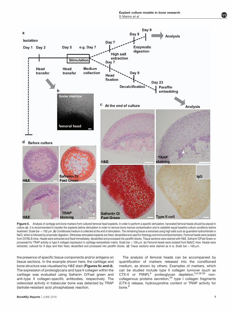
the presence of specific tissue components and/or antigens ontissue sections. In the example shown here, the cartilage andbone structure was visualised by H&E stain (Figures 5c and d).The expression of proteoglycans and type II collagen within thecartilage was evaluated using Safranin O/Fast green andanti-type II collagen-specific antibodies, respectively. Theosteoclast activity in trabecular bone was detected by TRAP(tartrate-resistant acid phosphatase) reaction.
The analysis of femoral heads can be accompanied byquantification of markers released into the conditionedmedium, as shown by others. Examples of markers, whichcan be studied include type II collagen turnover (such asCTX-II or PIINP),9 proteoglycan depletion,5,8,36,39 non-collagenous proteins secretion,39 type I collagen fragments(CTX-I) release, hydroxyproline content or TRAP activity forbone.9
Figure 5. Analysis of cartilage and bone markers from cultured femoral head explants. In order to perform a specific stimulation, harvested femoral heads should be placed inculture (a). It is recommended to transfer the explants before stimulation in order to remove bone marrow contamination and to establish equal baseline culture conditions beforetreatment. Scale bar¼ 100mm. (b) Conditioned medium is collected at the end of stimulation. The remaining tissue is extracted using high salts such as guanidine hydrochloride orNaCl, which is followed by enzymatic digestion. Otherwise stimulated explants are fixed, decalcified and used for histology and immunohistochemistry. Femoral heads were isolatedfrom C57BL/6 mice. Heads were extracted and fixed immediately, decalcified and processed into paraffin blocks. Tissue sections were stained with H&E, Safranin O/Fast Green orprocessed for TRAP activity or type II collagen expression in cartilage extracellular matrix. Scale bar¼ 100mm. (c) Femoral heads were isolated from Balb/C mice. Heads wereextracted, cultured for 9 days and then fixed, decalcified and processed into paraffin blocks. (d) Tissue sections were stained as in c. Scale bar¼ 100mm.
Explant culture models in bone research
S Marino et al
BoneKEy Reports | JUNE 2016 7

Method 3
The neonatal calvaria is an active, not fully calcified bone tissue,routinely used to study resorption and bone formation ex vivo.40
In neonatal calvaria, bone modelling is favoured over boneremodelling and therefore might not always be the best modelto study bonemetabolism. However, culturing calvaria explantsallow preserving cellular diversity and interactions within thebone microenvironment. Moreover, calvaria explants areexcellent models to study the effect of cancer cells or theirsecretome on skeletal structure.13 Here we provide a methodfor mouse calvaria isolation, culture and characterisation thatallows for the investigation of cancer cell-induced osteolysis.
Specific materials
Isolation and culture medium: MEMa medium with 10% fetalbovine serum (FBS), 2mM L-Glutamine, 100Uml� 1 penicillinand 100 mgml� 1 streptomycin; PBS, pH 7.2; equipment:scissors, tweezers and forceps (straight and curved fine tip),straight microscissors (12mm cutting edge) and stainless steelmesh (can be obtained at the local hardware store, stainlesssteel no. 30 mesh is normally used).NOTE 12: The entire mouse calvaria or the two halves can be
cultured in 48-well plates on the stainless steel grid, preparedbycutting rectangular 0.8� 1 cm pieces of stainless steel meshfrom the sheet. Meshes are made by bending both ends with aruler to make the bridge, as shown in Figure 6. The dimensionsof the meshes should be adapted accordingly to the dimensionof tissue culture plate. As described in Mohammad et al,11 fourhemi-calvaria can be cultured at the same time on rectangular1� 1.5 cm meshes in a 12-well plate. After each use, wash themeshes with detergent and rinse them with 70% ethanol. Thewashed meshes are then rinsed with PBS to remove anyresidual detergent, air-dried and autoclaved.
Animals. C57BL/6 mice at 4–7 days old.
NOTE 13: Different strains of mice can be used for this assaywith similar results. It is, however, important to comparetreatments using a single pup calvaria divided into two halvesdue to the variability in osteogenic responses among litters andeven among pups within the same litter.40
Calvarial isolation
1. Sterilise all tools and perform the isolation under sterileconditions.
2. Hold the pup with forceps and dip it in 95% ethanol for 2 s.3. Decapitate the pups and transfer the heads into a 50ml
collection tube filled with 45ml of sterile PBS. Keep theheads in PBS while dissecting one head at the time.
4. Using the curved forceps, firmly hold the head by placing theforceps on either side of the head.
5. Using the straight microscissors, remove most of the scalp.Expose the calvaria by incising both lateral sides of thecalvaria and by moving the scissors from the eyes to theback of the skull. At the end, flip the excessive skin overthe mouse forehead.
6. Identify the internal sutures of the calvaria as shown inFigure 7 and accurately cut the calvaria around the edges.
7. Gently remove the calvaria from the rest of the skull, removetheadherent connective tissueandwash the calvaria inPBS.
8. While holding the calvaria with straight fine tip forceps, cut itinto two halves along the median sagittal suture and placeeach half in fresh medium.
9. Repeat the steps for all remaining calvaria.
Calvarial culture
1. Place one sterilised mesh into each well of a 48-well tissueculture plate.
2. Using the fine tip forceps, transfer half of the calvaria fromthemedium onto the top of the grid. Make sure that the concaveor cranial side is facing down on the stainless steel mesh.
3. Add 1ml of culture medium into each well.NOTE 14: Avoid flushing the calvaria with themedium. It is
important that the calvaria half sits at the air–liquid interfacein order to prevent the tissue fromdrying out or floating in themedium; 1ml of medium is sufficient to cover calvaria whenculture is performed in a 48-well tissue culture plate.NOTE 15: Use at least five calvaria halves from the same
litter for each treatment group and an appropriate control.4. Replace the medium 24h later by carefully aspirating the
medium with a syringe and adding 1ml of medium containing
Figure 6. Preparation of stainless steel mesh for calvaria explant culture. Stainless steel meshes measuring 0.8� 1 cm are cut from stainless steel sheets, and the two edgesare bent to form a bridge that supports the calvaria. Meshes are then placed in the well of a 48-well plate.
Figure 7. Anatomy of the mouse calvaria and its dissection. The red dotted lineillustrates the isolation procedure.
Explant culture models in bone research
S Marino et al
8 JUNE 2016 | www.nature.com/bonekey

thedesired treatments.Change themediumevery48–72huntilthe endof the experiment (Figure 8). The standardprotocol is 7days of treatment, but the duration can vary from 4 to 14 days.
Calvaria fixation and paraffin embedding
1. At the end of the culture period, remove the calvaria halvesfrom the mesh, transfer into a 1.5ml collection tube anddirectly fix in 4% buffered formalin/saline (pH 7.4) for 24 h.
NOTE 16: After fixation, rinse samples with PBS and storein 70% ethanol.
2. Rinse samples with PBS and decalcify in 14% EDTA pH 7.2for 48 h.
3. Transfer samples into 70% ethanol before processing forparaffin embedding.
NOTE 17: Following the dehydration procedure, place thesamples in a freshly prepared paraffin-based infiltrationsolution for 4 h at 60 1C. Using forceps, transfer the calvariainto the molds filled with some wax. The sagittal sutureshould facedown, toward thebaseof themold, to ensure thecorrect orientation. Move the mold to a cold surface andmake sure that the calvaria maintain the desired orientationwhile the paraffin wax hardens.
4. Cut 5 mmsectionswithamicrotome, along themiddle suture,and collect them every 20 mm interval on glass slides. Allowthe slides to dry overnight before staining.
Analysis of cancer-bone metastases using calvaria
cultures
In order to study the effect of cancer cells on bone cells ex vivo,calvaria are divided into two halves, and each half is placed on astainless steel mesh in wells containing amediumwith either nocells or cancer cells (Figure 9a). Cancer cells are seeded 24 h inadvance to allow their adherence.NOTE 18: Seeding density of the cancer cells is cell specific
and has to be determined in advance.NOTE 19: Recently, Curtin41described a three-dimensional
model in which a single calvaria is cultured in 2ml of serum-freeDMEM in the presence of 5� 105 floating cancer cells per tube or25%of their conditionedmedium; 150mgml� 1 sodium ascorbatecan be added to the culture medium in order to study the effect ofcancer cells and exogenous factors on bone formation.Medium isrefreshed every 48–72h and the culture is terminated after 7 days.NOTE 20: In addition, calvaria can be grown in the presence
of a conditioned medium from cancer cells (20% v/v) or amedium containing either vehicle alone or exogenous factors tobe tested (Figures 9b and c). Conditionedmedium fromcancercells is obtained by culturing cancer cells in 6-well plates until
they reach 80%confluence.Completemedium is then replacedwith a serum-free medium. Cancer cells are cultured for anadditional 16 h, and the conditioned medium obtained isremoved and filtered through a 0.2mm filter. Control standardmedium is a serum-freemedium incubated for 16 h at 37 1Cand5% CO2 in a six-well plate without cancer cells.
Method 4
Previous work has demonstrated that rat mandibles can beextracted, sliced and kept viable in culture for at least 21days.16
This method was later modified to include the use of greenfluorescent protein (GFP) expressing DPSCs, enabling thevisualisation of transplanted cells following injection into ex vivo
mandible slice.17 Although much of the work on DPSCs hasbeen completed using monolayer culture systems, there is afundamental need to better understand how these cells behavein a three-dimensional system, providing a more accuratereflection of their in vivo characteristics.
Specific materials
DPSCs medium. MEMa with nucleosides supplemented with20% (v/v) FBS, 100Uml� 1 Penicillin, 100mgml� 1 streptomycinsulphate and 100mM L-ascorbic acid; PBS; collagenase-dispase(4mgml� 1 in DPSCs medium); fibronectin (10mgml� 1 solutiondiluted in PBS with 0.1mM MgCl2 and 0.1mM CaCl2); accutase;CyGel (Biostatus, Shepshed, UK); equipment: scissors, scalpel,tweezers, low-speed bone saw (IsoMet Buehler, Lake Bluff, IL,
Figure 8. Timeline for the calvarial explant culture. On day 0, calvaria are isolated, cut into 2 halves and placed into separate wells of a 48-well plate containing 1ml of completemedium. On day 1, the medium is replaced with a fresh medium either with or without cytokines, tested compounds or conditioned medium from cancer cell lines. On day 3 and 5,replace the medium with fresh medium either with or without cytokines, tested compounds or conditioned medium. On day 7, calvaria halves are removed, fixed and scanned bymicroCT for histological and histomorphometric examination.
Figure 9. Model for cancer-induced osteolysis of the calvaria. Calvaria are dividedinto two halves. Each half is placed in the 48-well plates on the stainless steel meshes.Calvaria are grown in the absence or presence of cancer cells (a), in the presence ofcancer cells with or without treatment (b), in standard medium or cancer cell-derivedconditioned medium (c). Note: cancer cells should be seeded 24 h in advance. Mediumis replaced every 48 h and the culture is terminated after 7 days.
Explant culture models in bone research
S Marino et al
BoneKEy Reports | JUNE 2016 9
USA); 40mm cell strainer, No 1.5 coverslips, blue agarose beads(BD Bioscience, Oxford, UK), nanofil syringe (World PrecisionInstruments, Sarasota, FL, USA).Animals. 28-day-old Wistar rats and GFP rats (SDT(CAG-
EGFP) CZ-004 Osb) produced as described elsewhere.42
Preparation of mandible slice culture
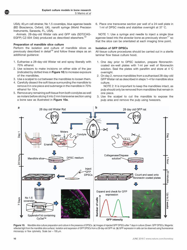
Perform the isolation and culture of mandible slices aspreviously described in detail16 and follow these steps as anadditional guidance:
1. Euthanise a 28-day-old Wistar rat and spray liberally with70% ethanol.
2. Use scissors to make incisions on either side of the jaw(indicated by dotted lines in Figure 10) to increase exposureof the mandibles.
3. Use a scalpel to cut between the mandibles to loosen them.4. Carefully dissect the soft tissue surrounding the mandible to
remove it in one piece and submerge in themandible in 70%ethanol for 10 s.
5. Removeany remaining soft tissue frombothcondyles aswellasmolars before slicing it into 2mm transverse section usinga bone saw as illustrated in Figure 10a.
6. Place one transverse section per well of a 24-well plate in1ml of DPSC media and stabilise overnight at 37 1C.
NOTE 1: Use a syringe and needle to inject a single blueagarose bead into the alveolar bone as previously shown17 sothat the slice can be orientated at each imaging time point.
Isolation of GFP DPSCs
All tissue culture procedures should be carried out in a sterilelaminar flow tissue culture hood.
1. One day prior to DPSC isolation, prepare fibronectin-coated six-well plates with 1ml per well of fibronectinsolution. Seal the plates with parafilm and store at 4 1Covernight.
2. On day 2, removemandibles from a euthanised 28-day-oldGFPWistar rat as described in steps 1–4 for mandible sliceculture.NOTE 2: It is important to keep the mandibles intact, as
pulp should only be removed frommandibles that remain inone piece.
3. Use the scalpel to cut the mandible to expose thepulp area and remove the pulp using tweezers.
Figure 10. Mandible slice culture preparation and culture in the presence of DPSCs. (a) Images of injected GFP DPSCs after 7 days in culture (Green: GFP DPSCs, Magenta:reflected light from the mandible slice surface). Isolation and expansion of GFP DPSCs from a 28-day-old GFP rat. (b) GFP expression in cells can be observed using fluorescencemicroscopy or flow cytometry. Scale bar¼ 100mm.
Explant culture models in bone research
S Marino et al
10 JUNE 2016 | www.nature.com/bonekey

NOTE 3: This can often be achieved by cutting off thetip of the incisor to expose the pulp cavity, squeezingthe mandible and removing the emerging pulp withtweezers.
4. Place thepulp in a 60mm tissue culture dish containing 5mlcollagenase-dispase solution and mince finely using ascalpel.
5. Place in the incubator for 1 h.6. Triturate the digested pulp using successively smaller
pipette tips (1ml, 200ml and 10 ml) to obtain a single-cellsuspension and finally pass through a cell strainer into a50ml collection tube to remove any remaining undigestedtissue.
7. Centrifuge the cell solution at 400g for 5min andre-suspend in 1ml serum-free DPSC medium.
8. Count the cells using a haemocytometer and prepare a cellsuspension of 1� 104cells per ml in the appropriate totalvolume.
9. Remove fibronectin solution from the six-well plates andreplace with 1ml of DPSC cell solution.
10. Incubate for 20min at 37 1C.11. Remove the medium and non-adherent cells, replace with
DPSC medium and return the plate in the incubator.12. Change the medium every 2–3 days until the cells reach
85–95% confluency.NOTE 4: It can take 15–18 days for cells to reach
confluence.13. Passage cells by removing the medium and washing
once with PBS. Aspirate PBS and replace with 500 mlaccutase and return the plate into the incubator for5min.
14. Once cells have detached, add 2ml of DPSC medium,collect the cell suspension in a 15ml collection tube andcentrifuge at 400 g for 5min to pellet the cells.
15. Aspirate the supernatant, re-suspend in 1ml DPSCmedium and count the cells. Re-seed at a density of4� 103 cells per cm2.
16. Check for the presence of GFP using fluorescencemicroscopy.
Analysis of DPSC behaviour in mandible slices
1. Dissociate GFP DPSCs at approximately 80–90%confluence using accutase as described above in steps13–16 for GFP cell isolation.
2. Count cells using a haemocytometer and prepare a cellsuspension of 2� 106cells per ml in the appropriate totalvolume.
3. Inject 1 ml of cell solution into the pulp of the mandible sliceusing a 35-gauge micro-needle and a nanofil syringe.
4. Return the mandible slice to the incubator until required forimaging, changing the media every 2–3 days.
The imaging of the mandible slice presents a challenge due tothe presence of both soft and hard tissue. CyGel is opticallyclear and is used to reversibly mount the mandible slice,allowing collection of both fluorescence and reflected lightimages. These reflective light images provide reference pointsfor imaging within the slice. This is especially useful forlongitudinal studies. This method used has previously beendescribed in detail.17
1. Remove the lid froma 1.5ml collection tube and attach it to a60mm 10-cm dish with adhesive.
2. Fill the collection lid with CyGel to suspend themandible slice.NOTE 5: Ensure that the mandible slice is suspended with
blue agarose beads facing up to allow the slice to beorientated and images collected with reference to the bead.
3. Cover the sample with a No 1.5 coverslip and place in theincubator for 3min.NOTE 6: CyGel is a thermo-reversible hydrogel that gels
rapidly at 37 1C, and therefore prepare the chamber with thegel using cooled pipette tips and keep the CyGel cool untilrequired.
4. Collect GFP fluorescence using 488 nm excitation and530/30 emission and reflection images at 488 nm usingan upright confocal microscope.
5. Following imaging, rinse the chamber with 4 1C PBS toliquefy the CyGel and remove the mandible slice. Return themandible slice to DPSC media in the 24-well plate.
NOTE 7: CyGel is biocompatible with live cells; therefore,cultures can be viably maintained following imaging.
Method 5
The bovine trabecular explant model described below repre-sents a system, in which osteocytes within their nativeenvironment can send signals to osteoblasts on the bonesurface.23,24 Bone cores are thoroughly cleaned to removebone marrow, disrupted nerves and vasculature, as studieshave demonstrated an influence of these systems on bonemodelling. This procedure has also been optimised to removeall surface cells from the trabecular bone explants and preserveonly the osteocytes, which remain viable and embedded withinthe bone matrix. Then, a controlled number of primaryosteoblasts are re-seeded onto the bone surface. Mechanicalloading is subsequently applied by a custom loadable perfusionbioreactor to evaluate long-term histological and mechanicalproperty changes within ex vivo grown bovine bone cores.
Specific materials
Culture media. MEMa medium with 10% FBS and antibiotics(100mgml� 1 streptomycin, 100Uml� 1 penicillin); PBS; 0.05and 0.25% trypsin-EDTA; WD-40 (San Diego, CA, USA);LIVE/DEAD Viability/Cytotoxicity kit (Invitrogen, Waltham, MA,USA); equipment: diamond-tipped coring tool, 7mm diameter(Starlite Industries, Rosemont, PA, USA); 60ml sterile syringeand needles (18 and 30 gauge); Roccal-D Plus detergent(Zoetis); custom cell seeder, small magnetic stirrer; largepronged forceps, scalpel and blades; hand drill with locking bitand key; small vise; Interplak water jet (Conair, Stamford, CT,USA); IsoMet low-speed saw with high carbon (HC) waferingblade (Buehler, Lake Bluff, IL, USA); magnetic stir plate;mechanical testing device such as Bose Electroforce (Bose,Eden Prairie, MN, USA)
Animals. Bovine fetlock joints from 6-week-old calves can beobtained from a local slaughterhouse and shipped overnight onice. The carpal-metacarpal joint contains the hoof, metacarpusand carpal joint and is usually cut at the radius and ulna. If thelimb was cut higher, youmay also see the elbow joint where theradius would connect with the humerus.
Explant culture models in bone research
S Marino et al
BoneKEy Reports | JUNE 2016 11

Calf joint dissection
1. If the joint arrived with the skin, begin by gently removing theskin from the underlying muscle tissue with the scalpel.Examine the carpal joint and ensure that the joint capsule isintact.While handling the joint, be careful not to puncture thejoint capsule to maintain sterility.
NOTE 1: If the joint capsule has been punctured, isolatethis joint from the others, as it may be more prone toinfection.
2. Remove the hoof at the fetlock joint, where the metacarpusmeets the hoof. To help determine the site of incision, lightlybend the joint and look for the space between the bones.There may already be an incision in the hoof from the killingprocess.
3. There are large tendons at the fetlock joint that need to be cut toremove the hoof. Also, trim these tendons along the shaft of themetacarpus,as theymaycause thebone tosliponce it isplacedin the vise for drilling the bone cores. If you wish to cut offadditional tissue at this point, you can remove some of themusclesurroundingtheradiusandulna,but this isnotnecessaryas this portion of the limb will not be used for the experiment.
4. Soak cut and skinned joints in a diluted antibacterialdetergent for 30min (use Roccal-D Plus or a similarproduct).
5. Transfer joints to 70% ethanol for another 30min.
Obtaining trabecular bone cores
1. Transfer the joints to a sterile biosafety cabinet.Wipe the jointarea with alcohol and hold the joint flexed. Cut open thecarpal-metacarpal joint; the space between the metacarpusand the adjacent (first) carpal (Figure 11a). Cut off the radiusand ulna.
2. The metacarpus should have a relatively flat articularsurface. Using gauze soaked in 70% ethanol, wipe awaythe synovial fluid and clean the surface of the joint.
3. Wrap gauze on the bottom end of the carpal bone (oppositethe end of the articular surface of interest). Put the end of thebone wrapped in gauze in the vise and tighten until the boneis stable.
4. Open a sterile syringe and fit it with a 18-gauge needle.Fill the syringe with PBS and rinse the surface of the bone.NOTE 2: Make sure that the exposed surface of the bone
does not dry out at any point during the isolation procedure.In the following step, the bone should be constantly irrigatedwith PBS during drilling.
5. Put the coring tool into the drill and tighten the chuckwith the key. Dip the coring tool in 70% ethanol and thenrinse well in PBS. To drill, enter at an angle and lightlysqueeze the trigger to drill at a slow speed (Figure 11b).The angle needs to be determined by looking at theoverall shape of the bone; for example, if the midshaft isnarrow and the articular surface is really wide, you must drillat a larger angle from the centre axis in order to avoid drillingthrough the cortical shell. Apply a light downward force onthe drill but take care to lift up at times to allow PBS to enterthe space between the drill bit and the bone. Stop drillingwhen the bone ‘‘gives’’ and hits the medullary cavity.NOTE3: It is important to not drill tooquickly, otherwise the
bone around the edge of the bit will burn and a hard shell willbe created.
6. Pull the coring tool out of the bone and remove the tool fromthe drill. Use a hex key to push the bone core out of thebacksideof the coring tool, pushingon thearticular cartilage.Push the core into a 10-cm dish with 15ml of pre-warmed,fully supplemented media. Repeat for as many bone coresas desired.
Figure 11. Bovine trabecular bone core isolation. (a) The carpal-metacarpal joint of a bovine calf fetlock. (b) Drilling bone cores through the medullary cavity of the metacarpus.(c) A trabecular bone sample. (d) Isomet low-speed saw assembly. (e) 7 mm trabecular bone explant cut from the bone core.
Explant culture models in bone research
S Marino et al
12 JUNE 2016 | www.nature.com/bonekey

Cutting bone cores
1. Sterilise the reservoir of the water jet with 70% ethanol, andthen rise well with PBS.
2. Pick up the bone corewith the pronged forceps (Figure 11c)and begin spraying PBS at the core to lightly clean the coresand remove some of the marrow.
3. Once all the cores are cleaned, set up the IsoMet. Set thespeed between 2 and 3 to cut the bone at a slow speed. Fillthe trough with enough PBS so that the blade is justskimming the surface of the PBS. Using sterile forceps, put abone core into the holder,with the articular surface facing theIsoMet and the trabecular bone facing outwards(Figure 11d). Turn the blade on first, then slowly lowerthe specimen onto the blade. Trim the ‘rough’ edge andremove the soft trabecular/marrow tissue.
4. Advance the micrometre to obtain a core that is 7mm inheight. Turn the blade on, then slowly lower the specimenonto the blade (Figure 11e). When the bone core isalmost completely cut, hold the core with forceps so itdoesnot fall.When thespecimen is completely cut, place it ina 10-cm dish with fresh, pre-warmed, fully supplementedmedia.
5. Repeat until nomore specimens can be cut from that core oruntil the specimen reaches the subchondral surface.
NOTE 4: The drill chuck, IsoMet components and vise shouldall be cleaned with water and WD-40 after use to removeresidual salt from the PBS and prevent rust.
Cleaning explants of bone marrow and surface cells
In the following steps, the individual bone explants are thenthoroughly cleaned using PBS rinsing and trypsin treatments toremove bone marrow components, damaged vasculature,nerves and any surface cells. The only cells that remain afterthese steps are osteocytes embedded within the bone matrix(Figure 12a). The following procedure is robust so that, evenafter 2 weeks, very few surface cells remain to re-populate thebone surface (Figure 12b).
1. Once cut, thoroughly clean the bone cores with the PBS jet.The cores should be entirely white once the bone marrow isremoved. Place the cleaned cores in a 12-well plate andcover with pre-warmed PBS.
2. To remove the surface cells, the cores must undergo aserial trypsin treatment. In each well, add B6ml 0.25%trypsin-EDTA (enough to cover the cores) and incubate for8min. After incubation, remove the trypsin, neutralise anyremaining trypsin by adding media to the wells andthoroughly rinse each core again with PBS. Repeat twomore times so each core is treated three times with trypsin.
Figure 12. Cleaned trabecular bone explants. (a) A confocal image of a cleaned trabecular bone explant stained with a LIVE/DEAD cytotoxicity/viability kit. Note the absence ofsurface cells. (b) Cleaned explant after 14 days in culture. Live osteocytes remain in the interior and few cells re-populate the bone surface.
Figure 13. Seeding of primary osteoblasts. (a) Schematic of the custom cell seeder. (b) A confocal image of a seeded trabecular bone explant stained with a LIVE/DEADcytotoxicity/viability kit. (c) Seeded explant after 6 days in culture.
Explant culture models in bone research
S Marino et al
BoneKEy Reports | JUNE 2016 13

3. Rinse a final time with PBS. The final PBS rinse should berather extensive to ensure that the detached cells on thebone surface are removed.
4. Put the cleaned cores in a new dish with pre-warmed, fullysupplemented media.
Seeding primary osteoblasts onto the explants
The bone cores may be used to obtain primary osteoblasts,but the ‘‘excess’’ bone cores pieces are also good for thisprocedure. Detailed methods for obtaining primary osteoblastsby explant outgrowth are described elsewhere.43
NOTE 6: The primary osteoblasts should be prepared in aprevious harvest, as they take B3 weeks to expand.The primary cells are used to seed a controlled number of
cells back onto the bone cores using a custom cell seeder(Figure 13a). This achieves a uniform distribution of cells alongthe bone surface (Figure 13b). Osteoblasts proliferate andeventually cover the bone surface after a few days in culture(Figure 13c).
1. Trypsinise cells to be seeded onto bone cores and centrifugeto collect a cell pellet.
2. Count the cells using a haemocytometer to obtain a cellconcentration. For osteoblast seeding onto 7mm bonecores, the optimal concentration is 105 cells per ml fullysupplemented media. Prepare at least 30ml of cellsuspension. Pipet the cell suspension in the cell seederjar with a magnetic stirrer.
3. Place the cell seeder lid on the benchtop so that the needlesface upwards. Using forceps, skewer the bone cores ontothe needles so that the flat side of the needles faces the lidand the cores are secure.
NOTE 7: When attaching the bone cores to the needles ofthe cell seeder, try to push the needle through the trabecularpores rather than break through trabeculae.
4. Carefully place the lid on the cell seeder jar. Place the stirplate in a CO2 incubator at 37 1C and put the cell seeder onthe stir plate. Stir slowly for 1 h.
5. Remove the lid from the jar, take the bone cores off theneedles and transfer to a new culture dish with fullysupplemented media.
6. Obtain the cell concentration of the remaining solution toestimate the seeding density per core, assuming uniformcellseeding for all cores.
Transferring explant into loadable perfusion bioreactor
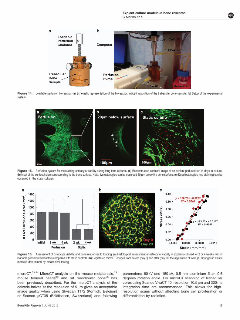
To sustain the viability of osteocytes in long-term culture, boneexplants should be placed in a loadable perfusion bioreactor(Figure 14). In addition to traditional histological assessment,live osteocytes can be imaged by confocal microscopy using afluorescent viability stain.NOTE 7: In order to image the interior of the bone cores, they
should be cut vertically in half using the Isomet saw prior to thisprocedure.
1. Prepare the LIVE/DEAD Cytotoxicity/Viability kit accordingto the manufacturer’s instructions and use 2mM calcein-AMand 4mM EthD-1.
2. Remove the media from the bone cores and rinse once withPBS. Transfer the bone cores to sterile microcentrifugetubes.
3. Add B1ml of the working solution to each bone core andcover the tubes with foil to protect from light. Incubate thecores at room temperature for 45min.
4. Image using a confocal microscope. Focus on the bottom ofthe bone core. You should see considerable numbers ofdead cells here from the cutting procedure. Using themicrometre, focus at least 100mm into the bone tissue toimage and assess an undamaged region.
This method can be then used to evaluate the influence ofperfusion on osteocyte viability and the general healthof the cultures (Figures 15a-c). After 2 weeks in culture, liveosteoblasts are confluent along the bone surface, and beneaththis surface layer live osteocytes can be identified (Figure 15b).Static cultures, in comparison, show a considerable number ofdead osteocytes (Figure 15c).
Analysis of osteocyte viability and bone formation
responses to mechanical loading
The perfusion bioreactor can be coupled to a mechanicaltesting device such as the Bose Electroforce. This setup can beused to apply dynamic, deformational loads to induce boneformation responses.To complement staining in living explants, traditional
histological techniques can be used to evaluate the bone.Explants can be fixed, embedded, sectioned and stained withtraditional assays suchasH&E.ComparingmicroCTscans frombefore and after the culture period using techniques such asimage registration enables quantification of bone volumeand microstructural changes (Figure 16a). Furthermore,mechanical testing of individual explants before and after theapplication of mechanical loading can be used to determine aneffect of applied load on the apparent elastic modulus(Figure 16b). Combined, all listed techniques can be used todemonstrate long-term changes to short-term mechanicalstimulation mediated by osteocytes (Figure 17).
Additionalmethods to analyse ex vivo grownbone explants
Histological examination. Explants can be frozen, paraffin orplastic embedded. Bone explants are commonly decalcifiedbefore paraffin embedding. However, metatarsal bones do notneed decalcifying for paraffin embedding and subsequentprocessing. Thus, histological methods such as von Kossa oralizarin red staining can be immediately adopted.33 Alkalinephosphatase (ALP) activitywithin themetatarsal bones canalsobe determined using a commercially bought assay for ALP.33
Similarly, calvarial bone can be embedded in plastic before vonKossaor TRAP reaction. In order to later detect TRAPactivity onplastic, it is, however, important toperform the initial embeddingand polymerisation steps at low temperature (4 1C), whichpartially preserves the enzymatic reactivity.44 Descriptions ofstaining for Toludine blue or Goldner’s Trichrome, von Kossa orTRAP are in Supplementary Information.
Proteomic analysis. Conditionedmedium from themouse femoralhead cultures can be monitored using Dimethylmethyleneblue assay (detects sulphated glygosaminoglycans),5,8,36,45–48
western blotting36,45,46,49–52 or specific ELISAs.9
MicroCT. The quantification of bone parameters andvisualisation of its microstructure can be performed by
Explant culture models in bone research
S Marino et al
14 JUNE 2016 | www.nature.com/bonekey
microCT.53,54 MicroCT analysis on the mouse metatarsals,33
mouse femoral heads38 and rat mandibular bone55 hasbeen previously described. For the microCT analysis of thecalvaria halves at the resolution of 5 mm gives an acceptableimage quality when using Skyscan 1172 (Kontich, Belgium)or Scanco mCT35 (Bruttisellen, Switzerland) and following
parameters: 60 kV and 150 mA, 0.5mm aluminium filter, 0.6degrees rotation angle. For microCT scanning of trabecularcores using Scanco VivaCT 40, resolution 10.5 mm and 300msintegration time are recommended. This allows for high-resolution scans without affecting bone cell proliferation ordifferentiation by radiation.
Figure 14. Loadable perfusion bioreactor. (a) Schematic representation of the bioreactor, indicating position of the trabecular bone sample. (b) Setup of the experimentalsystem.
Figure 15. Perfusion system for maintaining osteocyte viability during long-term cultures. (a) Reconstructed confocal image of an explant perfused for 14 days in culture.(b) Inset of the confocal slice corresponding to the bone surface. Note: live osteocytes can be observed 20 mm below the bone surface. (c) Dead osteocytes (red staining) can beobserved in the static cultures.
Figure 16. Assessment of osteocyte viability and bone responses to loading. (a) Histological assessment of osteocyte viability in explants cultured for 2 or 4 weeks (wk) inloadable perfusion bioreactors compared with static controls. (b) Registered microCT images from before (day 0) and after (day 28) the application of load. (c) Changes in elasticmodulus determined by mechanical testing.
Explant culture models in bone research
S Marino et al
BoneKEy Reports | JUNE 2016 15

Signalling mechanisms. Analysis of the signalling mechanismscan be successfully performed using the model culturesdescribed in this protocol. The femoral head ex vivomodel waseffectively applied to study cell responses and the effects ofcytokines (interleukin (IL)-1b,5,45,47,49,50,56,57 IL-1a,48 tumournecrosis factor-a9,49,50 or oncostatin M9), growth factorsinsulin-like growth factor 1 (IGF-19,49), proteases46, alarmins 49,51
or inhibitors49 on the cells and intact ECM. Similarly, metatarsalexplants can be supplemented with different peptides, growthfactors, recombinant proteins, antibodies or small-moleculeinhibitors to examine their effects on bone development or onprimary bone cells of the calvaria culture ex vivo.58,59 Finally,the inflammatory bone destruction can be studied using themandibular slice model.60
Gene expression. Adenovirus techniques can be adopted tomanipulate specific genes in metatarsal bones.61 RNA andprotein can also be extracted from metatarsal bones whenpooled together (B4 bones per group) to provide sufficientquantities.34 Likewise, gene expression can be studied byisolating the RNA from the calvaria at the end of the cultureperiod.62 In addition, defining the functional roles of individualgenes can be achieved by modulating their expression by viraland non-viral methods on the calvaria organ culture or bymanipulating primary osteoblasts prior to seeding ontotrabecular bone cores.63,64
Discussion
This protocol provides a detailed description of the mostcommonly used animal ex vivo bone culture models and givesexamples of their application across different areas of boneresearch. Remarkably some of these explants can be isolatedfrom genetically modified animals to greater our understandingof the role of specific genes in bone and cartilage biology.Although the main limitation of these models is a limited life-span, the appropriate culture conditions indicated in thisprotocol and media supplementation enhance their use over aset periodof time. Interestingly,ex vivogrowthofmetatarsal andfemoral head explants are grown in the absence of serum,which eliminates the need of serum batch testing and improves
reproducibility. However, for the long-term experiments, theserum supplementation and low-level perfusion driven by aperistaltic pump can be implemented to significantly extendculture periods and keep bone cells alive, as described here fortrabecular bone explants.The major advantage of these models is the ability to retain
native bone cell communication and to study cellular responsesin a physiological bone environment. For example, themetatarsal organ culture allows for direct examination ofchondrocyte interactions during linear bone growth, which isnot possible to observe using primary chondrocyte cultures. Italso allows for the separation of systemic and local factors thataffect bone development, therefore permitting the specificanalysis of the local effects on the growth plate dynamics.Similarly, the trabecular bone explant model is unique in that itisolates the interactions between osteocytes andosteoblasts ina controlled manner to delineate mechanisms underlying load-induced bone formation responses.Another advantage is that ex vivo culturemodels allow for the
study of bone extracellular matrix remodelling. The model ofmouse femoral head culture enables the study of matrixdegeneration or de novo synthesis occurring simultaneouslywithin the cartilage and bone. Likewise, the calvaria boneexplant model is useful to enhance our understanding ofmechanisms linked with bone resorption and formation. Finally,thesemodels can be used to analyse novel interactions such ascancer cell-induced bone osteolysis or stem cell behaviourduring bone repair. This indicates that there is a growing interestin the use of animal ex vivo bone culturemodels in several fieldshelping to increase our understanding about bone organotypicresponses in health and disease.
Recommended further reading
Comprehensive review of various models to study endo-
chondral ossification including the metatarsal organ culture
system: Andrade AC, Chrysis D, Audi L, Nilsson O. Methods tostudy cartilage and bone development. Endocr Dev 2011; 21:52–66.Detailed characterisation of bone and catrtilage dynamic
changes duringmouse development:MadsenSH,Goettrup aS,Thomsen G, Christensen ST, Schultz N, Henriksen K et al.
Figure 17. Experimental timeline for bovine explant culture and mechanical testing.
Explant culture models in bone research
S Marino et al
16 JUNE 2016 | www.nature.com/bonekey

Characterization of an ex vivo femoral head model assessedby markers of bone and cartilage turnover. Cartilage 2011; 2:265–278.Comprehensive review of a three-dimentional model for the
studies of cancer-bone metastasis ex vivo: Curtin P, Youm H,Erdjan S. Three-dimensional cancer-bone metastasis modelusing ex- vivo co-cultures of live calvarial bones and cancercells. Biomaterials 2012; 33: 1065–1078.Detailed protocol for establishing mandibular organotypic
cultures: Sloan AJ, Taylor SY, Smith EL. Organotypicmandibular cultures for the study of inflammatory bonepathology. In: Replacing Animal Models. John Wiley & Sons,Ltd, pp. 159–166.Application of loadable perfusion bioreactor to study
biochemical and biomechanical changes of these bone cores
over long-term cultures: David V, Guignandon A, Martin A,Malaval L, Lafage-Proust M-H, Rattner A et al. Ex vivo boneformation in bovine trabecular bone cultured in a dynamic 3Dbioreactor is enhanced by compressive mechanical strain.Tissue Eng Part A 2008; 14: 117–126.
Conflict of Interest
The authors declare no conflict of interest.
Acknowledgements
Dr SM acknowledges Dr Aymen I Idris for his valuable adviceand support from IBMS-ECTS Young Investigators. Ms GBacknowledges the support of her research advisor, Professor XEdward Guo, and funding from the National ScienceFoundation Graduate Research Fellowship Program. Dr RH-Jacknowledges Dr JS Colombo and Dr FI Young for providingimages for the figures herein. Dr KS thanks Professor ColinFarquharson for his assistance with the metatarsal organcultures and Arthritis Research UK for funding (20413).Dr MA acknowledges Arthritis Research UK for providingFoundation Fellowship funding (20512) and thanks Dr AlisonGartland for suggestions and support.
References
1. Raggatt LJ, Partridge NC. Cellular andmolecular mechanisms of bone remodeling. J Biol Chem
2010; 285: 25103–25108.
2. Burger EH, Van-Delft JL. Calcification of embryonic hypertrophic cartilage in vitro. Proc K Ned
Akad Wet Ser C Biol Med Sci 1976; 79: 309–322.
3. Staines KA, Pollard AS, McGonnell IM, Farquharson C, Pitsillides AA. Cartilage to bone
transitions in health and disease. J Endocrinol 2013; 219: R1–R12.
4. Chagin AS, Karimian E, Sundstrom K, Eriksson E, Savendahl L. Catch-up growth after
dexamethasone withdrawal occurs in cultured postnatal rat metatarsal bones. J Endocrinol
2010; 204: 21–29.
5. Kadri A, Funck-Brentano T, Lin H, Ea H-K, Hannouche D, Marty C et al. Inhibition of bone
resorption blunts osteoarthritis in mice with high bone remodelling. Ann Rheum Dis 2010; 69:
1533–1538.
6. Zhen G, Wen C, Jia X, Li Y, Crane JL, Mears SC et al. Inhibition of TGF-b signaling in
mesenchymal stem cells of subchondral bone attenuates osteoarthritis. Nat Med 2013; 19:
704–712.
7. Stanescu R, Leibovich SJ. The negative charge of articular cartilage surfaces. An electron
microscopic study using cationized ferritin. J Bone Joint Surg Am 1982; 64: 388–398.
8. Glasson SS, Askew R, Sheppard B, Carito B, Blanchet T, Ma H et al. Deletion of active
ADAMTS5 prevents cartilage degradation in a murine model of osteoarthritis. Nature 2005;
434: 644–648.
9. Madsen SH, Goettrup aS, Thomsen G, Christensen ST, Schultz N, Henriksen K et al.
Characterization of an ex vivo femoral head model assessed by markers of bone and cartilage
turnover. Cartilage 2011; 2: 265–278.
10. Sakai R, Eto Y, Hirafuji M, Shinoda H. Activin release from bone coupled to bone resorption in
organ culture of neonatal mouse calvaria. Bone 2000; 26: 235–240.
11. Mohammad KS, Chirgwin JM, Guise TA. Assessing new bone formation in neonatal calvarial
organ cultures. Methods Mol Biol 2008; 455: 37–50.
12. Wu X, Downes S, Watts DC. Evaluation of critical size defects of mouse calvarial bone:
An Organ Culture Study. Microsc Res Tech 2010; 73: 540–547.
13. Sophocleous A, Marino S, Logan JG, Mollat P, Ralston SH, Idris AI. Bone cell-autonomous
contribution of type 2 cannabinoid receptor to breast cancer induced osteolysis. J Biol Chem
2015; 290: 22049–22060.
14. Liu H, Yao C, Sun J, Lee C, Huang C, Lin F. Osteogenic evaluation of glutaraldehyde
crosslinked gelatin composite with fetal rat calvarial culture model. Artif Organs 2001; 25:
644–654.
15. Jacenko O, Tuan RS. Chondrogenic potential of chick embryonic calvaria: I. Low calcium
permits cartilage differentiation. Dev Dyn 1995; 202: 13–26.
16. Smith EL, Locke M, Waddington RJ, Sloan AJ. An ex vivo rodent mandible culture model for
bone repair. Tissue Eng Part C Methods 2010; 16: 1287–1296.
17. Colombo JS, Howard-Jones RA, Young FI, Waddington RJ, Errington RJ, Sloan AJ. A 3D
ex vivo mandible slice system for longitudinal culturing of transplanted dental pulp progenitor
cells. Cytom Part A 2015; 87: 921–928.
18. Chai Y, Jiang X, Ito Y, Bringas P, Han J, Rowitch DH et al. Fate of themammalian cranial neural
crest during tooth and mandibular morphogenesis. Development 2000; 127: 1671–1679.
19. Gronthos S, Mankani M, Brahim J, Robey PG, Shi S. Postnatal human dental pulp stem cells
(DPSCs) in vitro and in vivo. Proc Natl Acad Sci USA 2000; 97: 13625–13630.
20. Waddington RJ, Youde SJ, Lee CP, Sloan AJ. Isolation of distinct progenitor stem cell
populations from dental pulp. Cells Tissues Organs 2009; 189: 268–274.
21. Sloan AJ, Smith AJ. Stem cells and the dental pulp: potential roles in dentine regeneration and
repair. Oral Dis 2007; 13: 151–157.
22. Chen J-H, Liu C, You L, Simmons CA. Boning up on Wolff’s Law: mechanical regulation of the
cells that make and maintain bone. J Biomech 2010; 43: 108–118.
23. Takai E, Mauck RL, Hung CT, Guo XE. Osteocyte viability and regulation of osteoblast function
in a 3D trabecular bone explant under dynamic hydrostatic pressure. J bone Miner Res 2004;
19: 1403–1410.
24. Chan ME, Lu XL, Huo B, Baik AD, Chiang V, Robert E et al. A trabecular bone explant model of
osteocyte–osteoblast co- culture for bone mechanobiology. Cell Mol Bioeng 2009; 2: 405–415.
25. Jones DB, Broeckmann E, Pohl T, Smith EL. Development of a mechanical testing and
loading system for trabecular bone studies for long term culture. Eur Cells Mater 2003; 5:
48–60.
26. Davies CM, Jones DB, Stoddart MJ, Koller K, Smith E, Archer CW et al. Mechanically loaded
ex vivo bone culture system ‘‘ Zetos ’’: systems and culture preparation. Eur Cells Mater 2006;
11: 57–75.
27. David V, Guignandon A, Martin A, Malaval L, Lafage-Proust M-H, Rattner A et al. Ex vivo bone
formation in bovine trabecular bone cultured in a dynamic 3D bioreactor is enhanced by
compressive mechanical strain. Tissue Eng Part A 2008; 14: 117–126.
28. Vivanco J, Garcia S, Ploeg HL, Alvarez G, Cullen D, Smith EL. Apparent elastic modulus of
ex vivo trabecular bovine bone increases with dynamic loading. Proc Inst Mech Eng H 2013;
227: 904–912.
29. Zong ming W, Jian yu L, Rui xin L, Hao L, Yong G, Lu L et al. Bone formation in rabbit
cancellous bone explant culture model is enhanced by mechanical load. Biomed Eng Online
2013; 12: 35.
30. Davidson EH, Reformat DD, Allori A, Canizares O, Wagner IJ, Saadeh PB et al. Flow perfusion
maintains ex vivo bone viability: a novel model for bone biology research. J Tissue Eng Regen
Med 2012; 6: 769–776.
31. MacRae VE, Farquharson C, Ahmed SF. The restricted potential for recovery of growth plate
chondrogenesis and longitudinal bone growth following exposure to pro-inflammatory
cytokines. J Endocrinol 2006; 189: 319–328.
32. Mushtaq T, Bijman P, Ahmed S, Farquharson C. Insulin-like growth factor-I augments
chondrocyte hypertrophy and reverses glucocorticoid-mediated growth retardation in fetal mice
metatarsal cultures. Endocrinology 2004; 145: 2478–2486.
33. Staines KA, Mackenzie NCW, Clarkin CE, Zelenchuk L, Rowe PS, MacRae VE et al.MEPE is a
novel regulator of growth plate cartilage mineralization. Bone 2012; 51: 418–430.
34. Dobie R, Ahmed SF, Staines Ka, Pass C, Jasim S, MacRae VE et al. Increased linear bone
growth by GH in the absence of SOCS2 is independent of IGF-1. J Cell Physiol 2015; 230:
2796–2806.
35. Pass C, MacRae VE, Huesa C, Faisal Ahmed S, Farquharson C. SOCS2 is the critical
regulator of GH action in murine growth plate chondrogenesis. J Bone Miner Res 2012; 27:
1055–1066.
36. Stanton H, Golub SB, Rogerson FM, Last K, Little CB, Fosang AJ. Investigating
ADAMTS-mediated aggrecanolysis in mouse cartilage. Nat Protoc 2011; 6: 388–404.
37. Wilson R, Norris EL, Brachvogel B, Angelucci C, Zivkovic S, Gordon L et al. Changes in the
chondrocyte and extracellular matrix proteome during post-natal mouse cartilage development.
Mol Cell Proteomics 2012; 11: M111.014159.
38. Turner CH, Hsieh YF, Muller R, Bouxsein ML, Baylink DJ, Rosen CJ et al.Genetic regulation of
cortical and trabecular bone strength and microstructure in inbred strains of mice. J Bone Miner
Res 2000; 15: 1126–1131.
39. Cecil DL, Terkeltaub R. Transamidation by Transglutaminase 2 Transforms
S100A11 Calgranulin into a Procatabolic Cytokine for Chondrocytes. J Immunol 2008; 180:
8378–8385.
40. Garrett RI. Assessing bone formation using mouse calvarial organ cultures. In: Bone Research
Protocols: Methods in Molecular Medicine, Part III , vol. 80. Humana Press: New York City, NY,
USA, 2003.
Explant culture models in bone research
S Marino et al
BoneKEy Reports | JUNE 2016 17

41. Curtin P, Youm H, Erdjan S. Three-dimensional cancer-bone metastasis model using ex- vivo
co-cultures of live calvarial bones and cancer cells. Biomaterials 2012; 33: 1065–1078.
42. Okabe M, Ikawa M, Kominami K, Nakanishi T, Nishimune Y. ‘Green mice’ as a source of
ubiquitous green cells. FEBS Lett 1997; 407: 313–319.
43. Gartland A, Rumney RM, Dillon JP, Gallagher JA. Isolation and culture of human osteoblasts.
Methods Mol Biol 2012; 806: 337–355.
44. Erben RG. Embedding of bone samples in methylmethacrylate: an improved method suitable
for bone histomorphometry. Histochem Immunohistochem 1997; 45: 307–313.
45. Cecil DL, Terkeltaub R. Transamidation by transglutaminase 2 transforms S100A11 calgranulin
into a procatabolic cytokine for chondrocytes. J Immunol 2008; 180: 8378–8385.
46. Magarinos N, Bryant K, Fosang A, Adachi R, Stevens R, McNeil H. Mast cell-restricted,
tetramer-forming tryptases induce aggrecanolysis in articular cartilage by activating matrix
metalloproteinase-3 and -13 zymogens. J Immunol 2013; 3: 1404–1412.
47. Kobayashi H, Hirata M, Saito T, Itoh S, Chung U, Kawaguchi H. Transcriptional induction of
ADAMTS5 protein by nuclear factor-kB (NF-kB) family member RelA/p65 in chondrocytes
during osteoarthritis development. J Biol Chem 2013; 288: 28620–28629.
48. Ma HL, Blanchet TJ, Peluso D, Hopkins B, Morris Ea, Glasson SS. Osteoarthritis severity is sex
dependent in a surgical mouse model. Osteoarthr Cartil 2007; 15: 695–700.
49. Cecil DL, Appleton CTG, Polewski MD, Mort JS, Marie A, Bendele A et al. The pattern
recognition receptor CD36 is a chondrocyte hypertrophy marker associated with suppression of
catabolic responses and promotion of repair responses to inflammatory stimuli. J Immunol
2009; 182: 5024–5031.
50. Terkeltaub R, Yang B, Lotz M, Liu-Bryan R. Chondrocyte AMP-activated protein kinase activity
suppresses matrix degradation responses to inflammatory cytokines IL-1b and TNFa. Arthritis
Rheum 2012; 63: 1928–1937.
51. Liu-Bryan R, Terkeltaub R. Chondrocyte innate immune MyD88-dependent signaling drives
pro-catabolic effects of the endogenous TLR2/TLR4 ligands LMW-HA and HMGB1. Arthritis
Rheumatol 2011; 62: 2004–2012.
52. Raducanu A, Hunziker EB, Drosse I, Aszodi A. b1 integrin deficiency results in multiple
abnormalities of the knee joint. J Biol Chem 2009; 284: 23780–23792.
53. Campbell GM, Sophocleous A. Quantitative analysis of bone and soft tissue bymicro-computed
tomography: applications to ex vivo and in vivo studies. BoneKey Rep 2014; 3: 564.
54. van ‘t Hof RJ. Analysis of bone architecture in rodents using microcomputed tomography. In:
Bone Research Protocols: Methods in Molecular Medicine, vol. 816. Humana Press: New York
City, NY, USA, 2012.
55. Liu Z, Yan C, Kang C, Zhang B, Li Y. Distributional variations in trabecular architecture
of the mandibular bone: an in vivo micro-CT analysis in rats. PLoS ONE 2015; 10:
e0116194.
56. Miyaki S, Sato T, Inoue A, Otsuki S, Ito Y, Yokoyama S et al.MicroRNA-140 plays dual roles in
both cartilage development and homeostasis. Genes Dev 2010; 24: 1173–1185.
57. Zreiqat H, Belluoccio D, Smith MM,Wilson R, Rowley La, Jones K et al. S100A8 and S100A9 in
experimental osteoarthritis. Arthritis Res Ther 2010; 12: R16.
58. Huang Y, Harrison JR, Lorenzo JA, Kream BE. Parathyroid hormone induces interleukin-6
heterogeneous nuclear and messenger RNA expression in murine calvarial organ cultures.
Bone 1998; 23: 327–332.
59. Kusano K, Miyaura C, Inada M, Tamura T, Ito A, Nagase H et al. Regulation of matrix
metalloproteinases (MMP-2, -3, -9, and -13) by interleukin-1 and interleukin-6 in mouse
calvaria: association of MMP induction with bone resorption. Endocrinology 1998; 139:
1338–1345.
60. Sloan AJ, Taylor SY, Smith EL. Organotypic mandibular cultures for the study of inflammatory
bone pathology. In: (ed. Davies J) Replacing Animal Models: A Practical Guide to Creating and
Using Culture-Based Biomimetic Alternatives. John Wiley & Sons, Ltd: Chichester, UK, 2012.
61. Alvarez J, Sohn P, Zeng X, Doetschman T, Robbins DJ, Serra R. TGFbeta2 mediates the
effects of hedgehog on hypertrophic differentiation and PTHrP expression. Development
2002; 129: 1913–1924.
62. Mishina Y, Starbuck MW, Gentile Ma, Fukuda T, Kasparcova V, Seedor JG et al. Bone
morphogenetic protein type IA receptor signaling regulates postnatal osteoblast function and
bone remodeling. J Biol Chem 2004; 279: 27560–27566.
63. Gupta AK, Eshraghi Y, Gliniak C, Gosain AK. Nonviral transfection of mouse calvarial organ
in vitro using Accell-modified siRNA. Plast Reconstr Surg 2010; 125: 494–501.
64. Marino S, Logan JG, Mellis D, Capulli M. Generation and culture of osteoclasts. BoneKey Rep
2014; 3: 570.
This work is licensed under a Creative Commons Attribution 3.0Unported License. The images or other third party material in this
article are included in the article’s Creative Commons license, unless indicatedotherwiseinthecreditline;ifthematerialisnotincludedundertheCreativeCommonslicense,userswillneedtoobtainpermissionfromthelicenseholdertoreproducethematerial. To viewacopyof this license, visit http://creativecommons.org/licenses/by/3.0/
Supplementary Information accompanies the paper on the BoneKEy website (http://www.nature.com/bonekey)
Explant culture models in bone research
S Marino et al
18 JUNE 2016 | www.nature.com/bonekey

![Explant cultures of Rpe65 mouse retina: a model to ... · Medical Research, La Jolla, CA), and both were used at 1 µM as recommended [32]. We will refer to them as RAR- and RXR-agonists,](https://img.pdfslide.us/doc/110x75/5e2f3a7df475b167c2580549/explant-cultures-of-rpe65-mouse-retina-a-model-to-medical-research-la-jolla.jpg)