Embed Size (px)
Citation preview
Journal of Visualized Experiments www.jove.com
Copyright © 2017 Journal of Visualized Experiments June 2017 | 124 | e55704 | Page 1 of 8
Video Article
Neonatal Murine Cochlear Explant Technique as an In Vitro Screening Tool inHearing ResearchLukas D. Landegger1,2,3, Sonam Dilwali1,4, Konstantina M. Stankovic1,2,4
1Eaton Peabody Laboratories, Department of Otolaryngology, Massachusetts Eye and Ear2Department of Otolaryngology, Harvard Medical School3Department of Otolaryngology, Vienna General Hospital, Medical University of Vienna4Harvard Program in Speech and Hearing Bioscience and Technology
Correspondence to: Konstantina M. Stankovic at [email protected]
URL: https://www.jove.com/video/55704DOI: doi:10.3791/55704
Keywords: Medicine, Issue 124, Murine cochlear explant, organotypic culture, inner ear, outer hair cells, inner hair cells, spiral ganglion neurons,auditory synaptopathy, adeno-associated virus
Date Published: 6/8/2017
Citation: Landegger, L.D., Dilwali, S., Stankovic, K.M. Neonatal Murine Cochlear Explant Technique as an In Vitro Screening Tool in HearingResearch. J. Vis. Exp. (124), e55704, doi:10.3791/55704 (2017).
Abstract
While there have been remarkable advances in hearing research over the past few decades, there is still no cure for Sensorineural HearingLoss (SNHL), a condition that typically involves damage to or loss of the delicate mechanosensory structures of the inner ear. Sophisticated invitro and ex vivo assays have emerged in recent years, enabling the screening of an increasing number of potentially therapeutic compoundswhile minimizing resources and accelerating efforts to develop cures for SNHL. Though homogenous cultures of certain cell types continue toplay an important role in current research, many scientists now rely on more complex organotypic cultures of murine inner ears, also known ascochlear explants. The preservation of organized cellular structures within the inner ear facilitates the in situ evaluation of various componentsof the cochlear infrastructure, including inner and outer hair cells, spiral ganglion neurons, neurites, and supporting cells. Here we present thepreparation, culture, treatment, and immunostaining of neonatal murine cochlear explants. The careful preparation of these explants facilitatesthe identification of mechanisms that contribute to SNHL and constitutes a valuable tool for the hearing research community.
Video Link
The video component of this article can be found at https://www.jove.com/video/55704/
Introduction
Sensorineural Hearing Loss (SNHL) reflects damage to the inner ear or ascending auditory pathway. While hearing loss is the most commonsensory deficit in humans1, curative therapies do not yet exist2. Although cochlear or auditory brainstem implants can restore some degree ofhearing to patients with severe to profound SNHL, the hearing provided by these devices is still very different from "natural" hearing, especiallyduring attempts to understand speech in noise or to listen to music.
While hair cell degeneration has long been considered the primary consequence of traumatic auditory events (e.g., exposure to loud noise),there is growing evidence that the synapses transmitting information from hair cells to the auditory nerve are at least as vulnerable to acoustictrauma3,4,5,6. Since human audiometric thresholds, the current gold standard for the evaluation of hearing function, do not predict specific cellulardamage in the inner ear, more refined tools are needed to detect cellular degeneration as soon as possible and to initiate adequate treatment7.
Promising pharmaceutical treatments for hearing loss are often tested on homogenous cell cultures in vitro, but such systems do not accuratelymodel the cochlear microenvironment. Cochlear cells are known to secrete trophic factors that influence other cell types within the cochlea8,9, acrucial in vivo process that is lost when the organ of Corti10,11 or Spiral Ganglion Neurons (SGNs)12 are cultured in isolation or when molecularmarkers are analyzed13. However, in vivo studies that may be necessary for the validation of in vitro data to establish new, personalizedtreatments for hearing loss in the pursuit of "precision medicine" require significant resources and time. This is especially relevant whenconsidering how much effort is required to perfect and perform middle ear or round window membrane injections with hearing tests and thesubsequent dissection of cochlear whole-mounts. The efficient screening of promising compounds in organotypic ex vivo cultures known ascochlear explants provides an economic and reliable alternative14,15,16,17.
This article details a protocol by which to generate, maintain, and evaluate treated cochlear explants. Specific applications for this modelare emphasized, including its use in the screening of potentially therapeutic compounds and the comparative evaluation of viral vectors forgene therapy. An ex vivo explant approach allows researchers to visualize the effects of a given treatment on different cell populations in situ,facilitating the identification of cell type-specific mechanisms and the subsequent refinement of targeted therapeutics.
Journal of Visualized Experiments www.jove.com
Copyright © 2017 Journal of Visualized Experiments June 2017 | 124 | e55704 | Page 2 of 8
Overall, this technique provides a model to study the cochlea ex vivo while preserving vital cross-talk between the vastly different cell types thatcoexist within the cochlea.
Protocol
The study protocol was approved by the Institutional Animal Care and Use Committee (IACUC) of Massachusetts Eye and Ear. Experimentswere carried out according to the Code of Ethics of the World Medical Association.
1. Preparing the Dissection
1. Preparing the surgical table1. Use 70% ethanol to disinfect the surgical table.2. Place two nonsterile prep pads next to the microscope.3. Prepare the instrument tray, including heat-sterilized operating scissors, a scalpel handle, a micro knife, and forceps (two each of #4 &
#55). Place the instrument tray and a 50 mm, clear-walled, glass-bottomed petri dish onto one non-sterile pad.4. Obtain a sterile #15 scalpel blade; a sealable plastic bag or container (to dispose of the carcasses in line with institutional guidelines);
ice in a bucket; and cold, sterile Hank's Balanced Salt Solution (HBSS). Place these items onto the other nonsterile pad.5. Transfer the HBSS into a 50 mL plastic laboratory tube and put it on ice.
2. Preparing the culture1. Prepare all materials in a laminar flow hood. For every four specimens, put four autoclaved 10 mm round glass cover slips in the four
inlays of the 35 mm 4-well petri dish.2. For every 4 specimens, mix a total volume of 300 µL consisting of the following in a microcentrifuge tube: 10 µL of tissue adhesive, 285
µL of NaHCO3, and 5 µL of NaOH.3. Put 45 µL of the resulting solution on every cover slip and leave it there for at least 20 min at RT; this solution can be left on the
coverslip for several hours but cannot be left O/N.4. Place one or two 35 mm 4-well petri dish(es) inside a 10 cm Petri dish in order to create two barriers against contamination.5. Prepare culture medium containing 98% Dulbecco's Modified Eagle's Medium (DMEM), 1% N-2, and 1% ampicillin (65 µL per well in a
(micro)centrifuge tube). Warm the solution to 25 °C prior to use.
2. Murine Cochlear Explant Dissection
1. Decapitation of the neonatal mouse1. Obtain a 60 mm plastic petri dish and spray 70% ethanol in its lid so that the surface is wet.2. Pour cold HBSS from the plastic laboratory tube into the bottom part of the 60 mm plastic and the 50 mm clear-walled, glass-bottomed
petri dish. Store the plastic laboratory tube and both petri dishes on ice to keep the tissue cold whenever it is not being dissected.3. Place a P3 - P5-aged neonatal mouse on ice in a cutoff finger of a latex glove and wait until the hypothermia induces loss of
consciousness (approximately 1-3 min).4. Decapitate the mouse quickly with operating scissors and dispose of the body in the sealable plastic bag. Put the severed neonate
head in the 70% ethanol in the lid of the 60 mm plastic petri dish.
2. Macroscopic dissection of the temporal bone1. Remove the skin of the skull by making a superficial cut from the anterior to the posterior end (directly on top of the sagittal suture)
using the scalpel blade.2. Cut both external auditory canals with the scalpel blade and fold the skin anteriorly to expose the whole cranium.
NOTE: Although no dissection microscope is usually needed for this step, it can be used for improved visualization.3. Open the cranium along the sagittal suture using the #15 scalpel blade. Make sure to cut the cranium by gently moving the blade up
and down from anterior to posterior to avoid compression of the cochleae.
NOTE: The cranium should not be ossified at this age and should cut easily.4. Remove and discard the snout by making a vertical cut directly posterior to the orbits using the #15 scalpel blade.
NOTE: The posterior part of the skull should still contain the brain and cochleae.5. Dispose of the forebrain, cerebellum, and brainstem through blunt dissection with #4 forceps.
3. Microscopic extraction of the cochleae1. Adjust the microscope (6.5X magnification) and light source by placing forceps under the scope and optimizing the illumination and
focus.2. Place the two halves of the cranium into the 60 mm plastic petri dish filled with cold HBSS.3. Completely expose the cochlea/e, identifiable as being adjacent to the surrounding stapedial artery (a tortuous artery within the
temporal bone), and bluntly separate the organ from the temporal bone using #4 forceps.4. Transfer the cochlea into the 50 mm, clear-walled, glass-bottomed petri dish and position the dish under the microscope.5. Place the 60 mm plastic petri dish with the other half of the cranium on ice.6. Remove the cochlea from the vestibular system and carefully dissect the cochlear otic capsule (typically cartilaginous at this age) with
#4 forceps. Use #55 forceps for all further steps.
4. Final steps of tissue processing1. Continue with the processing of the inner ear tissue by carefully separating the spiral ligament adherent to the organ of Corti from the
rest of the cochlea and the modiolus using the micro knife or two forceps.
Journal of Visualized Experiments www.jove.com
Copyright © 2017 Journal of Visualized Experiments June 2017 | 124 | e55704 | Page 3 of 8
1. Cut into the darker, translucent area at the crevice between the spiral ligament and the hair cell region and proceed to thesubsequent removal of the spiral ligament by grasping it at the most basal aspect and gently unwinding it while moving apically.
2. Position the specimen to obtain a longitudinal, sagittal view of the cochlea and cut the cochlea into two to three sections using themicro knife, classifying these sections as the apical, (middle,) and basal turns. Remove the tissue superior and inferior to the actuallayer of hair cells and neurites in order to achieve a cochlear explant piece that can lay horizontally on the petri dish.
NOTE: An appropriate size for stable adhesion to the culture dish is approximately half of one cochlear turn.3. Remove the tectorial membrane, a very thin translucent layer immediately superior to the organ of Corti, by gently peeling it away with
forceps.4. Remove Reissner's membrane, immediately superior to the SGNs, by touching it with forceps on both sides and peeling it away.
NOTE: Remove any laterally-located damaged hair cell areas by cutting them away with the micro knife.
3. Transfer of Specimens to the Culture Plates
1. Remove the 45 µL of tissue adhesive solution from the four cover slips. Wash each cover slip with 50 µL of sterile H2O. Aspirate the water.2. Pick up the 1 mL pipette. Wet the pipette tip with approximately 120 µL of DMEM to prevent the specimen from adhering to the inside of the
tip.3. Transfer the specimens individually using the 1 mL pipette. Pipette a maximum of 70 µL from the HBSS-filled, small, clear-walled, glass-
bottomed petri dish and place the specimen along with the liquid on one of the 4 inlays (prepared with cover slips) in the 4-well petri dish.4. Check under the microscope (50X magnification) that the specimen is in the correct orientation. Ensure that the area of hair cells
from which the tectorial membrane was removed faces upwards. Ensure that the basilar membrane, together with the trimmed,basal part of the modiolus, faces downwards and adheres to the coverslip.
1. If the specimen is oriented upside-down upon inspection, deposit the HBSS that remains in the pipette tip into the inlay and repositionthe piece until it is correctly oriented.
NOTE: This will prevent hair cells from floating in culture medium without structural support from the coverslip, which could lead todegeneration.
5. Aspirate excess HBSS using the 1 mL pipette. Wait for 15 s.6. Pipette 60 µL of the prepared warm culture medium (98% DMEM, 1% N-2, and 1% ampicillin) directly onto the specimen. Take care not to
touch the specimen with the pipette. Replace the inner and outer petri dish covers and incubate O/N at 37 °C.7. Put all of the stock solutions back in their proper places, dispose of carcasses and residual waste, decontaminate the surgical table according
to institutional guidelines (e.g., using ethanol or hypochlorite), clean and autoclave the instruments, and discard the sharps in the appropriatecontainer.
4. Adding Final Culture Media and Substances of Interest
NOTE: Cochlear explants will be ready for use 10-16 h later.
1. Prepare a mixture of 97% DMEM, 1% fetal bovine serum (FBS), 1% N-2, and 1% ampicillin (65 µL per well in a microcentrifuge tube; warmthe solution to 25 °C prior to use).
2. Add other substances of interest to the solutions for the respective treatment groups. If fluorescent substances are added (e.g., greenfluorescent protein (GFP)-expressing viruses; in a recent publication, a concentration of 1010 GC of Adeno-Associated Viruses (AAVs) wasused for 48 h in 50 µL)18, protect from light.
3. Transfer the cochlear explant-containing petri dishes from the incubator and place them under the laminar flow hood.4. Aspirate the old culture medium (w/o FBS) with the pipette from the inlays.5. Add 60 µL per well of the prepared warm culture (treatment) medium (97% DMEM, 1% FBS, 1% N-2, 1% ampicillin, and the substances of
interest).6. Place the petri dish back into the incubator (37 °C).7. View regularly under the microscope to identify signs of contamination, cellular migration, or detachment of the cochlear explant and perform
staining after a maximum of 7 days.
5. Immunofluorescence - Day 1
1. Prepare blocking solution (94% Phosphate-Buffered Saline (PBS), 5% normal goat or horse serum (NGS/NHS, depending on the secondaryantibodies), and 1% non-ionic surfactant (see the Table of Materials)) and stock antibody solution (98.6% PBS, 1% NHS, and 0.4% non-ionic surfactant with the respective antibodies).
NOTE: Antibodies include rabbit anti-Myo7A at a concentration of 1:400+ (myosin 7A, for inner and outer hair cells) and mouse anti-TuJ11:200+ (β-tubulin, for SGNs) OR mouse(IgG1) anti-CtBP2 1:200+ (c-terminal binding protein, for presynapse), mouse(IgG2a) anti-PSD951:50+ (postsynaptic density, for postsynapse), and chicken anti-NF-H 1:1000+ (neurofilament, for SGNs and nerve fibers). "+" means "orhigher."
2. Aspirate the culture medium using a pipette. Take care not to touch the specimen.3. Under the laminar flow hood, rinse the cochlear explants twice with sterile PBS, placing the specimens on a shaker for 5 min after each wash.4. Fix the tissue in 4% paraformaldehyde (PFA - CAUTION) for 20 min on the shaker (under the hood). Aspirate the PFA.5. Apply 60 µL of PBS to the cochlear explants and place the petri dish on a shaker for 5 min. Aspirate the PBS. Repeat 3x.6. Place the cochlear explants in prepared blocking solution (94% PBS, 5% NHS, and 1% non-ionic surfactant) for 30 min on shaker.7. Place the cochlear explants in a prepared stock antibody solution (98.6% PBS, 1% NHS, and 0.4% non-ionic surfactant) with the respective
primary antibodies (vortex before use) O/N on a counter at RT.
Journal of Visualized Experiments www.jove.com
Copyright © 2017 Journal of Visualized Experiments June 2017 | 124 | e55704 | Page 4 of 8
NOTE: Wrap the petri dishes with damp paper towels enclosed in plastic wrap (or aluminum foil if the added solution contains fluorescentmaterials such as GFP-expressing viruses) to keep the dishes humid and to prevent evaporation of the antibody solution O/N.
6. Immunofluorescence - Day 2
1. Prepare the 4',6-diamidino-2-phenylindole (DAPI) stain (300 nM) and use the stock antibody solution to obtain the correct secondary antibodymixtures, complementary to the primary antibodies in step 5.1. (e.g., goat anti-rabbit 488 1:200+ and goat anti-mouse 568 1:200+ OR goatanti-mouse (IgG1) 568 1:1000+, goat anti-mouse (IgG2a) 488 1:500+, and goat anti-chicken 647 1:200+ OR Phalloidin 568 1:200+ (for innerand outer hair cells)).
2. Aspirate the primary antibody solution.3. Apply 60 µL of PBS to the cochlear explants and place the petri dish on a shaker for 5 min. Aspirate the PBS. Repeat 3x.4. Place the cochlear explants in prepared stock antibody solution (98.6% PBS, 1% NHS, and 0.4% non-ionic surfactant) with the respective
secondary antibodies (vortex before use) for 1.5 h on a counter and protect from light.5. Aspirate the secondary antibody solution and rinse the cochlear explants with a DAPI stain (placing on a shaker for 1 min and then aspirate).6. Apply 60 µL of PBS to the cochlear explants and place the petri dish on a shaker for 5 min. Aspirate the PBS. Repeat 3x.7. Use forceps to remove coverslips with specimens from the 4-well plate, dip them into a recipient filled with distilled H2O, and dry the
coverslips by vertically bringing them into contact with paper towels.8. Place a drop of antifade mounting medium onto the microscope slide and invert the coverslips to immerse the downward-facing tissue in the
medium.9. Seal the coverslip using clear nail polish circumferentially covering the edge (without crushing the specimen under the glass). Leave the
slides lying flat in a covered area away from light for 15 - 20 min until dry and then place them in a box or examine under the confocalmicroscope.
NOTE: Fluorescent staining is best preserved by storing the slides at 4 or -20 °C.
7. Confocal Imaging
1. Obtain an overview and zoomed-in pictures for the organ of Corti region, including neurites and SGNs.
NOTE: Standard settings for the imaging process: Format of 1,024 x 1,024 pixels, speed of 400 Hz, frame average of 3, 20% overall laserintensity, and usually 20X and 63X lens with oil immersion (potentially combined with 2X digital zoom). Channel settings for the DAPI, Myo7A,and TuJ1 staining protocol are outlined above: 405 5% DAPI, "Smart Gain" 766.8V, "Smart Offset" 0.0% - 488 15% fluorescein isothiocyanate(FITC), "Smart Gain" 622.2V, "Smart Offset" 0.0% - 561 13% tetramethylrhomadine isothiocyanate (TRITC), "Smart Gain" 562.4V, "SmartOffset" 0.0%.
2. Merge the images in a z-stack using the software to obtain a z-axis projection.
Representative Results
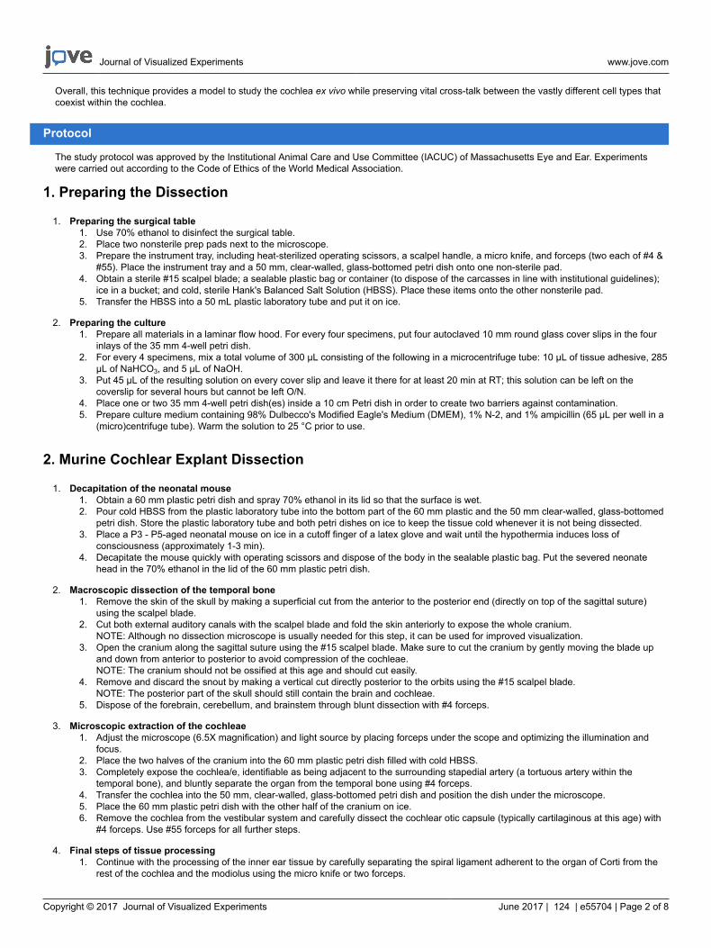
While many protocols focus on organ of Corti explants, this technique attempts to preserve the anatomy of the entire cochlear turn, includingthe SGNs. This gives researchers the opportunity to analyze the effects of a given treatment on neurites and somata of SGNs in addition to theorgan of Corti. Performing a dissection that preserves part of the modiolus, as described here, is more technically challenging than explanting theorgan of Corti alone. However, the neurite and SGN area is important, because significant pathological effects were observed in this region afterapplying vestibular schwannoma secretions (Figure 1) and extracellular vesicles in two recent publications using the same methodology19,20.Such changes could not have been revealed by using isolated organ of Corti explants.
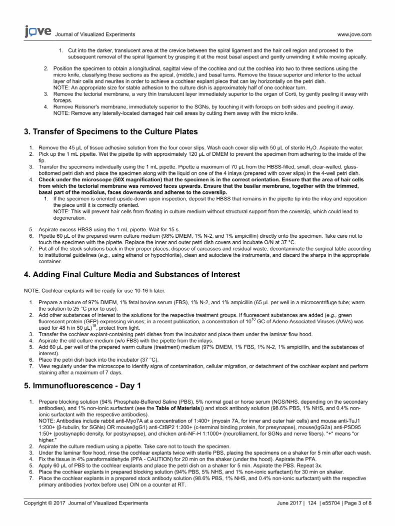
The quantification of damaged or GFP-expressing cells for inner and outer hair cells, supporting cells, and SGNs can be performed by manualcount or with an automated process, such as that built into the ImageJ plugin NeuronJ. Representative areas for these counts can be establishedafter confirming their correlation with total counts. 3D reconstructions are particularly useful to exclude overlay effects in the z-axis projections ofSGNs. Fiber organization, number of SGNs per given area, and SGN soma area can be evaluated. The staining and counting of synapses, haircells, and neurites is depicted in Figure 2; the same protocol can also be used for cochlear whole mounts and has been shown to be a reliablemodel for these purposes17,21,22. Other researchers have established elegant alternative methods that can also be incorporated into experimentalparadigms23,24.
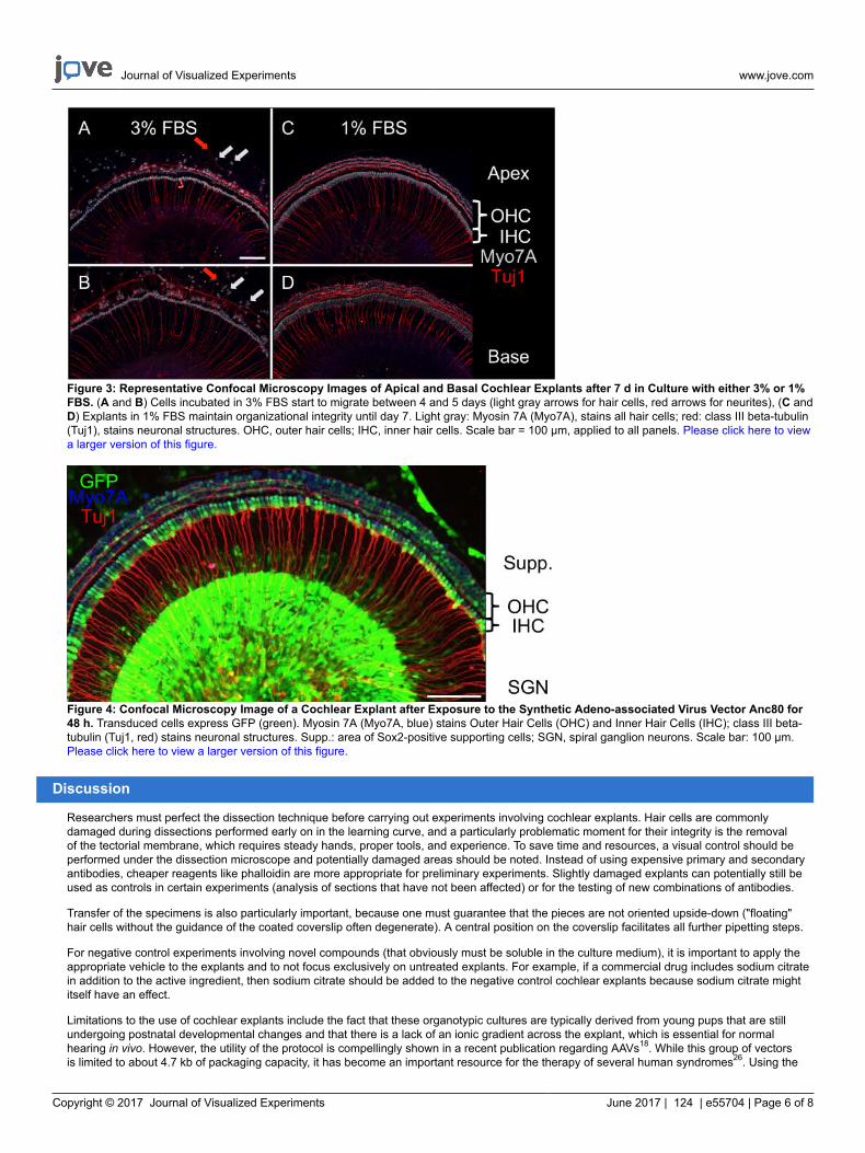
The maximum length of culture is usually limited by the start of cellular migration and depends upon the concentration of FBS in the culturemedium. When the proportion of FBS was reduced from five percent to three percent or one percent, the time period for maintaining ananatomically intact cochlear explant was extended to approximately one week (Figure 3).
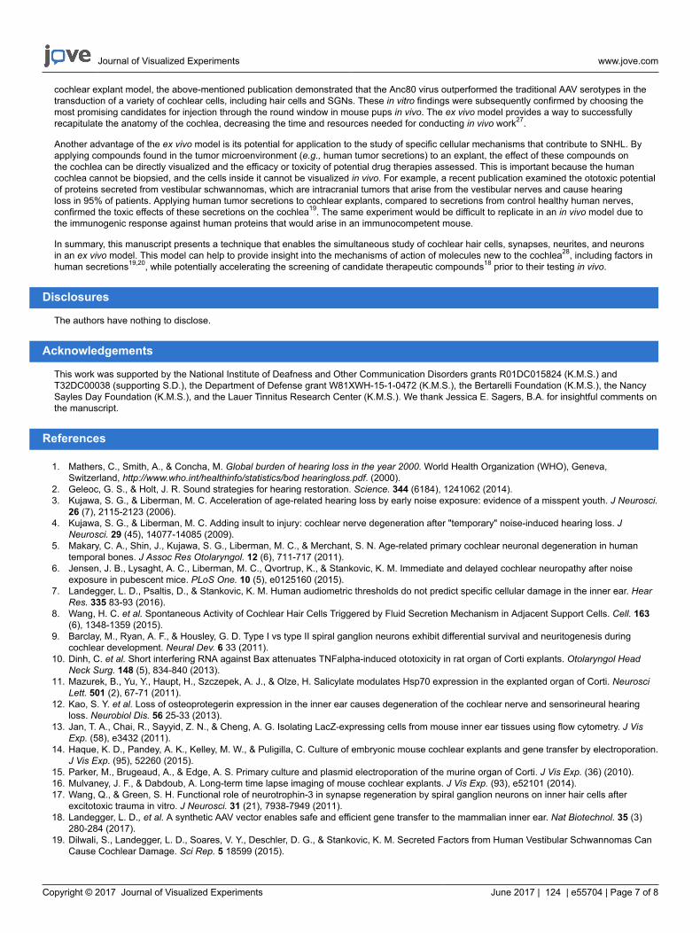
Figure 4 demonstrates GFP-positive cochlear explant cells after transduction with the synthetic adeno-associated virus vector Anc80 for 48 h25.Inner hair cells, outer hair cells, and supporting cells can be quantified in this view, while a 3D reconstruction can help to quantify GFP-positivecells in the SGN area. Different viral serotypes can be compared in terms of their transduction efficiency in certain cell types using the outlinedmethodology18.
Journal of Visualized Experiments www.jove.com
Copyright © 2017 Journal of Visualized Experiments June 2017 | 124 | e55704 | Page 5 of 8
Figure 1: Representative Confocal Microscopy Images of Cochlear Explants from the Apical and Basal Regions after Incubation withSecretions from Great Auricular Nerves or Vestibular Schwannomas for 48 h. (A) Overview image of an explant. The rectangle marks thearea in close-up images. Scale bar = 200 µm. (B-E) Zoomed-in views of hair cells and neurites. Scale bar = 50 µm. Green = Myosin 7A (Myo7A),stains hair cells; red = class III beta-tubulin (Tuj1), stains neuronal structures. Please click here to view a larger version of this figure.
Figure 2: Confocal Microscopy Image of the Cochlear Explant Hair Cell Region. (A) Intact synapses can be assessed by labelingpresynaptic (C-terminal binding protein, red) and postsynaptic (postsynaptic density-95, green) proteins together with the staining of neuronalstructures (neurofilament, blue). OHC, outer hair cells; IHC, inner hair cells. Scale bar = 10 µm. (B) Focus on the inner hair cell region with thecount of inner hair cells (red arrowheads) and neurites (blue arrowheads). Due to the ramification of the neuronal structures, it is important tostandardize the distance from the inner hair cells during counting (highlighted with the white frame, directly connected to the synapses). Scalebar = 5 µm. (C) Close-up of separate synapses (2 pre- and postsynaptic pairs marked with white arrowheads). Scale bar = 1 µm. Please clickhere to view a larger version of this figure.
Journal of Visualized Experiments www.jove.com
Copyright © 2017 Journal of Visualized Experiments June 2017 | 124 | e55704 | Page 6 of 8
Figure 3: Representative Confocal Microscopy Images of Apical and Basal Cochlear Explants after 7 d in Culture with either 3% or 1%FBS. (A and B) Cells incubated in 3% FBS start to migrate between 4 and 5 days (light gray arrows for hair cells, red arrows for neurites), (C andD) Explants in 1% FBS maintain organizational integrity until day 7. Light gray: Myosin 7A (Myo7A), stains all hair cells; red: class III beta-tubulin(Tuj1), stains neuronal structures. OHC, outer hair cells; IHC, inner hair cells. Scale bar = 100 µm, applied to all panels. Please click here to viewa larger version of this figure.
Figure 4: Confocal Microscopy Image of a Cochlear Explant after Exposure to the Synthetic Adeno-associated Virus Vector Anc80 for48 h. Transduced cells express GFP (green). Myosin 7A (Myo7A, blue) stains Outer Hair Cells (OHC) and Inner Hair Cells (IHC); class III beta-tubulin (Tuj1, red) stains neuronal structures. Supp.: area of Sox2-positive supporting cells; SGN, spiral ganglion neurons. Scale bar: 100 µm.Please click here to view a larger version of this figure.
Discussion
Researchers must perfect the dissection technique before carrying out experiments involving cochlear explants. Hair cells are commonlydamaged during dissections performed early on in the learning curve, and a particularly problematic moment for their integrity is the removalof the tectorial membrane, which requires steady hands, proper tools, and experience. To save time and resources, a visual control should beperformed under the dissection microscope and potentially damaged areas should be noted. Instead of using expensive primary and secondaryantibodies, cheaper reagents like phalloidin are more appropriate for preliminary experiments. Slightly damaged explants can potentially still beused as controls in certain experiments (analysis of sections that have not been affected) or for the testing of new combinations of antibodies.
Transfer of the specimens is also particularly important, because one must guarantee that the pieces are not oriented upside-down ("floating"hair cells without the guidance of the coated coverslip often degenerate). A central position on the coverslip facilitates all further pipetting steps.
For negative control experiments involving novel compounds (that obviously must be soluble in the culture medium), it is important to apply theappropriate vehicle to the explants and to not focus exclusively on untreated explants. For example, if a commercial drug includes sodium citratein addition to the active ingredient, then sodium citrate should be added to the negative control cochlear explants because sodium citrate mightitself have an effect.
Limitations to the use of cochlear explants include the fact that these organotypic cultures are typically derived from young pups that are stillundergoing postnatal developmental changes and that there is a lack of an ionic gradient across the explant, which is essential for normalhearing in vivo. However, the utility of the protocol is compellingly shown in a recent publication regarding AAVs18. While this group of vectorsis limited to about 4.7 kb of packaging capacity, it has become an important resource for the therapy of several human syndromes26. Using the
Journal of Visualized Experiments www.jove.com
Copyright © 2017 Journal of Visualized Experiments June 2017 | 124 | e55704 | Page 7 of 8
cochlear explant model, the above-mentioned publication demonstrated that the Anc80 virus outperformed the traditional AAV serotypes in thetransduction of a variety of cochlear cells, including hair cells and SGNs. These in vitro findings were subsequently confirmed by choosing themost promising candidates for injection through the round window in mouse pups in vivo. The ex vivo model provides a way to successfullyrecapitulate the anatomy of the cochlea, decreasing the time and resources needed for conducting in vivo work27.
Another advantage of the ex vivo model is its potential for application to the study of specific cellular mechanisms that contribute to SNHL. Byapplying compounds found in the tumor microenvironment (e.g., human tumor secretions) to an explant, the effect of these compounds onthe cochlea can be directly visualized and the efficacy or toxicity of potential drug therapies assessed. This is important because the humancochlea cannot be biopsied, and the cells inside it cannot be visualized in vivo. For example, a recent publication examined the ototoxic potentialof proteins secreted from vestibular schwannomas, which are intracranial tumors that arise from the vestibular nerves and cause hearingloss in 95% of patients. Applying human tumor secretions to cochlear explants, compared to secretions from control healthy human nerves,confirmed the toxic effects of these secretions on the cochlea19. The same experiment would be difficult to replicate in an in vivo model due tothe immunogenic response against human proteins that would arise in an immunocompetent mouse.
In summary, this manuscript presents a technique that enables the simultaneous study of cochlear hair cells, synapses, neurites, and neuronsin an ex vivo model. This model can help to provide insight into the mechanisms of action of molecules new to the cochlea28, including factors inhuman secretions19,20, while potentially accelerating the screening of candidate therapeutic compounds18 prior to their testing in vivo.
Disclosures
The authors have nothing to disclose.
Acknowledgements
This work was supported by the National Institute of Deafness and Other Communication Disorders grants R01DC015824 (K.M.S.) andT32DC00038 (supporting S.D.), the Department of Defense grant W81XWH-15-1-0472 (K.M.S.), the Bertarelli Foundation (K.M.S.), the NancySayles Day Foundation (K.M.S.), and the Lauer Tinnitus Research Center (K.M.S.). We thank Jessica E. Sagers, B.A. for insightful comments onthe manuscript.
References
1. Mathers, C., Smith, A., & Concha, M. Global burden of hearing loss in the year 2000. World Health Organization (WHO), Geneva,Switzerland, http://www.who.int/healthinfo/statistics/bod hearingloss.pdf. (2000).
2. Geleoc, G. S., & Holt, J. R. Sound strategies for hearing restoration. Science. 344 (6184), 1241062 (2014).3. Kujawa, S. G., & Liberman, M. C. Acceleration of age-related hearing loss by early noise exposure: evidence of a misspent youth. J Neurosci.
26 (7), 2115-2123 (2006).4. Kujawa, S. G., & Liberman, M. C. Adding insult to injury: cochlear nerve degeneration after "temporary" noise-induced hearing loss. J
Neurosci. 29 (45), 14077-14085 (2009).5. Makary, C. A., Shin, J., Kujawa, S. G., Liberman, M. C., & Merchant, S. N. Age-related primary cochlear neuronal degeneration in human
temporal bones. J Assoc Res Otolaryngol. 12 (6), 711-717 (2011).6. Jensen, J. B., Lysaght, A. C., Liberman, M. C., Qvortrup, K., & Stankovic, K. M. Immediate and delayed cochlear neuropathy after noise
exposure in pubescent mice. PLoS One. 10 (5), e0125160 (2015).7. Landegger, L. D., Psaltis, D., & Stankovic, K. M. Human audiometric thresholds do not predict specific cellular damage in the inner ear. Hear
Res. 335 83-93 (2016).8. Wang, H. C. et al. Spontaneous Activity of Cochlear Hair Cells Triggered by Fluid Secretion Mechanism in Adjacent Support Cells. Cell. 163
(6), 1348-1359 (2015).9. Barclay, M., Ryan, A. F., & Housley, G. D. Type I vs type II spiral ganglion neurons exhibit differential survival and neuritogenesis during
cochlear development. Neural Dev. 6 33 (2011).10. Dinh, C. et al. Short interfering RNA against Bax attenuates TNFalpha-induced ototoxicity in rat organ of Corti explants. Otolaryngol Head
Neck Surg. 148 (5), 834-840 (2013).11. Mazurek, B., Yu, Y., Haupt, H., Szczepek, A. J., & Olze, H. Salicylate modulates Hsp70 expression in the explanted organ of Corti. Neurosci
Lett. 501 (2), 67-71 (2011).12. Kao, S. Y. et al. Loss of osteoprotegerin expression in the inner ear causes degeneration of the cochlear nerve and sensorineural hearing
loss. Neurobiol Dis. 56 25-33 (2013).13. Jan, T. A., Chai, R., Sayyid, Z. N., & Cheng, A. G. Isolating LacZ-expressing cells from mouse inner ear tissues using flow cytometry. J Vis
Exp. (58), e3432 (2011).14. Haque, K. D., Pandey, A. K., Kelley, M. W., & Puligilla, C. Culture of embryonic mouse cochlear explants and gene transfer by electroporation.
J Vis Exp. (95), 52260 (2015).15. Parker, M., Brugeaud, A., & Edge, A. S. Primary culture and plasmid electroporation of the murine organ of Corti. J Vis Exp. (36) (2010).16. Mulvaney, J. F., & Dabdoub, A. Long-term time lapse imaging of mouse cochlear explants. J Vis Exp. (93), e52101 (2014).17. Wang, Q., & Green, S. H. Functional role of neurotrophin-3 in synapse regeneration by spiral ganglion neurons on inner hair cells after
excitotoxic trauma in vitro. J Neurosci. 31 (21), 7938-7949 (2011).18. Landegger, L. D., et al. A synthetic AAV vector enables safe and efficient gene transfer to the mammalian inner ear. Nat Biotechnol. 35 (3)
280-284 (2017).19. Dilwali, S., Landegger, L. D., Soares, V. Y., Deschler, D. G., & Stankovic, K. M. Secreted Factors from Human Vestibular Schwannomas Can
Cause Cochlear Damage. Sci Rep. 5 18599 (2015).
Journal of Visualized Experiments www.jove.com
Copyright © 2017 Journal of Visualized Experiments June 2017 | 124 | e55704 | Page 8 of 8
20. Soares, V. Y. et al. Extracellular vesicles derived from human vestibular schwannomas associated with poor hearing damage cochlear cells.Neuro Oncol. (2016).
21. Tong, M., Brugeaud, A., & Edge, A. S. Regenerated synapses between postnatal hair cells and auditory neurons. J Assoc Res Otolaryngol.14 (3), 321-329 (2013).
22. Yuan, Y. et al. Ouabain-induced cochlear nerve degeneration: synaptic loss and plasticity in a mouse model of auditory neuropathy. J AssocRes Otolaryngol. 15 (1), 31-43 (2014).
23. Fernandez, K. A., Jeffers, P. W., Lall, K., Liberman, M. C., & Kujawa, S. G. Aging after noise exposure: acceleration of cochlear synaptopathyin "recovered" ears. J Neurosci. 35 (19), 7509-7520 (2015).
24. Barclay, M., Constable, R., James, N. R., Thorne, P. R., & Montgomery, J. M. Reduced sensory stimulation alters the molecular make-up ofglutamatergic hair cell synapses in the developing cochlea. Neuroscience. 325 50-62 (2016).
25. Zinn, E. et al. In Silico Reconstruction of the Viral Evolutionary Lineage Yields a Potent Gene Therapy Vector. Cell Rep. 12 (6), 1056-1068(2015).
26. Wu, Z., Yang, H., & Colosi, P. Effect of genome size on AAV vector packaging. Mol Ther. 18 (1), 80-86 (2010).27. Shu, Y. et al. Identification of Adeno-Associated Viral Vectors That Target Neonatal and Adult Mammalian Inner Ear Cell Subtypes. Hum
Gene Ther. (2016).28. Kao, S. Y., Soares, V. Y., Kristiansen, A. G., & Stankovic, K. M. Activation of TRAIL-DR5 pathway promotes sensorineural degeneration in the
inner ear. Aging Cell. 15 (2), 301-308 (2016).