Embed Size (px)
Citation preview
Mobilization of Sialidase from Intracellular Stores to the Surface of HumanNeutrophils and Its Role in Stimulated Adhesion Responses of These CellsAlan S. Cross and Daniel G. WrightDepartments of Bacterial Diseases and Hematology, Walter Reed Army Institute of Research, Washington, D.C. 20307-5100
Abstract
Desialation of cell surfaces has been associated with the initia-tion or modification of diverse cellular functions. In these stud-ies we have examined the subcellular distribution of sialidase(SE) in human neutrophils as well as the mobilization of thisenzyme following neutrophil activation. Separation of subcellu-lar fractions by density gradient centrifugation showed that SEis present not only in neutrophil primary and secondary granulepopulations, like lysozyme, but also in plasma membrane frac-tions. Neutrophil activation was associated with a redistribu-tion of SE from secondary granule-enriched fractions to theplasma membrane. Furthermore, SE activity detected on thesurface of intact neutrophils with a fluorescent SE substrateincreased rapidly after activation with kinetics that matchedboth the loss of total cell-associated sialic acid and release offree sialic acid from the cells. These activation-dependentevents were in each case blocked by incubation of neutrophilswith the SE inhibitor, 2-deoxy-N-acetyl-neuraminic acid. Ag-gregation responses of neutrophils as well as adhesion re-sponses to nylon and plastic surfaces were also inhibited by2-deoxyNANA. Our findings indicate that the activation-de-pendent desialation of the neutrophil surface is associated withmobilization of an endogenous SE to the plasma membrane andhas a role in stimulated adhesion responses of these cells. (J.Clin. Invest. 1991.88:2067-2076.) Keywords: sialicacideneur-aminidase * cell activation * granulocytes * aggregation
Introduction
The presence of sialic acids on cell surfaces, as terminal sugars
of glycosylated cell surface molecules, has been highly con-served in evolution (1). Furthermore, there is substantial evi-dence that sialic acids are structural determinants for a varietyof important cell-cell interactions and cellular functions, suchas the adhesiveness and metastatic potential of neoplastic cells(2-4) and the phenomenon of contact inhibition in tissue cul-ture (5).
Immature myeloid cells have been shown to be rich in cellsurface sialic acid (6). Moreover, there is evidence that this highdegree of sialation contributes to the negative surface charge,rigidity, and lack of adhesiveness of these cells (7, 8), for as
myeloid precursor cells mature, they become less negatively
Address correspondence to Dr. Alan S. Cross, Department of BacterialDiseases, Walter Reed Army Institute of Research, Washington, DC20307-5100.
Received for publication 7 December 1989 and in revised form 6August 1991.
The Journal of Clinical Investigation, Inc.Volume 88, December 1991, 2067-2076
charged, more deformable and more adhesive, coincident witha progressive loss of cell surface sialic acid (6).
Cells may be desialated by sialidases (or neuraminidases)which cleave n-glycosidic-linked sialic acids in a one-step reac-tion (9). These enzymes, like sialic acids, are widely distributedin nature and exist in a variety of forms that maydiffer substan-tially in electrophysical properties and also in their ability tocleave distinct glycosidic linkages (10-12). Sialidases of mam-malian cells are generally membrane bound and have been lesswell characterized than microbial neuraminidases (13). None-theless, it is evident that these enzymes may provide a mecha-nism for rapidly modulating cell function by modifying glyco-sylated cell surface structures ( 14).
Humanneutrophils have been shown by several laborato-ries to contain sialidase activity (15, 16). Moreover, studies ofthe appearance of sialidase activity during induced maturationof the myeloid leukemia cell line, HL60, have suggested thatneutrophil sialidase is acquired during the latter phases of neu-trophilic maturation (15). Because desialation of cells has beenassociated with changes in various cell functions that areknown to occur with neutrophil activation (e.g., increased adhe-siveness [17] and decreased surface charge [18, 19]), we wereattracted to the possibility that mobilization of an endogenoussialidase in neutrophils might have a role in the functionalactivation of these cells. To explore this possibility, we exam-ined the intracellular compartmentalization of sialidase in hu-man neutrophils, together with the mobilization and redistri-bution of this enzyme during cell activation.
Methods
Isolation of human blood neutrophils and preparation of subcellularfractions. Neutrophils were isolated from 50-200-ml samples of hepa-rinized venous blood obtained from healthy volunteers by combiningHypaque-Ficoll density gradient centrifugation and dextran sedimenta-tion procedures, followed by hypotonic lysis of residual erythrocytes, asdescribed previously (20, 21). For certain studies that required largenumbers of cells, up to 5 x 1O' neutrophils were isolated by the samemethods from 200 to 400-ml leukocyte concentrates obtained by con-tinuous flow, centrifugation leukapharesis of normal volunteers (2997continuous flow centrifuge; IBM Corp., Danbury, CT). In both cases,final leukocyte preparations contained > 95% neutrophils and were> 99% viable as determined by trypan blue dye exclusion.
For preparation of subcellular fractions, neutrophils were sus-pended in calcium-free 10 mMpiperazine-N,NT-bis(2-ethane sulfonicacid (Pipes) buffer with 1.0 mMATP and 3.5 mMMgCl2 at a cellconcentration of 5 X 10⁷ neutrophils/ml. Cells were then disrupted bynitrogen cavitation at 40C using a precooled pressure homogenizer(Parr Instrument Co., Moline, IL) as described previously (21). Cavi-tates were then centrifuged at 1,400 g for 10 min to remove nuclei andunbroken cells, and supernatants were applied to continuous sucrosegradients (21, 22) or to discontinuous PercollR (Pharmacia, Uppsala,Sweden) gradients (21, 23) and centrifuged in a refrigerated (40C) ultra-centrifuge at 95,000 g for 4 h or at 48,000 g for 30 min, respectively.Stepwise elution of gradients from the bottom of centrifuge tubes was
Neutrophil Sialidase 2067

then carried out. Granule-rich and plasma membrane vesicle-rich frac-tions were identified by direct turbidometric measurements of frac-tions at 450 nm(21, 22) and by the measurement of previously charac-terized granule and plasma membrane markers: lysozyme (24), myelo-peroxidase (25), vitamin B12 binding protein (26), and alkalinephosphatase (27), after the addition of 0.1% Triton X-100 (vol/vol,final concentration) to fractions. For certain studies that addressed thesubcellular distribution of neutrophil sialidase with and without prioractivation of the cells, fractions separated on discontinuous PercollRgradients were collected by aspiration of the gradients from the topusing a fine-tipped Pasteur pipette; aspirated fractions that were col-lected at the separate visible interfaces of gradients, where plasmamembrane vesicles, secondary granules, or primary granules were con-centrated, were saved as pooled fractions enriched in these distinctsubcellular components.
Cellular extracts of intact neutrophils (1-5 x 10⁷ cells/ml), sus-pended either in HBSS, pH 7.3, or in 0.34 Msucrose, were also pre-pared by sonication or rapid freeze-thawing in the presence of 0.1%Triton X-100 and protease inhibitors (aprotinin 100 Mg/ml, leupeptin100 Mg/ml, and PMSF1.0 mM,all obtained from Sigma Chemical Co.,St. Louis, MO), followed by centrifugation at 12,000 g for 2 min.
For certain studies, neutrophil extraction or subcellular fraction-ation was carried out after first incubating the neutrophils (suspendedat a concentration of I07 cells/ml in HBSS) in a shaking water bath at370C for 5 min with or without 5 jtg/ml cytochalasin B (Sigma Chemi-cal Co.) and then for 0.5-30 min with or without 100 ng/ml phorbolmyristate acetate (PMA), 1.0 IM fMet-Leu-Phe (fMLF),' 1.0MuM iono-mycin (IMN), or 1.0 MMA23187 (all obtained from Sigma). Afterincubation, cells were centrifuged at 250 g for 10 min in a refrigeratedcentrifuge, supernatants saved for subsequent assays of free sialic acidand enzymatic activities, and the pelleted cells resuspended in mediaappropriate for extraction or for subcellular fractionation procedures.
Measurement ofneutrophil sialidase and neutrophil sialic acid con-tent. Neutrophil sialidase activity was measured and compared withVibrio cholerae neuraminidase (Sigma) by three separate methods: (a)by the release of free sialic acid from the artificial substrate, neuramin-yllactose (Sigma, also referred to as N-acetylneuramin-lactose), frompurified glycoproteins (e.g., transferrin, orosomucoid, fetuin [Sigma])(13) or from intact neutrophils; (b) by release of the fluorescent moiety,4-methylumbelliferone (4-MU) from 4-methylumbelliferyl-alpha-D-N-acetyl-neuraminic acid (4MU-NANA) (15); and (c) by the ability ofpeanut lectin to agglutinate human erythrocytes pretreated with siali-dase (28).
With the first method, cell extracts to be measured for sialidaseactivity were suspended in sodium acetate buffer (0.8 M, pH 4.5) andadded to substrates. Following incubation at 37°C for various periodsof time, samples were centrifuged at 12,000 g for 2 min in a microfuge(Eppendorf Inc., Fremont, CA) and the supernatants assayed for freesialic acid as described below. To control for the spontaneous release offree sialic acid from substrate molecules or "substrate" neutrophils, aswell as from cell extracts that served as a source of sialidase, we mea-sured the free sialic acid released from cell extracts when mixed withbuffer alone, as well as the free sialic acid released by the substratemolecules and by substrate neutrophils alone when incubated underidentical conditions but without an exogenous source of sialidase. Thesialic acid released under these control conditions was then subtractedfrom the total amount of sialic acid released by incubation of substratemolecules, or "substrate" cells, with neuraminidase or with neutrophil
1. Abbreviations used in this paper: ELAM-1, endothelial leukocyteadhesion molecule 1; fMLF, fMet-Leu-Phe; GMP-140, guanosine5'-monophosphate; ICAM- 1, intercellular adhesion molecule 1; IMN,ionomycin; KDO, 2-keto-3-deoxyoctulosonic acid; LEC-CAM, lectincellular adhesion molecule; LFA-l, lymphocyte function-associatedantigen; 4-MU, 4-methylumbellifereone; 4-MU-NANA, 4-methyl-umbelliferyl-alpha-D-N-acetyl-neuraminic acid; NA, neuraminidase;NANA, N-acetyl neuraminic acid; 2 deoxy-NANA, 2,3-dehydro-2-deoxy-N-acetyl-neuraminic acid.
extracts. For some studies, serine esterase inhibitors (PMSF [fromSigma], TPCK [N-tosyl-L-phenylalanine chloromethyl ketone] orTLCK [N-tosyl-L-lysine chloromethyl ketone], obtained from Calbio-chem Corp., La Jolla, CA) were added to reaction mixtures.
To examine the possibility that neutrophils contained or generatedan inhibitor of sialidase activity, we carried out mixing experiments todetermine whether intact neutrophils or neutrophil extracts modifiedthe ability of microbial neuraminidase to release sialic acid from neura-minyllactose. Release of sialic acid from neuraminyllactose mediatedby neuraminidase and by neutrophils or neutrophil extracts were mea-sured both separately and together to determine whether the sialidaseactivities detected individually were additive when neuraminidase andneutrophils or neutrophil extracts were mixed together. Controls in-cluded neuraminyllactose alone, neutrophil extracts alone, and cells orextracts together with neuraminidase.
Total cell-associated sialic acid (or N-acetyl neuraminic acid[NANA]) and cell-free NANAreleased from sialidase substrates weremeasured by both the Warren thiobarbituric acid assay and by theNANAaldolase, or lyase, assay. Each assay was performed as previ-ously described (29, 30). In the latter assay, samples to be measuredwere mixed with NANA-aldolase (Sigma) which specifically cleavessialic acid into pyruvate and acylmannosamine; pyruvate, in the pres-ence of lactic dehydrogenase, reduces NADHto NADwhich was mea-sured spectrofluorimetrically at an excitation wavelength of 320 nmand emission of 470 nm. Sialic acid was then measured indirectly bythe amount of pyruvate generated by the aldolase. Each sample was runwith and without aldolase. Controls included a standard curve of pyru-vate as positive control, a standard curve of sialic acid (Sigma), a con-trol deficient in sample, and a tube with NADHalone. The amount ofsialic acid was determined by comparing the relative differences be-tween samples assayed with and without aldolase in relationship to astandard sialic acid curve.
Intact- neutrophils to be assayed for total sialic acid content wereextracted in H2SO4 (0.1 N) at 80°C for 1 h. Readjustment of pH wasnot necessary in the Warren assay; however, in the aldolase assay, thesample was titrated to pH 7.4 with 0.1 NNaOHbefore the addition ofNANAaldolase.
The 4MU-NANAassay of sialidase in neutrophil extracts was per-formed as described previously (15). For these assays, 100 MMleupep-tin, I mMPMSF, and 100 Mg/ml aprotinin were added to cell prepara-tions before extraction procedures. Freshly prepared 4MU-NANAwasadded at a final concentration of 0.5 mMto sodium acetate buffer,0.075 mM, pH 4.5. After incubation at 37°C for 2 h, the release ofNANAfrom the substrate was determined by measurement of fluores-cence at 440 nmafter the addition of ethanol and NaOHto the reactionmixture (1 5); measurements were then correlated with a 4MUfluores-cence standard.
For studies designed to detect sialidase activity on the neutrophilcell surface, intact neutrophils, after incubation in HBSS, pH 7.4, at37°C (I0' cells/ml) for 0.5-10 min with or without 100 ng/ml PMA,1.0 MMIMN, or 1.0 MMfMLF, were resuspended in the 4MU-NANAassay buffer at pH 7.0 and incubated for 30 min, followed by measure-ment of 4MUfluorescence in the reaction mixture. In certain studiessialidase activity detected with intact cells incubated in buffer contain-ing 4MU-NANAwas measured in the presence of the sialidase inhibi-tor, 2,3-dehydro-2-deoxy-N-acetyl-neuraminic acid (2deoxy-NANA,Boehringer-Mannheim Corp., Indianapolis, IN) or a structurally simi-lar compound lacking sialidase inhibitory activity, 2-keto-3-deoxyoc-tulosonic acid (KDO, Sigma).
For the third sialidase assay procedure, samples to be measured forsialidase activity were incubated with a suspension of human erythro-cytes according to the method of Pereira (28). The ability of peanutlectin (Arachis hypogaea, Sigma) to induce agglutination ofthe erythro-cytes following incubation was then used as a measure of desialationmediated by sialidase activity in the sample.
To determine the extent to which neutrophil sialidase remainsmembrane associated or is released as a soluble enzyme after disrup-tion of cells or subcellular organelles, extracts were prepared from neu-
2068 A. S. Cross and D. G. Wright
trophils or subcellular fractions that were suspended in HBSSand pro-tease inhibitors in the presence or absence of 0.1% Triton X-100 andsonicated. After centrifugation at 10,000 g for 10 min at 4VC, superna-tants were assayed for sialidase activity by incubation with the artificialsubstrate, neuraminyllactose, for 1 h at 370C. Release of sialic acid wasthen determined by the Warren assay.
Measurement of radiolabelled sialic acid binding to neutrophils.Neutrophils were prepared from human peripheral blood as describedand adjusted to 8 X 10' in HBSS. Radiolabelled sialic acid (cytidine5'-monophospho['4C]sialic acid, ammonium salt, Amersham Corp.,Arlington Heights, IL) was added to replicate aliquots of cells in thepresence or absence of excess (100 ug/ml) unlabelled sialic acid. Theneutrophils were retained on ice or at 370C without stimulation, orwere stimulated for up to 10 min with 1 MMIMN at 370C. Aliquots(100 Ml) were then centrifuged in triplicate through silicone oil (Versi-lube; General Electric Co., Waterford, NY) and cell pellets were treatedovernight with tissue solubilizer (Solusol; National Diagnostics, Inc.,Somerville, NJ) and counted in a scintillation counter.
Measurement of stimulated neutrophil aggregation and spontane-ous adhesion of neutrophils to nylon fibers or plastic surfaces. Homoty-pic neutrophil aggregation was measured by a standard procedure de-scribed previously (31) using a Lumi-aggregometer (Chrono-LogCorp., Havertown, PA). Cells were incubated with and without 1.0 uMfMLF, 1.0 MMIMN, or 100 ng/ml PMA, and with or without freshlyprepared 3-250 Mg/ml 2-deoxyNANA or KDO, which was added tostirred neutrophil suspensions immediately before addition of thestimuli.
Neutrophil adhesion to nylon fibers or to albumin-coated plastictissue culture plates was measured using assay systems that had beenadapted from methods described previously (31). For measurements ofadhesion to nylon fibers, isolated neutrophils (see above) were resus-pended at a final concentration of 5 X 10⁶ cells/ml in heparinizedautologous plasma that had been separated from venous blood samplesused for neutrophil isolation and had been diluted 1:1 with HBSS. 1-mlaliquots of neutrophil suspensions, prewarmed to 37°C, were deliveredto nylon fiber-packed mini-columns, with or without 3-250 Mg/ml2-deoxyNANA, KDO, or NANA, and with or without 0.1 MMfMLF,which had been added to cell suspensions immediately before deliveryto the mini-columns. Replicate nylon fiber mini-columns were pre-pared from disposable tuberculin syringes, with needles and plungersremoved, by packing each with 20 mgof sterile nylon fibers (type 200;Fenwall Laboratories, Morton Grove, IL). Each column was then sus-pended in a sterile polypropylene test tube to collect cells that passedthrough the columns. Once aliquots of neutrophil suspensions weredelivered to the columns, they were placed in a humidified incubator at37°C for 5 min after which cells that had passed through individualcolumns were counted. The percent of cells that had adhered to thenylon fibers and had not passed through the mini-columns was thencalculated.
For measurement of neutrophil adhesion to plastic, isolated neutro-phils were resuspended at a final concentration of 5 X 10⁶/ml in HBSS.Replicate 1.0-ml aliquots of cell suspensions, prewarmed to 37°C, weredelivered to flat bottom plastic culture wells (six-well plastic culturedishes; Costar Corp., Cambridge, MA) that had been previously incu-bated with HBSS + 2% BSA and then rinsed with HBSSand dried.Cells were incubated in the plastic wells in a humidified 37°C incuba-tor for precisely 3 min, with or without 3-250 Mg/ml 2-deoxyNANA,KDO, or NANA, and with or without 0.1 MMfMLF, which had beenadded to cell suspensions immediately before delivery to the plasticculture wells, after which nonadherent cells were aspirated from thewells and counted. The percent of cells that remained adherent to theplastic wells was then calculated.
ResultsDetection and characterization of neutrophil sialidaseAs reported previously by others (15, 16), sialidase activitycould be detected readily in extracts of purified human blood
neutrophils. This activity was maximal at pH 4.5-5.0 and was,like other mammalian sialidases (32), a heat labile enzyme thatwas not inhibited by serine esterase inhibitors (PMSF, TPCK,or TLCK). Greater than 60% of activity was lost when neutro-phil extracts were incubated > 60 min at 560C, and > 80% ofactivity was lost with incubations at 100IC for 5 min. In addi-tion, substantial amounts of sialidase activity were lost fromcell extracts with a single cycle of freeze-thawing, which hin-dered the study of stored samples.
In Table I, substrate specificities of neutrophil sialidase arecompared with those of V. cholerae neuraminidase. Both werefound to be active against the artificial sialidase substrate, neur-aminyllactose, and the activity recoverable from 107 neutro-phils was roughly equivalent to 20 ,U of neuraminidase. Siali-dase in neutrophil extracts was relatively more active in me-diating the release of free sialic acid from intact neutrophils andin cleavage of 4MU-NANA, but unlike neuraminidase, lackedactivity against a number of other substrates.
Neutrophils did not appear to have a sialidase inhibitoryactivity that might account for differences in the apparent activ-ities in neutrophil sialidase vs. microbial neuraminidase, inthat the addition of intact neutrophils (resting or stimulated),or neutrophil extracts, to a mixture of V. cholerae neuramini-dase and neuraminyllactose did not decrease the release of freesialic acid from neuraminyllactose that was mediated by neur-aminidase alone (data not shown).Subcellular localization of neutrophil sialidaseSialidase activity was recoverable from subcellular fractions ofnitrogen-cavitated, resting neutrophils separated by ultracentri-fugation on both continuous sucrose gradients (21, 22) or ondiscontinuous PercollR gradients (21, 23). As is illustrated inFigs. 1 and 2, and as has been well documented previously (22,23), both techniques permitted the separation of cell fractionsenriched in plasma membrane vesicles, peroxidase-negative
Table I. Comparison of Neuraminidase and Neutrophil SialidaseActivities
Source of sialidase activity'
Substrate* Assayt V. cholera NA Neutrophils
Neuraminyllactose (W) 79.30 1.38(L) 17.10 0.37
Intact neutrophils (W) 3.71 1.31(L) 1.86 0.25
Mucin (W) 0.63 0Transferrin (W) 0.09 0Fetuin (W) 0.60 0Orosomucoid (W) 1.29 04MU-NANA (F1) 0.74 0.13RBC/peanut agglutinin'l 4+ 0
* Neuraminyllactose, mucin, transferrin, fetuin, and orosomucoid, allat 1 mg/ml. Intact neutrophils at 4 x 1 07/ml. * W, Warren assay; L,lyase assay; Fl, 4MUfluorescence assay. § Data expressed as micro-moles of free sialic acid released/1 mUper h for V. cholera neur-aminidase, and as micromoles of free sialic acid released/107 PMNsper h for sialidase activity derived from neutrophil extracts. 11 Positiveagglutination of sialidase-treated red blood cells (2% suspension) in0.1 MPBS, pH 6.8 at 8n sg/ml lectin (see Methods, ref. 28).
Neutrophil Sialidase 2069

0.6
0.5
C4 0.4uj Eo ~-.j 0.3. c,
.c Len 3
l c 0.2
0.1
na
i
cc
x
CL)
-i
LLUcooIC
J=-n
1° 2° M(a) (0) (r)
2 4 6 8 10 12 14 16
1'I A BI
1.2
1.0
0.8
0.6
0.4
0.2
-.
EE-
ui
Cl-
: -
o C_a
.J -
0.2~~O L
0.6
0.5
0.4
0.3
0.2
0.1
AE _iM Ei
L
cm cmcccc C,)
_ a:
_
mco _m ,
E
a.
anCD
0-
LUJMJ.
FRACTION NUMBER
Figure 1. Separation of cell fractions by PercollR gradient from nitro-gen-cavitated, resting neutrophils. Upper panel shows separation intoplasma membrane vesicles (M), peroxidase-negative secondary ((3), or
peroxidase-positive primary (a) granules by turbidity and proteinmeasurements. Middle panel shows distribution of myeloperoxidase(.), lysozyme (o), and vitamin B,2-binding protein (A) activities.Lower panel demonstrates localization of sialidase activity (-) andalkaline phosphatase (o) activities. 1 U of sialidase activity = 1.0 figsialic acid release from neuraminyllactose/h. Results of a representa-tive cell fractionation study are shown.
secondary granules, or peroxidase-positive primary granules.However, the sucrose gradient technique also resolved two dis-tinct primary granule populations with different densities.
Shown in Fig. are representative results of fractionation
studies using the PercollR gradient technique, with elution ofgradients from the bottom of ultracentrifuge tubes. Elution ofgranule or membrane-rich fractions is indicated in the toppanel of the figure by turbidity and protein measurements.Shown in the middle and bottom panels of the figure are thelocalizations of myeloperoxidase (in the dense, primary gran-ule-rich fractions), B12-binding activity (in the lighter density,secondary granule-rich fractions), lysozyme (in both primaryand secondary granule fractions), alkaline phosphatase (in the
11-i
E
Lu~1
-C)
C0
2'C M
2 4 6 8 10 12 14 16
1.2
1.0
0.8
0.6
0.4
0.2
-6
E
a-
L.
-EMI-
car
C-
2E
CD
co
orMcoTo
C2or
-i
FRACTION NUMBER
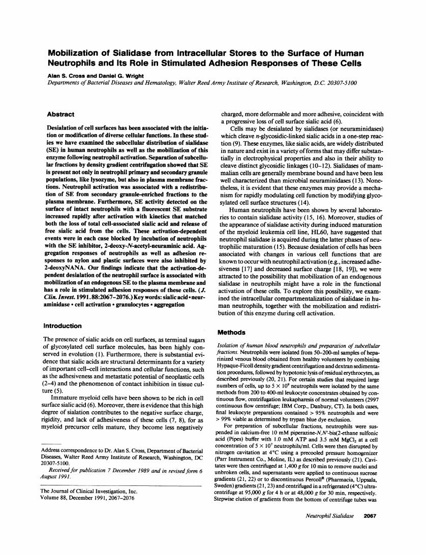
Figure 2. Separation of cell fractions by continuous sucrose gradientfrom nitrogen-cavitated, resting neutrophils. Upper panel shows sep-
aration into plasma membrane vesicles (M), secondary granules (C),and two populations (A and B) of primary granules by turbidity andprotein measurements. The middle panel shows the distribution ofmyeloperoxidase (o), lysozyme (o), and vitamin B12-binding protein(A) activities. The lower panel shows the distribution of sialidase (-)and alkaline phosphatase (o) activities, demonstrating that sialidaseactivity is recovered only from the more dense of the twoperoxidase-positive primary granule population. 1 U of sialidase ac-
tivity = 1.0 ig sialic acid released/h. Results of a representative cellfractionation study are shown.
plasma membrane vesicle-rich, light density fractions), and sia-lidase activity (shown in the bottom panel) which was recov-
ered from both primary and secondary granule fractions, as
well as from plasma membrane fractions.Fig. 2, which illustrates cell fractionation using the continu-
ous sucrose gradient technique, shows very similar results forthe subcellular localization of sialidase activity. However, inthese studies it was evident that sialidase, like lysozyme, is re-
coverable only from the more dense of the two peroxidase posi-
tive, primary granule populations, resolved by this technique.
2070 A. S. Cross and D. G. Wright
2 4 6 8 10 12 14 16
2 4 6 8 10 12 14 16

Translocation of sialidase activity in activated neutrophilsWhen comparative PercollR gradient fractionation studieswere carried out with both resting and activated neutrophils, itwas evident that activation of the cells by PMA, by the iono-phore, A23 187, or by the chemotactic peptide fMLF both withand without preincubation with cytochalasin B, were in eachcase associated with an apparent translocation of sialidase activ-ity from secondary granule-rich fractions to fractions contain-ing plasma membrane vesicles (Fig. 3). This translocation ofsialidase was evident both when enzyme activities in fractionswere expressed in terms of total numbers of cells fractionated(Fig. 3 A) and when expressed in terms of total protein recov-ered in the fractions (Fig. 3 B). The decline in sialidase specificactivity in secondary granule fractions (Fig. 3 B), in addition tothe decline in total sialidase activity detected in the fractions,suggested that the sialidase translocated to the plasma mem-brane is derived from a subpopulation of the organelles present
A
Up
00
0T-
la
0(A00
.
a-.
co(U
0)-i0
m
a
.) 350.
0 0
i 25-100) 20-U)co0 15-
0 10-._
0)co._
en
PrimaryGranules
Secondary MembraneGranules
B
T
PrimaryGranules
ni T
1 WM IE EM-Secondary MembraneGranules
Figure 3. Sialidase activity recovered from pooled primary granule,secondary granule, and plasma membrane fractions (obtained byPercoll gradient fractionation) of resting neutrophils (control) andneutrophils stimulated by PMA, 100 ng/ml, the calcium ionophoreA23 187 (1.0 AM), and fMLF (1.0MM) in the presence of cytochalasinB (5 Mg/ml). Translocation of sialidase activity from secondary gran-ule to plasma membrane fractions is evident when sialidase is ex-pressed in terms of total number of cells fractionated (A) and in termsof total protein recovered in fractions, or specific activity (B). Resultsrepresent mean values±SEM for three replicate studies. o, Control; u,PMA; o, A23187; a, fMLP.
in the secondary granule rich fractions. It was noteworthy inthis regard that translocation of sialidase present in "second-ary" granule fractions to the plasma membrane fractions wasfound to occur with fMLF stimulation in the absence of cyto-chalasin, conditions with which little or no granule exocytosiswas evident by release of the primary and secondary granulemarkers, beta-glucuronidase and B12-binding protein.
The relative distribution of sialidase activities in the pri-mary, secondary, and plasma membrane fractions varied some-what among separate cell preparations; the distribution of siali-dase also varied depending upon whether all fractions of a den-sity gradient were analyzed, as in Figs. 1 and 2, or whetherpooled fractions were examined, Fig. 3. The relative recoveryof sialidase from the secondary granules was consistentlygreater than that from primary granules when pooled fractionsof gradients were examined (e.g., Fig. 3). Variations in the rela-tive recoveries of sialidase activity from secondary granule andplasma membrane fractions, on the other hand, were observedwith different preparations of unstimulated neutrophils (as inthe separate gradient studies shown in Fig. 1 and Fig. 2) regard-less of the fractionation procedure used, and we believe thatthese variations are likely to have been caused by differences inthe partial activation of cells that occurred during their prepara-tion.
Detection of sialidase activity on the surface of intactneutrophilsWhen intact resting neutrophils were incubated with the artifi-cial sialidase substrate 4MU-NANAunder conditions in whichcell viability (e.g., dye exclusion) was preserved (30-min incu-bations in acetate buffer containing 4MU-NANA,pH 7.0), lowbut detectable levels of sialidase were measured by release ofthe fluorescent 4MU-NANA cleavage product, 4MU. Asshown in Fig. 4, this apparent cell surface sialidase activity wasincreased substantially when neutrophils were incubatedbriefly in HBSSat pH 7.4 with fMLF, the ionophore A23 187,or PMAbefore being resuspended in acetate buffer containing4MU-NANA. The ionophore and fMLF were found to causerelatively rapid but transient increases in this sialidase activitydetected with intact cells, while PMAcaused a more gradualbut persistent increase in this activity. In all cases, the additionof 100 ,uM (or 30 ug/ml) 2-deoxyNANA, a specific, competi-tive inhibitor of sialidase (33) to both stimulated and unstimu-lated cells exposed to the 4MU-NANAsubstrate eliminated alldetectable cell surface sialidase activity (data not shown).
It was anticipated that a rapid cell surface expression ofsialidase, which interacts with both exogenous substrate andinhibitor following neutrophil activation, would be associatedwith an equally rapid expression of sialate binding. We, there-fore, examined the binding of exogenous, radiolabelled sialicacid to PMNsthat had been activated with the ionophore IMN.Weobserved a rapid, transient increase in the binding of radio-labelled sialate to neutrophils which was apparent within 1 minafter IMN stimulation to levels > 150% of baseline and whichwas blocked by the addition of excess unlabelled sialic acid. Ofnote, the transience of this IMN-stimulated increase in sialatebinding was kinetically very similar to that observed with theincrease in apparent cell surface sialidase activity stimulated bythe ionophore A23187 (Fig. 4) or by IMN (data not shown)under identical conditions.
There was no detectable release of soluble sialidase activityfrom neutrophils (e.g., enzymatic activity that remained in the
Neutrophil Sialidase 2071

fMLF (1.0 pM) XS
.
0
0
-
0
-
O.-
. I . a . II .-
t'6 -64% 0
c
_ E
b.
OEa.
c
2 E
_W a
0-a_;
1.0 lonophore A23187 (1.0 uM)0.8
O.S.~~~~~~~~~~~~~0.4 -
0.2- / I _.0.2
0 12 3 4 5 6 7 9 10 _
1.0 - _PMA (100 ng/mL)
0.8 -
0.6-
- t 02aU.
CL0.0; I ,I . , I . , . , , I I I
%;O2
0 1 2 3 4 5 6 7 8 9Time (Minutes)
Figure 4. Sialidase activity detected with intact neutrophils incubatedwith the sialidase substrate 4MU-NANA. Increases in cell-associatedsialidase activity were observed after stimulation of intact neutrophilswith fMLF (1.0 MM), ionomycin (1.0MM), or PMA(100 ng/ml). Re-sults represent mean values±SEM for four replicate measurements.
supernatant following centrifugation of the cells at 10,000 g for
10 min) under any of the cell activation conditions that wereexamined. Moreover, sonication of intact neutrophils, by itself,also failed to release soluble sialidase activity from the cells.Rather, soluble sialidase activity was detected only upon treat-ment of neutrophils or cell fractions with detergents (e.g., 0.1%Triton X-100 [data not shown]).
Loss of sialic acidfrom activated neutrophilsActivation of neutrophils by PMA, IMN, and fMLF was alsofound to be associated with both a release of sialic acid into thesurrounding medium (Fig. 5 A) and a rapid loss of cell-asso-ciated sialic acid (Fig. 5 B). Furthermore, this desialation phe-nomenon, illustrated in Fig. 5, occurred with kinetics that weresimilar to those observed with increases in apparent cell surfacesialidase activity (as described above) in neutrophils exposed tothese different activating stimuli. As was the case with the ex-
Time (minutes)
B
4 6
Time (minutes)
Figure 5. Release of free sialic acid (A) and loss of cell-associated sialicacid (B) after activation of neutrophils by PMA(100 ng/ml, A); iono-mycin (1.0 MM, 0); or fMLF (1.0 MM, o). Neutrophils were exposedto activating stimuli or to incubation buffer alone (.) for 0-10 min,and then centrifuged, 1,000 g for 2 min at 40C. Sialic acid levels were
then determined in the supernatants and in the cell pellets after hy-drolysis in 0.1 N H2SO4by the thiobarbituric acid assay. Results rep-
resent mean values±SEM for four replicate measurements.
pression of cell surface sialidase, loss of sialic acid was more
rapid and short-lived with fMLF and ionophore activationthan with PMA. It should be noted that not all of the sialic acid
lost from the cells after activation could be accounted for as
free sialic acid recovered in extracellular media, suggesting thatsome degree of sialate degradation may occur as a consequenceof neutrophil activation.
Effects of the sialidase inhibitor, 2-deoxyNANA, onstimulated neutrophil aggregation and adhesion responses
As illustrated in Fig. 6, homotypic aggregation responses ofneutrophils activated by PMA, IMN, or fMLF, were found tobe kinetically similar under each activation condition to theappearance of cell surface sialidase activity and loss of cell-asso-ciated sialic acid. Aggregation responses were also found to beinhibited in a dose-responsive fashion by 2-deoxyNANA. Incontrast, 2-keto-3-deoxyoctulosonic acid (or KDO), which haselectrophysical properties similar to 2-deoxyNANA but doesnot inhibit sialidase activity, did not interfere with neutrophil
2072 A. S. Cross and D. G. Wright
12
3%
0a
0
A
I

AControl
50)fELF
Ba _,* sopm
__~~~1000Pi
I Ien
B A
Stimulus
NA (lOOmU/mL)
NA (1OOmU/mL)+ 2deoxyNANA
(100pM)
HBSS
1 min1 min
Control
CPEA (lOOnWmL)
+2deoxyNANA
I min
Figure 6. Homotypic aggregation responses of neutrophils activatedby fMLF (A), ionomycin (B), or PMA(C). Neutrophils were incu-bated in either buffer alone (control) or in buffer to which the specificsialidase inhibitor, 2-deoxyNANA (10-100 MM, or 3-30 ,ug/ml), orKDOhad been added. Changes in light transmission over time areshown by chart recorder tracings taken from representative studies.
aggregation at comparable concentrations. Free sialic acid(NANA) was also found to inhibit aggregation responses butwas less active in this regard than 2-deoxyNANA (results notshown).
In separate studies, microbial neuraminidase was found toinduce a gradual neutrophil aggregation response, illustrated inFig. 7 A, and this effect of neuraminidase could be blockedcompletely by 2-deoxyNANA. Furthermore, when the com-bined effects of neuraminidase and various concentrations offMLF were examined, it was found that pretreatment of neutro-phils with neuraminidase enhanced the aggregation responsetriggered by a subsequent stimulation with fMLF. This effectwas most noticeable with suboptimal stimulatory doses of pep-tide (Fig. 7 B). The addition of neuraminidase 1-2 min afterexposure of neutrophils to these same doses of fMLF, on theother hand, did not alter the character of the gradual aggrega-tion response induced by neuraminidase alone (data notshown).
As shown in Table II, 2-deoxyNANA and NANAwere alsofound to inhibit the adhesion of neutrophils, both with andwithout concomitant stimulation by fMLF (0.1 MM), to nylonfiber mini-columns (see Methods) and also to the surfaces ofplastic tissue culture wells; inhibition was dose responsive andwas observed at the same concentrations as those found toinhibit homotypic neutrophil aggregation. In contrast, KDOatcomparable concentrations had little or no effect on neutrophiladhesion to nylon fibers or plastic. The inhibitory effects of2-deoxyNANA and NANA, shown in Table II, were mostreadily detected under experimental conditions with which ad-hesion was limited or incomplete. These effects became lessapparent when conditions were modified to enhance the extent
B
fMLFm.OnM
1 min
+ NA (1OOmU/mL)
Control
+ NA (OOmU/mL)
Control
Figure 7. Homotypic aggregation responses of neutrophils incubatedwith buffer alone or with neuraminidase (NA, 100 mU/ml), with orwithout 2-deoxyNANA, are shown in A. Aggregation responses ofneutrophils to suboptimal or optimal concentrations of fMLF, withor without prior addition of neuraminidase (NA), are shown in B.Changes in light transmission over time are shown by chart recordertracings taken from representative studies.
of adhesion (e.g., increasing cell concentrations > 5 x 10⁶/mland/or nylon fiber packing > 30 mg/column in assays of nylonfiber adhesion, or increasing incubation times to 2 5 min inassays of adherence to plastic culture wells).
Discussion
Subcellular fractionation studies of human neutrophils, de-scribed here, indicate that neutrophil sialidase is stored, likelysozyme, in both primary and secondary neutrophil granules.These studies also show that sialidase activity is detectable inplasma membrane-rich fractions of neutrophils, and that thisactivity increases substantially after activation of the cells withsoluble stimuli, reflecting a translocation of sialidase to theplasma membrane from a subcellular compartment that co-sediments with secondary granules. All sialidase activity recov-ered from granule fractions in general, and from secondarygranule fractions in particular, was not equally accessible formobilization to the plasma membrane. Moreover, transloca-tion of sialidase activity to the plasma membrane was observedunder conditions of neutrophil activation (e.g., fMLF stimula-tion without cytochalasin) with which little or no exocytosis of
Neutrophil Sialidase 2073
MA
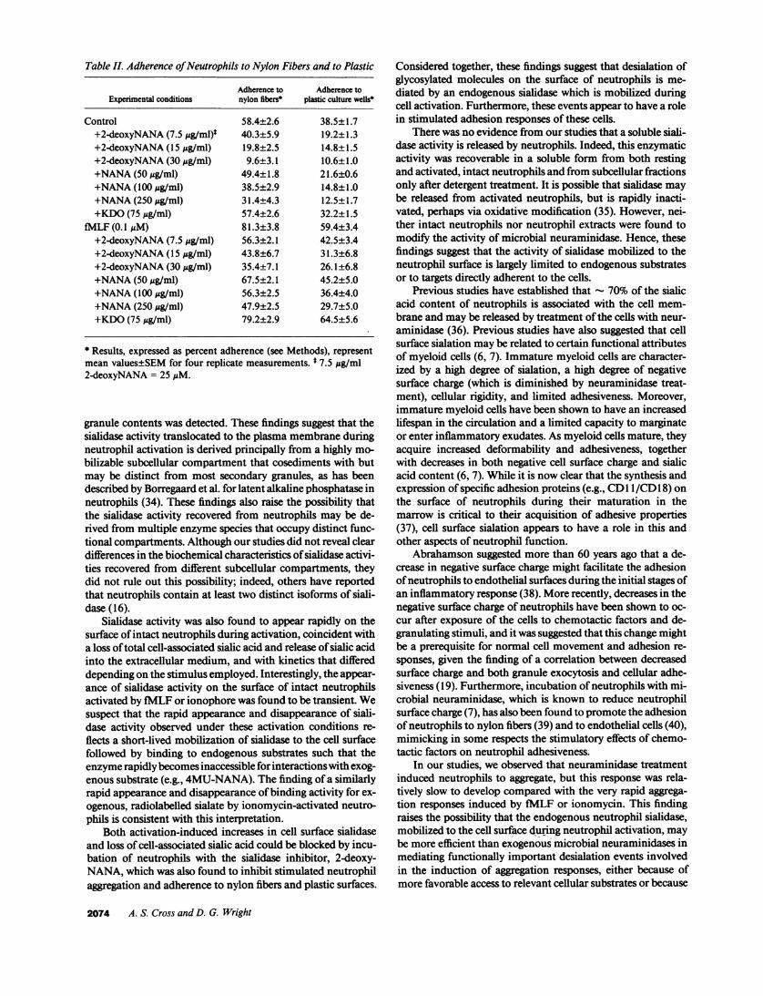
Table IL Adherence of Neutrophils to Nylon Fibers and to Plastic
Adherence to Adherence toExperimental conditions nylon fibers* plastic culture wells*
Control 58.4±2.6 38.5± 1.7+2-deoxyNANA (7.5 gg/ml)t 40.3±5.9 19.2±1.3+2-deoxyNANA (15 gg/ml) 19.8±2.5 14.8±1.5+2-deoxyNANA (30 ,g/ml) 9.6±3.1 10.6±1.0+NANA(50 Ag/ml) 49.4±1.8 21.6±0.6+NANA(100 gg/ml) 38.5±2.9 14.8±1.0+NANA(250 ,g/ml) 31.4±4.3 12.5±1.7+KDO(75 ug/ml) 57.4±2.6 32.2±1.5
fMLF (0.1 M) 81.3±3.8 59.4±3.4+2-deoxyNANA (7.5 ,g/ml) 56.3±2.1 42.5±3.4+2-deoxyNANA (15 tg/ml) 43.8±6.7 31.3±6.8+2-deoxyNANA (30 ug/ml) 35.4±7.1 26.1±6.8+NANA(50 ;g/ml) 67.5±2.1 45.2±5.0+NANA(100 ,ug/ml) 56.3±2.5 36.4±4.0+NANA(250 ,g/ml) 47.9±2.5 29.7±5.0+KDO(75 jg/ml) 79.2±2.9 64.5±5.6
* Results, expressed as percent adherence (see Methods), representmean values±SEM for four replicate measurements. * 7.5 jAg/ml2-deoxyNANA = 25 ,uM.
granule contents was detected. These findings suggest that thesialidase activity translocated to the plasma membrane duringneutrophil activation is derived principally from a highly mo-bilizable subcellular compartment that cosediments with butmay be distinct from most secondary granules, as has beendescribed by Borregaard et al. for latent alkaline phosphatase inneutrophils (34). These findings also raise the possibility thatthe sialidase activity recovered from neutrophils may be de-rived from multiple enzyme species that occupy distinct func-tional compartments. Although our studies did not reveal cleardifferences in the biochemical characteristics of sialidase activi-ties recovered from different subcellular compartments, theydid not rule out this possibility; indeed, others have reportedthat neutrophils contain at least two distinct isoforms of siali-dase (16).
Sialidase activity was also found to appear rapidly on thesurface of intact neutrophils during activation, coincident witha loss of total cell-associated sialic acid and release of sialic acidinto the extracellular medium, and with kinetics that differeddepending on the stimulus employed. Interestingly, the appear-ance of sialidase activity on the surface of intact neutrophilsactivated by fMLF or ionophore was found to be transient. Wesuspect that the rapid appearance and disappearance of siali-dase activity observed under these activation conditions re-flects a short-lived mobilization of sialidase to the cell surfacefollowed by binding to endogenous substrates such that theenzyme rapidly becomes inaccessible for interactions with exog-enous substrate (e.g., 4MU-NANA). The finding of a similarlyrapid appearance and disappearance of binding activity for ex-ogenous, radiolabelled sialate by ionomycin-activated neutro-phils is consistent with this interpretation.
Both activation-induced increases in cell surface sialidaseand loss of cell-associated sialic acid could be blocked by incu-bation of neutrophils with the sialidase inhibitor, 2-deoxy-NANA, which was also found to inhibit stimulated neutrophilaggregation and adherence to nylon fibers and plastic surfaces.
Considered together, these findings suggest that desialation ofglycosylated molecules on the surface of neutrophils is me-diated by an endogenous sialidase which is mobilized duringcell activation. Furthermore, these events appear to have a rolein stimulated adhesion responses of these cells.
There was no evidence from our studies that a soluble siali-dase activity is released by neutrophils. Indeed, this enzymaticactivity was recoverable in a soluble form from both restingand activated, intact neutrophils and from subcellular fractionsonly after detergent treatment. It is possible that sialidase maybe released from activated neutrophils, but is rapidly inacti-vated, perhaps via oxidative modification (35). However, nei-ther intact neutrophils nor neutrophil extracts were found tomodify the activity of microbial neuraminidase. Hence, thesefindings suggest that the activity of sialidase mobilized to theneutrophil surface is largely limited to endogenous substratesor to targets directly adherent to the cells.
Previous studies have established that - 70% of the sialicacid content of neutrophils is associated with the cell mem-brane and may be released by treatment of the cells with neur-aminidase (36). Previous studies have also suggested that cellsurface sialation may be related to certain functional attributesof myeloid cells (6, 7). Immature myeloid cells are character-ized by a high degree of sialation, a high degree of negativesurface charge (which is diminished by neuraminidase treat-ment), cellular rigidity, and limited adhesiveness. Moreover,immature myeloid cells have been shown to have an increasedlifespan in the circulation and a limited capacity to marginateor enter inflammatory exudates. As myeloid cells mature, theyacquire increased deformability and adhesiveness, togetherwith decreases in both negative cell surface charge and sialicacid content (6, 7). While it is now clear that the synthesis andexpression of specific adhesion proteins (e.g., CD11/CD 18) onthe surface of neutrophils during their maturation in themarrow is critical to their acquisition of adhesive properties(37), cell surface sialation appears to have a role in this andother aspects of neutrophil function.
Abrahamson suggested more than 60 years ago that a de-crease in negative surface charge might facilitate the adhesionof neutrophils to endothelial surfaces during the initial stages ofan inflammatory response (38). More recently, decreases in thenegative surface charge of neutrophils have been shown to oc-cur after exposure of the cells to chemotactic factors and de-granulating stimuli, and it was suggested that this change mightbe a prerequisite for normal cell movement and adhesion re-sponses, given the finding of a correlation between decreasedsurface charge and both granule exocytosis and cellular adhe-siveness (19). Furthermore, incubation of neutrophils with mi-crobial neuraminidase, which is known to reduce neutrophilsurface charge (7), has also been found to promote the adhesionof neutrophils to nylon fibers (39) and to endothelial cells (40),mimicking in some respects the stimulatory effects of chemo-tactic factors on neutrophil adhesiveness.
In our studies, we observed that neuraminidase treatmentinduced neutrophils to aggregate, but this response was rela-tively slow to develop compared with the very rapid aggrega-tion responses induced by fMLF or ionomycin. This findingraises the possibility that the endogenous neutrophil sialidase,mobilized to the cell surface during neutrophil activation, maybe more efficient than exogenous microbial neuraminidases inmediating functionally important desialation events involvedin the induction of aggregation responses, either because ofmore favorable access to relevant cellular substrates or because
2074 A. S. Cross and D. G. Wright

of differences in substrate specificities. Indeed, differences insubstrate specificities among sialidases are well recognized (41),and our studies documented such differences in comparingneutrophil sialidase and V. cholerae neuraminidase. In particu-lar, we found that the sialidase present in neutrophil extractswas more active relative to the microbial neuraminidase inreleasing free sialic acid from intact neutrophils than it was inreleasing sialic acid from other substrates.
Free sialic acid is known to inhibit the activity of sialidases(42), and in our studies sialic acid was found to inhibit neutro-phil aggregation and adhesion responses, although it was lesspotent in this regard than 2-deoxyNANA. This finding is ofinterest in light of studies reported by Gorog et al. (43) whoobserved that intravenous infusions of free sialic acid into rab-bits caused a decrease in the adherence of granulocytes to endo-thelial walls of postcapillary venules examined in ear chamberpreparations in vivo. These investigators also found that sialicacid infusions induced a granulocytosis in rats that was asso-ciated with the release of marginated leukocytes into the circu-lation (43).
The glycosylated molecules on the surface of neutrophilsthat are substrates for neutrophil sialidase and maybe involvedin neutrophil adhesion responses remain to be identified. How-ever, it is possible that either the beta-2 integrin adhesion mole-cules, CD11/CD18 (44, 45), or the ligands on neutrophils thatbind to CDl 1/CD 1 8 are functionally important targets for de-sialation associated with neutrophil activation. The recent find-ing that deglycosylated forms of intercellular adhesion mole-cule 1 (ICAM-1) (CD54) bind more avidly to CDl lb/CD 1 8(Mac-l) lends credence to this possibility (46), since there isnow substantial evidence that ICAM- 1, which has been shownto bind lymphocyte function-associated antigen (LFA-1) andto mediate homotypic aggregation of lymphocytes, is a cellularcounterreceptor or ligand for CDl lb/CD 1 8 that is involved inmediating CD 8-dependent neutrophil adhesion phenomenaincluding homotypic aggregation (45, 46). Although a mobili-zation of CD11/CD 1 8 adhesion molecules to the cell surfaceoccurs during neutrophil activation, recent studies have shownthat an increased density of CDl 1/CD 1 8 at the cell surface isneither necessary nor sufficient for the increased adhesivenessof activated neutrophils (47). Hence, it has been suggested thata posttranslational modification of the CDl l/CD1 8 mole-cules, such as an activation-dependent phosphorylation event,may be involved in the expression of the adhesive properties ofCDl 1/CD 1 8 heterodimers (48). Our studies raise the addi-tional possibility that desialation of these glycoprotein com-plexes, or other glycoproteins that participate in adhesion re-sponses, may promote or reveal the adhesive functions of thesecell surface molecules. This possibility is of particular interestin light of the finding that LFA-l molecules on lymphocytes,which are structurally related to neutrophil CDI1 /CD 1 8 adhe-sion molecules, express differing degrees of sialylation amongfunctionally distinct lymphocyte subpopulations (49).
Adhesion of neutrophils to platelets and endothelial cellshas also been shown recently to be mediated in part by interac-tions with lectinlike molecules that are exposed on the surfaceof activated platelets and endothelial cells (50-53). These lec-tin-like receptors (endothelial-leukocyte adhesion molecules 1)(ELAM-1) on endothelial cells [53], and guanosine S'-mono-phosphate-140 (GMP- 140) [50, 51], or platelet activation de-pendent granule-external membrane protein [52] on plateletsand endothelial cells are members of a structurally related fam-ily of adhesion proteins (lectin cellular adhesion molecules
[LEG-CAMs] or selectins) which include the murine lympho-cyte gp90"'4 (54) and are known to mediate cell-cell adhe-sion by binding to complex cell surface carbohydrates. Alectin-like adhesion protein (gpl00'4), structurally related toGMP-140 and ELAM-1, has also been identified on neutro-phils, although its role in neutrophil aggregation and adhesionresponses has yet to be fully defined (55). Recently, the ligandfor ELAM-l has been identified as sialyl-Lewis X, a terminalcarbohydrate structure found on cell-surface glycoproteins andglycolipids of neutrophils (56, 57). Studies using specific mono-clonal antibodies that recognize either the sialated or asialoforms of this carbohydrate structure have indicated that thesialic acid residue may be required for efficient binding toELAM-l and related lectin adhesion molecules (56). Hence,we infer from these studies that the enhancement of neutrophiladhesion phenomena associated with neutrophil desialation,observed in our studies, does not involve the ELAM-I ligand,since desialation of sialyl-Lewis X molecules on neutrophilsmight be anticipated to reduce (not promote) intercellular ad-hesion events dependent upon these structures. Nonetheless,desialation of the ELAM-I ligand by an endogenous sialidasemobilized to the surface of activated neutrophils could modu-late adhesion events mediated by the selectin family of adhe-sion molecules following the binding of neutrophils to endothe-lial cells and during transendothelial migration of neutrophilsin response to inflammatory signals.
Desialation of glycosylated molecules on the surface of neu-trophils may affect functions other than those related to stimu-lated adhesion. Indeed, neuraminidase treatment of neutro-phils has been reported to modify stimulated superoxide andhydrogen peroxide generation by these cells, although reportsof such effects have been conflicting (9, 58-61). Recently Perezet al. have reported that neuraminidase treatment inhibits che-motactic responses of neutrophils, and they have related thiseffect to an inhibition of the recycling of chemotactic factorreceptors (62). Our studies support the conclusion that effortsto identify functionally important desialation targets on thesurface of neutrophils should contribute to a more completeunderstanding of molecular mechanisms involved in the in-duction of neutrophil functional responses by inflammatorysignals, for it is evident that desialation of the neutrophil sur-face, mediated by an endogenous sialidase, is a component ofneutrophil activation.
Acknowledgments
The authors wish to thank Lynnette Young for her superb technicalassistance.
References1. Corfield, A. P., and R. Schauer. 1982. Occurrence of sialic acids. In Sialic
Acids: Chemistry, Metabolism and Function. Cell Biology Monographs. Vol. 10.R. Schauer, editor. Springer-Verlag, Vienna, NewYork. 5-50.
2. Dennis, J., C. Waller, R. Timpl, and V. Schirrmacher. 1982. Surface sialicacid reduces attachment of metastatic tumor cells to collagen type IV and fibro-nectin. Nature (Lond.) 300:274-276.
3. Fogel, M., P. Altevogt, and V. Schirrmacher. 1983. Metastatic potentialseverely altered by changes in tumor cell adhesiveness and cell surface sialylation.J. Exp. Med. 157:371-376.
4. Pearlstein, E., P. L. Salk, G. Yogeeswaran, and S. Karpatkin. 1980. Correla-tion between spontaneous metastatic potential, platelet-aggregating activity ofcell surface extracts, and cell surface sialylation in 10 metastatic-variant deriva-tives of a rat renal sarcoma cell line. Proc. Nad. Acad. Sci. USA. 77:4336-4339.
5. Lloyd, C. W. 1975. Sialic acid and the social behavior of cells. Biol. Rev.50:325-350.
6. Lichtman, M. A., and R. I. Weed. 1972. Alteration of the cell peripheryduring granulocyte maturation: relationship to cell function. Blood. 39:301-316.
Neutrophil Sialidase 2075

7. Lichtman, M. A., and R. I. Weed. 1970. Electrophoretic mobility andN-acetyl neuraminic acid content of human normal and leukemic lymphocytesand granulocytes. Blood. 35:12-22.
8. Weiss, L. 1965. Studies on cell deformability. I. Effect of surface charge. J.Cell Biol. 26:735-739.
9. Henricks, P. A. J., M. E. Van Erne-van der Tol, and J. Verhoef. 1982.Partial removal of sialic acid enhances phagocytosis and the generation of super-oxide and chemiluminescence by polymorphonuclear leukocytes. J. Immunol.129:745-750.
10. Corfield, A. P., and R. Schauer. 1982. Metabolism of sialic acids. In SialicAcids: Chemistry, Metabolism, and Function. Cell Biology Monographs. Vol. 10.R. Schauer, editor. Springer-Verlag, Vienna, NY. 195-261.
1 1. Michalski, J.-C., A. P. Corfield, and R. Schauer. 1986. Properties of hu-man liver lysosomal sialidase. Biol. Chem. Hoppe-Seyler. 367:715-722.
12. Samollow, P. A., J. L. VandeBerg, H. W. Kunz, and T. J. Gill III. 1985.Analysis of neuraminidase isozyme phenotypes in mammalian tissues: an electro-phoretic approach. Biochem. Biophys. Res. Commun. 126:1182-1188.
13. Miyagi, T., and S. Tsuiki. 1985. Purification and characterization of cyto-solic sialidase from rat liver. J. Biol. Chem. 260:6710-6716.
14. Schauer, R. 1985. Sialic acids and their role as biological masks. TrendsBiochem. Sci. 10:357-360.
15. Nojiri, H., F. Takaku, T. Tetsuka, and M. Saito. 1982. Stimulation ofsialidase activity during cell differentiation of human promyelocytic leukemiacell line HL-60. Biochem. Biophys. Res. Commun. 104:1239-1246.
16. Verheijen, F. W., H. C. Janse, 0. P. van Diggelen, H. D. Bakker, M. C. B.Loonen, P. Durand, and H. Galjaard. 1983. Two genetically different MU-NANAneuraminidases in human leucocytes. Biochem. Biophys. Res. Commun.117:470-478.
17. Gallin, J. I., D. G. Wright, and E. Schiffmann. 1978. Role of secretoryevents in modulating human neutrophil chemotaxis. J. Clin. Invest. 62:1364-1374.
18. Gallin, J. I., J. R.Durocher, and A. P. Kaplan. 1975. Interaction of leuko-cyte chemotactic factors with the cell surface. I. Chemotactic factor-inducedchanges in human granulocyte surface charge. J. Clin. Invest. 55:967-974.
19. Gallin, J. 1. 1980. Degranulating stimuli decrease the negative surfacecharge and increase the adhesiveness of human neutrophils. J. Clin. Invest.65:298-306.
20. Cross, A. S., G. H. Lowell, J. Palmblad, J. C. Sadoff, L. Young, and M.Berger. 1985. Mechanism of priming of human neutrophils by a soluble lympho-blastoid cell factor. J. Immunol. 135:2074-2083.
21. Wright, D. G. 1988. Neutrophil degranulation. Methods Enzymol.162:538-551.
22. Wright, D. G., and J. I. Gallin. 1979. Secretory responses of human neutro-phils. J. Immunol. 123:285-294.
23. Borregaard, N., J. M. Heiple, E. R. Simons, and R. A. Clark. 1983. Subcel-lular localization of the b-cytochrome component of the human microbicidaloxidase: translocation during activation. J. Cell Biol. 97:52-61.
24. Wright, D. G., D. Bralove, and J. I. Gallin. 1977. The differential mobili-zation of human neutrophil granules. Am. J. Pathol. 87:272-284.
25. Baggiolini, M., J. G. Hirsch, and C. DeDuve. 1969. Resolution of granulesfrom rabbit heterophil leukocytes into distinct populations by zonal sedimenta-tion. J. Cell Biol. 40:529-541.
26. Kane, S. P., and T. J. Peters. 1975. Analytical subcellular fractionation ofhuman granulocytes with reference to the localization of vitamin B12 bindingprotein. Clin. Sci. Mol. Med. 49:171-182.
27. DeChatelet, L. R., and M. R. Cooper. 1970. A modified procedure for thedetermination of leukocyte alkaline phosphatase. Biochem. Med. 4:61-68.
28. Pereira, M. E. A. 1983. A rapid and sensitive assay for neuraminidaseusing peanut lectin hemagglutination: application to Vibrio cholera and Trypano-soma cruzi. J. Immunol. Meth. 63:25-34.
29. Warren, L. 1959. The thiobarbituric acid assay of sialic acids. J. Biol.Chem. 234:1971-1975.
30. Brunetti, P., A. Swanson, and S. Roseman. 1963. Enzymatic determina-tion of sialic acids. In Methods in Enzymology. VI. Academic Press, NewYork,London. pp. 465-473.
31. Metcalf, J. A., J. I. Gallin, W. M. Nauseef, and R. K. Root. LaboratoryManual of Neutrophil Function. 1986. Raven Press, Ltd., NY.
32. Spaltro, J., and J. A. Alhadeff. 1984. Solubilization, stabilization andisoelectric focusing of human liver neuraminidase activity. Biochim. Biophys.Acta. 800:159-165.
33. Kumar, V., J. Kessler, M. E. Scott, B. H. Patwardhan, S. W. Tanenbaum,and M. Flashner. 1981. 2,3-dehydro-4-epi-N-acetylneurminic acid; a neuramini-dase inhibitor. Carbohydr. Res. 94:123-130.
34. Borregaard, N., L. Christensen, 0. W. Bejerrum, H. S. Birgens, and I.Clemmensen. 1990. Identification of a highly mobilizable subset of human neu-trophil intracellular vesicles that contain tetranectin and latent alkaline phospha-tase. J. Clin. Invest. 85:408-416.
35. Clark, R. A., and N. Borregaard. 1985. Neutrophils autoinactivate secre-tory products by myeloperoxidase-catalyzed oxidation. Blood. 65:375-381.
36. DePierre, J. W., J. Lazdins, and M. L. Karnovsky. 1980. The determina-tion and localization of sialic acid in guinea-pig granulocytes. Biochem. J.192:543-550.
37. Anderson, D. C., F. C. Schmalsteig, M. J. Finegold, B. J. Hughes, R.Rothlein, L. J. Miller, S. Kohl, M. F. Tosi, R. L. Jacobs, T. C. Waldrop, et al.1985. The severe and moderate phenotypes of heritable Mac-i, LFA-1 deficiency:their quantitative definition and relation to leukocyte dysfunction and clinicalfeatures. J. Infect. Dis. 152:668-689.
38. Abrahamson, H. A. 1927. The mechanism of the inflammatory process. I.The electrophoresis of the blood cells of the horse and its relation to leukocyteemigration. J. Exp. Med. 46:987-1002.
39. Kownatzki, E., S. Uhrich, and B. Weil. 1984. Adherence and migration ofguinea pig granulocytes after enzyme treatment of the cell surface. Immunobiol-ogy. 166:111-117.
40. Hoover, R. L., R. T. Briggs, and M. J. Karnovsky. 1978. The adhesiveinteraction between polymorphonuclear leukocytes and endothelial cells in vitro.Cell. 14:423-428.
41. Michalski, J. C., A. P. Corfield, and R. Schauer. 1982. Properties of hu-man liver lysosomal sialidase. Hoppe-Seyler's Z. Physiol. Chem. 363:1097-1102.
42. Mendla, K., and M. Cantz. 1984. Specificity studies on the oligosaccha-ride neuraminidase of human fibroblasts. Biochem. J. 218:625-628.
43. Gorog, P., I. B. Kovacs, and G. V. R. Born. 1980. Suppression of theintravascular adherence of granulocytes by N-acetyl neuraminic (sialic) acid. Br.J. Exp. Pathol. 61:490-496.
44. Sanchez-Madrid, F., J. Nagy, E. Robbins, P. Simon, and T. A. Springer.1983. A human leukocyte differentiation antigen family with distinct alpha sub-units and a commonbeta subunit: the lymphocyte function associated antigen-l(LFA-1), the C3bi complement receptor (OKMl/Mac-l), and the p150.95 mole-cule. J. Exp. Med. 158:1785-1803.
45. Carlos, T. M., and J. M. Harlan. 1990. Membrane proteins involved inphagocyte adherence to endothelium. Immunol. Rev. 114:5-28.
46. Diamond, M. S., D. E. Staunton, S. D. Marlin, and T. A. Springer. 1991.Binding of the integrin Mac-l (CDl lb/CD18) to the third immunoglobulin-likedomain of ICAM- I (CD54) and its regulation by glycosylation. Cell. 65:961-971.
47. Vedder, N. B., and J. M. Harlan. 1988. Increased surface expression ofCDl lb/CD18 (Mac-l) is not required for stimulated neutrophil adherence tocultured endothelium. J. Clin. Invest. 81:676-682.
48. Buyon, J. P., S. G. Slade, J. Reibman, S. B. Abramson, M. Philips, G.Weissmann, and R. Winchester. 1990. Constitutive and induced phosphoryla-tion of the a- and ,B-chains ofthe CDI I /CD 18 leukocyte integrin family: relation-ship to adhesion-dependent functions. J. Immunol. 144:191-197.
49. Takeda, A. 1987. Sialylation patterns of lymphocyte function-associatedantigen 1 (LFA- 1) differ between T and B lymphocytes. Eur. J. Immunol.17:28 1-286.
50. McEver, R. P., J. H. Beckstead, K. L. Moore, L. Marshall-Carlson, andD. F. Bainton. 1989. GMP-140, a platelet alpha-granule membrane protein, isalso synthesized by vascular endothelial cells and is localized in Weibel-Paladebodies. J. Clin. Invest. 84:92-99.
51. Johnston, G., R. G. Cook, and R. P. McEver. 1989. Cloning of GMP-140,a granule membrane protein of platelets and endothelium: sequence similarity toproteins involved in cell adhesion and inflammation. Cell. 56:1033-1044.
52. Larsen, E., A. Celi, G. E. Gilbert, B. C. Furie, J. K. Erban, R. Bonfanti,D. D. Wagner, and B. Furie. 1989. PADGEMprotein: a receptor that mediatesthe interaction of activated platelets with neutrophils and monocytes. Cell.59:305-3 12.
53. Bevilacqua, M. P., J. S. Pober, D. L. Mendrick, R. S. Cotran, and M. A.Gimbrone, Jr. 1987. Identification of an inducible endothelial-leukocyte adhe-sion molecule, ELAM-1. Proc. Natl. Acad. Sci. USA. 84:9238-9242.
54. Lewinsohn, D. M., R. F. Bargatze, and E. C. Butcher. 1987. Leukocyte-en-dothelial cell recognition: evidence of a common molecular mechanism sharedby neutrophils, lymphocytes, and other leukocytes. J. Immunol. 138:4313-432 1.
55. Kishimoto, T. K., M. A. Jutila, E. L. Berg, and E. C. Butcher. 1989.Neutrophil Mac- 1 and MEL-14 adhesion proteins inversely regulated by chemo-tactic factors. Science (Wash. DC). 245:1238-1241.
56. Phillips, M. L., E. Nudelman, F. C. A. Gaeta, M. Perez, A. K. Singhal, S.-I.Hakomori, and J. C. Paulson. 1990. ELAM-1 mediates cell adhesion by recogni-tion of a carbohydrate ligand, sialyl-Lex. Science (Wash. DC) 250:1130-1132.
57. Walz, G., A. Aruffo, W. Kolanus, M. Bevilacqua, and B. Seed. 1990.Recognition by ELAM-1 of the sialyl-LeX determinant on myeloid and tumorcells. Science (Wash. DC). 250:1132-1135.
58. Tsan, M. F., and P. A. McIntyre. 1976. The requirement for membranesialic acid in the stimulation of superoxide production during phagocytosis byhuman polymorphonuclear leukocytes. J. Immunol. 129:745-750.
59. Suzuki, H., T. Kurita, and K. Kakinuma. 1982. Effects of neuraminidaseon 0- consumption and release of °- and H202 from phagocytosing humanpolymorphonuclear leukocytes. Blood. 60:446-453.
60. Noseworthy, J., H. Korchak, and M. L. Karnovsky. 1972. Phagocytosisand the sialic acid of the surface of polymorphonuclear leukocytes. J. Cell. Phys-iol. 79:91-96.
61. Perez, H. D., R. R. Ong, and F. Elfman. 1985. Removal or oxidation ofsurface membrane sialic acid inhibits formyl-peptide-induced polymorphonu-clear leukocyte chemotaxis. J. Immunol. 134:1902-1908.
62. Perez, H. -D., F. Elfman, and E. Lobo. 1987. Removal of human polymor-phonuclear leukocyte surface sialic acid inhibits reexpression (or recycling) offormyl peptide receptors. J. Immunol. 139:1978-1984.
2076 A. S. Cross and D. G. Wright


![Regulation of the intracellular Ca2+. Regulation of intracellular [H]:](https://img.pdfslide.us/doc/110x75/5a4d1b717f8b9ab0599b56a5/regulation-of-the-intracellular-ca2-regulation-of-intracellular-h.jpg)


![Mobilization of intracellular calcium stores participates ... · calcium channels do not participate in these effects, as: (i) the increase in [Ca21] i was not affected by NMDA receptor](https://img.pdfslide.us/doc/110x75/5f887ca9ab2e2b7b4d74c6dc/mobilization-of-intracellular-calcium-stores-participates-calcium-channels-do.jpg)