Embed Size (px)
Citation preview

NEU1 and NEU3 Sialidase Expression in Human Endothelia
1
NEU1 and NEU3 Sialidase Activity Expressed in Human Lung Microvascular Endothelia.
NEU1 Restrains Endothelial Cell Migration Whereas NEU3 Does Not.
Alan S. Cross,2,4
, Sang Won Hyun1, Alba Miranda-Ribera
1, Chiguang Feng
2, Anguo Liu
1, Chinh
Nguyen1,3
, Lei Zhang2, Irina G. Luzina
4, Sergei P. Atamas
4, William S. Twaddell
5, Wei Guang
1,6,
Erik P. Lillehoj1,6
, Adam C. Puché7, Wei Huang
8,9, Lai -Xi Wang
8,9, Antonino Passaniti
1,5, and
Simeon E. Goldblum1,3,4,5
1The Mucosal Biology Research Center
2Center for Vaccine Development
3Division of Infectious Diseases
4Department of Medicine
5Department of Pathology
6Department of Pediatrics
7Department of Anatomy and Neurobiology
8Institute of Human Virology
Department of Biochemistry and Molecular Biology 9University of Maryland, School of Medicine, Baltimore, MD 21201
*Running head: NEU1 and NEU3 Sialidase Expression in Human Endothelia
To whom correspondence should be addressed: Simeon E. Goldblum, M.D., Mucosal Biology Research
Center, University of Maryland School of Medicine, 20 Penn Street, Room 351, Baltimore, MD 21201,
USA; Tel: 410-706-5504, Fax: 410-706-5508, Email: [email protected]
Keywords: neuraminidase, sialidase, sialic acid, migration, wound healing
Background: The vascular endothelial surface is
highly sialylated.
Results: Vascular endothelia express catalytically-
active NEU1 and NEU3 sialidases and NEU1
restrains the endothelial migratory response to
wounding.
Conclusion: NEU1 regulates endothelial
remodeling in response to injury.
Significance: Learning how NEU1 and NEU3
regulate sialylated molecules on the endothelial
surface is key to understanding endothelial
receptor-ligand, cell-cell, and host-pathogen
interactions.
SUMMARY
The microvascular endothelial surface
expresses multiple molecules whose sialylation
state regulates multiple aspects of endothelial
function. To better regulate these sialoproteins,
we asked whether endothelial cell (EC)s might
express one or more catalytically active
sialidase(s). Human lung microvascular EC
lysates contained heat-labile sialidase activity
for a fluorogenic substrate, 2’-(4-
methylumbelliferyl)--D-N-acetylneuraminic
acid (4-MU-NANA), that was dose-dependently
inhibited by the competitive sialidase inhibitor,
2,3-dehydro-2-deoxy-N-acetylneuraminic acid
(2-deoxy-NANA), but not its negative control.
The EC lysates also contained sialidase activity
for a ganglioside mixture. Using real time RT-
PCR to detect mRNAs for the four known
mammalian sialidases, NEU1, 2, 3, and 4, NEU1
mRNA was expressed at levels 2,700-fold
higher that those found for NEU2, 3, or 4.
Western analyses indicated NEU1 and 3 protein
expression. Using confocal microscopy and flow
cytometry, NEU1 was immunolocalized to both
the plasma membrane and the perinuclear
region. NEU3 was detected both in the cytosol
and nucleus. Prior siRNA-mediated knockdown
of NEU1 and NEU3 each decreased EC
sialidase activity for 4-MU-NANA by >65%
and >17%, respectively, and for the ganglioside
mixture by 0% and 40%, respectively. NEU1
overexpression in ECs reduced their migration
into a wound by >40% whereas NEU3
overexpression did not. Immunohistochemical
http://www.jbc.org/cgi/doi/10.1074/jbc.M112.346817The latest version is at JBC Papers in Press. Published on March 8, 2012 as Manuscript M112.346817
Copyright 2012 by The American Society for Biochemistry and Molecular Biology, Inc.
by guest on May 27, 2018
http://ww
w.jbc.org/
Dow
nloaded from

NEU1 and NEU3 Sialidase Expression in Human Endothelia
2
studies of normal human tissues
immunolocalized NEU1 and NEU3 proteins to
both pulmonary and extrapulmonary vascular
endothelia. These combined data indicate that
human lung microvascular ECs, as well as
other endothelia, express catalytically-active
NEU1 and NEU3. NEU1 restrains EC
migration whereas NEU3 does not.
The vascular endothelium lines the
intravascular space and presents a selective barrier
that actively regulates paracellular movement of
circulating cells, macromolecules, and fluids into
extravascular tissues and compartments (1,2). In
eukaryotic cells, glycoproteins and glycolipids
expressed on the cell surface contain
oligosaccharide chains whose outermost positions
are terminated with sialic acids (3,4). The sialic
acid family of >50 closely related but structurally
diverse sugars, with their terminal location and
negative charge, are strategically positioned to
influence intermolecular and cell-cell interactions
through nonspecific steric hindrance and/or
electrostatic repulsion. Vascular endothelia are
highly sialylated (5). The sialic acid-bearing
surface structures on endothelia, together with
other membrane-bound polyanionic
macromolecules, constitute the endothelial cell
(EC) glycocalyx (6,7). This negatively-charged
zone can be extended through hydration and/or
acquisition of plasma proteins. The glycocalyx
might nonspecifically diminish cell-cell adhesion
through either negative charge-mediated repulsion
or the masking of specific adhesion molecules.
However, sialyl residues also can serve as
components of EC surface recognition motifs that
participate in specific intermolecular interactions.
For example, the selectins are C-type lectins that
bind sialyl Lewis X (sLex)-bearing molecules
(1,8). ECs can surface express or secrete selectins
that recognize their sialylated ligands on
circulating leukocytes (1,8) and tumor cells (1,8-
10). Removal of sialic acid from the surface of
HL60 myeloid cells diminishes their adhesion to
EC-expressed E-selectin (9). Increased 2,6-
sialylation of the EC surface increases endothelial
adhesiveness for CD22-bearing B cells (11,12)
and lymphocyte function-associated antigen
(LFA)-1 expressing T cells (13). Vascular
endothelial (VE)-cadherin is enriched to
intercellular boundaries where it regulates EC-EC
homophilic adhesion and paracellular pathway
function (14). VE-cadherin is sialylated and its
desialylation results in its profound reorganization.
Other sialylated molecules at the EC surface
regulate components of both the clotting (15,16)
and complement (17) pathways. The sialylation
state of these EC surface molecules influences
their tertiary conformation, intermolecular
interactions, resistance to proteolysis, and function
(1,9,10,12-14).
The state of sialylation of these EC- and
leukocyte-associated glycoproteins are tightly and
dynamically regulated through the net catalytic
activities of both sialyltransferase (ST)s (18,19)
and sialidases (20,21). An ST family of at least 15
members, each containing the ~50 amino acid
sialyl motif thought to be involved in substrate
binding, catalyzes the transfer of sialic acid
molecules onto an acceptor substrate (18,19). The
endothelial surface reportedly expresses ST
activity (11). Whether this same endothelial
surface also expresses sialidase activity is
unknown. At least 4 mammalian sialidases have
been identified (20,21). NEU1 is reportedly
localized to lysosomes where it associates with
cathepsin A and -galactosidase which are
required for correct sialidase conformation
(22,23). NEU2 is found in the cytosol (24) and
NEU3 is associated with the plasma membrane
(25,26). The most recently described sialidase,
NEU4, is associated with the mitochondria (27).
As members of the neuraminidase/sialidase
superfamily, each contains one or more of the
conserved Asp box, an amino acid sequence
comprised of –S-X-D-X-G-X-T-W-, together with
the F/YRIP motif, comprised of –X-P-R-P-, where
X represents variable residues (20,21). Although
the overall sequence identities between the four
sialidases are low, their catalytic domains share a
common “six-bladed β-propeller fold”
architecture. Each of these enzymes selectively
hydrolyzes specific glycosidic linkages between
sialic acid molecules and the subterminal sugar of
glycoconjugates.
We previously found that treatment of ECs
with exogenous neuraminidase increased
polymorphonuclear leukocyte (PMNL) adhesion
to and migration across the endothelium and that
preincubation of cells with the selective sialidase
inhibitor, 2,3-dehydro-2-deoxy-N-acetyl-
neuraminic acid (2-deoxy-NANA), inhibited
by guest on May 27, 2018
http://ww
w.jbc.org/
Dow
nloaded from

NEU1 and NEU3 Sialidase Expression in Human Endothelia
3
PMNL-mediated desialylation of the endothelial
surface (28). In other studies, we found that
infusion of neutralizing anti-neuraminidase
antibodies in mice blocked PMNL recruitment to
the pulmonary microvasculature in response to
either systemic complement activation or
intratracheal interleukin (IL)-8 installation (29).
These combined studies indicate that endogenous
sialidase activity is operative during PMNL-EC
interactions within the pulmonary
microvasculature. However, the identity of the
responsible sialidase molecule(s) is unknown. In
the current report, we have asked whether human
pulmonary microvascular endothelia, and/or other
vascular endothelia, express one or more
catalytically active sialidase(s) and whether such
expression regulates EC behavior.
EXPERIMENTAL PROCEDURES Cell Culture- Human lung microvascular EC
(HMVEC-L)s and human pulmonary artery EC
(HPAEC)s, (Lonza; Rockland, ME) as well as
HMEC-1 cells, a simian virus (SV) 40 T antigen
transformed human dermal microvascular EC line
(CDC, Atlanta, GA), were cultured in EC growth
medium (EBM-2, Lonza) containing 5% fetal
bovine serum (FBS) (Hyclone Laboratories,
Logan, UT), human recombinant epidermal
growth factor (EGF), human recombinant insulin-
like growth-factor-1, human basic fibroblast
growth factor, vascular endothelial growth factor,
hydrocortisone, ascorbic acid, gentamicin, and
amphotericin B as described (30). HMVEC-Ls and
HPAECs were studied in passages 2 to 7. Human
bone marrow EC (HBME)s (31) and human
umbilical vein retroviral telemorized EC (Hrvt)s
(generously provided by A.P.) were similarly
cultured.
Fluorometric Assay for Sialidase Activity-
Sialidase activity was measured in HMVEC-Ls,
HPAECs, HBMEs, Hrvts, and HMEC-1s, in the
presence of 2’-(4-methylumbelliferyl)--D-N-
acetylneuraminic acid sodium salt hydrate (4-MU-
NANA) (Sigma, St. Louis, MO) as the substrate as
described (32). ECs (~106
cells/tube) were
suspended in 200 l of buffer containing 500 mM
sodium acetate, 0.1% Triton X-100, supplemented
with protease inhibitor cocktail (Roche), and then
incubated for 1h at 37oC in the presence of 4-MU-
NANA, mixing tubes every 15 min. The sialidase
reaction was terminated by the addition of glycine-
NaOH buffer, pH 10.3, containing 133 mM
glycine, 60 mM NaCl, and 0.083 M Na2CO3, after
which fluorescence intensity was measured with a
Bio-Rad fluorometer (excitation at 355 nm;
emission at 460 nm). In selected experiments, EC
lysates were boiled (100oC, 10 min). In other
experiments, EC lysates were preincubated with
the competitive neuraminidase inhibitor, 2-
deoxyNANA (5-500 g/ml) (Calbiochem, La
Jolla, CA) (28,29). A molecule with comparable
MW and charge to 2-deoxyNANA, 2-keto-3-
deoxyoctulosonic acid (KDO) (Sigma), was used
as a negative control (28).
Sialidase Activity for Ganglioside
Substrate- Since NEU3 preferentially hydrolyzes
sialic acid linkages within gangliosides (25),
sialidase activity for a ganglioside substrate was
assayed as described (33). HMVEC-Ls were
suspended in 200 l of 500 mM sodium acetate,
pH 4.4 containing 0.1% Triton X-100 and protease
inhibitor cocktail and the total cell lysates mixed
with 25 l ganglioside mixture (2 mg/ml)
(Calbiochem). The HMVEC-L ganglioside
substrate mixtures were incubated for 1h at 37oC.
The reaction was terminated by the addition of 25
l glycine buffer, pH 10.3 containing 1.33 M
glycine, 0.6 M NaCl, and 0.42 M Na2CO3.
Released sialic acid in each reaction mixture was
quantified by high pH anion-exchange
chromatography with pulsed amperometric
detection (HPAEC-PAD) as described (29). For
each assay, serial dilutions of known
concentrations of pure sialic acid (Sigma) were
measured and a standard curve generated. The
sialic acid concentration in each sample was
interpolated from the standard curve using
GraphPad Prism 4 (GraphPad Software, Inc., La
Jolla, CA). The concentration of sialic acid
spontaneously released from the simultaneous
cell-free ganglioside control, i.e. the background,
was subtracted from each value.
Real-Time RT-PCR for NEU1, 2, 3, and 4-
Total cellular RNA was extracted from HMVEC-
Ls using the Qiagen RNA isolation kit according
to the manufacturer’s protocol (30). RNA purity
was established with the 260 nm/280 nm
absorption ratio. For real time quantitative RT-
PCR (qPCR), total RNA was reverse-transcribed
using AMV reverse transcriptase (Promega,
Madison, WI) and poly-T primer, as recommended
by guest on May 27, 2018
http://ww
w.jbc.org/
Dow
nloaded from

NEU1 and NEU3 Sialidase Expression in Human Endothelia
4
by the manufacturer. The resulting cDNA was
quantified by using real-time PCR using SYBR
Green PCR Master Mix (Applied
Biosystems/Invitrogen; Carlsbad, CA) and ABI
Prism 7900HT cycler. Primers for detection of
NEU1, NEU2, NEU3, NEU4, and hypoxanthine
phosphoribosyltransferase (HPRT) mRNAs were
designed using the Primer Express 2.0 program
(Applied Biosystems) and are indicated in Table 1.
Relative gene expression was calculated using the
Ct method, where Ct refers to the cycle number
at which the PCR product for a particular gene is
detected by the light cycler. The housekeeping
gene, HPRT, was used as an internal control and
relative gene expression was normalized to the
HPRT gene expression by the following formula
(1.8[CtHOUSEKEEPING GENE-CtGENE OF INTEREST]).
Immunoblotting for NEU1 and 3- Cells
were thoroughly rinsed with ice-cold HEPES
buffer, and solubilized with ice-cold lysis buffer
containing 50 mM Tris-HC1 (pH 8.0), 1% Nonidet
P-40, 0.5% sodium dodecyl sulfate (SDS), 150
mM NaCl, 0.1 mM phenylmethylsulfonyl fluoride,
5 g/ml leupeptin, 1 mg/ml pepstatin A, 1 mg/ml
aprotinin, 1 mM vanadate, 1 mM sodium fluoride,
10 mM disodium pyrophosphate, 500 M
paranitrophenol, and 1 mM phenylarsine oxide (all
purchased from Sigma) as described (29,30). The
cell lysates were assayed for protein concentration
with a Bio-Rad Protein Assay Dye Reagent (Bio-
Rad, Richmond, CA). Equal amounts of protein
were resolved by electrophoresis on an 8-16%
SDS-polyacrylamide gel (Novex, San Diego, CA)
and transferred to polyvinylidene fluoride (PVDF)
membranes (Millipore, Bedford, MA). The blots
were blocked for 1h using 5% nonfat milk in TBS-
Tween buffer and probed with rabbit anti-human
NEU1 antibody (Rockland Immunochemicals;
Gilbertsville, PA) or anti-human NEU3 antibody
(Strategic Diagnostics Inc., Newark, DE) each
followed by horseradish peroxidase (HRP)-
conjugated goat anti-rabbit antibody in 5% milk,
TBS-T and developed with enhanced
chemiluminescence (ECL) (Amersham,
Piscataway, NJ) (30). To confirm equivalent
protein loading and transfer, blots were stripped
with 100 mM 2-mercaptoethanol, 2% SDS, 62.5
mM Tris-HCI, pH 6.7, and reprobed with 0.5
ng/ml of murine anti-Physarum β-tubulin IgG2b
(Boehringer-Mannheim, Indianapolis, IN)
followed by HRP-conjugated anti-mouse IgG
(Transduction Laboratories) and again, developed
with ECL. In selected experiments, NEU3
immunoblotting was performed with cytoplasmic
and nuclear subcellular fractions isolated using the
NE-PER Nuclear and Cytoplasmic Extraction Kit
(Thermo Fisher Scientific, Rockford, IL). To
verify subcellular fractionation, the blot was
stripped and reprobed for the cytoplasmic marker
protein, IκBα, and the nuclear marker protein,
lamin B1.
Flow Cytometry for NEU1 and 3
Expression in HMVEC-Ls- HMVEC-Ls were
detached using 0.25% trypsin-EDTA, in some
cases permeabilized with 0.1% Triton X-100,
incubated for 30 min at 4oC with anti-human
NEU1 or NEU3 antibodies or a species-matched
control antibody (rabbit IgG, Invitrogen). The cells
were washed and incubated with fluorescein
isothiocyanate (FITC)-conjugated goat anti-rabbit
antibody (BD Pharmingen, Franklin Lakes, NJ).
Antibody binding to the intact and permeabilized
cells was evaluated using a flow cytometer
(FACSCAN, BD Biosciences) and the data were
analyzed with CELLQUEST Software (BD
Biosciences) as described (29,30).
Adenoviral Constructs Encoding for
Epitope-Tagged NEU1 and NEU3- To regulate
NEU1 and NEU3 expression in HMVEC-Ls,
recombinant adenovirus (Ad) encoding for human
FLAG-tagged wild-type NEU1 (Ad-NEU1-
FLAG) and human hemagglutinin (HA)-tagged
wild-type NEU3 (Ad-NEU3-HA) were generated
as described for another gene product (34). The
human NEU1 (NM__000434.3) and NEU3
(NM__006656.5) sequences were cloned by RT-
PCR using PCR primers synthesized by Primm
Biotech (Cambridge, MA), after which the 3 x
FLAG tag and HA tag sequences were inserted
prior to the stop codon at the 3’ end of NEU1 and
NEU3 sequences, respectively. The Ad-NEU1-
FLAG and Ad-NEU3-HA were generated using
the AdEasy Adenoviral Vector System
(Stratagene, La Jolla, CA) according to the
manufacturer’s recommendation. Briefly, the
NEU1-FLAG and NEU3-HA each were subcloned
into a shuttle vector (pShuttle-IREs-hrGFP-1)
using restriction enzyme digestion and ligation.
Each resultant shuttle plasmid was linearized
through Pmel digestion and, with the Ad backbone
plasmid (pAdEasy-1, Qbiogene), was used to
by guest on May 27, 2018
http://ww
w.jbc.org/
Dow
nloaded from

NEU1 and NEU3 Sialidase Expression in Human Endothelia
5
cotransform electrocompetent E. coli BJ5183 cells
to produce the recombinant plasmids, Ad-NEU1-
FLAG and Ad-NEU3-HA. Recombinants were
selected for kanamycin resistance and screened for
recombination by Pac1 restriction enzyme analysis
and agarose gel electrophoresis. The correct
recombinant plasmids were used to transform
XL10-Gold cells and bacterial lysates were passed
through Maxiprep columns (Qiagen) for
purification. Ad-NEU1-FLAG and Ad-NEU3-HA
each was linearized with Pac1 digestion and
transfected, in the presence of Lipofectamine
(Invitrogen), into AD-293 cells. After 7-10 days,
cells were scraped off flasks with a rubber
policeman, subjected to 3 freeze-thaw cycles, and
virus harvested in the supernatants for presentation
to fresh AD-293 cells and titration in a plaque-
forming assay. HMVEC-Ls were transiently
infected with packaged Ad-NEU1-FLAG or Ad-
NEU3-HA at increasing MOIs, and after 48h,
were lysed and the lysates processed for FLAG or
HA immunoblotting. In selected experiments, Ad-
GFP was used as a vector control, as described
(35).
Immunolocalization of NEU1 and 3 in
HMVEC-Ls- HMVEC-Ls were cultured overnight
in 8-well glass chamber slides (Nunc/Thermo
Fisher, Waltham, MA), washed 3 times with PBS,
fixed (4% paraformaldehyde in PBS, 10 min, RT),
washed, permeabilized (0.5% Triton X-100 in
HEPES buffer, 10 min, 4oC), blocked (super-block
buffer, 30 min), and incubated overnight at 4oC
separately with anti-human NEU1 or NEU3
antibodies (1:500 dilution, overnight, 4oC) with
1% donkey serum. The chamber slides were then
washed and incubated with Cy3-conjugated goat
anti-rabbit antibody (Jackson Immunoresearch).
DAPI was used to counterstain nuclei. After
immediate mounting with mounting media
(Vectashield; Vector Laboratories), the
immunostained HMVEC-Ls were analyzed and
photographed using an Olympus FluoView 500
laser scanning confocal fluorescence microscope
fitted with a 60X, N.A. 1.4 objective and standard
excitation/emission filters for detection of DAPI
and Cy3. Images were cropped and assembled into
panels with Adobe Photoshop 4.0.
Knockdown of NEU1 and NEU3 Through
siRNA Technology- First, HMVEC-Ls were
transfected with small interfering RNA (siRNA)
duplex products designed to specifically target
NEU1 or NEU3, or an irrelevant control siRNA
duplex not corresponding to any known sequence
in the human genome (Dharmacon, Lafayette, CO)
as described (36). For transfection, 5 x 10⁵
HMVEC-Ls were centrifuged (200g, 10 min) and
the HMVEC-L pellet resuspended in 100 l
Nucleofactor solution (Amaxa Biosystems) with
2.7 g siRNA duplexes. The HMVEC-L-siRNA
mixture was transferred to an Amaxa-certified
cuvette and subjected to programmed
electroporation (program S-005) (Amaxa
Biosystems). The transfected cells were cultured
for 24-72h after which time they were lysed and
the lysates processed for NEU1 and NEU3
immunoblotting. In other experiments, HMVEC-
Ls were transiently infected with packaged Ad-
NEU1-FLAG, Ad-NEU3-HA, or Ad-null vector
control, each at MOI = 100. After 48h, NEU1-
targeting or control siRNAs were introduced into
HMVEC-Ls overexpressing FLAG-tagged NEU1.
Similarly, HMVEC-Ls overexpressing HA-tagged
NEU3 were transfected with NEU3-targeting or
control siRNAs. To exclude off-target effects,
HMVEC-Ls overexpressing FLAG-tagged NEU1
were transfected with NEU3-targeting siRNAs and
HMVEC-Ls overexpressing HA-tagged NEU3
were transfected with NEU1-targeting siRNAs.
After 24-72h, the transfected cells were lysed and
processed for immunoblotting with either murine
monoclonal anti-FLAG antibody (Sigma) or rabbit
anti-HA antibody (Cell Signaling, Danvers, MA).
To confirm equivalent protein loading, blots were
stripped and reprobed for -tubulin. These siRNA-
transfected HMVEC-Ls were studied for sialidase
activity.
Endothelial Cell Migration in a Wounding
Assay – HMVEC-Ls were transfected with NEU1-
targeting, NEU3-targeting, or control siRNAs, or
infected with increasing MOIs of Ad-NEU1-
FLAG, Ad-NEU3-HA, or Ad-GFP as the vector
control. The transfected/infected cells were
cultured to confluence in the wells of 24-well
plates (Corning, Corning, NY). After 48h, a sterile
200l pipette tip was used to place a single wound
across the diameter of each monolayer, after which
the medium was replaced to remove cell debris, as
described (34, 36) with minor modifications. The
cells were then incubated for 24h with
serum/growth factor-enriched medium. At 0h, 2h,
16h, and 24h, images of each monolayer were
by guest on May 27, 2018
http://ww
w.jbc.org/
Dow
nloaded from

NEU1 and NEU3 Sialidase Expression in Human Endothelia
6
captured using a Nikon Eclipse TS100
microscope coupled to a Nikon Cool pix 4300
camera. Cell migration into the wound was
calculated using ImageJ Software (Rasband, WS,
ImageJ, US National Institutes of Health,
Bethesda, MD, USA, http://rsb.info.nih.gov/ij/).
Cell migration into each wound after 2h, 16h, and
24h was compared to that observed in the same
wounded monolayer at 0h.
Immunostaining of NEU1 and NEU3 in
Human Tissues- Human vascular tissues were
harvested and processed through an IRB-approved
protocol at the University of Maryland, Baltimore.
More specifically, vascular endothelia from lung,
liver, and kidney, as well as aorta and carotid and
cerebral arteries, were each obtained from normal
tissues from >2 subjects. The sections were
deparaffinized in xylene and rehydrated in graded
series of ethanol. Sections were pretreated with
heat-induced epitope retrieval using a pressure
cooker and Target Retrieval Solution pH 6.1
(Dako TRS, S1699/1700), followed by
endogenous peroxidase blocking for 5 min with
0.3% hydrogen peroxide. The sections were then
incubated overnight with rabbit anti-NEU1 or anti-
NEU3 antibodies at 1:250 dilution at 4oC degrees
in a hydration chamber. Antibody detection was
performed by incubation with biotinylated goat
anti-rabbit secondary antibody (Dako, Carpinteria,
CA) for 30 min at room temperature. Slides were
developed for 5 min using diaminobenzidine as
the chromagen (Dako) and were counterstained
with hematoxylin. As a negative control, tissue
sections were reacted with non-immune rabbit IgG
plus the secondary antibody. For selected
specimen, lung sections were probed with
antibodies against CD31 (Ventana Med. Syst.;
Tucson, AZ), a marker for endothelia (37).
Staining was performed on a Dako automatic
stainer using EnVision+ (Dako), a biotin-free
detection system that consists of a secondary
antibody covalently linked to peroxidase coated
dextrose polymers.
Statistical Methods- For those outcomes
measured once per sample, a Student's t-test was
used to compare experimental vs control groups.
For those outcomes measured more than once per
sample, a repeated measures ANOVA was applied
using SAS PROC MIXED with a random effects
model (random intercept) and an unstructured
covariance matrix to account for the serial
autocorrelation of repeated measures from the
same sample. A 2-tailed p<0.05 was considered
significant.
RESULTS EC Sialidase Activity- Dynamic and
specific interactions between sialoproteins
expressed on the endothelial surface and their
counter-ligands expressed on the PMNL surface
regulate PMNL recruitment to inflamed
extravascular tissues (1,8). PMNLs express
sialidase activity that influences their adhesion to
and migration across the endothelial barrier
(28,29,32). We asked whether the endothelium
might also express such sialidase activity.
Increasing HMVEC-L cell numbers were assayed
for sialidase activity for the fluorogenic substrate,
4-MU-NANA (Figure 1A) (n=2) and for a
ganglioside mixture (Figure 1B) (n=2). These ECs
contained sialidase activity for 4-MU-NANA at
~626 catalytic units per 106 cells or ~1.17 catalytic
units per g cellular protein. For the ganglioside
mixture, these same ECs expressed sialidase
activity that released ~0.677 M sialic acid per 106
cells or ~0.001 M sialic acid per g cellular
protein. The lysates contained sialidase activity
that was destroyed by boiling and was dose-
dependently inhibited by the competitive sialidase
inhibitor, 2-deoxyNANA, but not the KDO
negative control (n=2) (Figure 1C). To our
knowledge, this is the first report of sialidase
activity in endothelia. We then asked whether
sialidase activity was expressed in other
endothelia. Equivalent numbers (106 cells) of
HMVEC-Ls, HPAECs, HBMEs, Hrvts, and
HMEC-1s, were assayed for sialidase activity for
the 4-MU-NANA substrate (n=2) (Figure 1D).
Sialidase activity for the 4-MU-NANA substrate
was detected in all endothelia studied and their
relative sialidase activities were: HMVEC-L>
Hrvt HPAEC> HMEC-1> HBME. These same
endothelia also contained sialidase activity for the
ganglioside substrate (Figure 1E), and here, their
relative sialidase activities were: HMVEC-L
HPAEC> HMEC-1> HBME Hrvt. These
findings indicate that sialidase activity could be
detected in all endothelia tested and that HMVEC-
Ls contain as much or more sialidase activity than
did equivalent numbers of the other primary and
immortalized endothelia studied. Further,
by guest on May 27, 2018
http://ww
w.jbc.org/
Dow
nloaded from
NEU1 and NEU3 Sialidase Expression in Human Endothelia
7
expression of sialidase activities for both 4-MU-
NANA and ganglioside substrates were relatively
abundant in pulmonary vascular endothelia.
EC Expression of NEU1, 2, 3 and 4- Since
HMVEC-Ls contain sialidase activity (Figure 1A-
B), we asked whether one or more of the already
reported mammalian sialidases (20,21) might be
expressed in these same cells. In HMVEC-Ls, a
real-time RT-PCR approach was adopted to detect
mRNA for NEU1, 2, 3, and 4 (Figure 2A). Each of
the four sialidase mRNAs was normalized to
HPRT mRNA in the same sample. NEU1 mRNA
was expressed at the highest levels of 3.76 (+/-
0.63) copies relative to HPRT expression. In
contrast, the mRNAs for NEU2, 3, and 4 each was
expressed at <0.002 copies relative to HPRT
expression. Normalized NEU3 mRNA expression
was >4-fold higher than was either NEU2 or
NEU4 expression. Therefore, at the mRNA level,
NEU1 was expressed at levels 2,700-fold higher
than were NEU2, 3, and 4, with NEU3 at a distant
second. We then used antibodies specific for the
NEU protein most abundantly expressed at the
mRNA level, NEU1, to probe lysates of HMVEC-
Ls transfected with NEU1-targeting or control
siRNAs (Figure 2B). NEU1 was detected at the
protein level at its anticipated gel mobility (lanes 1
and 3) and prior siRNA-induced silencing reduced
the intensity of this band (lanes 2 and 4). Although
NEU3 mRNA was expressed at extremely low
levels (Figure 2A), HMVEC-Ls clearly contained
sialidase activity for the ganglioside substrate
(Figure 1B). Accordingly, we processed lysates of
HMVEC-Ls transfected with NEU3-targeting or
control siRNAs for NEU3 immunoblotting (Figure
2C). Like NEU1, NEU3 also could be detected at
the protein level, again, at its anticipated gel
mobility (lanes 1 and 3), and prior siRNA-induced
silencing modestly but reproducibly reduced the
intensity of this band (lanes 2 and 4). These
findings clearly indicate that both NEU1 and
NEU3 proteins are expressed in HMVEC-Ls.
Subcellular Localization of NEU1 and
NEU3- Since the mammalian sialidases, NEU1
and NEU3, have been previously immunolocalized
in various host cells to specific subcellular
compartments (23,25,26), we utilized flow
cytometry of permeabilized and nonpermeabilized
HMVEC-Ls (Figure 3) together with confocal
fluorescence microscopy (Figure 4) to probe for
NEU1 and 3 proteins. Flow cytometry of
nonpermeabilized HMVEC-Ls detected greater
surface expression of NEU1 with a mean
fluorescence index (MFI) of 57.88 compared to
NEU3 with an MFI of 13.97 (Figure 3A).
Simultaneous studies of these same
nonpermeabilized cells with a species- and
isotype-matched control antibody generated an
MFI of 8.03. Flow cytometry of permeabilized
HMVEC-Ls revealed large intracellular pools of
both NEU1 with an MFI of 431.72 and NEU3 with
an MFI of 550.86 (Figure 3B). Here, incubation of
these same permeabilized cells with the species-
and isotype-matched antibody control generated
an MFI of 12.1. These results indicate that
HMVEC-Ls express both NEU1 and NEU3
proteins and that NEU1 is expressed on the EC
surface at a higher level than is NEU3. Although it
is NEU3 that is usually associated with the plasma
membrane (25, 26), several reports have localized
NEU1 to the cell surface (33, 38-40). More
specifically, NEU1 has been detected on the
surface of COS-7 cells (38), human aortic smooth
muscle cells (39), human monocytes undergoing
differentiation in response to a phorbol myristate
acetate stimulus (40), and activated human T
lymphocytes (33). Confocal fluorescence
microscopy of immunostained HMVEC-Ls
showed granular/punctuate staining for NEU1,
with maximal signal in the perinuclear region
(Figure 4Ai). Punctate NEU3 immunostaining was
present more uniformly throughout the cytosol
(Figure 4Aii). NEU1 was not detected within
nuclei (Figure 4Aiii, see arrows). In contrast,
NEU3 signal was abundant within nuclei (Figure
4Aiv, see arrows). To confirm nuclear expression
of NEU3, immunoblots containing the nuclear
fraction of HMVEC-Ls were probed with anti-
NEU3 antibody and NEU3 immunoreactivity was
seen (Figure 4B). These combined studies
establish the presence of a relatively abundant
intracellular pool of NEU3 in HMVEC-Ls
together with localization to the nucleus. Relevant
to this finding, Wang et al have demonstrated
NEU3 within the inner nuclear membranes of both
rodent and human neuroblastoma cell lines (41).
These combined studies establish the presence of
relatively abundant intracellular pools in HMVEC-
Ls for both NEU1 and NEU3. NEU1 was more
abundant on the HMVEC-L surface than was
NEU3, whereas NEU3 could be immunolocalized
to the nucleus.
by guest on May 27, 2018
http://ww
w.jbc.org/
Dow
nloaded from

NEU1 and NEU3 Sialidase Expression in Human Endothelia
8
EC activation increases surface expression
of the adhesion molecules, E-selectin and
intercellular adhesion molecule (ICAM)-1 (1). We
asked whether EC sialidase mRNA and/or activity
might also be upregulated upon activation. In
HMVEC-Ls incubated for 1h with TNF (100
ng/ml), LPS (100 g/ml), or medium alone, real
time RT-PCR revealed no differences between
these three conditions for either NEU1 mRNA or
NEU3 mRNA (data not shown). Lysates of
HMVEC-Ls preincubated for 0.5-2h with TNF
(100 ng/ml), LPS (100 g/ml) or media alone were
assayed for sialidase activity for the 4-MU-NANA
substrate. Total sialidase activity in activated ECs
was not increased compared to the medium
controls (data not shown). In another study, PMN
sialidase activity could be mobilized to the plasma
membrane upon activation (32). Accordingly, we
asked whether EC sialidase activity might be
translocated to the cell surface upon activation.
Nonpermeabilized HMVEC-Ls were studied by
flow cytometry for surface expression of NEU1
and NEU3, prior to and following exposure for
increasing times to TNF (100 ng/ml), LPS (100
g/ml), or medium alone. No increases in surface
expression of either NEU1 or NEU3 could be
detected for up to 2h after activation compared to
the simultaneous medium controls (data not
shown). Collectively, these combined data indicate
that HMVEC-Ls express NEU1 and NEU3 both
intracellularly and on the EC surface and that their
total activity and surface expression was not
altered in response to activation.
NEU1 and NEU3 Sialidase Activity in
Endothelia- On the basis of real-time RT-PCR
(Figure 2A) and immunoblotting experiments
(Figures 2B and C), NEU1 and NEU3 are
expressed in HMVEC-Ls. We asked whether
HMVEC-L-associated sialidase activity might be,
in part, explained through NEU1 and/or NEU3.
Although transfection of the NEU1-targeting
siRNA clearly knocked down NEU1 protein
(Figure 2B), NEU3 depletion was less convincing
(Figure 2C). To unambiguously establish siRNA-
induced knockdown of NEU1 and NEU3, epitope-
tagged proteins were ectopically expressed in
HMVEC-Ls (Figures 5A and C). HMVEC-Ls
overexpressing FLAG-tagged NEU1 were
transfected with NEU1-targeting or control
siRNAs, after which the cells were lysed and the
lysates processed for FLAG tag immunoblotting
(Figure 5B). At 48h, NEU1 was reduced >95%
relative to control siRNA-transfected cells (lane 4
vs 3). When HMVEC-Ls overexpressing HA-
tagged NEU3 were transfected with NEU3-
targeting siRNA, at 48h and 72h, NEU3 was
reduced >95% relative to control siRNA-
transfected cells (Figure 5D, lanes 4 vs 3 and 6 vs
5). Silencing of NEU1 did not knockdown NEU3
(lane 6) and silencing of NEU3 did not influence
NEU1 protein expression (lane 3) (Figure 5E).
These combined data clearly indicate successful
and selective siRNA-induced depletion of either
NEU1 or NEU3 in HMVEC-Ls. To establish
whether the sialidase activity in HMVEC-Ls could
be explained, at least in part, through either NEU1
or NEU3 expression, HMVEC-Ls transfected with
NEU1-targeting, NEU3-targeting, or control
siRNAs were lysed at 48h and the lysates
fluorometrically assayed for sialidase activity
(Figure 5F). Prior siRNA-mediated knockdown of
NEU1 decreased sialidase activity for the 4-MU-
NANA substrate by >65%. Prior siRNA-mediated
NEU 3 depletion diminished sialidase activity by
~17%. In the ganglioside assay, prior siRNA-
induced knockdown of NEU3 decreased sialidase
activity for the ganglioside mixture by 40%
(Figure 5G). In contrast, prior knockdown of
NEU1 did not alter sialidase activity for the
ganglioside substrate. These findings indicate that
much of the sialidase activity expressed in
HMVEC-Ls can be ascribed to NEU1, and to a
lesser degree, NEU3.
The Effect of NEU1 and NEU3 Expression
on HMVEC-L Migration in Wounding Assay –
Since endothelia express multiple sialylated
receptors that participate in cell migration (14), we
asked whether changes in either NEU1 and/or
NEU3 expression could be coupled to alterations
in the injury-induced migratory response.
HMVEC-Ls transfected with NEU1-targeting,
NEU3-targeting, or control siRNAs (Figure 6), or
transiently infected with increasing MOIs of Ad-
NEU1-FLAG, Ad-NEU3-HA, or Ad-GFP (Figure
7), were studied for migration in a wounding
assay. In cells transfected with either NEU1-
targeting or NEU3-targeting siRNAs, migration
was not different from control siRNA-transfected
cells (Figure 6). These results suggest that neither
NEU1 nor NEU3 is absolutely required for the EC
migratory response to injury, 2) extremely low
by guest on May 27, 2018
http://ww
w.jbc.org/
Dow
nloaded from

NEU1 and NEU3 Sialidase Expression in Human Endothelia
9
levels of either of these two molecules are
sufficient, 3) the function of either sialidase
overlaps with or is redundant with the function of
the other, or 4) the resting EC surface is already
maximally sialylated. In cells in which NEU1 was
overexpressed, migration was dose-dependently
inhibited by 29-44% compared to the simultaneous
Ad-GFP controls (Figure 7A-C). At 2h after
wounding, no inhibition was seen (Figure 7A). At
both 16h (Figure 7B) and 24h (Figure 7C),
transient infection with Ad-NEU1 at MOIs >30
inhibited EC migration compared to the Ad-GFP
infected cells. In contrast, transient infection of
cells with Ad-NEU3 at MOIs up to 150 did not
diminish migration. Representative images of EC
migration into the wound at 24h, using HMVEC-
Ls infected with Ad-GFP at an MOI of 150 (i),
and HMVEC-Ls infected with Ad-NEU1-FLAG at
MOIs of 30 (ii), 100 (iii), and 150 (iv) are
presented (Figure 7D). Again, of all the
manipulations of NEU1 or NEU3 expression in
HMVEC-Ls, only NEU1 overexpression altered
cell migration into the wound. These combined
data indicate that NEU1 restrains EC migration
whereas NEU3 does not.
NEU1 and NEU3 Immunostaining in
Vascular Endothelia in Human Tissues- Sialidase
activity can be detected in cultured ECs derived
from various human endothelia (Figures 1D and
E) and NEU1 and NEU3 are expressed in
HMVEC-Ls (Figures 2 and 5). We asked whether
NEU1 and NEU3 proteins were also expressed in
normal human tissues and other vascular
endothelia using immunohistochemical techniques
in human lung (A,B,E, and F), aorta (G and H),
carotid (I and J) and cerebral (K and L) arteries,
liver (M and N) and kidney (O and P) (Figure 8).
In the pulmonary microvasculature, NEU1 (A) and
NEU3 (B) proteins and CD31 (C), a marker of
endothelia (37), each was detected within alveolar
septal wall capillaries whereas sections probed
with a species- and isotype-matched control
antibody were completely nonreactive (D). Of
note, NEU1 and NEU3 signals were also detected
in the alveolar epithelium. We then asked whether
NEU1 and/or NEU3, might be detected in larger
caliber vessels within the same tissues. NEU1 (E)
and NEU3 (F) each could be seen in the
endothelium lining the pulmonary artery.
Therefore, staining of neither NEU1 nor NEU3
was restricted to the endothelium of either
microvascular or macrovascular endothelia.
Immunostaining for NEU1 and NEU3 were both
found in vascular endothelia within the aorta (G
and H), carotid artery (I and J), cerebral artery (K
and L), hepatic vein (E and F), and renal artery (O
and P) (Figure 8). In all the larger caliber vascular
endothelia, neither NEU1 or NEU3 signal could be
detected in the subendothelial adventitia (E-P).
Therefore, NEU1 and NEU3 proteins were
selectively expressed in both pulmonary and
extrapulmonary vascular endothelia, including
vessels within the central nervous system (K and
L).
DISCUSSION
We now have demonstrated sialidase
activity in endothelia (Figures 1A, 1B, 1D, and
1E). The EC-associated sialidase activity could be
detected in the presence of either 4-MU-NANA or
ganglioside substrates, was heat-labile, and was
dose-dependently inhibited by the sialidase
inhibitor, 2-deoxy-NANA, but not by its negative
control, KDO (Figure 1C). In HMVEC-Ls, NEU1
mRNA was expressed at far greater levels than
were mRNAs for NEU2, NEU3, and NEU4
(Figure 2A). At the protein level, NEU1 and
NEU3 each was detected (Figures 2B-C). The
application of flow cytometry to both
permeabilized and nonpermeabilized cells
localized more NEU1 than NEU3 signal to the cell
surface (Figures 3A-B). Using confocal
fluorescence microscopy, an intracellular pool of
NEU1 could be immunolocalized to the
perinuclear region (Figure 4Ai) whereas NEU3
was detected in both the cytosol and nucleus
(Figure 4Aii-iv). NEU3 protein was also detected
in the nucleus through immunoblotting of nuclear
fractions (Figure 4B). Prior siRNA-induced
knockdown of NEU1 and NEU3 diminished
sialidase activity in the 4-MU-NANA assay by
>65% and >17%, respectively (Figure 5F), and in
the ganglioside assay, by 0% and 40%,
respectively (Figure 5G). Overexpression of
NEU1 inhibited EC migration into a wound
whereas overexpression of NEU3 did not (Figure
7). Finally, NEU1 and NEU3 proteins both were
detected in the vascular endothelia of normal
human tissues (Figure 8). Our combined data
indicate that HMVEC-Ls, and likely all
endothelia, express sialidase activity that can be
ascribed, in part, to NEU1, which restrains the EC
by guest on May 27, 2018
http://ww
w.jbc.org/
Dow
nloaded from

NEU1 and NEU3 Sialidase Expression in Human Endothelia
10
migratory response to wounding, and to a lesser
extent, to NEU3, which does not.
Although we are not aware of any previous
studies of EC expression of one or more
sialidase(s), there was a single report in 1988 of
desialylation of transferrin by rat liver
endothelium (42). However, sialidase activity in
various organs, tissues, and cells has been
described (20). It has been detected in brain, liver,
intestines, skin, kidney, breast, and chorionic
tissues (20). Sialidase activity has also been
described in formed elements of the blood,
including erythrocytes, neutrophils, monocytes,
lymphocytes, and platelets (20,32). In those
studies where total tissue sialidase activity was
determined, the relative activity of specific
sialidases is unclear. Each sialidase has been
localized to one or more discrete subcellular
compartments where they display differential
catalytic activity for various sialic acid linkages
which can be further influenced by flanking
residues. NEU1 or so-called lysosomal sialidase
must operate within a multiprotein complex that
includes cathepsin A, also known as protective
protein, and -galactosidase, for optimal catalytic
activity (22,23). The activity of membrane-bound
NEU3 is enhanced by detergents (26). Divalent
ions (20,21), pH (20,21,26), and free sialic acid
(29) each can influence sialidase activity. More
recently, tissue surveys of mRNA expression of
each of the four known mammalian sialidases
have been reported (20,21). NEU1 is usually
expressed at much higher levels than either NEU3
or NEU4. NEU1 was expressed in pancreas >
skeletal muscle > kidney > heart > placenta > liver
> lung > brain (20,21,43). NEU3 and NEU4
expression each was ubiquitous (20,21). NEU3
was expressed at higher levels in adrenal gland,
skeletal muscle, testis, and thymus (26,44). The
highest expression of NEU4 was detected in liver
(21). NEU2 was expressed at much lower levels,
preferentially in muscle (45). The higher
expression of NEU1 relative to the other three
sialidases in endothelia (Figures 2A and 5F) is
compatible with these reported tissue surveys. We
demonstrated both NEU1 and NEU3 staining in all
endothelia examined (Figure 8). Since vascular
endothelia course through all organ systems, they
surely contribute to the overall sialidase
expression and activity detected in each of these
various tissues.
The highly sialylated endothelial surface
participates in a number of physiological processes
and disease states. Multiple key ligand-counter
ligand interactions that involve sialic acid-binding
lectins tightly regulate extravasation of circulating
leukocytes into tissues (1). These EC-leukocyte
interactions are central to the host response to
septic and other inflammatory processes.
Sialylated structures appear to also regulate
endothelial adhesiveness for tumor cells (10).
NEU1 and NEU3 sialidase-overexpressing tumor
cells each displayed reduced experimental
pulmonary metastasis (46-48). Metastatic potential
did not correlate with overall cellular sialic acid
content but in N17 mouse colon adenocarcinoma
cells, did so with sLex and ganglioside GM3
levels (48). Of interest, sialic acid-containing
glycosphingolipids, ganglioside GD1a and GM3,
each influence vascular endothelial growth factor
(VEGF)-driven angiogenesis (49, 50). GD1a
dramatically reduced the threshold for VEGF
activation (49) whereas GM3 was anti-angiogenic
(50). Components of the clotting (15,16) and
complement (17) pathways are sialylated and
respond to changes in their sialylation states.
Arterial sialic acid attenuates binding of low
density lipoproteins to the vessel wall and arterial
desialylation is associated with increased smooth
muscle cell proliferation and neointimal formation
(51). It is conceivable that EC sialidases regulate
one or more of these biological processes.
The mechanism(s) through which NEU1
overexpression might restrain HMVEC-L
migration is unclear. EC migration requires
responsiveness to multiple endogenous mediators,
EC disengagement from adjacent ECs, and
dynamic alternating adhesion/detachment from the
underlying extracellular matrix (ECM) (52).
Multiple sialoproteins known to participate in the
EC migratory response are expressed both on the
EC surface and within the underlying ECM (53-
59). Potential NEU1 substrates might include the
receptors for vascular endothelial growth factors
(VEGF)s, fibroblast growth factor (FGF)s,
hepatocyte growth factor (HGF), insulin-like
growth factor (IGF)-1, platelet-derived growth
factor (PDGF), epidermal growth factor,
angiopoietins, insulin growth factor, interleukins,
tumor necrosis factor α, and platelet-activating
factor (52). In fact, desialylation of the receptor for
HGF, c-Met, expressed on HCT116 colonic cancer
by guest on May 27, 2018
http://ww
w.jbc.org/
Dow
nloaded from

NEU1 and NEU3 Sialidase Expression in Human Endothelia
11
cells reportedly abolishes their motility (58). Two
other growth factor receptors, VEGFR and FGFR
each is regulated by gangliosides (60, 61).
However, in our wounding assays, overexpression
of NEU3, which displays substrate preference for
gangliosides (25), did not inhibit EC migration
(Figure 7B-C). Other sialylated receptors extend
across the paracellular space to homophilically
interact with identical molecules on neighboring
cells (11). The sialylation state of two such
receptors, vascular endothelial (VE)-cadherin and
platelet endothelial cellular adhesion molecule
(PECAM)-1, regulates their subcellular
distribution (59, 62) and homophilic adhesion
(59). ECM components such as collagens,
laminins, fibronectin, and fibrin also regulate EC
motility (52). During early steps of angiogenesis,
EC-expressed matrix metalloprotease (MMP)s
proteolytically modify the ECM initiating
promigratory signals through either generation of
proteolytic cleavage products and/or liberation of
embedded stimulatory molecules (52). ECs also
express multiple heterodimeric adhesion receptors
or integrins that bind to and tether ECM proteins
to the force-generating actin cytoskeleton (52).
Actually, desialylation of β1 integrins has been
shown to inhibit EC motility (54, 55). Endothelia
express a migration-associated phenotype
characterized by upregulation of binding sites for
the lectin, wheat germ agglutinin, and after
neuraminidase treatment, peanut agglutinin,
implicating sialic acid residues linked to galactose
(63). Proinflammatory cytokines known to
stimulate EC migration (52) also upregulate α-2,
6-sialyltransferase in ECs (64). Recently, NEU1
has been shown to desialylate the receptors for
PDGF-BB and insulin growth factor (IGF)-2 (56)
and to physically associate with MMP-9 (65).
NEU1 also desialylates integrin β4 in human colon
cancer HT-29 cells and suppresses their metastasis
(66). Finally, NEU1-mediated desialylation of
insulin receptors and IGF-1R in arterial smooth
muscle cells regulates their proliferative response
to insulin (67). It is conceivable that NEU1-
mediated inhibition of EC migration might be
explained through such desialylation of one or
more of these molecules.
EC-derived sialidase(s) may not only
desialylate carbohydrate structures on the
endothelium but may also modify such structures
on neighboring cells in a paracrine manner. In fact,
plasma membrane-associated NEU3 has been
shown to modify gangliosides on adjacent cells
(68). Further, we have found that activated PMNs
can directly desialylate the endothelial surface
(28). Each of the two sialidases expressed in
human lung microvascular endothelia, NEU1 and
NEU3, either singularly or in concert may provide
an additional level of control over sialic acid-
based intermolecular and intercellular interactions
that occur at the endothelial surface.
REFERENCES
1. Smith, C.W. (2008) J. Allergy Clin. Immunol. 121, S375-S379.
2. Stevens, T., Garcia, J.G., Shasby, D.M., Bhattacharya, J., and Malik, A.B. (2000) Am. J. Physiol.
Lung Cell. Mol. Physiol. 279, L419-L422.
3. Varki A. (2008) Trends Mol. Med. 14, 351-360.
4. Schauer R. (2009) Curr. Opin. Struct. Biol. 19, 507-514.
5. Born, G.V. and Palinski, W. (1985) Br. J. Exp. Pathol. 66, 543-549.
6. Constantinescu, A.A., Vink, H., and Spaan, J.A. (2003) Arterioscler. Thromb. Vasc. Biol. 23,1541-
1547.
7. Pahakis, M.Y., Kosky, J.R., Dull, R.O., and Tarbell, J.M. (2007) Biochem. Biophys. Res. Commun.
355, 228-233.
8. Varki A. (2007) Nature 446, 1023-1029.
9. Phillips, M.L., Nudelman, E., Gaeta, F.C., Perez, M., Singhal, A.K., Hakomori, S., and Paulson,
J.C. (1990) Science 250, 1130-1132.
10. Walz, G., Aruffo, A., Kolanus, W., Bevilacqua, M., and Seed, B. (1990) Science 1990; 250, 1132-
1135.
11. Hanasak,i K., Varki, A., Stamenkovic, I., and Bevilacqua, M.P. (1994) J Biol Chem. 1994; 269,
10637-10643.
12. Hanasaki, K., Varki, A., and Powell, .LD. (1995) J. Biol. Chem. 270, 7533-7542.
by guest on May 27, 2018
http://ww
w.jbc.org/
Dow
nloaded from

NEU1 and NEU3 Sialidase Expression in Human Endothelia
12
13. Weber, K.S., Alon, R., and Klickstein, L.B. (2004) Inflammation 28, 177-88.
14. Geyer, H., Geyer, R., Odenthal-Schnittler, M., Schnittler, H.J. (1999) Glycobiology 1999; 9, 915-
925.
15. Conway, E.M., Van de Wouwer, M., Pollefeyt, S., Jurk, K., Van Aken, H., De Vriese, A., Weitz,
J.I., Weiler, H., Helling,s P.W., Schaeffer, P., Herbert, J.M., Collen, D., and Theilmeier, G. (2002)
J. Exp. Med. 196, 565-577.
16. Ellies, L.G., Ditto, D., Levy, G.G., Wahrenbrock, M., Ginsburg, D., Varki, A., Le, D.T., and
Marth, J.D. (2002) Proc. Natl. Acad. Sci. U.S.A. 99, 10042-10047.
17. Ram, S., Sharma, A.K., Simpson, S.D., Gulati, S., McQuillen, D.P., Pangburn, M.K., Rice, P.A.
(1998) J. Exp. Med. 187, 743-752.
18. Broquet, P., Baubichon-Corta,y H., George, P., and Louisot, P. (1991) Int. J. Biochem. 23, 385-
389.
19. Rifat, S., Kang, T.J., Mann, D., Zhang, L., Puche, A.C., Stamatos, N.M., Goldblum, S.E.,
Brossmer, R., and Cross, A.S. (2008) J. Leukoc. Biol. 84,1075-1081.
20. Komandoor, A.E. and Achyuthan, A.M. (2001) Comp. Biochem. Physiol. B. Biochem. Mol. Biol.
129, 29-64.
21. Monti, E., Preti, A., Venerando, B., and Borsani, G. (2002) Neurochem. Res. 27, 649-663.
22. Pshezhetsky, A.V., Richard, C., Michaud, L., Igdoura, S., Wang, S., Elsliger, M.A., Qu, J.,
Leclerc, D., Gravel, R., Dallaire, L., and Potier, M. (1997) Nat. Genet. 15, 316-320.
23. Bonten, E.J. and d'Azzo, A. (2000) J. Biol. Chem. 275, 37657-37663.
24. Monti, E., Preti, A., Nesti, C., Ballabio, A., and Borsani, G. (1999) Glycobiology 9, 1313-1321.
25. Miyagi, T., Wada, T., Iwamatsu, A., Hata, K., Yoshikawa, Y., Tokuyama, S., and Sawada, M.
(1999) J. Biol. Chem. 274, 5004-5011.
26. Monti, E, Bassi, M.T., Papini, N., Riboni, M., Manzoni, M., Venerando, B., Croci, G., Preti, A.,
Ballabio, A., Tettamanti, G., and Borsani, G. (2000) Biochem. J. 349, 343-351.
27. Comelli, E.M., Amado, M., Lustig, S.R., and Paulson, J.C. (2003) Gene 321, 155-161.
28. Sakarya, S., Rifat, S., Zhou, J., Bannerman, D.D., Stamatos, N.M., Cross, A.S., and Goldblum,
S.E. (2004) Glycobiology 14, 481-494.
29. Cross, A.S., Sakarya, S., Rifat, S., Held, T.K., Drysdale, B.E., Grange, P.A., Cassels, .FJ., Wang,
L.X., Stamatos, N., Farese, A., Casey, D., Powell, J., Bhattacharjee, A.K., Kleinberg, M., and
Goldblum, S.E. (2003) J. Biol. Chem. 278, 4112-4120.
30. Gong, P., Angelini, D.J., Yang, S., Xia, G., Cross, A.S., Mann, D., Bannerman, D.D., Vogel, S.N.,
and Goldblum, S.E. (2008) J. Biol. Chem. 283, 13437-13449.
31. D'Souza, D.R., Salib, M.M., Bennett, J., Mochin-Peters, M., Asrani, K., Goldblum, S.E., Renoud,
K.J., Shapiro, P., and Passaniti, A. (2009) J. Biol. Chem. 284, 17947-17955.
32. Cross, A.S. and Wright, D.G. (1991) J. Clin. Invest. 88, 2067-2076.
33. Nan X, Carubelli I, and Stamatos NM. (2007) J. Leukoc. Biol. 81, 284-296.
34. Hyun, S.W., Anglin, I.E., Liu , A., Yang, S., Sorkin, J.D., Lillehoj, E., Tonks, N.K., Passaniti, A.,
and Goldblum, S.E. (2011) Exp. Lung Res. 37, 327-343.
35. Yang, C., Cirielli, C., Capogrossi, M.C., and Passaniti, A. (1995) Cancer Res. 55, 4210-4213.
36. Liu, A., Garg, P., Yang, S., Gong, P., Pallero, M.A., Annis, D.S., Liu, Y., Passaniti, A., Mann, D.,
Mosher, D.F., Murphy-Ullrich, J.E., and Goldblum, S.E. (2009) J. Biol. Chem. 284, 6389-6402.
37. Muller, W.A., Ratti, C.M., McDonnell, S.L,. and Cohn, Z.A. (1989) J. Exp. Med. 170, 399-414.
38. Lukong, K.E., Seyrantepe, V., Landry, K., Trudel, S., Ahmad, A., Gahl, W.A., Lefrancois ,S.,
Morales, C.R., and Pshezhetsky, A.V. (2001) J. Biol. Chem. 276, 46172-46181
39. Hinek, A., Pshezhetsky, A.V., von Itzstein, M., and Starcher ,B. ( 2006 ) J. Biol. Chem. 281, 3698-
3710.
40. Liang, F., Seyrantepe, V., Landry, K., Ahmad, R., Ahmad, A., Stamatos, N.M., and Pshezhetsky,
A.V. (2006) J. Biol. Chem. 281, 27526-27538.
41. Wang, J., Wu, G., Miyagi, T., Lu, Z.H., and Ledeen, R.W. (2009) J. Neurochem. 111, 547-554.
42. Irie, S., Kishimoto, T., and Tavassol,i M. (1988) J. Clin. Invest. 82, 508-513.
by guest on May 27, 2018
http://ww
w.jbc.org/
Dow
nloaded from

NEU1 and NEU3 Sialidase Expression in Human Endothelia
13
43. Bonten, E., van der Spoel, A., Fornerod, M., Grosveld, G., and d'Azzo, A. (1996) Genes Dev. 10,
3156-169.
44. Wada, T., Yoshikawa, Y., Tokuyama, S., Kuwabara, M., Akita, H., and Miyagi, T. (1999)
Biochem. Biophys. Res. Commun. 261, 21-27.
45. Monti, E., Preti, A., Rossi, E., Ballabio, A., and Borsani, G. (1999) Genomics 57, 137-143.
46. Tokuyama, S., Moriya, S., Taniguchi, S., Yasui, A., Miyazaki, J., Orikasa,S., and Miyagi, T.
(1997) Int. J. Cancer 73, 410-415.
47. Kato, T., Wang, Y., Yamaguchi, K., Milner, C.M., Shineha, R., Satomi, S., and Miyagi, T. (2001)
Int. J. Cancer 92, 797-804.
48. Sawada, M., Moriya, S., Saito, S., Shineha, R., Satomi, S., Yamori, T., Tsuruo, T., Kannagi R, and
Miyagi T. (2002) Int. J. Cancer 97,180-185.
49. Liu, Y., McCarthy, J., and Ladisch, S. (2006) Cancer Res. 66, 10408-10414.
50. Mukherjee, P., Faber, A.C., Shelton, L.M., Baek, R.C., Chiles, T.C., and Seyfried, T.N. (2008) J.
Lipid Res. 49, 929-938.
51. Cuniberti, L.A., Martinez, V., Schachter, J., Magariños, G., Meckert, P.C., Laguens, R.P.,
Levenson, J., and Werba, J.P. (2005) Atherosclerosis 181, 225-231.
52. Lamalice, L., Le Boeuf, F., Huot, J. (2007) Circ. Res. 100, 782-794.
53. Lin, H.Y., Moustakas, A., Knaus, P., Wells, R.G., Henis, Y.I., Lodish, H.F. (1995) J. Biol. Chem.
270, 2747-2754.
54. Semel, A.C., Seales, E.C., Singhal, A., Eklund, E.A., Colley, K.J., Bellis, S.L. (2002) J. Biol.
Chem. 277, 32830-32836.
55. Seales, E.C., Jurado, G.A., Brunson, B.A., Wakefield, J.K., Frost, A.R., Bellis, S.L. (2005) Cancer
Res. 65, 4645-4652.
56. Hinek, A., Bodnaruk, T.D., Bunda, S., Wang, Y., Liu, K. (2008) Am. J. Pathol. 173, 1042-1056.
57. Nightingale, T.D., Frayne, M.E., Clasper, S., Banerji, S., Jackson, D.G. (2009) J. Biol. Chem. 284,
3935-3945.
58. Qian, J., Zhu, C.H., Tang, S., Shen, A.J., Ai, J., Li, J., Geng, M.Y., Ding, J. (2009) Acta
Pharmacol. Sin. 30, 1039-1045.
59. Kitazume, S. Imamaki, R., Ogawa, K., Komi, Y., Futakawa, S., Kojima, S., Hashimoto, Y., Marth,
J.D., Paulson, J.C., Taniguchi, N. (2010) J. Biol. Chem. 285, 6515-6521.
60. Lin, H.Y., Moustakas, A., Knaus, P., Wells, R.G., Henis, Y.I., Lodish, H.F. J. (1995) Biol. Chem.
270, 2747-2754.
61. Rusnati, M., Tanghetti, E., Urbinati, C., Tulipano, G., Marchesini, S., Ziche, M., Presta, M. (1999)
Mol. Biol. Cell. 10, 313-327.
62. Geyer, H., Geyer, R., Odenthal-Schnittler, M., Schnittler, H.J. (1999) Glycobiology 9, 915-925.
63. Augustin-Voss, H.G., Pauli, B.U. (1992) J. Cell. Biol. 119, 483-491.
64. Hanasaki, K., Varki, A., Stamenkovic, I., Bevilacqua, M.P. (1994) J. Biol. Chem. 269, 10637-
10643.
65. Jayanth, P., Amith, S.R., Gee, K., Szewczuk, M.R. (2010) Cell Signal. 22, 1193-1205.
66. Uemura, T., Shiozaki, K., Yamaguchi, K., Miyazaki, S., Satomi, S., Kato, K., Sakuraba, H.,
Miyagi, T. (2009) Oncogene 28, 1218-1229.
67. Arabkhari, M., Bunda, S., Wang, Y., Wang, A., Pshezhetsky, A.V., Hinek, A. (2010)
Glycobiology 20, 603-616
68. Papini, N., Anastasia, L., Tringali, C., Croci, G., Bresciani, R., Yamaguchi, K., Miyagi, T., Preti,
A., Prinetti, A., Prioni, S., Sonnino, S., Tettamanti, G., Venerando, B., Monti, E. (2004) J. Biol.
Chem. 279, 16989-16995.
Acknowledgements-We thank Ms. Shirley Taylor for excellent secretarial support, and Ms. Kimberly
Tuttle for her immunohistochemistry expertise.
FOOTNOTES
by guest on May 27, 2018
http://ww
w.jbc.org/
Dow
nloaded from

NEU1 and NEU3 Sialidase Expression in Human Endothelia
14
* This work was supported in part by grants HL086933 (ASC) and HL084223 (SEG) from the National
Institutes of Health.
Abbreviations- 2-deoxy-NANA, 2,3-dehydro-2-deoxy-N-acetyl-neuraminic acid; 4-MU-NANA, 2'-(4-
methylumbelliferyl)-α-D-N-acetylneuraminic acid; Ad, adenovirus; DAPI 4', 6-diamidino-2-
phenylindole; EC, endothelial cell; ECL, enhanced chemiluminescence; EGF, epidermal growth factor;
FBS, fetal bovine serum; FITC, fluorescein isothiocyanate; HA, hemagglutinin; HBME, human bone
marrow endothelial cell; HMVEC-L, human lung microvascular endothelial cell; HPAEC, human
pulmonary artery endothelial cell; HPRT, hypoxanthine phosphoribosyltransferase; HRP, horseradish
peroxidase; Hrvt, human umbilical vein retroviral telemorized endothelial cell; ICAM-1, intercellular
adhesion molecule-1; IL-8, interleukin-8; KDO, 2-keto-3-deoxyoctulosonic acid; LFA-1, lymphocyte
function-associated antigen-1; LPS, lipopolysaccharide; MFI, mean fluorescence index; MOI, multiplicity
of infection; PCR, polymerase chain reaction; PMN, polymorphonuclear leukocyte; PVDF,
polyvinylidene fluoride; qPCR, real time quantitative RT-PCR; SDS, sodium dodecyl sulfate; siRNA,
small interfering RNA; sLex, sialyl Lewis X; ST, sialyltransferase; SV, simian virus; TNFα, tumor
necrosis factor α; VE-cadherin, vascular endothelial-cadherin; VEGF, vascular endothelial growth factor;
Table 1. Oligonucleotide primers used for quantitative RT-PCR.
RNA
Target Primer Sequence
PCR Product
Size (bp)
GenBank
Accession No.
NEU1 F: 5'-TGTGACCTTCGACCCTGAGC-3’
R: 5’-TCGCAGGGTCAGGTTCACTC-3’ 124 NM_000434
NEU2 F: 5’-AGTGGTCCACCTTTGCAGTG-3’
R: 5’-ATGGCTGAGGAAGCAGAAGG-3’ 143 NM_005383
NEU3 F: 5’-AATGTGAAGTGGCAGAGGTGA-3’
R: 5’-TCACAGAGCTGTCGACTCAGG-3’ 148 NM_006656
NEU4 F: 5’-TGCTGGTACCCGCCTACAC-3’
R: 5’-CCGTGGTCATCGCTGTAGAA-3’ 103 NM_080741
HPRT F: 5’-ACCAGTCAACAGGGGACATAAAAG-3’
R: 5’-GTCTGCATTGTTTGCCAGTGTC -3’ 102 NM_000194
F, forward primer
R, reverse primer
FIGURE LEGENDS
FIGURE 1. EC Sialidase Activity. Increasing HMVEC-L cell numbers were assayed for sialidase
activity as liberation of sialic acid from the fluorogenic substrate, 4-MU-NANA (A) or from a ganglioside
mixture (B). For comparison, sialidase activity for a known concentration of purified clostridial
neuraminidase is indicated (A). In (C), HMVEC-Ls were assayed for sialidase activity prior to and after
boiling and in the presence of increasing concentrations (5-500 g/ml) of the competitive sialidase
inhibitor, 2-deoxy-NANA, or its negative control, KDO (500 g/ml). Equivalent cell numbers of
HMVEC-Ls, HPAECs, HBMEs, Hrvts, and HMEC-1s were assayed for sialidase activity for the 4-MU-
NANA substrate (D) and the ganglioside mixture (E). In A-E, Vertical bars represent mean (+/-SE)
sialidase activity, expressed as either arbitrary fluorescence units (A,C, and D) or sialic acid release in M
by guest on May 27, 2018
http://ww
w.jbc.org/
Dow
nloaded from

NEU1 and NEU3 Sialidase Expression in Human Endothelia
15
(B and E). The n for each experimental and control group is >2. *, significantly increased compared to
background sialidase activity in the cell-free control at p<0.05. **, significantly decreased compared to
sialidase activity in total HMVEC-L lysates at p<0.05.
FIGURE 2. Expression of NEU1-4 in Endothelia. (A) Total RNA isolated from HMVEC-Ls was
reverse-transcribed and the resulting cDNA was used as a template for amplification with primers
corresponding to NEU1, NEU2, NEU3, and NEU4, as well as HPRT as a housekeeping gene control. The
mRNAs for each sialidase was normalized to HPRT (n=2). (B) HMVEC-Ls were transfected with NEU1-
targeting or control siRNAs, cultured for 48h or 72h, lysed, and the lysates processed for NEU1
immunoblotting. (C) HMVEC-Ls were transfected with NEU3-targeting or control siRNAs, cultured for
24h or 48h, lysed, and the lysates processed for NEU3 immunoblotting. (B and C) To control for protein
loading and transfer, blots were stripped and reprobed for -tubulin. IB = immunoblot; IB* immunoblot
after stripping. MW in kDa is indicated on left. Each blot is representative of >3 independent
experiments.
FIGURE 3. Surface and Intracellular Pools of NEU1 and NEU3. (A) Nonpermeabilized HMVEC-Ls
were studied by flow cytometry for surface NEU1 and NEU3. (B) Permeabilized HMVEC-Ls were
studied by flow cytometry for total NEU1 and NEU3. HMVEC-Ls, some of which were permeabilized
with Triton X-100, were incubated with rabbit polyclonal antibodies raised against human NEU1 or
NEU3 or a species-matched control antibody, washed, and incubated with FITC-conjugated goat anti-
rabbit antibody. Each plot is representative of >3 independent experiments.
FIGURE 4. (A) Immunolocalization of NEU1 and 3 in HMVEC-Ls. (i) Subconfluent HMVEC-Ls were
fixed, permeabilized, blocked, and incubated with rabbit polyclonal antibodies raised against NEU1 (i and
iii) or NEU3 (ii and iv) and counterstained with DAPI (overlay in I and ii). Arrows indicate the
perinuclear regions in NEU1-stained (iii) and nuclear localization in NEU3-stained (iv) cells. In each
panel, the scale bar represents 50 m. Each photomicrograph is representative of >2 independent
experiments. (B) The nuclear fraction of HMVEC-Ls was probed with anti-NEU3 antibody. To verify
nuclear fractionation, the blot was stripped and reprobed for the cytoplasmic marker protein, IκBα, and
the nuclear marker protein, lamin B1. IB = immunoblot; IB* = immunoblot after stripping. MW in kDa is
indicated on left. Each blot is representative of 2 independent experiments.
FIGURE 5. NEU1 and NEU3 Sialidase Activity in HMVEC-Ls. (A) HMVEC-Ls were transiently
infected with increasing MOI’s of Ad-NEU1-FLAG. At 48h, cells were lysed and the lysates processed
for immunoblotting with anti-FLAG antibodies. (B) HMVEC-Ls were transiently infected with Ad-
NEU1-FLAG at an MOI = 100. After 48h, the HMVEC-Ls overexpressing FLAG-tagged NEU1 were
transfected with either NEU1-targeting or control siRNAs, cultured for 24h or 48h, lysed, and the lysates
processed for immunoblotting with anti-FLAG antibodies. (C) HMVEC-Ls were transiently infected with
increasing MOI’s of Ad-NEU3-HA. At 48h, cells were lysed and the lysates processed for
immunoblotting with anti-HA antibodies. (D) HMVEC-Ls were transiently infected with Ad-NEU3-HA
at an MOI = 100. After 48h, the HMVEC-Ls overexpressing HA-tagged NEU3 were transfected with
either NEU3-targeting or control siRNAs, cultured for 24h, 48h, or 72h, lysed, and the lysates processed
for immunoblotting with anti-HA antibodies. (E) HMVEC-Ls were transiently infected with either Ad-
NEU1-FLAG (lanes 1-3) or Ad-NEU3-HA (lanes 4-6), each at MOI=100. After 48h, the cells were
transfected with NEU1-targeting (lanes 2 and 6), NEU3-targeting (lanes 3 and 5), or control (lanes 1 and
4) siRNAs, cultured for 48h, lysed, and the lysates processed for immunoblotting with either anti-FLAG
(lanes 1-3) or anti-HA (lanes 4-6) antibodies. In A-E, to control for protein loading and transfer, blots
were stripped and reprobed for -tubulin. IB = immunoblot; IB* = immunoblot after stripping. MW in
kDa is indicated on left. Each blot is representative of >3 independent experiments. (F and G) After
transfection with NEU1-targeting, NEU3-targeting, and control siRNAs, HMVEC-Ls were
fluorometrically assayed for sialidase activity for the 4-MU-NANA substrate (F) or the ganglioside
by guest on May 27, 2018
http://ww
w.jbc.org/
Dow
nloaded from
NEU1 and NEU3 Sialidase Expression in Human Endothelia
16
mixture (G). In F and G, vertical bars represent mean (+/-SE) sialidase activity, expressed as either
arbitrary fluorescence units (F) or sialic acid release in M (G). The n for each experimental and control
group is indicated below the bars. *, significantly increased compared to background sialidase activity in
the cell-free control. **, significantly decreased compared to sialidase activity in lysates of control
siRNA-transfected HMVEC-Ls at p<0.05.
FIGURE 6. Effect of NEU1/NEU3 Depletion on HMVEC-L Migration in a Wounding Assay. HMVEC-
Ls transfected with NEU1-targeting, NEU3-targeting, or control siRNAs were cultured to confluence,
after which a single wound was made across each monolayer. At 0h, 2h, 16h, and 24h, images of each
monolayer were captured and cell migration into the wound after 2h, 16h, and 24h was compared to that
observed in the same wounded monolayer at 0h. Vertical bars represent mean (+/-SE) migration of cells
into the wound at 2h, 16h, and 24h. n, the number of independent experiments, are indicated.
FIGURE 7. Effect of NEU1/NEU3 Overexpression on HMVEC-L Migration in a Wounding Assay.
HMVEC-Ls infected with increasing MOIs of Ad-NEU1-FLAG, Ad-NEU3-HA, or Ad-GFP, were
cultured to confluence, after which a single wound was made across each monolayer. At 0h, 2h, 16h, and
24h, images of each monolayer were captured and cell migration into the wound after 2h, 16h, and 24h
was compared to that observed in the same wounded monolayer at 0h. In (A), (B), and (C), vertical bars
represent mean (+/-SE) migration of cells into the wound at 2h (A), 16h (B), and 24h (C). n, the number
of independent experiments, are indicated. *significantly decreased compared to the Ad-GFP infected
cells at p<0.05. (D) At 24h, representative images of wounded HMVEC-L monolayers, including (i) Ad-
NEU1, MOI = 30; (ii) Ad-NEU1, MOI = 100; (iii) Ad-NEU1, MOI = 150; (iv) Ad-GFP, MOI = 150.
Dotted lines indicate wound boundaries at 0h.
FIGURE 8. NEU1 and NEU3 Immunostaining in Vascular Endothelia in Human Tissues. Sections of
human tissues were probed with rabbit antibodies raised against either human NEU1 or NEU3 followed
by biotinylated peroxidase-conjugated goat anti-rabbit IgG antibody, and developed with
diaminobenzidine and counterstained with hematoxylin. NEU1 and NEU3 immunostaining appears
brown and is indicated by arrows. (A) Expanded pulmonary alveoli-NEU1. (B) Expanded pulmonary
alveoli-NEU3. (C) CD31 immunostaining of expanded pulmonary alveoli. (D) As a negative control,
sections of expanded lung were incubated with nonimmune rabbit IgG in lieu of the anti-NEU1/3
antibody followed by secondary goat anti-rabbit antibody. (E) Pulmonary artery-NEU1. (F) Pulmonary
artery-NEU3. (G) Aorta-NEU1. (H) Aorta-NEU3. (I) Carotid artery-NEU1. (J) Carotid artery-NEU3. (K)
Cerebral artery-NEU1. (L) Cerebral artery-NEU3. (M) Hepatic vein-NEU1. (N) Hepatic vein-NEU3. (O)
Renal artery-NEU1. (P) Renal artery-NEU3. Each tissue section was photographed at either 400x or
1000x (see inserts). In each panel, the scale bar represents 50 m. Arrows indicate immunostaining for
either NEU1 or 3.
by guest on May 27, 2018
http://ww
w.jbc.org/
Dow
nloaded from
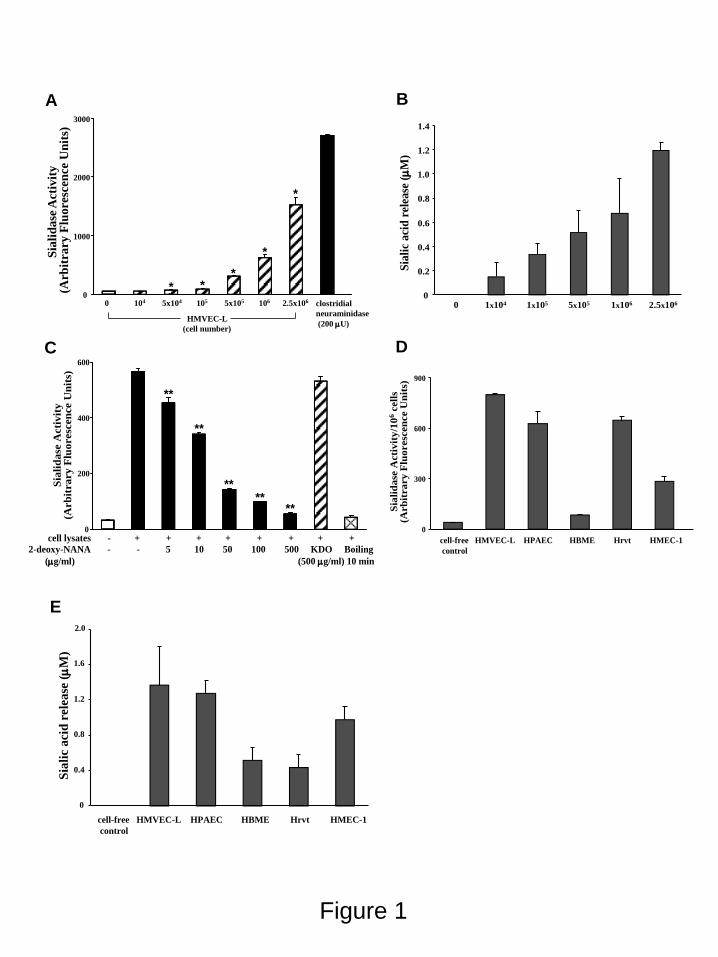
Figure 1
0 104 5x10⁴ 105 5x10⁵ 106 2.5x10⁶
Sia
lid
ase
Act
ivit
y(A
rbit
rary
Flu
ore
scen
ce U
nit
s)
HMVEC-L
(cell number)
clostridial
neuraminidase
(200 U)
0
1000
2000
3000
* **
*
*
0
300
600
900
cell-free HMVEC-L HPAEC HBME Hrvt HMEC-1
control
Sia
lid
ase
Acti
vit
y/1
06
cell
s(A
rb
itra
ry
Flu
oresc
en
ce U
nit
s)
A B
C D
Sia
lid
ase
Acti
vit
y(A
rb
itra
ry
Flu
oresc
en
ce U
nit
s)
cell lysates - + + + + + + + +
2-deoxy-NANA - - 5 10 50 100 500 KDO Boiling
(g/ml) (500 g/ml) 10 min
0
200
400
600
**
**
****
**
0
0.2
0.4
0.6
0.8
1.0
1.2
1.4
0 1x10⁴ 1x10⁵ 5x10⁵ 1x10⁶ 2.5x10⁶
Sia
lic
aci
d r
elea
se (
M)
cell-free HMVEC-L HPAEC HBME Hrvt HMEC-1
control
E
0
0.4
0.8
1.2
1.6
2.0
Sia
lic
aci
d r
ele
ase
(
M)
by guest on May 27, 2018
http://ww
w.jbc.org/
Dow
nloaded from

Figure 2
A
B C
60kDa
50kDa
40kDa
1 2 3 4
siRNA control control NEU1NEU1
Time 48h 72h
IB* -tubulin
IB NEU1
60kDa
50kDa
40kDa
1 2 3 4
siRNA control control NEU3NEU3
Time 24h 48h
IB*
IB NEU3
0
NEU1 NEU2 NEU4
Rel
ati
ve
mR
NA
Cop
y N
um
ber
(NE
U1,2
,3,4
/HP
RT
)
NEU3
0.01
0.02
3.0
4.0
5.0
-tubulin
by guest on May 27, 2018
http://ww
w.jbc.org/
Dow
nloaded from

Figure 3
2 3 1040 210 310 410
200
400
600
800
100
0120
0 A. Nonpermeabilized
Surface NEU1
Surface NEU3
Surface Isotype
control
2 3 1040 210 310 410
200
400
600
800
100
0120
0 A. Nonpermeabilized
Surface NEU1
Surface NEU3
Surface Isotype
control
200
400
600
800
100
0120
0 B. Permeabilized
Total NEU3
Total NEU1Total isotype
control
Mean Fluoresence Intensity
2 3 1040 210 310 410
200
400
600
800
100
0120
0 B. Permeabilized
Total NEU3
Total NEU1Total isotype
control
Mean Fluoresence Intensity
2 3 1040 210 310 410
by guest on May 27, 2018
http://ww
w.jbc.org/
Dow
nloaded from

Figure 4
52kDa
34kDa
72kDa
IB : NEU3
IB* : IB
IB*: Lamin B1
A B
i ii
iii iv
NEU1 NEU3
(+) DAPI
(-) DAPI
by guest on May 27, 2018
http://ww
w.jbc.org/
Dow
nloaded from

Rel
ati
ve
siali
dase
act
ivit
y (
%)
HMVEC-L lysates - + + + +
siRNA - - control NEU1 NEU3
n 2 2 3 3 3
0
20
40
60
80
100
120
**
**
**
0
20
40
60
80
100
120
**
**
**
0
20
40
60
80
100
120
**
**
**
A B
C D
E F
60kDa
50kDa
40kDa
Ad-NEU1(MOI) 0 10 25 50 100 300
IB Flag
IB* -tubulin
1 2 3 4 5 660kDa
50kDa
40kDa
1 2 3 4
siRNA control control NEU1NEU1
Time 24h 48h
IB*
IB Flag
Ad-NEU1(MOI=100) + ++ +
Ad-NEU3(MOI) 0 10 25 50 100 300
HA
1 2 3 4 5 6
IB
IB*
60kDa
50kDa
40kDaAd-NEU3(MOI=100) +
1 2 3 4
++ +siRNA control control NEU3NEU3
Time 24h 48h
5 6
++control NEU3
72h
HAIB
IB*
60kDa
50kDa
40kDa
98
55
38
siRNA control NEU1 NEU3 control NEU3 NEU1
Ad-NEU1
IB Flag (NEU1) HA (NEU3)
1 2 3 4 5 6kDa
IB* -tubulin
Ad
G
HMVEC-L lysates + + + +siRNA - control NEU1 NEU3
n 2 5 3 5
0
20
40
60
80
100
120
Rel
ati
ve
siali
caci
d r
elea
se (
%)
**
-tubulin
-tubulin
-tubulin
Ad-NEU3
Figure 5
by guest on May 27, 2018
http://ww
w.jbc.org/
Dow
nloaded from

Figure 6
siRNA --- ctrl NEU1 NEU3
2h 24h16h
--- ctrl NEU1 NEU3 --- ctrl NEU1 NEU3
by guest on May 27, 2018
http://ww
w.jbc.org/
Dow
nloaded from

Figure 7
* **
* * *
i) ad-NEU1 MOI30 24h ii) ad-NEU1 MOI100 24h
iii) ad-NEU1 MOI150 24h iv) ad-GFP MOI150 24h
D. 24h
by guest on May 27, 2018
http://ww
w.jbc.org/
Dow
nloaded from

Figure 8
Pulmonary Artery
NEU1 NEU3
NEU1 NEU3
Lung Capillaries
CD31 Negative Control
E F
A B C D
Aorta
NEU1
Carotid Artery
NEU3
NEU1 NEU3
G H
I J
Cerebral Artery
NEU1 NEU3
K L
Hepatic Vein Renal Artery
NEU1 NEU3 NEU1 NEU3
M N O P
by guest on May 27, 2018
http://ww
w.jbc.org/
Dow
nloaded from

E. GoldblumErik P. Lillehoj, Adam C. Puche, Wei Huang, Lai-Xi Wang, Antonino Passaniti and SimeonNguyen, Lei Zhang, Irina G. Luzina, Sergei P. Atamas, William S. Twaddell, Wei Guang, Alan S. Cross, Sang Won Hyun, Alba Miranda-Ribera, Chiguang Feng, Anguo Liu, Chinh
NEU1 restrains endothelial cell migration whereas NEU3 does notNEU1 and NEU3 sialidase activity expressed in human lung microvascular endothelia.
published online March 8, 2012J. Biol. Chem.
10.1074/jbc.M112.346817Access the most updated version of this article at doi:
Alerts:
When a correction for this article is posted•
When this article is cited•
to choose from all of JBC's e-mail alertsClick here
by guest on May 27, 2018
http://ww
w.jbc.org/
Dow
nloaded from