Embed Size (px)
Citation preview
Mitosis
Onions have larger chromosomes than most plants and they stain dark.
The chromosomes are easily observed through a compound light microscope.
The apical meristem is an area of a plant where cell division takes place at a rapid rate.
Why Onions?
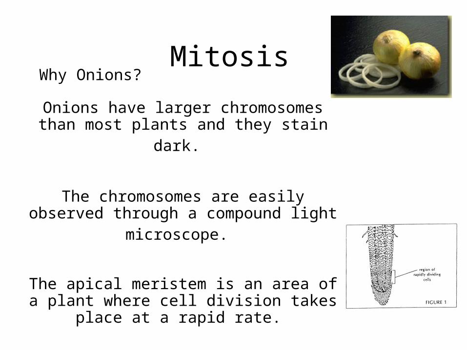
Stages of MitosisThe numbers to the left correspond to the numbered pictures on the right. For example
picture 1 is a cell in interphase.
1 interphase2 Beginning of prophase3 early prophase4 mid prophase5 late prophase6 metaphase 7 early anaphase8 anaphase9 telophase10 cytokinesis
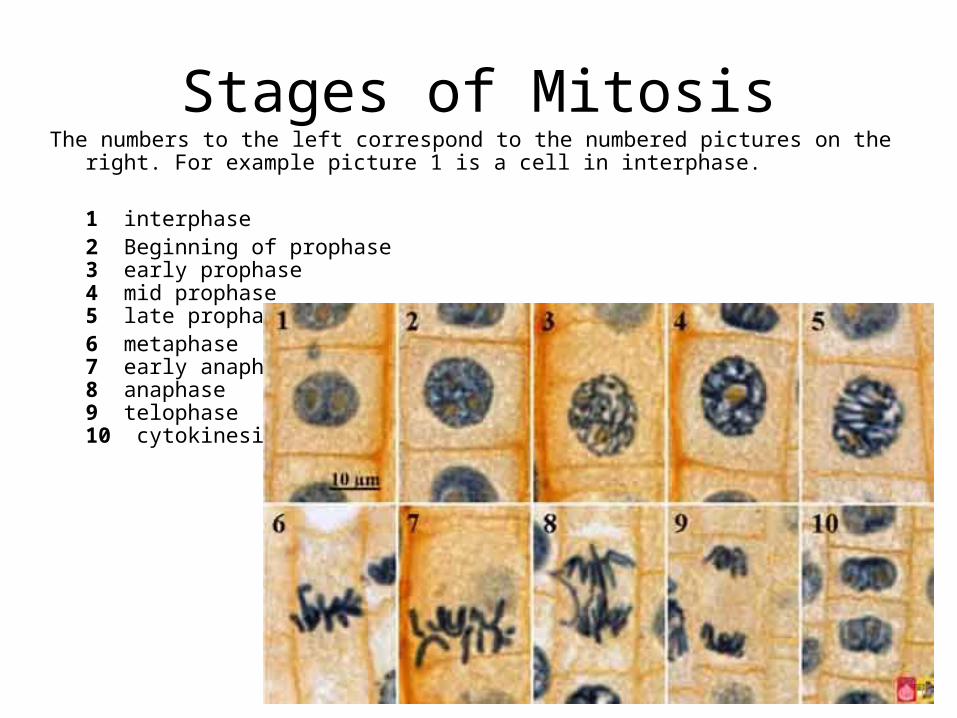
Interphase
The highlighted area shows a cell in interphase. Draw this cell in the space provided.
-The cell is growing in size
- The cell is replicating its DNA in preparation for division.
-The nucleus is apparent.
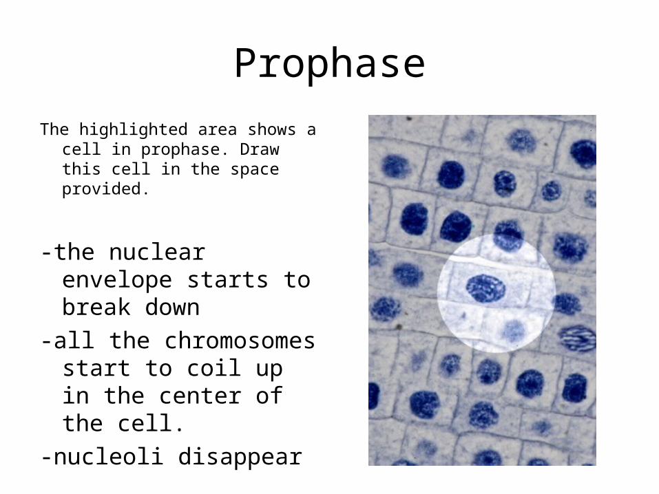
Prophase
The highlighted area shows a cell in prophase. Draw this cell in the space provided.
-the nuclear envelope starts to break down
-all the chromosomes start to coil up in the center of the cell.
-nucleoli disappear
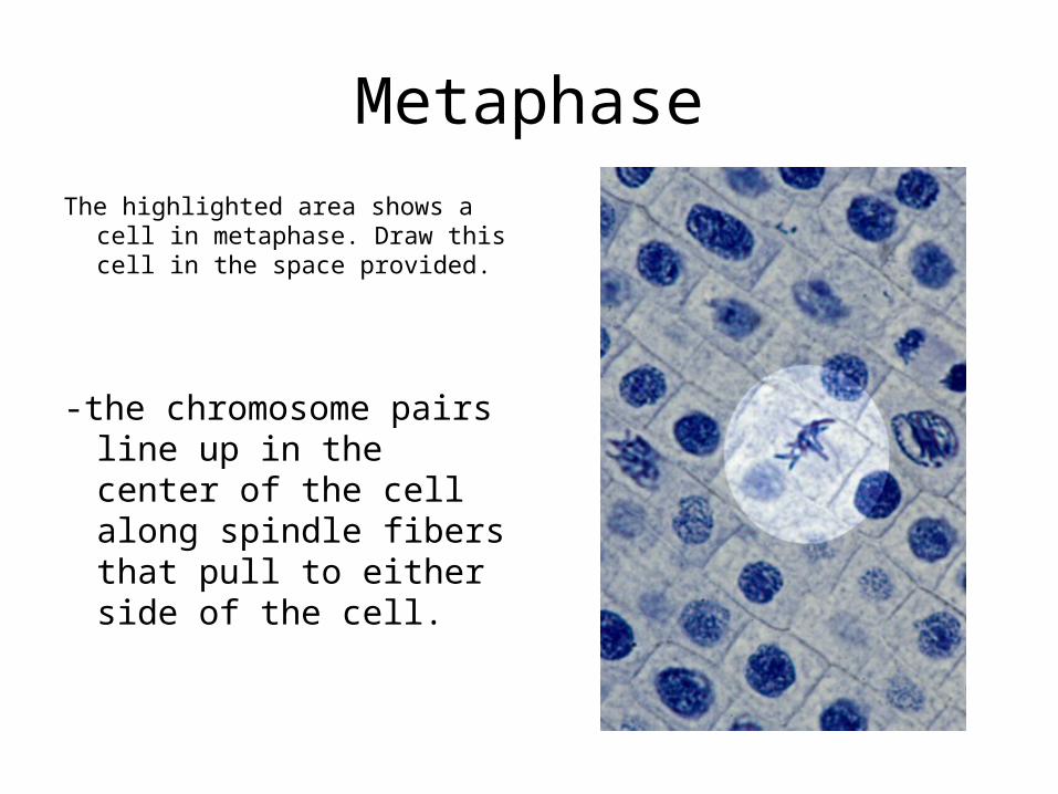
Metaphase
The highlighted area shows a cell in metaphase. Draw this cell in the space provided.
-the chromosome pairs line up in the center of the cell along spindle fibers that pull to either side of the cell.
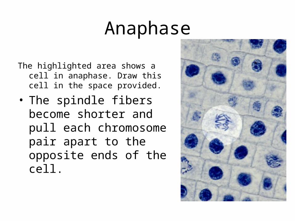
Anaphase
The highlighted area shows a cell in anaphase. Draw this cell in the space provided.
• The spindle fibers become shorter and pull each chromosome pair apart to the opposite ends of the cell.
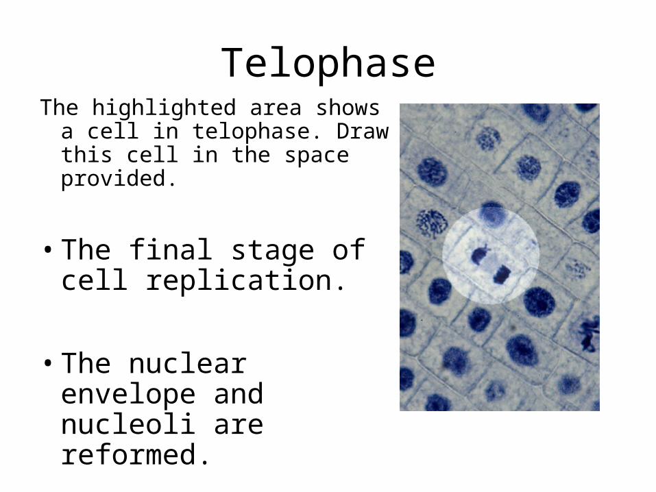
TelophaseThe highlighted area shows a
cell in telophase. Draw this cell in the space provided.
• The final stage of cell replication.
• The nuclear envelope and nucleoli are reformed.
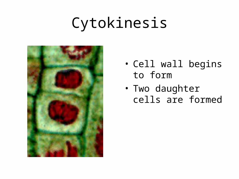
Cytokinesis
• Cell wall begins to form
• Two daughter cells are formed
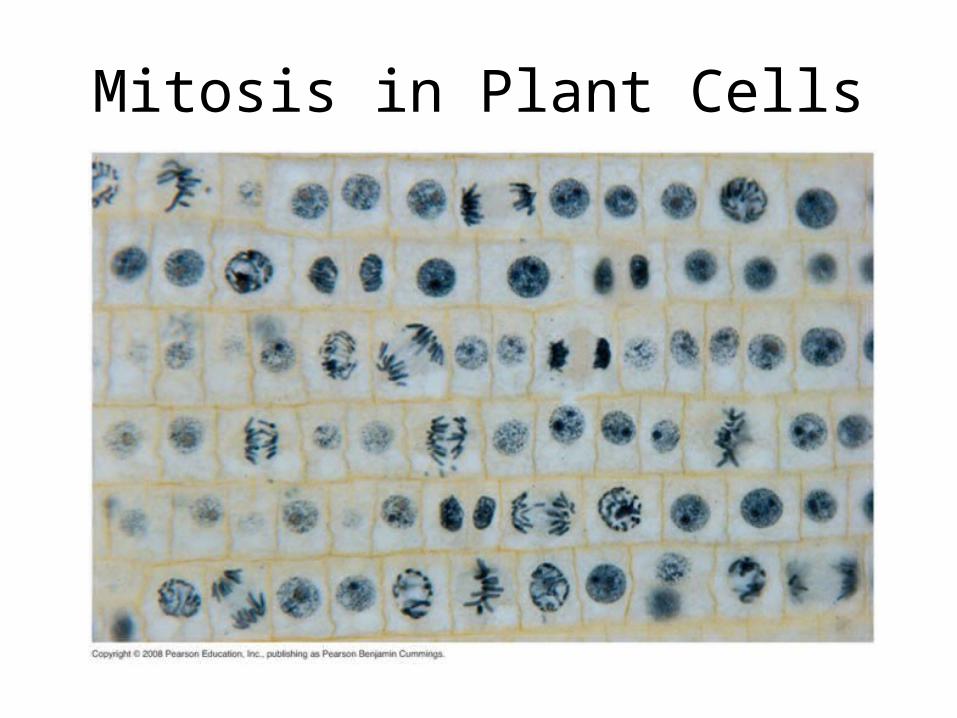
Mitosis in Plant Cells
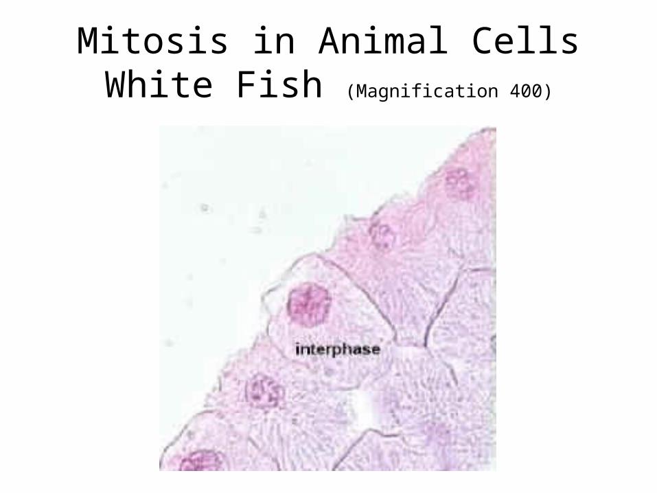
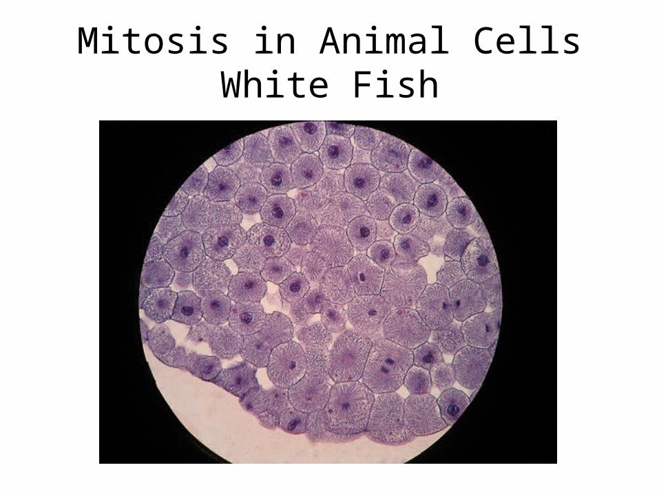
Mitosis in Animal CellsWhite Fish (Magnification 400)
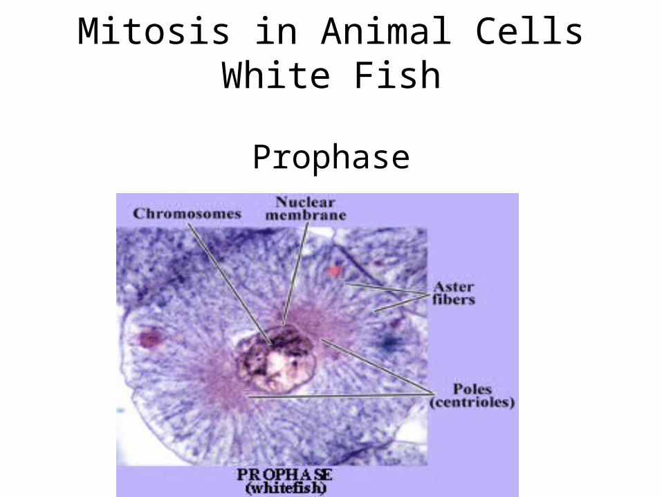
Mitosis in Animal CellsWhite Fish
Prophase
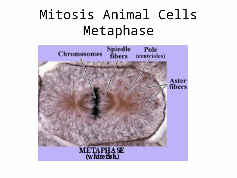
Mitosis Animal CellsMetaphase
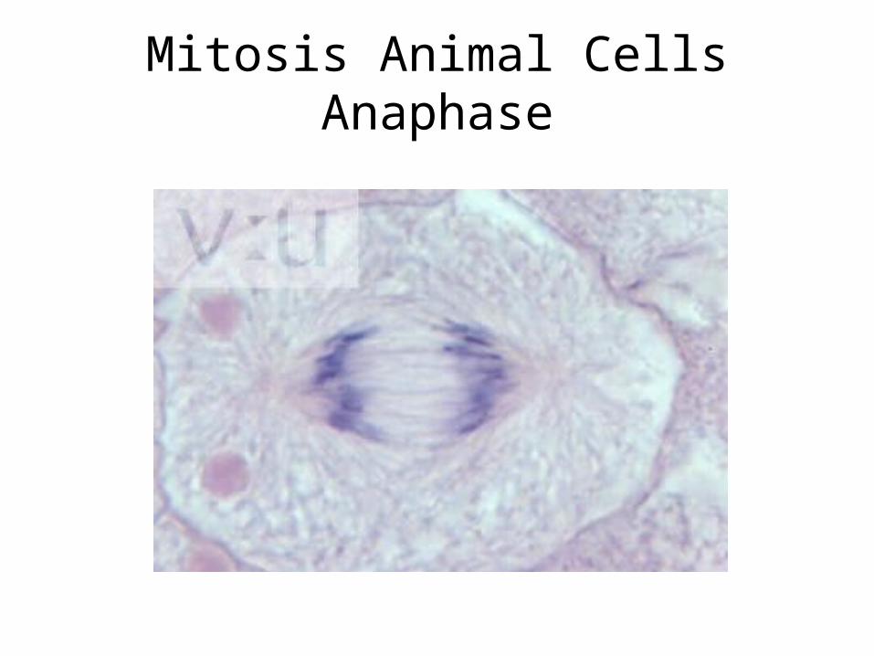
Mitosis Animal CellsAnaphase
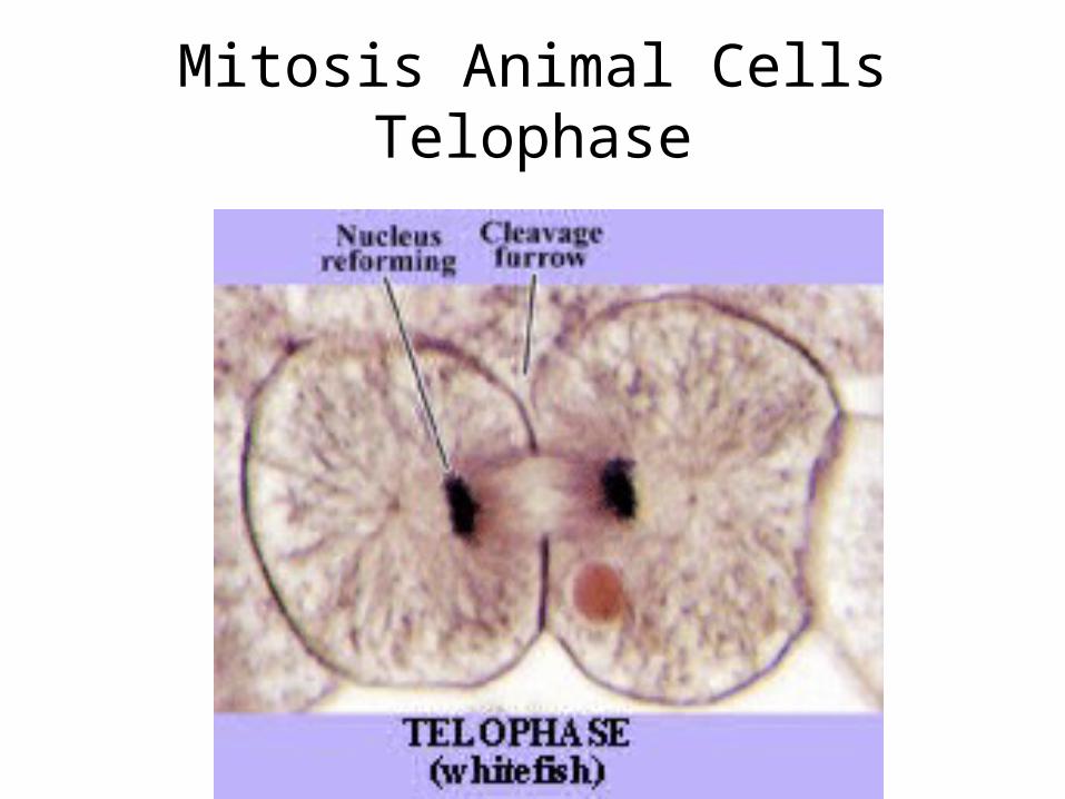
Mitosis Animal CellsTelophase
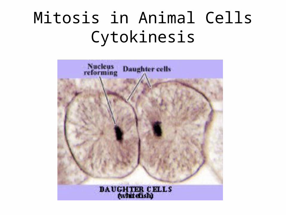
Mitosis in Animal CellsCytokinesis
Mitosis in Animal CellsWhite Fish
Resources
• http://www.microscopy-uk.org.uk/mag/artnov04macro/jronionroot.html• http://www.vcbio.science.ru.nl/en/image-gallery/show/PL0096/labels/
• http://biog-101-104.bio.cornell.edu/BioG101_104/tutorials/cell_division/onion_review_fs.html
• White Fish Cell Mitosis Pictures: http://www.linkpublishing.com/video-mitosis.htm#MITOSIS_-_WHITEFISH_BLASTULA
• White Fish Mitosis Cells Pic: http://www.flickr.com/photos/10517539@N07/1554957435/
• Mitosis World: http://www.bio.unc.edu/faculty/salmon/lab/mitosis/mitosislinks.html
• www.cellsalive.com