Embed Size (px)
Citation preview
Cell Cycle Mitosis Meiosis
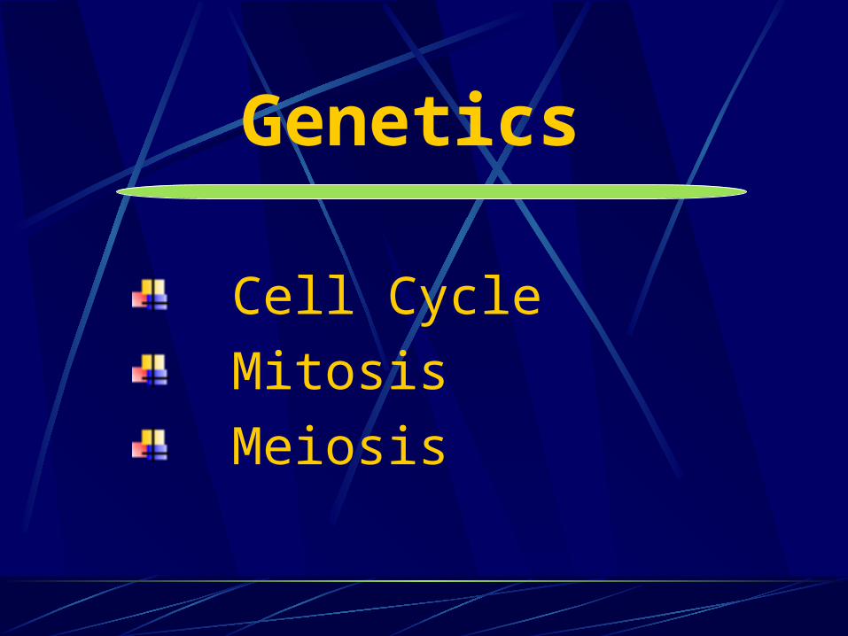
Genetics

Eukaryotic chromosomes contain DNA and protein
The chromosomes carry the genetic information

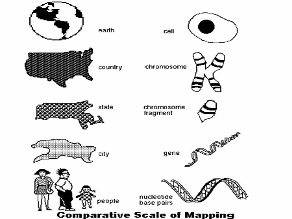


When a cell divides, chromatin fibers are very highly folded, and become visible in the light microscope as chromosomes.
During interphase (between divisions), chromatin is more extended, a form used for expression genetic information.

• DNA is organized into informational units called genes
• Chromosomes contain hundreds to thousands of genes
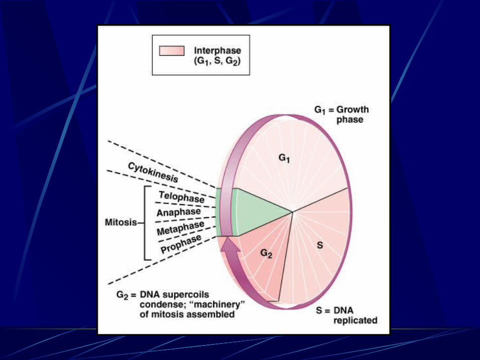
Cell Cycle
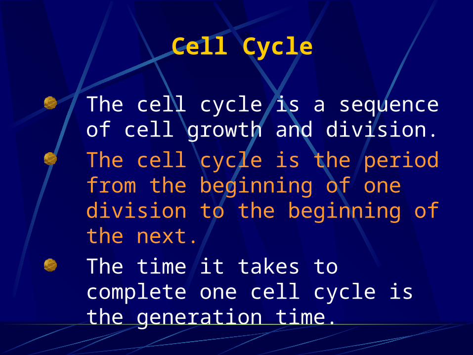
The cell cycle is a sequence of cell growth and division.The cell cycle is the period from the beginning of one division to the beginning of the next.The time it takes to complete one cell cycle is the generation time.

Cells divide when they reach a certain size NO (nerve, skeletal muscle and red blood cells)
Cell division involves mitosis and cytokinesis. Mitosis involves division of the chromosomes. Cytokinesis involves division of the cytoplasm. Mitosis without cytokinesis results in multinucleate cells.

Eukaryotic cell cycleBeginning of one division to beginning of nextStages in eukaryotic cell cycle Interphase
First gap phaseSynthesis phaseSecond gap phase
M phaseMitosisCytokinesis

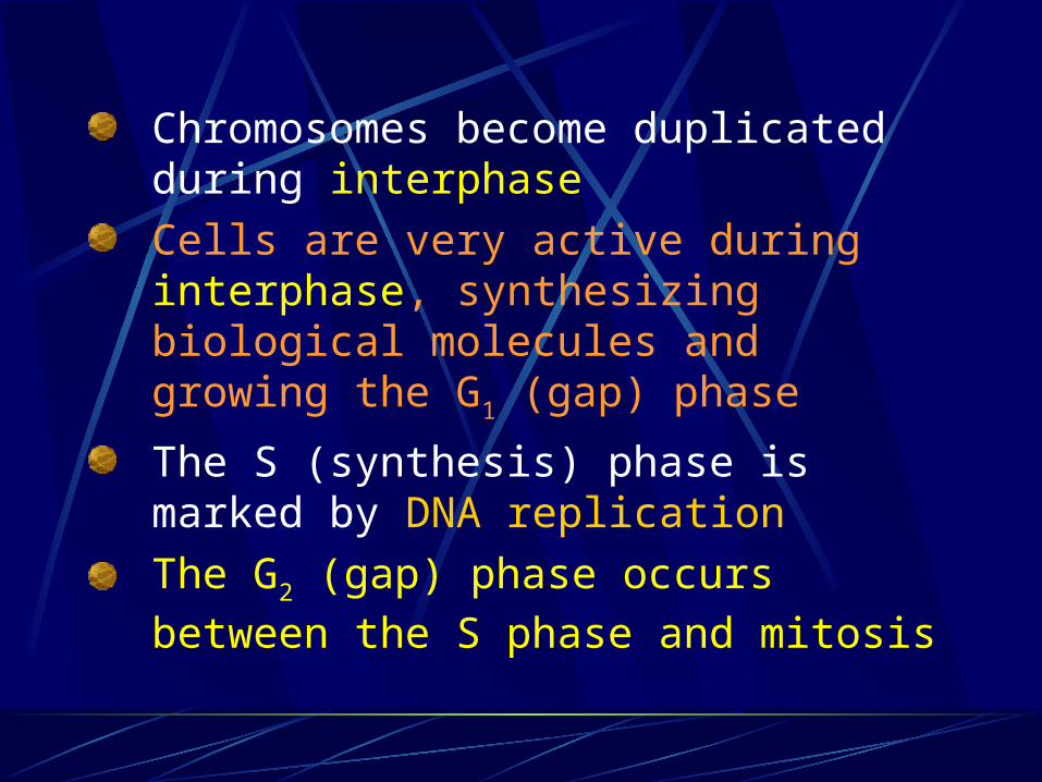
Chromosomes become duplicated during interphase Cells are very active during interphase, synthesizing biological molecules and growing the G1 (gap) phase
The S (synthesis) phase is marked by DNA replication The G2 (gap) phase occurs between the S phase and mitosis
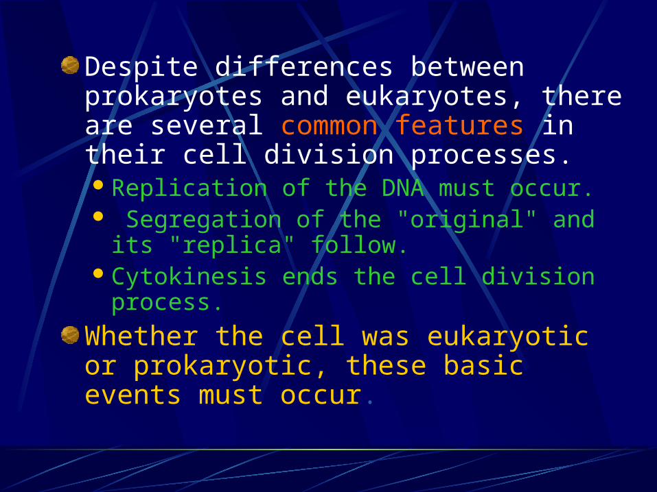
Despite differences between prokaryotes and eukaryotes, there are several common features in their cell division processes. Replication of the DNA must occur. Segregation of the "original" and its
"replica" follow. Cytokinesis ends the cell division
process.
Whether the cell was eukaryotic or prokaryotic, these basic events must occur.

Hereditary material is passed on to new cells by mitosis or meiosis
Cell division, growth, and reproduction
InterphaseMitosisCytokinesisMeiosis

Cell division
• Chromosomal packaging of DNA allows efficient distribution of genetic material during cell division
• Life cycle requires two distinct types of cell division processes: mitosis and meiosis
• Cell division: one cell becomes two cells during an organism’s life cycle

Mitosis
Mitosis is nuclear division plus cytokinesis, and produces two identical daughter cells during the following steps: Prophase Metaphase Anaphase Telophase.
Interphase is often included in discussions of mitosis, but interphase is technically not part of mitosis, but rather encompasses stages G1, S, and G2 of the cell cycle.

Interphase
The cell is engaged in metabolic activity and performing its prepare for mitosis (the next four phases that lead up to and include nuclear division). Chromosomes are not clearly discerned in the nucleus, although a dark spot called the nucleolus may be visible. The cell may contain a pair of centrioles (or microtubule organizing centers in plants) both of which are organizational sites for microtubules.

Prophase
Chromatin in the nucleus begins to condense and becomes visible in the light microscope as chromosomes.
The nucleolus disappears.
Centrioles begin moving to opposite ends of the cell and fibers extend from the centromeres.
Some fibers cross the cell to form the mitotic spindle.
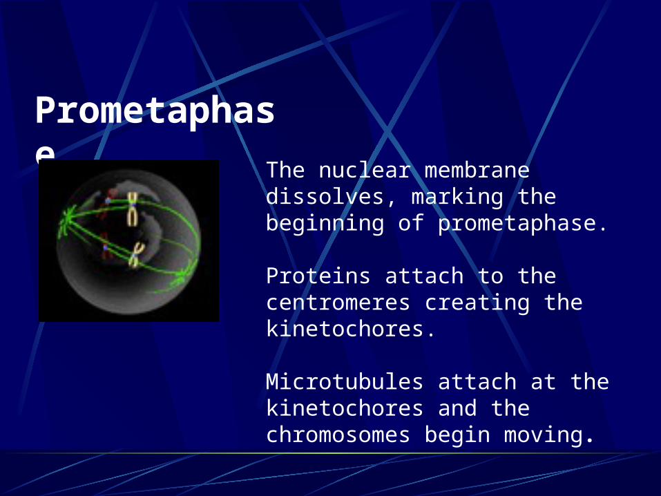
Prometaphase
The nuclear membrane dissolves, marking the beginning of prometaphase.
Proteins attach to the centromeres creating the kinetochores.
Microtubules attach at the kinetochores and the chromosomes begin moving.

Metaphase
Spindle fibers line the chromosomes along the middle of the cell nucleus. This line is referred to as the metaphase plate. Polar microtubules extend from the pole to the equator, and typically overlap Kinetochore microtubules extend from the pole to the kinetochores
This organization helps to ensure that in the next phase, when the chromosomes are separated, each new nucleus will receive one copy of each chromosome.
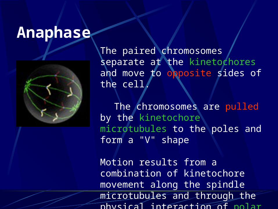
Anaphase
The paired chromosomes separate at the kinetochores and move to opposite sides of the cell.
The chromosomes are pulled by the kinetochore microtubules to the poles and form a "V" shape
Motion results from a combination of kinetochore movement along the spindle microtubules and through the physical interaction of polar microtubules.

Telophase
Chromatids arrive at opposite poles of cell, and new membranes form around the daughter nuclei.
The chromosomes disperse and are no longer visible under the light microscope.
The spindle fibers disperse, and cytokinesis will start.

Cytokinesis
In animal cells, cytokinesis results when a fiber ring composed of a protein called actin around the center of the cell contracts pinching the cell into two daughter cells, each with one nucleus.
In plant cells, synthesis of new cell wall between two daughter cells rather than cleavage furrow in
cytoplasm

Interphase

Prophase

Prometaphase

Metaphase

Anaphase

Telophase

Cytokinesis


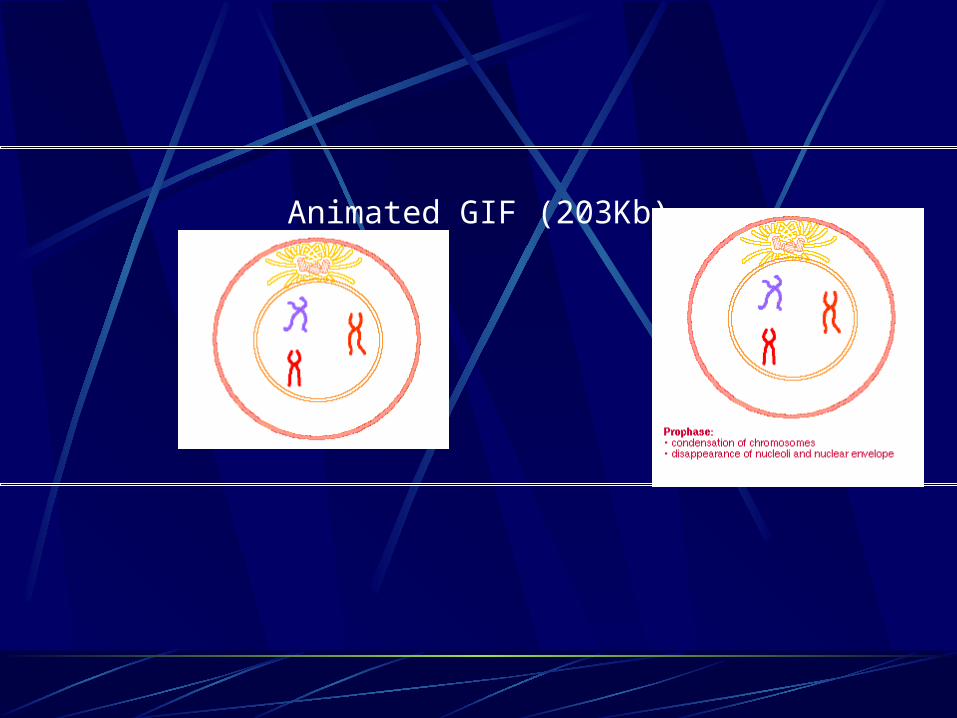
Animated GIF (203Kb)

Reproduction
Asexual reproduction
Sexual reproduction

Asexual Reproduction
A form of duplication using only mitosis.
Example, a new plant grows out of the root or a shoot from an existing plant.
Produces only genetically identical offspring since all divisions are by mitosis.

Sexual reproduction
Formation of new individual by a combination of two haploid sex cells (gametes). Fertilization- combination of genetic information from two separate cells that have one half the original genetic information Gametes for fertilization usually come from separate parents

1. Female- produces an egg 2. Male produces sperm
Both gametes are haploid, with a single set of chromosomes The new individual is called a zygote, with two sets of chromosomes (diploid). Meiosis is a process to convert a diploid cell to a haploid gamete, and cause a change in the genetic information to increase diversity in the offspring.

Chromosomes in a Diploid Cell
Summary of chromosome characteristics
Diploid set for humans; 2n = 46 Autosomes; homologous chromosomes, one from each parent (humans = 22 sets of 2) Sex chromosomes (humans have 1 set)
1. Female-sex chromosomes are homologous (XX)
2. Male-sex chromosomes are non-homologous (XY)

Number of sets of chromosomes in a cell
Haploid (n)-- one set chromosomes Diploid (2n)-- two sets chromosomes Most plant and animal adults are diploid (2n) Eggs and sperm are haploid (n)

Most cells in the human body are produced by mitosis. These are the somatic (or vegetative) line cells.
Cells that become gametes are referred to as germ line cells. The vast majority of cell divisions in the human body are mitotic, with meiosis being restricted to the gonads.

Diploid cells Characteristic number of
chromosome pairs per cellHomologous chromosomesSimilar in length, shape, other features,
and carry similar attributes
Haploid cellsContain only one member of each
homologous chromosome pair


Meiosis
Diploid cells undergo meiosis to form haploid cells
Meiosis potentially produces four haploid cells
Meiosis involves two separate divisions
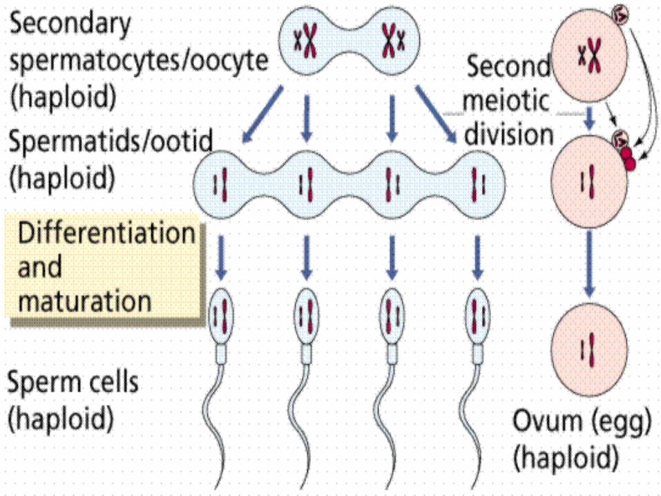
Two successive nuclear divisions occur, Meiosis I (Reduction) and Meiosis II (Division).
Meiosis I reduces the ploidy level from 2n to n (reduction) while Meiosis II divides the remaining set of chromosomes in a mitosis-like process (division).
In meiosis, homologous chromosomes are separated into different daughter cells
Meiosis I and meiosis II each include prophase, metaphase, anaphase, and telophase
The First Division Meiosis I
Prophase I is one of the most important stages of meiosis.
During this stage, many crucial events occur.

In prophase I,
The spindle appears.
Nuclear envelopes disappear.
The DNA of the chromosomes begin to twist and condense, making the DNA visible to the microscope.
Each chromosome actively seeks out its homologous pair (which also has a sister chromatid).

The two replicated homologous pairs find each other and form a synapse. The structure formed is referred to as a tetrad (four chromatids).
The point at which the two non-sister chromatids intertwine is called a chiasma. Sometimes a process known as crossing over occurs at this point.
This is where two non-sister chromatids exchange genetic material. This exchange does not become evident, however, until the two homologous pairs separate.


Prophase I includes synapsis and crossing over
Homologous chromosomes pair and undergo synapsis
One member of a pair is the maternal homologue, the other is the paternal homologue
Synapsis is the association of four chromatids (two from each homologue)
In metaphase I, the tetrads line up along the equator.
Anaphase I results in the separation of homologous pairs. Cells are haploid at this point.
Telophase I results in a brief reappearance of nuclear envelopes, and the spindle disappears. The cell waits momentarily during interkinesis.
Interkinesis separates meiosis I and II; no DNA synthesis occurs

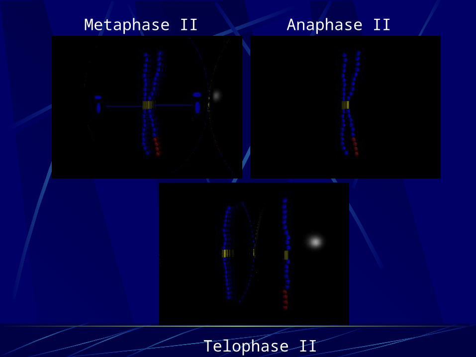
The Second Division Meiosis II
In prophase II, the spindle reappears, and the nuclear membrane fragments.
In metaphase II, the chromosomes align at the equator.
In anaphase II, sister chromatids separate.
In telophase II, the nuclear envelopes reappear, and four haploid cells are the result.


Prophase I
Interphase

Prophase IITelophase I
Anaphase IMetaphase I
Metaphase II Anaphase II
Telophase II

Germ line cells undergo gametogenesis
Spermatogenesis produces sperm
Oogenesis typically produces eggs, or a single ovum and two or more polar bodies