Embed Size (px)
Citation preview
CASE REPORT Open Access
Mirror therapy for an adult with centralpost-stroke pain: a case reportDavide Corbetta1*, Elisabetta Sarasso1,2, Federica Agosta2, Massimo Filippi3 and Roberto Gatti4
Abstract
Background : Treatment of central post-stroke pain (CPSP) after a thalamic-capsular stroke is generally based onpharmacological approach as it is low responsive to physiotherapy. In this case report, the use of mirror therapy(MT) for the reduction of CPSP in a subject after a stroke involving thalamus is presented.
Case presentation: Five years after a right lenticular-capsular thalamic stroke, despite a good recovery of voluntarymovement that guaranteed independence in daily life activities, a 50-year-old woman presented with mildweakness and spasticity, an important sensory loss and a burning pain in the left upper limb. MT for reducing armpain was administered in 45-min sessions, five days a week, for two consecutive weeks. MT consisted in performingsymmetrical movements of both forearms and hands while watching the image of the sound limb reflected by aparasagittal mirror superimposed to the affected limb. Pain severity was assessed using visual analogue scale (VAS)before and after the intervention and at one-year follow-up. After the two weeks of MT, the patient demonstrated4.5 points reduction in VAS pain score of the hand at rest and 3.9 points during a maximal squeeze left handcontraction. At one-year follow-up, pain reduction was maintained and also extended to the shoulder.
Conclusion: This case report shows the successful application of a motor training with a sensory confoundingcondition (MT) in reducing CPSP in a patient with a chronic thalamic stroke.
Keywords: Stroke, Physical therapy modalities, Pain perception, Case reports
BackgroundStroke often causes impairment in movement controlbut can also affect perception [1, 2]. Alterations ofstimulus integration are common after a stroke, withvariable reported prevalence ranging from 11 to 85% [3],and sometimes these alterations of perception result inpain. Pain relates with the site of lesion and it is com-pletely distinct from other painful conditions such asshoulder pain or spasticity [4]. It typically emerges fromhemispheric lesions that involve the spinothalamic andthalamocortical pathways, leading patients to complainof sharping, stabbing, or burning through an experienceof hyperpathia and allodynia [5, 6]. This associationbetween sensory abnormalities and constant or intermit-tent central neuropathic pain, arising from damage ofthe sensory tracts, is known as the central post-stroke
pain (CPSP) syndrome [7, 8]. The estimated incidence ofCPSP comes up to 1 every 6 patients presenting a vascu-lar lesion in the thalamus [8, 9], but its prevalence isdifficult to estimate because of the co-occurrence ofother painful conditions, such as spasticity or shoulderpain [4]. The pathophysiological mechanisms underlyingthe development of CPSP are thought to be related tothe hyperexcitability or to the spontaneous discharge ofdamaged neurons located in the thalamus or in the cor-tex [10]. The CPSP syndrome is one of the less respon-sive conditions to physiotherapy treatment and it usuallyrequires a pharmacological approach through the use ofAmitriptyline, Gabapentin and Pregabalin [2].Mirror therapy (MT), defined as the use of a mirror
reflection of unaffected limb movements superimposedon the affected extremity, is often used to treat motorand perception problems [11, 12]. This technique wasdescribed for the first time in 1995 in studies reportingthe reduction of phantom limb pain in arm amputees[13]; more recently, its use was described also for recov-ery of motor function after stroke [14, 15], for the
* Correspondence: [email protected] of Analysis and Rehabilitation of Motor Function, San RaffaeleScientific Institute, Vita-Salute San Raffaele University, Via Olgettina 60, 20132Milan, ItalyFull list of author information is available at the end of the article
© The Author(s). 2018 Open Access This article is distributed under the terms of the Creative Commons Attribution 4.0International License (http://creativecommons.org/licenses/by/4.0/), which permits unrestricted use, distribution, andreproduction in any medium, provided you give appropriate credit to the original author(s) and the source, provide a link tothe Creative Commons license, and indicate if changes were made. The Creative Commons Public Domain Dedication waiver(http://creativecommons.org/publicdomain/zero/1.0/) applies to the data made available in this article, unless otherwise stated.
Corbetta et al. Archives of Physiotherapy (2018) 8:4 https://doi.org/10.1186/s40945-018-0047-y
treatment of complex regional pain syndrome type I [12]and other painful conditions (e.g., brachial plexus avul-sion and after surgery) [16, 17].This case report describes the beneficial effect of MT for
the reduction of pain of the upper limb in a subject pre-senting CPSP in the left body side combined to sensoryloss and mild movement disorders after a right haemor-rhagic lenticular-capsular, thalamic stroke occurred fiveyears before. To the best of our knowledge, the effect ofMT for the treatment of CPSP has never been observeddespite it has been defined deserving to be explored [11].
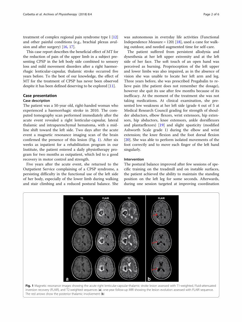
Case presentationCase descriptionThe patient was a 50-year old, right-handed woman whoexperienced a haemorrhagic stroke in 2010. The com-puted tomography scan performed immediately after theacute event revealed a right lenticular-capsular, lateralthalamic and intraparenchymal hematoma, with a mid-line shift toward the left side. Two days after the acuteevent a magnetic resonance imaging scan of the brainconfirmed the presence of this lesion (Fig. 1). After sixweeks as inpatient for a rehabilitation program in ourInstitute, the patient entered a daily physiotherapy pro-gram for two months as outpatient, which led to a goodrecovery in motor control and strength.Five years after the acute event, she returned to the
Outpatient Service complaining of a CPSP syndrome, apersisting difficulty in the functional use of the left sideof her body, especially of the lower limb during walkingand stair climbing and a reduced postural balance. She
was autonomous in everyday life activities (FunctionalIndependence Measure = 120) [18], used a cane for walk-ing outdoor, and needed augmented time for self-care.The patient suffered from persistent allodynia and
dysesthesia at her left upper extremity and at the leftside of her face. The soft touch of an open hand wasperceived as burning. Proprioception of the left upperand lower limbs was also impaired, as in the absence ofvision she was unable to locate her left arm and leg.Three years before, she was prescribed Pregabalin to re-lieve pain (the patient does not remember the dosage),however she quit its use after few months because of itsinefficacy. At the moment of the treatment she was nottaking medications. At clinical examination, she pre-sented low weakness at her left side (grade 4 out of 5 atMedical Research Council grading for strength of shoul-der abductors, elbow flexors, wrist extensors, hip exten-sors, hip abductors, knee extensors, ankle dorsiflexorsand plantarflexors) [19] and slight spasticity (modifiedAshworth Scale grade 1) during the elbow and wristextension; the knee flexion and the foot dorsal flexion[20]. She was able to perform isolated movements of thefoot correctly and to move each finger of the left handsingularly.
InterventionThe postural balance improved after few sessions of spe-cific training on the treadmill and on instable surfaces,the patient achieved the ability to maintain the standingposition on the left leg for some seconds. Afterwards,during one session targeted at improving coordination
Fig. 1 Magnetic resonance images showing the acute right lenticular-capsular-thalamic stroke lesion assessed with T1-weighted, Fluid-attenuatedinversion recovery (FLAIR), and T2-weighted sequences (a); one-year follow-up MRI showing the lesion evolution assessed with FLAIR sequence.The red arrows show the posterior thalamic involvement (b)
Corbetta et al. Archives of Physiotherapy (2018) 8:4 Page 2 of 6
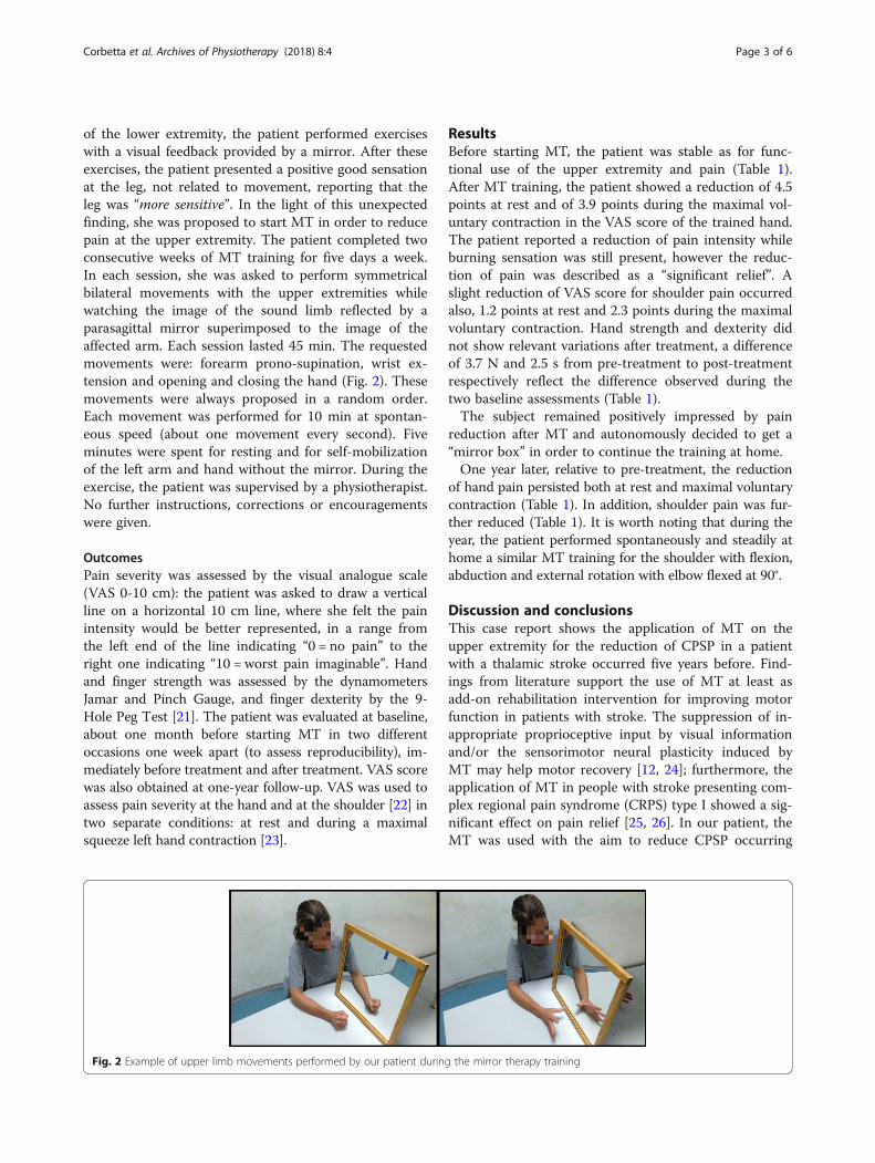
of the lower extremity, the patient performed exerciseswith a visual feedback provided by a mirror. After theseexercises, the patient presented a positive good sensationat the leg, not related to movement, reporting that theleg was “more sensitive”. In the light of this unexpectedfinding, she was proposed to start MT in order to reducepain at the upper extremity. The patient completed twoconsecutive weeks of MT training for five days a week.In each session, she was asked to perform symmetricalbilateral movements with the upper extremities whilewatching the image of the sound limb reflected by aparasagittal mirror superimposed to the image of theaffected arm. Each session lasted 45 min. The requestedmovements were: forearm prono-supination, wrist ex-tension and opening and closing the hand (Fig. 2). Thesemovements were always proposed in a random order.Each movement was performed for 10 min at spontan-eous speed (about one movement every second). Fiveminutes were spent for resting and for self-mobilizationof the left arm and hand without the mirror. During theexercise, the patient was supervised by a physiotherapist.No further instructions, corrections or encouragementswere given.
OutcomesPain severity was assessed by the visual analogue scale(VAS 0-10 cm): the patient was asked to draw a verticalline on a horizontal 10 cm line, where she felt the painintensity would be better represented, in a range fromthe left end of the line indicating “0 = no pain” to theright one indicating “10 = worst pain imaginable”. Handand finger strength was assessed by the dynamometersJamar and Pinch Gauge, and finger dexterity by the 9-Hole Peg Test [21]. The patient was evaluated at baseline,about one month before starting MT in two differentoccasions one week apart (to assess reproducibility), im-mediately before treatment and after treatment. VAS scorewas also obtained at one-year follow-up. VAS was used toassess pain severity at the hand and at the shoulder [22] intwo separate conditions: at rest and during a maximalsqueeze left hand contraction [23].
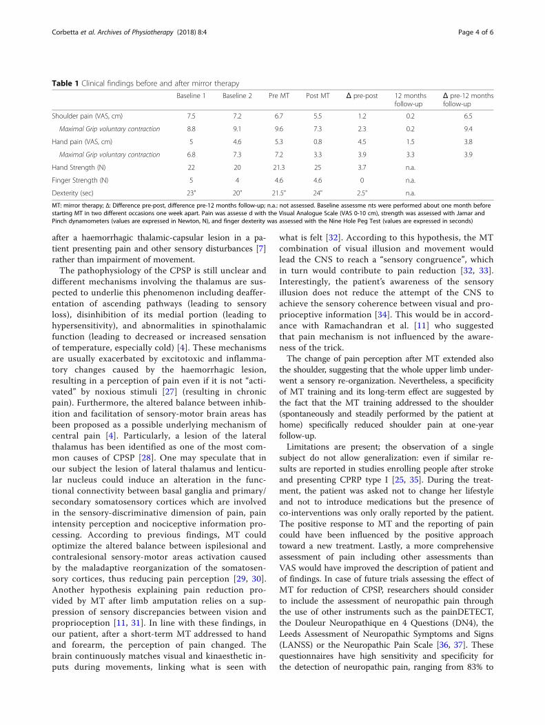
ResultsBefore starting MT, the patient was stable as for func-tional use of the upper extremity and pain (Table 1).After MT training, the patient showed a reduction of 4.5points at rest and of 3.9 points during the maximal vol-untary contraction in the VAS score of the trained hand.The patient reported a reduction of pain intensity whileburning sensation was still present, however the reduc-tion of pain was described as a “significant relief”. Aslight reduction of VAS score for shoulder pain occurredalso, 1.2 points at rest and 2.3 points during the maximalvoluntary contraction. Hand strength and dexterity didnot show relevant variations after treatment, a differenceof 3.7 N and 2.5 s from pre-treatment to post-treatmentrespectively reflect the difference observed during thetwo baseline assessments (Table 1).The subject remained positively impressed by pain
reduction after MT and autonomously decided to get a“mirror box” in order to continue the training at home.One year later, relative to pre-treatment, the reduction
of hand pain persisted both at rest and maximal voluntarycontraction (Table 1). In addition, shoulder pain was fur-ther reduced (Table 1). It is worth noting that during theyear, the patient performed spontaneously and steadily athome a similar MT training for the shoulder with flexion,abduction and external rotation with elbow flexed at 90°.
Discussion and conclusionsThis case report shows the application of MT on theupper extremity for the reduction of CPSP in a patientwith a thalamic stroke occurred five years before. Find-ings from literature support the use of MT at least asadd-on rehabilitation intervention for improving motorfunction in patients with stroke. The suppression of in-appropriate proprioceptive input by visual informationand/or the sensorimotor neural plasticity induced byMT may help motor recovery [12, 24]; furthermore, theapplication of MT in people with stroke presenting com-plex regional pain syndrome (CRPS) type I showed a sig-nificant effect on pain relief [25, 26]. In our patient, theMT was used with the aim to reduce CPSP occurring
Fig. 2 Example of upper limb movements performed by our patient during the mirror therapy training
Corbetta et al. Archives of Physiotherapy (2018) 8:4 Page 3 of 6
after a haemorrhagic thalamic-capsular lesion in a pa-tient presenting pain and other sensory disturbances [7]rather than impairment of movement.The pathophysiology of the CPSP is still unclear and
different mechanisms involving the thalamus are sus-pected to underlie this phenomenon including deaffer-entation of ascending pathways (leading to sensoryloss), disinhibition of its medial portion (leading tohypersensitivity), and abnormalities in spinothalamicfunction (leading to decreased or increased sensationof temperature, especially cold) [4]. These mechanismsare usually exacerbated by excitotoxic and inflamma-tory changes caused by the haemorrhagic lesion,resulting in a perception of pain even if it is not “acti-vated” by noxious stimuli [27] (resulting in chronicpain). Furthermore, the altered balance between inhib-ition and facilitation of sensory-motor brain areas hasbeen proposed as a possible underlying mechanism ofcentral pain [4]. Particularly, a lesion of the lateralthalamus has been identified as one of the most com-mon causes of CPSP [28]. One may speculate that inour subject the lesion of lateral thalamus and lenticu-lar nucleus could induce an alteration in the func-tional connectivity between basal ganglia and primary/secondary somatosensory cortices which are involvedin the sensory-discriminative dimension of pain, painintensity perception and nociceptive information pro-cessing. According to previous findings, MT couldoptimize the altered balance between ispilesional andcontralesional sensory-motor areas activation causedby the maladaptive reorganization of the somatosen-sory cortices, thus reducing pain perception [29, 30].Another hypothesis explaining pain reduction pro-vided by MT after limb amputation relies on a sup-pression of sensory discrepancies between vision andproprioception [11, 31]. In line with these findings, inour patient, after a short-term MT addressed to handand forearm, the perception of pain changed. Thebrain continuously matches visual and kinaesthetic in-puts during movements, linking what is seen with
what is felt [32]. According to this hypothesis, the MTcombination of visual illusion and movement wouldlead the CNS to reach a “sensory congruence”, whichin turn would contribute to pain reduction [32, 33].Interestingly, the patient’s awareness of the sensoryillusion does not reduce the attempt of the CNS toachieve the sensory coherence between visual and pro-prioceptive information [34]. This would be in accord-ance with Ramachandran et al. [11] who suggestedthat pain mechanism is not influenced by the aware-ness of the trick.The change of pain perception after MT extended also
the shoulder, suggesting that the whole upper limb under-went a sensory re-organization. Nevertheless, a specificityof MT training and its long-term effect are suggested bythe fact that the MT training addressed to the shoulder(spontaneously and steadily performed by the patient athome) specifically reduced shoulder pain at one-yearfollow-up.Limitations are present; the observation of a single
subject do not allow generalization: even if similar re-sults are reported in studies enrolling people after strokeand presenting CPRP type I [25, 35]. During the treat-ment, the patient was asked not to change her lifestyleand not to introduce medications but the presence ofco-interventions was only orally reported by the patient.The positive response to MT and the reporting of paincould have been influenced by the positive approachtoward a new treatment. Lastly, a more comprehensiveassessment of pain including other assessments thanVAS would have improved the description of patient andof findings. In case of future trials assessing the effect ofMT for reduction of CPSP, researchers should considerto include the assessment of neuropathic pain throughthe use of other instruments such as the painDETECT,the Douleur Neuropathique en 4 Questions (DN4), theLeeds Assessment of Neuropathic Symptoms and Signs(LANSS) or the Neuropathic Pain Scale [36, 37]. Thesequestionnaires have high sensitivity and specificity forthe detection of neuropathic pain, ranging from 83% to
Table 1 Clinical findings before and after mirror therapy
Baseline 1 Baseline 2 Pre MT Post MT Δ pre-post 12 monthsfollow-up
Δ pre-12 monthsfollow-up
Shoulder pain (VAS, cm) 7.5 7.2 6.7 5.5 1.2 0.2 6.5
Maximal Grip voluntary contraction 8.8 9.1 9.6 7.3 2.3 0.2 9.4
Hand pain (VAS, cm) 5 4.6 5.3 0.8 4.5 1.5 3.8
Maximal Grip voluntary contraction 6.8 7.3 7.2 3.3 3.9 3.3 3.9
Hand Strength (N) 22 20 21.3 25 3.7 n.a.
Finger Strength (N) 5 4 4.6 4.6 0 n.a.
Dexterity (sec) 23" 20" 21.5" 24" 2.5" n.a.
MT: mirror therapy; Δ: Difference pre-post, difference pre-12 months follow-up; n.a.: not assessed. Baseline assessme nts were performed about one month beforestarting MT in two different occasions one week apart. Pain was assesse d with the Visual Analogue Scale (VAS 0-10 cm), strength was assessed with Jamar andPinch dynamometers (values are expressed in Newton, N), and finger dexterity was assessed with the Nine Hole Peg Test (values are expressed in seconds)
Corbetta et al. Archives of Physiotherapy (2018) 8:4 Page 4 of 6
85% for the sensitivity, for DN4 and LANSS respectively,and from 80% to 90% for specificity, for painDETECTand DN4 respectively [38].In conclusion, this case report suggests that MT after
stroke may affect the perception of pain resulting fromCNS lesions and it could be considered as an additionalapproach to treat people presenting CPSP.
AbbreviationsCNS: Central nervous system; CPSP: Central post stroke pain; CRPS: ComplexRegional Pain Syndrome; MT: Mirror therapy; VAS: Visual analogue scale
FundingNone
Availability of data and materialsAll data generated or analysed during this study are included in thispublished article.
Authors’ contributionsAll authors designed the study, collected and interpreted the data, drafted orcritically revised the article and approved the final draft.
Ethics approval and consent to participateNo ethical approval needed for the use of a treatment already in use withpeople after a stroke.
Consent for publicationWritten permission from the patient to participate and to publish results ofthis report is obtained.
Competing interestsThe authors declare that they have no competing interests.
Publisher’s NoteSpringer Nature remains neutral with regard to jurisdictional claims inpublished maps and institutional affiliations.
Author details1Laboratory of Analysis and Rehabilitation of Motor Function, San RaffaeleScientific Institute, Vita-Salute San Raffaele University, Via Olgettina 60, 20132Milan, Italy. 2Neuroimaging Research Unit, Institute of ExperimentalNeurology, Division of Neuroscience, San Raffaele Scientific Institute, Milan,Italy. 3Department of Neurology and Neuroimaging Research Unit, Instituteof Experimental Neurology, Division of Neuroscience, San Raffaele ScientificInstitute, Vita-Salute San Raffaele University, Milan, Italy. 4Humanitas Clinicaland Research Center, Humanitas University, Rozzano, Italy.
Received: 7 September 2017 Accepted: 14 February 2018
References1. Déjerine JJ, Roussy G. Le syndrome thalamique. Rev Neurol (Paris). 1906;14:
521–32.2. Flaster M, Meresh E, Rao M, Biller J. Central postroke pain: current diagnosis
and treatment. Top Stroke Rehabil. 2013;20:116–23.3. Yekutiel M. Sensory re-education of the hand after stroke. Whurr:
Philadelphia PA; 2000.4. Klit H, Finnerup NB, Jensen TS. Central post-stroke pain: clinical
characteristics, pathophysiology, and management. Lancet Neurol.2009;8:857–68.
5. Hong JH, Choi BY, Chang CH, et al. The prevalence of central poststrokepain according to the integrity of the spinothalamo-cortical pathway. EurNeurol. 2012;67:12–7.
6. Lee IH, Kim YN, Son CS, et al. Clinical aspects of screening test tools forcentral neuropathic pain in patients with thalamic stroke. J Phys Ther Sci.2011;23:749–52.
7. Andersen G, Vestergaard K, Ingeman-Nielsen M, Jensen TS. Incidence ofcentral post-stroke pain. Pain. 1995;61:187–93.
8. Sposato LA, Sharma HA, Khan AR, et al. Thalamic cramplike pain. J NeurolSci. 2014;336:269–72.
9. Nasreddine ZS, Saver JL. Pain after thalamic stroke: right diencephalicpredominance and clinical features in 180 patients. Neurology. 1997;48:1196–9.
10. Vestergaard K, Nielsen J, Andersen G, et al. Sensory abnormalities inconsecutive, unselected patients with central post-stroke pain. Pain. 1995;61:177–86.
11. Ramachandran VS, Altschuler EL. The use of visual feedback, in particularmirror visual feedback, in restoring brain function. Brain. 2009;132:1693–710.
12. Thieme H, Mehrholz J, Pohl M, et al. Mirror therapy for improving motorfunction after stroke. Cochrane Database Syst Rev. 2012;3:CD008449.
13. Ramachandran VS, Rogers-Ramachandran D. Synaesthesia in phantom limbsinduced with mirrors. Proc Biol Sci. 1996;263:377–86.
14. Yavuzer G, Selles R, Sezer N, et al. Mirror therapy improves hand function insubacute stroke: a randomized controlled trial. Arch Phys Med Rehabil. 2008;89:393–8.
15. Sutbeyaz S, Yavuzer G, Sezer N, Koseoglu BF. Mirror therapy enhanceslower-extremity motor recovery and motor functioning after stroke: arandomized controlled trial. Arch Phys Med Rehabil. 2007;88:555–9.
16. Giraux P, Sirigu A. Illusory movements of the paralyzed limb restore motorcortex activity. NeuroImage. 2003;20:S107–11.
17. Moseley GL, Gallace A, Spence C. Is mirror therapy all it is cracked up to be?Current evidence and future directions. Pain. 2008;138:7–10.
18. Keith RA, Granger CV, Hamilton BB, Sherwin FS. The functionalindependence measure: a new tool for rehabilitation. Adv Clin Rehabil.1987;1:6–18.
19. Gregson JM, Leathley MJ, Moore AP, et al. Reliability of measurements ofmuscle tone and muscle power in stroke patients. Age Ageing. 2000;29:223–8.
20. Bohannon RW, Smith MB. Interrater reliability of a modified Ashworth scaleof muscle spasticity. Phys Ther. 1987;67:206–7.
21. Beebe JA, Lang CE. Relationships and responsiveness of six upper extremityfunction tests during the first six months of recovery after stroke. J NeurolPhys Ther. 2009;33:96–103.
22. Dworkin RH, Turk DC, Wyrwich KW, et al. Interpreting the clinicalimportance of treatment outcomes in chronic pain clinical trials: IMMPACTrecommendations. J Pain. 2008;9:105–21.
23. Bohannon RW, Schaubert K. Test-retest reliability of grip-strength measuresobtained over a 12-week interval from community dwelling elders. J HandTher. 2005;18:426–8.
24. Altschuler EL, Wisdom SB, Stone L, et al. Rehabilitation of hemiparesis afterstroke with a mirror. Lancet. 1999;353:2035–6.
25. Cacchio A, De Blasis E, De Blasis V, Santilli V, Spacca G. Mirror therapy incomplex regional pain syndrome type 1 of the upper limb in strokepatients. Neurorehabil Neural Repair. 2009;23(8):792–9.
26. Pervane Vural S, Nakipoglu Yuzer GF, Sezgin Ozcan D, Demir Ozbudak S,Ozgirgin N. Effects of mirror therapy in stroke patients with complexregional pain syndrome type 1: a randomized controlled study. Arch PhysMed Rehabil. 2016;97(4):575–81.
27. Woolf C. Central sensitization: implications for the diagnosis and treatmentof pain. Pain. 2011;152:S2–1.
28. Sprenger T, Seifert CL, Valet M, Andreou AP, Foerschler A, Zimmer C, CollinsDL, Goadsby PJ, Tölle TR. Chakravarty MM assessing the risk of central post-stroke pain of thalamic origin by lesion mapping. Brain. 2012;135(Pt 8):2536–45.
29. Foell J, Bekrater-Bodmann R, Diers M. Flor H mirror therapy for phantomlimb pain: brain changes and the role of body representation. Eur J Pain.2014;18(5):729–39.
30. Franz EA, Fu Y, Moore M, Winter T, Mayne T, Debnath R, Stringer C. Foolingthe brain by mirroring the hand: brain correlates of the perceptual captureof limb ownership. Restor Neurol Neurosci. 2016;34(5):721–32.
31. Harris AJ. Cortical origin of pathological pain. Lancet. 2000;355:318–9.32. Proske U, Gandevia SC. The kinaesthetic senses. J Physiol. 2009;587:4139–46.33. Metral M, Chancel M, Brun C, et al. Kinaesthetic mirror illusion and spatial
congruence. Exp Brain Res. 2015;233:1463–70.34. Tesio L. Secondo Rapporto sull’ictus. Disabilità, riabilitazione, ricerca.
Masson 2006.35. Cacchio A, De Blasis E, Necozione S, Di Orio F, Santilli V. Mirror therapy for
chronic complex regional pain syndrome type 1 and stroke. N Engl J Med.2009;361(6):634–6.
Corbetta et al. Archives of Physiotherapy (2018) 8:4 Page 5 of 6
36. May S, Serpell M. Diagnosis and assessment of neuropathic pain. F1000Med Rep. 2009;1:76.
37. Haanpää M, Attal N, Backonja M, Baron R, Bennett M, Bouhassira D,Cruccu G, Hansson P, Haythornthwaite JA, Iannetti GD, Jensen TS,Kauppila T, Nurmikko TJ, Rice AS, Rowbotham M, Serra J, Sommer C,Smith BH, Treede RD. NeuPSIG guidelines on neuropathic painassessment. Pain. 2011;152(1):14–27.
38. Dach Eckeli F, Teixeira RA, Gouvêa AL. Neuropathic pain evaluation tools.Rev Dor São Paulo. 2016;17(Suppl 1):S20–2.
• We accept pre-submission inquiries
• Our selector tool helps you to find the most relevant journal
• We provide round the clock customer support
• Convenient online submission
• Thorough peer review
• Inclusion in PubMed and all major indexing services
• Maximum visibility for your research
Submit your manuscript atwww.biomedcentral.com/submit
Submit your next manuscript to BioMed Central and we will help you at every step:
Corbetta et al. Archives of Physiotherapy (2018) 8:4 Page 6 of 6