Embed Size (px)
Citation preview
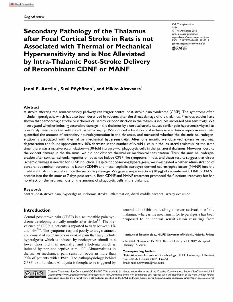
Original Article
Secondary Pathology of the Thalamusafter Focal Cortical Stroke in Rats is notAssociated with Thermal or MechanicalHypersensitivity and is Not Alleviatedby Intra-Thalamic Post-Stroke Deliveryof Recombinant CDNF or MANF
Jenni E. Anttila1, Suvi Poyhonen1, and Mikko Airavaara1
AbstractA stroke affecting the somatosensory pathway can trigger central post-stroke pain syndrome (CPSP). The symptoms ofteninclude hyperalgesia, which has also been described in rodents after the direct damage of the thalamus. Previous studies haveshown that hemorrhagic stroke or ischemia caused by vasoconstriction in the thalamus induces increased pain sensitivity. Weinvestigated whether inducing secondary damage in the thalamus by a cortical stroke causes similar pain hypersensitivity as haspreviously been reported with direct ischemic injury. We induced a focal cortical ischemia-reperfusion injury in male rats,quantified the amount of secondary neurodegeneration in the thalamus, and measured whether the thalamic neurodegen-eration is associated with thermal or mechanical hypersensitivity. After one month, we observed extensive neuronaldegeneration and found approximately 40% decrease in the number of NeuNþ cells in the ipsilateral thalamus. At the sametime, there was a massive accumulation—a 30-fold increase—of phagocytic cells in the ipsilateral thalamus. However, despitethe evident damage in the thalamus, we did not observe thermal or mechanical sensitization. Thus, thalamic neurodegen-eration after cortical ischemia-reperfusion does not induce CPSP-like symptoms in rats, and these results suggest that directischemic damage is needed for CPSP induction. Despite not observing hyperalgesia, we investigated whether administration ofcerebral dopamine neurotrophic factor (CDNF) and mesencephalic astrocyte-derived neurotrophic factor (MANF) into theipsilateral thalamus would reduce the secondary damage. We gave a single injection (10 mg) of recombinant CDNF or MANFprotein into the thalamus at 7 days post-stroke. Both CDNF and MANF treatment promoted the functional recovery but hadno effect on the neuronal loss or the amount of phagocytic cells in the thalamus.
Keywordscentral post-stroke pain, hyperalgesia, ischemic stroke, inflammation, distal middle cerebral artery occlusion
Introduction
Central post-stroke pain (CPSP) is a neuropathic pain syn-
drome developing typically months after stroke1,2. The pre-
valence of CPSP in patients is reported to vary between 1%and 14%2–4. The symptoms respond poorly to drug treatment
and consist of spontaneous or evoked pain that may include
hyperalgesia which is induced by nociceptive stimuli at a
lower threshold than normally, and allodynia which is
induced by non-nociceptive stimuli3,4. Abnormalities in
thermal or mechanical pain sensation occur in more than
90% of patients with CPSP3. The pathophysiology behind
CPSP is still unclear. Allodynia is thought to be triggered by
central disinhibition leading to over-activation of the
thalamus, whereas the mechanism for hyperalgesia has been
proposed to be central sensitization resulting from
1 Institute of Biotechnology, HiLIFE, University of Helsinki, Helsinki, Finland
Submitted: November 15, 2018. Revised: February 13, 2019. Accepted:
February 14, 2019.
Corresponding Author:
Mikko Airavaara, Institute of Biotechnology, HiLIFE, University of Helsinki,
P.O. Box 56, Helsinki 00014, Finland.
Email: [email protected]
Cell Transplantation1–14ª The Author(s) 2019Article reuse guidelines:sagepub.com/journals-permissionsDOI: 10.1177/0963689719837915journals.sagepub.com/home/cll
Creative Commons Non Commercial CC BY-NC: This article is distributed under the terms of the Creative Commons Attribution-NonCommercial 4.0License (http://www.creativecommons.org/licenses/by-nc/4.0/) which permits non-commercial use, reproduction and distribution of the work without furtherpermission provided the original work is attributed as specified on the SAGE and Open Access pages (https://us.sagepub.com/en-us/nam/open-access-at-sage).
deafferentation of neurons4. Patients suffering from thalamic
infarcts at the ventral posterolateral thalamic nucleus (VPL)4
or the ventral posterior nucleus-pulvinar (in rodents: poster-
ior thalamic nuclear group; Po) border zone have a higher
risk for developing CPSP5. However, non-thalamic infarcts
anywhere in the somatosensory pathways, including the cor-
tex, can also induce CPSP3,4.
In rodents, the development of CPSP has been shown to
be region specific after hemorrhagic lesion, and to be asso-
ciated with a lesion of the VPL/ventral posteromedial thala-
mic nucleus (VPM)-Po system6. Several studies using
hemorrhagic lesions of VPL/VPM have found mechanical
and thermal hyperalgesia starting 7 days post-hemorrhage
and lasting at least several weeks7–9. In some studies, only
mechanical hyperalgesia was found after VPL/VPM hemor-
rhage6,10. Similarly to rodents, primates develop mechanical
and thermal hyperalgesia after a hemorrhagic stroke of the
VPL11. In addition, in rodent ischemic stroke model, sensi-
tization to mechanical12 and electrical12,13 stimuli has been
shown to occur 3 days after intraluminal middle cerebral
artery occlusion and to persist at least 2 weeks. Furthermore,
focal thalamic ischemia caused by endothelin-1 injection is
known to induce local ischemia and neuronal loss in the
thalamus and sensitization to thermal, but not mechanical,
stimuli 4 weeks after the endothelin-1 injection14. In all of
these studies, the lesion extended to the thalamus causing
direct ischemic or hemorrhagic damage in the thalamus.
A cortical infarct induces delayed neuronal loss in the
ipsilateral thalamus as a secondary effect due to connecting
thalamocortical and corticothalamic pathways15,16. This
phenomenon, called “exo-focal post-ischemic neuronal
death,” was originally described by Nagasawa and Kogure
in 199016. Secondary atrophy of the thalamus after a middle
cerebral artery infarct has been observed in patients as
well17–20. Moreover, microglia are activated in the thalamus
after cortical stroke in rodents15 and humans21,22. Microglial
activation seems to play a role in the development of hyper-
algesia and neuropathic pain, since treatment with microglial
inhibitor minocycline has been shown to reduce hyperalgesia
in a hemorrhagic CPSP model23, and microglial activation in
the spinal cord has been shown to play a role in the mainte-
nance of chronic pain that develops after spinal cord
injury24–26. Also, transient activation of VPL microglia with
a local chemokine injection induces mechanical and thermal
hypersensitivity27. In patients, spinal cord injury can lead to
delayed loss of connected neurons in the thalamus and sec-
ondary somatosensory cortex, but the severity of neurode-
generation does not correlate with neuropathic pain, and
sometimes more severe disruption of the somatosensory
pathway can lead to numbness and loss of pain sensation
rather than hypersensitivity28. Given the many uncertainties
in the cause of neuropathic pain after injury in the somato-
sensory pathway, and the intriguing findings on the role of
microglial activation in pain sensitization, we wanted to fur-
ther extend the studies to cortical ischemia. Thus, our study
aimed to investigate whether pure cortical ischemia caused
by distal middle cerebral artery occlusion (dMCAo) and the
following secondary thalamic neurodegeneration and micro-
glial activation is associated with thermal or mechanical
hypersensitivity. We are the first to study pain sensitization
in the dMCAo ischemia-reperfusion model, and show here
extensive thalamic neurodegeneration and inflammation
which is not related to CPSP-like symptoms. These data
indicate that neither neuronal loss nor microglial activation
per se cause CPSP, and may thus bring more insight into the
complex mechanism of CPSP development.
As an attempt to reduce the secondary damage, we tar-
geted proteins with known in vivo neuroprotective effects to
the thalamus where the delayed secondary damage occurs.
Since the secondary damage occurs in a delayed manner, it
would be an attractive target from a clinical point of view as
the time window for acute neuroprotective treatment is
extremely limited. Cerebral dopamine neurotrophic factor
(CDNF) and mesencephalic astrocyte-derived neurotrophic
factor (MANF) are endoplasmic reticulum (ER) resident
proteins and form a family of proteins that differs structurally
and functionally from classical neurotrophic factors29–33. The
mechanism of action of CDNF and MANF is still unclear, but
MANF has been shown to be important factor for ER home-
ostasis34,35 and to protect from ER stress-induced cell death in
vitro36. MANF and CDNF are neuroprotective in vivo in
ischemic cerebral injury37–40 and protect mouse primary
cultures from oxygen glucose deprivation in vitro39,41.
Both CDNF and MANF protect dopaminergic neurons and
restore dopaminergic neurocircuitry in Parkinson’s
disease toxin model42,43. CDNF has been shown to
decrease microglial activation in the substantia nigra of
6-hydroxydopamine treated rats44. Also, MANF has been
shown to have anti-inflammatory effects by downregulat-
ing cytokine expression45 and the NF-kB pathway46,47, and
to mediate tissue repair by increasing the alternative M2-
like activation of innate immune cells after retinal damage
in Drosophila and mouse48. We have shown before that
when MANF is targeted into the peri-infarct area with an
adeno-associated viral (AAV) vector 2 days after dMCAo,
the number of phagocytic cells is transiently increased at 4
days post-stroke and the functional recovery of the rats is
hastened49. Overall, there is rather strong evidence that
MANF, and possibly CDNF, can modulate inflammation
and reduce neuronal cell death. Thus, the aim was to test
if post-stroke delivery of recombinant human CDNF
(rhCDNF) or MANF (rhMANF) directly into the thalamus
is able to alleviate the secondary pathology and promote
behavioral recovery after dMCAo.
Materials and Methods
Animals
A total of 59 male Sprague Dawley rats (age 8–9 weeks,
weight 250–300 g, Envigo, Netherlands) were used for the
experiments. Rats were housed in groups of 4 animals in
2 Cell Transplantation
individually ventilated cages with ad libitum access to food
and water under a 12h/12h dark–light cycle. The wellbeing
of the animals was monitored daily. All behavioral experi-
ments were performed during the light phase and the animals
in different experimental groups were assessed in a random
order by a blinded investigator. All animal experiments were
conducted according to the 3 R principles of EU directive
2010/63/EU on the care and use of experimental animals,
local laws and regulations, and were approved by the
national Animal Experiment Board of Finland (protocol
approval number ESAVI/7812/04.10.07/2015). The sample
size was calculated based on a pilot experiment on Har-
greaves’ test, with 0.05 significance level, 0.95 power, and
the calculated Cohen’s d (2.21) as the effect size. All beha-
vioral experiments and analyses were performed in a blinded
manner, and the results are reported according to the
ARRIVE guidelines. One animal from the stroke group was
excluded because it did not have observable lesion. One
animal died due to anesthesia during dMCAo surgery and
one animal from the sham group died from an unknown
reason 3 days after the sham operation. There was no further
mortality.
Distal Middle Cerebral Artery Occlusion Model
To model cortical ischemia-reperfusion injury, a dMCAo
was performed as described before37,38,50. Briefly, the rats
were anesthetized with an intraperitoneal injection of 4%chloral hydrate (0.4 g/kg; Sigma Aldrich, St. Louis, MO,
USA). Both common carotid arteries (CCAs) were isolated
through a cervical incicion, and a craniotomy was made on
the right hemisphere to expose the middle cerebral artery
(MCA). The distal branch of the right MCA was ligated with
10-0 suture and the CCAs were occluded with non-traumatic
arterial clips. After 90 min the suture and clips were removed
to allow reperfusion. Sham-operated rats went through all
the same procedures as stroke rats but the arteries were not
occluded. Lidocaine (10 mg/ml, Orion Pharma, Espoo, Fin-
land) was used locally on the skin during surgery and a single
dose of carprofen (5 mg/kg s.c.; Rimadyl, Zoetis, Louvain-
la-Neuve, Belgium) was given after the surgery for post-
operative pain. Saline (4–5 ml s.c.) was given after the
surgery to prevent dehydration. Body temperature of the
rats was maintained at 37�C until the animals had recovered
from anesthesia and were returned to their home cages.
Although chloral hydrate anesthesia is still used in stroke
research51–56, the use has been criticized for ethical and
safety reasons57. Therefore, we would like to point out that
chloral hydrate is not an optimal anesthetic for surgical pro-
cedures in rodents and the anesthesia protocol should be
further refined.
Neurological Tests
Body asymmetry test58 and modified Bederson’s score59
were used to monitor the level of neurological deficits
caused by ischemia37,38. The body asymmetry was analyzed
from 20 consecutive trials by lifting the rats above the testing
table by the tails and counting the frequency of initial turn-
ings of the head or upper body contralateral to the ischemic
side (the maximum impairment in stroke animals is 20 con-
tralateral turns whereas naıve animals turn in each direction
with equal frequency resulting in 10 contralateral turns). In
modified Bederson’s score the neurological deficits were
scored according to the following criteria: 0¼ no observable
deficit; 1 point ¼ rats show decreased resistance to lateral
push; 2 points ¼ rats keep the contralateral forelimb to the
breast and extend the other forelimb straight when lifted by
the tail in addition to behavior in score 1; 3 points ¼ rats
twist the upper half of their body toward the contralateral
side when lifted by the tail in addition to behavior in other
scores.
Hargreaves’ Test
Hargreaves’ test was used to measure thermal hyperalgesia
from all the paws. The rats were placed into Plexiglas boxes
on Plexiglas floor, and were allowed to habituate for 10–15
min until they ceased exploratory behavior. An infrared
beam (intensity 70 mW/cm2) was positioned beneath the
plantar surface of each paw using an automated device with
a switch-off function as a response to moving the paw (Plan-
tar test 37370, Ugo Basile, Gemonio, Italy). The cut-off time
was set to 30 s. Each measurement was repeated three times
with several minutes in between trials. A rapid withdrawal of
the paw followed by shaking and/or licking the paw was
interpreted as a reaction to pain. An average of three trials
was used for the data analysis. We included an additional
group of naıve rats in the experiments to control for the
effect of repeated testing, and the animals were assigned to
different groups (naıve, sham, stroke) based on the basal
latency to withdraw paw in Hargreaves’ test to form equiv-
alent groups.
Mechanical Stimulation with Dynamic PlantarAesthesiometer
Dynamic Plantar Aesthesiometer was used to measure sen-
sitivity toward light mechanical touch from the left (contral-
ateral) hindpaw. The rats were placed into Plexiglas boxes
on a metal mesh surface and were allowed to habituate for
10–15 min until they ceased exploratory behavior. A stain-
less steel probe (diameter 0.5 mm) attached to a touch sti-
mulator (Dynamic Plantar Aesthesiometer 37450, Ugo
Basile) was lifted toward the plantar surface of the left hind-
paw from below. The touch stimulator had an automated
switch-off function as a response to movement of the paw.
A rapid withdrawal of the paw was interpreted as a reaction
to the stimulus. Maximum force exerted was set to 50 g in 20
s. As in Hargreaves’ test, each measurement was repeated
three times with several minutes in between trials.
Anttila et al 3
Intracranial Administration of rhCDNF and rhMANF
The rats were balanced into groups based on the body swing
and Bederson’s score on day 4 post-stroke. We chose to give
the protein injection on day 7 post-stroke since there are not
yet many phagocytic cells in the thalamus at that time point60.
The stereotaxic surgery was performed under isoflurane
anesthesia (4.5% during induction, 2.5% during mainte-
nance). After placing the animal in a stereotaxic frame (Stoelt-
ing, Wood Dale, IL, USA), the skull was exposed and a small
hole was made with a dental drill. Using coordinates accord-
ing to The Rat Brain in Stereotaxic Coordinates61, 4 ml of
vehicle (phosphate buffered saline; PBS), rhCDNF (2.5 mg/
ml) or rhMANF (2.5 mg/ml) was injected into the right thala-
mus (A/P –3.0; M/L –3.0; D/V –6.0) at speed 0.5 ml/min with
33G blunt needle (Nanofil; World Precision Instruments, Sar-
asota, FL, USA). The needle was kept in place for 4 min after
the injection to prevent backflow. RhCDNF and rhMANF
(Icosagen, Tartu, Estonia) were produced in a Chinese ham-
ster ovarian (CHO)-based cell line.
The distribution of rhCDNF in the brain after intra-
thalamic injection was tested in naıve rats by injecting
rhCDNF as described above, and sacrificing the animals
2 h after the stereotaxic injection for immunohistochemistry
with anti-hCDNF antibody.
Immunohistochemistry
The rats were anesthetized with a lethal dose of pentobarbital
(90 mg/kg i.p.; Mebunat, Orion Pharma) and transcardially
perfused with 200 ml of PBS followed by 500 ml of 4%paraformaldehyde (Sigma Aldrich) in PBS. The brains were
post-fixed in 4% paraformaldehyde for at least 2 days before
they were dehydrated in a series of ethanol and xylene, and
embedded in paraffin. Brains were cut into 5 mm thick coronal
sections using a Leica HM355 S microtome and mounted on
Labsolute microscope slides (Th. Geyer, Renningen, Ger-
many). Sections were deparaffinized in xylene, rehydrated
in a series of ethanol, and heated in 0.05% citraconic anhy-
dride (Sigma Aldrich), pH 7.4, for antigen retrieval. Endogen-
ous peroxidase activity was blocked with 0.6% hydrogen
peroxide (Sigma Aldrich). Non-specific antibody binding was
blocked with 1.5% normal horse or goat serum (Vector
Laboratories, Burlingame, CA, USA), followed by incubation
with primary antibody (mouse anti-CD68 1:500,
cat#MCA341 R, AbD Serotec, Kidlington, UK; mouse anti-
NeuN 1:200, cat#MAB377, Millipore, Billerica, MA, USA;
rabbit anti-MBP 1:500, cat#40390, Abcam, Cambridge, UK;
rabbit anti-hCDNF 1:500, DDV1, a gift from Dr. Johan Per-
anen, University of Helsinki, Finland) at 4�C overnight. The
next day, sections were incubated with horse anti-mouse bio-
tinylated secondary antibody (1:200, Vector Laboratories),
followed by incubation with avidin-biotin complex (ABC kit,
Vector Laboratories). Color was developed using peroxidase
reaction with 3’,3’-diaminobenzidine (Vector Laboratories).
Anti-CD68 immunostained sections were additionally stained
with cresyl violet to visualize the whole section for cell count-
ing. For immunofluorescence staining, goat anti-rabbit Alexa
Fluor 488 (1:500, cat#A11034, Invitrogen, Carlsbad, CA,
USA) and goat anti-mouse Alexa Fluor 568 (1:500,
cat#A11004, Invitrogen) were used as secondary antibodies.
Immunofluorescence was imaged with a Leica TCS SP5 MP
confocal microscope.
Image Analysis
The stained slides were scanned with a 3DHISTECH Pan-
noramic 250 FLASH II digital slide scanner (Budapest, Hun-
gary; scanning service provided by the Institute of
Biotechnology, University of Helsinki; https://www.hel
sinki.fi/en/infrastructures/histotechnology-and-laboratory-
animal-pathology/bi-histoscanner) and images of stained
sections were taken with Pannoramic Viewer version
1.15.3 using 10� magnification. The thalamus was outlined
according to The Rat Brain in Stereotaxic Coordinates61 and
the number of NeuNþ and CD68þ cells in the thalamus was
quantified with Image-Pro Analyzer version 7.0. The cells
were counted from a minimum of three coronal sections per
brain between –2.9 and –3.9 mm relative to bregma. The
average was counted for each animal and was used for fur-
ther analysis. The number of NeuNþ cells in the ipsilateral
thalamus was expressed as a ratio of the number of NeuNþcells in the contralateral thalamus of each section to decrease
variation due to differences in the background staining. The
neuronal loss in the thalamus, outlined according to The Rat
Brain in Stereotaxic Coordinates61, on day 7 post-stroke was
quantified from three sagittal sections per hemisphere (every
0.5 mm between 2.4 and 3.4 mm, and –2.4 and –3.4 mm
relative to bregma) stained with cresyl violet by manually
counting the Nisslþ neurons. The neuronal counts were
expressed as a ratio of the contralateral thalamus. The aver-
age infarction size was quantified from a minimum of four
anti-NeuN stained sections of the caudal brain, between –2.3
and –4.4 relative to bregma. The area devoid of NeuNþ cells
was delineated in Pannoramic Viewer version 1.15.3 and the
infarction size was expressed as a percentage of the whole
section. In the CDNF/MANF experiment, the infarct size
was similarly quantified from four sections stained with cre-
syl violet between 1.2 and –3.6 relative to bregma.
Statistical Analysis
All the statistical analysis was done with IBM SPSS Statis-
tics version 24 and all values are reported as mean + stan-
dard deviation (SD). Hargreaves’ test, Dynamic Plantar
Aesthesiometer and body weight were analyzed with two-
way repeated measures ANOVA followed by Bonferroni’s
post hoc test. Body asymmetry and Bederson’s score were
analyzed with Kruskall–Wallis test and pairwise Mann–
Whitney U test using the exact p-value. Immunohistological
data were analyzed by one-way ANOVA followed by Bon-
ferroni’s post hoc test. The unit of analysis was a single
4 Cell Transplantation
animal in all analyses. Statistical significance was consid-
ered at p < 0.05.
Results
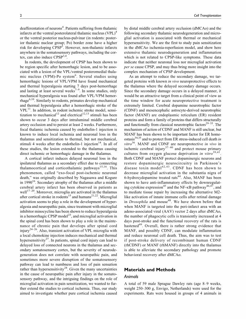
Neurodegeneration and Microglial Activation in theIpsilateral Thalamus at Day 28 Post-Stroke
We characterized the secondary thalamic neurodegeneration
by immunostaining with anti-NeuN (a marker for neurons)
and anti-CD68 (a marker for activated, phagocytic micro-
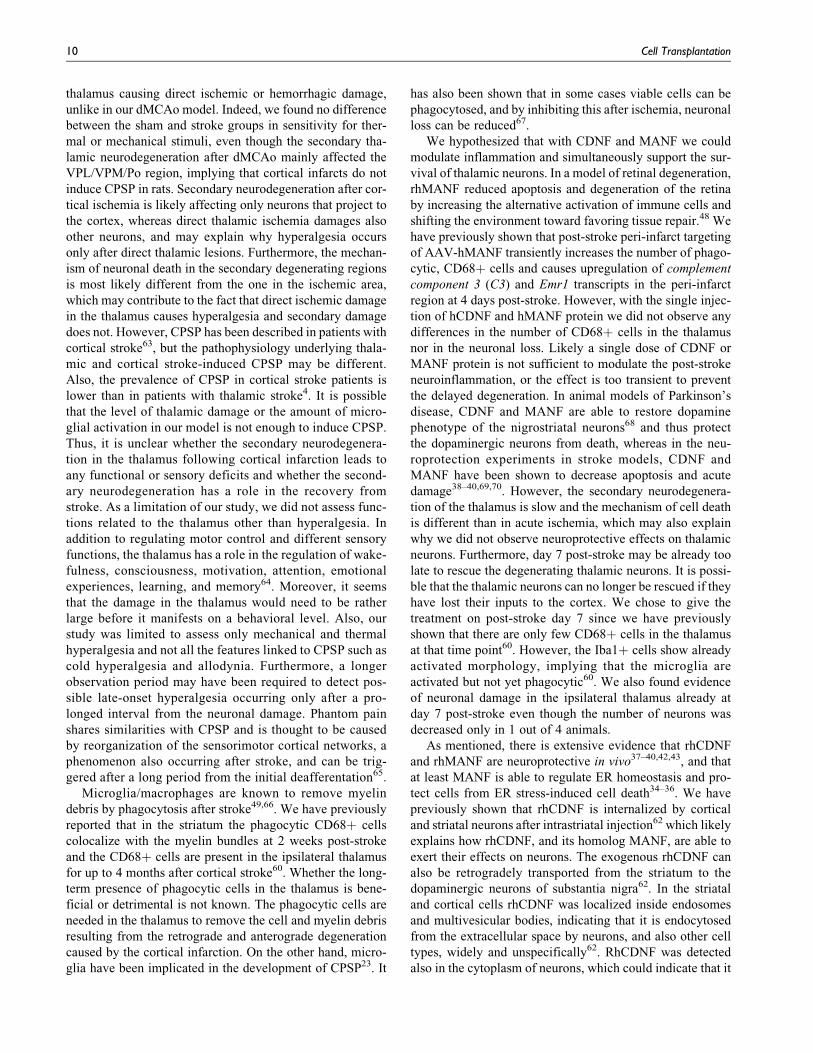
glia/macrophages) antibodies (Fig. 1a). At 28 days post-
stroke, 38% of the neurons in the ipsilateral thalamus were
lost (Fig. 1b; d–i). The amount of NeuNþ cells in the ipsi-
lateral thalamus was 98%, 100%, and 62% of the amount in
the contralateral thalamus in the naıve, sham, and stroke
groups, respectively. The stroke rats had significantly fewer
neurons in the ipsilateral thalamus (F2,21 ¼ 26.11, p <
0.0001, one-way ANOVA) than the rats in naıve (p <
Fig. 1. Delayed neuronal loss and phagocytosis occurs in the ipsilateral thalamus after cortical ischemia-reperfusion injury. (a) Experimentaltimeline. D ¼ indicated post-stroke day; B ¼ behavioral experiment; dMCAo ¼ distal middle cerebral artery occlusion; IHC ¼ immuno-histochemistry. (b) The ratio of NeuNþ cells in the ipsilateral thalamus compared with the contralateral thalamus in naıve rats and 28 daysafter cortical stroke or sham operation. (c) The number of phagocytic CD68þ cells in the ipsilateral thalamus in naıve rats and 28 days aftercortical stroke or sham operation. Representative images of anti-NeuN (d–i) and anti-CD68 (j–o) immunostained brain sections from naıve(d, g, j, m), sham (e, h, k, n), and stroke (f, i, l, o) groups. The delineated area in d–f; j–l indicates the area analyzed. Scale bar is 500 mm in lowmagnification images and 100 mm in high magnification images. Naıve n¼ 6, sham n¼ 9, stroke n¼ 9. ****(p < 0.0001) indicates comparisonwith the sham group, ####(p < 0.0001) indicates comparison with the naıve group. (p) Pearson correlation with 95% confidence intervals ofneuronal loss (b) and the number of phagocytic cells (c) in the thalamus 28 days post-stroke. (q) The average infarct size in the caudal brain(between –2.3 and –4.4 relative to bregma) at 28 days post-stroke expressed as a percentage of the whole section. (r) Representativeimage of anti-NeuN stained section showing the NeuN negative infarct area on the cortex. Scale bar is 2000 mm. All values are reported asmean + SD.
Anttila et al 5
0.0001) and sham (p < 0.0001) groups. There was no differ-
ence between the naıve and sham groups, and sham opera-
tion did not cause any detectable damage to the brain. Also,
the stroke rats had significantly more CD68þ cells in the
ipsilateral thalamus (640 cells/mm2; F2,21 ¼ 22.57, p <
0.0001, one-way ANOVA) when compared with the naıve
(20 cells/mm2; p < 0.0001) and sham (16 cells/mm2; p <
0.0001) groups (Fig. 1c; j–o). There was a negative correla-
tion between the number of CD68þ cells and NeuNþ cells
in the ipsilateral thalamus at 28 days post-stroke (Pearson
correlation R ¼ –0.698, p ¼ 0.036; Fig. 1p). The average
infarct size in the caudal brain was 3.9% of the brain section
and the infarct was restricted to the cortex in all animals (Fig.
1q–r).
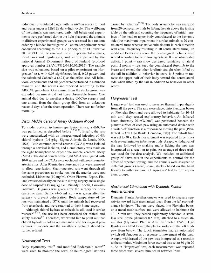
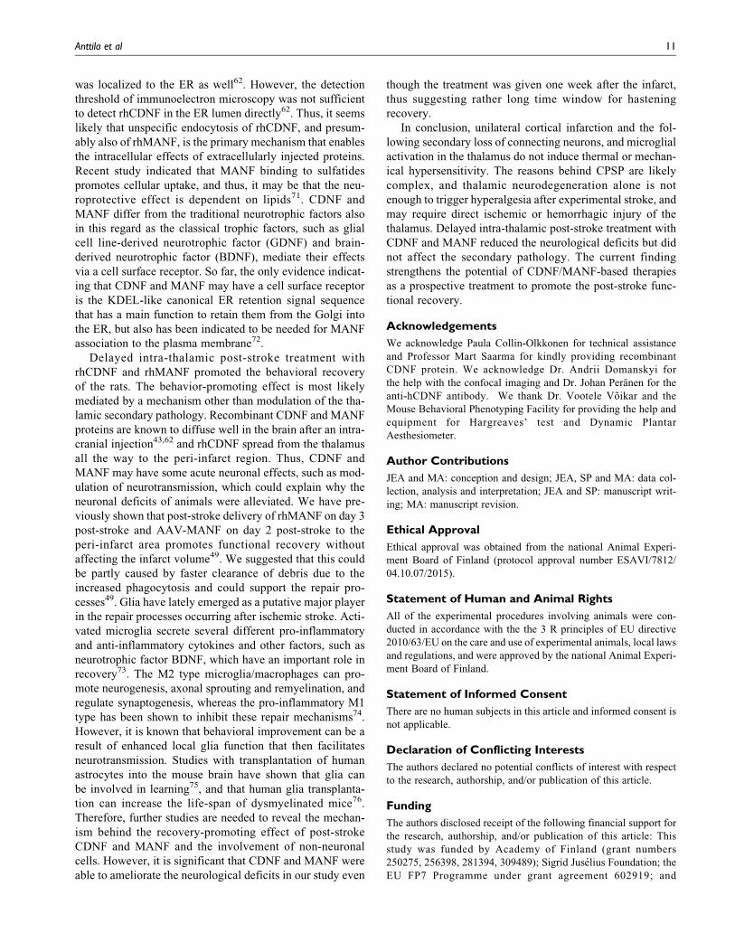
Next, we clarified the location of phagocytic cells in rela-
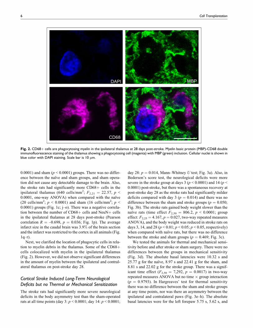
tion to myelin debris in the thalamus. Some of the CD68þcells colocalized with myelin in the ipsilateral thalamus
(Fig. 2). However, we did not observe significant differences
in the amount of myelin between the ipsilateral and contral-
ateral thalamus on post-stroke day 28.
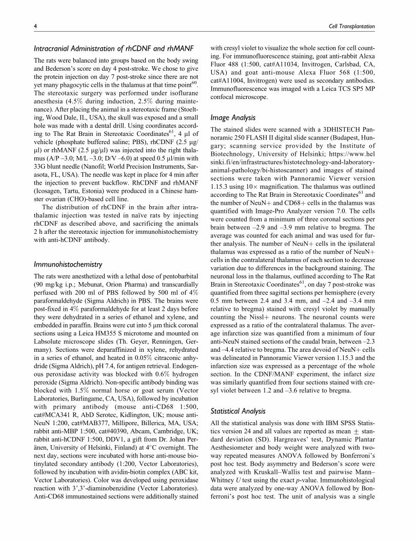
Cortical Stroke Induced Long-Term NeurologicalDeficits but no Thermal or Mechanical Sensitization
The stroke rats had significantly more severe neurological
deficits in the body asymmetry test than the sham-operated
rats at all time points (day 3: p < 0.0001; day 14: p < 0.0001;
day 28: p ¼ 0.014, Mann–Whitney U test; Fig. 3a). Also, in
Bederson’s score test, the neurological deficits were more
severe in the stroke group at days 3 (p < 0.0001) and 14 (p <
0.0001) post-stroke, but there was a spontaneous recovery at
post-stroke day 28 as the stroke rats had significantly milder
deficits compared with day 3 (p ¼ 0.014) and there was no
difference between the sham and stroke groups (p ¼ 0.050;
Fig. 3b). The stroke rats gained body weight slower than the
naıve rats (time effect F2,50 ¼ 806.2, p < 0.0001; group
effect: F2,25¼ 4.167, p¼ 0.027, two-way repeated measures
ANOVA), and the body weight was reduced in stroke rats on
days 3, 14, and 28 (p < 0.01; p < 0.05; p < 0.05, respectively)
when compared with naıve rats, but there was no difference
between the stroke and sham groups (p ¼ 0.469; Fig. 3c).
We tested the animals for thermal and mechanical sensi-
tivity before and after stroke or sham surgery. There were no
differences between the groups in mechanical sensitivity
(Fig. 3d). The absolute basal latencies were 10.32 s and
25.77 g for the naıve, 8.97 s and 22.41 g for the sham, and
8.81 s and 22.02 g for the stroke group. There was a signif-
icant time effect (F2,50 ¼ 7,292, p ¼ 0.0017) in two-way
repeated measures ANOVA but no time � group interaction
(p ¼ 0.9793). In Hargreaves’ test for thermal sensitivity
there was no difference between the sham and stroke groups
at any time points, nor was there an asymmetry between the
ipsilateral and contralateral paws (Fig. 3e–h). The absolute
basal latencies were for the left forepaw 5.75 s, 5.62 s, and
Fig. 2. CD68þ cells are phagocytosing myelin in the ipsilateral thalamus at 28 days post-stroke. Myelin basic protein (MBP)-CD68 doubleimmunofluorescence staining of the thalamus showing a phagocytosing cell (magenta) with MBP (green) inclusion. Cellular nuclei is shown inblue color with DAPI staining. Scale bar is 10 mm.
6 Cell Transplantation
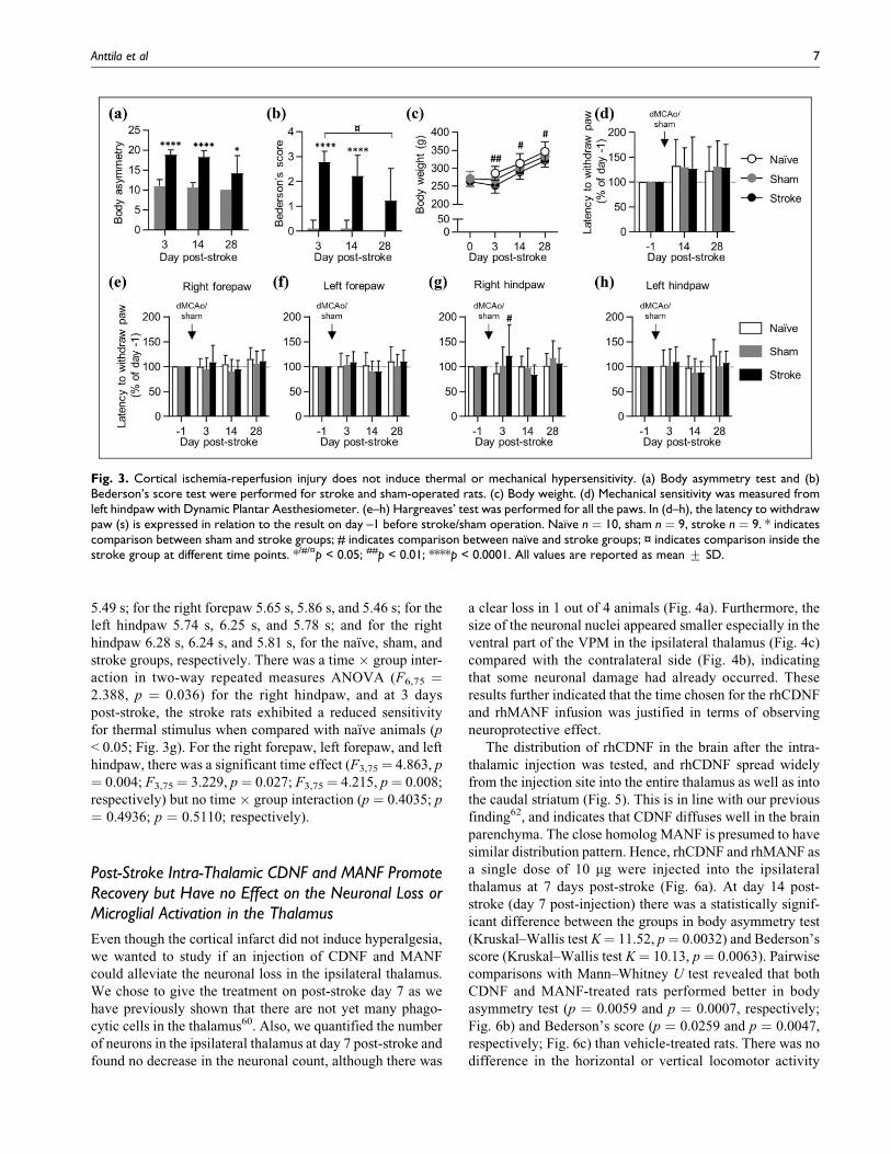
5.49 s; for the right forepaw 5.65 s, 5.86 s, and 5.46 s; for the
left hindpaw 5.74 s, 6.25 s, and 5.78 s; and for the right
hindpaw 6.28 s, 6.24 s, and 5.81 s, for the naıve, sham, and
stroke groups, respectively. There was a time � group inter-
action in two-way repeated measures ANOVA (F6,75 ¼2.388, p ¼ 0.036) for the right hindpaw, and at 3 days
post-stroke, the stroke rats exhibited a reduced sensitivity
for thermal stimulus when compared with naıve animals (p
< 0.05; Fig. 3g). For the right forepaw, left forepaw, and left
hindpaw, there was a significant time effect (F3,75¼ 4.863, p
¼ 0.004; F3,75¼ 3.229, p¼ 0.027; F3,75¼ 4.215, p¼ 0.008;
respectively) but no time � group interaction (p ¼ 0.4035; p
¼ 0.4936; p ¼ 0.5110; respectively).
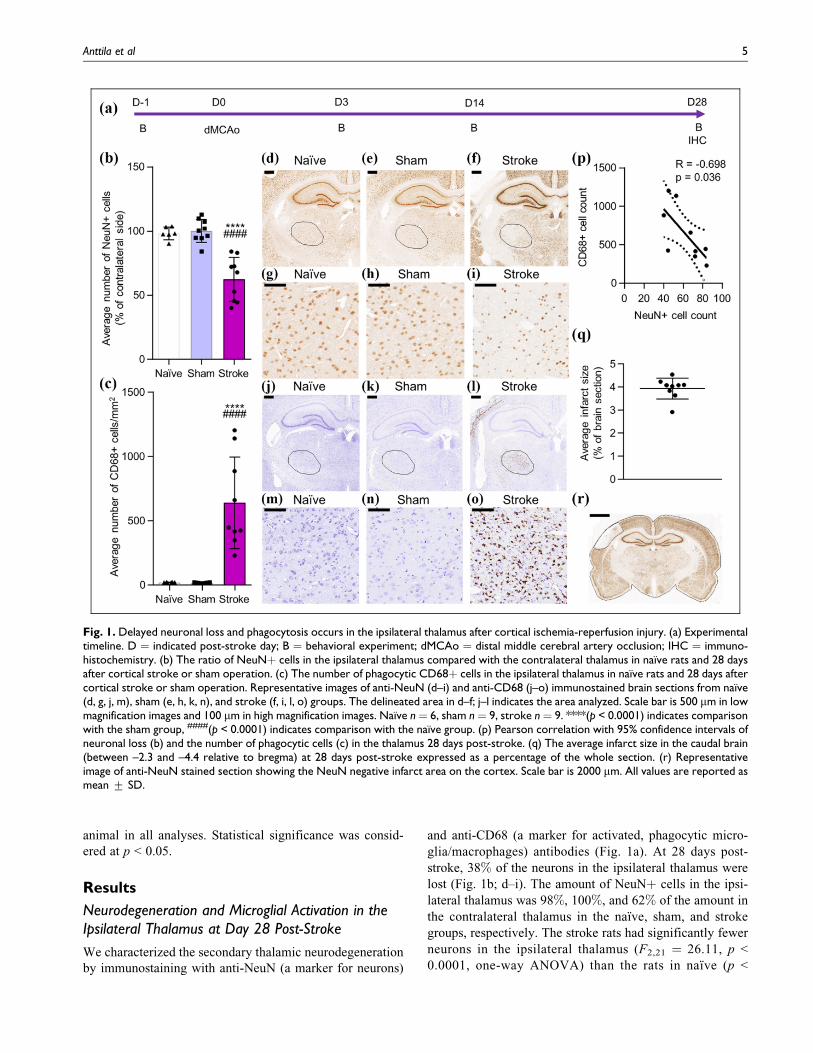
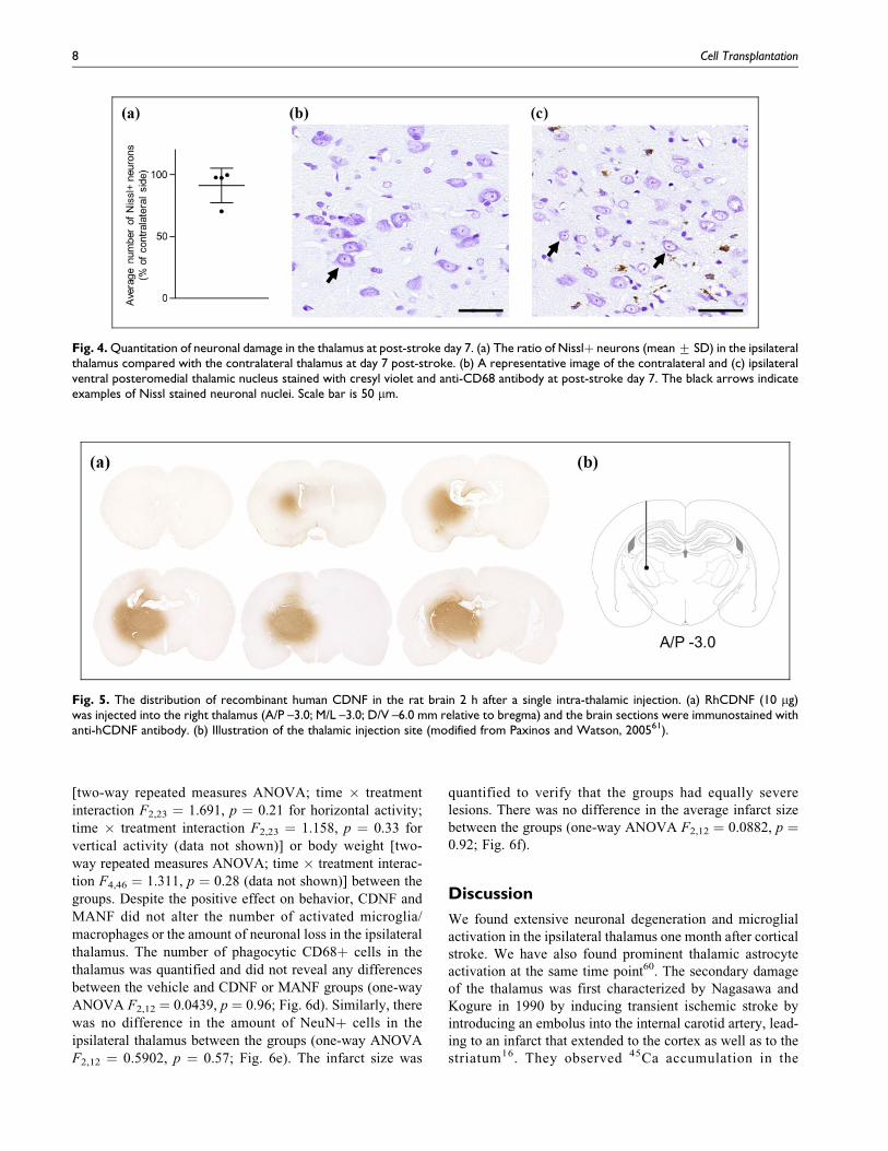
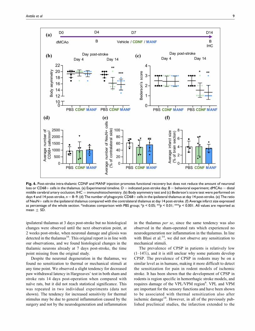
Post-Stroke Intra-Thalamic CDNF and MANF PromoteRecovery but Have no Effect on the Neuronal Loss orMicroglial Activation in the Thalamus
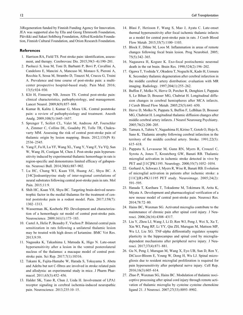
Even though the cortical infarct did not induce hyperalgesia,
we wanted to study if an injection of CDNF and MANF
could alleviate the neuronal loss in the ipsilateral thalamus.
We chose to give the treatment on post-stroke day 7 as we
have previously shown that there are not yet many phago-
cytic cells in the thalamus60. Also, we quantified the number
of neurons in the ipsilateral thalamus at day 7 post-stroke and
found no decrease in the neuronal count, although there was
a clear loss in 1 out of 4 animals (Fig. 4a). Furthermore, the
size of the neuronal nuclei appeared smaller especially in the
ventral part of the VPM in the ipsilateral thalamus (Fig. 4c)
compared with the contralateral side (Fig. 4b), indicating
that some neuronal damage had already occurred. These
results further indicated that the time chosen for the rhCDNF
and rhMANF infusion was justified in terms of observing
neuroprotective effect.
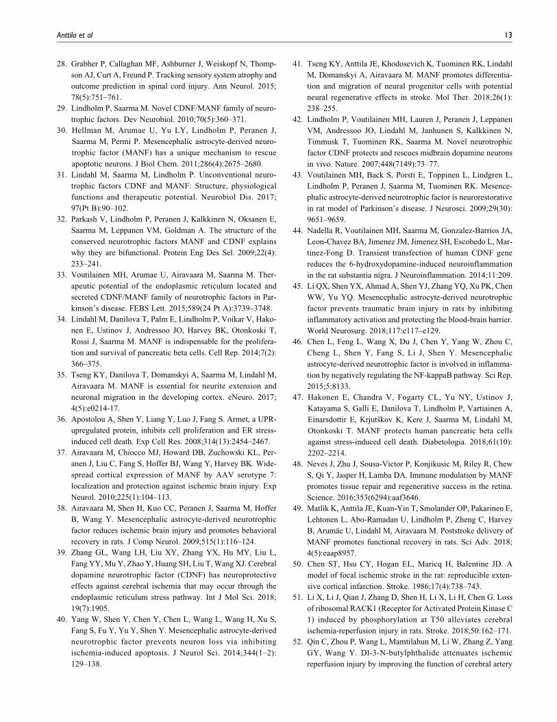
The distribution of rhCDNF in the brain after the intra-
thalamic injection was tested, and rhCDNF spread widely
from the injection site into the entire thalamus as well as into
the caudal striatum (Fig. 5). This is in line with our previous
finding62, and indicates that CDNF diffuses well in the brain
parenchyma. The close homolog MANF is presumed to have
similar distribution pattern. Hence, rhCDNF and rhMANF as
a single dose of 10 mg were injected into the ipsilateral
thalamus at 7 days post-stroke (Fig. 6a). At day 14 post-
stroke (day 7 post-injection) there was a statistically signif-
icant difference between the groups in body asymmetry test
(Kruskal–Wallis test K¼ 11.52, p¼ 0.0032) and Bederson’s
score (Kruskal–Wallis test K¼ 10.13, p¼ 0.0063). Pairwise
comparisons with Mann–Whitney U test revealed that both
CDNF and MANF-treated rats performed better in body
asymmetry test (p ¼ 0.0059 and p ¼ 0.0007, respectively;
Fig. 6b) and Bederson’s score (p ¼ 0.0259 and p ¼ 0.0047,
respectively; Fig. 6c) than vehicle-treated rats. There was no
difference in the horizontal or vertical locomotor activity
Fig. 3. Cortical ischemia-reperfusion injury does not induce thermal or mechanical hypersensitivity. (a) Body asymmetry test and (b)Bederson’s score test were performed for stroke and sham-operated rats. (c) Body weight. (d) Mechanical sensitivity was measured fromleft hindpaw with Dynamic Plantar Aesthesiometer. (e–h) Hargreaves’ test was performed for all the paws. In (d–h), the latency to withdrawpaw (s) is expressed in relation to the result on day –1 before stroke/sham operation. Naıve n ¼ 10, sham n ¼ 9, stroke n ¼ 9. * indicatescomparison between sham and stroke groups; # indicates comparison between naıve and stroke groups; ¤ indicates comparison inside thestroke group at different time points. */#/¤p < 0.05; ##p < 0.01; ****p < 0.0001. All values are reported as mean + SD.
Anttila et al 7
[two-way repeated measures ANOVA; time � treatment
interaction F2,23 ¼ 1.691, p ¼ 0.21 for horizontal activity;
time � treatment interaction F2,23 ¼ 1.158, p ¼ 0.33 for
vertical activity (data not shown)] or body weight [two-
way repeated measures ANOVA; time � treatment interac-
tion F4,46 ¼ 1.311, p ¼ 0.28 (data not shown)] between the
groups. Despite the positive effect on behavior, CDNF and
MANF did not alter the number of activated microglia/
macrophages or the amount of neuronal loss in the ipsilateral
thalamus. The number of phagocytic CD68þ cells in the
thalamus was quantified and did not reveal any differences
between the vehicle and CDNF or MANF groups (one-way
ANOVA F2,12 ¼ 0.0439, p ¼ 0.96; Fig. 6d). Similarly, there
was no difference in the amount of NeuNþ cells in the
ipsilateral thalamus between the groups (one-way ANOVA
F2,12 ¼ 0.5902, p ¼ 0.57; Fig. 6e). The infarct size was
quantified to verify that the groups had equally severe
lesions. There was no difference in the average infarct size
between the groups (one-way ANOVA F2,12 ¼ 0.0882, p ¼0.92; Fig. 6f).
Discussion
We found extensive neuronal degeneration and microglial
activation in the ipsilateral thalamus one month after cortical
stroke. We have also found prominent thalamic astrocyte
activation at the same time point60. The secondary damage
of the thalamus was first characterized by Nagasawa and
Kogure in 1990 by inducing transient ischemic stroke by
introducing an embolus into the internal carotid artery, lead-
ing to an infarct that extended to the cortex as well as to the
striatum16. They observed 45Ca accumulation in the
Fig. 4. Quantitation of neuronal damage in the thalamus at post-stroke day 7. (a) The ratio of Nisslþ neurons (mean + SD) in the ipsilateralthalamus compared with the contralateral thalamus at day 7 post-stroke. (b) A representative image of the contralateral and (c) ipsilateralventral posteromedial thalamic nucleus stained with cresyl violet and anti-CD68 antibody at post-stroke day 7. The black arrows indicateexamples of Nissl stained neuronal nuclei. Scale bar is 50 mm.
Fig. 5. The distribution of recombinant human CDNF in the rat brain 2 h after a single intra-thalamic injection. (a) RhCDNF (10 mg)was injected into the right thalamus (A/P –3.0; M/L –3.0; D/V –6.0 mm relative to bregma) and the brain sections were immunostained withanti-hCDNF antibody. (b) Illustration of the thalamic injection site (modified from Paxinos and Watson, 200561).
8 Cell Transplantation
ipsilateral thalamus at 3 days post-stroke but no histological
changes were observed until the next observation point, at
2 weeks post-stroke, when neuronal damage and gliosis was
detected in the thalamus16. This original report is in line with
our observations, and we found histological changes in the
thalamic neurons already at 7 days post-stroke, the time
point missing from the original study.
Despite the neuronal degeneration in the thalamus, we
found no sensitization to thermal or mechanical stimuli at
any time point. We observed a slight tendency for decreased
paw withdrawal latency in Hargreaves’ test in both sham and
stroke rats 14 days post-operation when compared with
naıve rats, but it did not reach statistical significance. This
was repeated in two individual experiments (data not
shown). The tendency for increased sensitivity for thermal
stimulus may be due to general inflammation caused by the
surgery and not by the neurodegeneration and inflammation
in the thalamus per se, since the same tendency was also
observed in the sham-operated rats which experienced no
neurodegeneration nor inflammation in the thalamus. In line
with Blasi et al.14, we did not observe any sensitization to
mechanical stimuli.
The prevalence of CPSP in patients is relatively low
(1–14%), and it is still unclear why some patients develop
CPSP. The prevalence of CPSP in rodents may be on a
similar level as in humans, making it more difficult to detect
the sensitization for pain in rodent models of ischemic
stroke. It has been shown that the development of CPSP in
rodents is region specific in hemorrhagic stroke models, and
requires damage of the VPL/VPM region6. VPL and VPM
are important for the sensory functions and have been shown
to be associated with thermal sensitization also after
ischemic damage14. However, in all of the previously pub-
lished preclinical studies, the infarction extended to the
Fig. 6. Post-stroke intra-thalamic CDNF and MANF injection promotes functional recovery but does not reduce the amount of neuronalloss or CD68þ cells in the thalamus. (a) Experimental timeline. D¼ indicated post-stroke day; B¼ behavioral experiment; dMCAo¼ distalmiddle cerebral artery occlusion; IHC¼ immunohistochemistry. (b) Body asymmetry test and (c) Bederson’s score test were performed ondays 4 and 14 post-stroke, n¼ 8–9. (d) The number of phagocytic CD68þ cells in the ipsilateral thalamus at day 14 post-stroke. (e) The ratioof NeuNþ cells in the ipsilateral thalamus compared with the contralateral thalamus at day 14 post-stroke. (f) Average infarct size expressedas percentage of the whole section. *indicates comparison with PBS group; *p < 0.05; **p < 0.01; ***p < 0.001. All values are reported asmean + SD.
Anttila et al 9
thalamus causing direct ischemic or hemorrhagic damage,
unlike in our dMCAo model. Indeed, we found no difference
between the sham and stroke groups in sensitivity for ther-
mal or mechanical stimuli, even though the secondary tha-
lamic neurodegeneration after dMCAo mainly affected the
VPL/VPM/Po region, implying that cortical infarcts do not
induce CPSP in rats. Secondary neurodegeneration after cor-
tical ischemia is likely affecting only neurons that project to
the cortex, whereas direct thalamic ischemia damages also
other neurons, and may explain why hyperalgesia occurs
only after direct thalamic lesions. Furthermore, the mechan-
ism of neuronal death in the secondary degenerating regions
is most likely different from the one in the ischemic area,
which may contribute to the fact that direct ischemic damage
in the thalamus causes hyperalgesia and secondary damage
does not. However, CPSP has been described in patients with
cortical stroke63, but the pathophysiology underlying thala-
mic and cortical stroke-induced CPSP may be different.
Also, the prevalence of CPSP in cortical stroke patients is
lower than in patients with thalamic stroke4. It is possible
that the level of thalamic damage or the amount of micro-
glial activation in our model is not enough to induce CPSP.
Thus, it is unclear whether the secondary neurodegenera-
tion in the thalamus following cortical infarction leads to
any functional or sensory deficits and whether the second-
ary neurodegeneration has a role in the recovery from
stroke. As a limitation of our study, we did not assess func-
tions related to the thalamus other than hyperalgesia. In
addition to regulating motor control and different sensory
functions, the thalamus has a role in the regulation of wake-
fulness, consciousness, motivation, attention, emotional
experiences, learning, and memory64. Moreover, it seems
that the damage in the thalamus would need to be rather
large before it manifests on a behavioral level. Also, our
study was limited to assess only mechanical and thermal
hyperalgesia and not all the features linked to CPSP such as
cold hyperalgesia and allodynia. Furthermore, a longer
observation period may have been required to detect pos-
sible late-onset hyperalgesia occurring only after a pro-
longed interval from the neuronal damage. Phantom pain
shares similarities with CPSP and is thought to be caused
by reorganization of the sensorimotor cortical networks, a
phenomenon also occurring after stroke, and can be trig-
gered after a long period from the initial deafferentation65.
Microglia/macrophages are known to remove myelin
debris by phagocytosis after stroke49,66. We have previously
reported that in the striatum the phagocytic CD68þ cells
colocalize with the myelin bundles at 2 weeks post-stroke
and the CD68þ cells are present in the ipsilateral thalamus
for up to 4 months after cortical stroke60. Whether the long-
term presence of phagocytic cells in the thalamus is bene-
ficial or detrimental is not known. The phagocytic cells are
needed in the thalamus to remove the cell and myelin debris
resulting from the retrograde and anterograde degeneration
caused by the cortical infarction. On the other hand, micro-
glia have been implicated in the development of CPSP23. It
has also been shown that in some cases viable cells can be
phagocytosed, and by inhibiting this after ischemia, neuronal
loss can be reduced67.
We hypothesized that with CDNF and MANF we could
modulate inflammation and simultaneously support the sur-
vival of thalamic neurons. In a model of retinal degeneration,
rhMANF reduced apoptosis and degeneration of the retina
by increasing the alternative activation of immune cells and
shifting the environment toward favoring tissue repair.48 We
have previously shown that post-stroke peri-infarct targeting
of AAV-hMANF transiently increases the number of phago-
cytic, CD68þ cells and causes upregulation of complement
component 3 (C3) and Emr1 transcripts in the peri-infarct
region at 4 days post-stroke. However, with the single injec-
tion of hCDNF and hMANF protein we did not observe any
differences in the number of CD68þ cells in the thalamus
nor in the neuronal loss. Likely a single dose of CDNF or
MANF protein is not sufficient to modulate the post-stroke
neuroinflammation, or the effect is too transient to prevent
the delayed degeneration. In animal models of Parkinson’s
disease, CDNF and MANF are able to restore dopamine
phenotype of the nigrostriatal neurons68 and thus protect
the dopaminergic neurons from death, whereas in the neu-
roprotection experiments in stroke models, CDNF and
MANF have been shown to decrease apoptosis and acute
damage38–40,69,70. However, the secondary neurodegenera-
tion of the thalamus is slow and the mechanism of cell death
is different than in acute ischemia, which may also explain
why we did not observe neuroprotective effects on thalamic
neurons. Furthermore, day 7 post-stroke may be already too
late to rescue the degenerating thalamic neurons. It is possi-
ble that the thalamic neurons can no longer be rescued if they
have lost their inputs to the cortex. We chose to give the
treatment on post-stroke day 7 since we have previously
shown that there are only few CD68þ cells in the thalamus
at that time point60. However, the Iba1þ cells show already
activated morphology, implying that the microglia are
activated but not yet phagocytic60. We also found evidence
of neuronal damage in the ipsilateral thalamus already at
day 7 post-stroke even though the number of neurons was
decreased only in 1 out of 4 animals.
As mentioned, there is extensive evidence that rhCDNF
and rhMANF are neuroprotective in vivo37–40,42,43, and that
at least MANF is able to regulate ER homeostasis and pro-
tect cells from ER stress-induced cell death34–36. We have
previously shown that rhCDNF is internalized by cortical
and striatal neurons after intrastriatal injection62 which likely
explains how rhCDNF, and its homolog MANF, are able to
exert their effects on neurons. The exogenous rhCDNF can
also be retrogradely transported from the striatum to the
dopaminergic neurons of substantia nigra62. In the striatal
and cortical cells rhCDNF was localized inside endosomes
and multivesicular bodies, indicating that it is endocytosed
from the extracellular space by neurons, and also other cell
types, widely and unspecifically62. RhCDNF was detected
also in the cytoplasm of neurons, which could indicate that it
10 Cell Transplantation
was localized to the ER as well62. However, the detection
threshold of immunoelectron microscopy was not sufficient
to detect rhCDNF in the ER lumen directly62. Thus, it seems
likely that unspecific endocytosis of rhCDNF, and presum-
ably also of rhMANF, is the primary mechanism that enables
the intracellular effects of extracellularly injected proteins.
Recent study indicated that MANF binding to sulfatides
promotes cellular uptake, and thus, it may be that the neu-
roprotective effect is dependent on lipids71. CDNF and
MANF differ from the traditional neurotrophic factors also
in this regard as the classical trophic factors, such as glial
cell line-derived neurotrophic factor (GDNF) and brain-
derived neurotrophic factor (BDNF), mediate their effects
via a cell surface receptor. So far, the only evidence indicat-
ing that CDNF and MANF may have a cell surface receptor
is the KDEL-like canonical ER retention signal sequence
that has a main function to retain them from the Golgi into
the ER, but also has been indicated to be needed for MANF
association to the plasma membrane72.
Delayed intra-thalamic post-stroke treatment with
rhCDNF and rhMANF promoted the behavioral recovery
of the rats. The behavior-promoting effect is most likely
mediated by a mechanism other than modulation of the tha-
lamic secondary pathology. Recombinant CDNF and MANF
proteins are known to diffuse well in the brain after an intra-
cranial injection43,62 and rhCDNF spread from the thalamus
all the way to the peri-infarct region. Thus, CDNF and
MANF may have some acute neuronal effects, such as mod-
ulation of neurotransmission, which could explain why the
neuronal deficits of animals were alleviated. We have pre-
viously shown that post-stroke delivery of rhMANF on day 3
post-stroke and AAV-MANF on day 2 post-stroke to the
peri-infarct area promotes functional recovery without
affecting the infarct volume49. We suggested that this could
be partly caused by faster clearance of debris due to the
increased phagocytosis and could support the repair pro-
cesses49. Glia have lately emerged as a putative major player
in the repair processes occurring after ischemic stroke. Acti-
vated microglia secrete several different pro-inflammatory
and anti-inflammatory cytokines and other factors, such as
neurotrophic factor BDNF, which have an important role in
recovery73. The M2 type microglia/macrophages can pro-
mote neurogenesis, axonal sprouting and remyelination, and
regulate synaptogenesis, whereas the pro-inflammatory M1
type has been shown to inhibit these repair mechanisms74.
However, it is known that behavioral improvement can be a
result of enhanced local glia function that then facilitates
neurotransmission. Studies with transplantation of human
astrocytes into the mouse brain have shown that glia can
be involved in learning75, and that human glia transplanta-
tion can increase the life-span of dysmyelinated mice76.
Therefore, further studies are needed to reveal the mechan-
ism behind the recovery-promoting effect of post-stroke
CDNF and MANF and the involvement of non-neuronal
cells. However, it is significant that CDNF and MANF were
able to ameliorate the neurological deficits in our study even
though the treatment was given one week after the infarct,
thus suggesting rather long time window for hastening
recovery.
In conclusion, unilateral cortical infarction and the fol-
lowing secondary loss of connecting neurons, and microglial
activation in the thalamus do not induce thermal or mechan-
ical hypersensitivity. The reasons behind CPSP are likely
complex, and thalamic neurodegeneration alone is not
enough to trigger hyperalgesia after experimental stroke, and
may require direct ischemic or hemorrhagic injury of the
thalamus. Delayed intra-thalamic post-stroke treatment with
CDNF and MANF reduced the neurological deficits but did
not affect the secondary pathology. The current finding
strengthens the potential of CDNF/MANF-based therapies
as a prospective treatment to promote the post-stroke func-
tional recovery.
Acknowledgements
We acknowledge Paula Collin-Olkkonen for technical assistance
and Professor Mart Saarma for kindly providing recombinant
CDNF protein. We acknowledge Dr. Andrii Domanskyi for
the help with the confocal imaging and Dr. Johan Peranen for the
anti-hCDNF antibody. We thank Dr. Vootele Voikar and the
Mouse Behavioral Phenotyping Facility for providing the help and
equipment for Hargreaves’ test and Dynamic Plantar
Aesthesiometer.
Author Contributions
JEA and MA: conception and design; JEA, SP and MA: data col-
lection, analysis and interpretation; JEA and SP: manuscript writ-
ing; MA: manuscript revision.
Ethical Approval
Ethical approval was obtained from the national Animal Experi-
ment Board of Finland (protocol approval number ESAVI/7812/
04.10.07/2015).
Statement of Human and Animal Rights
All of the experimental procedures involving animals were con-
ducted in accordance with the the 3 R principles of EU directive
2010/63/EU on the care and use of experimental animals, local laws
and regulations, and were approved by the national Animal Experi-
ment Board of Finland.
Statement of Informed Consent
There are no human subjects in this article and informed consent is
not applicable.
Declaration of Conflicting Interests
The authors declared no potential conflicts of interest with respect
to the research, authorship, and/or publication of this article.
Funding
The authors disclosed receipt of the following financial support for
the research, authorship, and/or publication of this article: This
study was funded by Academy of Finland (grant numbers
250275, 256398, 281394, 309489); Sigrid Juselius Foundation; the
EU FP7 Programme under grant agreement 602919; and
Anttila et al 11
3iRegeneration funded by Finnish Funding Agency for Innovation.
JEA was supported also by Ella and Georg Ehrnrooth Foundation,
Paivikki and Sakari Sohlberg Foundation, Alfred Kordelin Founda-
tion, Finnish Cultural Foundation, and Orion Research Foundation.
References
1. Harrison RA, Field TS. Post stroke pain: identification, assess-
ment, and therapy. Cerebrovasc Dis. 2015;39(3–4):190–201.
2. Paolucci S, Iosa M, Toni D, Barbanti P, Bovi P, Cavallini A,
Candeloro E, Mancini A, Mancuso M, Monaco S, Pieroni A,
Recchia S, Sessa M, Strambo D, Tinazzi M, Cruccu G, Truini
A. Prevalence and time course of post-stroke pain: a multi-
center prospective hospital-based study. Pain Med. 2016;
17(5):924–930.
3. Klit H, Finnerup NB, Jensen TS. Central post-stroke pain:
clinical characteristics, pathophysiology, and management.
Lancet Neurol. 2009;8(9):857–868.
4. Kumar B, Kalita J, Kumar G, Misra UK. Central poststroke
pain: a review of pathophysiology and treatment. Anesth
Analg. 2009;108(5):1645–1657.
5. Sprenger T, Seifert CL, Valet M, Andreou AP, Foerschler
A, Zimmer C, Collins DL, Goadsby PJ, Tolle TR, Chakra-
varty MM. Assessing the risk of central post-stroke pain of
thalamic origin by lesion mapping. Brain. 2012;135(Pt 8):
2536–2545.
6. Yang F, Fu H, Lu YF, Wang XL, Yang Y, Yang F, Yu YQ, Sun
W, Wang JS, Costigan M, Chen J. Post-stroke pain hypersen-
sitivity induced by experimental thalamic hemorrhage in rats is
region-specific and demonstrates limited efficacy of gabapen-
tin. Neurosci Bull. 2014;30(6):887–902.
7. Lu HC, Chang WJ, Kuan YH, Huang AC, Shyu BC. A
[14C]iodoantipyrine study of inter-regional correlations of
neural substrates following central post-stroke pain in rats. Mol
Pain. 2015;11:9.
8. Shih HC, Kuan YH, Shyu BC. Targeting brain-derived neuro-
trophic factor in the medial thalamus for the treatment of cen-
tral poststroke pain in a rodent model. Pain. 2017;158(7):
1302–1313.
9. Wasserman JK, Koeberle PD. Development and characteriza-
tion of a hemorrhagic rat model of central post-stroke pain.
Neuroscience. 2009;161(1):173–183.
10. Castel A, Helie P, Beaudry F, Vachon P. Bilateral central pain
sensitization in rats following a unilateral thalamic lesion
may be treated with high doses of ketamine. BMC Vet Res.
2013;9:59.
11. Nagasaka K, Takashima I, Matsuda K, Higo N. Late-onset
hypersensitivity after a lesion in the ventral posterolateral
nucleus of the thalamus: a macaque model of central post-
stroke pain. Sci Rep. 2017;7(1):10316.
12. Takami K, Fujita-Hamabe W, Harada S, Tokuyama S. Abeta
and Adelta but not C-fibres are involved in stroke related pain
and allodynia: an experimental study in mice. J Pharm Phar-
macol. 2011;63(3):452–456.
13. Halder SK, Yano R, Chun J, Ueda H. Involvement of LPA1
receptor signaling in cerebral ischemia-induced neuropathic
pain. Neuroscience. 2013;235:10–15.
14. Blasi F, Herisson F, Wang S, Mao J, Ayata C. Late-onset
thermal hypersensitivity after focal ischemic thalamic infarcts
as a model for central post-stroke pain in rats. J Cereb Blood
Flow Metab. 2015;35(7):1100–1103.
15. Block F, Dihne M, Loos M. Inflammation in areas of remote
changes following focal brain lesion. Prog Neurobiol. 2005;
75(5):342–365.
16. Nagasawa H, Kogure K. Exo-focal postischemic neuronal
death in the rat brain. Brain Res. 1990;524(2):196–202.
17. Ogawa T, Yoshida Y, Okudera T, Noguchi K, Kado H, Uemura
K. Secondary thalamic degeneration after cerebral infarction in
the middle cerebral artery distribution: evaluation with MR
imaging. Radiology. 1997;204(1):255–262.
18. Buffon F, Molko N, Herve D, Porcher R, Denghien I, Pappata
S, Le Bihan D, Bousser MG, Chabriat H. Longitudinal diffu-
sion changes in cerebral hemispheres after MCA infarcts.
J Cereb Blood Flow Metab. 2005;25(5):641–650.
19. Herve D, Molko N, Pappata S, Buffon F, LeBihan D, Bousser
MG, Chabriat H. Longitudinal thalamic diffusion changes after
middle cerebral artery infarcts. J Neurol Neurosurg Psychiatry.
2005;76(2):200–205.
20. Tamura A, Tahira Y, Nagashima H, Kirino T, Gotoh O, Hojo S,
Sano K. Thalamic atrophy following cerebral infarction in the
territory of the middle cerebral artery. Stroke. 1991;22(5):
615–618.
21. Pappata S, Levasseur M, Gunn RN, Myers R, Crouzel C,
Syrota A, Jones T, Kreutzberg GW, Banati RB. Thalamic
microglial activation in ischemic stroke detected in vivo by
PET and [11C]PK1195. Neurology. 2000;55(7):1052–1054.
22. Gerhard A, Schwarz J, Myers R, Wise R, Banati RB. Evolution
of microglial activation in patients after ischemic stroke: a
[11C](R)-PK11195 PET study. Neuroimage. 2005;24(2):
591–595.
23. Hanada T, Kurihara T, Tokudome M, Tokimura H, Arita K,
Miyata A. Development and pharmacological verification of a
new mouse model of central post-stroke pain. Neurosci Res.
2014;78:72–80.
24. Hains BC, Waxman SG. Activated microglia contribute to the
maintenance of chronic pain after spinal cord injury. J Neu-
rosci. 2006;26(16):4308–4317.
25. Liu Y, Zhou LJ, Wang J, Li D, Ren WJ, Peng J, Wei X, Xu T,
Xin WJ, Pang RP, Li YY, Qin ZH, Murugan M, Mattson MP,
Wu LJ, Liu XG. TNF-alpha differentially regulates synaptic
plasticity in the hippocampus and spinal cord by microglia-
dependent mechanisms after peripheral nerve injury. J Neu-
rosci. 2017;37(4):871–881.
26. Gu N, Peng J, Murugan M, Wang X, Eyo UB, Sun D, Ren Y,
DiCicco-Bloom E, Young W, Dong H, Wu LJ. Spinal micro-
gliosis due to resident microglial proliferation is required for
pain hypersensitivity after peripheral nerve injury. Cell Rep.
2016;16(3):605–614.
27. Zhao P, Waxman SG, Hains BC. Modulation of thalamic noci-
ceptive processing after spinal cord injury through remote acti-
vation of thalamic microglia by cysteine cysteine chemokine
ligand 21. J Neurosci. 2007;27(33):8893–8902.
12 Cell Transplantation
28. Grabher P, Callaghan MF, Ashburner J, Weiskopf N, Thomp-
son AJ, Curt A, Freund P. Tracking sensory system atrophy and
outcome prediction in spinal cord injury. Ann Neurol. 2015;
78(5):751–761.
29. Lindholm P, Saarma M. Novel CDNF/MANF family of neuro-
trophic factors. Dev Neurobiol. 2010;70(5):360–371.
30. Hellman M, Arumae U, Yu LY, Lindholm P, Peranen J,
Saarma M, Permi P. Mesencephalic astrocyte-derived neuro-
trophic factor (MANF) has a unique mechanism to rescue
apoptotic neurons. J Biol Chem. 2011;286(4):2675–2680.
31. Lindahl M, Saarma M, Lindholm P. Unconventional neuro-
trophic factors CDNF and MANF: Structure, physiological
functions and therapeutic potential. Neurobiol Dis. 2017;
97(Pt B):90–102.
32. Parkash V, Lindholm P, Peranen J, Kalkkinen N, Oksanen E,
Saarma M, Leppanen VM, Goldman A. The structure of the
conserved neurotrophic factors MANF and CDNF explains
why they are bifunctional. Protein Eng Des Sel. 2009;22(4):
233–241.
33. Voutilainen MH, Arumae U, Airavaara M, Saarma M. Ther-
apeutic potential of the endoplasmic reticulum located and
secreted CDNF/MANF family of neurotrophic factors in Par-
kinson’s disease. FEBS Lett. 2015;589(24 Pt A):3739–3748.
34. Lindahl M, Danilova T, Palm E, Lindholm P, Voikar V, Hako-
nen E, Ustinov J, Andressoo JO, Harvey BK, Otonkoski T,
Rossi J, Saarma M. MANF is indispensable for the prolifera-
tion and survival of pancreatic beta cells. Cell Rep. 2014;7(2):
366–375.
35. Tseng KY, Danilova T, Domanskyi A, Saarma M, Lindahl M,
Airavaara M. MANF is essential for neurite extension and
neuronal migration in the developing cortex. eNeuro. 2017;
4(5):e0214-17.
36. Apostolou A, Shen Y, Liang Y, Luo J, Fang S. Armet, a UPR-
upregulated protein, inhibits cell proliferation and ER stress-
induced cell death. Exp Cell Res. 2008;314(13):2454–2467.
37. Airavaara M, Chiocco MJ, Howard DB, Zuchowski KL, Per-
anen J, Liu C, Fang S, Hoffer BJ, Wang Y, Harvey BK. Wide-
spread cortical expression of MANF by AAV serotype 7:
localization and protection against ischemic brain injury. Exp
Neurol. 2010;225(1):104–113.
38. Airavaara M, Shen H, Kuo CC, Peranen J, Saarma M, Hoffer
B, Wang Y. Mesencephalic astrocyte-derived neurotrophic
factor reduces ischemic brain injury and promotes behavioral
recovery in rats. J Comp Neurol. 2009;515(1):116–124.
39. Zhang GL, Wang LH, Liu XY, Zhang YX, Hu MY, Liu L,
Fang YY, Mu Y, Zhao Y, Huang SH, Liu T, Wang XJ. Cerebral
dopamine neurotrophic factor (CDNF) has neuroprotective
effects against cerebral ischemia that may occur through the
endoplasmic reticulum stress pathway. Int J Mol Sci. 2018;
19(7):1905.
40. Yang W, Shen Y, Chen Y, Chen L, Wang L, Wang H, Xu S,
Fang S, Fu Y, Yu Y, Shen Y. Mesencephalic astrocyte-derived
neurotrophic factor prevents neuron loss via inhibiting
ischemia-induced apoptosis. J Neurol Sci. 2014;344(1–2):
129–138.
41. Tseng KY, Anttila JE, Khodosevich K, Tuominen RK, Lindahl
M, Domanskyi A, Airavaara M. MANF promotes differentia-
tion and migration of neural progenitor cells with potential
neural regenerative effects in stroke. Mol Ther. 2018;26(1):
238–255.
42. Lindholm P, Voutilainen MH, Lauren J, Peranen J, Leppanen
VM, Andressoo JO, Lindahl M, Janhunen S, Kalkkinen N,
Timmusk T, Tuominen RK, Saarma M. Novel neurotrophic
factor CDNF protects and rescues midbrain dopamine neurons
in vivo. Nature. 2007;448(7149):73–77.
43. Voutilainen MH, Back S, Porsti E, Toppinen L, Lindgren L,
Lindholm P, Peranen J, Saarma M, Tuominen RK. Mesence-
phalic astrocyte-derived neurotrophic factor is neurorestorative
in rat model of Parkinson’s disease. J Neurosci. 2009;29(30):
9651–9659.
44. Nadella R, Voutilainen MH, Saarma M, Gonzalez-Barrios JA,
Leon-Chavez BA, Jimenez JM, Jimenez SH, Escobedo L, Mar-
tinez-Fong D. Transient transfection of human CDNF gene
reduces the 6-hydroxydopamine-induced neuroinflammation
in the rat substantia nigra. J Neuroinflammation. 2014;11:209.
45. Li QX, Shen YX, Ahmad A, Shen YJ, Zhang YQ, Xu PK, Chen
WW, Yu YQ. Mesencephalic astrocyte-derived neurotrophic
factor prevents traumatic brain injury in rats by inhibiting
inflammatory activation and protecting the blood-brain barrier.
World Neurosurg. 2018;117:e117–e129.
46. Chen L, Feng L, Wang X, Du J, Chen Y, Yang W, Zhou C,
Cheng L, Shen Y, Fang S, Li J, Shen Y. Mesencephalic
astrocyte-derived neurotrophic factor is involved in inflamma-
tion by negatively regulating the NF-kappaB pathway. Sci Rep.
2015;5:8133.
47. Hakonen E, Chandra V, Fogarty CL, Yu NY, Ustinov J,
Katayama S, Galli E, Danilova T, Lindholm P, Vartiainen A,
Einarsdottir E, Krjutskov K, Kere J, Saarma M, Lindahl M,
Otonkoski T. MANF protects human pancreatic beta cells
against stress-induced cell death. Diabetologia. 2018;61(10):
2202–2214.
48. Neves J, Zhu J, Sousa-Victor P, Konjikusic M, Riley R, Chew
S, Qi Y, Jasper H, Lamba DA. Immune modulation by MANF
promotes tissue repair and regenerative success in the retina.
Science. 2016;353(6294):aaf3646.
49. Matlik K, Anttila JE, Kuan-Yin T, Smolander OP, Pakarinen E,
Lehtonen L, Abo-Ramadan U, Lindholm P, Zheng C, Harvey
B, Arumae U, Lindahl M, Airavaara M. Poststroke delivery of
MANF promotes functional recovery in rats. Sci Adv. 2018;
4(5):eaap8957.
50. Chen ST, Hsu CY, Hogan EL, Maricq H, Balentine JD. A
model of focal ischemic stroke in the rat: reproducible exten-
sive cortical infarction. Stroke. 1986;17(4):738–743.
51. Li X, Li J, Qian J, Zhang D, Shen H, Li X, Li H, Chen G. Loss
of ribosomal RACK1 (Receptor for Activated Protein Kinase C
1) induced by phosphorylation at T50 alleviates cerebral
ischemia-reperfusion injury in rats. Stroke. 2018;50:162–171.
52. Qin C, Zhou P, Wang L, Mamtilahun M, Li W, Zhang Z, Yang
GY, Wang Y. Dl-3-N-butylphthalide attenuates ischemic
reperfusion injury by improving the function of cerebral artery
Anttila et al 13
and circulation. J Cereb Blood Flow Metab. 2018:
271678X18776833.
53. Xu WW, Zhang YY, Su J, Liu AF, Wang K, Li C, Liu YE,
Zhang YQ, Lv J, Jiang WJ. Ischemia reperfusion injury after
gradual versus rapid flow restoration for middle cerebral artery
occlusion rats. Sci Rep. 2018;8(1):1638.
54. Zhong CJ, Chen MM, Lu M, Ding JH, Du RH, Hu G.
Astrocyte-specific deletion of Kir6.1/K-ATP channel aggra-
vates cerebral ischemia/reperfusion injury through endoplas-
mic reticulum stress in mice. Exp Neurol. 2019;311:225–233.
55. Xing S, Pan N, Xu W, Zhang J, Li J, Dang C, Liu G, Pei Z,
Zeng J. EphrinB2 activation enhances angiogenesis, reduces
amyloid-beta deposits and secondary damage in thalamus at
the early stage after cortical infarction in hypertensive rats. J
Cereb Blood Flow Metab. 2018:271678X18769188.
56. Delattre C, Bournonville C, Auger F, Lopes R, Delmaire C,
Henon H, Mendyk AM, Bombois S, Devedjian JC, Leys D,
Cordonnier C, Bordet R, Bastide M. Hippocampal deforma-
tions and entorhinal cortex atrophy as an anatomical signature
of long-term cognitive impairment: from the MCAO rat model
to the stroke patient. Transl Stroke Res. 2018;9(3):294–305.
57. Baxter MG, Murphy KL, Taylor PM, Wolfensohn SE.
Chloral hydrate is not acceptable for anesthesia or euthana-
sia of small animals. Anesthesiology. 2009;111(1):209;
author reply 209–210.
58. Borlongan CV, Tajima Y, Trojanowski JQ, Lee VM, Sanberg
PR. Cerebral ischemia and CNS transplantation: differential
effects of grafted fetal rat striatal cells and human neurons
derived from a clonal cell line. Neuroreport. 1998;9(16):
3703–3709.
59. Bederson JB, Pitts LH, Tsuji M, Nishimura MC, Davis RL,
Bartkowski H. Rat middle cerebral artery occlusion: evaluation
of the model and development of a neurologic examination.
Stroke. 1986;17(3):472–476.
60. Anttila JE, Albert K, Wires ES, Matlik K, Loram LC, Watkins
LR, Rice KC, Wang Y, Harvey BK, Airavaara M. Post-stroke
intranasal (þ)-naloxone delivery reduces microglial activation
and improves behavioral recovery from ischemic injury.
eNeuro. 2018;5(2):e0395-17.
61. Paxinos G, Watson C. The Rat Brain in Stereotaxic Coordi-
nates. San Diego: Elsevier Academic Press; 2005.
62. Matlik K, Vihinen H, Bienemann A, Palgi J, Voutilainen MH,
Booms S, Lindahl M, Jokitalo E, Saarma M, Huttunen HJ,
Airavaara M, Arumae U. Intrastriatally infused exogenous
CDNF is endocytosed and retrogradely transported to substan-
tia nigra. eNeuro. 2017;4(1):e0128-16.
63. Garcia-Larrea L, Perchet C, Creac HC, Convers P, Peyron R,
Laurent B, Mauguiere F, Magnin M. Operculo-insular pain
(parasylvian pain): a distinct central pain syndrome. Brain.
2010;133(9):2528–2539.
64. Schmahmann JD. Vascular syndromes of the thalamus. Stroke.
2003;34(9):2264–2278.
65. Acerra NE, Souvlis T, Moseley GL. Stroke, complex regional
pain syndrome and phantom limb pain: can commonalities
direct future management? J Rehabil Med. 2007;39(2):
109–114.
66. Marin MA, Carmichael ST. Mechanisms of demyelination and
remyelination in the young and aged brain following white
matter stroke. Neurobiol Dis. 2018.
67. Neher JJ, Emmrich JV, Fricker M, Mander PK, Thery C,
Brown GC. Phagocytosis executes delayed neuronal death
after focal brain ischemia. Proc Natl Acad Sci U S A. 2013;
110(43):E4098–E4107.
68. Domanskyi A, Saarma M, Airavaara M. Prospects of neu-
rotrophic factors for Parkinson’s disease: comparison of
protein and gene therapy. Hum Gene Ther. 2015;26(8):
550–559.
69. Li T, Xu W, Gao L, Guan G, Zhang Z, He P, Xu H, Fan L, Yan
F, Chen G. Mesencephalic astrocyte-derived neurotrophic fac-
tor affords neuroprotection to early brain injury induced by
subarachnoid hemorrhage via activating Akt-dependent pro-
survival pathway and defending blood-brain barrier integrity.
FASEB J. 2019;33(2):1727–1741.
70. Xu W, Gao L, Li T, Zheng J, Shao A, Zhang J. Mesencephalic
astrocyte-derived neurotrophic factor (MANF) protects against
neuronal apoptosis via activation of Akt/MDM2/p53 signaling
pathway in a rat model of intracerebral hemorrhage. Front Mol
Neurosci. 2018;11:176.
71. Bai M, Vozdek R, Hnizda A, Jiang C, Wang B, Kuchar L, Li T,
Zhang Y, Wood C, Feng L, Dang Y, Ma DK. Conserved roles
of C. elegans and human MANFs in sulfatide binding and
cytoprotection. Nat Commun. 2018;9(1):897.
72. Henderson MJ, Richie CT, Airavaara M, Wang Y, Harvey BK.
Mesencephalic astrocyte-derived neurotrophic factor (MANF)
secretion and cell surface binding are modulated by KDEL
receptors. J Biol Chem. 2013;288(6):4209–4225.
73. Lambertsen KL, Finsen B, Clausen BH. Post-stroke
inflammation-target or tool for therapy? Acta Neuropathol.
2018.
74. Wang X, Xuan W, Zhu ZY, Li Y, Zhu H, Zhu L, Fu DY, Yang
LQ, Li PY, Yu WF. The evolving role of neuro-immune inter-
action in brain repair after cerebral ischemic stroke. CNS Neu-
rosci Ther. 2018;24(12):1100–1114.
75. Han X, Chen M, Wang F, Windrem M, Wang S, Shanz S, Xu
Q, Oberheim NA, Bekar L, Betstadt S, Silva AJ, Takano T,
Goldman SA, Nedergaard M. Forebrain engraftment by human
glial progenitor cells enhances synaptic plasticity and learning
in adult mice. Cell Stem Cell. 2013;12(3):342–353.
76. Lyczek A, Arnold A, Zhang J, Campanelli JT, Janowski M,
Bulte JW, Walczak P. Transplanted human glial-restricted pro-
genitors can rescue the survival of dysmyelinated mice inde-
pendent of the production of mature, compact myelin. Exp
Neurol. 2017;291:74–86.
14 Cell Transplantation