Embed Size (px)
Citation preview
MIDDLE EAR MUSCLE DYSFUNCTION AS THE CAUSE OF MENIERE’S DISEASEAndrew BellADEF
John Curtin School of Medical Research, Australian National University, Canberra, Australia
Corresponding author: Andrew Bell, JCSMR, 131 Garran Road, Australian National University, Canberra, ACT 2601, Australia, e-mail: [email protected]
Abstract
The symptoms of Meniere’s disease form a distinct cluster: bouts of vertigo, fluctuating hearing loss, low-frequency tinnitus, and a feeling of pressure in the ear. Traditionally, these signature symptoms have pointed to some sort of pathology within the inner ear itself, but here the focus is shifted to the middle ear muscles. These muscles, the tensor tympani and the stapedius, have generally been seen as serving only a secondary protective role in hearing, but in this paper they are identified as vigilant gate-keepers – constantly monitoring acoustic input and dynamically adjusting hearing sensitivity so as to enhance external sounds and suppress internally generated ones. The case is made that this split-second adjustment is accomplished by regulation of inner ear pressure: when the middle ear muscles contract they push the stapes into the oval window and increase the pressure of fluids inside the otic capsule. In turn, hydraulic pressure squeezes hair cells, instantly adjusting their sensitivity. If the middle ear muscles should malfunction – such as from cramp, spasm, or dystonia – the resulting abnormal pressure will disrupt hair cells and produce Meniere’s symptoms.A wide-ranging review of Meniere’s disease and the middle ear muscles reinforces the link between the two. Since every striated muscle is prone to dystonia – an involuntary contraction involving derangement of its underlying control loop – middle ear muscle dystonia would lead to elevated pressure and abnormal hair cell function. The hypothesis is based on recognizing that the inner ear is a hydrostat – a cavi-ty filled with fluid whose pressure is controlled by the middle ear muscles. Since the fluid is incompressible, even a slight contraction of the muscles can increase the pressure in the labyrinth to 3 kPa. The effect of such a pressure on the sensing cells within is crucial. Outer hair cells carry an internal turgor pressure of about 1 kPa, behaving physically like inflated balloons, and hence contraction of the middle ear muscles can instantly overcome internal cellular pressure, switch off ion channels, and reduce hearing sensitivity.
This paper brings together supporting evidence and sets out major implications for Meniere’s disease, including possible treatments.
Key words: hydrostat • intralabyrinthine pressure • middle ear muscles • Meniere’s disease • dystonia • cochlear amplifier
TRASTORNOS DE LOS MÚSCULOS DEL OÍDO MEDIO COMO CAUSA DEL SÍNDROME DE MÉNIÈRE
Resumen
Dentro del grupo de los síntomas del síndrome de Ménière pueden diferenciarse: vértigo, pérdida progresiva de la audición, acúfenos en el rango de frecuencias bajas, sensación de presión en los oídos. Los síntomas antes mencionados, característicos de esta enfermedad, tradicio-nalmente se han asociado a cierta patología dentro del oído medio, en este caso se pone hincapié sobre los músculos del oído medio.
Dichos músculos – el músculo tensor del tímpano y el músculo tensor del estribo – se percibían hasta ahora como unos que cumplen la fun-ción protectora del aparato auditivo de forma meramente secundaria, sin embargo en este artículo se reconocen como controladores que vi-gilan constantemente los estímulos acústicos y reajustan de forma dinámica la sensibilidad auditiva con el fin de reforzar los sonidos exter-nos y atenuar los generados internamente.
En el caso mencionado, el ajuste inmediato se realiza regulando la presión en el oído interno; cuando los músculos del oído medio se con-traen, empujan el estribo hacia la ventana oval y causan el aumento de presión del líquido dentro del laberinto. Por su parte, la presión hi-dráulica reduce el diámetro de los capilares, ajustando de manera constante su sensibilidad. Si los músculos del oído medio experimentan problemas con un funcionamiento correcto – por ejemplo aparecen contracciones, espasmos o distonía – los capilares se deterioran a conse-cuencia de esa presión atípica, lo cual conduce a la aparición de los síntomas del síndrome de Ménière. Observando de forma integral el sín-drome de Ménière y los músculos de oído medio se puede reafirmar la relación que existe entre ellos. Como cada músculo estriado es suscep-tible a la distonía – las contracciones involuntarias por trastornos del principal bucle de control – la distonía de los músculos del oído medio causaría una presión elevada y el funcionamiento incorrecto de los capilares.
La hipótesis se basa en el reconocimiento del hecho de que el oído medio es un presostato: una cavidad rellena de líquido cuya presión vie-ne controlada por los músculos del oído medio. Como el líquido es incompresible, incluso una contracción minúscula de los músculos pue-de aumentar la presión dentro del laberinto óseo hasta do 3 kPa. El impacto de esa presión dentro de los capilares es crítico. Los capilares ex-ternos que mantienen la turgencia a nivel de 1kPa se comportan como unos globos hinchados, por tanto la contracción de los músculos del oído medio puede rápidamente superar la turgencia, desactivar los canales iónicos y reducir la sensibilidad auditiva.
El presente trabajo relaciona las pruebas científicas existentes y presenta las principales causas del síndrome de Ménière, así como las posi-bilidades de su tratamiento.Palabras clave: presostato • presión dentro del laberinto óseo • músculos del oído medio • síndrome de Ménière • distonía, amplificador coclear
9
© J Hear Sci, 2017; 7(3): 9–25DOI: 10.17430/904674
Contributions:A Study design/planningB Data collection/entryC Data analysis/statisticsD Data interpretationE Preparation of manuscriptF Literature analysis/searchG Funds collection
НАРУШЕНИЯ МЫШЦ СРЕДНЕГО УХА КАК ПРИЧИНА БОЛЕЗНИ МЕНЬЕРА
Изложение
Среди симптомов болезни Меньера можно выделить головокружения, прогрессирующую потерю слуха, низкочастотный шум в ушах, чувство давления в ушах. Традиционно вышеуказанные характерные симптомы указывали на определённую патоло-гию в области среднего уха, в данном случае делается упор на мышцы среднего уха.
Данные мышцы – мышца, напрягающая барабанную перепонку, и стременная мышца – до сих пор считались играющими только второстепенную роль защиты органа слуха, однако в данной статье они будут исследованы как чувтствительные кон-тролеры, постоянно контролирующие акустические раздражители и динамично адаптирующие слуховую чувствительность, чтобы усилить внешние звуки и подавить возникающие внутри.
В вышеуказанном случае немедленная адаптация происходит путём регулировки давления во внутреннем ухе – когда мыш-цы среднего уха сокращаются, выталкивают стремя в овальное окно и увеличивают давление жидкостей внутри лабиринта. Давление жидкостей, в свою очередь, сужает капилляры, неустанно адаптируя их чувствительность. Если мышцы среднего уха имеют проблемы с правильным функционированием, в частности, из-за сокращений, спазмов или дистонии, капилляры повреждаются вследствие нетипичного давления, что приводит к появлению симптомов синдрома Меньера. Комплексное на-блюдение за падалексимией и мышцами среднего уха усиливает связь между ними. Поскольку каждая поперечнополосатая мышца восприимчива к дистонии – непроизвольным сокращениям, включающим нарушения их основной регулировочной петли – дистония мышц среднего уха привела бы к повышенному давлению и неправильному функционированию капилляров.
Гипотеза опирается на определении, что внутреннее ухо является гидростатом – выемкой, заполненной жидкостью, давление которой контролируется мышцами среднего уха. Поскольку жидкость несжимаема, даже небольшое сокращение мышц мо-жет увеличить давление в костном лабиринте до 3 кПа. Воздействие такого давления в области капилляров является ключе-вым. Наружные капилляры, поддерживающие внутриклеточное давление (тургор) на уровне 1 кПа, ведут себя как надутые воздушные шары, таким образом, сокращение мышц среднего уха может мгновенно преодолеть внутриклеточное давление, выключить ионные каналы и редуцировать слуховую чувствительность.
Настоящая работа сочетает существующие доказательства и представляет главные причины болезни Меньера, а также воз-можное лечение.Ключевые слова: гидростат • давление внутри костного лабиринта • мышцы среднего уха • болезнь Меньера • дистония • улитковый усилитель
ZABURZENIA MIĘŚNI UCHA ŚRODKOWEGO JAKO PRZYCZYNA CHOROBY MENIERE'A
Streszczenie
W zespole objawów choroby Meniere'a można wyróżnić: zawroty głowy, postępujący ubytek słuchu, szumy uszne o niskich częstotliwościach, uczucie ciśnienia w uchu. Tradycyjnie powyższe charakterystyczne objawy wskazywały na pewną patologię w obrębie ucha środkowego, w tym przypadku kładzie się nacisk na mięśniach ucha środkowego.
Mięśnie te – napinacz błony bębenkowej oraz strzemiączkowy – były do tej pory postrzegane jako pełniące jedynie drugorzędną rolę ochron-ną narządu słuchu, jednak w tym artykule zostają rozpoznane jako czujni kontrolerzy, stale monitorujący bodźce akustyczne i dynamicznie dostosowujący wrażliwość słuchową w celu wzmocnienia zewnętrznych dźwięków i stłumienia wytwarzanych wewnętrznie.
W powyższym przypadku błyskawiczne dostosowanie następuje poprzez regulację ciśnienia w uchu wewnętrznym – gdy mięśnie ucha środ-kowego kurczą się, wypychają strzemiączko do okienka owalnego i zwiększają ciśnienie płynów wewnątrz błędnika. Z kolei ciśnienie hydrau-liczne zwęża naczynia włosowate, nieustannie dostosowując ich wrażliwość. Jeśli mięśnie ucha środkowego mają problemy z prawidłowym funkcjonowaniem – np. przez skurcze, spazmy czy dystonię, naczynia włosowate zostaną uszkodzone w następstwie nietypowego ciśnienia, co doprowadzi do pojawienia się objawów choroby Maniere’a. Kompleksowa obserwacja choroby Maniere’a oraz mięśni ucha środkowego wzmacnia związek między nimi. Jako, że każdy mięsień prążkowany jest podatny na dystonię – mimowolne skurcze obejmujące zaburzenia ich podstawowej pętli regulacyjnej – dystonia mięśni ucha środkowego prowadziłaby do podniesionego ciśnienia i nieprawidłowego funk-cjonowania naczyń włosowatych.
Hipoteza opiera się na rozpoznaniu, że ucho wewnętrzne jest hydrostatem - wnęką wypełnioną płynem, którego ciśnienie jest kontrolowane przez mięśnie ucha środkowego. Ponieważ płyn jest nieściśliwy, nawet niewielki skurcz mięśni może zwiększyć ciśnienie w błędniku kostnym do 3 kPa. Wpływ takiego ciśnienia w obrębie naczyń włosowatych jest kluczowy. Zewnętrzne naczynia włosowate utrzymujące wewnątrzko-mórkowe ciśnienie (turgor) na wysokości 1kPa zachowują się jak napompowane balony, zatem skurcz mięśni ucha środkowego może błyska-wicznie pokonać ciśnienie wewnątrzkomórkowe, wyłączyć kanały jonowe i zredukować wrażliwość słuchową.
Niniejsza praca łączy istniejące dowody i prezentuje główne przyczyny choroby Meniere'a, w tym możliwe leczenie.
Słowa kluczowe: hydrostat • ciśnienie wewnątrz błędnika kostnego • mięśnie ucha środkowego • choroba Meniere'a • dystonia • wzmac-niacz ślimakowy
Review papers • 9–25
10 © Journal of Hearing Science® · 2017 Vol. 7 · No. 3
DOI: 10.17430/904674
Introduction
Meniere’s disease (MD) is a chronic, incapacitating, and so far incurable inner ear disorder affecting hearing and balance. The four distinctive symptoms of MD – vertigo, fluctuating hearing loss, low-frequency tinnitus, and au-ral fullness – occur in a unique cluster, strongly suggest-ing a common origin [1]. Distinctively, the person afflicted with MD experiences sudden, random attacks of unusu-al severity, often lasting hours at a time, which eventual-ly subside, leaving the sufferer to wait for the next disa-bling attack, days or weeks or months later. In the long term, hearing and balance progressively deteriorate. Re-markably, ever since Prosper Meniere in 1861 first iden-tified the cluster of symptoms named after him, the cause of MD has proven elusive, despite more than 150 years of medical research [2–4].
Here, a fresh approach to the disorder is taken and, after examining symptoms of the disease and their circumstanc-es, a single largely invisible factor is identified which fits the diverse features of the complaint. That factor is tak-en to be raised hydrostatic pressure within the fluids of the labyrinth caused by hyperactivity of the tensor tympa-ni, a muscle hidden away within the middle ear. Normal-ly, the tensor tympani operates silently and effectively as a gate-keeper within the middle ear to regulate the sensi-tivity of the cochlea to sound. However, after recognizing how cochlear sensitivity can be adjusted by regulating hy-draulic pressure within the labyrinth, it becomes possible to appreciate that if the control system should malfunction – perhaps by muscle spasm or loss of its set-point – then it could lead to elevated intralabyrinthine pressure and a Meniere’s attack. Over time, increased pressure resulting from overactivity of the muscle would lead to damaged hair cells and compromised hearing and balance.
Emphasis is placed here on a functional fit between the or-gan-scale hydrostat and thousands of other much smaller hydrostats – the outer hair cells (OHCs) contained within it. Specifically, the body of each OHC is itself a hydrostat – a pressure vessel which, like an inflated balloon, carries an internal pressure of 1 kPa [5]. As later calculations show, if the tensor tympani contracts with a force of 0.3 gram weight, it will raise the hydraulic pressure of the cochlear fluids by 1 kPa; this pressure will in turn overcome the OHC’s inter-nal turgor pressure, causing the inflated cell to become flac-cid and thus switching off the cochlear amplifier. Whereas the middle ear muscles normally act quietly and efficiently to control pressure and hearing sensitivity, any focal dysto-nia – a muscle cramp or spasm – could lead to abnormal-ly high pressure and a Meniere’s attack. Each symptom of MD – vertigo, fluctuating hearing loss, low-frequency tin-nitus, a feeling of pressure in the ear – is compatible with this muscular dysfunction theory. A wide range of sup-porting evidence is put forward, and some promising av-enues for treating this debilitating affliction are suggested.
Background to MD: Suspected hydrops
The symptoms of MD are puzzling, in that they are clear-ly emanating from within the inner ear, and yet, ana-tomically and pathologically, nothing much appears to be out of place. There are indications of distension of
membranes [6,7], which for many years has been sugges-tive of an excess of endolymph, which in turn might ex-plain the pressure-related symptoms. For decades “endo-lymphatic hydrops” was synonymous with MD [8]. In the hydrops model, an attack was typically seen as the effect of rupture of Reissner’s membrane, allowing endolymph and perilymph to mix. As will be discussed later, endolymphat-ic hydrops can no longer be considered the fundamental cause of MD, although it may be a symptom [9,10]. The main problem with the hydrops theory is that not all MD cases show hydrops, and not all hydropic ears (notably those induced in animals) show vertigo [6,11]. Distinctive-ly, MD is a uniquely human disease. Over the years, many theories have been proposed, but none has been general-ly accepted and the cause of MD remains a medical mys-tery [12]. However, a range of triggers have been identified, and these provide additional clues: fatigue, stress, virus-es, allergic reactions (including foods), migraine, various psychosomatic factors, and maybe excessive salt intake.
As expressed by a number of authors, endolymphatic hy-drops may largely be an epiphenomenon [13] for which only weak and often contradictory evidence can be found [8,10,14,15]. A recent evaluation in the Cochrane data base [15] found no evidence that surgical interventions to alleviate endolymphatic hydrops were successful. Howev-er, the hydrops idea persists, perhaps because of its long history and because alternative mechanisms are lacking. That is not to say that distensions in the inner ear cannot be seen in MD; there are now high-resolution MRI scans which indicate a correlation between MD and an elevated volume of endolymph [4], but the problem remains that a high proportion of asymptomatic ears also show enlarged endolymphatic spaces [6,16]. It seems that a focus on en-dolymph is not based on clear-cut evidence (see [10] for evidence against the hydrops hypothesis; see also [17]). There might be a common factor that leads to both ele-vated pressure and an enlarged volume of endolymph, but the endolymphatic sac itself does not appear to be the pri-mary locus. In this context, what else could give rise to raised pressure inside the inner ear?
The proposal put forward here is that all the symptoms of MD are consistent with dystonia of the middle ear mus-cles – manifesting as loss of control and elevated pressure in the labyrinth. The explanation relies first on recognising the aptness of the intralabyrinthine pressure (ILP) theory of middle ear muscle function – the theory that the mid-dle ear muscles are constantly at work, adjusting the hy-draulic pressure within the cochlea and thereby controlling the gain of the cochlear amplifier [18]. The known rela-tionship between MD and pressure invites a closer look at how pressure affects the labyrinth and the crucial role the middle ear muscles play in controlling it.
No hydrops, but elevated pressure
Elevated pressure in the labyrinth has, from a wide range of circumstantial evidence, long been considered a possi-ble cause of MD attacks. Ever since Hallpike and Cairns reported what they thought was distension of Reissner’s membrane in thawed specimens of post-mortem tempo-ral bones [19], the rise in pressure has been attributed to excess production of endolymph, and for this reason MD
Bell – Middle ear muscles and Meniere's
11© Journal of Hearing Science® · 2017 Vol. 7 · No. 3
DOI: 10.17430/904674

has become almost synonymous with endolymphatic hy-drops, even though hydrops does not now seem to be the cause [9,11,20]. Clinicians have long searched for the cause of the hydrops, blaming it for the pressure which disturbs hearing and balance and seeking ways of overcoming the problem by reducing production of endolymph. These methods, involving either drainage or even surgical re-moval of the endolymphatic sac, have been tried, but a common view now is that it is mistaken to equate endo-lymphatic hydrops with MD [8]. The newly proposed def-inition of MD by an international team [6] does not in-clude endolymphatic hydrops [4]. Nevertheless, there is now a wide literature describing attempts to mimic MD by inducing hydrops in experimental animals through de-struction of the endolymphatic sac or other means. Again, the relevance of this work has also been questioned [8].
The difficulty facing experimenters is that the inner ear is about the size of a pea and the volume of fluids within it is so small that the fluid contents (endolymph and perilymph, which together occupy just 2 drops or 0.1 mL) are virtual-ly incompressible, making accurate pressure measurement difficult. The fluids are contained within incompressible bone, with the major pressure relief points being the oval window (within which sits the stapes) and the round win-dow. Looked at another way, it only requires a minute dis-placement of the stapes for the pressure within the laby-rinthine fluids to change considerably – a stapes motion of just 0.1 mm will cause the pressure to rise to 3 kPa (based on a round window compliance of 10–13 m5/N [21]). Tak-ing meaningful long-term measurements on this tiny, in-accessible system is a challenge.
Compounding the problem, the function of the middle ear muscles has long been assigned a secondary role, and
it is only over the last few decades that the cochlea has moved on from being considered a passive linear receiv-er of sound. Only since Kemp’s ground-breaking discov-ery of otoacoustic emissions nearly 40 years ago [22] has the cochlea emerged as an active, electrically driven de-vice whose action is part of a sophisticated feedback loop. We are learning that the cochlear amplifier, the uniden-tified device within the cochlea which serves to amplify weak sounds, is a sensitive mechanism with many sub-tle properties.
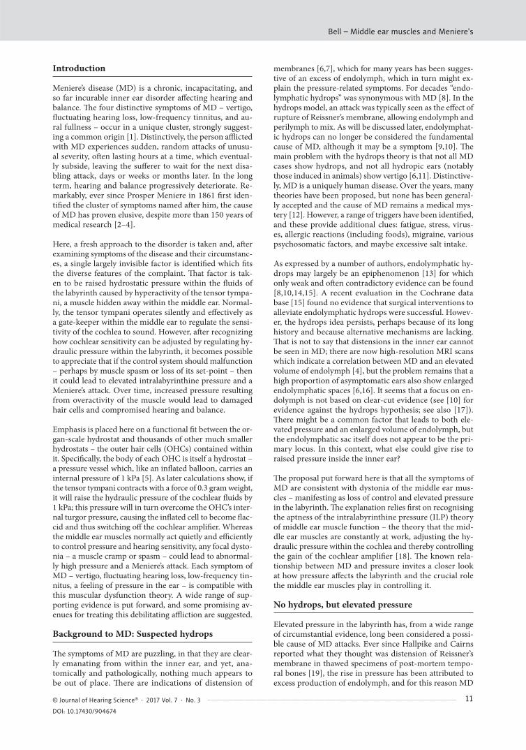
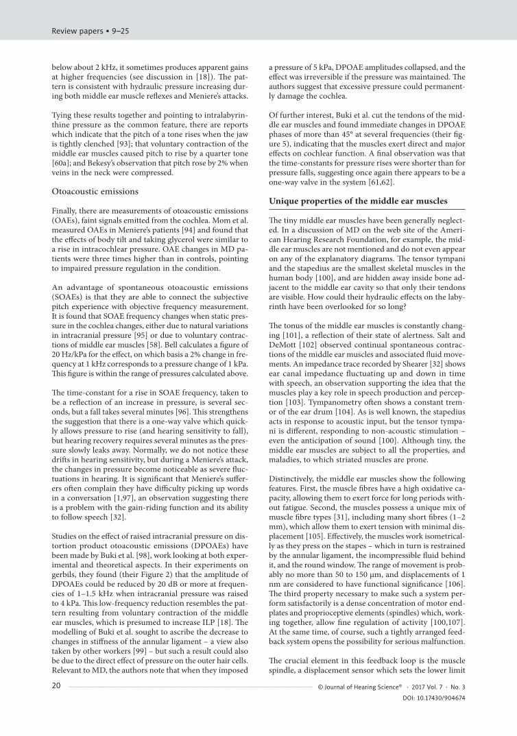
A noteworthy feature of the cochlear amplifier is an inbuilt resistance to overload. The cochlea has a dynamic range of 120 dB, meaning it can detect sound over an extraor-dinary intensity range of a million, million times. Strate-gically placed at the entrance to the cochlea, the tensor tympani and the stapedius stand guard. These tiny mus-cles, the smallest skeletal muscles in the human body, can be seen as part of a fast-acting gain control circuit (the acoustic reflex) which prevents damage to the deli-cate hair cells. Connected to the middle ear bones via ten-dons (Figure 1), the muscles spring into action whenever sound levels approach damaging levels and, through co-ordinated contraction, physically attenuate cochlear sen-sitivity. How they do so is not completely understood, but the case that it involves regulation of inner ear hydraulic pressure is discussed in the next section.
As an indication of the important role that pressure plays in normal cochlear function, there are long-standing ob-servations of what can happen if hydraulic pressure within the inner ear is lost, such as through a labyrinthine fistula. Sometimes encountered as an unwanted outcome of sur-gery for otosclerosis, leakage of fluid from the otic capsule produces sudden vertigo, tinnitus, and severe sensorineural
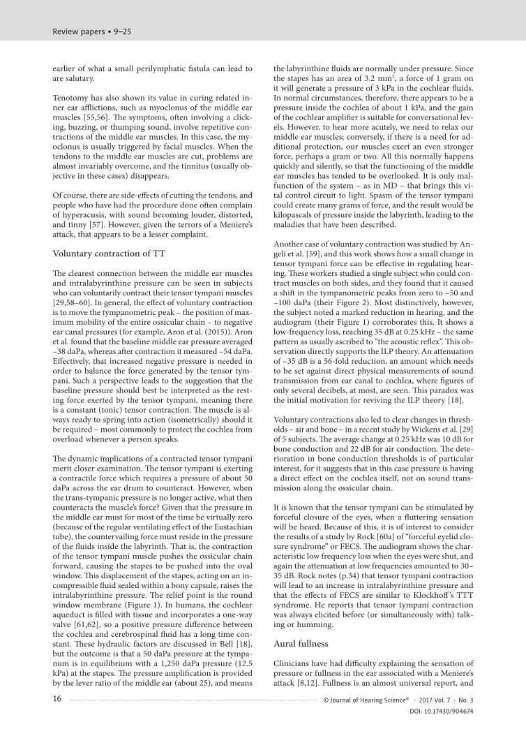
Figure 1. The middle ear muscles. (A) Diagram showing the tensor tympani (red) attached to the malleus, and the stape-dius (grey) attached to the stapes. When the tensor tympani contracts, the ossicular chain is pulled forward, forcing the stapes into the cochlea and raising the pressure of the fluids within. Adapted from Kirikae [23]. (B) 3D reconstruction from histopathology, showing the middle ear muscles in red, ossicles in yellow, and the tympanic membrane in green. Arrows indicate motion resulting from tensor tympani contraction. Image kindly supplied by R. Pennings and M. Bance, Dalhousie University
A B
Review papers • 9–25
12 © Journal of Hearing Science® · 2017 Vol. 7 · No. 3
DOI: 10.17430/904674
hearing loss [24] – the same distinctive symptoms as seen in MD. Goodhill reports that sudden hearing loss occa-sioned by barotrauma can also sometimes be traced to simple rupture of the oval or round windows, leading to loss of intralabyrinthine pressure.
The following text investigates evidence for a direct struc-ture–function relationship between the middle ear mus-cles and pressure. The conclusion is that the intralabyrin-thine pressure theory provides a unifying account of the role of the middle ear muscles and, significantly, how their dysfunction might lead to MD.
The ILP theory of middle ear muscle function
Knowledge of how the middle ear muscles function re-mains rudimentary. For many years, it was thought that contraction of the muscles simply stiffened the chain of acoustic transmission through the middle ear bones [25]. However, a fresh perspective on how the middle ear muscles act was put forward in 2011 when the case was made [18] for reviving an alternative theory: that contrac-tion of the middle ear muscles, particularly the tensor tym-pani, pushes the stapes into the oval window and raises the hydraulic pressure of the fluids in the otic capsule, as shown in Figure 1. The core of the intralabyrinthine pres-sure (ILP) theory is that the gain of the cochlear amplifier is directly controlled by pressure in the labyrinth and that the role of the middle ear muscles is to regulate this pres-sure. The ILP theory sees the outer hair cells as directly sensitive to the hydraulic pressure in the fluids surround-ing them, so, when the pressure rises, the result is an in-stant reduction in cochlear amplifier gain – effectively pro-tecting the hair cells against acoustic overload. Explicitly, if it is assumed that the tensor tympani muscle exerts a maximum contractile force of 1 g wt, this will be transmit-ted via the ossicular chain to the stapes (area of 3.2 mm2), where its piston-like action will generate a pressure of 3 kPa in the labyrinthine fluids. Such a figure for inner ear pressure is consistent with a range of observations on in-tracochlear pressures [26,27]. According to the ILP theo-ry, such a pressure causes a reduction in cochlear ampli-fier gain (usually interpreted as sound attenuation when electrode measurements are made in live animals) of up to 30 dB, with most of it occurring at low frequencies (be-low 1 kHz). In this way, the acoustic reflex exerts a pow-erful effect, although the standard explanation by which the reflex “stiffens” the acoustic chain produces only mi-nor changes – physical measurements show an acoustic transmission loss of only a few decibels [18]. The acous-tic reflex even works with bone-conducted sound, produc-ing the familiar pattern of reduced sensitivity at low fre-quencies [28,29]; such an observation is consistent with an effect on the cochlea itself, not just the ossicular chain.
The ILP theory, originally formulated by Gellé in the 1880s, carries good explanatory power, although it must be ac-knowledged it is not the current textbook explanation for how the acoustic reflex works. Silently and swiftly, and with minimum effort, contraction of the muscles immediately increases pressure and reduces the gain of the 30,000 outer hair cells. Because the labyrinthine fluids are virtually in-compressible and the stapes works against the compliance (10–13 m5/N) of the round window membrane [21], the
motion is minute – of the order of a tenth of a millimeter or less (see Helmholtz [30]) – so it is not surprising that the pressure rise, itself difficult to measure, has been overlooked. As will be shown, the middle ear muscles are well adapt-ed to the task of providing sustained and finely graded iso-metric contraction, with many small-diameter fibres [31].
Normally, the middle ear muscles function as intended and our hearing is constantly protected against overload. The muscles play a vital role, springing into action if a loud external sound reaches the cochlea, or whenever a loud internal sound – speaking, yawning, or chewing – is produced [28,32,33]. In this way, the muscles allow exter-nal sounds to be clearly heard even when we are speak-ing. The system is similar to that which operates in bats, where the animal’s middle ear muscles rapidly attenuate self-generated ultrasonic calls (perhaps more than 100 per second) while at the same time allowing intervening faint echoes to be perceived. Even a whisper or a touch to the cheek will trigger the muscle [32]. The surprise is that, subjectively, we do not notice this constant gain-rid-ing. It is rather like our vision filling in the gaps whenev-er we blink or move our eyes, making the outside world appear stable. The middle ear muscles regulate sound in-put to the ear not unlike the way in which the iris muscles regulate light input to the eye. Subjectively, the fluttering sound one hears when yawning or tightly closing the eyes is the sound of the tensor tympani at work.
Ordinarily, the muscles sit poised ready to contract in re-sponse to a loud sound. Under abnormal conditions, how-ever, it is possible for muscle control to be lost. In particu-lar, like all muscles, the middle ear muscles might suffer from a cramp or spasm, creating a sudden spike in inner ear pressure – a Meniere’s attack. Extended over longer time-spans, elevated pressure is likely to produce damage to the pressure-sensitive hair cells responsible for hearing and balance. This prediction, which follows from the ILP theory, provides a unified understanding of MD, and the following text elaborates the hypothesis.
Middle ear muscles, raised pressure, and MD – the evidence
It has been known for many years that elevated pressure in the ear canal of normal subjects can produce vestibular symptoms such as nystagmus and vertigo [34–36]. Indeed, pressure applied this way can also cause other Meniere-like symptoms: subjective tinnitus [37]; hearing loss at low frequencies [18]; pitch anomalies [38], and, of course, a feeling of fullness in the ear [12]. In each case, the ap-plied ear canal pressure leads to the stapes pressing on the oval window and increasing intralabyrinthine pressure, a connection that has been reasonably well documented and acknowledged [39]. However, if similar symptoms to MD occur from simply elevating ear canal pressure, this prompts the question: what other mechanism could ex-ist for driving the tympanic membrane inwards and in-creasing intralabyrinthine pressure? The answer suggest-ed here is that it is contraction of the middle ear muscles, predominantly the tensor tympani.
This paper makes the case that hyperactivity of the mid-dle ear muscles can lead to a rise in labyrinthine pressure
Bell – Middle ear muscles and Meniere's
13© Journal of Hearing Science® · 2017 Vol. 7 · No. 3
DOI: 10.17430/904674
and, in certain susceptible individuals (perhaps 1 in 1000 of the population), a calamitous Meniere’s attack. The case is based on the following observations.
Sudden attacks
It is remarkable how people with MD can go for long pe-riods, sometimes years, without problems, and then sud-denly be struck down within minutes by an attack. The si-multaneous involvement of balance and hearing disorders during a Meniere’s attack – typically explosive in onset and sometimes abrupt in termination – point to a single physi-cal factor rather than a biochemical imbalance. Pressure is put forward as the likely physical factor. House [3] provides an important clue. In the midst of an attack, accompanied by intense vomiting, patients sometimes report hearing a snap, at which point their symptoms are suddenly gone, perhaps for months. This quick resolution again supports some sort of direct physical effect, and the idea that an at-tack is brought on by dysfunction of the middle ear mus-cles is consistent with this observation. The explanation is that the inner ear muscle feedback loop is suddenly up-set and is gradually restored – just like with most muscle cramps – after which pressure returns to normal and the symptoms disappear.
The accompanying symptoms of aural fullness, headaches, pain in the occipital muscles, and nystagmus (Ch 2 of [40]) also support the pressure explanation. Indeed, the whole argument linking MD with endolymphatic hydrops tacitly assumes it is pressure itself which is the provoking agent. According to the present hypothesis, however, it is the ac-tion of the middle ear muscles which deranges pressure levels, not an excessive rate of fluid production (hydrops).
In overview, the inner ear is an exquisitely sensitive pres-sure-sensing mechanism [3], and this paper suggests that the middle ear muscles, although not anatomically a part of the inner ear, play a key role in regulating its internal pressure and responsiveness to sound.
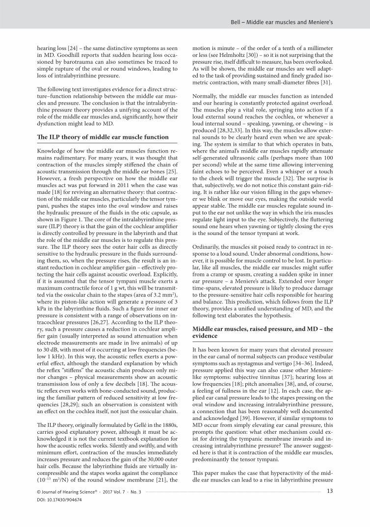
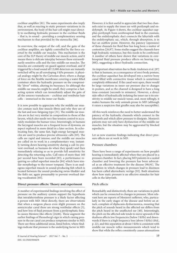
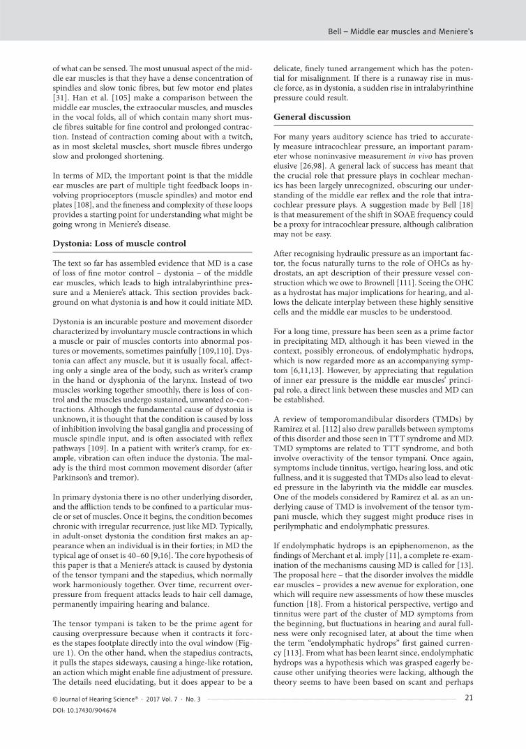
It has been well recognised by a range of authors that con-traction of the middle ear muscles can affect inner ear pressure (see [18]), and a description of how this affects hearing sensitivity – the intralabyrinthine pressure theory – has already been sketched above. It is important to rec-ognise, however, that the pressure rise can, if sustained, be sufficient to disable and, in the long term, cause per-manent damage to hair cells concerned with hearing and balance. The labyrinth is an almost sealed hydraulic ves-sel (Figure 2), and the cochlear and vestibular aqueducts (discussed later) have insufficient hydraulic conductivity to dissipate a sudden plunger action of the stapes.
Fluctuating hearing experienced by Meniere’s sufferers is a day-to-day reminder of how delicately poised the inner ear system is. Usually a Meniere’s attack begins unilater-ally, but over time it tends to become bilateral [41]. Of-ten a sudden reduction in hearing is a premonition of a Meniere’s attack, and tinnitus is another common warn-ing sign [40]. Almost inevitably, MD progresses to in-volve both ears [1], and interestingly in such cases Kitaha-ra found that fluctuations in hearing sometimes occurred simultaneously in both ears, or sometimes they alternated
between the ears [42]. This makes it highly unlikely that membrane rupture is responsible but it does lend support to the idea of tensor tympani malfunction, since the mus-cles are part of a bilateral system. Cases where Meniere-like symptoms have resulted from acoustic overload [43] also indicate middle ear muscle involvement.
When a muscle spasm begins, there will be a sudden rise of pressure, and the typical MD symptoms will start. It is important to note that, in the early stages, the hearing loss in MD has the same typical pattern of low-frequency hear-ing loss (upsloping audiogram) as occurs with the acoustic reflex (loss of sensitivity below about 1 kHz: p.142 of [20]; p.20 of [8]; [18]) and with sudden hearing loss [40,44].
In the long term, sustained elevated pressure will damage hair cells. As a later section describes, hair cells are pressure sensors, a property used to detect small oscillating chang-es in pressure (i.e., sound). The clearest example of such pressure sensing is the dogfish [45], which senses sound pressure directly, without requiring stereocilia deflection. The interplay between pressure detection and particle dis-placement is a subtle one [46], and the interested read-er is referred to earlier work [18] for a fuller discussion.
TTT syndrome and its similarity to MD
It is remarkable how similar the symptoms of MD are to another disorder, tonic tensor tympani (TTT) syndrome, a complaint first described by Klockhoff which characteris-tically involves spontaneous fluctuation in middle ear im-pedance but is subjectively associated with vertigo, tinni-tus, fullness in the ear, tension headache, and dysacusis [47]. Klockhoff ascribed the syndrome to an elevated to-nus of the tensor tympani, which he thought was due to mental stress. Of interest, he notes the difficulty of distin-guishing the syndrome from Meniere’s disease, although he himself thought they were two separate complaints (because there were no sudden attacks and the prevailing view that MD must somehow be due to “hydrops”). Klock-hoff makes clear that contraction of the tensor tympani is more of a startle reflex than an acoustic reflex, and can be
Cochlea
EndolymphStapes
Oval window
Vestibule
Perilymph
SacculeUtricle
Semicircular canalRound
window
Figure 2. The otic capsule is a cavity within solid bone, and is an almost hydraulically sealed system. When the stapes is pushed in by the tensor tympani, the round window bulges out by an equal amount, as endolymph and perilymph are incompressible. Pressure of the fluids therefore depends almost totally on the force generated by contraction of the tensor tympani. [Modified from http://www.eneurosurgery.com]
Review papers • 9–25
14 © Journal of Hearing Science® · 2017 Vol. 7 · No. 3
DOI: 10.17430/904674
activated by a puff of air to the eye, touch to the outer ear, yawning, chewing, swallowing, speaking, or even anticipa-tion of a loud sound. However, unlike with the stapedius, sound alone does not appear to elicit a tensor reflex [47].
Although Klockhoff ’s original work on TTT syndrome was based on more than 200 cases, TTT syndrome was subse-quently regarded as largely an oddity. It is only in recent years that the wider implications of the syndrome have been recognized and interest has been revived. In 2006, an audiologist specializing in treating tinnitus and hyperacusis [48] recognized the relevance of TTT syndrome to patients who were suffering these complaints, who had temporo-mandibular problems, or were call-centre workers who had experienced acoustic shock from wearing head-sets. Westcott and her colleagues began a more intensive study of 345 patients [49] which identified TTT syndrome as an involuntary, anxiety-based condition where the threshold for reflex activity of the tensor tympani had been reduced, causing frequent spasm. It was found to affect some 60% of patients in total, and many of the symptoms could be seen in other patient groups as well. The study concluded there was a basic relationship between tinnitus, hypera-cusis, and TTT syndrome, and raised a number of clini-cal implications. According to the hypothesis here, one of these is MD. Both TTT syndrome and MD involve dys-function of the TT, although in slightly different ways. TTT syndrome can be said to involve a somewhat overactive TT, but it is suggested that a full MD attack is caused by spasm of the muscle, causing an overwhelming rise in in-tralabyrinthine pressure. With raised pressure, there will be a direct sensation of pressure in the ear (aural fullness), there will be fluctuating hearing loss, and, as pressure af-fects the semicircular canals as well, there will be vertigo. As for tinnitus, the connection between tinnitus and ear canal pressure has already been mentioned.
Effect of sectioning muscle tendons
What may be regarded as the best evidence that malfunc-tion of the middle ear muscles causes MD comes from studies in which the tendons to the muscles have been cut (tenotomy). In patients with Meniere’s disease, the sim-ple result of tenotomy of the stapedius and tensor tympa-ni muscles is that attacks cease almost completely [50,51] and dizziness is markedly reduced [52]. Surprisingly, al-though these papers have led to new interest in the role of the middle ear muscles, they have not brought about a revolution in treatment of MD, as might be expected. The reasons for the reticence are illuminating, and will be ex-amined below, but first it is worth noting that tenotomy of the middle ear muscles is a procedure with a long his-tory. It was advocated as a treatment for MD back in the 19th century [51,53], but fell out of favour because, with-out antibiotics, there was risk of serious infection. It was only revived in 2003 by Franz and colleagues, who re-ported outstanding results when tried on a group of 20 Meniere’s patients. In this study, the frequency of vertigo attacks was reduced to zero in 70% of them, hearing im-proved by an average of 13 dB, tinnitus was markedly re-duced, and the sensation of pressure and fullness in the ear was relieved. Tellingly, when scar tissue occasionally caused the stumps of the tendons to reconnect, the symp-toms returned. In two follow-up studies [50,52], there were
similarly dramatic results: 26 of 30 subjects had zero verti-go attacks following the procedure and the dizziness hand-icap inventory fell from 52 to 4.
These powerful demonstrations have been cautiously received, although the potential of tenotomy has been supported by Albu et al. [54]. The main reason for the reticence seems to be a real difficulty in seeing how sec-tioning a mere tendon could affect the inner ear, where the Meniere’s symptoms arise. Franz et al. speculated that perhaps “tenotomy prevents muscle-induced mechanical irritations of the hydropic inner ear.” Here the authors ap-pear to be taking the hydrops as the starting point, and that the muscles are only responsible for aggravating what they describe as “primary endolymphatic hydrops”. At yet an-other point, they suggest how pathological cramps of the muscles might “intensify” the symptoms of MD. In a later paper [50], they acknowledge that the tensor tympani has the ability to increase pressure in the labyrinth, but only beyond a certain “trigger point”, and that the preexisting hydrops of MD is the primary factor in raising cochlear pressure. In Loader et al. [52], the idea is expressed some-what differently: it is said that tenotomy prevents the ele-vated pressure, due to hydrops, from displacing the ossi-cles and causing reflex action of the tensor, which would then add to the pressure and trigger symptoms.
To contrast this position with that taken by the present paper, the model being put forward here is that the mus-cles themselves are fully and directly responsible for MD. In accordance with the ILP theory, their role is, via contrac-tion, to precisely control intralabyrinthine pressure at all times – when speaking, chewing, swallowing, in response to loud sound – and if control is lost, such as through mus-cle spasm or general dystonia, MD is the immediate re-sult. As expressed earlier, endolymphatic hydrops could basically be a roundabout way of ascribing a cause to the raised pressure whose damaging effects are widely evident throughout the labyrinth. The raised pressure, however, is not the sign of a metabolic imbalance but of the aber-rant functioning – dystonia – of the middle ear muscles.
As in all things, preconceptions are hard to shift, and this is especially the case with the hydrops basis of MD, proba-bly because no other theory has emerged to take its place. A similar example is the ILP theory itself, which has long been put aside because auditory science could not see a way by which pressure surrounding the basilar membrane could affect a mechanical travelling wave [18]. The travel-ling wave rose to become the core causal entity, and it re-quired the emergence of the cochlear amplifier before it became possible to see that static pressure could indeed have an effect on such an active device without necessari-ly involving a mechanical travelling wave. Likewise, with-out entertaining the possible validity of the ILP theory, it is not possible to appreciate how small muscles, via their mechanical and fluid connections, can have major effects on hearing and balance. The end result has been that, in response to Franz’s findings, clinicians have found it diffi-cult to conceive that small cryptic muscles, buried in bone some distance from the inner ear, could possibly produce the catastrophic symptoms of MD. If a single example of the potent effect of pressure is needed, the reports cited
Bell – Middle ear muscles and Meniere's
15© Journal of Hearing Science® · 2017 Vol. 7 · No. 3
DOI: 10.17430/904674
earlier of what a small perilymphatic fistula can lead to are salutary.
Tenotomy has also shown its value in curing related in-ner ear afflictions, such as myoclonus of the middle ear muscles [55,56]. The symptoms, often involving a click-ing, buzzing, or thumping sound, involve repetitive con-tractions of the middle ear muscles. In this case, the my-oclonus is usually triggered by facial muscles. When the tendons to the middle ear muscles are cut, problems are almost invariably overcome, and the tinnitus (usually ob-jective in these cases) disappears.
Of course, there are side-effects of cutting the tendons, and people who have had the procedure done often complain of hyperacusis, with sound becoming louder, distorted, and tinny [57]. However, given the terrors of a Meniere’s attack, that appears to be a lesser complaint.
Voluntary contraction of TT
The clearest connection between the middle ear muscles and intralabyrinthine pressure can be seen in subjects who can voluntarily contract their tensor tympani muscles [29,58–60]. In general, the effect of voluntary contraction is to move the tympanometric peak – the position of max-imum mobility of the entire ossicular chain – to negative ear canal pressures (for example, Aron et al. (2015)). Aron et al. found that the baseline middle ear pressure averaged –38 daPa, whereas after contraction it measured –54 daPa. Effectively, that increased negative pressure is needed in order to balance the force generated by the tensor tym-pani. Such a perspective leads to the suggestion that the baseline pressure should best be interpreted as the rest-ing force exerted by the tensor tympani, meaning there is a constant (tonic) tensor contraction. The muscle is al-ways ready to spring into action (isometrically) should it be required – most commonly to protect the cochlea from overload whenever a person speaks.
The dynamic implications of a contracted tensor tympani merit closer examination. The tensor tympani is exerting a contractile force which requires a pressure of about 50 daPa across the ear drum to counteract. However, when the trans-tympanic pressure is no longer active, what then counteracts the muscle’s force? Given that the pressure in the middle ear must for most of the time be virtually zero (because of the regular ventilating effect of the Eustachian tube), the countervailing force must reside in the pressure of the fluids inside the labyrinth. That is, the contraction of the tensor tympani muscle pushes the ossicular chain forward, causing the stapes to be pushed into the oval window. This displacement of the stapes, acting on an in-compressible fluid sealed within a bony capsule, raises the intralabyrinthine pressure. The relief point is the round window membrane (Figure 1). In humans, the cochlear aqueduct is filled with tissue and incorporates a one-way valve [61,62], so a positive pressure difference between the cochlea and cerebrospinal fluid has a long time con-stant. These hydraulic factors are discussed in Bell [18], but the outcome is that a 50 daPa pressure at the tympa-num is in equilibrium with a 1,250 daPa pressure (12.5 kPa) at the stapes. The pressure amplification is provided by the lever ratio of the middle ear (about 25), and means
the labyrinthine fluids are normally under pressure. Since the stapes has an area of 3.2 mm2, a force of 1 gram on it will generate a pressure of 3 kPa in the cochlear fluids. In normal circumstances, therefore, there appears to be a pressure inside the cochlea of about 1 kPa, and the gain of the cochlear amplifier is suitable for conversational lev-els. However, to hear more acutely, we need to relax our middle ear muscles; conversely, if there is a need for ad-ditional protection, our muscles exert an even stronger force, perhaps a gram or two. All this normally happens quickly and silently, so that the functioning of the middle ear muscles has tended to be overlooked. It is only mal-function of the system – as in MD – that brings this vi-tal control circuit to light. Spasm of the tensor tympani could create many grams of force, and the result would be kilopascals of pressure inside the labyrinth, leading to the maladies that have been described.
Another case of voluntary contraction was studied by An-geli et al. [59], and this work shows how a small change in tensor tympani force can be effective in regulating hear-ing. These workers studied a single subject who could con-tract muscles on both sides, and they found that it caused a shift in the tympanometric peaks from zero to –50 and –100 daPa (their Figure 2). Most distinctively, however, the subject noted a marked reduction in hearing, and the audiogram (their Figure 1) corroborates this. It shows a low-frequency loss, reaching 35 dB at 0.25 kHz – the same pattern as usually ascribed to “the acoustic reflex”. This ob-servation directly supports the ILP theory. An attenuation of –35 dB is a 56-fold reduction, an amount which needs to be set against direct physical measurements of sound transmission from ear canal to cochlea, where figures of only several decibels, at most, are seen. This paradox was the initial motivation for reviving the ILP theory [18].
Voluntary contractions also led to clear changes in thresh-olds – air and bone – in a recent study by Wickens et al. [29] of 5 subjects. The average change at 0.25 kHz was 10 dB for bone conduction and 22 dB for air conduction. The dete-rioration in bone conduction thresholds is of particular interest, for it suggests that in this case pressure is having a direct effect on the cochlea itself, not on sound trans-mission along the ossicular chain.
It is known that the tensor tympani can be stimulated by forceful closure of the eyes, when a fluttering sensation will be heard. Because of this, it is of interest to consider the results of a study by Rock [60a] of “forceful eyelid clo-sure syndrome” or FECS. The audiogram shows the char-acteristic low frequency loss when the eyes were shut, and again the attenuation at low frequencies amounted to 30–35 dB. Rock notes (p.34) that tensor tympani contraction will lead to an increase in intralabyrinthine pressure and that the effects of FECS are similar to Klockhoff ’s TTT syndrome. He reports that tensor tympani contraction was always elicited before (or simultaneously with) talk-ing or humming.
Aural fullness
Clinicians have had difficulty explaining the sensation of pressure or fullness in the ear associated with a Meniere’s attack [8,12]. Fullness is an almost universal report, and
Review papers • 9–25
16 © Journal of Hearing Science® · 2017 Vol. 7 · No. 3
DOI: 10.17430/904674
is often a warning sign of an impending attack and goes away again when the attack ends [63]. And yet, as Mat-tox points out, there is no recognised mechanism for sens-ing hydraulic pressure in the inner ear, so where does the sensation come from? If one assumes that the middle ear muscles are responsible for the excess pressure, then sens-ing by primary spindles within the muscles could explain the effect. Relevant here are the experiments of Densert and colleagues, who were able to decrease the sensation of fullness in Meniere’s patients by applying static pressure of about 15 daPa to the ear canal [64].
Hennebert’s sign and the Tullio phenomenon
When pressure is applied to the ear canal and vestibular symptoms such as vertigo and nystagmus occur, the re-action is called a positive Hennebert’s sign and is rare in normal subjects. In Meniere’s patients, however, a positive reaction is frequent, and in terms of the present hypothe-sis this is significant. Nadol [65] found that 29 of 81 ears suffering from MD exhibited Hennebert’s sign, compared to zero of 100 normal ears. Nadol ascribed the reaction to fibrous connections between the stapes and the labyrinth (vestibulofibrosis), but a more plausible connection is hy-draulic: inward motion of the stapes causes fluid pressure to rise, and this directly affects the vestibular hair cells. This hydraulic mechanism can also explain why even just inserting vents in the tympanic membrane (grommets) can reduce symptoms in a proportion of Meniere’s pa-tients [66,67]: a grommet will equalize pressure between the middle ear and the atmosphere, avoiding the possibil-ity that inadvertent underpressure in the middle ear will, through inward deflection of the ear drum, cause a cor-responding overpressure in the inner ear.
Another curious phenomenon is sometimes seen when the ear is subjected to high-intensity, low-frequency sound. In normal subjects, there is an acoustic reflex but there are no vestibular effects. However, among Meniere’s patients, it is notable that an enhanced vestibular reaction is frequently seen. Stephens and Ballam [68] found that 7 of 25 Meniere’s patients had an enhanced vestibular reaction (Tullio phe-nomenon or sono-ocular reaction), an effect which was absent in the controls. Given that the sound stimuli used are likely to produce a reflex contraction of the middle ear muscles, this provides an indication that MD patients have poor control of the middle ear muscles (or have impaired flow through the channels conducting fluid in and out of the labyrinth, a possibility discussed later).
Tinnitus
Tinnitus is a symptom of many hearing disorders. Given the size of the subject matter it will not be discussed in detail here. It is sufficient for our purposes to simply note that one of the effects of raising pressure in the ear canal is subjective tinnitus [37], and the simplest and clearest explanation must be that the increased pressure has, like tensor tympani contraction, forced the stapes into the otic capsule and increased fluid pressure. It has been not-ed [69] that tinnitus can result from voluntary contraction of the middle ear muscles. It is of interest that the tinni-tus associated with MD is of the low-frequency type (be-low about 2 kHz [70,71]), and so it is supposed that the
hair cells at the apex are the most sensitive to pressure, an idea which in turn connects to observations that raised ILP (as in the acoustic reflex) reduces hearing sensitivity below about 2 kHz [18].
It is significant that low-frequency OHCs are much long-er than high-frequency ones [72], so that the former are expected to be more susceptible to changes in static pres-sure than the latter; consequently it is understandable why the acoustic reflex mostly affects low frequencies, and why MD first impairs these same frequencies.
Voluntary control of the tensor tympani and psy-chosomatic aspects of MD
The middle ear muscles are striated, or skeletal, muscles. This means that, like all such muscles, they are not just triggered by reflexes but are also, to varying degrees, un-der voluntary control. It is therefore significant that the symptoms of TTT syndrome include psychological factors such as anxiety, fear, and fright, and even anticipation can cause contraction of the tensor tympani. The sight of a toy gun has been cited as a stimulus [48].
A number of authors have pointed out that MD attacks are often triggered by stress, and that emotional compo-nents play a strong part [9,73]. Indeed, some have even suggested that MD is a purely psychosomatic ailment, ex-plaining why many treatments, through a placebo effect, have produced favourable outcomes [17,74]. It is diffi-cult to gauge the strength of these suggestions, but the fact that Meniere’s attacks are episodic, and that the mid-dle ear muscles are capable of being contracted voluntar-ily, provides a framework which supports the hypothe-sis that MD is due to dystonia of the middle ear muscles. Making the case that there is a real, physical basis for MD opens up a promising avenue for understanding and per-haps curing the condition.
Two linked hydrostats: The labyrinth and the outer hair cell
The text to this point has emphasized that the labyrinth is a hydrostat in which the middle ear muscles control the hydraulic pressure of fluids within. However, so far only a general statement has been made that pressure controls the sensitivity of the outer hair cells. This section presents a specific physical model of how pressure could control that sensitivity: that is, outer hair cells normally carry an internal pressure of about 1 kPa, so when the labyrinthine pressure is increased beyond that point, the cells can no longer counteract the pressure and they momentarily de-flate. At that point, electromotility, and cochlear amplifier gain, drops to zero. The middle ear muscles can therefore be powerful controllers of hearing sensitivity.
According to Brownell, the test-tube shaped OHC is a hy-drostat [75]. At the same time, it is, according to anoth-er view [46], an exquisitely sensitive detector of minute pressure variations – that is, sound. In this case the body of the cell can be taken to be the sensing element rather than the stereocilia, although sterocilia are necessary to complete the feedback loop between neighbouring cells to create a surface acoustic wave (SAW) resonator [76].
Bell – Middle ear muscles and Meniere's
17© Journal of Hearing Science® · 2017 Vol. 7 · No. 3
DOI: 10.17430/904674
The SAW resonator involves a standing wave between the three rows of OHCs, and the standing wave establishes the cochlear amplifier in a physical form. Acting as both detec-tors and motors, and cooperating in triplets, 12,000 OHCs can create 4,000 highly tuned cochlear amplifiers, boost-ing hearing levels a thousand-fold (60 dB). The OHCs do not signal the brain directly; instead they are pre-ampli-fiers which pass their output to the adjoining inner hair cells (IHCs), where signaling to higher centres begins. If a person should lose their delicate OHCs, perhaps from age or loud noise, leaving only the more robust IHCs, then hearing levels decline by about 60 dB.
Although there have been several suggestions, the physical basis of the cochlear amplifier is still not settled [76,77]. The SAW model, and its dependence on OHC electromo-tility, provides a direct account of how hydraulic pressure is able to control cochlear amplifier gain. However, there may be other models which can provide a similar out-come, and more research is needed. Irrespective of which model of cochlear mechanics is favoured, all depend on strong OHC electromotility.
There are various efferent connections to the OHCs, and these provide another way of adjusting the cochlea’s sensi-tivity [78]. However, the drawback of neural mechanisms is that they involve considerable delay. There is a way to short-circuit this delay, and this is where the middle ear muscles play a crucial role. Any force exerted by the mid-dle ear muscles is conveyed immediately to the fluids of the inner ear, increasing hydraulic pressure. As the calcu-lations above have shown, the tensor tympani, exerting a force of 1 gram, can cause a corresponding pressure of 3 kPa in the inner ear. According to the ILP theory, this will cause a reduction of hearing sensitivity of up to 30 dB, and this raises the question of how a mild hydraulic pressure can have such a large effect.
Let us look closer at what happens if a sudden increase in pressure is applied to a hydrostat. The following experi-mental findings were derived from the outer hair cells of
guinea pigs rather than humans, but it is assumed the re-sults are generally applicable.
In a unique arrangement, the body of the OHC is sur-rounded on all sides by fluid, meaning that the OHC is in direct hydraulic communication with the stapes – that is, we have a hydrostat or pressure vessel surrounded on all sides by cochlear fluid. The OHC is piezoelectric and able to change length cycle by cycle, elongating and contracting in time with oscillating sound pressure [75]. Its outer walls are corrugated and are reinforced by helical fibres which encircle the cylindrical body in both clockwise and anti-clockwise directions. The fibres are oriented at the famil-iar winding angle of 55° which means that length chang-es are accompanied by minimal volume changes (that is, it has a Poisson ratio close to 0.5), making length chang-es possible at up to 100 kHz (as in bats).
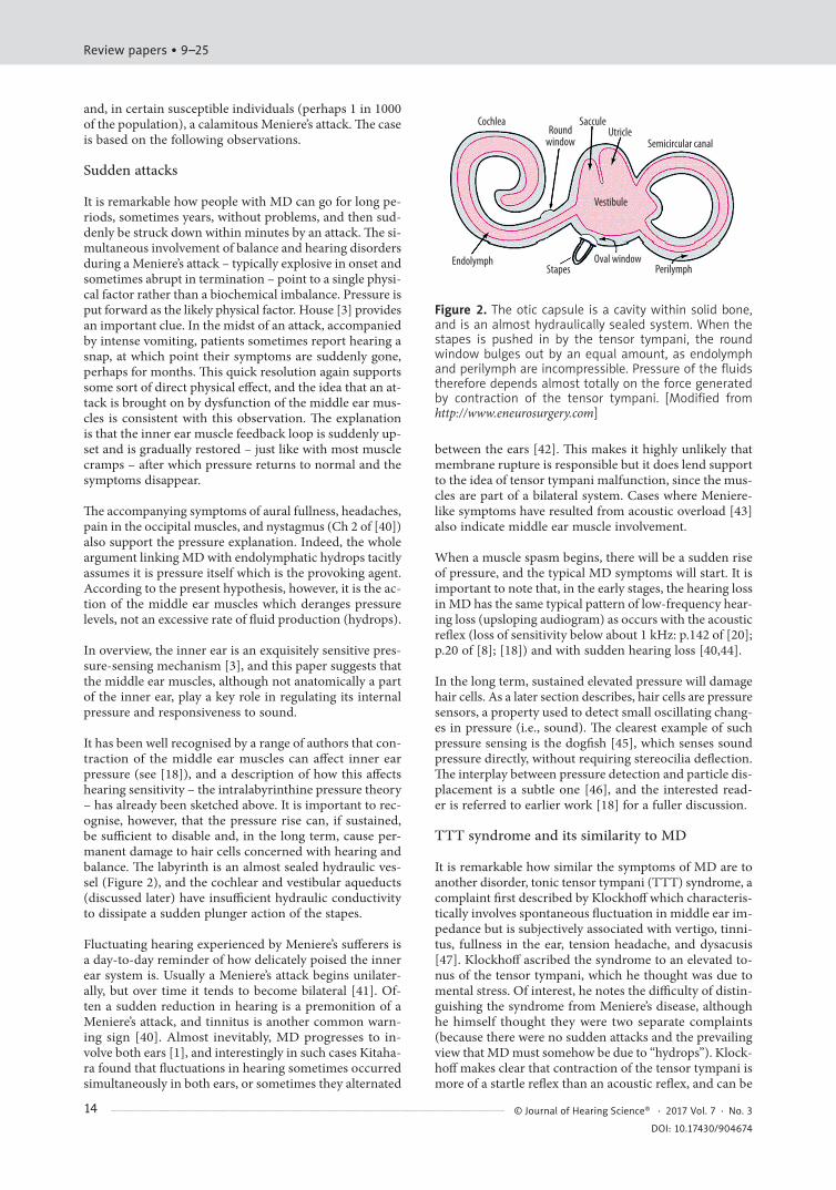
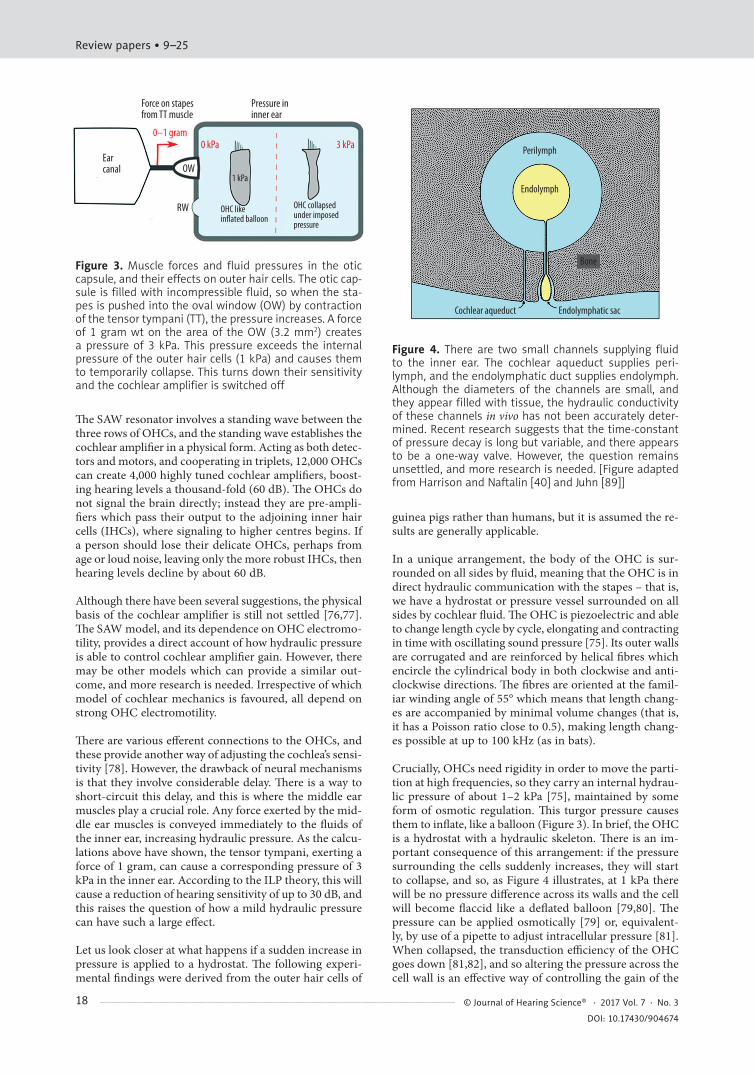
Crucially, OHCs need rigidity in order to move the parti-tion at high frequencies, so they carry an internal hydrau-lic pressure of about 1–2 kPa [75], maintained by some form of osmotic regulation. This turgor pressure causes them to inflate, like a balloon (Figure 3). In brief, the OHC is a hydrostat with a hydraulic skeleton. There is an im-portant consequence of this arrangement: if the pressure surrounding the cells suddenly increases, they will start to collapse, and so, as Figure 4 illustrates, at 1 kPa there will be no pressure difference across its walls and the cell will become flaccid like a deflated balloon [79,80]. The pressure can be applied osmotically [79] or, equivalent-ly, by use of a pipette to adjust intracellular pressure [81]. When collapsed, the transduction efficiency of the OHC goes down [81,82], and so altering the pressure across the cell wall is an effective way of controlling the gain of the
Earcanal
Force on stapesfrom TT muscle
Pressure ininner ear
OHC likeinflated balloon
1 kPa
OHC collapsedunder imposedpressure
RW
0–1 gram0 kPa 3 kPa
OW
Figure 3. Muscle forces and fluid pressures in the otic capsule, and their effects on outer hair cells. The otic cap-sule is filled with incompressible fluid, so when the sta-pes is pushed into the oval window (OW) by contraction of the tensor tympani (TT), the pressure increases. A force of 1 gram wt on the area of the OW (3.2 mm2) creates a pressure of 3 kPa. This pressure exceeds the internal pressure of the outer hair cells (1 kPa) and causes them to temporarily collapse. This turns down their sensitivity and the cochlear amplifier is switched off
Cochlear aqueduct Endolymphatic sac
Perilymph
Endolymph
Bone
Figure 4. There are two small channels supplying fluid to the inner ear. The cochlear aqueduct supplies peri-lymph, and the endolymphatic duct supplies endolymph. Although the diameters of the channels are small, and they appear filled with tissue, the hydraulic conductivity of these channels in vivo has not been accurately deter-mined. Recent research suggests that the time-constant of pressure decay is long but variable, and there appears to be a one-way valve. However, the question remains unsettled, and more research is needed. [Figure adapted from Harrison and Naftalin [40] and Juhn [89]]
Review papers • 9–25
18 © Journal of Hearing Science® · 2017 Vol. 7 · No. 3
DOI: 10.17430/904674
cochlear amplifier [81]. The same experiments also imply that, as well as reacting to static pressure variations in its environment, the body of the hair cell might react direct-ly to oscillating hydraulic pressure in the cochlear fluids – that is, to sound – providing a complementary sensing mechanism to that provided by the stereocilia [46].
In overview, the output of the cell, and the gain of the cochlear amplifier, are tightly controlled by the force ex-erted by the middle ear muscles. This mechanism is the core of the ILP theory of middle ear muscle action, and it means there is delicate interplay between these extraordi-narily sensitive cells and the tiny middle ear muscles. The muscles are constantly on-guard against overload, and if triggered by a loud sound all 12,000 detectors are instantly switched off by an all-enveloping 3 kPa pressure. A physi-cal analogy might be the Cartesian diver, where a change of force on the flexible membrane covering a water-filled container alters the hydraulic pressure on the compressi-ble “diver” within, altering its buoyancy. So although the middle ear muscles might be small, they comprise a fast-acting system which can immediately adjust the gain of all the sensory transducers – cochlear and vestibular hair cells – immersed in the inner ear fluids.
It is now possible to appreciate why the middle ear mus-cles contain such fine muscle fibres and why they are of the fast and non-fatiguing type [31]. The middle ear mus-cles are in fact very similar in composition to those of the larynx, which also needs very fine tension control to accu-rately modulate the human voice. Interestingly, in humans the laryngeal muscles are a frequent locus of dystonia, an aspect explored in more detail in a later section. In echo-locating bats, the same fast, high-energy laryngeal mus-cles are used to produce precise ultrasonic calls [83]. The calls are rapid and intense, and the middle ear muscles are called on to work in a complementary way, instant-ly turning down hearing sensitivity during a call (to pre-vent overload, as humans do when they speak) and then immediately relaxing so as to provide full sensitivity for detecting the returning echo. Call rates of more than 160 per second have been recorded [83], a performance re-quiring so-called superfast muscles [84] which have sim-ilar morphology to the tensor tympani. There is an anal-ogous superfast muscle in sound-producing fish which is located between the sound-producing swim-bladder and the fish’s ear, again presumably to prevent overload dur-ing vocalization [85].
Direct pressure effects: Theory and experiment
A number of experimental findings involving the effect of pressure on the auditory system support the hypothesis that intralabyrinthine pressure is a likely factor affecting a person with MD. Most directly, there are observations that when a surgeon places even slight pressure on the semicircular canal there are strong vestibular effects [3], and how loss of fluid pressure from a perilymphatic fistu-la causes Meniere-like effects [24,86]. These augment the earlier findings of Henneberg’s sign in which raising pres-sure in the ear canal can produce symptoms similar to MD. There are three additional areas, listed below, where find-ings indicate that pressure is the underlying factor in MD.
However, it is first useful to appreciate that two fine chan-nels exist to supply the inner ear with perilymph and en-dolymph. As Figure 4 shows, the cochlear aqueduct sup-plies perilymph from cerebrospinal fluid in the cranium, and the endolymphatic duct connects the labyrinth with the endolymphatic sac, which, through absorption, is in fact an outflow point. However, the patency (openness) of these channels for fluid flow has long been a matter of contention [26,87]. Some studies suggest the channels have high hydraulic resistance, but this needs to be confirmed. A number of others have shown that variations in cere-brospinal fluid pressure produce effects on hearing (e.g. [88]), suggesting a direct hydraulic connection.
An important observation due to Rask-Andersen et al. [87] is that in humans, as a consequence of erect body posture, the cochlear aqueduct has developed into a narrow bony canal filled with connective tissue which is sometimes completely obliterated. If the canal were open, there would be large variations in inner ear pressure due to changes in posture, and so the channel is designed to have a long time-constant (seconds to minutes). However, a direct side- effect of hydraulically isolating the inner ear is its sen-sitivity to middle ear muscle tonus, and, most tellingly, it makes humans the only animals prone to MD (although it raises a suspicion that giraffes may also be susceptible).
All this work reinforces the need to better understand the patency of the hydraulic channels which connect to the labyrinth and which allow pressure to dissipate. Meniere’s patients may not only have dysfunction of the middle ear muscles, but the situation may be aggravated by blocked aqueducts.
Let us now examine findings indicating that direct pres-sure effects are at work in MD.
Pressure chambers
There have been a range of experiments on how people’s hearing is immediately affected when they are placed in a pressure chamber. In fact, placing MD patients in a sealed chamber and lowering the pressure has been advocat-ed as an effective treatment for the disease [90,91]. The condition in which changes in pressure lead to dizziness has been called alternobaric vertigo [92]. Both situations show how static pressure is an effective stimulus for hair cells in the labyrinth.
Pitch effects
Remarkably and distinctively, there are variations in pitch which can be connected to changes in pressure. Most rele-vant here are reports of Meniere’s sufferers, who, particu-larly in the early stages of the disease and before an at-tack, complain of diplacusis dysharmonica, meaning that the pitch of sounds heard in the affected ear differs from the pitch heard in the unaffected ear [40]. Interestingly, the pitch on the affected side tends to move upwards if the deafness affects low frequencies (below 3 kHz) and down-wards if there is a high frequency loss (above 5 kHz) (ibid., p. 22), and this separation at about 3 kHz is consistent with middle ear muscle reflex measurements which tend to show that while the reflex consistently causes attenuations
Bell – Middle ear muscles and Meniere's
19© Journal of Hearing Science® · 2017 Vol. 7 · No. 3
DOI: 10.17430/904674
below about 2 kHz, it sometimes produces apparent gains at higher frequencies (see discussion in [18]). The pat-tern is consistent with hydraulic pressure increasing dur-ing both middle ear muscle reflexes and Meniere’s attacks.
Tying these results together and pointing to intralabyrin-thine pressure as the common feature, there are reports which indicate that the pitch of a tone rises when the jaw is tightly clenched [93]; that voluntary contraction of the middle ear muscles caused pitch to rise by a quarter tone [60a]; and Bekesy’s observation that pitch rose by 2% when veins in the neck were compressed.
Otoacoustic emissions
Finally, there are measurements of otoacoustic emissions (OAEs), faint signals emitted from the cochlea. Mom et al. measured OAEs in Meniere’s patients [94] and found that the effects of body tilt and taking glycerol were similar to a rise in intracochlear pressure. OAE changes in MD pa-tients were three times higher than in controls, pointing to impaired pressure regulation in the condition.
An advantage of spontaneous otoacoustic emissions (SOAEs) is that they are able to connect the subjective pitch experience with objective frequency measurement. It is found that SOAE frequency changes when static pres-sure in the cochlea changes, either due to natural variations in intracranial pressure [95] or due to voluntary contrac-tions of middle ear muscles [58]. Bell calculates a figure of 20 Hz/kPa for the effect, on which basis a 2% change in fre-quency at 1 kHz corresponds to a pressure change of 1 kPa. This figure is within the range of pressures calculated above.
The time-constant for a rise in SOAE frequency, taken to be a reflection of an increase in pressure, is several sec-onds, but a fall takes several minutes [96]. This strengthens the suggestion that there is a one-way valve which quick-ly allows pressure to rise (and hearing sensitivity to fall), but hearing recovery requires several minutes as the pres-sure slowly leaks away. Normally, we do not notice these drifts in hearing sensitivity, but during a Meniere’s attack, the changes in pressure become noticeable as severe fluc-tuations in hearing. It is significant that Meniere’s suffer-ers often complain they have difficulty picking up words in a conversation [1,97], an observation suggesting there is a problem with the gain-riding function and its ability to follow speech [32].
Studies on the effect of raised intracranial pressure on dis-tortion product otoacoustic emissions (DPOAEs) have been made by Buki et al. [98], work looking at both exper-imental and theoretical aspects. In their experiments on gerbils, they found (their Figure 2) that the amplitude of DPOAEs could be reduced by 20 dB or more at frequen-cies of 1–1.5 kHz when intracranial pressure was raised to 4 kPa. This low-frequency reduction resembles the pat-tern resulting from voluntary contraction of the middle ear muscles, which is presumed to increase ILP [18]. The modelling of Buki et al. sought to ascribe the decrease to changes in stiffness of the annular ligament – a view also taken by other workers [99] – but such a result could also be due to the direct effect of pressure on the outer hair cells. Relevant to MD, the authors note that when they imposed
a pressure of 5 kPa, DPOAE amplitudes collapsed, and the effect was irreversible if the pressure was maintained. The authors suggest that excessive pressure could permanent-ly damage the cochlea.
Of further interest, Buki et al. cut the tendons of the mid-dle ear muscles and found immediate changes in DPOAE phases of more than 45° at several frequencies (their fig-ure 5), indicating that the muscles exert direct and major effects on cochlear function. A final observation was that the time-constants for pressure rises were shorter than for pressure falls, suggesting once again there appears to be a one-way valve in the system [61,62].
Unique properties of the middle ear muscles
The tiny middle ear muscles have been generally neglect-ed. In a discussion of MD on the web site of the Ameri-can Hearing Research Foundation, for example, the mid-dle ear muscles are not mentioned and do not even appear on any of the explanatory diagrams. The tensor tympani and the stapedius are the smallest skeletal muscles in the human body [100], and are hidden away inside bone ad-jacent to the middle ear cavity so that only their tendons are visible. How could their hydraulic effects on the laby-rinth have been overlooked for so long?
The tonus of the middle ear muscles is constantly chang-ing [101], a reflection of their state of alertness. Salt and DeMott [102] observed continual spontaneous contrac-tions of the middle ear muscles and associated fluid move-ments. An impedance trace recorded by Shearer [32] shows ear canal impedance fluctuating up and down in time with speech, an observation supporting the idea that the muscles play a key role in speech production and percep-tion [103]. Tympanometry often shows a constant trem-or of the ear drum [104]. As is well known, the stapedius acts in response to acoustic input, but the tensor tympa-ni is different, responding to non-acoustic stimulation – even the anticipation of sound [100]. Although tiny, the middle ear muscles are subject to all the properties, and maladies, to which striated muscles are prone.
Distinctively, the middle ear muscles show the following features. First, the muscle fibres have a high oxidative ca-pacity, allowing them to exert force for long periods with-out fatigue. Second, the muscles possess a unique mix of muscle fibre types [31], including many short fibres (1–2 mm), which allow them to exert tension with minimal dis-placement [105]. Effectively, the muscles work isometrical-ly as they press on the stapes – which in turn is restrained by the annular ligament, the incompressible fluid behind it, and the round window. The range of movement is prob-ably no more than 50 to 150 μm, and displacements of 1 nm are considered to have functional significance [106]. The third property necessary to make such a system per-form satisfactorily is a dense concentration of motor end-plates and proprioceptive elements (spindles) which, work-ing together, allow fine regulation of activity [100,107]. At the same time, of course, such a tightly arranged feed-back system opens the possibility for serious malfunction.
The crucial element in this feedback loop is the muscle spindle, a displacement sensor which sets the lower limit
Review papers • 9–25
20 © Journal of Hearing Science® · 2017 Vol. 7 · No. 3
DOI: 10.17430/904674
of what can be sensed. The most unusual aspect of the mid-dle ear muscles is that they have a dense concentration of spindles and slow tonic fibres, but few motor end plates [31]. Han et al. [105] make a comparison between the middle ear muscles, the extraocular muscles, and muscles in the vocal folds, all of which contain many short mus-cle fibres suitable for fine control and prolonged contrac-tion. Instead of contraction coming about with a twitch, as in most skeletal muscles, short muscle fibres undergo slow and prolonged shortening.
In terms of MD, the important point is that the middle ear muscles are part of multiple tight feedback loops in-volving proprioceptors (muscle spindles) and motor end plates [108], and the fineness and complexity of these loops provides a starting point for understanding what might be going wrong in Meniere’s disease.
Dystonia: Loss of muscle control
The text so far has assembled evidence that MD is a case of loss of fine motor control – dystonia – of the middle ear muscles, which leads to high intralabyrinthine pres-sure and a Meniere’s attack. This section provides back-ground on what dystonia is and how it could initiate MD.
Dystonia is an incurable posture and movement disorder characterized by involuntary muscle contractions in which a muscle or pair of muscles contorts into abnormal pos-tures or movements, sometimes painfully [109,110]. Dys-tonia can affect any muscle, but it is usually focal, affect-ing only a single area of the body, such as writer’s cramp in the hand or dysphonia of the larynx. Instead of two muscles working together smoothly, there is loss of con-trol and the muscles undergo sustained, unwanted co-con-tractions. Although the fundamental cause of dystonia is unknown, it is thought that the condition is caused by loss of inhibition involving the basal ganglia and processing of muscle spindle input, and is often associated with reflex pathways [109]. In a patient with writer’s cramp, for ex-ample, vibration can often induce the dystonia. The mal-ady is the third most common movement disorder (after Parkinson’s and tremor).
In primary dystonia there is no other underlying disorder, and the affliction tends to be confined to a particular mus-cle or set of muscles. Once it begins, the condition becomes chronic with irregular recurrence, just like MD. Typically, in adult-onset dystonia the condition first makes an ap-pearance when an individual is in their forties; in MD the typical age of onset is 40–60 [9,16]. The core hypothesis of this paper is that a Meniere’s attack is caused by dystonia of the tensor tympani and the stapedius, which normally work harmoniously together. Over time, recurrent over-pressure from frequent attacks leads to hair cell damage, permanently impairing hearing and balance.
The tensor tympani is taken to be the prime agent for causing overpressure because when it contracts it forc-es the stapes footplate directly into the oval window (Fig-ure 1). On the other hand, when the stapedius contracts, it pulls the stapes sideways, causing a hinge-like rotation, an action which might enable fine adjustment of pressure. The details need elucidating, but it does appear to be a
delicate, finely tuned arrangement which has the poten-tial for misalignment. If there is a runaway rise in mus-cle force, as in dystonia, a sudden rise in intralabyrinthine pressure could result.
General discussion
For many years auditory science has tried to accurate-ly measure intracochlear pressure, an important param-eter whose noninvasive measurement in vivo has proven elusive [26,98]. A general lack of success has meant that the crucial role that pressure plays in cochlear mechan-ics has been largely unrecognized, obscuring our under-standing of the middle ear reflex and the role that intra-cochlear pressure plays. A suggestion made by Bell [18] is that measurement of the shift in SOAE frequency could be a proxy for intracochlear pressure, although calibration may not be easy.
After recognising hydraulic pressure as an important fac-tor, the focus naturally turns to the role of OHCs as hy-drostats, an apt description of their pressure vessel con-struction which we owe to Brownell [111]. Seeing the OHC as a hydrostat has major implications for hearing, and al-lows the delicate interplay between these highly sensitive cells and the middle ear muscles to be understood.
For a long time, pressure has been seen as a prime factor in precipitating MD, although it has been viewed in the context, possibly erroneous, of endolymphatic hydrops, which is now regarded more as an accompanying symp-tom [6,11,13]. However, by appreciating that regulation of inner ear pressure is the middle ear muscles’ princi-pal role, a direct link between these muscles and MD can be established.
A review of temporomandibular disorders (TMDs) by Ramirez et al. [112] also drew parallels between symptoms of this disorder and those seen in TTT syndrome and MD. TMD symptoms are related to TTT syndrome, and both involve overactivity of the tensor tympani. Once again, symptoms include tinnitus, vertigo, hearing loss, and otic fullness, and it is suggested that TMDs also lead to elevat-ed pressure in the labyrinth via the middle ear muscles. One of the models considered by Ramirez et al. as an un-derlying cause of TMD is involvement of the tensor tym-pani muscle, which they suggest might produce rises in perilymphatic and endolymphatic pressures.
If endolymphatic hydrops is an epiphenomenon, as the findings of Merchant et al. imply [11], a complete re-exam-ination of the mechanisms causing MD is called for [13]. The proposal here – that the disorder involves the middle ear muscles – provides a new avenue for exploration, one which will require new assessments of how these muscles function [18]. From a historical perspective, vertigo and tinnitus were part of the cluster of MD symptoms from the beginning, but fluctuations in hearing and aural full-ness were only recognised later, at about the time when the term “endolymphatic hydrops” first gained curren-cy [113]. From what has been learnt since, endolymphatic hydrops was a hypothesis which was grasped eagerly be-cause other unifying theories were lacking, although the theory seems to have been based on scant and perhaps
Bell – Middle ear muscles and Meniere's
21© Journal of Hearing Science® · 2017 Vol. 7 · No. 3
DOI: 10.17430/904674
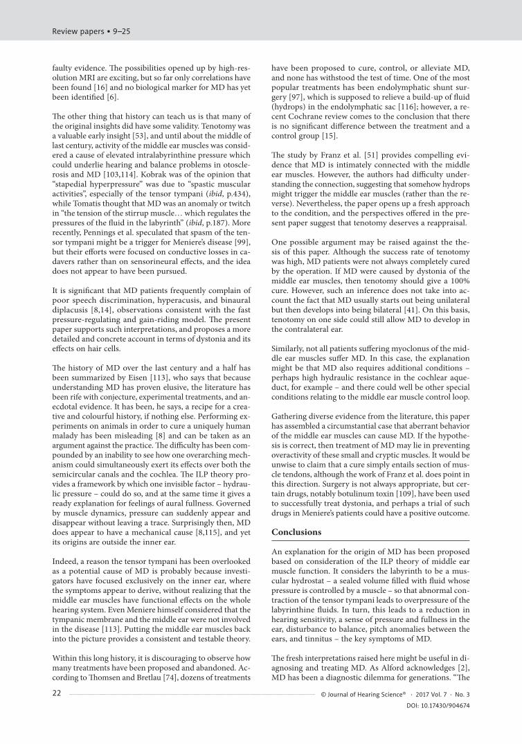
faulty evidence. The possibilities opened up by high-res-olution MRI are exciting, but so far only correlations have been found [16] and no biological marker for MD has yet been identified [6].
The other thing that history can teach us is that many of the original insights did have some validity. Tenotomy was a valuable early insight [53], and until about the middle of last century, activity of the middle ear muscles was consid-ered a cause of elevated intralabyrinthine pressure which could underlie hearing and balance problems in otoscle-rosis and MD [103,114]. Kobrak was of the opinion that “stapedial hyperpressure” was due to “spastic muscular activities”, especially of the tensor tympani (ibid, p.434), while Tomatis thought that MD was an anomaly or twitch in “the tension of the stirrup muscle… which regulates the pressures of the fluid in the labyrinth” (ibid, p.187). More recently, Pennings et al. speculated that spasm of the ten-sor tympani might be a trigger for Meniere’s disease [99], but their efforts were focused on conductive losses in ca-davers rather than on sensorineural effects, and the idea does not appear to have been pursued.
It is significant that MD patients frequently complain of poor speech discrimination, hyperacusis, and binaural diplacusis [8,14], observations consistent with the fast pressure-regulating and gain-riding model. The present paper supports such interpretations, and proposes a more detailed and concrete account in terms of dystonia and its effects on hair cells.
The history of MD over the last century and a half has been summarized by Eisen [113], who says that because understanding MD has proven elusive, the literature has been rife with conjecture, experimental treatments, and an-ecdotal evidence. It has been, he says, a recipe for a crea-tive and colourful history, if nothing else. Performing ex-periments on animals in order to cure a uniquely human malady has been misleading [8] and can be taken as an argument against the practice. The difficulty has been com-pounded by an inability to see how one overarching mech-anism could simultaneously exert its effects over both the semicircular canals and the cochlea. The ILP theory pro-vides a framework by which one invisible factor – hydrau-lic pressure – could do so, and at the same time it gives a ready explanation for feelings of aural fullness. Governed by muscle dynamics, pressure can suddenly appear and disappear without leaving a trace. Surprisingly then, MD does appear to have a mechanical cause [8,115], and yet its origins are outside the inner ear.
Indeed, a reason the tensor tympani has been overlooked as a potential cause of MD is probably because investi-gators have focused exclusively on the inner ear, where the symptoms appear to derive, without realizing that the middle ear muscles have functional effects on the whole hearing system. Even Meniere himself considered that the tympanic membrane and the middle ear were not involved in the disease [113]. Putting the middle ear muscles back into the picture provides a consistent and testable theory.
Within this long history, it is discouraging to observe how many treatments have been proposed and abandoned. Ac-cording to Thomsen and Bretlau [74], dozens of treatments
have been proposed to cure, control, or alleviate MD, and none has withstood the test of time. One of the most popular treatments has been endolymphatic shunt sur-gery [97], which is supposed to relieve a build-up of fluid (hydrops) in the endolymphatic sac [116]; however, a re-cent Cochrane review comes to the conclusion that there is no significant difference between the treatment and a control group [15].
The study by Franz et al. [51] provides compelling evi-dence that MD is intimately connected with the middle ear muscles. However, the authors had difficulty under-standing the connection, suggesting that somehow hydrops might trigger the middle ear muscles (rather than the re-verse). Nevertheless, the paper opens up a fresh approach to the condition, and the perspectives offered in the pre-sent paper suggest that tenotomy deserves a reappraisal.
One possible argument may be raised against the the-sis of this paper. Although the success rate of tenotomy was high, MD patients were not always completely cured by the operation. If MD were caused by dystonia of the middle ear muscles, then tenotomy should give a 100% cure. However, such an inference does not take into ac-count the fact that MD usually starts out being unilateral but then develops into being bilateral [41]. On this basis, tenotomy on one side could still allow MD to develop in the contralateral ear.
Similarly, not all patients suffering myoclonus of the mid-dle ear muscles suffer MD. In this case, the explanation might be that MD also requires additional conditions – perhaps high hydraulic resistance in the cochlear aque-duct, for example – and there could well be other special conditions relating to the middle ear muscle control loop.
Gathering diverse evidence from the literature, this paper has assembled a circumstantial case that aberrant behavior of the middle ear muscles can cause MD. If the hypothe-sis is correct, then treatment of MD may lie in preventing overactivity of these small and cryptic muscles. It would be unwise to claim that a cure simply entails section of mus-cle tendons, although the work of Franz et al. does point in this direction. Surgery is not always appropriate, but cer-tain drugs, notably botulinum toxin [109], have been used to successfully treat dystonia, and perhaps a trial of such drugs in Meniere’s patients could have a positive outcome.
Conclusions
An explanation for the origin of MD has been proposed based on consideration of the ILP theory of middle ear muscle function. It considers the labyrinth to be a mus-cular hydrostat – a sealed volume filled with fluid whose pressure is controlled by a muscle – so that abnormal con-traction of the tensor tympani leads to overpressure of the labyrinthine fluids. In turn, this leads to a reduction in hearing sensitivity, a sense of pressure and fullness in the ear, disturbance to balance, pitch anomalies between the ears, and tinnitus – the key symptoms of MD.
The fresh interpretations raised here might be useful in di-agnosing and treating MD. As Alford acknowledges [2], MD has been a diagnostic dilemma for generations. “The
Review papers • 9–25
22 © Journal of Hearing Science® · 2017 Vol. 7 · No. 3
DOI: 10.17430/904674
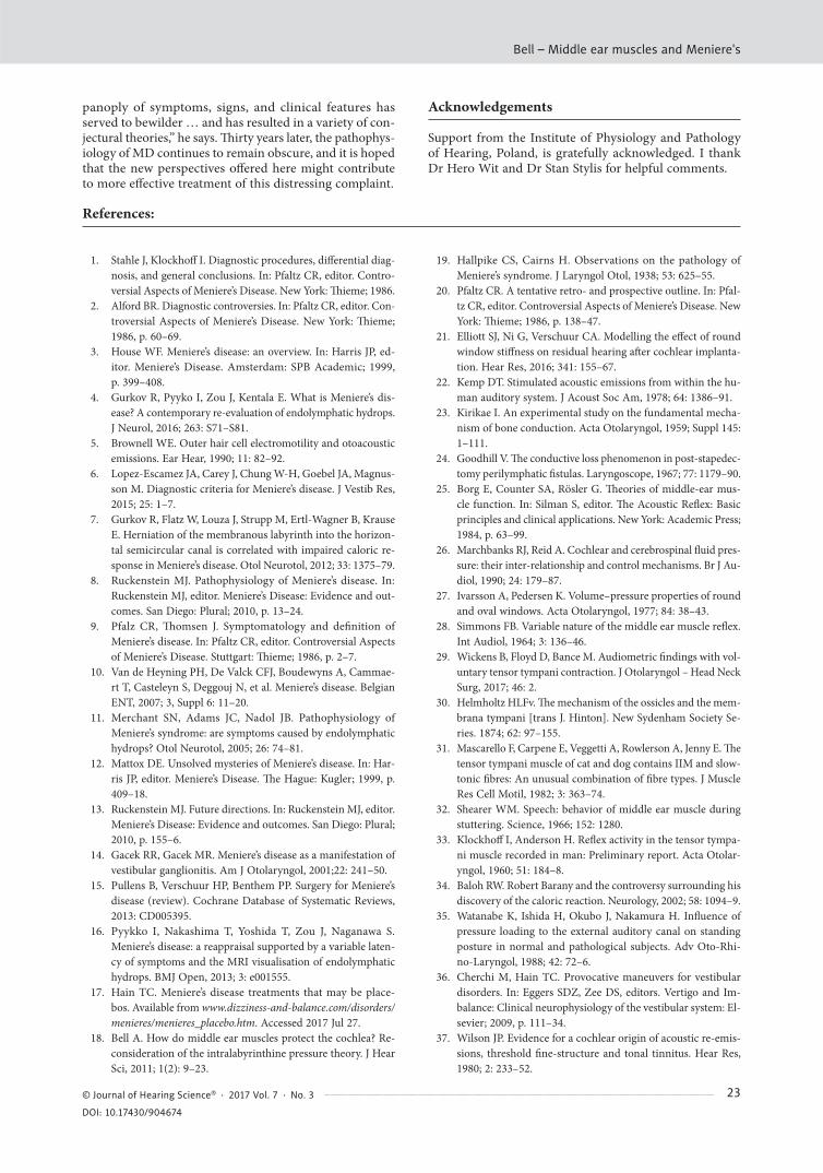
panoply of symptoms, signs, and clinical features has served to bewilder … and has resulted in a variety of con-jectural theories,” he says. Thirty years later, the pathophys-iology of MD continues to remain obscure, and it is hoped that the new perspectives offered here might contribute to more effective treatment of this distressing complaint.
Acknowledgements
Support from the Institute of Physiology and Pathology of Hearing, Poland, is gratefully acknowledged. I thank Dr Hero Wit and Dr Stan Stylis for helpful comments.
1. Stahle J, Klockhoff I. Diagnostic procedures, differential diag-nosis, and general conclusions. In: Pfaltz CR, editor. Contro-versial Aspects of Meniere’s Disease. New York: Thieme; 1986.
2. Alford BR. Diagnostic controversies. In: Pfaltz CR, editor. Con-troversial Aspects of Meniere’s Disease. New York: Thieme; 1986, p. 60–69.
3. House WF. Meniere’s disease: an overview. In: Harris JP, ed-itor. Meniere’s Disease. Amsterdam: SPB Academic; 1999, p. 399–408.
4. Gurkov R, Pyyko I, Zou J, Kentala E. What is Meniere’s dis-ease? A contemporary re-evaluation of endolymphatic hydrops. J Neurol, 2016; 263: S71–S81.
5. Brownell WE. Outer hair cell electromotility and otoacoustic emissions. Ear Hear, 1990; 11: 82–92.
6. Lopez-Escamez JA, Carey J, Chung W-H, Goebel JA, Magnus-son M. Diagnostic criteria for Meniere’s disease. J Vestib Res, 2015; 25: 1–7.
7. Gurkov R, Flatz W, Louza J, Strupp M, Ertl-Wagner B, Krause E. Herniation of the membranous labyrinth into the horizon-tal semicircular canal is correlated with impaired caloric re-sponse in Meniere’s disease. Otol Neurotol, 2012; 33: 1375–79.
8. Ruckenstein MJ. Pathophysiology of Meniere’s disease. In: Ruckenstein MJ, editor. Meniere’s Disease: Evidence and out-comes. San Diego: Plural; 2010, p. 13–24.
9. Pfalz CR, Thomsen J. Symptomatology and definition of Meniere’s disease. In: Pfaltz CR, editor. Controversial Aspects of Meniere’s Disease. Stuttgart: Thieme; 1986, p. 2–7.
10. Van de Heyning PH, De Valck CFJ, Boudewyns A, Cammae-rt T, Casteleyn S, Deggouj N, et al. Meniere’s disease. Belgian ENT, 2007; 3, Suppl 6: 11–20.
11. Merchant SN, Adams JC, Nadol JB. Pathophysiology of Meniere’s syndrome: are symptoms caused by endolymphatic hydrops? Otol Neurotol, 2005; 26: 74–81.
12. Mattox DE. Unsolved mysteries of Meniere’s disease. In: Har-ris JP, editor. Meniere’s Disease. The Hague: Kugler; 1999, p. 409–18.
13. Ruckenstein MJ. Future directions. In: Ruckenstein MJ, editor. Meniere’s Disease: Evidence and outcomes. San Diego: Plural; 2010, p. 155–6.
14. Gacek RR, Gacek MR. Meniere’s disease as a manifestation of vestibular ganglionitis. Am J Otolaryngol, 2001;22: 241–50.
15. Pullens B, Verschuur HP, Benthem PP. Surgery for Meniere’s disease (review). Cochrane Database of Systematic Reviews, 2013: CD005395.
16. Pyykko I, Nakashima T, Yoshida T, Zou J, Naganawa S. Meniere’s disease: a reappraisal supported by a variable laten-cy of symptoms and the MRI visualisation of endolymphatic hydrops. BMJ Open, 2013; 3: e001555.
17. Hain TC. Meniere’s disease treatments that may be place-bos. Available from www.dizziness-and-balance.com/disorders/menieres/menieres_placebo.htm. Accessed 2017 Jul 27.
18. Bell A. How do middle ear muscles protect the cochlea? Re-consideration of the intralabyrinthine pressure theory. J Hear Sci, 2011; 1(2): 9–23.
References:
19. Hallpike CS, Cairns H. Observations on the pathology of Meniere’s syndrome. J Laryngol Otol, 1938; 53: 625–55.
20. Pfaltz CR. A tentative retro- and prospective outline. In: Pfal-tz CR, editor. Controversial Aspects of Meniere’s Disease. New York: Thieme; 1986, p. 138–47.
21. Elliott SJ, Ni G, Verschuur CA. Modelling the effect of round window stiffness on residual hearing after cochlear implanta-tion. Hear Res, 2016; 341: 155–67.
22. Kemp DT. Stimulated acoustic emissions from within the hu-man auditory system. J Acoust Soc Am, 1978; 64: 1386–91.
23. Kirikae I. An experimental study on the fundamental mecha-nism of bone conduction. Acta Otolaryngol, 1959; Suppl 145: 1–111.
24. Goodhill V. The conductive loss phenomenon in post-stapedec-tomy perilymphatic fistulas. Laryngoscope, 1967; 77: 1179–90.
25. Borg E, Counter SA, Rösler G. Theories of middle-ear mus-cle function. In: Silman S, editor. The Acoustic Reflex: Basic principles and clinical applications. New York: Academic Press; 1984, p. 63–99.
26. Marchbanks RJ, Reid A. Cochlear and cerebrospinal fluid pres-sure: their inter-relationship and control mechanisms. Br J Au-diol, 1990; 24: 179–87.
27. Ivarsson A, Pedersen K. Volume–pressure properties of round and oval windows. Acta Otolaryngol, 1977; 84: 38–43.
28. Simmons FB. Variable nature of the middle ear muscle reflex. Int Audiol, 1964; 3: 136–46.
29. Wickens B, Floyd D, Bance M. Audiometric findings with vol-untary tensor tympani contraction. J Otolaryngol – Head Neck Surg, 2017; 46: 2.
30. Helmholtz HLFv. The mechanism of the ossicles and the mem-brana tympani [trans J. Hinton]. New Sydenham Society Se-ries. 1874; 62: 97–155.
31. Mascarello F, Carpene E, Veggetti A, Rowlerson A, Jenny E. The tensor tympani muscle of cat and dog contains IIM and slow-tonic fibres: An unusual combination of fibre types. J Muscle Res Cell Motil, 1982; 3: 363–74.
32. Shearer WM. Speech: behavior of middle ear muscle during stuttering. Science, 1966; 152: 1280.
33. Klockhoff I, Anderson H. Reflex activity in the tensor tympa-ni muscle recorded in man: Preliminary report. Acta Otolar-yngol, 1960; 51: 184–8.
34. Baloh RW. Robert Barany and the controversy surrounding his discovery of the caloric reaction. Neurology, 2002; 58: 1094–9.
35. Watanabe K, Ishida H, Okubo J, Nakamura H. Influence of pressure loading to the external auditory canal on standing posture in normal and pathological subjects. Adv Oto-Rhi-no-Laryngol, 1988; 42: 72–6.
36. Cherchi M, Hain TC. Provocative maneuvers for vestibular disorders. In: Eggers SDZ, Zee DS, editors. Vertigo and Im-balance: Clinical neurophysiology of the vestibular system: El-sevier; 2009, p. 111–34.
37. Wilson JP. Evidence for a cochlear origin of acoustic re-emis-sions, threshold fine-structure and tonal tinnitus. Hear Res, 1980; 2: 233–52.
Bell – Middle ear muscles and Meniere's
23© Journal of Hearing Science® · 2017 Vol. 7 · No. 3
DOI: 10.17430/904674
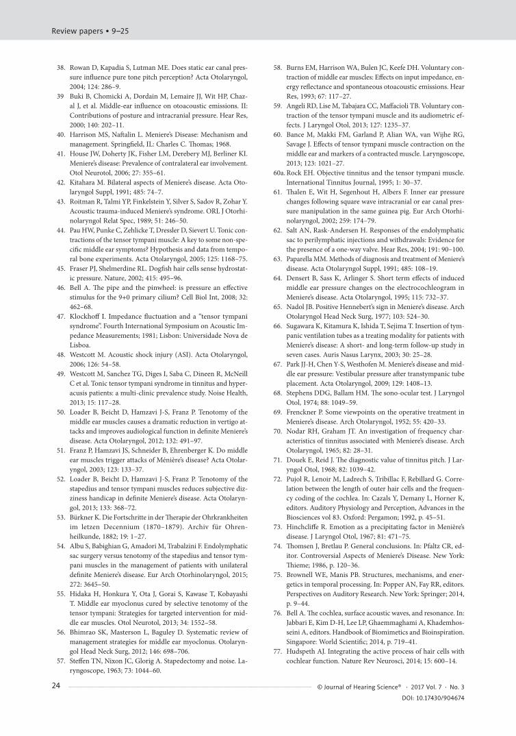
38. Rowan D, Kapadia S, Lutman ME. Does static ear canal pres-sure influence pure tone pitch perception? Acta Otolaryngol, 2004; 124: 286–9.
39 Buki B, Chomicki A, Dordain M, Lemaire JJ, Wit HP, Chaz-al J, et al. Middle-ear influence on otoacoustic emissions. II: Contributions of posture and intracranial pressure. Hear Res, 2000; 140: 202–11.
40. Harrison MS, Naftalin L. Meniere’s Disease: Mechanism and management. Springfield, IL: Charles C. Thomas; 1968.
41. House JW, Doherty JK, Fisher LM, Derebery MJ, Berliner KI. Meniere’s disease: Prevalence of contralateral ear involvement. Otol Neurotol, 2006; 27: 355–61.
42. Kitahara M. Bilateral aspects of Meniere’s disease. Acta Oto-laryngol Suppl, 1991; 485: 74–7.
43. Roitman R, Talmi YP, Finkelstein Y, Silver S, Sadov R, Zohar Y. Acoustic trauma-induced Meniere’s syndrome. ORL J Otorhi-nolaryngol Relat Spec, 1989; 51: 246–50.
44. Pau HW, Punke C, Zehlicke T, Dressler D, Sievert U. Tonic con-tractions of the tensor tympani muscle: A key to some non-spe-cific middle ear symptoms? Hypothesis and data from tempo-ral bone experiments. Acta Otolaryngol, 2005; 125: 1168–75.
45. Fraser PJ, Shelmerdine RL. Dogfish hair cells sense hydrostat-ic pressure. Nature, 2002; 415: 495–96.
46. Bell A. The pipe and the pinwheel: is pressure an effective stimulus for the 9+0 primary cilium? Cell Biol Int, 2008; 32: 462–68.
47. Klockhoff I. Impedance fluctuation and a “tensor tympani syndrome”. Fourth International Symposium on Acoustic Im-pedance Measurements; 1981; Lisbon: Universidade Nova de Lisboa.
48. Westcott M. Acoustic shock injury (ASI). Acta Otolaryngol, 2006; 126: 54–58.
49. Westcott M, Sanchez TG, Diges I, Saba C, Dineen R, McNeill C et al. Tonic tensor tympani syndrome in tinnitus and hyper-acusis patients: a multi-clinic prevalence study. Noise Health, 2013; 15: 117–28.
50. Loader B, Beicht D, Hamzavi J-S, Franz P. Tenotomy of the middle ear muscles causes a dramatic reduction in vertigo at-tacks and improves audiological function in definite Meniere’s disease. Acta Otolaryngol, 2012; 132: 491–97.
51. Franz P, Hamzavi JS, Schneider B, Ehrenberger K. Do middle ear muscles trigger attacks of Ménière’s disease? Acta Otolar-yngol, 2003; 123: 133–37.
52. Loader B, Beicht D, Hamzavi J-S, Franz P. Tenotomy of the stapedius and tensor tympani muscles reduces subjective diz-ziness handicap in definite Meniere’s disease. Acta Otolaryn-gol, 2013; 133: 368–72.
53. Bürkner K. Die Fortschritte in der Therapie der Ohrkrankheiten im letzen Decennium (1870–1879). Archiv für Ohren-heilkunde, 1882; 19: 1–27.
54. Albu S, Babighian G, Amadori M, Trabalzini F. Endolymphatic sac surgery versus tenotomy of the stapedius and tensor tym-pani muscles in the management of patients with unilateral definite Meniere’s disease. Eur Arch Otorhinolaryngol, 2015; 272: 3645–50.
55. Hidaka H, Honkura Y, Ota J, Gorai S, Kawase T, Kobayashi T. Middle ear myoclonus cured by selective tenotomy of the tensor tympani: Strategies for targeted intervention for mid-dle ear muscles. Otol Neurotol, 2013; 34: 1552–58.
56. Bhimrao SK, Masterson L, Baguley D. Systematic review of management strategies for middle ear myoclonus. Otolaryn-gol Head Neck Surg, 2012; 146: 698–706.
57. Steffen TN, Nixon JC, Glorig A. Stapedectomy and noise. La-ryngoscope, 1963; 73: 1044–60.
58. Burns EM, Harrison WA, Bulen JC, Keefe DH. Voluntary con-traction of middle ear muscles: Effects on input impedance, en-ergy reflectance and spontaneous otoacoustic emissions. Hear Res, 1993; 67: 117–27.
59. Angeli RD, Lise M, Tabajara CC, Maffacioli TB. Voluntary con-traction of the tensor tympani muscle and its audiometric ef-fects. J Laryngol Otol, 2013; 127: 1235–37.
60. Bance M, Makki FM, Garland P, Alian WA, van Wijhe RG, Savage J. Effects of tensor tympani muscle contraction on the middle ear and markers of a contracted muscle. Laryngoscope, 2013; 123: 1021–27.
60a. Rock EH. Objective tinnitus and the tensor tympani muscle. International Tinnitus Journal, 1995; 1: 30–37.
61. Thalen E, Wit H, Segenhout H, Albers F. Inner ear pressure changes following square wave intracranial or ear canal pres-sure manipulation in the same guinea pig. Eur Arch Otorhi-nolaryngol, 2002; 259: 174–79.
62. Salt AN, Rask-Andersen H. Responses of the endolymphatic sac to perilymphatic injections and withdrawals: Evidence for the presence of a one-way valve. Hear Res, 2004; 191: 90–100.
63. Paparella MM. Methods of diagnosis and treatment of Meniere’s disease. Acta Otolaryngol Suppl, 1991; 485: 108–19.
64. Densert B, Sass K, Arlinger S. Short term effects of induced middle ear pressure changes on the electrocochleogram in Meniere’s disease. Acta Otolaryngol, 1995; 115: 732–37.
65. Nadol JB. Positive Hennebert’s sign in Meniere’s disease. Arch Otolaryngol Head Neck Surg, 1977; 103: 524–30.
66. Sugawara K, Kitamura K, Ishida T, Sejima T. Insertion of tym-panic ventilation tubes as a treating modality for patients with Meniere’s disease: A short- and long-term follow-up study in seven cases. Auris Nasus Larynx, 2003; 30: 25–28.
67. Park JJ-H, Chen Y-S, Westhofen M. Meniere’s disease and mid-dle ear pressure: Vestibular pressure after transtympanic tube placement. Acta Otolaryngol, 2009; 129: 1408–13.
68. Stephens DDG, Ballam HM. The sono-ocular test. J Laryngol Otol, 1974; 88: 1049–59.
69. Frenckner P. Some viewpoints on the operative treatment in Meniere’s disease. Arch Otolaryngol, 1952; 55: 420–33.
70. Nodar RH, Graham JT. An investigation of frequency char-acteristics of tinnitus associated with Meniere’s disease. Arch Otolaryngol, 1965; 82: 28–31.
71. Douek E, Reid J. The diagnostic value of tinnitus pitch. J Lar-yngol Otol, 1968; 82: 1039–42.
72. Pujol R, Lenoir M, Ladrech S, Tribillac F, Rebillard G. Corre-lation between the length of outer hair cells and the frequen-cy coding of the cochlea. In: Cazals Y, Demany L, Horner K, editors. Auditory Physiology and Perception, Advances in the Biosciences vol 83. Oxford: Pergamon; 1992, p. 45–51.
73. Hinchcliffe R. Emotion as a precipitating factor in Menière’s disease. J Laryngol Otol, 1967; 81: 471–75.
74. Thomsen J, Bretlau P. General conclusions. In: Pfaltz CR, ed-itor. Controversial Aspects of Meniere’s Disease. New York: Thieme; 1986, p. 120–36.
75. Brownell WE, Manis PB. Structures, mechanisms, and ener-getics in temporal processing. In: Popper AN, Fay RR, editors. Perspectives on Auditory Research. New York: Springer; 2014, p. 9–44.
76. Bell A. The cochlea, surface acoustic waves, and resonance. In: Jabbari E, Kim D-H, Lee LP, Ghaemmaghami A, Khademhos-seini A, editors. Handbook of Biomimetics and Bioinspiration. Singapore: World Scientific; 2014, p. 719–41.
77. Hudspeth AJ. Integrating the active process of hair cells with cochlear function. Nature Rev Neurosci, 2014; 15: 600–14.
Review papers • 9–25
24 © Journal of Hearing Science® · 2017 Vol. 7 · No. 3
DOI: 10.17430/904674
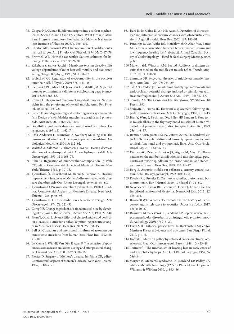
78. Cooper NP, Guinan JJ. Efferent insights into cochlear mechan-ics. In: Shera CA and Olson ES, editors. What Fire is in Mine Ears: Progress in Auditory Biomechanics. Melville, NY: Amer-ican Institute of Physics; 2005, p. 396–402.
79. Chertoff ME, Brownell WE. Characterization of cochlear outer hair cell turgor. Am J Physiol Cell Physiol, 1994; 35: C467–79.
80. Brownell WE. How the ear works: Nature’s solutions for lis-tening. Volta Review, 1997; 99: 9–28.
81. Kakehata S, Santos-Sacchi J. Membrane tension directly shifts voltage dependence of outer hair cell motility and associated gating charge. Biophys J, 1995; 68: 2190–97.
82. Frolenkov GI. Regulation of electromotility in the cochlear outer hair cell. J Physiol, 2006; 576.1: 43–48.
83. Elemans CPH, Mead AF, Jakobsen L, Ratcliffe JM. Superfast muscles set maximum call rate in echolocating bats. Science, 2011; 333: 1885–88.
84. Rome LC. Design and function of superfast muscles: New in-sights into the physiology of skeletal muscle. Annu Rev Phys-iol, 2006; 68: 193–221.
85. Ladich F. Sound-generating and -detecting motor system in cat-fish: Design of swimbladder muscles in doradids and pimelo-dids. Anat Rec, 2001; 263: 297–306.
86. Goodhill V. Sudden deafness and round window rupture. La-ryngoscope, 1971; 81: 1462–74.
87. Rask-Andersen H, Kinnefors A, Svedberg M, Illing R-B. The human round window: A perilymph pressure regulator? Au-diological Medicine, 2004; 3: 182–92.
88. Walsted A, Salomon G, Thomsen J, Tos M. Hearing decrease after loss of cerebrospinal fluid: A new hydrops model? Acta Otolaryngol, 1991; 111: 468–76.
89. Juhn SK. Regulation of inner ear fluids composition. In: Pfalz CR, editor. Controversial Aspects of Meniere’s Disease. New York: Thieme; 1986, p. 10–15.
90. Tjernström Ö, Casselbrant M, Harris S, Ivarsson A. Hearing improvement in attacks of Meniere’s disease treated with pres-sure chamber. Adv Oto-Rhino-Laryngol, 1979; 25: 54–60.
91. Tjernström Ö. Pressure chamber treatment. In: Pfaltz CR, ed-itor. Controversial Aspects of Meniere’s Disease. New York: Thieme; 1986, p. 96–98.
92. Tjernstrom O. Further studies on alternobaric vertigo. Acta Otolaryngol, 1974; 78: 221–31.
93. Corey VB. Change in pitch of sustained musical note by clench-ing of the jaws of the observer. J Acoust Soc Am, 1950; 22: 640.
94. Mom T, Gilain L, Avan P. Effects of glycerol intake and body tilt on otoacoustic emissions reflect labyrinthine pressure chang-es in Meniere’s disease. Hear Res, 2009; 250: 38–45.
95. Bell A. Circadian and menstrual rhythms of spontaneous otoacoustic emissions from human ears. Hear Res, 1992; 58: 91–100.
96. de Kleine E, Wit HP, Van Dijk P, Avan P. The behavior of spon-taneous otoacoustic emissions during and after postural chang-es. J Acoust Soc Am, 2000; 107: 3308–16.
97. Plester D. Surgery of Meniere’s disease. In: Pfaltz CR, editor. Controversial Aspects of Meniere’s Disease. New York: Thieme; 1986, p. 104–12.
98. Buki B, de Kleine E, Wit HP, Avan P. Detection of intracoch-lear and intracranial pressure changes with otoacoustic emis-sions: A gerbil model. Hear Res, 2002; 167: 180–91.
99. Pennings R, Van Wijhe RG, Majdalawieh O, Alian WA, Bance M. Is there a correlation between tensor tympani spasm and low-frequency hearing loss? [abstract]. Annual Canadian Soci-ety of Otolaryngology – Head & Neck Surgery Meeting, 2009, p. 63.
100. Mukerji SM, Windsor AM, Lee DJ. Auditory brainstem cir-cuits that mediate the middle ear muscle reflex. Trends Amp-lif, 2010; 14: 170–91.
101. Simmons FB. Perceptual theories of middle ear muscle func-tion. Ann Otol, 1964; 73: 724–29.
102. Salt AN, DeMott JE. Longitudinal endlolymph movements and endocochlear potential changes induced by stimulation at in-frasonic frequencies. J Acoust Soc Am, 1999; 106: 847–56.
103. Tomatis AA. The Conscious Ear. Barrytown, NY: Station Hill Press; 1992.
104. Yonovitz A, Harris JD. Eardrum displacement following sta-pedius muscle contraction. Acta Otolaryngol, 1976; 81: 1–15.
105. Han Y, Wang J, Fischman DA, Biller HF, Sanders I. Slow ton-ic muscle fibers in the thyroarytenoid muscles of human vo-cal folds: A possible specialization for speech. Anat Rec, 1999; 256: 146–57.
106. Ramirez Aristeguieta LM, Ballesteros Acuna LE, Sandoval Or-tiz GP. Tensor veli palatine and tensor tympani muscles: ana-tomical, functional and symptomatic links. Acta Otorrinola-ringol Esp, 2010; 61: 26–33.
107. Kierner AC, Zelenka I, Lukas JR, Aigner M, Mayr R. Obser-vations on the number, distribution and morphological pecu-liarities of muscle spindles in the tensor tympani and stapedi-us muscle of man. Hear Res, 1999; 135: 71–77.
108. Borg E. Acoustic middle ear reflexes: a sensory-control sys-tem. Acta Otolaryngol Suppl, 1972; 304: 1–34.
109. Rosales RL, Dressler D. On muscle spindles, dystonia and bot-ulinum toxin. Eur J Neurol, 2010; 17 (Suppl 1): 71–80.
110. Neychev VK, Gross RE, Lehericy S, Hess EJ, Jinnah HA. The functional anatomy of dystonia. Neurobiol Dis, 2011; 42: 185–201.
111. Brownell WE. What is electromotility? The history of its dis-covery and its relevance to acoustics. Acoustics Today, 2017; 13(1): 20–27.
112. Ramirez LM, Ballesteros LE, Sandoval GP. Topical review: Tem-poromandibular disorders in an integral otic symptom mod-el. Audiology, 2008; 47: 215–27.
113. Eisen MD. Historical perspectives. In: Ruckenstein MJ, editor. Meniere’s Disease: Evidence and outcomes. San Diego: Plural; 2010, p. 1–6.
114. Kobrak F. Study on pathophysiological factors in clinical oto-sclerosis. Pract Otorhinolaryngol (Basel). 1948; 10: 423–48.
115. Tonndorf J. The mechanism of hearing loss in early cases of endolymphatic hydrops. Ann Otol Rhinol Laryngol, 1957; 66: 766–84.
116. Storper IS. Meniere’s syndrome. In: Rowland LP, Pedley TA, editors. Merritt’s Neurology (12th ed). Philadelphia: Lippincott Williams & Wilkins; 2010, p. 963–66.
Bell – Middle ear muscles and Meniere's
25© Journal of Hearing Science® · 2017 Vol. 7 · No. 3
DOI: 10.17430/904674