Embed Size (px)
Citation preview
MHC class-I associated phosphopeptides are the targets of memory-like immunity
in leukemia
Running Head
Immunological targeting of the leukemia phosphoproteome
Authors and Affiliations
Mark Cobbold1,2*
Hugo De La Peña2*
Andrew Norris3
Joy M. Polefrone3
Jie Qian3
A. Michelle English3
Kara L. Cummings1
Sarah Penny2
James E. Turner2
Jennifer Cottine3
Jennifer G Abelin3
Stacy A Malaker3
Angela L Zarling1
Hsing-Wen Huang2
Oliver Goodyear2
Sylvie D. Freeman2
Jeffrey Shabanowitz3
Guy Pratt4
Charles Craddock5,6
Michael E Williams7
Donald F Hunt3,8
Victor H Engelhard1†
1 Carter Immunology Center and Department of Microbiology, Immunology and Cancer
Biology, University of Virginia, Charlottesville, Virginia 22908
2 Medical Research Council Centre for Immune Regulation and Clinical Immunology Service,
School of Immunity and Infection, College of Medicine and Dental Sciences, University of
Birmingham, Birmingham, UK.
3 Department of Chemistry, University of Virginia, Charlottesville, Virginia 22901, USA.
4 Cancer Research UK Institute for Cancer Studies, School of Cancer Sciences, University of
Birmingham, Birmingham, UK
5 Cancer Research UK Clinical Trials Unit, School of Cancer Sciences, University of
Birmingham, Birmingham, UK;
6 Centre for Clinical Haematology, Queen Elizabeth Hospital, Birmingham, UK,
7 Division of Hematology/Oncology and Hematologic Malignancy Program, University of
Virginia School of Medicine, Charlottesville, VA 22908
8 Department of Pathology, University of Virginia, Charlottesville, Virginia 22908, USA.
†Email: [email protected], [email protected]
Abstract
Deregulation of signaling pathways involving phosphorylation is a hallmark of malignant
transformation. Degradation of phosphoproteins generates cancer-specific phosphopeptides that
are associated with MHC-I and II molecules and recognized by T-cells. We identified 95
phosphopeptides presented on the surface of primary hematological tumors and normal tissues,
including 61 that were tumor-specific. Phosphopeptides were more prevalent on more
aggressive and malignant samples. CD8 T-cell lines specific for these phosphopeptides
recognized and killed both leukemia cell lines and HLA-matched primary leukemia cells ex vivo.
Healthy individuals showed surprisingly high levels of CD8 T-cell responses against many of
these phosphopeptides within the circulating memory compartment. This immunity was
significantly reduced or absent in some leukemia patients, which correlated with clinical
outcome, and was restored following allogeneic stem cell transplantation. These results suggest
that phosphopeptides may be targets of cancer immune surveillance in humans, and point to their
importance for development of vaccine-based and T-cell adoptive transfer immunotherapies..
One-sentence summary
In this work we characterize 95 leukemia-associated phosphopeptide antigens and show that
healthy donors exhibit unexpected immunity against many of these which is lacking in patients
with leukemia but can be restored following stem cell transplantation.
Introduction
Hematologic malignancies are susceptible to curative immunological therapies, such as stem cell
transplantation, donor lymphocyte infusion and manipulation of endogenous immunity through
immunostimulatory cytokines (1-4). The clinical utility of cellular immunotherapies provides
powerful direct evidence that the adaptive immune response can control and eradicate tumors(5-
9). However, immunotherapies targeting identified tumor antigens have met with limited
success, suggesting the optimal antigens remain undiscovered (10-12). Moreover, the vast
majority of identified tumor antigens are not derived from oncoproteins orchestrating the
transformation process. Efforts targeting these antigens may be compromised by tolerance
mechanisms and immune escape.
The importance of signal-transduction pathway deregulation in cancer pathogenesis is
well-established (13-17). Small molecule therapies targeting these pathways have met with
considerable clinical success, providing a powerful argument for immunotherapies that target
similar deregulated pathways (18-20). Protein phosphorylation is the dominant mechanism
involved in oncogenic signaling processes (21). We previously showed that phosphorylation is
preserved on peptides during antigen processing for presentation by both MHC-I and –II
molecules (22-24). This suggests that phosphopeptide antigens derived from cancer-related
phosphoproteins could serve as immunological signatures of 'transformed self' (22-26).
Moreover, phosphorylation can enhance the binding of peptides to MHC-I molecules, creating
“neo-antigens”. Collectively these results suggest that phosphopeptides are attractive targets for
cancer immunotherapy (27).
To date, no studies have examined MHC-I-bound phosphopeptides displayed on primary
human tumor samples, nor has the human immune response against MHC-I restricted
phosphopeptides been characterized. Here, we identify novel phosphopeptides from primary
leukemia samples and show that healthy individuals display immune responses with memory
characteristics against many of them. Immunity is lost in some patients with leukemia, but can be
restored. This unexpected pre-existing immunity to phosphopeptides suggests that they could be
targets of immune surveillance, and play a role in enabling immunological control of
malignancies in humans.
Results
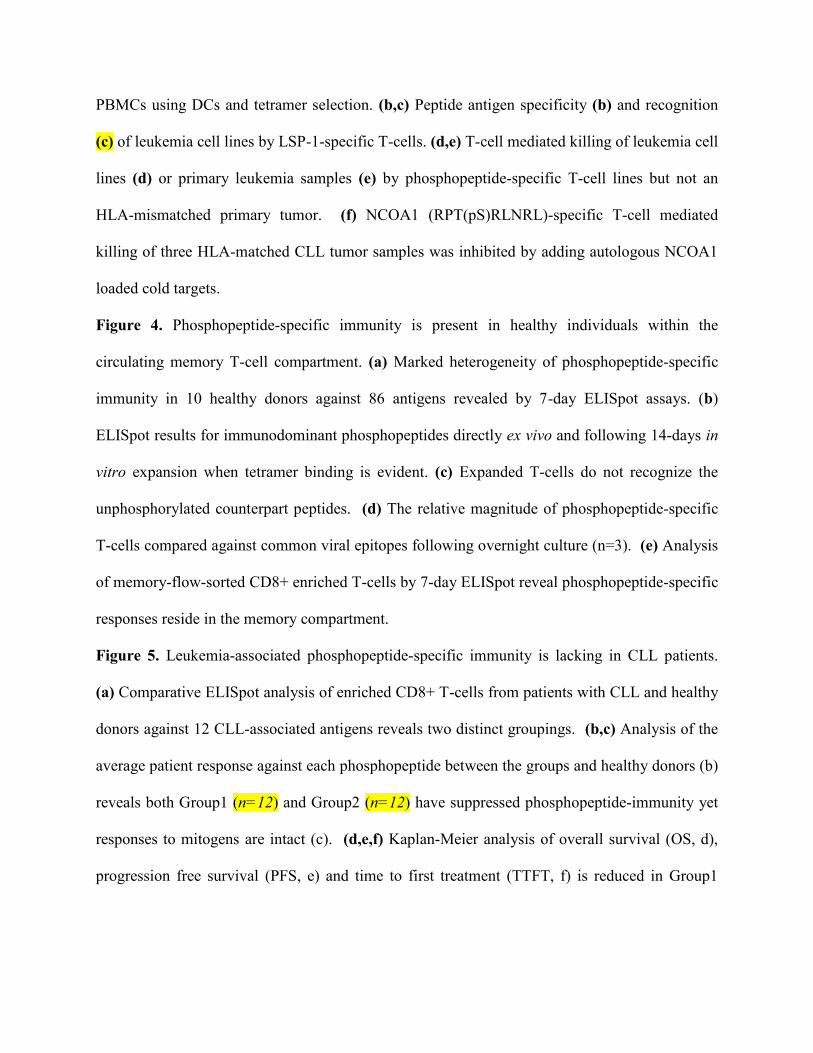
Characterization of Leukemia-Associated MHC Class-I Restricted Phosphopeptides
To identify tumor-associated phosphopeptides, we affinity-isolated HLA-A*0201 (HLA-A2) and
HLA-B*0702 (HLA-B7) peptide complexes from 4 primary chronic lymphocytic leukemia
(CLL) tumors, a primary hairy-cell leukemia (HCL), a primary mantle cell lymphoma (MCL), a
primary acute lymphoblastic leukemia (ALL), and a primary acute myeloid leukemia (AML), in
addition to normal splenic T and B-cells, bone marrow cells (BM), and cultured B-
lymphoblastoid cell lines (B-LCL). Collectively, 10 HLA-A2-restricted and 85 HLA-B7-
restricted phosphopeptides were identified (Fig. 1a). All tumor types and normal tissues
expressed a greater number of HLA-B7 than HLA-A2 phosphopeptides (Fig. 1b).
The large number of HLA-B7-restricted phosphopeptides enabled us to compare their
representation on different tumor types and healthy tissue. On average more than twice as many
were found on aggressive (AML and ALL) as on indolent (CLL and HCL) tumors or normal
tissue (Fig. 1c). Of 56 HLA-B7-restricted phosphopeptides identified on AML, 36 were not
found on any other leukemia and only two were found on bone marrow (Figs. 1a, 2a). Of 45
phosphopeptides identified on ALL, 19 were not found on other leukemias and 13/19 were also
not observed on T- or B-cells (Fig. 2a). Seven were derived from oncogenes implicated in
leukemogenesis: MYC, EP300, SKI, GFI-1, Bcl-11A, MEF2D, and MLL (Fig. 1a). Twenty-
seven HLA-B7 phosphopeptides were identified on two CLL tumors, all of which were shared
with either AML or ALL. Sixteen were observed on normal tissue but seven were common to
AML, ALL and CLL and not normal tissue (CCDC45, GRK2, SETD2, C17orf85, TSC22D4 and
SPR(pS)PGKPM, derived from an unknown protein). These proteins have not been previously
associated with leukemic malignancies and the functions of most remain undefined. Twenty-six
of 27 CLL peptides were also found on HCL, which itself expressed one additional unique
phosphopeptide. Twenty-four of these were also shared by B-LCL (Fig. 2b). These results
identify a cohort of phosphopeptides expressed on multiple leukemic malignancies, but not
normal tissue, which represent potential immunotherapeutic targets.
Characteristics of the HLA-A2 bound phosphopeptides were similar to those previously
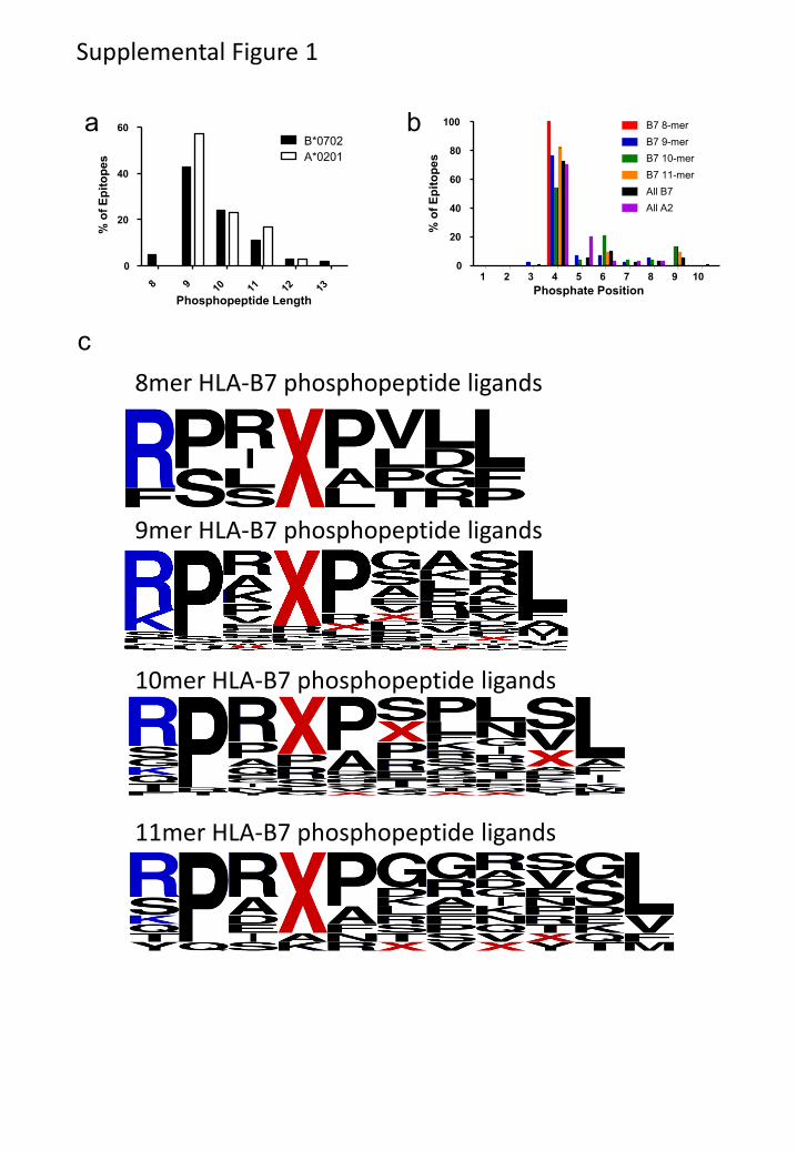
reported (22, 23). Of 85 HLA-B7-restricted phosphopeptides, two were dually phosphorylated
and the remainder monophosphorylated, with 77/85 containing phosphoserine and 9/85
containing phosphothreonine. The phosphate was found at position four in 72% of HLA-B7
phosphopeptides, similar to the distribution in HLA-A2 phosphopeptides (Fig. S1a,b) (27).
When compared with 1038 non-phosphorylated HLA-B7-restricted 9mer peptides in the
ImmuneEpitope database (28), HLA-B7-restricted phosphopeptides showed a similar strong
preference for proline at P2 and common hydrophobic C-terminal anchor residues (Fig. 2c, d).
However, they showed an unusual bias for basic residues at P1 and proline at P5 (Fig 2c, Fig.
S1c). To test whether these biases were imposed by an underlying kinase recognition motif,
potential HLA-B7 binding 9mer peptides were identified in the Phosphosite dataset (29) of
known serine phosphorylation sites. The position of the phosphoserine within 1031

phosphopeptides predicted to bind to HLA-B7 was not skewed towards P4 (Fig. 2e). Thus, these
biases likely reflect roles in binding to HLA-B7 analogous to those demonstrated for HLA-A2-
associated phosphopeptides (27). Of 164 predicted HLA-B7 binding peptides that had
phosphoserine at P4 there was no apparent bias for basic residues at P1 (Fig. 2f). However 87%
contained a proline at P5 suggesting this bias reflects an underlying kinase motif, rather than
being imposed by HLA binding.
Phosphopeptide-specific T-cell responses in healthy donors
Three HLA-A2 and two HLA-B7 phosphopeptides were derived from LSP-1, a lymphoma
marker (30). One, RQA(pS)IELPSMAV, is present on all HLA-A2+ tumor samples at high copy
number. RQA(pS)IELPSMAV-pulsed dendritic cells were used to prime autologous T-cells.
Responses to this phosphopeptide could be elicited in 3/3 healthy individuals. Specific T-cells
were enriched using HLA-A2- RQA(pS)IELPSMAV tetramers to produce T-cell lines (Fig. 3a).
These lines secreted IFNγ in response to stimulators pulsed with RQA(pS)IELPSMAV, but not
with unphosphorylated RQASIELPSMAV, nor to other phosphopeptides, including the closely
related RQA(pS)IELPSM (Fig. 3b). Recognition was therefore both phosphate-dependent and
peptide sequence-specific. These lines also killed the HLA-A2+ AML cell line THP-1 and the
HLA-A2 transfected ALL cell line, Jurkat-A2, but not untransfected Jurkat (Fig. 3c-d). Most
importantly, RQA(pS)IELPSMAV-specific T-cells killed HLA-A2+ primary AML and CLL
tumors, but not an HLA-A2neg
CLL tumor (Fig. 3e). T-cell lines were also elicited against an
HLA-B7 restricted phosphopeptide (RPT(pS)RLNRL) derived from NCOA-1 and showed
remarkable phosphopeptide-specific killing of three primary CLL tumors (Fig. 3f).
We next evaluated immunity in 10 healthy donors against 10 HLA-A2 and 76 HLA-B7
leukemia-associated phosphopeptides (Fig. 4a). Immune responses to 50/76 HLA-B7 and 9/10
HLA-A2 phosphopeptides were observed. Each individual responded to an average of 16/76
(range 11-22) HLA-B7 and 2/10 (range 0-4) HLA-A2 phosphopeptides. Importantly, these
responses were observed in 7-day in vitro cultures without addition of exogenous cytokines. No
responses of a similar magnitude were observed for several phosphopeptides encoded by well-
established leukemia oncogenes, most notably MYC, BCL-11A and EP300. However, the MYC
and BCL-11A phosphopeptides were also present on normal T-cells (Fig. 1a). Phosphopeptides
from other leukemia oncogenes (MLL, LPP, SKI, GFI-1 and MEF2D) elicited strong immune
responses in several individuals. There was substantial donor to donor variation as to which
phosphopeptides stimulated these unusually strong responses: the GFI1 phosphopeptide
stimulated responses in 9/10 individuals; the MLL, LPP, and MAP3K11 phosphopeptides in
8/10; and those from LSP1, SCAP and RBM15 in 7/10. For four phosphopeptides that were
recognized by most healthy donors, T-cell specificity was confirmed in 14-day cultures using
HLA-phosphopeptide multimers (Fig. 4b). Furthermore, these T-cells did not recognize the
unphosphorylated counterpart peptides (Fig. 4c, Fig. S2). Collectively, these data demonstrate
that the majority of leukemia-associated phosphopeptides elicited surprisingly strong and
specific responses in a significant fraction of healthy individuals.
Leukemia-associated phosphopeptide-specific immunity in healthy donors.
Because responses to phosphopeptides were observed after only 7 days of in vitro culture in the
absence of exogenous cytokines, we compared them with responses to immunodominant
epitopes from three persistent viruses (CMV, EBV and HSV) and two non-persistent viruses
(influenza and adenovirus). Responses against four phosphopeptides were even evident in ex
vivo ELISpot analysis of peripheral blood mononuclear cells (PBMC) from some donors (Fig.
4b,d). These responses were similar to, or higher than, those to both non-persistent viral
epitopes, but lower than responses to CMV and EBV epitopes. This high level of
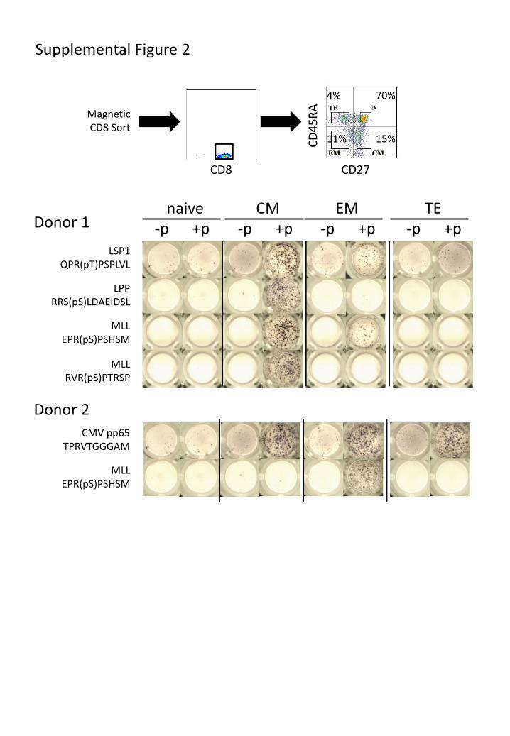
phosphopeptide-specific T-cells led us to investigate whether the responding cells resided within
memory or naïve T-cell compartments. Enriched CD8 T-cells from peripheral blood of two
HLA-B7+ healthy donors were flow-sorted into naïve (TN), central memory (TCM), effector
memory (TEM) and terminal effector memory cells (TEMRA) based on expression of CD45RA and
CD27 (31). After 7-day in vitro culture, ELISpot analysis demonstrated that T-cells responding
to individual phosphopeptides were exclusively in the memory compartment, and predominantly
had a TCM phenotype (Fig. 4e, Fig. S3). This suggests that the majority of healthy individuals
have been previously exposed to a stimulus that establishes immunological memory to tumor-
associated phosphopeptides.
Absent phosphopeptide-specific immunity in leukemia patients.
We next evaluated the level of phosphopeptide-specific CD8 T-cell immunity in
immunocompetent patients with early-stage CLL (Table S1). As tumor cells are present at high
levels in PBMC from CLL patients, comparison with responses in PBMC from healthy
individuals is problematic. Therefore, responses against a group of 12 CLL-associated
phosphopeptides (identified by red dots in Fig. 1) were assessed by direct ex vivo ELISpot of
purified CD8 T-cells (Fig. S4). As expected, we detected immune responses to most of these
phosphopeptides in all 10 healthy HLA-B7+ individuals (Fig. 5a). However, 9/14 CLL patients
(Group 1) had low or absent immunity to all 12 phosphopeptides (less than a combined total of
10 spots for all 12 phosphopeptides/200,000 CD8 T-cells). Five CLL patients (Group 2) showed

breadth of recognition of different phosphopeptides similar to that of the healthy donors.
However, average responses to all phosphopeptides were significantly lower in Group 2 patients
than in healthy individuals (Fig 5b).
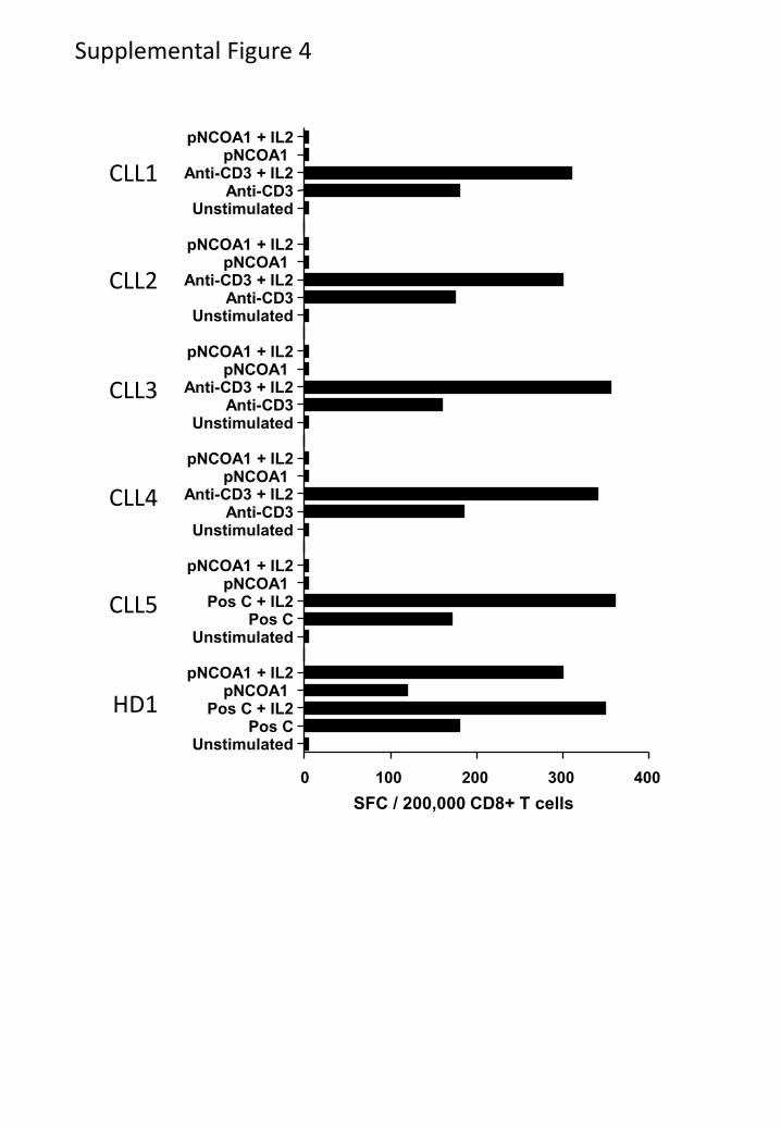
This lack of phosphopeptide-specific immunity in CLL patients might have been due to
an overall depression of T-cell immunity. However, bulk CD8 T-cell responses to low level anti-
CD3 were comparable in Group 1 and 2 patients and healthy donors (Fig. 5c). To assess
whether the lack of phosphopeptide-specific immunity in Group 1 patients reflected T-cell
anergy, we analyzed patient responses to the pNCOA-1 phosphopeptide, which is immunogenic
in 9/10 healthy donors, in the presence of IL-2 as this cytokine has been shown to reverse the
anergic state (32). Responses to anti-CD3 increased in the presence of IL-2, but no anti-
phosphopeptide immunity was detected (Fig. S5). This suggests that pre-existing phosphopeptide
immune T-cells in Group 1 patients had been deleted, or had never developed, rather than being
anergized.
Although CLL is a less aggressive tumor than either AML or ALL, the disease is more
aggressive in a subgroup of patients. Thus, we assessed whether the level of phosphopeptide-
specific immunity was associated with patient outcome. Despite selecting patients with early
stage disease, there were large differences in progression-free survival, overall survival and time
to first treatment between Group 1 and Group 2. Group 2 patients survived longer and required
less treatment than Group 1 patients (Figs. 5d-f). However, due to the small study size these
differences did not reach statistical significance.
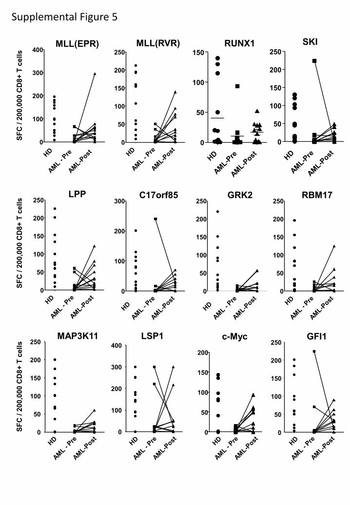
We extended these studies to a cohort of 12 HLA-B7+ AML patients in complete
remission (Table S2) using a panel of 12 AML-specific phosphopeptides (green dots in Fig. 1).
Responses to most of these phosphopeptides were detected in 12 healthy donors after 7-day in
vitro culture (Fig. 6a, Fig. S6). However, as with CLL patients, the average responses of AML
patients to individual phosphopeptides were significantly lower (Fig 6b, P<0.0001). AML
patients could also be stratified: Group 1 patients (10/12) showed a profound lack of
phosphopeptide-specific immunity, while Group 2 (2/12) showed responses that were similar to
those of normal donors. Again, responses to low dose anti-CD3 were similar between the groups,
indicating that the lack pre-existing anti-phosphopeptide responses in patients is not due to
immune incompetence (Fig. S6). In keeping with this, absolute lymphocyte counts for all 12
AML patients were within the normal range pre-transplant (Table S2). Collectively, these
findings demonstrate that the majority of patients with either CLL or AML have reduced pre-
existing immunity specific for leukemia-associated phosphopeptides.
Restoration of phosphopeptide-specific immunity after stem cell transplantation
(SCT).
A graft versus leukemia (GvL) response following allogeneic SCT correlates with a positive
clinical outcome in AML patients. While it is believed that GvL is directed to minor
histocompatibility antigens (mHAgs), the targets have been only partly identified (32). We
hypothesised that SCT might also reconstitute potentially protective immunity against leukemia-
associated phosphopeptides in AML patients. All 12 AML patients went on to have allogeneic
SCT. Immunity against the 12 AML-specific phosphopeptides was at least partly restored in the
majority of patients studied (Fig. 6c, Fig. S5). Marked expansion of immune responses to six
different phosphopeptides was observed in patient AML2 (Fig. 6d), while patient AML4 showed
dramatic expansion of responses to MLL(EPR) and LSP1(QPR) phosphopeptides, and modestly
increased responses to C17orf85 and SKI (Fig 6e). Some of these responses were as large as
those against an immunodominant CMV epitope (pp65) and the resulting T-cells bound HLA-

phosphopeptide tetramers. Furthermore, in vitro expanded MLL(EPR)-specific T-cells from
patient AML4 killed AML cells (Fig. 6f) confirming their functional relevance. These data
reveal a linkage between immunity to leukemia-associated phosphopeptides and GvL following
allogeneic SCT
Discussion
Posttranslationally modified antigens are increasingly being shown to play important roles in
human disease (33-37). Here we have tested the hypothesis that phosphopeptide neoantigens
could be identified on primary human malignant tissue and would be immunogenic in healthy
donors or patients with cancer. We now report 95 phosphopeptides displayed on the surface of
primary hematological malignant tissue in association with two dominant human MHC-I
molecules. We observed substantial differences in the number of phosphopeptides presented by
different HLA alleles, although the underlying mechanism for these differences is unclear. Our
results establish that MHC-I displayed phosphopeptides are over-represented on multiple
leukemic malignancies, and that more aggressive malignancies display a greater diversity of
them. Many of these tumor-associated phosphopeptides are derived from oncogenes linked to
leukemogenesis, making these of particular interest as immunotherapeutic targets.
Unexpectedly, many of these phosphopeptides were the targets of pre-existing immunity,
based on high levels of responding CD8 T-cells with a predominantly central memory
phenotype. The levels of responding cells were similar to those directed against
immunodominant epitopes from some non-persistent viruses. These T-cells, when expanded in
vitro, bound to HLA-B7-phosphopeptide tetramers, and recognized and killed primary tumor
cells, suggests they were high avidity. These observations contrast with those reported for other
tumor-associated antigens. Immunity to cancer-testis antigens is generally not present in healthy
individuals, and becomes detectable in patients with cancer, but is associated with a poor
prognosis (38, 39). Immunity to tissue-associated differentiation antigens, particularly those
defined as targets for melanoma-specific T-cells, is also elevated in melanoma patients (40, 41),
but is believed to be compromised by self-tolerance (42, 43). Importantly, none of the healthy
donors in which these memory-compartment associated immune responses were evident showed
any signs of autoimmune disease. This suggests that these phosphopeptides are displayed on
normal tissue at levels that are substantially lower than those on tumors. In keeping with this, T-
cells specific for LSP1 (RQAV) and NCOA-1 recognize tumors, but not resting B-cells, despite
the presence of these epitopes on the latter cells.
Although a small study, these findings suggest that the majority of healthy individuals
have been previously exposed to a stimulus that establishes immunological memory to
substantial numbers of phosphopeptides. For a small number of these phosphopeptides,
immunological memory is prevalent among most individuals examined. The mechanism that
underlies the development of phosphopeptide-specific memory CD8 T-cells is of great interest.
While it is possible that a distinct, cross-reactive stimulus could be responsible for the
development of a generic phosphate-directed immunity, all of the T-cells studied to date are
specific for the phosphate, the specific peptide sequence, and the MHC molecule that presents it.
On the other hand, hematolymphoid transformation by Epstein-Barr virus (EBV) is ubiquitous in
humans, and T cell immunosuppressed individuals commonly develop EBV-related
malignancies (44). This points to ongoing immune surveillance directed against virally-
transformed, and not just virally infected, cells. While much of this is directed at latent EBV
gene products, T-cells directed against EBV transformed cells also recognize EBV negative
tumors (45). We hypothesize that phosphopeptides that are shared by different kinds of
malignancies are among the targets of these T-cells. In addition, molecular techniques have
shown that 69% of healthy individuals have detectable myeloid-transforming transcripts in
peripheral blood (46), and 5-12% of healthy individuals show a monoclonal B-cell
lymphocytosis, a recognized precursor of CLL (47, 48). Finally, since some of these
phosphopeptides are also found on cell lines from solid tumor malignancies (22) , immune
surveillance against a broad range of early-stage non-hematopoietic cancers may also lead to the
development of phosphopeptide-specific immune memory. The extensive variation among
individuals in which phosphopeptides are the targets of this pre-existing immunity is consistent
with immune surveillance operating on the unique tumors that arise in each person. While it is
difficult to prove definitively in humans, we hypothesize that phosphopeptide-specific memory
cells are evidence of previous encounters with nascent tumors that have deregulated some of
their phosphorylation-based signaling cascades.
Interestingly, the majority of patients with CLL and AML lacked evidence of pre-existing
immunity to substantial subsets of phosphopeptides, and the clinical outcome was more
favorable in CLL patients where phosphopeptide immunity was present. It is possible that the
lack of phosphopeptide-specific immunity reflects a lack of exposure during the immune
surveillance phase preceding tumor development. Alternatively, it may reflect the operation of
immunosuppressive mechanisms, or the occurrence of clonal exhaustion, in conjunction with
tumor outgrowth. Regardless of the exact mechanism, the results point to a role for
phosphopeptide-specific immunity as a component of tumor recognition and control.
Although the antigenic targets for the GvL response include mHAgs (49-51), multiple
studies have revealed that SCT between identical twins, where mHAg differences are not
present, can lead to curative outcomes (52-54). In AML, deficient phosphopeptide-specific
immunity could be restored following adoptive transfer of T-cells from a stem cell donor. This
indicates that immunity against phosphopeptide tumor antigens may define a component of the
GvL response (1, 5). It is known that survival of CMV+ patients can be enhanced by pre-
selecting donors for SCT who have pre-existing CMV-specific immunity (55). It is therefore
possible SCT outcome may also be improved by matching donors with pre-existing immunity to
particular phosphopeptides based on the display of phosphopeptides on leukemia cells.
In this present study we have been unable to demonstrate a direct causal relationship between the
lack of cellular immunity against phosphorylated antigens in patients, with disease development
and/or progression. Neither have we been able to connect, at the molecular level, deregulated
oncogenic signaling present in each primary tumor, to the phosphorylation events present within
the defined antigens. Larger, more complex and extensive studies, will be needed to define these
relationships. Collectively, however, our results suggest that the display of MHC-associated
phosphopeptides may play a role in preventing either the development or progression of
malignant disease. Thus, enhancing immunity to these tumor-associated antigens should become
a focus of future cancer immunotherapeutic strategies.
Materials and Methods
Tumor samples and cell lines. Blood or leukapheresis samples were taken from patients with
high-burden leukemia in heparin, and tumor cells were isolated on Ficoll density gradients. The
purity of the tumor was >98% pure in all cases as assessed by flow cytometry. Healthy T and B-
cell populations were isolated from normal spleen and tonsil samples processed by mechanical
disruption, followed by density gradient separation and enrichment using anti-CD19 or anti-CD3
microbeads (Miltenyi Biotec) to >98% purity. Bone marrow sample was obtained from an
elective orthopedic procedure and red cell depleted using hypotonic lysis. All cell lines were
grown at 37°C with 5% CO2 in medium consisting of RPMI-1640 supplemented with 10% Fetal
Bovine Serum (FBS) and 2 mM L-glutamine (all from Sigma-Aldrich).
Isolation of HLA-associated peptides. Class-I MHC molecules were immunoaffinity purified
from samples, and their associated peptides were extracted, as described(22). Briefly 1.2 to 14
x109 cells were lysed in 20 mM Tris-HCl (pH 8.0), 150 mM NaCl, 1% CHAPS, 1 mM PMSF, 5
μg/mL aprotinin, 10 μg/mL leupeptin, 10 μg/mL pepstatin A, 1 µg/ml calyculin A (Sigma-
Aldrich) and phosphatase inhibitor cocktails I and II (Sigma-Aldrich). For primary tumors or the
HLA-A2, B7 homozygote B-LCL JY the mixture was centrifuged at 100, 000 x g for 1 hour and
the resulting supernatant was passed over protein A Sepharose pre-loaded with the HLA-A2-
specific antibody BB7.2 or HLA-B7-specific antibody ME1. For the HLA-A3, B7 homozygous
B-LCL GM03107 columns were loaded with the HLA-B, C specific antibody B123.2 to recover
HLA-B7 molecules. Peptides were eluted from the purified class-I MHC molecules with 10%
acetic acid and separated by ultrafiltration (ULTRAFREE-MC, Millipore).
Sequence analysis of HLA-Associated Phosphopeptides. Immunoaffinity-purified class I
peptides were converted to d0- or d3-methyl esters and subjected to Fe+3
-immobilized metal-
affinity chromatography to isolate phosphopeptides, as described(22). Phosphopeptide methyl
esters were then analyzed by a combination of nanoflow HPLC, microelectrospray ionization,
and collision activated dissociation on LTQ/FT or Orbitrap tandem mass spectrometers (Thermo
Scientific)(22). Electron transfer dissociation (ETD) spectra were acquired on an in-house
modified LTQ mass spectrometer(56). Peptide sequences were determined by manual
interpretation of CAD and ETD spectra recorded on the above peptide esters. If necessary,
phosphopeptide sequences were confirmed by recording tandem mass spectra on the
corresponding synthetics.
Epitope Prediction. This was performed using SYFPEITHI and a threshold score of 20.
Peptides. Peptides used in this study were synthesized with Fmoc chemistry, isolated by HPLC
to >90% purity, and validated with mass spectrometry (EZ-Biolabs and Genscript).
HLA-phosphopeptide Tetramers. HLA tetramers were produced as described (57).
Generation of Human Phosphopeptide-Specific CD8 Cytotoxic T-cell lines. HLA-A2
restricted RQA(pS)IELPSMAV phosphopeptide-specific cytotoxic CD8 T-cells were generated
from healthy donors as described previously (58). All cytokines were from PeproTech except
where stated. Briefly, PBMCs were cultured in flat-bottom 6-well plates at 107 cells per well in
RPMI-1640 plus 10% heat-inactivated human AB serum (Biosera) (10% media). GM-CSF (800
IU/ml) and IL-4 (1000U/mL) were added on day 0 to generate dendritic cells (DC). On day 1, a
maturation cocktail containing 100ng/ml TNF, 100ng/ml IL-1β, 10,000IU/ml IL-6, 8000 IU/ml
GM-CSF and 10 µg/ml PGE2 (Sigma-Aldrich) was added. On day 2, DCs were harvested and
loaded with phosphopeptide (20µg/ml) for 4 hours in the absence of FBS or human serum.
Following three washes, T-cells were added at a ratio of five T-cells per DC in 10% media.
Recombinant IL-7 (10ng/ml) and IL-15 (10ng/ml) were added on day 5. On day 9, T-cells were
harvested and re-stimulated by adding 107 irradiated PBMCs and 10
6 irradiated autologous LCLs
pulsed with phosphopeptide with IL-7 (5ng/ml), IL-15 (5ng/ml) and IL-2 (20IU/ml). Cultures
were re-stimulated every 7 days thereafter in the same manner. At each re-stimulation T-cells
were enriched using either anti-CD8 microbeads (Miltenyi Biotec) or by labeling with HLA-
phosphopeptide tetramers and using anti-PE microbeads (Miltenyi Biotec).
HLA-B7 restricted anti-phosphopeptide T-cells were grown in the absence of dendritic
cells by plating 5x10⁶ PBMCs in 48 well plates in 10% media with individual phosphopeptides
at 10µg/ml for 7 days without cytokines. Re-stimulations with irradiated phosphopeptide-pulsed
autologous PBMCs took place every 7 days with cytokines added 3 days after each re-
stimulation (final concentration 20IU/ml IL-2, 5ng/ml IL-7 and 5ng/ml IL-15). Functional and
cytotoxicity assays were then performed from day 13.
T-cell recognition assays. For ELISpot analysis, PBMCs or CD8 T-cells were isolated fresh
from in heparinized blood taken from healthy donors and patients. 1x10⁶ PBMCs were isolated
from both AML patients and healthy donors and re-suspended in AIM-V media (Invitrogen) with
10% human AB serum (Biosera) in a 96 well plate. For 7-day assays peptide or phosphopeptides
were added individually at 10g/ml and placed at 37°C in CO2 incubator for 7 days. For some
experiments (figure 4b) the 7-day culture was restimulated by adding irradiated phosphopeptide-
pulsed autologous dendritic cells for a further 7-days. For the ELISpot, cells were then harvested,
washed 4 times in AIM-V and incubated for 16 hours with either phosphopeptides (10ug/ml),
peptides (10ug/ml), or anti-CD3 (OKT3, 100ng/ml, Mabtech). Cytokine-producing cells were
identified as dark spots after a 15 min reaction with 5-bromo-4-chloro-3-indolyl phosphate and
NBT by means of an alkaline phosphatase conjugate substrate (Mabtech). Spots were counted
using an automated reader (AID-Diagnostika), and results displayed as number of spot-forming
cells (SFC) per 105
CD8 T-cells or 106 PBMCs.
For CLL patients a different approach had to be taken as PBMCs contain largely tumor
cells. Therefore CD8 T-cells were magnetically enriched using anti-CD8 microbeads (Miltenyi
Biotec) to a purity of >99% for both healthy donors and patients with CLL. 200,000 CD8 T-cells
from both patients and healthy donors were used for ELISpot analysis as described above.
Cytotoxicity assays. Cytotoxic activity was evaluated in a standard 4 hour 51
Cr release assay, as
previously described(57).
Statistical Analysis
We analyzed the data using the Kaplan-Meier for the survival rate, the unpaired t-test for two-
group comparisons. We performed the statistical analyses using Prism version 5 (GraphPad). P
values <0.05 were considered significant.
Ethical approval. The University of Virginia Institutional Review Board approved all protocols.
Patients with CLL were recruited from specialist clinics at the University Hospital NHS Trust
and Heart of England NHS Trust (Birmingham, United Kingdom). The study received approval
from the local ethics committees at South Birmingham, Birmingham East, North, and Solihull,
and informed written consent was obtained in accordance with the Declaration of Helsinki in all
cases.
Supplementary Materials
Fig. S1. Detailed HLA binding motif analysis.
Fig. S2. Immunity against phosphopeptides in healthy donors is phosphate dependent.
Fig. S3. T-cell memory subset mapping of anti-phosphopeptide responses in healthy donors.
Fig. S4. Leukemia-associated phosphopeptide-specific immunity is lacking in CLL patients.
Fig. S5. Deletion (rather than anergy) of anti-phosphopeptide immunity in patients with CLL.
Fig. S6. Leukemia-associated phosphopeptide-specific immunity is lacking in AML patients but
restored following SCT.

Fig. S6. Immunocompetence of patients with AML in complete remission prior to
transplantation.
Table S1. CLL patient characteristics.
Table S2. AML patient characteristics.
Reference list and notes.
1. H. J. Kolb. Graft-versus-leukemia effects of transplantation and donor lymphocytes. Blood 112,
4371-4383 (2008).
2. C. Preudhomme, J. Guilhot, F. E. Nicolini, A. Guerci-Bresler, F. Rigal-Huguet, F. Maloisel, V.
Coiteux, M. Gardembas, C. Berthou, A. Vekhoff, D. Rea, E. Jourdan, C. Allard, A. Delmer, P.
Rousselot, L. Legros, M. Berger, S. Corm, G. Etienne, C. Roche-Lestienne, V. Eclache, F. X.
Mahon, and F. Guilhot. Imatinib plus peginterferon alfa-2a in chronic myeloid leukemia.
N.Engl.J.Med. 363, 2511-2521 (2010).
3. M. Talpaz, H. M. Kantarjian, K. McCredie, J. M. Trujillo, M. J. Keating, and J. U. Gutterman.
Hematologic remission and cytogenetic improvement induced by recombinant human interferon
alpha A in chronic myelogenous leukemia. N.Engl.J.Med. 314, 1065-1069 (1986).
4. M. M. Horowitz, R. P. Gale, P. M. Sondel, J. M. Goldman, J. Kersey, H. J. Kolb, A. A. Rimm, O.
Ringden, C. Rozman, B. Speck, and . Graft-versus-leukemia reactions after bone marrow
transplantation. Blood 75, 555-562 (1990).
5. H. J. Kolb, J. Mittermuller, C. Clemm, E. Holler, G. Ledderose, G. Brehm, M. Heim, and W.
Wilmanns. Donor leukocyte transfusions for treatment of recurrent chronic myelogenous
leukemia in marrow transplant patients. Blood 76, 2462-2465 (1990).
6. F. Baron, M. B. Maris, B. M. Sandmaier, B. E. Storer, M. Sorror, R. Diaconescu, A. E. Woolfrey,
T. R. Chauncey, M. E. Flowers, M. Mielcarek, D. G. Maloney, and R. Storb. Graft-versus-tumor
effects after allogeneic hematopoietic cell transplantation with nonmyeloablative conditioning.
J.Clin.Oncol. 23, 1993-2003 (2005).
7. S. A. Rosenberg, P. Spiess, and R. Lafreniere. A new approach to the adoptive immunotherapy of
cancer with tumor-infiltrating lymphocytes. Science 233, 1318-1321 (1986).
8. P. W. Kantoff, C. S. Higano, N. D. Shore, E. R. Berger, E. J. Small, D. F. Penson, C. H. Redfern,
A. C. Ferrari, R. Dreicer, R. B. Sims, Y. Xu, M. W. Frohlich, and P. F. Schellhammer.
Sipuleucel-T immunotherapy for castration-resistant prostate cancer. N.Engl.J.Med. 363, 411-422
(2010).
9. S. A. Rosenberg and M. E. Dudley. Adoptive cell therapy for the treatment of patients with
metastatic melanoma. Curr.Opin.Immunol. 21, 233-240 (2009).
10. S. A. Rosenberg, J. C. Yang, and N. P. Restifo. Cancer immunotherapy: moving beyond current
vaccines. Nat.Med. 10, 909-915 (2004).
11. Waun Ki Hong, Robert C.Bast Jr, William Hait, Donald W.Kufe, Raphael E.Pollock, Ralph
R.Weichselbaum, James F.Holland, and Emil Frei Iii. Holland-Frei Cancer Medicine. (10 A.D.).
McGraw-Hill Medical.
12. K. R. Chi. Cancer research: Promise of protection. Nature 471, 537-538 (2011).
13. T. J. Ley, E. R. Mardis, L. Ding, B. Fulton, M. D. McLellan, K. Chen, D. Dooling, B. H.
Dunford-Shore, S. McGrath, M. Hickenbotham, L. Cook, R. Abbott, D. E. Larson, D. C. Koboldt,
C. Pohl, S. Smith, A. Hawkins, S. Abbott, D. Locke, L. W. Hillier, T. Miner, L. Fulton, V.
Magrini, T. Wylie, J. Glasscock, J. Conyers, N. Sander, X. Shi, J. R. Osborne, P. Minx, D.
Gordon, A. Chinwalla, Y. Zhao, R. E. Ries, J. E. Payton, P. Westervelt, M. H. Tomasson, M.
Watson, J. Baty, J. Ivanovich, S. Heath, W. D. Shannon, R. Nagarajan, M. J. Walter, D. C. Link,
T. A. Graubert, J. F. DiPersio, and R. K. Wilson. DNA sequencing of a cytogenetically normal
acute myeloid leukaemia genome. Nature 456, 66-72 (2008).
14. S. Jones, X. Zhang, D. W. Parsons, J. C. Lin, R. J. Leary, P. Angenendt, P. Mankoo, H. Carter, H.
Kamiyama, A. Jimeno, S. M. Hong, B. Fu, M. T. Lin, E. S. Calhoun, M. Kamiyama, K. Walter,
T. Nikolskaya, Y. Nikolsky, J. Hartigan, D. R. Smith, M. Hidalgo, S. D. Leach, A. P. Klein, E.
M. Jaffee, M. Goggins, A. Maitra, C. Iacobuzio-Donahue, J. R. Eshleman, S. E. Kern, R. H.
Hruban, R. Karchin, N. Papadopoulos, G. Parmigiani, B. Vogelstein, V. E. Velculescu, and K. W.
Kinzler. Core signaling pathways in human pancreatic cancers revealed by global genomic
analyses. Science 321, 1801-1806 (2008).
15. D. W. Parsons, M. Li, X. Zhang, S. Jones, R. J. Leary, J. C. Lin, S. M. Boca, H. Carter, J.
Samayoa, C. Bettegowda, G. L. Gallia, G. I. Jallo, Z. A. Binder, Y. Nikolsky, J. Hartigan, D. R.
Smith, D. S. Gerhard, D. W. Fults, S. Vandenberg, M. S. Berger, S. K. Marie, S. M. Shinjo, C.
Clara, P. C. Phillips, J. E. Minturn, J. A. Biegel, A. R. Judkins, A. C. Resnick, P. B. Storm, T.
Curran, Y. He, B. A. Rasheed, H. S. Friedman, S. T. Keir, R. McLendon, P. A. Northcott, M. D.
Taylor, P. C. Burger, G. J. Riggins, R. Karchin, G. Parmigiani, D. D. Bigner, H. Yan, N.
Papadopoulos, B. Vogelstein, K. W. Kinzler, and V. E. Velculescu. The Genetic Landscape of the
Childhood Cancer Medulloblastoma. Science 331, 435-439 (2011).
16. D. R. van, W. H. Zoutman, R. Dijkman, R. X. de Menezes, S. Commandeur, A. A. Mulder, P. A.
van der Velden, M. H. Vermeer, R. Willemze, P. S. Yan, T. H. Huang, and C. P. Tensen.
Epigenetic profiling of cutaneous T-cell lymphoma: promoter hypermethylation of multiple
tumor suppressor genes including BCL7a, PTPRG, and p73. J.Clin.Oncol. 23, 3886-3896 (2005).
17. E. Tiacci, V. Trifonov, G. Schiavoni, A. Holmes, W. Kern, M. P. Martelli, A. Pucciarini, B.
Bigerna, R. Pacini, V. A. Wells, P. Sportoletti, V. Pettirossi, R. Mannucci, O. Elliott, A. Liso, A.
Ambrosetti, A. Pulsoni, F. Forconi, L. Trentin, G. Semenzato, G. Inghirami, M. Capponi, R. F.
Di, C. Patti, L. Arcaini, P. Musto, S. Pileri, C. Haferlach, S. Schnittger, G. Pizzolo, R. Foa, L.
Farinelli, T. Haferlach, L. Pasqualucci, R. Rabadan, and B. Falini. BRAF mutations in hairy-cell
leukemia. N.Engl.J.Med. 364, 2305-2315 (2011).
18. B. J. Druker, F. Guilhot, S. G. O'brien, I. Gathmann, H. Kantarjian, N. Gattermann, M. W.
Deininger, R. T. Silver, J. M. Goldman, R. M. Stone, F. Cervantes, A. Hochhaus, B. L. Powell, J.
L. Gabrilove, P. Rousselot, J. Reiffers, J. J. Cornelissen, T. Hughes, H. Agis, T. Fischer, G.
Verhoef, J. Shepherd, G. Saglio, A. Gratwohl, J. L. Nielsen, J. P. Radich, B. Simonsson, K.
Taylor, M. Baccarani, C. So, L. Letvak, and R. A. Larson. Five-year follow-up of patients
receiving imatinib for chronic myeloid leukemia. N.Engl.J.Med. 355, 2408-2417 (2006).
19. C. Harrison. Trial watch: BTK inhibitor shows positive results in B cell malignancies.
Nat.Rev.Drug Discov. 11, 96 (2012).
20. A. Pardanani, J. R. Gotlib, C. Jamieson, J. E. Cortes, M. Talpaz, R. M. Stone, M. H. Silverman,
D. G. Gilliland, J. Shorr, and A. Tefferi. Safety and efficacy of TG101348, a selective JAK2
inhibitor, in myelofibrosis. J.Clin.Oncol. 29, 789-796 (2011).
21. D. Hanahan and R. A. Weinberg. Hallmarks of cancer: the next generation. Cell 144, 646-674
(2011).
22. A. L. Zarling, J. M. Polefrone, A. M. Evans, L. M. Mikesh, J. Shabanowitz, S. T. Lewis, V. H.
Engelhard, and D. F. Hunt. Identification of class I MHC-associated phosphopeptides as targets
for cancer immunotherapy. Proc.Natl.Acad.Sci.U.S.A 103, 14889-14894 (2006).
23. A. L. Zarling, S. B. Ficarro, F. M. White, J. Shabanowitz, D. F. Hunt, and V. H. Engelhard.
Phosphorylated peptides are naturally processed and presented by major histocompatibility
complex class I molecules in vivo. J.Exp.Med. 192, 1755-1762 (2000).
24. F. R. Depontieu, J. Qian, A. L. Zarling, T. L. McMiller, T. M. Salay, A. Norris, A. M. English, J.
Shabanowitz, V. H. Engelhard, D. F. Hunt, and S. L. Topalian. Identification of tumor-associated,
MHC class II-restricted phosphopeptides as targets for immunotherapy. Proc.Natl.Acad.Sci.U.S.A
106, 12073-12078 (2009).
25. K. T. Hogan, D. P. Eisinger, S. B. Cupp, III, K. J. Lekstrom, D. D. Deacon, J. Shabanowitz, D. F.
Hunt, V. H. Engelhard, C. L. Slingluff, Jr., and M. M. Ross. The peptide recognized by HLA-
A68.2-restricted, squamous cell carcinoma of the lung-specific cytotoxic T lymphocytes is
derived from a mutated elongation factor 2 gene. Cancer Res. 58, 5144-5150 (1998).
26. V. S. Meyer, O. Drews, M. Gunder, J. Hennenlotter, H. G. Rammensee, and S. Stevanovic.
Identification of natural MHC class II presented phosphopeptides and tumor-derived MHC class I
phospholigands. J.Proteome.Res. 8, 3666-3674 (2009).
27. F. Mohammed, M. Cobbold, A. L. Zarling, M. Salim, G. A. Barrett-Wilt, J. Shabanowitz, D. F.
Hunt, V. H. Engelhard, and B. E. Willcox. Phosphorylation-dependent interaction between
antigenic peptides and MHC class I: a molecular basis for the presentation of transformed self.
Nat.Immunol. 9, 1236-1243 (2008).
28. J. E. Beaver, P. E. Bourne, and J. V. Ponomarenko. EpitopeViewer: a Java application for the
visualization and analysis of immune epitopes in the Immune Epitope Database and Analysis
Resource (IEDB). Immunome.Res. 3, 3 (2007).
29. P. V. Hornbeck, I. Chabra, J. M. Kornhauser, E. Skrzypek, and B. Zhang. PhosphoSite: A
bioinformatics resource dedicated to physiological protein phosphorylation. Proteomics. 4, 1551-
1561 (2004).
30. T. Marafioti, C. Mancini, S. Ascani, E. Sabattini, P. L. Zinzani, M. Pozzobon, K. Pulford, B.
Falini, E. S. Jaffe, H. K. Muller-Hermelink, D. Y. Mason, and S. A. Pileri. Leukocyte-specific
phosphoprotein-1 and PU.1: two useful markers for distinguishing T-cell-rich B-cell lymphoma
from lymphocyte-predominant Hodgkin's disease. Haematologica 89, 957-964 (2004).
31. D. Hamann, P. A. Baars, M. H. Rep, B. Hooibrink, S. R. Kerkhof-Garde, M. R. Klein, and R. A.
van Lier. Phenotypic and functional separation of memory and effector human CD8+ T cells.
J.Exp.Med. 186, 1407-1418 (1997).
32. R. H. Schwartz. T cell anergy. Annu.Rev.Immunol. 21, 305-334 (2003).
33. E. Girbal-Neuhauser, J. J. Durieux, M. Arnaud, P. Dalbon, M. Sebbag, C. Vincent, M. Simon, T.
Senshu, C. Masson-Bessiere, C. Jolivet-Reynaud, M. Jolivet, and G. Serre. The epitopes targeted
by the rheumatoid arthritis-associated antifilaggrin autoantibodies are posttranslationally
generated on various sites of (pro)filaggrin by deimination of arginine residues. J.Immunol. 162,
585-594 (1999).
34. H. Arentz-Hansen, R. Korner, O. Molberg, H. Quarsten, W. Vader, Y. M. Kooy, K. E. Lundin, F.
Koning, P. Roepstorff, L. M. Sollid, and S. N. McAdam. The intestinal T cell response to alpha-
gliadin in adult celiac disease is focused on a single deamidated glutamine targeted by tissue
transglutaminase. J.Exp.Med. 191, 603-612 (2000).
35. D. M. Wuttge, M. Bruzelius, and S. Stemme. T-cell recognition of lipid peroxidation products
breaks tolerance to self proteins. Immunology 98, 273-279 (1999).
36. P. J. Utz, M. Hottelet, P. H. Schur, and P. Anderson. Proteins phosphorylated during stress-
induced apoptosis are common targets for autoantibody production in patients with systemic
lupus erythematosus. J.Exp.Med. 185, 843-854 (1997).
37. M. J. Mamula, R. J. Gee, J. I. Elliott, A. Sette, S. Southwood, P. J. Jones, and P. R. Blier.
Isoaspartyl post-translational modification triggers autoimmune responses to self-proteins.
J.Biol.Chem. 274, 22321-22327 (1999).
38. M. J. Scanlan, A. O. Gure, A. A. Jungbluth, L. J. Old, and Y. T. Chen. Cancer/testis antigens: an
expanding family of targets for cancer immunotherapy. Immunol.Rev. 188, 22-32 (2002).
39. T. Okada, M. Akada, T. Fujita, T. Iwata, Y. Goto, K. Kido, T. Okada, Y. Matsuzaki, K.
Kobayashi, S. Matsuno, M. Sunamura, and Y. Kawakami. A novel cancer testis antigen that is
frequently expressed in pancreatic, lung, and endometrial cancers. Clin.Cancer Res. 12, 191-197
(2006).
40. S. A. Rosenberg and D. E. White. Vitiligo in patients with melanoma: normal tissue antigens can
be targets for cancer immunotherapy. J.Immunother.Emphasis.Tumor Immunol. 19, 81-84 (1996).
41. P. R. Dunbar, J. L. Chen, D. Chao, N. Rust, H. Teisserenc, G. S. Ogg, P. Romero, P. Weynants,
and V. Cerundolo. Cutting edge: rapid cloning of tumor-specific CTL suitable for adoptive
immunotherapy of melanoma. J.Immunol. 162, 6959-6962 (1999).
42. C. E. Touloukian, W. W. Leitner, R. E. Schnur, P. F. Robbins, Y. Li, S. Southwood, A. Sette, S.
A. Rosenberg, and N. P. Restifo. Normal tissue depresses while tumor tissue enhances human T
cell responses in vivo to a novel self/tumor melanoma antigen, OA1. J.Immunol. 170, 1579-1585
(2003).
43. T. A. Colella, T. N. Bullock, L. B. Russell, D. W. Mullins, W. W. Overwijk, C. J. Luckey, R. A.
Pierce, N. P. Restifo, and V. H. Engelhard. Self-tolerance to the murine homologue of a
tyrosinase-derived melanoma antigen: implications for tumor immunotherapy. J.Exp.Med. 191,
1221-1232 (2000).
44. S. Gottschalk, C. M. Rooney, and H. E. Heslop. Post-transplant lymphoproliferative disorders.
Annu.Rev.Med. 56, 29-44 (2005).
45. H. M. Long, J. Zuo, A. M. Leese, N. H. Gudgeon, H. Jia, G. S. Taylor, and A. B. Rickinson.
CD4+ T-cell clones recognizing human lymphoma-associated antigens: generation by in vitro
stimulation with autologous Epstein-Barr virus-transformed B cells. Blood 114, 807-815 (2009).
46. S. Bose, M. Deininger, J. Gora-Tybor, J. M. Goldman, and J. V. Melo. The presence of typical
and atypical BCR-ABL fusion genes in leukocytes of normal individuals: biologic significance
and implications for the assessment of minimal residual disease. Blood 92, 3362-3367 (1998).
47. W. G. Nieto, J. Almeida, A. Romero, C. Teodosio, A. Lopez, A. F. Henriques, M. L. Sanchez, M.
Jara-Acevedo, A. Rasillo, M. Gonzalez, P. Fernandez-Navarro, T. Vega, and A. Orfao. Increased
frequency (12%) of circulating chronic lymphocytic leukemia-like B-cell clones in healthy
subjects using a highly sensitive multicolor flow cytometry approach. Blood 114, 33-37 (2009).
48. A. C. Rawstron, F. L. Bennett, S. J. O'Connor, M. Kwok, J. A. Fenton, M. Plummer, T. R. de, R.
G. Owen, S. J. Richards, A. S. Jack, and P. Hillmen. Monoclonal B-cell lymphocytosis and
chronic lymphocytic leukemia. N.Engl.J.Med. 359, 575-583 (2008).
49. J. M. den Haan, L. M. Meadows, W. Wang, J. Pool, E. Blokland, T. L. Bishop, C. Reinhardus, J.
Shabanowitz, R. Offringa, D. F. Hunt, V. H. Engelhard, and E. Goulmy. The minor
histocompatibility antigen HA-1: a diallelic gene with a single amino acid polymorphism. Science
279, 1054-1057 (1998).
50. D. Bonnet, E. H. Warren, P. D. Greenberg, J. E. Dick, and S. R. Riddell. CD8(+) minor
histocompatibility antigen-specific cytotoxic T lymphocyte clones eliminate human acute
myeloid leukemia stem cells. Proc.Natl.Acad.Sci.U.S.A 96, 8639-8644 (1999).
51. A. G. Brickner, E. H. Warren, J. A. Caldwell, Y. Akatsuka, T. N. Golovina, A. L. Zarling, J.
Shabanowitz, L. C. Eisenlohr, D. F. Hunt, V. H. Engelhard, and S. R. Riddell. The
immunogenicity of a new human minor histocompatibility antigen results from differential
antigen processing. J.Exp.Med. 193, 195-206 (2001).
52. S. Z. Pavletic, G. Zhou, K. Sobocinski, G. Marti, K. Doney, J. DiPersio, W. Feremans, L. Foroni,
S. Goodman, G. Prentice, C. LeMaistre, G. Bandini, A. Ferrant, N. Jacobsen, I. Khouri, R. P.
Gale, A. Wiestner, S. Giralt, E. Montserrat, W. C. Chan, and C. Bredeson. Genetically identical
twin transplantation for chronic lymphocytic leukemia. Leukemia 21, 2452-2455 (2007).
53. R. P. Gale, M. M. Horowitz, R. C. Ash, R. E. Champlin, J. M. Goldman, A. A. Rimm, O.
Ringden, J. A. Stone, and M. M. Bortin. Identical-twin bone marrow transplants for leukemia.
Ann.Intern.Med. 120, 646-652 (1994).
54. N. Kroger, R. Brand, B. A. van, D. Bron, D. Blaise, E. Hellstrom-Lindberg, G. Gahrton, R.
Powles, T. Littlewood, B. Chapuis, A. Zander, V. Koza, D. Niederwieser, and W. T. de. Stem cell
transplantation from identical twins in patients with myelodysplastic syndromes. Bone Marrow
Transplant. 35, 37-43 (2005).
55. E. Ozdemir, R. M. Saliba, R. E. Champlin, D. R. Couriel, S. A. Giralt, L. M. de, I. F. Khouri, C.
Hosing, S. M. Kornblau, P. Anderlini, E. J. Shpall, M. H. Qazilbash, J. J. Molldrem, R. F.
Chemaly, and K. V. Komanduri. Risk factors associated with late cytomegalovirus reactivation
after allogeneic stem cell transplantation for hematological malignancies. Bone Marrow
Transplant. 40, 125-136 (2007).
56. J. E. Syka, J. J. Coon, M. J. Schroeder, J. Shabanowitz, and D. F. Hunt. Peptide and protein
sequence analysis by electron transfer dissociation mass spectrometry. Proc.Natl.Acad.Sci.U.S.A
101, 9528-9533 (2004).
57. M. Cobbold, N. Khan, B. Pourgheysari, S. Tauro, D. McDonald, H. Osman, M. Assenmacher, L.
Billingham, C. Steward, C. Crawley, E. Olavarria, J. Goldman, R. Chakraverty, P. Mahendra, C.
Craddock, and P. A. Moss. Adoptive transfer of cytomegalovirus-specific CTL to stem cell
transplant patients after selection by HLA-peptide tetramers. J.Exp.Med. 202, 379-386 (2005).
58. W. Y. Ho, H. N. Nguyen, M. Wolfl, J. Kuball, and P. D. Greenberg. In vitro methods for
generating CD8+ T-cell clones for immunotherapy from the naive repertoire. J.Immunol.Methods
310, 40-52 (2006).
Figure legends
Figure 1. HLA-bound phosphopeptides are differentially displayed on tumor and matched
healthy tissue. (a) Phosphopeptide display isolated from HLA-A2 and HLA-B7 molecules from
11 primary tumor samples (ALL1, AML1, CLL1-4, HCL1, MCL), EBV-transformed B-cells (B-
LCL) and HLA-matched healthy tissue (T-cells, B-cells and bone marrow). Red dots and green
dots indicate phosphopeptide antigens selected for further study in patients with CLL and AML
respectively. (b) Comparison of the number of individual phosphopeptides identified between
HLA-A2 and HLA-B7 in both normal and malignant tissue. (c) Comparison of the number of
unique phosphopeptides identified between normal, indolent malignant and aggressive malignant
tissue.
Figure 2. HLA-B7 associated phosphopeptides have characteristics that distinguish them from
non-phosphorylated peptides. (a, b) Euler diagrams depicting the distribution of HLA-B7-
restricted phosphopeptides among different leukemias and normal tissues (a) and within different
B-cell malignancies (b). (c,d,f) Logoplots of residue frequency at each position of all 9mer
HLA-B7 phosphopeptides (c); 9mer non-phosphorylated HLA-B7 peptides from the
ImmuneEpitope database (d); and predicted HLA-B7 phosphopeptide binders with a pSer at
position 4 (f). The position of phosphoserine for all B7-predicted binders (e).
Figure 3. Phosphopeptide-specific T-cells from healthy donors recognize and kill leukemic
targets. (a) Expansion of LSP-1 (RQA(pS)IELPSMAV) specific T-cells from healthy donor
PBMCs using DCs and tetramer selection. (b,c) Peptide antigen specificity (b) and recognition
(c) of leukemia cell lines by LSP-1-specific T-cells. (d,e) T-cell mediated killing of leukemia cell
lines (d) or primary leukemia samples (e) by phosphopeptide-specific T-cell lines but not an
HLA-mismatched primary tumor. (f) NCOA1 (RPT(pS)RLNRL)-specific T-cell mediated
killing of three HLA-matched CLL tumor samples was inhibited by adding autologous NCOA1
loaded cold targets.
Figure 4. Phosphopeptide-specific immunity is present in healthy individuals within the
circulating memory T-cell compartment. (a) Marked heterogeneity of phosphopeptide-specific
immunity in 10 healthy donors against 86 antigens revealed by 7-day ELISpot assays. (b)
ELISpot results for immunodominant phosphopeptides directly ex vivo and following 14-days in
vitro expansion when tetramer binding is evident. (c) Expanded T-cells do not recognize the
unphosphorylated counterpart peptides. (d) The relative magnitude of phosphopeptide-specific
T-cells compared against common viral epitopes following overnight culture (n=3). (e) Analysis
of memory-flow-sorted CD8+ enriched T-cells by 7-day ELISpot reveal phosphopeptide-specific
responses reside in the memory compartment.
Figure 5. Leukemia-associated phosphopeptide-specific immunity is lacking in CLL patients.
(a) Comparative ELISpot analysis of enriched CD8+ T-cells from patients with CLL and healthy
donors against 12 CLL-associated antigens reveals two distinct groupings. (b,c) Analysis of the
average patient response against each phosphopeptide between the groups and healthy donors (b)
reveals both Group1 (n=12) and Group2 (n=12) have suppressed phosphopeptide-immunity yet
responses to mitogens are intact (c). (d,e,f) Kaplan-Meier analysis of overall survival (OS, d),
progression free survival (PFS, e) and time to first treatment (TTFT, f) is reduced in Group1
patients, but not statistically significant. **P < 0.01 by Student’s t test comparing average
responses for each phosphopeptide between HD and either Group 1 or Group 2.
Figure 6. Phosphopeptide-specific immunity is lacking in patients with AML and restored
following stem cell transplantation. (a) AML-associated phosphopeptide-specific immunity is
lacking in AML patients in remission compared against healthy individuals using a panel of 12
phosphopeptide antigens. (b) Analysis of the average patient response against each
phosphopeptide (n=12) between the groups and healthy donors reveals patients have suppressed
immunity. (c) Analysis of total immunity against all 12 antigens between healthy donors (n=12)
and each patient pre- and post-transplant (n=12) reveal the recovery of AML-associated
phosphopeptide immunity following SCT. (d,e) Immune reconstitution of donor anti-
phosphopeptide immunity in two AML patients demonstrating large expansions of
phosphopeptide-specific T-cells. (f) T-cell line specific to MLL(EPR) generated from patient
AML4 is able to kill the HLA-B7 transfected KG-1a leukemia cell line. ***P < 0.001,
**P<0.01 by Student’s t test.
Acknowledgments: We thank Dr Peter Nightingale for his valuable input on statistical analysis.
Funding: Supported by NIH grants AI33993 to D.F.H., AI20963 and CA134060 to V.H.E, Kay
Kendall Leukaemia Research grant KKL3227 and Leukaemia Lymphoma Research Fund grant
08038 to M.C. and by NIH Cancer Center Support Grant P30 CA45579 to the University of
Virginia.
Author contributions: M.C., V.H.E and D.F.H. formulated the hypothesis and designed the
experiments; M.C., H.d.l.P., A.N., J.P., J.Q., A.M.E, J.E.T., J.C., J.G.A., S.A.M., H-W.H., S.A.P.
and O.C.G. performed all experiments; M.E.W., C.C., G.P. and S.F. provided patient samples
and clinical input into the study; A.N., J.P., J.Q., A.M.E., J.C., J.G.A. and J.S. performed and
analysed mass spectrometry studies; M.C., H.d.l.P., S.A.P. J.E.T., H-W. H. and O.C.G
performed and analysed human in vitro experiments; M.C. and A.L.Z. conducted MHC-peptide
extraction.
Competing interests: The authors D.F.H., J.S., V.H.E., A.L.Z., K.L.C., A.N., J.C. and M.C.
have equity interest in PhosImmune Inc. company.
Figure 1
b c
a
CLL2
CLL4
HCL1
B-LCL
Spleen B Cell
Spleen T Cell
Tonsil B Cell
BM
Number of Phosphopeptides identified
Malignant
Non-Malignant
B-LCL AML CLL T cells B cells0
20
40
60
HLA-A*0201
HLA-B*0702
Ph
osp
ho
pep
tid
es
Iden
tified
Gene
Name CL
LA
ML
Sequence AM
L1
AL
L1
CL
L2
CL
L4
HC
L1
B-L
CL
BM
B C
ell
T C
ell
HLA-B*0702
RBM14 FRR(pS)PTKSSLDY
GPSM3 GPRSASLL(pS)L
RBM17 RPR(pS)PTGP
MKI67IP RRK(pS)QVAEL
SFRS7 SPRR(pS)RSISL
LSP1 ● QPR(pT)PSPLVL
GFI1 ● QPR(pS)PGPDYSL
MAP3K11 ● TPR(pS)PPLGLI
MAP3K11 TPR(pS)PPLGL
GPSM3 GPRSA(pS)LL(pS)L
LSP1 QPR(pT)P(pS)PLVL
AKAP13 RPR(pS)AVLL
MYL9 RPQRAT(pS)NVF
ANKRD17 SPS(pS)PSVRRQL
RBM14 FRR(pS)PTKSSL
ZC3H14 KPA(pS)PKFIVTL
ATXN2 RPR(pS)PRQNSI
GRM5 RPR(pS)PSPIS
RREB1 RPRAN(pS)GGVDL
MIIP RPRPH(pS)APSL
SYNPO RPSRS(pS)PGL
CHAF1A SPR(pS)PSTTYL
GIGYF2 YQR(pS)FDEVEGVF
RBM17 ● RPR(pS)PTG(pS)NSF
MLL ● RVR(pS)PTRSP
MLL ● EPR(pS)PSHSM
LPP ● RRS(pS)LDAEIDSL
ARHGAP17 APRRY(pS)SSL
HMGN1 EPKRR(pS)ARL
MAP7D1 LPA(pS)PRARL
EP300 RPKL(pS)SPAL
MEFV RPR(pS)LEVTI
ZNF219 RPR(pS)PAARL
GTF2IRD1 RPR(pS)PGSNSKV
SIK1 RPRPV(pS)PSSL
CCDC88B SPEKAGRR(pS)SL
ZFP106 SPGLARKR(pS)L
C1orf63 SPSTSRSGG(pS)SRL
SVIL VPR(pS)PKHAHSSSL
GIGYF2 YQR(pS)FDEVEGV
unknown SPR(pS)PGKPM
unknown RPA(pS)PQRAQL
RCSD1 ● SPK(pS)PGLKA
TSC22D4 ● RP(pS)SPALYF
C17orf85 ● ● RPH(pS)PEKAF
GRK2 ● ● KPR(pS)PVVEL
NCOA1 ● RPT(pS)RLNRL
CCDC45 ● KPPYRSH(pS)L
FAM53C RPY(pS)PPFFSL
AM
L A
sso
cia
ted
Gene
Name CL
LA
ML
Sequence AM
L1
AL
L1
CL
L2
CL
L4
HC
L1
B-L
CL
BM
B C
ell
T C
ell
HLA-B*0702
SETD2 RPD(pS)RLGKTEL
KIAA1310 RPA(pS)PAAKL
unknown RPV(pS)PFQEL
MYL9 RPQRA(pT)SNVF
LUZP1 RPF(pS)PREAL
ARID1B RPS(pS)LPDL
MEF2D RPA(pS)AGAML
SON FSI(pS)PVRL
KLF10 RPV(pT)PVSDL
MYC ● KPPH(pS)PLVL
SRRM2 RAP(pS)PSSRM
RERE RPP(pS)SEFLDL
SPEN KPP(pT)PGASF
BCL11A KPP(pS)PSPIEM
PCBP2 ● RPKPSS(pS)PV
NFATC2 KPY(pS)PLASL
ARHGAP30 RPAK(pS)MDSL
VPS13D RSL(pS)PLLF
SCAP ● RPP(pS)PGPVL
TLK1 ● RPN(pS)PSPTAL
MACF1 ● KPR(pS)PDHVL
MAP1A ● KPA(pS)PARRL
ATXN2L ● SPA(pS)PKISL
SKI ● RPW(pS)PAVSA
TBC1D10C KPQTRGK(pT)F
RUNX1 ● TPI(pS)PGRASGM
unknown RPA(pT)GGPGVA
PPP1CA RPI(pT)PPRNSA
KLF2 RPPPPPD(pT)PP
HIVEP2 RPV(pS)PGKDI
IP6K1 RSE(pS)KDRKL
KIAA0889 RSG(pS)LERKV
RBM15 RSL(pS)PGGAA
AKAP13 RTE(pS)DSGLKK
TPX2 TPAQPQRR(pS)L
MORC2 KPP(pS)PEHQSL
HLA-A*0201
NCOR2 KAK(pS)PAPGL
LSP1 KLIDRTE(pS)L
N4BP2 KMD(pS)FLDMQL
LSP1 RQA(pS)IELPSM
LSP1 RQA(pS)IELPSMAV
HSPB1 RQL(pS)SGVSEI
SYNM RTF(pS)PTYGL
IRS2 RVA(pS)PTSGV
SRSF8 SMTR(pS)PPRV
TNS3 VMIG(pS)PKKV
AM
L A
sso
cia
ted
B-L
CL
AM
L1
MC
L
B C
ell
T C
ell
CL
L1
CL
L2
CL
L3
CL
L4
Not observed
<1 copy per cell
>1 copy per cell
>5 copies per cell
>10 copies per cell
>25 copies per cell
Present but not quantified
AML1
ALL1
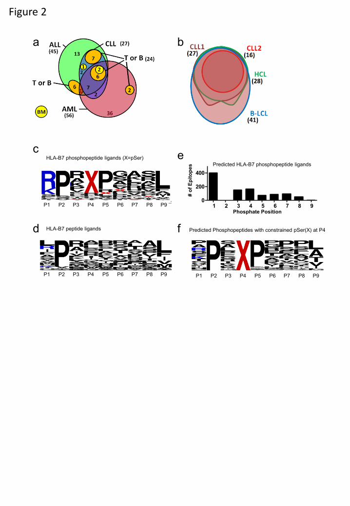
Figure 2
c
All Predicted pSer Phosphopeptides
(SYFPEITHI)
e
d f
b a
1 2 3 4 5 6 7 8 90
200
400
Phosphate Position
# o
f E
pit
op
es
Predicted Phosphopeptides with constrained pSer(X) at P4
P1 P2 P3 P4 P5 P6 P7 P8 P9 P1 P2 P3 P4 P5 P6 P7 P8 P9
P1 P2 P3 P4 P5 P6 P7 P8 P9
HLA-B7 phosphopeptide ligands (X=pSer)
HLA-B7 peptide ligands
Predicted HLA-B7 phosphopeptide ligands
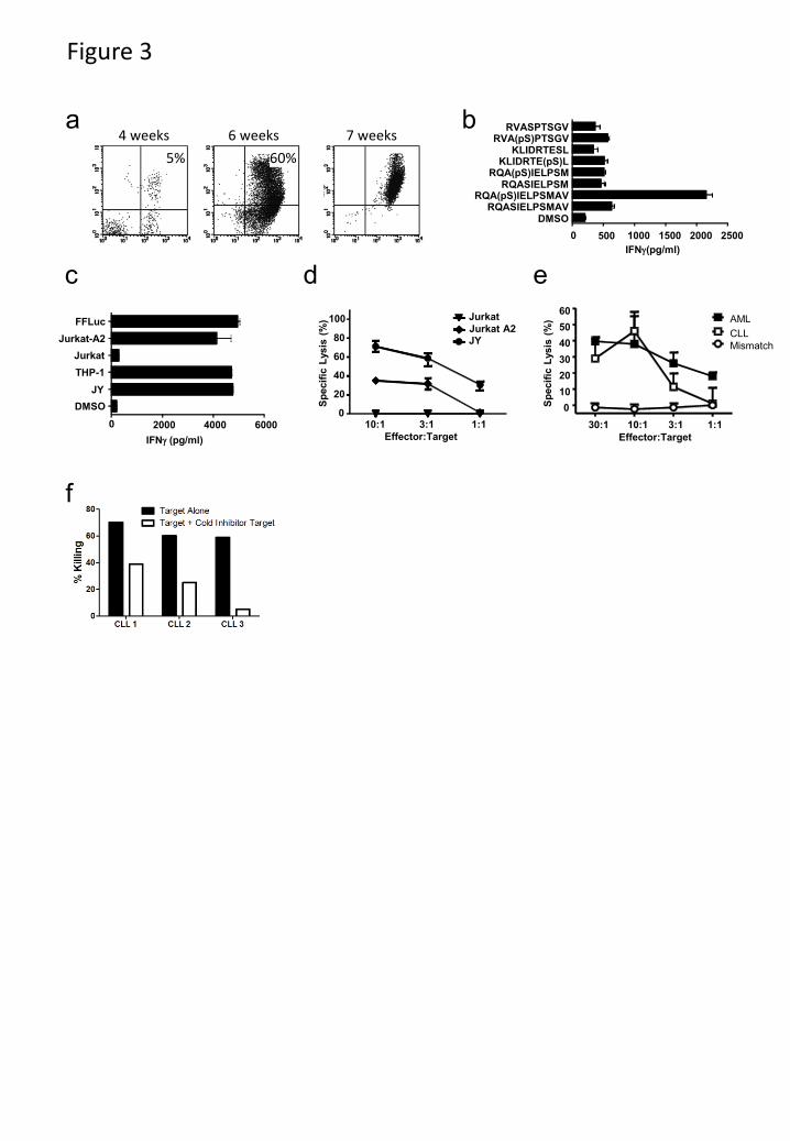
Figure 3
a
c
f
d
b
0 2000 4000 6000
DMSO
JY
THP-1
Jurkat
Jurkat-A2
FFLuc
IFNg (pg/ml)
0
20
40
60
80
100 Jurkat
Jurkat A2 JY
Sp
ecif
ic L
ysis
(%
)
10:1 3:1 1:1
Effector:Target
e
0 500 1000 1500 2000 2500
DMSO RQASIELPSMAV
RQA(pS)IELPSMAV RQASIELPSM
RQA(pS)IELPSM KLIDRTE(pS)L
KLIDRTESL RVA(pS)PTSGV
RVASPTSGV
IFNg(pg/ml)
30:1 10:1 3:1 1:1
0
10
20
30
40
50
60 AML
CLL
Mismatch
Effector:Target
Sp
ecif
ic L
ysis
(%
)
5% 60%
4 weeks 6 weeks 7 weeks
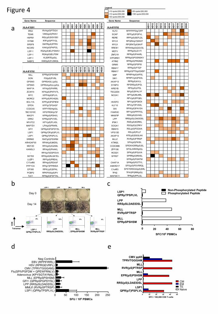
Figure 4
a
c b
d
0 20 40 60 80
MLL
EPRpSPSHSM
MLL
RVRpSPTRSP
LPP
RRSpSLDAEIDSL
LSP1
QPRpTPSPLVL
Non-Phosphorylated Peptide Phosphorylated Peptide Day 0
Day 14
e
<10 spots/200,000 >50 spots/200,000
>10 spots/200,000 >100 spots/200,000
>30 spots/200,000
Legend
SFC/105 PBMCs
LSP-1
QPRpTPSPLVL
LPP
RRSpSLDAEIDSL
MLL
EPRpSPSHSM
MLL
RVRpSPTRSP
LSP-1 LPP MLL MLL
8% 10% 6% 4%
CD
8
Tetramer
0 50 100 150 200 250
LSP1 (QPRpTPSPLVL) MMLtf (RVRpSPTRSP)
LPP (RRSpSLDAEIDSL) GFI1 (QPRpSPGPDYSL)
MLL (EPRpSPSHSM) Adenovirus (KPYSGTAYNAL)
Flu(SPIVPSFDM + QPEWFRNLV) CMV (TPRVTGGGAM))
HSV (RPRGEVRFL) EBV (RPPIFIRRL)
Neg Controls
SFU / 106 PBMCs
0 10 20 30 40 50
LSP1
QPR(pT)PSPLVL
LPP
RRS(pS)LDAEIDSL
MLL
EPR(pS)PSHSM
MLL
RVR(pS)PTRSP
CMV pp65
TPRVTGGGAM
CM EM TE Naive
SFC / 100,000 CD8 T-cells
Gene Name Sequence
HLA-A*0201 HD
1
HD
2
HD
3
HD
4
HD
5
HD
6
HD
7
HD
8
HD
9
HD
10
IRS2 RVA(pS)PTSGV
TEM6 VMIG(pS)PKKV
HSPB1 RQL(pS)SGVSEI
SYNM RTF(pS)PTYGL
SRSF8 SMTR(pS)PPRV
NCOR2 KAK(pS)PAPGL
LSP-1 RQA(pS)IELPSMAV
LSP-1 RQA(pS)IELPSM
LSP-1 KLIDRTE(pS)L
N4BP2 KMD(pS)FLDMQL
HLA-B*0702
MLL EPR(pS)PSHSM
SON FSI(pS)PVRL
GPSM3 GPRSASLL(pS)L
GPSM3 GPRSA(pS)LL(pS)L
MAP1A KPA(pS)PARRL
ZC3H14 KPA(pS)PKFIVTL
MYC KPPH(pS)PLVL
MORC2 KPP(pS)PEHQSL
BCL11A KPP(pS)PSPIEM
SPEN KPP(pT)PGASF
CCDC45 KPPYRSH(pS)L
TBC1D10C KPQTRGK(pT)F
MACF1 KPR(pS)PDHVL
GRK2 KPR(pS)PVVEL
NFATC2 KPY(pS)PLASL
MAP7D1 LPA(pS)PRARL
GFI1 QPR(pS)PGPDYSL
LSP1 QPR(pT)P(pS)PLVL
LSP1 QPR(pT)PSPLVL
SRRM2 RAP(pS)PSSRM
ARHGAP30 RPAK(pS)MDSL
MEF2D RPA(pS)AGAML
KANSL3 RPA(pS)PAAKL
- RPA(pT)GGPGVA
SETD2 RPD(pS)RLGKTEL
LUZP1 RPF(pS)PREAL
C17orf85 RPH(pS)PEKAF
PPP1CA RPI(pT)PPRNSA
EP300 RPKL(pS)SPAL
PCBP2 RPKPSS(pS)PV
TLK1 RPN(pS)PSPTAL
HD
11
HD
12
HD
13
HD
14
HD
15
HD
16
HD
17
HD
18
HD
19
HD
20
Gene Name Sequence
HLA-B*0702 HD
11
HD
12
HD
13
HD
14
HD
15
HD
16
HD
17
HD
18
HD
19
HD
20
KLF2 RPPPPPD(pT)PP
SCAP RPP(pS)PGPVL
RERE RPP(pS)SEFLDL
MYL9 RPQRA(pT)SNVF
MYL9 RPQRAT(pS)NVF
RREB1 RPRAN(pS)GGVDL
MEFV RPR(pS)LEVTI
ZNF219 RPR(pS)PAARL
GTF2IRD1 RPR(pS)PGSNSKV
ATXN2 RPR(pS)PRQNSI
GRM5 RPR(pS)PSPIS
RBM17 RPR(pS)PTGP
RBM17 RPR(pS)PTG(pS)NSF
MIIP RPRPH(pS)APSL
SIK1 RPRPV(pS)PSSL
AKAP13 RPR(pS)AVLL
SYNPO RPSRS(pS)PGL
ARID1B RPS(pS)LPDL
TSC22D4 RP(pS)SPALYF
NCOA1 RPT(pS)RLNRL
- RPV(pS)PFQEL
HIVEP2 RPV(pS)PGKDI
KLF10 RPV(pT)PVSDL
SKI RPW(pS)PAVSA
FAM53C RPY(pS)PPFFSL
MKI67IP RRK(pS)QVAEL
LPP RRS(pS)LDAEIDSL
IP6K1 RSE(pS)KDRKL
SOGA1 RSG(pS)LERKV
RBM15 RSL(pS)PGGAA
VPS13D RSL(pS)PLLF
AKAP13 RTE(pS)DSGLKK
MLLtf RVR(pS)PTRSP
ATXN2L SPA(pS)PKISL
CCDC88B SPEKAGRR(pS)SL
ZFP106 SPGLARKR(pS)L
RCSD1 SPK(pS)PGLKA
SFRS7 SPRR(pS)RSISL
- SPR(pS)PGKPM
CHAF1A SPR(pS)PSTTYL
ANKRD17 SPS(pS)PSVRRQL
C1orf63 SPSTSRSGG(pS)SRL
TPX2 TPAQPQRR(pS)L
MAP3K11 TPR(pS)PPLGL
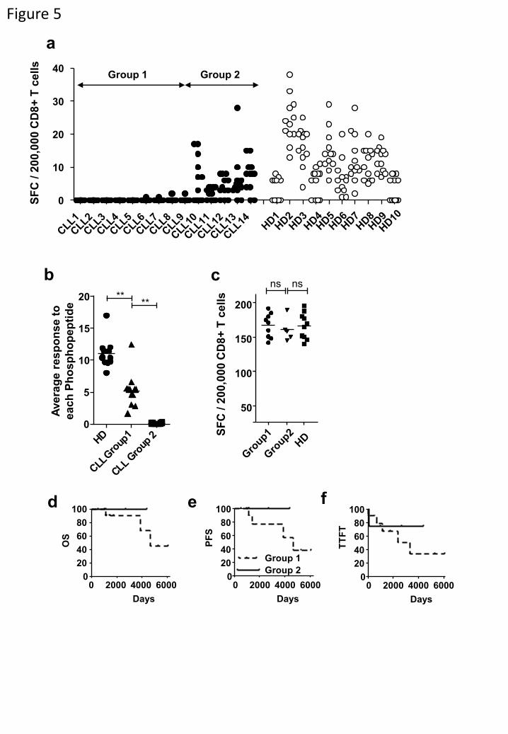
Figure 5
a
0
10
20
30
40
SF
C / 2
00
,00
0 C
D8
+ T
ce
lls
Group 1 Group 2
100
150
200
ns ns c
0 2000 4000 6000 0
20
40
60
80
100
Days
OS
TT
FT
0 2000 4000 6000 0
20
40
60
80
100
Days
0 2000 4000 6000 0
20
40
60
80
100
Days
PF
S
Group 1
Group 2
50
d e f
b
SF
C / 2
00
,00
0 C
D8
+ T
ce
lls
0
5
10
15
20 ** **
Ave
rag
e r
es
po
nse
to
ea
ch
Ph
osp
ho
pep
tid
e
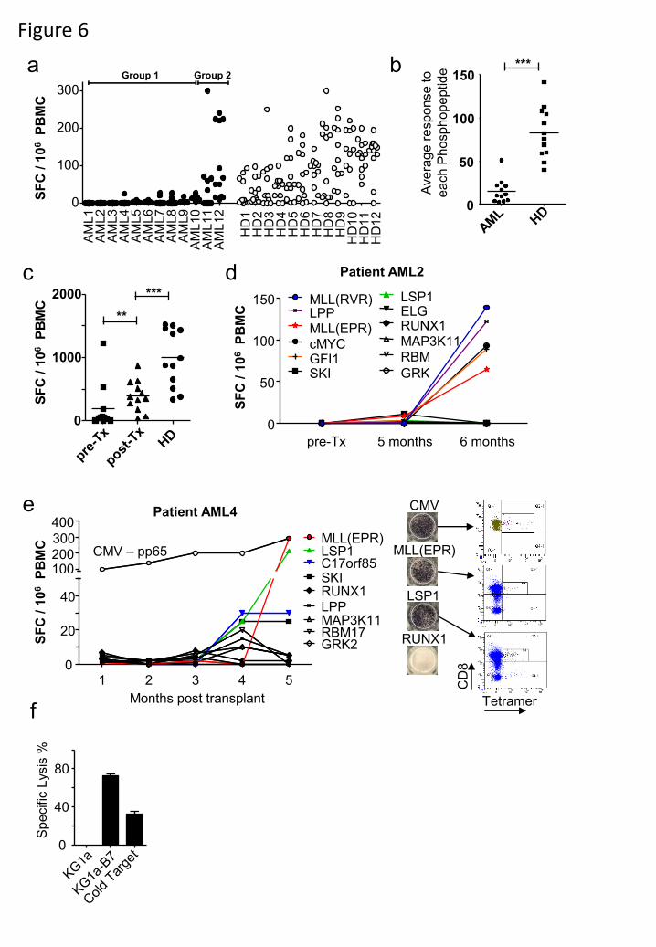
Figure 6
a
c
b
**
SF
C /
10
6 P
BM
C
0
40
80
Sp
ecific
Lysis
%
d
MLL(EPR)
LSP1
Tetramer
CD
8
e
0
1000
2000
1 2 3 4 5 0
20
40
100 200
300
400
LSP1
SKI C17orf85
RUNX1
CMV – pp65
MAP3K11 RBM17 GRK2
MLL(EPR)
LPP
SF
C /
10
6 P
BM
C
Patient AML4
SF
C /
10
6 P
BM
C
pre-Tx 5 months 6 months
0
50
100
150
cMYC MLL(EPR)
GFI1
LPP MLL(RVR)
SKI
LSP1 ELG RUNX1 MAP3K11 RBM
GRK
Patient AML2
Group 1 Group 2
AM
L1
A
ML
2
AM
L3
A
ML
4
AM
L5
A
ML
6
AM
L7
A
ML
8
AM
L9
A
ML
10
A
ML
11
A
ML
12
HD
1
HD
2
HD
3
HD
4
HD
5
HD
6
HD
7
HD
8
HD
9
HD
10
H
D1
1
HD
12
0
100
200
300
SF
C /
10
6 P
BM
C
Months post transplant
***
CMV
RUNX1
f
0
50
100
150
Ave
rag
e r
espo
nse
to
each
Ph
osp
ho
pe
ptid
e ***
Supplemental Figure 1
8mer HLA-B7 phosphopeptide ligands
10mer HLA-B7 phosphopeptide ligands
11mer HLA-B7 phosphopeptide ligands
c
9mer HLA-B7 phosphopeptide ligands
a
0
20
40
60
B*0702
A*0201
Phosphopeptide Length
% o
f E
pit
op
es
b
1 2 3 4 5 6 7 8 9 10 0
20
40
60
80
100 B7 8-mer
B7 9-mer
B7 10-mer
B7 11-mer
All B7
All A2
% o
f E
pit
op
es
Phosphate Position
Supplemental Figure 2
LSP1 QPR(pT)PSPLVL
LPP RRS(pS)LDAEIDSL
MLL EPR(pS)PSHSM
MLL RVR(pS)PTRSP
MLL EPR(pS)PSHSM
CMV pp65 TPRVTGGGAM
Donor 1
Donor 2
70%
15%
4%
11%
-p +p -p +p -p +p -p +p naive CM EM TE
CD8 CD27
CD
45
RA
Magnetic CD8 Sort

Supplemental Figure 3
Supplemental Figure 4
SFC / 200,000 CD8+ T cells
100 200 300 400
Unstimulated Pos C
Pos C + IL2 pNCOA1
pNCOA1 + IL2
Unstimulated Pos C
Pos C + IL2 pNCOA1
pNCOA1 + IL2
Unstimulated Anti-CD3
Anti-CD3 + IL2 pNCOA1
pNCOA1 + IL2
Unstimulated Anti-CD3
Anti-CD3 + IL2 pNCOA1
pNCOA1 + IL2
Unstimulated Anti-CD3
Anti-CD3 + IL2 pNCOA1
pNCOA1 + IL2
Unstimulated Anti-CD3
Anti-CD3 + IL2 pNCOA1
pNCOA1 + IL2
0
HD1
CLL1
CLL2
CLL3
CLL4
CLL5
MLL(EPR)
0
100
200
300
400 MLL(RVR)
0
50
100
150
200
250
RUNX1
0
50
100
150
SKI
0
50
100
150
200
250
GFI1
0
50
100
150
200
250
LPP
0
50
100
150
200
250
SF
C /
200
,00
0 C
D8
+ T
cell
s C17orf85
0
100
200
300 GRK2
0
50
100
150
200
250 RBM17
0
50
100
150
200
250
MAP3K11
0
50
100
150
200
250
0
50
100
150
200
c-Myc
SF
C /
200
,00
0 C
D8
+ T
cell
s
SF
C /
200
,00
0 C
D8
+ T
cell
s
0
100
200
300
400
LSP1
Supplemental Figure 5
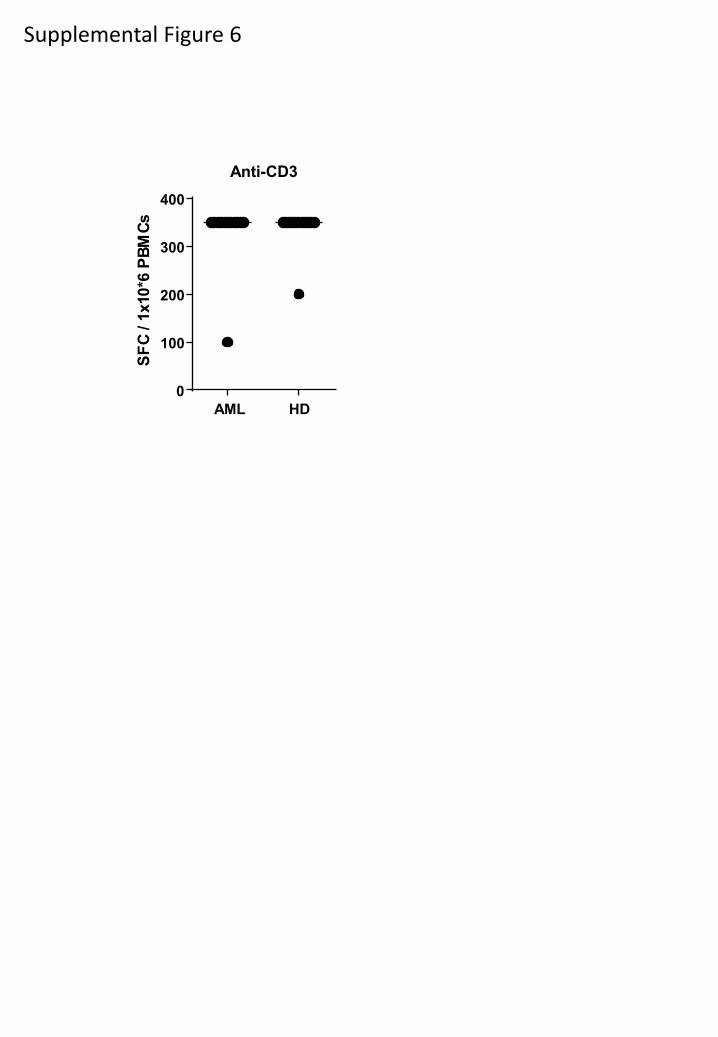
Supplemental Figure 6
AML HD
0
100
200
300
400
Anti-CD3
S F
C / 1
x 1 0 * 6
P B
M C
s
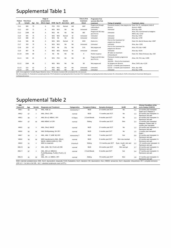
Supplemental Table 1
Supplemental Table 2
Patient Time Since
Stage at
presentation Genetic
Time to first
treatmentID Sample Age Sex (Binet stage) CD38 Zap-70 aberration IgVH (days)
CLL1 890 79 A POS POS Normal UM 2387Progressed through
chlorambucil
Previously treated, slow progressive
disease
Died, Chl, FC, methylpred, died of
disease +4695
CLL2 420 71 A NEG NEG NA NA Untreated Untreated Untreated Alive day +506
CLL3 1048 64 A NEG NA NA NA 685Progressed 301 days
post first txUntreated
Alive, FCR, transformed to Hodgkins
ABVD, day +1142
CLL4 945 75 A NEG NEG Normal M Untreated Untreated Untreated Alive day +5341
CLL5 688 63 A NEG NA NA NA Untreated Untreated Untreated Alive day +1749
CLL6 586 86 A NEG NEG Normal M 3334 Died on txPrior to first treatment for
progressive diseaseDied, Chl, died of disease day +3467
CLL7 359 70 A NEG NEG NA NA Untreated Untreated untreated Alive, day +1201
CLL8 359 67 A NEG NA 13q NA 1141 Not progressedPrior to first treatment for
progressive diseaseAlive, FCR, day +1460
CLL9 897 78 A NEG NEG Normal M Untreated Untreated Untreated Alive day +6133
CLL10 1091 89 C NA NA NA NA 0 Partial responseAll taken on treatment
(chlorambucil)Died, Chl, ?died of disease day +362
CLL11 540 65 B NEG POS NA NA 30Progressed 631 days
post first tx
Previously treated, progressive
diseaseAlive, Chl, FCO, day +1399
CLL12 1064 84 C NEG NEG NA NA 68 Not progressed
18/2/09 – Prior to first treatment
for progressive disease
6/1/10 – 4 months post treatment
Alive, Chl/R, day +1129
CLL13 523 63 A NEG NEG NA NA Untreated Untreated 6/1/10 – 4 months post treatment Alive, day +993
CLL14 579 63 A POS NEG NA M Untreated Untreated Untreated Alive day +2708
Progression free
survival after first
treatment Timing of sample(s) Treatment, status
IgVH, Immunoglobulin Variable Region Mutation status:- M indicates mutated; UM, unmutated; NA, not assessed;
NA, Not available; FC, fludarabine+cyclophosphamide; FCR, fludarabine+cyclophopshamide+rituximab; FCO, fludarabine+cyclophopshamide+ofatumumab; Chl, chlorambucil; Chl/R, chlorambucil+rituximab; Methylpred,
methylprednisolone
AML
Patient ID Age Gender Diagnosis and Treatment Cytogenetics Transplant History Samples Analyzed GvHD ALC
Clinical Condition at the
end of study (12/2012)
AML1 65 M AML, ADE x2 monosomy 7 MUD 21 months post SCT No 1.2 23 months post transplant:
Death due to Relapse †
AML2 67 F AML, DAx3, CR1 normal MUD 11 months post SCT No 1.3 26 months post transplant: In
Remission and well
AML3 64 F AML DA x2, MIDAC, CR1 FLT3pos 2 Cord Bloods 9 months post SCT No 1.3 26 months post transplant: In
Remission and well
AML4 67 M AML MIDAC x3 CR1 normal Sibling 10 months post SCT Skin 2.3 10 months post transplant:
Relapsed. Patient died 13
months post trasnplant
AML5 62 F AML, DAx2, MACE normal MUD 21 months post SCT No 1.3 39 months post transplant: In
Remission and well
AML6 65 M AML DA/Myelotarg, DA CR1 normal MUD 3 months post SCT No 1.5 3 months post transplant:
Death †
AML7 54 F AML, AML 17 (ADE X2) CR1 monosomy 7 MUD 7 months post SCT Gut 0.9 25 months post transplant: In
Remission and well
AML8 56 M MDS transformed to AML, [DAx2,
MACE, MIDAC] [ARA-C], CR2normal MUD 6 months post SCT Skin now resolved 1 25 months post transplant: In
Remission and well
AML9 51 F MDS no treatment trisomy 6 Sibling 113 months post SCT Eyes, mouth, skin and
liver2.2 131 months post transplant: In
Remission and well
AML10 66 F AML, [AML 16] [ FLAG x2] CR2 normal MUD 34 months post SCT Skin and gut 2.7 46 months post transplant: In
Remission and well
AML11 67 F AML, [DA x2, MIDAC] [
FLAG/Myelotarg, FLAG] [ FLAG x 2]
CR3
normal 2 Cord Bloods 9 months post SCT Gut 5.4 27 months post transplant: In
Remission and well
AML12 41 M AML, DA x 2, MIDAC, CR1 normal Sibling 15 months post SCT Skin 1.8 31 months post transplant: In
Remission and well
MUD, matched unrelated donor; ADE, Ara-C, daunorubicin, etoposide; FLAG, fludarabine, Ara-C, idarubicin; DA, daunorubicin, Ara-c; MIDAC; amsacrine, Ara-C, etoposide, mitozantrone; CR = complete remission.
(CR1,2,3 = 1st 2nd or 3rd CR). ALC = absolute lymphocyte count (x109/L)


![Mini HI-FI Component System · Mini HI-FI Component System Manual de instrucciones MHC-GTR88 MHC-GTR77 MHC-GTR55 MHC-GTR33. model name [MHC-GTR88] [4-165-654-33(2)] ES 2ES filename[D:\NORM'S](https://img.pdfslide.us/doc/110x75/5fdb9723a8509a11bd58c844/mini-hi-fi-component-system-mini-hi-fi-component-system-manual-de-instrucciones.jpg)