Embed Size (px)
Citation preview
Covalent capture of kinase-specific phosphopeptidesreveals Cdk1-cyclin B substratesJustin D. Blethrow*†, Joseph S. Glavy‡§, David O. Morgan¶, and Kevan M. Shokat*�
*Howard Hughes Medical Institute and Department of Cellular and Molecular Pharmacology, University of California, San Francisco, CA 94158; ‡Laboratoryof Cell Biology, Howard Hughes Medical Institute, Rockefeller University, 1230 York Avenue, New York, NY 10065; and ¶Departments of Physiology andBiochemistry and Biophysics, University of California, San Francisco, CA 94158
Edited by Benjamin Cravatt, The Scripps Research Institute, La Jolla, CA, and accepted by the Editorial Board November 29, 2007 (received for reviewSeptember 21, 2007)
We describe a method for rapid identification of protein kinasesubstrates. Cdk1 was engineered to accept an ATP analog thatallows it to uniquely label its substrates with a bio-orthogonalphosphate analog tag. A highly specific, covalent capture-and-release methodology was developed for rapid purification oftagged peptides derived from labeled substrate proteins. Applica-tion of this approach to the discovery of Cdk1-cyclin B substratesyielded identification of >70 substrates and phosphorylation sites.Many of these sites are known to be phosphorylated in vivo, butmost of the proteins have not been characterized as Cdk1-cyclin Bsubstrates. This approach has the potential to expand our under-standing of kinase–substrate connections in signaling networks.
chemical biology � chemical genetics � cyclin-dependent �phosphorylation � signaling
Protein kinases regulate a vast array of biological processesthrough phosphorylation of protein substrates. A comprehen-
sive map of all phosphorylation sites and kinase–substrate pairswould greatly facilitate the study of signaling networks. This goalfaces two fundamental challenges. First, all protein kinases use ATPas a cofactor to phosphorylate their targets, and thus the directsubstrates of a single kinase cannot be easily traced in proteinmixtures containing multiple kinases. Second, phosphorylationoften occurs at low stoichiometry and on low-abundance proteins.This makes substrate and phosphorylation site identification verychallenging. Powerful methods have been developed to addressthese dual problems (1, 2). Kinase–substrate pairs can be tested inmultiplexed phosphorylation assays by using immobilized arrays ofpurified proteins. These high-throughput chip-based assays havethe added benefit of presenting low-abundance proteins at easilydetectable levels and have provided a first-generation map ofprotein phosphorylation in Saccharomyces cerevisiae (3). However,these assays do not currently allow identification of phosphorylationsites and have not been adapted to organisms with more complexproteomes. Prediction of high-likelihood kinase substrates cansometimes be achieved by using knowledge of the substrate se-quence motif preferences of individual kinases, but only when thesepreferences are strong (4, 5). Finally, thousands of in vivo phos-phorylation sites from metazoan organisms have been identified inproteomic screens (6–9), but for most of these sites the responsibleupstream kinases remain unknown.
We have demonstrated a chemical and genetic solution to thecommon use of ATP by all kinases. Our approach relies onengineering a kinase to accept unnatural ATP analogs by modifi-cation of the ATP-binding pocket (10, 11). The analogs are verypoor substrates for wild-type kinases; thus, an analog-sensitivekinase (or as-kinase) can be used to specifically radiolabel itssubstrates in cell extracts while preserving important aspects ofbiological context, such as the integrity of protein complexes.Coupling this approach with the use of libraries of geneticallyencoded affinity-tagged proteins has facilitated identification oflow-abundance S. cerevisiae Cdk1 and Pho85 substrate proteins (12,13). However, this approach is not currently tractable in organismswhere genetic libraries present technical limits. Also, precise phos-
phorylation sites are not readily determined by this approach,because proteins are purified by virtue of a protein tag rather thana phospho-dependent tagging strategy.
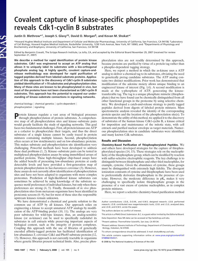
Here, we report a method in which the as-kinase uses a ATPanalog to deliver a chemical tag to its substrates, obviating the needto genetically pretag candidate substrates. The ATP analog con-tains two distinct modifications. Prior work has demonstrated thatmodification of the adenine moiety allows unique binding to anengineered kinase of interest (Fig. 1A). A second modification ismade at the �-phosphate of ATP, generating the kinase-transferable tag. The tag is a unique phosphate mimetic (thiophos-phate) that we have found can be chemically distinguished from allother functional groups in the proteome by using selective chem-istry. We developed a catch-and-release strategy to purify taggedpeptides derived from digests of labeled protein mixtures. Massspectrometric analysis revealed the identity of the correspondingprotein species and the location of the phosphorylation site(s). Todemonstrate the utility of this method, we applied it to the discoveryof substrates of the human kinase Cdk1-cyclin B, a kinase criticalfor imposition and maintenance of the mitotic state, with cellextracts and purified nuclear envelope as target materials. Numer-ous phosphorylation sites in candidate substrates were identified,and many known Cdk substrates.
Results and DiscussionChemistry-Based Purification of Thiophosphorylated Peptides. Weand others have developed strategies for the capture of thiophos-phorylated species (14, 15). These strategies rely on the nucleophi-licity of the thiophosphate group, which can be modified by reactionwith sulfur-selective electrophilic reagents. The key challenge is todistinguish between thiophosphate and other thiol nucleophiles, forexample, cysteine. Given the abundance of cysteine, these groupsmust be distinguished with extremely high fidelity. The divergentionization constants of cysteine and thiophosphate have been usedto preferentially derivatize thiophosphate in the presence of cys-teine. However, the moderate difference in pKa makes it verychallenging to specifically isolate thiophosphate groups in thepresence of a vast excess of cysteine nucleophiles, as in complexprotein mixtures.
Here, we report a selective chemistry-based purification method
Author contributions: J.D.B., D.O.M., and K.M.S. designed research; J.D.B. performedresearch; J.D.B. contributed new reagents/analytic tools; J.D.B. and J.S.G. analyzed data;and J.D.B. and K.M.S. wrote the paper.
The authors declare no conflict of interest.
This article is a PNAS Direct Submission. B.C. is a guest editor invited by the Editorial Board.
Data Deposition: Raw MS data can be accessed at ftp://acbishop.ucsf.edu.
†Present address: ThermoFisher Scientific, San Jose, CA 95134.
§Present address: Stevens Institute of Technology, Department of Chemistry and ChemicalBiology, Hoboken, NJ 07030.
�To whom correspondence should be addressed. E-mail: [email protected].
This article contains supporting information online at www.pnas.org/cgi/content/full/0708966105/DC1.
© 2008 by The National Academy of Sciences of the USA
1442–1447 � PNAS � February 5, 2008 � vol. 105 � no. 5 www.pnas.org�cgi�doi�10.1073�pnas.0708966105
Dow
nloa
ded
by g
uest
on
Nov
embe
r 22
, 202
1
that provides efficient and extremely specific recovery of thiophos-phorylated peptides. This approach relies on the fact that thiophos-phate and cysteine form qualitatively different products on alkyla-tion. These can subsequently be distinguished with total specificityin a second chemical step. This approach is conceptually analogousto the method of Aebersold and colleagues for purification ofphosphopeptides (42).
Our approach is detailed in Fig. 1B. A protein mixture containingtagged substrates is digested to peptides, separating the majority ofthiophosphate tags from cysteine. The peptide mixture is incubatedwith iodoacetyl-agarose resin, resulting in covalent capture oftagged peptides and peptides containing cysteine. Cysteine reacts toform thioether linkages, whereas tagged peptides form a chemicallydistinct phosphate diester. This diester linkage is normally quitestable but it can be made to rapidly hydrolyze by oxidation of thesulfur atom (16, 17). The resin is treated with the peroxide agentOxone, resulting in oxidation of all sulfur atoms present (thiophos-phate, cysteine, and methionine). The thiophosphate linkagesspontaneously hydrolyze, releasing phosphopeptides, whereas thethioether linkages, although oxidized, remain stable (18, 19). Thus,tagged peptides are rapidly and specifically eluted.
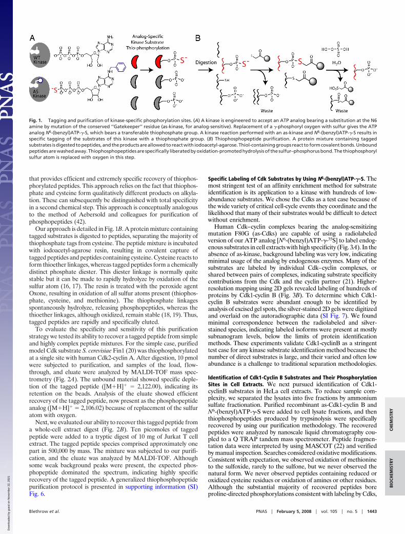
To evaluate the specificity and sensitivity of this purificationstrategy we tested its ability to recover a tagged peptide from simpleand highly complex peptide mixtures. For the simple case, purifiedmodel Cdk substrate S. cerevisiae Fin1 (20) was thiophosphorylatedat a single site with human Cdk2-cyclin A. After digestion, 10 pmolwere subjected to purification, and samples of the load, flow-through, and eluate were analyzed by MALDI-TOF mass spec-trometry (Fig. 2A). The unbound material showed specific deple-tion of the tagged peptide ([M�H]� � 2,122.00), indicating itsretention on the beads. Analysis of the eluate showed efficientrecovery of the tagged peptide, now present as the phosphopeptideanalog ([M�H]� � 2,106.02) because of replacement of the sulfuratom with oxygen.
Next, we evaluated our ability to recover this tagged peptide froma whole-cell extract digest (Fig. 2B). Ten picomoles of taggedpeptide were added to a tryptic digest of 10 mg of Jurkat T cellextract. The tagged peptide species comprised approximately onepart in 500,000 by mass. The mixture was subjected to our purifi-cation, and the eluate was analyzed by MALDI-TOF. Althoughsome weak background peaks were present, the expected phos-phopeptide dominated the spectrum, indicating highly specificrecovery of the tagged peptide. A generalized thiophosphopeptidepurification protocol is presented in supporting information (SI)Fig. 6.
Specific Labeling of Cdk Substrates by Using N6-(benzyl)ATP-�-S. Themost stringent test of an affinity enrichment method for substrateidentification is its application to a kinase with hundreds of low-abundance substrates. We chose the Cdks as a test case because ofthe wide variety of critical cell-cycle events they coordinate and thelikelihood that many of their substrates would be difficult to detectwithout enrichment.
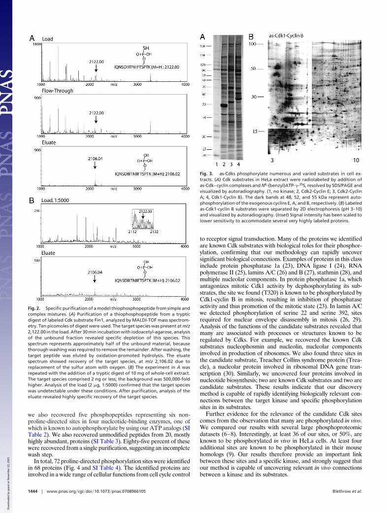
Human Cdk–cyclin complexes bearing the analog-sensitizingmutation F80G (as-Cdks) are capable of using a radiolabeledversion of our ATP analog [N6-(benzyl)ATP-�-35S] to label endog-enous substrates in cell extracts with high specificity (Fig. 3A). In theabsence of as-kinase, background labeling was very low, indicatingminimal usage of the analog by endogenous enzymes. Many of thesubstrates are labeled by individual Cdk–cyclin complexes, orshared between pairs of complexes, indicating substrate specificitycontributions from the Cdk and the cyclin partner (21). Higher-resolution mapping using 2D gels revealed labeling of hundreds ofproteins by Cdk1-cyclin B (Fig. 3B). To determine which Cdk1-cyclin B substrates were abundant enough to be identified byanalysis of excised gel spots, the silver-stained 2D gels were digitizedand overlaid on the autoradiographic data (SI Fig. 7). We foundminimal correspondence between the radiolabeled and silver-stained species, indicating labeled isoforms were present at mostlysubnanogram levels, below the limits of protein identificationmethods. These experiments validate Cdk1-cyclinB as a stringenttest case for any kinase substrate identification method because thenumber of direct substrates is large, and their varied and often lowabundance is a challenge to traditional separation methodologies.
Identification of Cdk1-Cyclin B Substrates and Their PhosphorylationSites in Cell Extracts. We next pursued identification of Cdk1-cyclinB substrates in HeLa cell extracts. To reduce sample com-plexity, we separated the lysates into five fractions by ammoniumsulfate fractionation. Purified recombinant as-Cdk1-cyclin B andN6-(benzyl)ATP-�-S were added to cell lysate fractions, and thenthiophosphopeptides produced by trypsinolysis were specificallyrecovered by using our purification methodology. The recoveredpeptides were analyzed by nanoscale liquid chromatography cou-pled to a Q TRAP tandem mass spectrometer. Peptide fragmen-tation data were interpreted by using MASCOT (22) and verifiedby manual inspection. Searches considered oxidative modifications.Consistent with expectation, we observed oxidation of methionineto the sulfoxide, rarely to the sulfone, but we never observed thenatural form. We never observed peptides containing reduced oroxidized cysteine residues or oxidation of amines or other residues.Although the substantial majority of recovered peptides boreproline-directed phosphorylations consistent with labeling by Cdks,
Fig. 1. Tagging and purification of kinase-specific phosphorylation sites. (A) A kinase is engineered to accept an ATP analog bearing a substitution at the N6amine by mutation of the conserved ‘‘Gatekeeper’’ residue (as-kinase, for analog-sensitive). Replacement of a �-phosphoryl oxygen with sulfur gives the ATPanalog N6-(benzyl)ATP-�-S, which bears a transferable thiophosphate group. A kinase reaction performed with an as-kinase and N6-(benzyl)ATP-�-S results inspecific tagging of the substrates of this kinase with a thiophosphate group. (B) Thiophosphopeptide purification. A protein mixture containing taggedsubstrates is digested to peptides, and the products are allowed to react with iodoacetyl-agarose. Thiol-containing groups react to form covalent bonds. Unboundpeptides are washed away. Thiophosphopeptides are specifically liberated by oxidation-promoted hydrolysis of the sulfur–phosphorus bond. The thiophosphorylsulfur atom is replaced with oxygen in this step.
Blethrow et al. PNAS � February 5, 2008 � vol. 105 � no. 5 � 1443
CHEM
ISTR
YBI
OCH
EMIS
TRY
Dow
nloa
ded
by g
uest
on
Nov
embe
r 22
, 202
1
we also recovered five phosphopeptides representing six non-proline-directed sites in four nucleotide-binding enzymes, one ofwhich is known to autophosphorylate by using our ATP analogs (SITable 2). We also recovered unmodified peptides from 20, mostlyhighly abundant, proteins (SI Table 3). Eighty-five percent of thesewere recovered from a single purification, suggesting an incompletewash step.
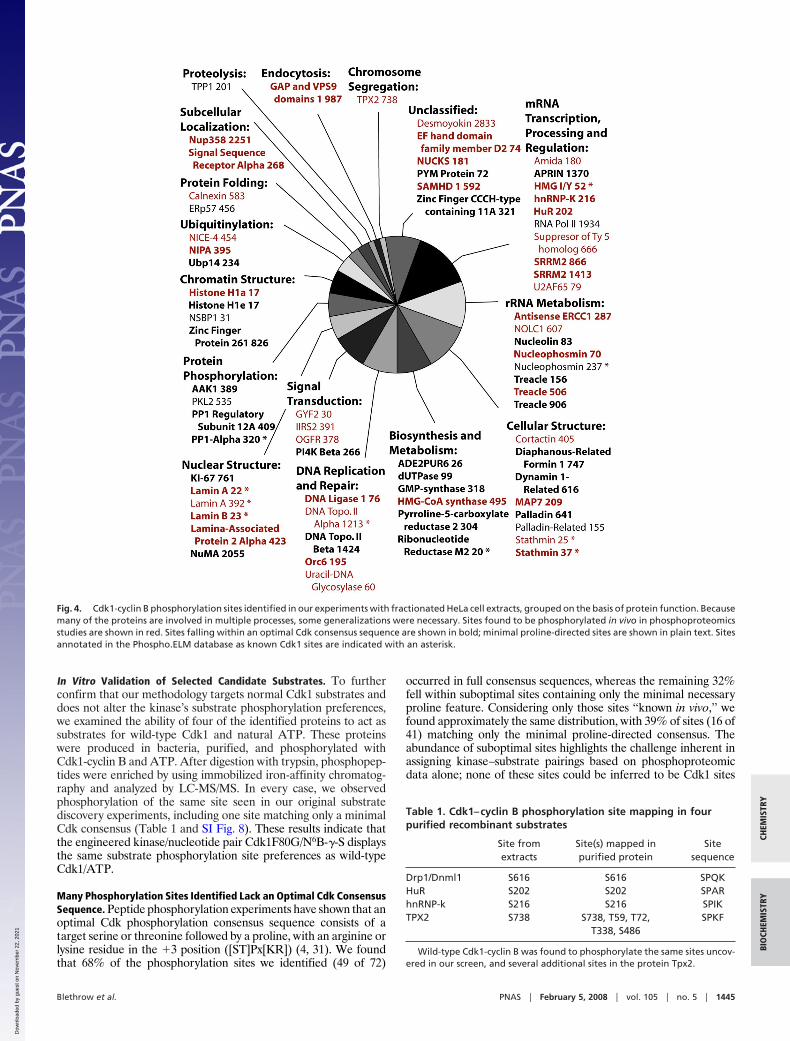
In total, 72 proline-directed phosphorylation sites were identifiedin 68 proteins (Fig. 4 and SI Table 4). The identified proteins areinvolved in a wide range of cellular functions from cell cycle control
to receptor signal transduction. Many of the proteins we identifiedare known Cdk substrates with biological roles for their phosphor-ylation, confirming that our methodology can rapidly uncoversignificant biological connections. Examples of proteins in this classinclude protein phosphatase 1a (23), DNA ligase I (24), RNApolymerase II (25), lamins A/C (26) and B (27), stathmin (28), andmultiple nucleolar components. In protein phosphatase 1a, whichantagonizes mitotic Cdk1 activity by dephosphorylating its sub-strates, the site we found (T320) is known to be phosphorylated byCdk1-cyclin B in mitosis, resulting in inhibition of phosphataseactivity and thus promotion of the mitotic state (23). In lamin A/Cwe detected phosphorylation of serine 22 and serine 392, sitesrequired for nuclear envelope disassembly in mitosis (26, 29).Analysis of the functions of the candidate substrates revealed thatmany are associated with processes or structures known to beregulated by Cdks. For example, we recovered the known Cdksubstrates nucleophosmin and nucleolin, nucleolar componentsinvolved in production of ribosomes. We also found three sites inthe candidate substrate, Treacher Collins syndrome protein (Trea-cle), a nucleolar protein involved in ribosomal DNA gene tran-scription (30). Similarly, we uncovered four proteins involved innucleotide biosynthesis; two are known Cdk substrates and two arecandidate substrates. These results indicate that our discoverymethod is capable of rapidly identifying biologically relevant con-nections between the target kinase and specific phosphorylationsites in its substrates.
Further evidence for the relevance of the candidate Cdk sitescomes from the observation that many are phosphorylated in vivo.We compared our results with several large phosphoproteomicdatasets (6–8). Interestingly, at least 36 of our sites, or 50%, areknown to be phosphorylated in vivo in HeLa cells. At least fouradditional sites are known to be phosphorylated in their mousehomologs (9). Our results therefore provide an important linkbetween these sites and a specific kinase, and strongly suggest thatour method is capable of uncovering relevant in vivo connectionsbetween a kinase and its substrates.
Fig. 2. Specific purification of a model thiophosphopeptide from simple andcomplex mixtures. (A) Purification of a thiophosphopeptide from a trypticdigest of labeled Cdk substrate Fin1, analyzed by MALDI-TOF mass spectrom-etry. Ten picomoles of digest were used. The target species was present at m/z2,122.00 in the load. After 30 min incubation with iodoacetyl-agarose, analysisof the unbound fraction revealed specific depletion of this species. Thisspectrum represents approximately half of the unbound material, becausethorough washing was required to remove the remainder. After washing, thetarget peptide was eluted by oxidation-promoted hydrolysis. The eluatespectrum showed recovery of the target species, at m/z 2,106.02 due toreplacement of the sulfur atom with oxygen. (B) The experiment in A wasrepeated with the addition of a tryptic digest of 10 mg of whole-cell extract.The target species comprised 2 ng or less; the background was 500,000-foldhigher. Analysis of the load (2 �g, 1:5000) confirmed that the target specieswas undetectable under these conditions. After purification, analysis of theeluate revealed highly specific recovery of the target species.
Fig. 3. as-Cdks phosphorylate numerous and varied substrates in cell ex-tracts. (A) Cdk substrates in HeLa extract were radiolabeled by addition ofas-Cdk–cyclin complexes and N6-(benzyl)ATP-�-35S, resolved by SDS/PAGE andvisualized by autoradiography. (1, no kinase; 2, Cdk2-Cyclin E; 3, Cdk2-CyclinA; 4, Cdk1-Cyclin B). The dark bands at 48, 52, and 55 kDa represent auto-phosphorylation of the exogenous cyclins E, A, and B, respectively. (B) Labeledas-Cdk1-cyclin B substrates were separated by 2D electrophoresis (pH 3–10)and visualized by autoradiography. (Inset) Signal intensity has been scaled tolower sensitivity to accommodate several very highly labeled proteins.
1444 � www.pnas.org�cgi�doi�10.1073�pnas.0708966105 Blethrow et al.
Dow
nloa
ded
by g
uest
on
Nov
embe
r 22
, 202
1
In Vitro Validation of Selected Candidate Substrates. To furtherconfirm that our methodology targets normal Cdk1 substrates anddoes not alter the kinase’s substrate phosphorylation preferences,we examined the ability of four of the identified proteins to act assubstrates for wild-type Cdk1 and natural ATP. These proteinswere produced in bacteria, purified, and phosphorylated withCdk1-cyclin B and ATP. After digestion with trypsin, phosphopep-tides were enriched by using immobilized iron-affinity chromatog-raphy and analyzed by LC-MS/MS. In every case, we observedphosphorylation of the same site seen in our original substratediscovery experiments, including one site matching only a minimalCdk consensus (Table 1 and SI Fig. 8). These results indicate thatthe engineered kinase/nucleotide pair Cdk1F80G/N6B-�-S displaysthe same substrate phosphorylation site preferences as wild-typeCdk1/ATP.
Many Phosphorylation Sites Identified Lack an Optimal Cdk ConsensusSequence. Peptide phosphorylation experiments have shown that anoptimal Cdk phosphorylation consensus sequence consists of atarget serine or threonine followed by a proline, with an arginine orlysine residue in the �3 position ([ST]Px[KR]) (4, 31). We foundthat 68% of the phosphorylation sites we identified (49 of 72)
occurred in full consensus sequences, whereas the remaining 32%fell within suboptimal sites containing only the minimal necessaryproline feature. Considering only those sites ‘‘known in vivo,’’ wefound approximately the same distribution, with 39% of sites (16 of41) matching only the minimal proline-directed consensus. Theabundance of suboptimal sites highlights the challenge inherent inassigning kinase–substrate pairings based on phosphoproteomicdata alone; none of these sites could be inferred to be Cdk1 sites
Fig. 4. Cdk1-cyclin B phosphorylation sites identified in our experiments with fractionated HeLa cell extracts, grouped on the basis of protein function. Becausemany of the proteins are involved in multiple processes, some generalizations were necessary. Sites found to be phosphorylated in vivo in phosphoproteomicsstudies are shown in red. Sites falling within an optimal Cdk consensus sequence are shown in bold; minimal proline-directed sites are shown in plain text. Sitesannotated in the Phospho.ELM database as known Cdk1 sites are indicated with an asterisk.
Table 1. Cdk1–cyclin B phosphorylation site mapping in fourpurified recombinant substrates
Site fromextracts
Site(s) mapped inpurified protein
Sitesequence
Drp1/Dnml1 S616 S616 SPQKHuR S202 S202 SPARhnRNP-k S216 S216 SPIKTPX2 S738 S738, T59, T72,
T338, S486SPKF
Wild-type Cdk1-cyclin B was found to phosphorylate the same sites uncov-ered in our screen, and several additional sites in the protein Tpx2.
Blethrow et al. PNAS � February 5, 2008 � vol. 105 � no. 5 � 1445
CHEM
ISTR
YBI
OCH
EMIS
TRY
Dow
nloa
ded
by g
uest
on
Nov
embe
r 22
, 202
1
based solely on sequence analysis. Given the large number ofproline-directed kinases, it’s tempting to speculate that such sitesmay be substrates for multiple kinases, perhaps serving as signalintegrators. Indeed, even optimal sites may serve such a function.For example, we found an optimal site in hnRNP-K (S216) that wasdetermined to be phosphorylated by JNK (32). Phosphorylation ofthis site along with S353 was found to increase the transcriptionalactivity of hnRNPk from AP1-bearing promoters. We speculatethat Cdk1-cyclin B activity may similarly regulate this protein.
Identification of Cdk1-Cyclin B Substrates in Purified Nuclear Enve-lope. We next sought to evaluate the utility of our method for thediscovery of kinase substrates in purified subcellular compartments.This approach serves to greatly enrich a subset of the proteomewhile preserving the structural context of the enriched proteins. Wechose to focus on the nuclear envelope. Mitotic disassembly of thenuclear lamina and of the nuclear pore complex (NPC) are knownto be imposed in large part by Cdk activity (33, 34).
Rat liver nuclei were prepared as described in ref. 35 and nuclearenvelope was prepared by DNase/RNase digestion followed byheparin treatment as described in ref. 36. This material contains thenuclear membrane, transmembrane and tightly associated proteins,and the NPC. Purified recombinant as-Cdk1-cyclin B and N6B-�-Swere added to the preparation; three experiments were performedand recovered phosphopeptides were analyzed by LC-MS/MS.
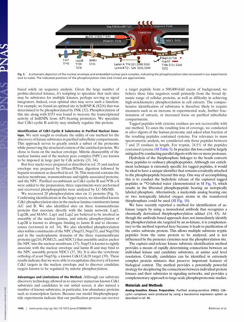
We recovered 20 phosphorylation sites on 11 proteins (Fig. 5).Confirming identifications in the larger screen, we identified knownCdk1 phosphorylation sites in the nuclear lamina constituents laminA/C and B. We also identified sites on three transmembraneproteins that associate directly with the lamin network: Lap1,Lap2B, and MAN1. Lap1 and Lap2 are believed to be involved inassembly of the nuclear lamina, and mitotic phosphorylation ofLap2B is known to abrogate binding to lamin B and to chromo-somes (reviewed in ref. 34). We also identified phosphorylationsites within constituents of the NPC (Nup53, Nup133, and Nup358)and in the nucleoplasmic domains of the three transmembraneproteins (gp210, POM121, and NDC1) that assemble and/or anchorthe NPC into the nuclear membrane (37). Nup53 is known to tightlyassociate with the nuclear envelope and lamin B and may bind tothe NPC assembly protein NDC1 (37, 38). It is also the vertebrateortholog of yeast Nup53p, a known Cdk1/Cdc28 target (39). Theseresults indicate that we were able to recapitulate discovery of knownCdk1 targets in the nuclear envelope and to discover candidatetargets known to be regulated by mitotic phosphorylation.
Advantages and Limitations of the Method. Although our substratediscovery technology allowed us to uncover numerous known Cdk1substrates and candidates in our initial screen, it also missed anumber of known substrates, in particular, low-abundance proteinssuch as transcription factors. Because our model thiophosphopep-tide experiments indicate that our purification process can recover
a target peptide from a 500,000-fold excess of background, webelieve these false negatives result primarily from the broad dy-namic range of cellular proteins, as well as difficulty in achievinghigh-stoichiometry phosphorylation in cell extracts. The compre-hensive identification of substrates is therefore likely to requiremeasures such as an increase in experimental scale, further frac-tionation of extracts, or increased focus on purified subcellularcompartments.
Tagged peptides with cysteine residues are not recoverable withour method. To asses the resulting loss of coverage, we conductedin silico digests of the human proteome and asked what fraction ofthe resulting peptides contained cysteine. For relevance to massspectrometric analysis, we considered only those peptides between7 and 25 residues in length. For trypsin, 24.5% of the peptidescontained cysteine (SI Table 5). In practice this loss could be largelymitigated by conducting parallel digests with two or more proteases.
Hydrolysis of the thiophosphate linkages to the beads convertsthese peptides to ordinary phosphopeptides. Although our enrich-ment technique is extremely specific for tagged peptides, it wouldbe ideal to have a unique identifier that remains covalently attachedto the phosphopeptide beyond this step. One way of accomplishingthis is to conduct the hydrolytic release of the captured taggedpeptides in 18O-labeled water (demonstrated in SI Fig. 9), whichresults in the liberated phosphopeptide bearing an isotopicallylabeled phosphate. Alternatively, a nucleotide analog bearing oneor two isotopically labeled oxygen atoms in the transferredthiophosphate could be used (SI Fig. 10).
We have recently reported a method for identification of as-kinase targets by using a monoclonal antibody that recognizes achemically derivatized thiophosphorylation adduct (14, 43). Al-though the antibody-based approach does not immediately identifythe phosphorylation site targeted by an as-kinase, it is complemen-tary to the method reported here because it leads to purification ofthe entire substrate protein. This allows multiple substrate trypticpeptides from the same protein to be analyzed, and is notinfluenced by the presence cysteines near the phosphorylation site.
The capture-and-release kinase substrate identification methodprovides a means of rapidly determining connections between anindividual kinase and candidate substrates, at amino acid levelresolution. Critically, candidates can be identified in extremelycomplex protein mixtures that preserve important features ofbiological context. The method provides a potentially powerfulstrategy for deciphering the connections between individual proteinkinases and their substrates in signaling networks, and provides acomplementary approach to large-scale phosphoproteomic studies.
Materials and MethodsAnalog-Sensitive Kinase Preparation. Purified analog-sensitive (F80G) Cdk–cyclin complexes were produced by using a baculovirus expression system asdescribed in ref. 40.
Fig. 5. A schematic depiction of the nuclear envelope and embedded nuclear pore complex, indicating the phosphorylation sites recovered in our experiments(not to scale). The indicated positions of the phosphorylation sites (red circles) are approximate.
1446 � www.pnas.org�cgi�doi�10.1073�pnas.0708966105 Blethrow et al.
Dow
nloa
ded
by g
uest
on
Nov
embe
r 22
, 202
1
Synthesis of N6-(Benzyl)ATP-�-phosphorothioate. The synthesis of this reagentfollows the procedure described for ATP-�-S by Goody and Eckstein (41) withthe exception of chromatographic separations (SI Fig. 11). In brief, sodiumthiophosphate (Sigma) was protected at sulfur with 3-chloropropionamide.The product was activated with diphenyl phosphorochloridate and coupled toN6-(benzyl)ADP. The protected product was isolated by ion-exchange chro-matography. Treatment with 0.1 M sodium hydroxide at 100°C for 10 min gavethe deprotected species. The product was again isolated by ion-exchangechromatography. Mass and structure were verified by MS/MS analysis.
Synthesis of N6-(Benzyl)ATP-�-35S-Phosphorothioate. N6-(Benzyl)ATP was con-verted to N6-(benzyl)ATP-�-35S by using an enzymatic system as described inref. 13.
Autoradiographic Analysis of Cdk Substrates. For 1D analysis, 20 �g of total HeLacell extract were labeled by the addition of 5 �Ci of N6-(benzyl)ATP-�-35S, 1 mMmagnesium chloride, and either buffer or purified recombinant as-Cdk–cyclincomplexes (200 ng), at room temperature for 30 min. The samples were resolvedby PAGE on 7.5–15% gradient gels, dried, and imaged by using a Typhoon system(GE Healthcare). For 2D analysis of as-Cdk1-cyclin B substrates, the reaction wasscaled 10-fold, and proteins were separated over an immobilized pH 3–10 gra-dient by using an IPGphor system (Amersham Biosciences). Separated proteinswere resolved by SDS/PAGE on a 7.5–15% gel.
Model Thiophosphopeptide Studies. Purified, bacterially expressed Fin1 proteinwas labeled by incubation with Cdk2-cyclin A, ATP-�-S, and 5 mM MgCl2, thendigestedwithtrypsin.Tenpicomolesofdigestwereaddedto100�lof iodoacetyl-agarose beads (SulfoLink gel, Pierce) in 100 �l of 50% acetonitrile, alone or with10 mg of Jurkat cell extract digest. The beads were incubated overnight in thedark with rotation, and then loaded in a 1-ml disposable column. The flow-through fraction from the Fin1-only sample was set aside, and the beads werewashed with 10 ml each of water, 5 M NaCl, 50% acetonitrile, and 5% formic acidin water. The beads were treated with 500 �l of a 1 mg/ml solution of Oxone.Eluting phosphopeptides were concentrated by capture on 1 �l of Poros R3 resin(Applied Biosystems) immobilized in a gel-loading pipette tip, and eluted directlyonto a MALDI plate by using a standard DHB MALDI matrix solution, for analysiswith a PrOTOF instrument (PerkinElmer).
Preparation of HeLa Cell Total Cell Extract. Pelleted cells were purchased fromtheNationalCellCultureCenter (NCCC)andresuspended inhypotonic lysisbuffer(20 mM HEPES, pH 7.4, 1 mM EDTA, 1 mM DTT, 0.1% Nonidet P-40, plus proteaseinhibitors) and lysed by douncing. Nuclei and membranous fractions were pel-leted by centrifugation and resuspended in lysis buffer containing 500 mM KCl.After centrifugation, the supernatant was added to the initial lysate.
Preparation of Nuclear Envelope. Rat liver nuclei were prepared as described inref. 35 and stored at �80°C in 100-U (3 � 108 nuclei) aliquots. Nuclear envelopewas prepared as described in ref. 36.
Purification of Cdk1-Cyclin B Substrate-Derived Phosphopeptides from HeLa CellExtracts. Ammonium sulfate was added to HeLa cell total extract and proteinsprecipitating at 25%, 40%, 50%, 60%, and 75% of saturation were separated by
centrifugation and stored frozen as pellets until needed. Pellets were thoroughlydialyzed against HEPES-buffered saline before further use (20 mM HEPES, pH 7.4,150 mM NaCl). Extract fractions (0.1–1 mg) were labeled by addition of purifiedrecombinant as-Cdk1-cyclin B to 1% by mass of total protein, 1 mM N6-(benzyl)ATP-�-phosphorothioate, and 5 mM MgCl2. After 30 min of labeling, thereactions were stopped by addition of EDTA and then digested with trypsin.Thiophosphopeptides were recovered by using our purification methodology, asdescribed for the model thiophosphopeptide experiments. The recovered pep-tides were analyzed by nanoscale LC-MS/MS. Proteins and phosphorylation sitesidentified over the course of several experiments were combined in a MicrosoftAccess database.
LC-MS/MS Analysis. Phosphopeptide samples purified from HeLa extract frac-tions were analyzed by nanoscale liquid chromatography coupled on-line to a QTrap tandem mass spectrometer (Sciex). The peptides were separated over thecourse of 100 min by using a nonlinear gradient of 5% to 30% acetonitrilecontaining 0.1% formic acid, at a flow rate of 150 nl/min. A 75 �m i.d. by 15 cmRP column (LC Packings) was used to resolve peptides, and an upstream trapcolumn was used to facilitate sample concentration and transfer from the au-tosampler. Survey scans were performed by using the ‘‘Enhanced MultiCharge’’mode. Neutral loss survey scans targeting loss of phosphate were occasionallyused. Fragmentation spectra were acquired automatically in information-dependent acquisition (IDA) mode and interpreted both manually and withMASCOT (Matrix Science).
Database Searching. MS data files were searched against the Uniprot and NCBInrdatabases by using MASCOT. First-pass searches considered one missed cut andoxidation of methionine to the sulfoxide or sulfone, and phosphorylation ofserine or threonine. Further searches considered two missed cuts, the presence ofone nontryptic end, and rarer modifications such pyroglutamic acid formation.Peptides with MASCOT expect scores of �0.05 were included; most of thereported hits had substantially better scores (SI Tables 2–4).
In Vitro Phosphorylation Assays. ORFs encoding Drp1 and TPX2 were purchasedas Ultimate ORF Gateway vectors (Invitrogen) and shuttled into the bacterialexpression vector pDest17 by site-specific recombination. pDest17 encodes anin-frame N-terminal 6x His tag. ORFs encoding HuR and hnRNP-k were purchasedascDNAs(Invitrogen)andclonedintopDONR221byPCR-mediatedincorporationof terminal recombination sequences, followed by recombination. The ORFswere then shuttled into pDEST17 by recombination. All four proteins wereexpressed in Escherichia coli strain BL21(DE3) pLysS (Novagen), and purified byIDA-Cobalt affinity chromatography. Five micrograms of each purified proteinwere labeled with 20 ng of Cdk1-cyclin B and 100 mM ATP for 30 min, thenreactions were stopped with 5 mM EDTA and digested. Phosphopeptides wereenrichedbyusingimmobilizediron(Phos-Selectbeads;Sigma),elutedwithEDTA,and analyzed by nanoscale LC-MS/MS using the QTrap instrument.
ACKNOWLEDGMENTS. This work was supported in part by National Institutesof Health Grants R01EB001987 (to K.M.S.) and R01GM69901 (to D.O.M.) anda Graduate Research and Education in Adaptive Biotechnology fellowshipfrom the University of California Biotechnology Research Education Program(to J.D.B.). We also thank the Sandler Family Foundation for supportingpurchase of mass spectrometers used in this work.
1. Manning BD, Cantley LC (2002) Sci STKE 2002:pe49.2. Ptacek J, Snyder M (2006) Trends Genet 22:545–554.3. Ptacek J, et al. (2005) Nature 438:679–684.4. Songyang Z, et al. (1994) Curr Biol 4:973–982.5. Obenauer JC, Cantley LC, Yaffe MB (2003) Nucleic Acids Res 31:3635–3641.6. Beausoleil SA, et al. (2004) Proc Natl Acad Sci USA 101:12130–12135.7. Nousiainen M, Sillje HH, Sauer G, Nigg EA, Korner R (2006) Proc Natl Acad Sci USA
103:5391–5396.8. Olsen JV, et al. (2006) Cell 127:635–648.9. Villen J, Beausoleil SA, Gerber SA, Gygi SP (2007) Proc Natl Acad Sci USA 104:1488–1493.
10. Shah K, Liu Y, Deirmengian C, Shokat KM (1997) Proc Natl Acad Sci USA 94:3565–3570.11. Liu Y, Shah K, Yang F, Witucki L, Shokat KM (1998) Chem Biol 5:91–101.12. Dephoure N, Howson RW, Blethrow JD, Shokat KM, O’Shea EK (2005) Proc Natl Acad
Sci USA 102:7940–17945.13. Ubersax JA, et al. (2003) Nature 425:859–864.14. Allen JJ, Lazerwith SE, Shokat KM (2005) J Am Chem Soc 127:5288–5289.15. Kwon SW, Kim SC, Jaunbergs J, Falck JR, Zhao Y (2003) Mol Cell Proteomics 2:242–247.16. Yang YC, Szafraniec LL, Beaudry WT, Rohrbaugh DK (1990) J Am Chem Soc 112:6621–6627.17. Blasko A, Bunton CA, Kumar A (1997) J Phys Org Chem 10:427–434.18. Webb KS (1994) Tetrahedron Lett 35:3457–3460.19. Kennedy RJ, Stock AM (1960) J Am Chem Soc 25:1901–1906.20. Woodbury EL, Morgan DO (2007) Nat Cell Biol 9:106–112.21. Loog M, Morgan DO (2005) Nature 434:104–108.
22. Perkins DN, Pappin DJ, Creasy DM, Cottrell JS (1999) Electrophoresis 20:3551–3567.23. Dohadwala M, et al. (1994) Proc Natl Acad Sci USA 91:6408–6412.24. Ferrari G, et al. (2003) J Biol Chem 278:37761–37767.25. Bregman DB, Pestell RG, Kidd VJ (2000) Front Biosci 5:D244–D257.26. Ward GE, Kirschner MW (1990) Cell 61:561–577.27. Goss VL, et al. (1994) J Biol Chem 269:19074–19080.28. Beretta L, Dobransky T, Sobel A (1993) J Biol Chem 268:20076–20084.29. Heald R, McKeon F (1990) Cell 61:579–589.30. Valdez BC, Henning D, So RB, Dixon J, Dixon MJ (2004) Proc Natl Acad Sci USA
101:10709–10714.31. Holmes J, Solomon M (1996) J Biol Chem 271:25240–25246.32. Habelhah H, et al. (2001) J Biol Chem 276:18090–18095.33. Onischenko EA, Gubanova NV, Kiseleva EV, Hallberg E (2005) Mol Biol Cell 16:5152–5162.34. Foisner R (2003) ScientificWorldJournal 3:1–20.35. Blobel G, Potter VR (1966) Science 154:1662–1665.36. Cronshaw JM, Krutchinsky AN, Zhang W, Chait BT, Matunis MJ (2002) J Cell Biol 158:915–927.37. Mansfeld J, et al. (2006) Mol Cell 22:93–103.38. Hawryluk-Gara LA, Shibuya EK, Wozniak RW (2005) Mol Biol Cell 16:2382–2394.39. Lusk CP, et al. (2007) Traffic 8:647–660.40. Polson AG, et al. (2001) J Virol 75:3175–3184.41. Goody RS, Eckstein F (1971) J Am Chem Soc 93:6252–6257.42. Zhou H, Watts JD, Aebersold R (2001) Nat Biotechnol 19:375–378.43. Allen J, et al. (2007) Nat Methods 4:511–516.
Blethrow et al. PNAS � February 5, 2008 � vol. 105 � no. 5 � 1447
CHEM
ISTR
YBI
OCH
EMIS
TRY
Dow
nloa
ded
by g
uest
on
Nov
embe
r 22
, 202
1