Embed Size (px)
Citation preview
Advances in Peritoneal Dialysis, Vol. 23, 2007
The goal of the present case report is to enhance
recognition of the incidence of tissue calcifications,
which are quite common in patients with end-stage
renal disease. We focus on pulmonary metastatic cal-
cifications and the potential progression of this con-
dition to tissue necrosis and lung cavitations in the
setting of severe electrolyte imbalance. This case
highlights the importance of early identification of
the causes and potential risk factors leading to vis-
ceral calciphylaxis.
Key words
Pulmonary calciphylaxis, metastatic pulmonary cal-
cification, high-resolution computed tomography,
HRCT, end-stage renal disease, ESRD, hemodialysis
Introduction
Ectopic pulmonary calcification is divided into two
types based on pathophysiologic and underlying his-
tologic characteristics. Dystrophic calcifications
occur when calcium salts are deposited into patho-
logically abnormal tissues. For instance, in granulo-
matous diseases, calcium salts precipitate in necrotic
tissue. On the other hand, metastatic calcification is
considered a metabolic disorder that occurs in the
setting of electrolyte imbalance and involves calcium
deposition into otherwise normal tissues. The meta-
static calcification process predominantly affects
blood vessels, periarticular soft tissues, lungs, stom-
ach, kidneys, and myocardium.
Metabolic Lung Disease:
Diffuse Metastatic
Pulmonary Calcifications
with Progression to
Calciphylaxis in End-Stage
Renal DiseaseArshan Beyzaei,1 Jean Francis,2 Herbert Knight,3 David B.
Simon,2 Frederic O. Finkelstein2
From: 1Department of Internal Medicine and Divisions of2Nephrology and 3Pulmonary Medicine, Hospital of St.
Raphael, 1450 Chapel Street, New Haven, Connecticut
06511 U.S.A.
Visceral calcification may develop silently over
many years and is often detected incidentally. Most
cases of metastatic pulmonary calcification occur in
patients with hypercalcemia, particularly those with
hyperparathyroidism secondary to chronic kidney
disease. Less common causes include primary hy-
perparathyroidism, extensive bone metastasis, hyper-
vitaminosis D, milk–alkali syndrome, and multiple
myeloma (1–3).
Multifocal patterns of pulmonary parenchymal
calcification may also be associated with infection (es-
pecially histoplasmosis and tuberculosis), silicosis,
diffuse parenchymal amyloidosis, alveolar proteinosis,
idiopathic pulmonary hemosiderosis, and alveolar
microlithiasis. It may also occur in metastatic malig-
nancies such as osteo genic sarcoma, chondrosarcoma,
and mucin-producing adenocarcinomas (3,4).
Histologically, metastatic pulmonary calcification
is an interstitial process characterized by deposition
of calcium in the elastic tissue of alveolar septa, ar-
teries, veins, bronchioles, and bronchi (3,5). As depo-
sition continues, the areas of involvement become
more fibrotic, and calcium deposition becomes more
plate-like (1).
Chest radiography is a relatively insensitive diag-
nostic tool. The most common radiologic manifesta-
tion consists of poorly defined nodular opacities (6,7).
More severe interstitial calcification can result in dense
areas of consolidation (2). Recently, high-resolution
computed tomography (HRCT), dual-energy digital
chest radiography, and isotope imaging using
hydroxymethylene diphosphonate (Tc-99m MDP)
have been used as effective tools to detect the pres-
ence of pulmonary calcifications (8,9). The most com-
mon parenchymal finding on HRCT is the presence

Beyzaei et al. 113
of centrilobular “ground glass” nodular opacities, with
numerous fluffy and poorly defined nodules measur-
ing 3 – 10 mm in diameter (2,7). Calcification, when
evident on CT, can be punctate within the nodular
opacities or ring-like, or they can involve the entire
nodule (2,3,6,7). Additionally, calcification in the ves-
sels of the chest wall is often seen on the CT scan as
well. The combination of pulmonary and vascular cal-
cification is characteristic of metastatic pulmonary
calcification (7). When the calcification pattern be-
comes confluent, it may mimic a consolidative pro-
cess and may be misdiagnosed as pulmonary edema
or pneumonia (2).
Most patients with metastatic pulmonary calcifi-
cation are asymptomatic. Pulmonary function tests are
usually normal in the early stages of the disease. With
disease progression, restrictive lung changes may de-
velop, with decrease in vital capacity and diffusion
capacity leading to hypoxemia (6).
Several types of tissue calcification have been
described from radiographic evaluation of patients
with end-stage renal disease (ESRD). Abnormal pul-
monary and cardiac calcifications have been reported
at autopsy in 40% – 80% of these patients (7,10).
Patients who have high Ca×P product have a higher
incidence of such findings. An ion product in excess
of 60 (mg/dL)2 has been associated with tissue cal-
cifications. Local and systemic changes in pH are
both responsible for inducing calcification in soft
tissues. Because of their relative alkaline environ-
ments, the lungs, kidneys, and stomach wall are pre-
disposed (11).
In lung, metastatic calcification tends to involve
mainly the upper zones. This distribution is related to
the lower partial pressure of CO2 in the upper zones
as a result of a higher ventilation–perfusion ratio and
thus a higher pH. A locally elevated pH favors the
deposition of calcium salts (6).
Calciphylaxis is a rare and life-threatening disor-
der characterized by small-vessel mural calcification
with intimal proliferation, fibrosis, and thrombosis,
resulting in ischemic necrosis of the tissue (8). It has
been viewed largely as a systemic disease involving
dermohypodermic arterioles, subcutaneous fat, or
muscles of the extremities. However calciphylaxis of
other organs has been reported occasionally in the lit-
erature. It occurs mostly in chronic kidney disease
patients with elevated Ca×P product in the setting of
secondary hyperparathyroidism (12).
The prevalence of calciphylaxis in dialysis patients
may be as high as 20% (13). Several risk factors con-
tributing to the syndrome have been identified, includ-
ing severe hyperparathyroidism, total or subtotal
parathyroidectomy, adynamic bone disease, and dia-
betes mellitus (13,14). Furthermore, local trauma, ste-
roid or warfarin use, protein C and S deficiencies, and
vitamin D excess have all been documented to con-
tribute to the development of calciphylaxis (8).
Case report
Our patient is a 22-year-old Hispanic male with ESRD
secondary to congenital dysplastic kidneys. When he
was 3 years old, an attempted kidney transplant failed
when complicated by immediate rejection. Subse-
quently, continuous ambulatory peritoneal dialysis
(CAPD) was initiated.
The patient’s medical history is also significant
for hypertension, anemia, asthma, obstructive sleep
apnea, and severe secondary hyperparathyroidism. He
underwent parathyroidectomy in 2003. After multiple
admissions for bacterial peritonitis, he was converted
to hemodialysis in December 2004. He also has dif-
fuse peritoneal calcifications with thickened calva-
rium, causing pseudotumor cerebri.
Since 2005, the patient has been suffering with nau-
sea and vomiting of unclear origin. An extensive gas-
trointestinal workup—including a gastric emptying
scan and multiple esophagogastroduodenoscopies—
failed to identify a cause of the persistent vomiting.
In 2004, patient was noted to have a normal chest
X-ray (CXR). In April 2005, the patient was admit-
ted to hospital with persistent fevers. Based on an
abnormal CXR, he underwent a chest HRCT, which
was significant for diffuse pulmonary calcifications
and renal osteodystrophy (Figure 1). At that point,
the patient’s calcium and vitamin D supplements
were discontinued. The fevers resolved, and he was
discharged home.
In August 2005, the patient was readmitted to hos-
pital with a primary diagnosis of pneumonia [CXR
showing right upper lobe (RUL) infiltrate]. He was
treated with a course of intravenous vancomycin and
piperacillin–tazobactam.
In January 2006, patient was readmitted with a
chief complaint of shortness of breath and worsening
of nausea and vomiting. Medications on admission
included sevelamer hydrochloride 800 mg 3 times
daily as a phosphate binder and metoclopramide 5 mg

114 Pulmonary Calciphylaxis in ESRD
daily. Physical exam revealed a blood pressure of 82/
50 mmHg, a heart rate of 86 bpm, and a temperature
of 98.2°F. A lung exam showed decreased breath
sounds with no evidence of wheezing. Heart was regu-
lar with no murmurs, rubs, or gallops. Livedo reticularis
and other signs of calciphylaxis were not seen. He-
matologic tests showed a white blood cell count of
12,200/µL with a differential of 73% neutrophils and
20% lymphocytes, a hemoglobin of 15.8 g/dL, a hema-
tocrit of 49%, and a platelet count of 159,000/µL. Blood
chemistry screening showed sodium, 152 mmol/L;
potassium, 3.8 mmol/L; chloride, 69 mmol/L; serum
bicarbonate, >50 mmol/L; calcium, 10.5 mg/dL; al-
bumin, 5.0 g/dL; phosphorus, 9.2 mg/dL; Ca×P prod-
uct, 96.6; and magnesium, 3.6 mg/dL. Renal function
tests showed a blood urea nitrogen of 38 mg/dL and a
serum creatinine of 10.1 mg/dL. Intact parathyroid
hormone measured during the course of that admis-
sion was 122 pg/mL (normal range: 10 – 65 pg/mL).
Liver function tests were unremarkable. Arterial blood
gas showed a pH of 7.39, a PO2
of 85 mmHg, and a
PCO2
of 59 mmHg. The CXR was significant for a
cavitary process in the RUL. Chest CT showed a large,
complexly septated cavity in the RUL, and some de-
struction of lung parenchyma in the left upper lobe
(LUL), although not as severe as that in the RUL (Fig-
ure 2).
The patient was started on broad spectrum antibi-
otics with intravenous vancomycin and piperacillin–
tazobactam for presumed necrotizing pneumonia.
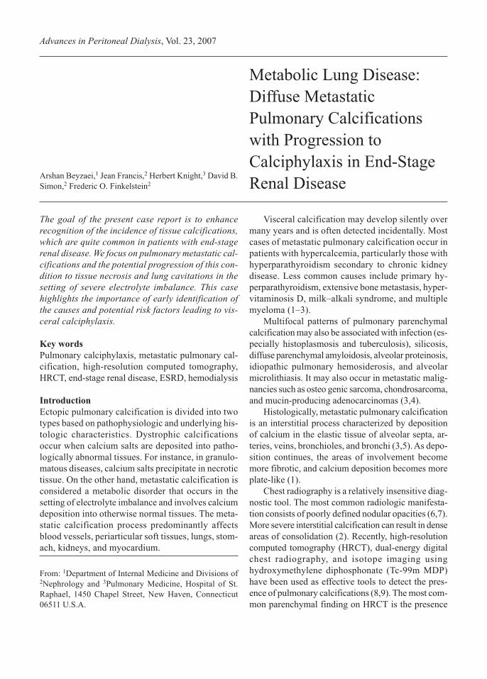
FIGURE 1 Diffuse pulmonary calcifications were initially noted on this high-resolution computed tomography chest scan from April 2005.

Beyzaei et al. 115
He improved clinically and was discharged in stable
condition.
In March 2006, a repeat chest CT scan showed a
septated cavitary right apical lesion and LUL struc-
tural distortion relatively unchanged as compared with
the chest CT scan from January 2006. Subsequently,
the patient was started on 4 weeks of intravenous
levofloxacin for a presumed RUL infectious process.
FIGURE 2 A complex septated thick-walled cavitary process was seen in the area of the right upper lobe, together with less severe
destruction of lung parenchyma in the left upper lobe, in a chest computed tomography scan from January 2006.

116 Pulmonary Calciphylaxis in ESRD
A follow-up chest CT scan in May 2006 showed a
large RUL cavitary lesion similar in appearance to
that uncovered in previous studies. In addition, he had
progressive structural distortion and fibrotic changes
of the LUL. Bronchoscopy with bronchoalveolar lav-
age was performed in July 2006, and cultures of the
washing fluid for bacteria, mycobacteria, and fungi
were negative. Staining for Pneumocystis carinii pneu-
monia was also negative. Bronchoscopic examination
showed a diffuse pattern of shiny, whitish, linear im-
ages in the bronchial tree of both lungs, suggestive of
submucosal pulmonary calcification.
Discussion
Metastatic pulmonary calcification is a relatively com-
mon complication in patients with ESRD. It mani-
fests as tissue calcification without ischemic and
necrotic changes. Calciphylaxis, on the other hand, is
associated with medial and intimal calcification of
small- and medium-sized arteries, with ischemic ne-
crosis of involved tissue (8,15).
Pulmonary calciphylaxis is a rare disorder that has
been associated with lung necrosis. The diagnosis of
pulmonary calciphylaxis requires high suspicion, and
a definite diagnosis requires pathology examination
of tissues. Although no tissue biopsy has been done
in the present case, the setting of electrolyte imbal-
ance, negative culture results, and the progressive
nature of the lung lesions on serial imaging studies
are highly suggestive of lung tissue necrosis second-
ary to pulmonary calciphylaxis.
We speculate that high calcium and phosphate lev-
els and an alkaline environment exacerbated by
chronic vomiting are the main culprits in accelerating
the development of diffuse metastatic calcifications
and ultimately fulminant pulmonary calciphylaxis.
Predilection for calcification in the lung, especially
the upper lobes, and ultimate necrosis in those areas
is consistent with the relative alkaline environment in
those areas as a result of higher ventilation-to-perfu-
sion ratios and hence lower partial pressures of CO2.
Conclusions
Although patients with metastatic pulmonary calcifi-
cations are usually asymptomatic, calcium, phosphate,
and parathyroid hormone levels should be monitored,
and abnormalities should be treated to avoid progres-
sion to calciphylaxis. Few options exist for treating
calciphylaxis, and the outcome is generally poor.
Several causes have been proposed to explain the
development of calciphylaxis at the molecular level.
Uremia is a proinflammatory state in which levels of
interleukin-6 and tumor necrosis factor α are reported
to be elevated in most patients with ESRD. These
proinflammatory cytokines are known to contribute
to endothelial dysfunction and vascular calcification,
which may lead to the development of calciphylaxis
(8,16). In addition, a decrease in calcification inhibi-
tors such as osteoprotegerin has also been reported in
patients on hemodialysis, possibly contributing fur-
ther to development of calciphylaxis (8,17).
References
1 Kobayashi T, Satoh K, Nakano S, Toyama Y,
Ohakawa M. A case of metastatic pulmonary calcifi-
cation after transient acute renal failure. Radiat Med
2005;23:435–8.
2 Marchiori E, Muller NL, Souza AS Jr, Escuissato DL,
Gasparetto EL, de Cerqueira EM. Unusual manifesta-
tions of metastatic pulmonary calcification. J Thorac
Imaging 2005;20:66–70.
3 Lingam RK, Teh J, Sharma A, Friedman E. Meta-
static pulmonary calcification in renal failure: a new
HRCT pattern. Br J Radiol 2002;75:74–7.
4 Brown K, Mund DF, Aberle DR, Batra P, Young DA. In-
trathoracic calcifications: radiographic features and dif-
ferential diagnosis. Radiographics 1994;14:1247–61.
5 Greenberg S, Suster B. Metastatic pulmonary calcifi-
cation: appearance on high-resolution CT. J Comput
Assist Tomogr 1994;18:497–9.
6 Ullmer E, Borer H, Sandoz P, Mayr M, Dalquen P,
Soler M. Diffuse pulmonary nodular infiltrates in a
renal transplant recipient. Chest 2001;120:1394–8.
7 Hartman TE, Muller NL, Primack SL, et al. Meta-
static pulmonary calcification in patients with hyper-
calcemia: findings on chest radiographs and CT
scans. AJR Am J Roentgenol 1994;162:799–802.
8 Li YJ, Tian YC, Chen YC, et al. Fulminant pulmo-
nary calciphylaxis and metastatic calcification caus-
ing acute respiratory failure in a uremic patient. Am J
Kidney Dis 2006;47:e47–53.
9 Nizami MA, Gerntholtz T, Swanepoel CR. The role
of bone scanning in the detection of metastatic cal-
cification: a case report. Clin Nucl Med
2000;25:407–9.
10 Conger JD, Hammond WS, Alfrey AC, Contiguglia
SR, Stanford RE, Huffer WE. Pulmonary calcifica-
tion in chronic dialysis patients: clinical and patho-
logical studies. Ann Intern Med 1975;83:330–6.
11 Matsuo T, Tsukamoto Y, Tamura M, et al. Acute res-
piratory failure due to “pulmonary calciphylaxis” in a

Beyzaei et al. 117
maintenance haemodialysis patient. Nephron
2001;87:75–9.
12 Strumia R, Lombardi AR, Bedani PL, Perini L. Be-
nign nodular calcification and calciphylaxis in a
haemodialysed patient. J Eur Acad Dermatol Venereol
1998;11:69–71.
13 Mazhar AR, Johnson RJ, Gillen D, et al. Risk factors
and mortality associated with calciphylaxis in end-
stage renal disease. Kidney Int 2001;60:324–32.
14 Dear J, Brookes J, Mansell M, Laing C. Calciphy-
laxis. Lancet 2003;362:1707.
15 Wilmer WA, Magro CM. Calciphylaxis: emerging
concepts in prevention, diagnosis, and treatment.
Semin Dial 2002;15:172–86.
16 Kato A, Odamaki M, Takita T, Maruyama Y,
Kumagai H, Hishida A. Association between
interleukin-6 and carotid atherosclerosis in hemodi-
alysis patients. Kidney Int 2002;61:1143–52.
17 Nitta K, Akiba T, Uchida K, et al. Serum
osteoprotegerin levels and the extent of vascular cal-
cification in haemodialysis patients. Nephrol Dial
Transplant 2004;19:1886–9.
Corresponding author:Fredric Finkelstein, MD, 136 Sherman Avenue, NewHaven, Connecticut 06511 U.S.A.E-mail:[email protected]


![REPID Program Program Newsletter 2013.pdf · related research careers focused on cardiovascular, pulmonary and hema-tologic disciplines. ... Department of Physiology. ... [in 2012]](https://img.pdfslide.us/doc/110x75/5fca64e7ebe92600841e5800/repid-program-newsletter-2013pdf-related-research-careers-focused-on-cardiovascular.jpg)