Embed Size (px)
Citation preview

1
Mesa CollegeBio 230 Human
AnatomyFall 2010
Tim Plagge,Instructor
Course Objectives
Upon successful completion of this course you should:
Know and be able to identify relevant tissues and microscopic structures of the human bodyKnow and be able to identify the relevant gross anatomical structures of the human bodyUnderstand the inter-relationships between the different systems of the body & how the structures of the systems relate to the functioning of the systems.
What I expect from you:
1. to be ready for class at the scheduled start time,2. read the assigned material prior to class, this allows
for more discussion & less blah, blah, blah….,3. to be prepared for quizzes,4. to fully utilize the lab time we have as well as open
labs… if you are planning on attending open labs, please consider being an open lab volunteer,
5. to handle and treat the lab materials care, the models are very expensive to replace.

2
Examinations & QuizzesThere will be 5 lecture exams and 6 lab practical exams.Each lecture exam will be 50 points.Each lab exam will be 50 points.I will hand out lab guides and exercises. They are designed to help you in lab as well as lecture.Practical exams will be on models, as well as preserved specimens & cadavers (if available).Quizzes will be unannounced and will be worth 5 points each. They will start at 6:00 promptly, and there are no make-ups for quizzes.
Lab protocol
You should not:Wear open toed shoes in labHave long hair that is not pulled back (so it doesn’t hang into specimens you may be working with)Eat or drink in labDissect the cadavers when they are out… that is another classes job.
You should:Bring your book and any hand outs that were given or emailed to youBe prepared to use the entire lab timeBring gloves, or better yet, keep a few pair in your book bag.
Some FAQ’s

3
1. What are your tests like?
Sample Lecture Exam QuestionLevel One (knowledge) Question
1. Intercalated discs are found in what tissue?a. intervertebral cartilageb. cardiac musclec. duodeno-jujenum junctiond. osseous tissue
Exams may consist of multiple choice, matching, true and false and short answer questions.
Sample Lecture Exam QuestionLevel 6 Question (evaluation)
1. The best tissue for increasing the stability of a diarthritic joint would be?
a. osseous tissueb. dense irregular tissuec. dense irregular tissued. hyaline cartilage
Exams may consist of multiple choice, matching, true and false and short answer questions.

4
Some Sample Lab Exam Items
1. This tissue would be best identified as _____.
2. Does spelling count?
3. Why?

5
4. I’m done… what should I do?
The Human Body:An Orientation
Today’s Topics. . . Overview of AnatomyStructural OrganizationSystem OverviewMicroscopic and Anatomical Study TechniquesGross AnatomyTerminologyPlanes & Sections, Regions & QuadrantsThe Body Plan & Body Cavities

6
How The Body is Studied . . .
Anatomical Studythe examination of the structures from
Microscopic to gross anatomical structuresUsing different “tools” such as:
Microscopy (light & electron), CT scans, MRI, X-rays, dissection . . . more later
Physiological Studythe study of how the body functions
Also uses “tools” such as:PET scans, ECG, sphygmomanometer . . .
An Overview of Anatomy
Divisions of anatomyDevelopmental anatomyEmbryology Pathological anatomy (pathology)Radiographic anatomyFunctional morphology
Menu
An Overview of Anatomy
Anatomical terminology – based on ancient Greek or Latin
Provides standard nomenclature worldwide

7
Microscopic Anatomy
Preparing human tissue for microscopySpecimen is fixed (preserved) and sectionedSpecimen is stained to distinguish anatomical structures
Acidic stain – negatively charged dye moleculesBasic stain – positively charged dye molecules
Specimen is then imaged
Microscopic Anatomy
Microscopy – examining small structures through a microscope
Light microscopy – illuminates tissue with a beam of light (lower magnification)Electron microscopy – uses beams of electrons (higher magnification)
May be SEM or TEMSEM (scanning electron microscopy)TEM (transmission electron microscopy)
Clinical Anatomy – An Introduction to Medical Imaging Techniques
X ray –electromagnetic waves of very short length
Best for visualizing bones and abnormal dense structures

8
Clinical Anatomy – An Introduction to Medical Imaging
TechniquesVariations of X ray
Fluoroscope – x rays emitted through the specimen and images are viewed on a fluorescent screen
Cineradiography – uses X-ray cinema film to record organ movements
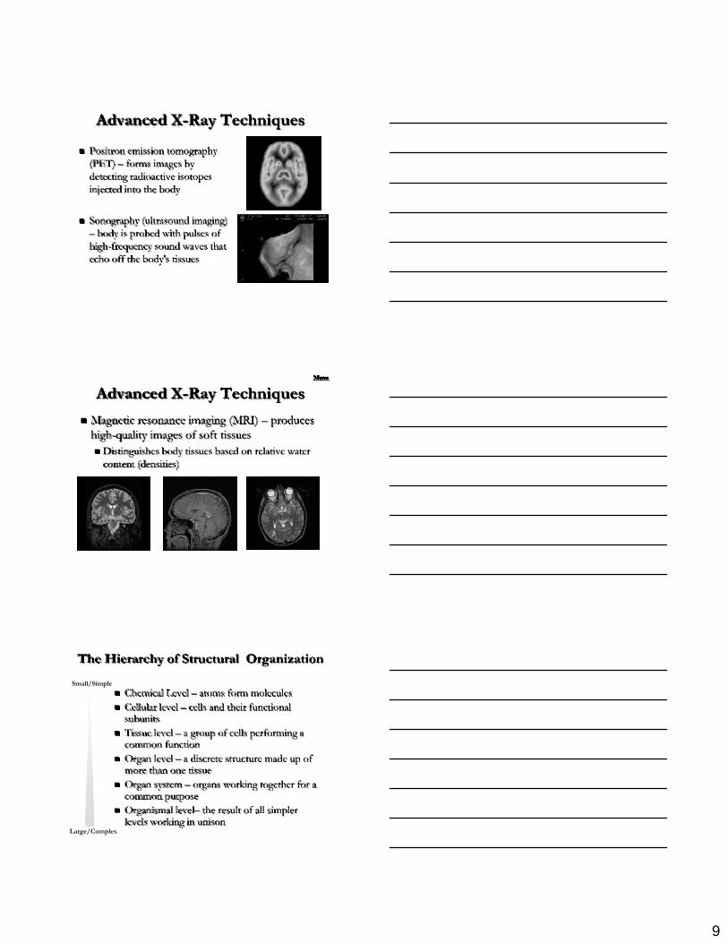
Advanced X-Ray Techniques
Computed (axial) tomography (CT or CAT) –takes successive X rays around a person's full circumference
Translates recorded information into a detailedpicture of the body section
Advanced X-Ray Techniques
Digital subtraction angiography imaging (DSA) – provides an unobstructed view of small arteries, used to find blockages, aneurisms . . .
9
Advanced X-Ray Techniques
Positron emission tomography (PET) – forms images by detecting radioactive isotopes injected into the body
Sonography (ultrasound imaging) – body is probed with pulses of high-frequency sound waves that echo off the body's tissues
Advanced X-Ray Techniques
Magnetic resonance imaging (MRI) – produces high-quality images of soft tissues
Distinguishes body tissues based on relative water content (densities)
Menu
The Hierarchy of Structural Organization
Chemical Level – atoms form moleculesCellular level – cells and their functional subunitsTissue level – a group of cells performing a common functionOrgan level – a discrete structure made up of more than one tissueOrgan system – organs working together for a common purposeOrganismal level– the result of all simpler levels working in unison
Small/Simple
Large/Complex

10
Levels of Structural Organization
Figure 1.1
Menu
Overview of Systems & General Functions
IntegumentarySkeletalMuscularNervousEndocrineCardiovascularLymphatic/ImmuneRespiratoryDigestiveUrinaryReproductive
Integumentary System
Forms external body coveringProtects deeper tissues from injurySynthesizes vitamin DSite of cutaneous receptors (pain, pressure, etc.) and sweat and oil glands

11
Skeletal System
Protects and supports body organsProvides a framework for musclesBlood cells formed within bonesStores fat (energy) & minerals
Muscular System
Allows for movementinternal movmentmovement of body
SupportFacial expressionMaintains postureThermogenesis
Nervous System
Fast-acting control systemIntegrates all sensory informationResponds to internal and external changes Developing neuron
12
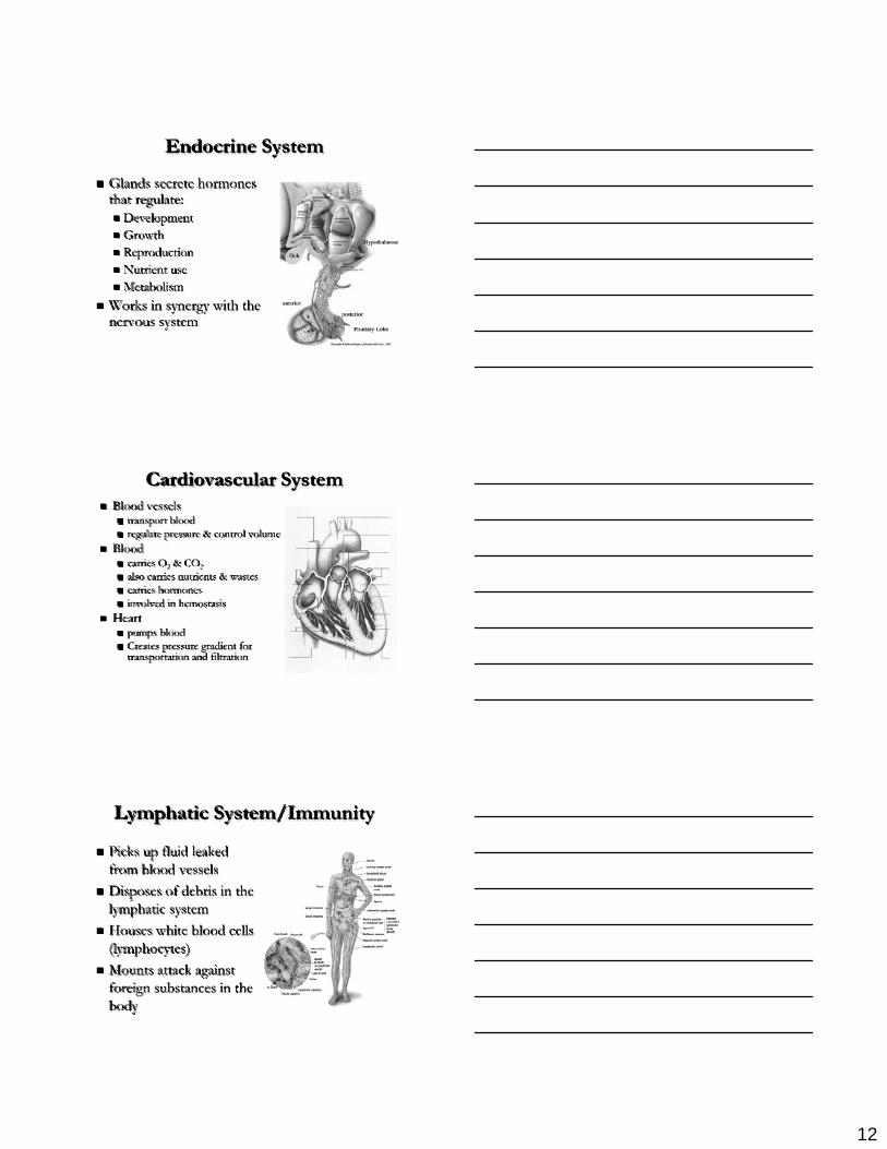
Endocrine System
Glands secrete hormones that regulate:
DevelopmentGrowthReproductionNutrient useMetabolism
Works in synergy with the nervous system
Cardiovascular SystemBlood vessels
transport bloodregulate pressure & control volume
Bloodcarries O2 & CO2also carries nutrients & wastescarries hormonesinvolved in hemostasis
Heart pumps bloodCreates pressure gradient for transportation and filtration
Lymphatic System/Immunity
Picks up fluid leaked from blood vesselsDisposes of debris in the lymphatic systemHouses white blood cells (lymphocytes)Mounts attack against foreign substances in the body

13
Respiratory System
Keeps blood supplied with oxygenRemoves carbon dioxideAir exchange & gas exchange occurs through walls of air sacs in the lungsProtectionHormone production
Digestive System
IngestionDigestion: breaks down food into absorbable unitsAbsorptionMotilityIndigestible foodstuffs eliminated as feces
Urinary System
Eliminates nitrogenous wastesRegulates water, electrolyte, and acid-base balance

14
Male & Female Reproductive Systems
Overall function is to produce offspringTestes produce sperm and male sex hormonesOvaries produce eggs and female sex hormonesMammary glands produce milk
Menu
Gross Anatomy – An Introduction
Anatomical position – a common visual reference point
Person stands erect with feet together and eyes forwardPalms face anteriorly with the thumbs pointed away from the bodyDirectional terminology always refers to the body in anatomical position
Gross Anatomy – An Introduction
Menu

15
Gross Anatomy – Terminology
Directional termsWill usually be relational (i.e. the eyes are medial to the nose . . . Or the nose is intermediate to the eyes).
Regional terms – names of specific body areasAxial region – the main axis of the bodyAppendicular region – the limbs
Orientation and Directional TermsSuperior
toward the upper part of a structure (or body), above
Inferior
toward the lower part of a structure (or body), below
Anterior
Toward (or at) the front of the body (or structure)
Posterior
Toward (or at) the back of the body (or structure)
The heart is superior to the diaphragm
The mouth is inferior to the nose
The trachea is anterior to the esophagus
The heart is posterior to the sternum
Orientation and Directional Termscontinued
MedialToward the midline of the body or structure
IntermediateBetween a more medial and a more lateral structure
LateralAway from the midline of the body or structure
ProximalCloser to the origin of the body part or the point of attachment of a limb to the body trunk
DistalFurther from the origin of the body part or the point of attachment of a limb to the body trunk
The sternum is medial to the scapula
The nose is intermediate to, or between, the eyes
The scapula are lateral to the vertebral column
The shoulder is proximal to the elbow
The ankle is distal to the knee

16
Orientation and Directional Terms continued
SuperficialNearer or closer to the surface of the body, external
DeepAway or further from the body surface, internal
The epidermis is superficial to the dermis
The muscles are deep to the skin
Regional TermsMenu
Body Planes and SectionsCoronal (frontal) plane
Lies vertically and divides body into anterior and posterior parts
Transverse planeRuns horizontally – divides body into superior and inferior parts
Median (midsagittal) planeSpecific sagittal plane that lies vertically in the midline

17
Abdominal Regions and Quadrants
Abdominal regionsdivides abdomen into nine regions
Abdominal quadrantsdivides abdomen into four quadrants
Abdominal Regions
Figure 1.11a, b
These regions are formed by two vertical planes and two horizontal planes. The two vertical planes are the lateral lines LLL and RLL. These lines are dropped from a point half way between the jugular notch and the acromion process. The two horizontal planes are the transpyloric plane TPP and the transtubercular plane TTP. The tubercles are the tubercles of the iliac crests.
The Human Body PlanThese are characteristics shared with all Vertebrates!
Tube-within-a-tubeBilateral symmetryDorsal hollow nerve cordNotochord and vertebraeSegmentationPharyngeal pouchesBody Cavities

18
Basic Human Body Plan and StructuresShared with all Vertebrates
Body Cavities and Membranes
Dorsal body cavityCranial cavityVertebral cavity
Ventral body cavityThoracic cavity – divided into three parts
Two lateral parts each containing a lung surrounded by a pleural cavity Mediastinum – contains the heart surrounded by the pericardial sac
Body Cavities and Membranes
Ventral cavity (continued)Abdominopelvic cavity – divided into two parts
Abdominal cavity – contains the liver, stomach, kidneys, and other organsPelvic cavity – contains the bladder, some reproductive organs, and rectum

19
Body Cavities
Body Cavities and Membranes
Body Cavities and Membranes
Serous cavities – a slit-like space lined by a serous membrane and include:
Pleura, pericardium, and peritoneum
The membranes lining the cavities are named:Parietal serosa – outer wall of the cavityVisceral serosa – covers visceral organs

20
Body Cavities and Membranes
outer layer = parietal serosa (membrane)
space in between = body cavity
inner layer = visceral serosa (membrane)
Body Cavities and Membranes
Pericardial Cavity
Pleural Cavity
Body Cavities and Membranes
Abdominal Cavity

21
Body Cavities and Membranes
Other cavitiesOral cavityNasal cavityOrbital cavitiesMiddle ear cavitiesSynovial cavities


![Furniture - Muebles - Mesa de Centro / Mesa baja [ Basics ]](https://img.pdfslide.us/doc/110x75/58a500a01a28abce778b6231/furniture-muebles-mesa-de-centro-mesa-baja-basics-.jpg)