Embed Size (px)
Citation preview
1
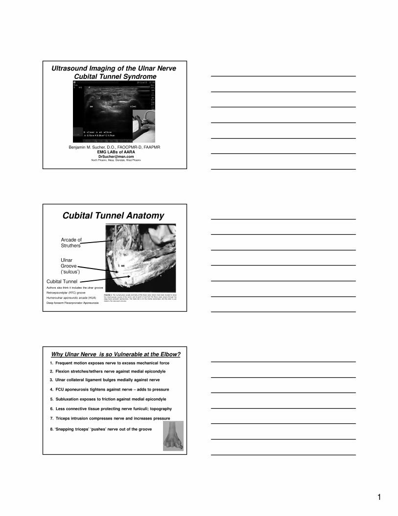
Ultrasound Imaging of the Ulnar Nerve
Cubital Tunnel Syndrome
Benjamin M. Sucher, D.O., FAOCPMR-D, FAAPMREMG LABs of [email protected]
North Phoenix, Mesa, Glendale, West Phoenix
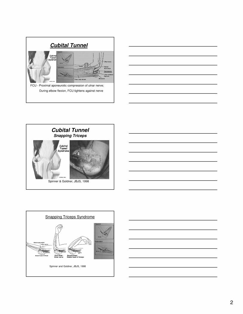
Cubital Tunnel Anatomy
Arcade of Struthers
Ulnar Groove (‘sulcus’)
Cubital TunnelAuthors also think it includes the ulnar groove
Retroepicondylar (RTC) groove
Humeroulnar aponeurotic arcade (HUA)
Deep forearm Flexorpronator Aponeurosis
ME
O
Why Ulnar Nerve is so Vulnerable at the Elbow?
1. Frequent motion exposes nerve to excess mechanical force
2. Flexion stretches/tethers nerve against medial epicondyle
3. Ulnar collateral ligament bulges medially against nerve
4. FCU aponeurosis tightens against nerve – adds to pressure
7. Triceps intrusion compresses nerve and increases pressure
5. Subluxation exposes to friction against medial epicondyle
8. ‘Snapping triceps’ ‘pushes’ nerve out of the groove
6. Less connective tissue protecting nerve funiculi; topography
2
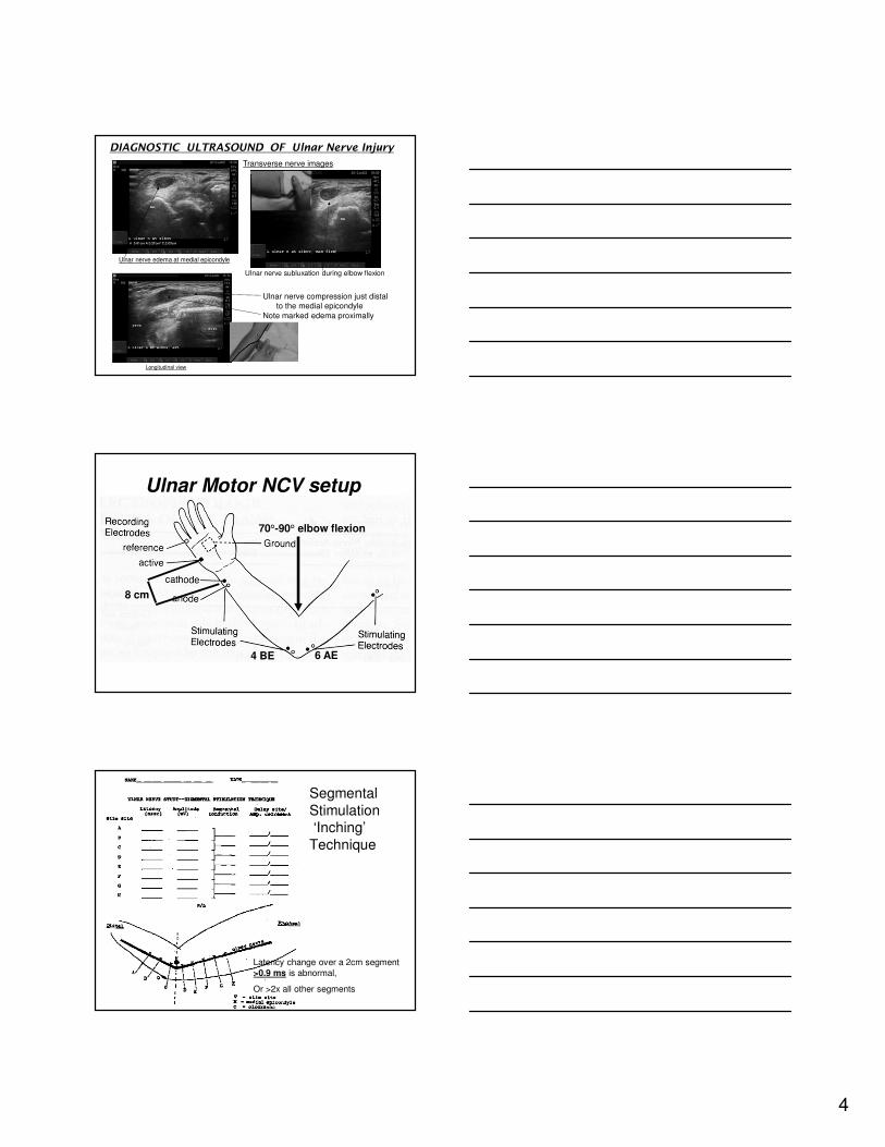
Cubital Tunnel
FCU - Proximal aponeurotic compression of ulnar nerve;
During elbow flexion, FCU tightens against nerve
Cubital TunnelSnapping Triceps
Spinner & Goldner, JBJS, 1998
Snapping Triceps Syndrome
Spinner and Goldner, JBJS, 1998
3
Triceps Intrusion Into the Ulnar Sulcus
and Ulnar Nerve Subluxation
Miller and Reinus, AJR, 2010
Extension
Flexion
Ulnar nerve
Ulnar nerve(subluxed)
DIAGNOSTIC ULTRASOUND of NORMAL Ulnar Nerves
Normal CSA:8-10mm2 maximum upper limit
[Mild = 10-14; Mod = 15-19; Severe >20]Axonal loss = larger nerve sizeBayrak, et al: M&N; 2010Beekman, et al: M&N, 2011Omejec and Podnar: M&N; 2015 (8-11mm2)
Normal CSA:
<7mm2 definitely normal in Females<8mm2 definitely normal in MalesPeer and Bodner, 2008Strakowski, 2014Normal CSA:
8-9mm2 maximum upper limit
[9 = males; 8 = females]Cartwright, et al: Arch Phys Med Rehabil; 2007
DIAGNOSTIC ULTRASOUND OF Ulnar Nerve Injury
NCS:
Ulnar motor amp = 1mV (median = 10mV)Forearm NCV = 49m/s
Complete CB at elbow (No response prox stim AE)Ulnar sensory response unobtainable (D5 and DUC)
Patient H&P:55 y/o male complains of pain, numbness and weakness in the hand for 4 months.
Exam revealed intrinsic atrophy and weakness, decreased sensation in the medial hand and positive Tinel at the cubital tunnel
Needle EMG:
2+Fibs FDI and FCUNeurogenic MUPs
4
DIAGNOSTIC ULTRASOUND OF Ulnar Nerve Injury
Longitudinal view
Ulnar nerve compression just distalto the medial epicondyle
Note marked edema proximally
Ulnar nerve edema at medial epicondyle
Transverse nerve images
Ulnar nerve subluxation during elbow flexion
Ulnar Motor NCV setup
70°-90° elbow flexion
8 cm
6 AE4 BE
Segmental
Stimulation ‘Inching’
Technique
Latency change over a 2cm segment
>0.9 ms is abnormal,
Or >2x all other segments
5
DIAGNOSTIC ULTRASOUND OF Ulnar Nerve Injury
NCS:
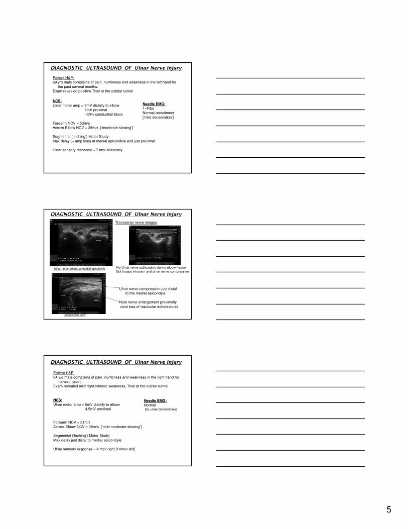
Ulnar motor amp = 9mV distally to elbow6mV proximal
~30% conduction block
Forearm NCV = 53m/sAcross Elbow NCV = 35m/s [‘moderate slowing’]
Segmental (‘Inching’) Motor Study:
Max delay (+ amp loss) at medial epicondyle and just proximal
Ulnar sensory response = 7 mcv bilaterally
Patient H&P:69 y/o male complains of pain, numbness and weakness in the left hand for
the past several months. Exam revealed positive Tinel at the cubital tunnel
Needle EMG:
1+FibsNormal recruitment
[‘mild denervation’]
DIAGNOSTIC ULTRASOUND OF Ulnar Nerve Injury
Longitudinal view
Ulnar nerve compression just distalto the medial epicondyle
Note nerve enlargement proximally
(and loss of fascicular echotexture)
Ulnar nerve edema at medial epicondyle
Transverse nerve images
No Ulnar nerve subluxation during elbow flexionBut triceps intrusion and ulnar nerve compression
DIAGNOSTIC ULTRASOUND OF Ulnar Nerve Injury
NCS:
Ulnar motor amp = 5mV distally to elbow4.5mV proximal
Forearm NCV = 51m/sAcross Elbow NCV = 38m/s [‘mild-moderate slowing’]
Segmental (‘Inching’) Motor Study:
Max delay just distal to medial epicondyle
Ulnar sensory response = 4 mcv right [14mcv left]
Patient H&P:84 y/o male complains of pain, numbness and weakness in the right hand for
several years. Exam revealed mild right intrinsic weakness, Tinel at the cubital tunnel
Needle EMG:
Normal[no ulnar denervation]
6
DIAGNOSTIC ULTRASOUND OF Ulnar Nerve Injury
Longitudinal view
Ulnar nerve compression just distal
to the medial epicondyle
Note marked edema proximally
Ulnar nerve edema at medial epicondyle
Transverse nerve images
No Ulnar nerve subluxation during elbow flexionBut triceps intrusion (no ulnar nerve compression)
DIAGNOSTIC ULTRASOUND OF Ulnar Nerve Injury
NCS:
Ulnar motor amp = 13mV distally to elbow12.9mV proximal
Forearm NCV = 62m/sAcross Elbow NCV = 55m/s [‘very mild, relative slowing’]
Segmental (‘Inching’) Motor Study:
Max delay at medial epicondyle (1.2ms; all other segments .3ms)No amplitude loss
Ulnar sensory response = 11 mcv left [13mcv right]
Patient H&P:64 y/o female complains of pain and numbness in the left hand for the past
several months. Exam revealed decreased sensation in the left medial hand, and a positive
Tinel test at the cubital tunnel
Needle EMG:
Normal[no ulnar denervation]
DIAGNOSTIC ULTRASOUND OF Ulnar Nerve Injury
Longitudinal view
Ulnar nerve edema medial epicondyleNote enlargement proximally
Ulnar nerve edema at medial epicondyle
Transverse nerve images
No Ulnar nerve subluxationduring elbow flexion, but triceps
intrusion (ulnar nerve compression)
Partial elbow flexion
7
DIAGNOSTIC ULTRASOUND OF Ulnar Nerve Injury
NCS:
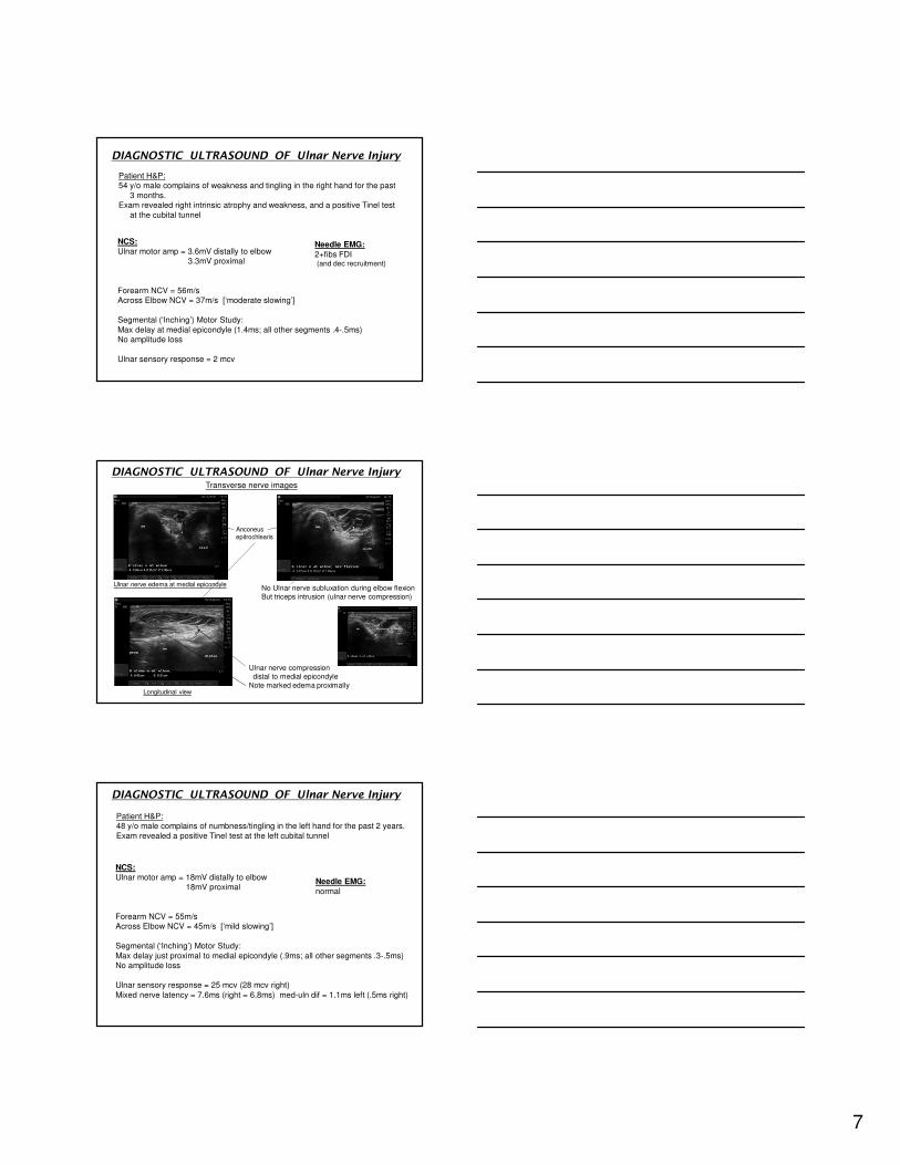
Ulnar motor amp = 3.6mV distally to elbow3.3mV proximal
Forearm NCV = 56m/sAcross Elbow NCV = 37m/s [‘moderate slowing’]
Segmental (‘Inching’) Motor Study:
Max delay at medial epicondyle (1.4ms; all other segments .4-.5ms)No amplitude loss
Ulnar sensory response = 2 mcv
Patient H&P:54 y/o male complains of weakness and tingling in the right hand for the past
3 months. Exam revealed right intrinsic atrophy and weakness, and a positive Tinel test
at the cubital tunnel
Needle EMG:
2+fibs FDI(and dec recruitment)
DIAGNOSTIC ULTRASOUND OF Ulnar Nerve Injury
Longitudinal view
Ulnar nerve compression distal to medial epicondyle
Note marked edema proximally
Ulnar nerve edema at medial epicondyle
Transverse nerve images
No Ulnar nerve subluxation during elbow flexionBut triceps intrusion (ulnar nerve compression)
Anconeusepitrochlearis
DIAGNOSTIC ULTRASOUND OF Ulnar Nerve Injury
NCS:
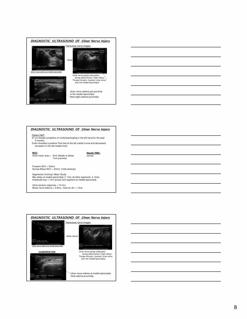
Ulnar motor amp = 18mV distally to elbow18mV proximal
Forearm NCV = 55m/sAcross Elbow NCV = 45m/s [‘mild slowing’]
Segmental (‘Inching’) Motor Study:
Max delay just proximal to medial epicondyle (.9ms; all other segments .3-.5ms)No amplitude loss
Ulnar sensory response = 25 mcv (28 mcv right)
Mixed nerve latency = 7.6ms (right = 6.8ms) med-uln dif = 1.1ms left (.5ms right)
Patient H&P:48 y/o male complains of numbness/tingling in the left hand for the past 2 years.
Exam revealed a positive Tinel test at the left cubital tunnel
Needle EMG:
normal
8
DIAGNOSTIC ULTRASOUND OF Ulnar Nerve Injury
Longitudinal view
Ulnar nerve edema just proximalto the medial epicondyle
Note slight edema proximally
Ulnar nerve edema at medial epicondyle
Transverse nerve images
Ulnar nerve partial subluxation during elbow flexion (‘high-riding’)
Triceps intrusion (‘pushes’ ulnar nerve over the medial epicondyle)
Ulnar nerve
DIAGNOSTIC ULTRASOUND OF Ulnar Nerve Injury
NCS:
Ulnar motor amp = 9mV distally to elbow7mV proximal
Forearm NCV = 53m/sAcross Elbow NCV = 43m/s [‘mild slowing’]
Segmental (‘Inching’) Motor Study:
Max delay at medial epicondyle (1.1ms; all other segments .3-.5ms)Amplitude loss 1.1mV across 2cm segment at medial epicondyle
Ulnar sensory response = 10 mcv
Mixed nerve latency = 5.8ms; med-uln dif = 1.3ms
Patient H&P:67 y/o female complains of numbness/tingling in the left hand for the past
2 months. Exam revealed a positive Tinel test at the left cubital tunnel and decreased
sensation in the left medial hand
Needle EMG:
normal
DIAGNOSTIC ULTRASOUND OF Ulnar Nerve Injury
Ulnar nerve edema at medial epicondyleNote edema proximally
Ulnar nerve edema at medial epicondyle
Transverse nerve images
Ulnar nerve partial subluxation during elbow flexion (‘high-riding’)
Triceps intrusion (‘pushes’ ulnar nerve over the medial epicondyle)
Ulnar nerve
Longitudinal view
9
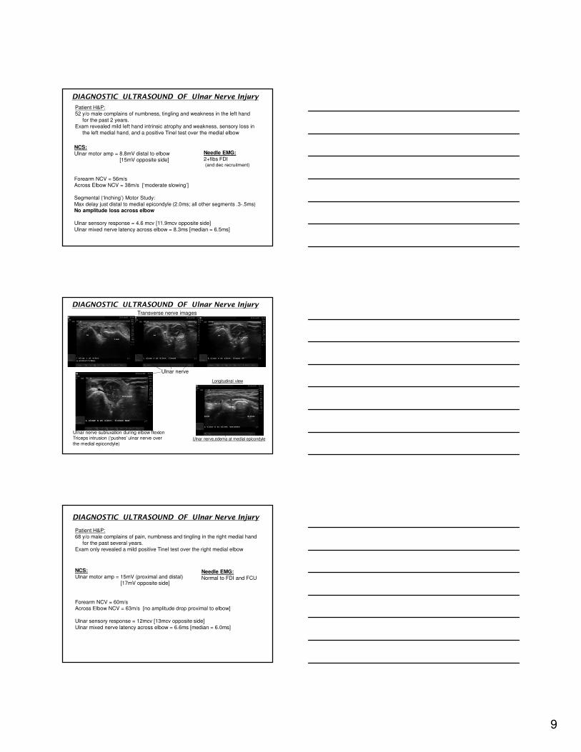
DIAGNOSTIC ULTRASOUND OF Ulnar Nerve Injury
NCS:
Ulnar motor amp = 8.8mV distal to elbow[15mV opposite side]
Forearm NCV = 56m/sAcross Elbow NCV = 38m/s [‘moderate slowing’]
Segmental (‘Inching’) Motor Study:
Max delay just distal to medial epicondyle (2.0ms; all other segments .3-.5ms)No amplitude loss across elbow
Ulnar sensory response = 4.6 mcv [11.9mcv opposite side]
Ulnar mixed nerve latency across elbow = 8.3ms [median = 6.5ms]
Patient H&P:52 y/o male complains of numbness, tingling and weakness in the left hand
for the past 2 years. Exam revealed mild left hand intrinsic atrophy and weakness, sensory loss in
the left medial hand, and a positive Tinel test over the medial elbow
Needle EMG:
2+fibs FDI(and dec recruitment)
DIAGNOSTIC ULTRASOUND OF Ulnar Nerve Injury
Longitudinal view
Ulnar nerve,edema at medial epicondyle
Transverse nerve images
Ulnar nerve subluxation during elbow flexion Triceps intrusion (‘pushes’ ulnar nerve over
the medial epicondyle)
Ulnar nerve
compression
DIAGNOSTIC ULTRASOUND OF Ulnar Nerve Injury
NCS:
Ulnar motor amp = 15mV (proximal and distal)[17mV opposite side]
Forearm NCV = 60m/sAcross Elbow NCV = 63m/s [no amplitude drop proximal to elbow]
Ulnar sensory response = 12mcv [13mcv opposite side]
Ulnar mixed nerve latency across elbow = 6.6ms [median = 6.0ms]
Patient H&P:68 y/o male complains of pain, numbness and tingling in the right medial hand
for the past several years. Exam only revealed a mild positive Tinel test over the right medial elbow
Needle EMG:
Normal to FDI and FCU
10
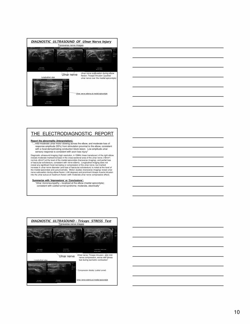
DIAGNOSTIC ULTRASOUND OF Ulnar Nerve Injury
Longitudinal view
Ulnar nerve edema at medial epicondyle
Transverse nerve images
Ulnar nerve subluxation during elbow flexion; Triceps intrusion (‘pushes’
ulnar nerve over the medial epicondyle)
Ulnar nerve
THE ELECTRODIAGNOSTIC REPORT
Report the abnormality (Interpretation):“…mild-moderate ulnar motor slowing across the elbow, and moderate loss of
response amplitude (55%) from stimulation proximal to the elbow, consistent with a focal demyelinating conduction block lesion. Low amplitude ulnar
sensory response is consistent with axon loss injury”
Summarize with ‘Impressions’ or ‘Conclusions’:“Ulnar mononeuropathy – localized at the elbow (medial epicondyle);
consistent with cubital tunnel syndrome; moderate, electrically”
Diagnostic ultrasound imaging (high resolution, 4-15MHz linear transducer) of the right elbow reveals moderate-marked increase in the cross-sectional area of the ulnar nerve (18mm2;
normal <9mm2) at the level of the medial epicondyle (transverse imaging), and partial loss of fascicular echotexture, consistent with nerve edema. Longitudinal imaging does not reveal any significant focal narrowing or compression of the ulnar nerve, but marked
increase in ulnar nerve diameter (and loss of fascicular echotexture) is noted at the level of the medial epicondyle and just proximally. Motion studies (transverse imaging) reveal ulnarnerve subluxation during elbow flexion (>90 degrees) and prominent triceps muscle intrusion
into the ulnar sulcus at maximum flexion (with moderate ulnar nerve compressive effect).
DIAGNOSTIC ULTRASOUND – Triceps STRESS Test
Longitudinal view
Ulnar nerve edema at medial epicondyle
Transverse nerve images
Ulnar nerve; Triceps intrusion, and mild nerve compression, worse with stress
test during isometric contraction
Ulnar nerve
Compression distally (cubital tunnel)coronoid tubercle
11
DIAGNOSTIC ULTRASOUND OF Ulnar Nerve Injury
NCS:
Ulnar motor amp = 6.3mV (4.8mV proximal; mild CB)[9.5mV opposite side]
Patient H&P:75 y/o male complains of pain, numbness and tingling in the left medial hand
for the past year. He admits to leaning heavily on his elbow frequently. Exam revealed intrinsic atrophy and weakness, positive Tinel at medial elbow
Needle EMG:
1+ fibs FDI Neurogenic firing
Forearm NCV = 53m/sAcross Elbow NCV = 36m/s [24% amplitude drop proximal to elbow]
Segmental (‘inching’) – max delay just distal to ME at cubital tunnel[1.3ms delay; other segments .3-.7ms]
Ulnar sensory response = 4mcv
Treatment Implications
Rampen, M&N 2011; Spinner, JBJS, 1998
Multi-faceted:
1. Avoid excessive elbow flexion (tape or brace into extension at night)
2. Avoid external pressure to medial elbow (padded sleeve for daytime use)
3. Avoid repetitive flexion-extension activities (‘friction neuritis’)
4. Discontinue triceps strengthening (no ‘bulk-building’)
5. Steroid injection (due to edema), just proximal to medial epicondyle
6. Surgery - decompression/release, transposition, muscle resection, osteotomy?
7. Consider Botox injections??
Ultrasound Imaging of the Ulnar Nerve
At the Wrist
Benjamin M. Sucher, D.O., FAOCPMR-D, FAAPMR
EMG LABs of [email protected]
North Phoenix, Mesa, Glendale, West Phoenix
12
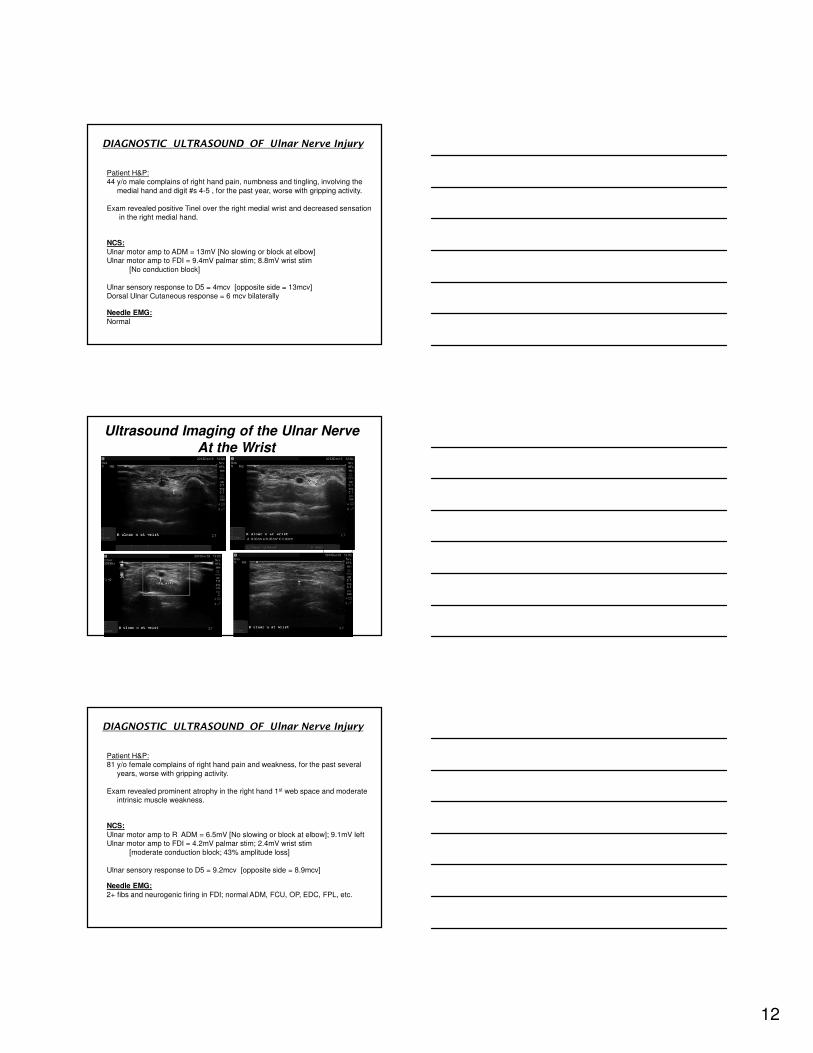
DIAGNOSTIC ULTRASOUND OF Ulnar Nerve Injury
NCS:
Ulnar motor amp to ADM = 13mV [No slowing or block at elbow]Ulnar motor amp to FDI = 9.4mV palmar stim; 8.8mV wrist stim
[No conduction block]
Ulnar sensory response to D5 = 4mcv [opposite side = 13mcv] Dorsal Ulnar Cutaneous response = 6 mcv bilaterally
Patient H&P:44 y/o male complains of right hand pain, numbness and tingling, involving the
medial hand and digit #s 4-5 , for the past year, worse with gripping activity.
Exam revealed positive Tinel over the right medial wrist and decreased sensationin the right medial hand.
Needle EMG:
Normal
Ultrasound Imaging of the Ulnar Nerve
At the Wrist
pisiform
Ulnar artery
Longitudinal View
Ulnar nerve
P
DIAGNOSTIC ULTRASOUND OF Ulnar Nerve Injury
NCS:
Ulnar motor amp to R ADM = 6.5mV [No slowing or block at elbow]; 9.1mV leftUlnar motor amp to FDI = 4.2mV palmar stim; 2.4mV wrist stim
[moderate conduction block; 43% amplitude loss]
Ulnar sensory response to D5 = 9.2mcv [opposite side = 8.9mcv]
Patient H&P:81 y/o female complains of right hand pain and weakness, for the past several
years, worse with gripping activity.
Exam revealed prominent atrophy in the right hand 1st web space and moderateintrinsic muscle weakness.
Needle EMG:
2+ fibs and neurogenic firing in FDI; normal ADM, FCU, OP, EDC, FPL, etc.
13
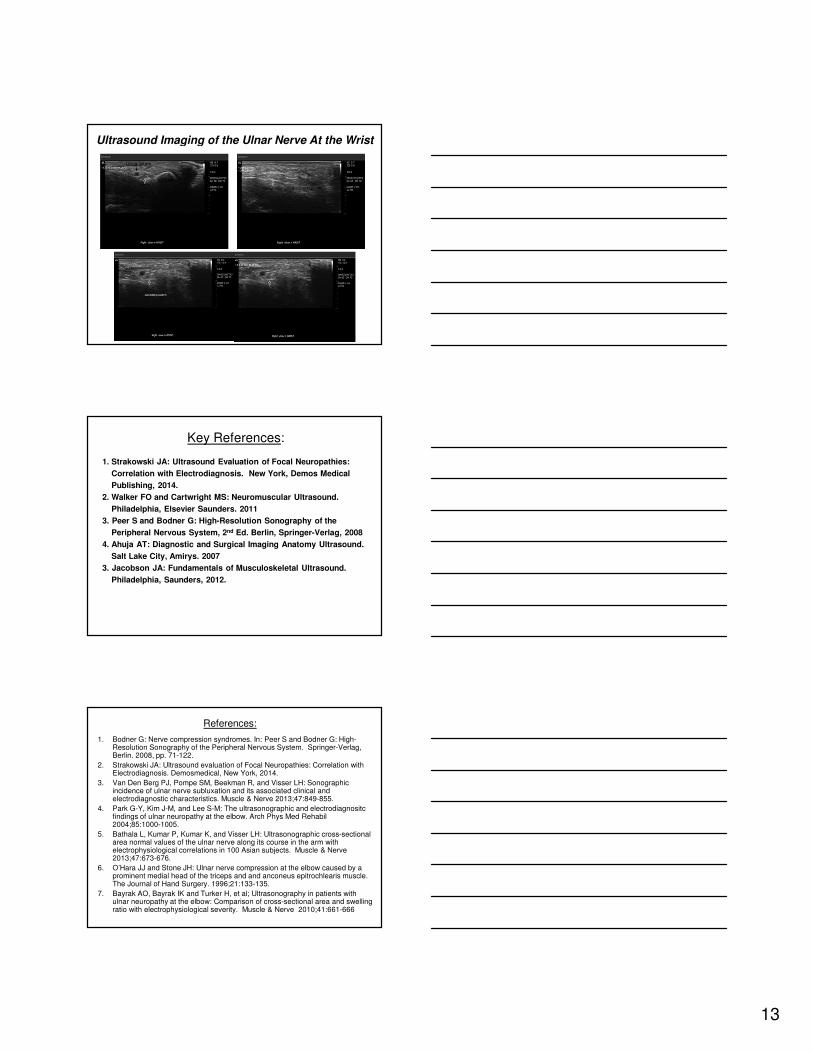
Ultrasound Imaging of the Ulnar Nerve At the Wrist
Ulnar artery
pisiform
Longitudinal View
nerveNerve enlarged distal to pisiform
Key References:
1. Strakowski JA: Ultrasound Evaluation of Focal Neuropathies:
Correlation with Electrodiagnosis. New York, Demos Medical
Publishing, 2014.
2. Walker FO and Cartwright MS: Neuromuscular Ultrasound.
Philadelphia, Elsevier Saunders. 2011
3. Peer S and Bodner G: High-Resolution Sonography of the
Peripheral Nervous System, 2nd Ed. Berlin, Springer-Verlag, 2008
4. Ahuja AT: Diagnostic and Surgical Imaging Anatomy Ultrasound.
Salt Lake City, Amirys. 2007
3. Jacobson JA: Fundamentals of Musculoskeletal Ultrasound.
Philadelphia, Saunders, 2012.
References:
1. Bodner G: Nerve compression syndromes. In: Peer S and Bodner G: High-Resolution Sonography of the Peripheral Nervous System. Springer-Verlag, Berlin. 2008, pp. 71-122.
2. Strakowski JA: Ultrasound evaluation of Focal Neuropathies: Correlation with Electrodiagnosis. Demosmedical, New York, 2014.
3. Van Den Berg PJ, Pompe SM, Beekman R, and Visser LH: Sonographic incidence of ulnar nerve subluxation and its associated clinical and electrodiagnostic characteristics. Muscle & Nerve 2013;47:849-855.
4. Park G-Y, Kim J-M, and Lee S-M: The ultrasonographic and electrodiagnositcfindings of ulnar neuropathy at the elbow. Arch Phys Med Rehabil2004;85:1000-1005.
5. Bathala L, Kumar P, Kumar K, and Visser LH: Ultrasonographic cross-sectional area normal values of the ulnar nerve along its course in the arm with electrophysiological correlations in 100 Asian subjects. Muscle & Nerve 2013;47:673-676.
6. O’Hara JJ and Stone JH: Ulnar nerve compression at the elbow caused by a prominent medial head of the triceps and and anconeus epitrochlearis muscle. The Journal of Hand Surgery. 1996;21:133-135.
7. Bayrak AO, Bayrak IK and Turker H, et al; Ultrasonography in patients with ulnar neuropathy at the elbow: Comparison of cross-sectional area and swelling ratio with electrophysiological severity. Muscle & Nerve 2010;41:661-666
14
References (cont):
8. Griffith JF and Paunipagar BK: Ulnar nerve. In: Ahuja AT, Antonio GE, Griffith JF, et al: Diagnostic and Surgical Imaging Anatomy Ultrasound. Amirsys, Salt
Lake City. 2007, pp.VI 158-167.
9. Cartwright MS: Ultrasound of focal neuropathies. In: Walker FO and Cartwright MS: Neuromuscular Ultrasound. Elsevier Saunders, Philadelphia. 2011, pp.72-
90.
10.Beekman R, Visser LH, and Verhagen WI: Ultrasonography in ulnar neuropathy
at the elbow: A critical review. Muscle & Nerve 2011;43:627-635.
11.Cartwright MS Shin HW, Passmore LV, and Walker FO: Ultrasonographic
findings of the normal ulnar nerve in adults. Arch Phys Med Rehabil. 2007;88:394-396.
12.Yoon JS, Walker FO, and Cartwright MS: Ultrasonographic swelling ratio in the diagnosis of ulnar neuropathy at the elbow. Muscle & Nerve 2008;38:1231-
1235.
13.Shaker Ali, et al: Which motor nerve conduction study is best in ulnar neuropathy
at the elbow? Muscle & Nerve 2004;29: 585-590.
14.Spinner RJ and Goldner RD: Snapping of the medial head of the triceps and recurrent dislocation of the ulnar nerve. JBJS 1998;80-A:239-247.
References (cont):
15. Miller TT and Reinus WR: Nerve entrapment syndromes of the elbow,
forearm, and wrist. Am J Roentgen 2010;195:585-594.
17. Campbell, William et al: Short Segment Incremental Studies in the
evaluation of Ulnar Neuropathy at the Elbow: Muscle and Nerve 1992;
15:1050-1054.
18. Campbell, W et al: Variations in Anatomy of the Ulnar Nerve at the Cubital
Tunnel: Pitfalls in the Diagnosis of the Ulnar Neuropathy at the Elbow. Muscle
& Nerve 1991;14:733-738.
19. Oh SJ: Clinical Electromyography, Nerve Conduction Studies,3rd edition, LWW
2003
20. Debase Zeki et al; New Near-Nerve needle Nerve Conduction Technique:
Differentiating Epicondylar from Cubital Tunnel Ulnar Neuropathy: Muscle &
Nerve 1999;22: 718-723.
21. Rampen AJJ, Wirtz PW, and Tavy DLJ: Ultrasound-guided steroid injection to
treat mild ulnar neuropathy at the elbow. Muscle & Nerve 2011;44:128-130.
22. Omejec G and Podnar S: Normative values for short-segment nerve conduction
studies and ultrasonography of the ulnar nerve at the elbow. Muscle & Nerve
2015;51:370-371.