Embed Size (px)
Citation preview

1521-0111/92/3/285–296$25.00 https://doi.org/10.1124/mol.116.107417MOLECULAR PHARMACOLOGY Mol Pharmacol 92:285–296, September 2017Copyright ª 2017 by The American Society for Pharmacology and Experimental Therapeutics
MOLECULAR PHARMACOLOGY IN CHINA
Megakaryocytic Smad4 Regulates Platelet Function through Sykand ROCK2 Expression s
Yanhua Wang, Lirong Jiang, Xi Mo, Yu Lan, Xiao Yang, Xinyi Liu, Jian Zhang, Li Zhu,Junling Liu, and Xiaolin WuDepartment of Laboratory Medicine, The Fifth People’s Hospital of Shanghai, Fudan University, Shanghai, People’s Republic ofChina (Y.W.); Institute for Pediatric Translational Medicine, Shanghai Children’s Medical Center, Shanghai, People’s Republicof China (L.J., X.M.); State Key Laboratory of Proteomics, Genetic Laboratory of Development and Diseases, Institute ofBiotechnology, Beijing, People’s Republic of China (Y.L., X.Y.); Department of Pathophysiology, Key Laboratory of CellDifferentiation and Apoptosis of National Ministry of Education, Shanghai Jiao Tong University School of Medicine, Shanghai,People’s Republic of China (X.L., J.Z.); Cyrus Tang Hematology Center, Soochow University, Suzhou, People’s Republic ofChina (L.Z.); Department of Biochemistry and Molecular Cell Biology, Shanghai Key Laboratory of Tumor Microenvironment andInflammation, Shanghai Jiao Tong University School of Medicine, Shanghai, People’s Republic of China (J.L.); and The CentralLaboratory of The Eighth People’s Hospital of Shanghai, Shanghai, People’s Republic of China (X.W.)
Received November 21, 2016; accepted June 21, 2017
ABSTRACTSmad4, a key transcription factor in the transforming growthfactor-b signaling pathway, is involved in a variety of cellphysiologic and pathologic processes. Here, we characterizedmegakaryocyte/platelet-specific Smad4 deficiency in miceto elucidate its effect on platelet function. We found thatmegakaryocyte/platelet-specific loss of Smad4 caused mildthrombocytopenia and significantly extended first occlusion timeand tail bleeding time in mice. Smad4-deficient platelets showedreduced agonist-induced platelet aggregation. Further studiesshowed that a severe defect was seen in integrin aIIbb3-mediatedbidirectional (inside-out and outside-in) signaling in Smad4-deficient platelets, as evidenced by reduced fibrinogen bindingand a-granule secretion, suppressed platelet spreading and clot
retraction. Microarray analysis showed that the expression levelsof multiple genes were altered in Smad4-deficient platelets.Among these genes, spleen tyrosine kinase (Syk) and Rho-associated coiled-coil containing protein kinase 2 (ROCK2) weredownregulated several times as confirmed by quantitativereverse-transcription polymerase chain reaction and immuno-blotting. Further research showed that Smad4 directly regu-lates ROCK2 transcription but indirectly regulates Syk.Megakaryocyte/platelet-specific Smad4 deficiency caused de-creased expression levels of Syk and ROCK2 in platelets.These results suggest potential links among Smad4 deficiency,attenuated Syk, and ROCK2 expression and defective plateletactivation.
IntroductionPlatelets play essential roles in hemostasis, angiogenesis,
inflammation, and metastasis (George, 2000; Gay andFelding-Habermann, 2011; Semple et al., 2011). Platelets in
circulating blood contain high concentrations of transforminggrowth factor (TGF)-b1 in their a-granules and release itduring platelet activation (Labelle et al., 2011). Platelet-released TGF-b1 substantially contributes to plasma levelsof TGF-b1 (Meyer et al., 2012), which can influence vascularfunction and hemostasis through the regulation of various cellfunctions (Redondo et al., 2012). A previous study demon-strated that TGF-b1 could regulate platelet activity through anontranscriptional signaling pathway. TGF-b1–deficientmiceexhibited a mild bleeding disorder and faulty platelet aggre-gation and fibrinogen (Fg) binding (Hoying et al., 1999).
This work was supported by the Program of National Natural ScienceFoundation of China [Grants 91439115, 81670129, and 81371922] and in partby the National Key Basic Research Program of China [Grant 2012CB518000].
J.L. and X.W. are co-corresponding authors.https://doi.org/10.1124/mol.116.107417.s This article has supplemental material available at molpharm.
aspetjournals.org.
ABBREVIATIONS: ANOVA, analysis of variance; CRP, collagen-related peptide; EP, epinephrine; Fg, fibrinogen; FITC, fluorescein isothiocyanate;GAPDH, glyceraldehyde-3-phosphate dehydrogenase; GPVI, glycoprotein VI; HEK293T, human embryonic kidney 293T; HHT, hereditaryhemorrhagic telangiectasia; NIH, National Institutes of Health; PBS, phosphate-buffered saline; PE, phycoerythrin; PF4, platelet factor 4; PRP,platelet-rich plasma; ROCK, Rho-associated coiled-coil containing protein kinase; RP, reticulated platelet; RT, room temperature; RT-qPCR, real-time quantitative reverse transcription polymerase chain reaction; Syk, spleen tyrosine kinase; TEM, transmission electron microscopy; TGF,transforming growth factor.
285
http://molpharm.aspetjournals.org/content/suppl/2017/06/29/mol.116.107417.DC1Supplemental material to this article can be found at:
at ASPE
T Journals on M
arch 13, 2019m
olpharm.aspetjournals.org
Dow
nloaded from

Generally, TGF-b ligands convey signals intracellularlythrough the Smad signaling pathway. Among the Smadproteins, Smad4 is the common mediator Smad that forms acomplex with the receptor-activated Smads (Smad1, 2, 3, 5,and 8). The activated complex accumulates in the nucleus andrecruits transcriptional cofactors to control gene expression(Shi and Massague, 2003). However, the effects of Smad4 onplatelet activation are still unknown. It has been reported thatthe specific inactivation of Smad4 in hematopoietic cellscaused severe polyposis and anemia in mice (Pan et al., 2007),even though the peripheral platelet counts were not affected.Since Smad4 is necessary for TGF-b signaling, Smad4-deficientmice could be expected to show severe defects in plateletfunctions. However, Smad4-deficient mice die at approximatelyembryonic day 7.5 (Sirard et al., 1998; Yang et al., 1998). Toinvestigate the role of Smad4 in platelets, we generated mega-karyocyte/platelet-specific Smad4-deficient mice. Our study re-veals a novel role for Smad4 as a positive regulator in plateletactivation. Megakaryocyte/platelet-specific Smad4 defici-ency indirectly interferedwith integrinaIIbb3-mediated bidirec-tional signaling and caused severe platelet dysfunction.
Materials and MethodsMaterials. Epinephrine (EP) (cat. no. E4250), apyrase (cat. no.
A6535), prostaglandin E1 (cat. no. P5515), Fg (cat. no. F3879), and thespleen tyrosine kinase (Syk)-specific inhibitor BAY61-3606 (cat. no.B9685) were purchased from Sigma-Aldrich (St. Louis,MO). ADP (cat.no. 0160) was purchased from Ameresco (Houston, TX). TheCHRONO-LUME reagent was from CHRONO-LOG Corporation(Havertown, PA). The collagen-related peptide (CRP) was a gift fromProfessor Li Zhu (Soochow University, Suzhou, People’s Republic ofChina). a-Thrombin (cat. no. HT4082A) was from Enzyme ResearchLaboratories (South Bend, IN). Fluorochrome-conjugated CD41 (cat.no. 558040), CD61 (cat. no. 553347), and P-selectin (CD62P; cat. no.553744) antibodies as well as the Annexin V kit (cat. no. 556547) werefrom BD Biosciences (San Jose, CA). The Alexa Fluor 647–labeledanti-mouse Fg monoclonal antibody (cat. no. F35200) was fromMolecular Probes (Life Technologies). The anti-Smad4 antibody(cat. no. D3M6U) was from Cell Signaling Technology (Danvers,MA). The anti-Syk antibody (cat. no. BS1344) was from BioworldTechnology (St. Louis Park, MN). Anti–Rho-associated coiled-coilcontaining protein kinase (ROCK) 1 (cat. no. 21850-1-AP) and anti-ROCK2 (cat. no. 21645-1-AP) antibodies were from Proteintech(Chicago, IL). The anti–glyceraldehyde-3-phosphate dehydrogenase(GAPDH) antibody (cat. no. KC-5G4) was from Kangchen (Shanghai,People’s Republic of China). The ROCK inhibitor Y-27632 (cat. no.IAD1011) was from Gene Operation (Ann Arbor, MI). Thiazole orange(CAS:107091-89-4) (cat. no. ES-SJ-S0265) was from EYSIN (Shanghai,People’s Republic of China). The Sulfo-NHS-LC-Biotin (cat. no. 21335)was from Thermo Fisher Scientific (Waltham,MA). The Dual-LuciferaseReporter Assay kit (cat. no. E1910) was from Promega (Madison, WI),and the real-time quantitative reverse transcription polymerase chainreaction (RT-qPCR) kit (cat. no. 4367659) was from Applied Biosystems(Foster City, CA). The human embryonic kidney 293T (HEK293T) cellline was obtained from the cell bank of the Shanghai Institutes forBiological Sciences, Chinese Academy of Sciences.
Generation ofMegakaryocyte/Platelet-Specific Smad4-DeficientMice. Mice carrying the Smad4 gene flanked by loxP recognition sites(Smad4f/f) (Yang et al., 2002)were crossedwith transgenicmice carrying theCre recombinase under the control of the platelet factor 4 (PF4) promoter(PF4-Cre) (Tiedt et al., 2007) to generate Smad4f/w PF4-Cre1 mice(Smad41/2). Furthermating gave rise to Smad4f/f PF4-Cre1 (Smad42/2)mice that exhibit Smad4 deficiency in platelets. The mice weregenotyped by PCR, and the Smad4 deficiency in the platelets was
confirmed by Western blotting. The animal research was approved by theShanghai Jiao Tong University School of Medicine Animal Care and UseCommittee.
Peripheral Platelet Counting and Platelet Preparation. Pe-ripheral platelet counting was performed using an HEMAVET950 Veterinary Multi-species Hematology System. Washed plateletswere prepared from mice as described previously (Weng et al., 2010).The platelet concentration was adjusted to 3 � 108 platelets/ml.
Platelet Aggregation. Aggregation experiments were performedwith 300 ml of washed platelets at 37°C with constant stirring in anoptical aggregometer (CHRONO-LOG Corporation). An inhibitor wasincubated with the platelets for 3 minutes prior to stimulation withdifferent agonists, and the platelet aggregation was recorded.
Analysis of Annexin V Binding to Platelets. To detect plateletapoptosis, 5 � 106 washed platelets from Smad4f/f, Smad42/2, andSmad41/2 mice were resuspended in annexin V binding buffer andpreincubated with fluorescein isothiocyanate (FITC)–conjugatedannexin V for 15 minutes. The annexin V binding was analyzed usinga FACSCalibur Flow Cytometer (BD Biosciences).
Bone Marrow Megakaryocyte Morphology and Counts.Bone marrow was collected from the bare femurs and tibiae of miceby flushing with a 10-ml empty syringe fitted with a 21-gauge needlerinsed with 10 mM EDTA. Drops of marrow were smeared on glassslides and stained with Wright and Giemsa stains. Images werecaptured with a Leica DM IRB Microscope (Leica Microsystems,Buffalo Grove, IL) in a 40� objective field.
To measure the number of megakaryocytes in the bonemarrow, redcells were lysed using and erythrocyte-lysing reagent, and the bonemarrow cells were washed twice in phosphate-buffered saline (PBS).The cells were stained with a CD41-phycoerythrin (PE) antibody, andthe stained cells were counted using a FACSCalibur Flow Cytometer(BD Biosciences).
Flow Cytometric Measurement of Reticulated Platelets. Tomeasure platelet reticulation, 5 ml of whole blood was added to a tubecontaining sodium citrate. Platelet-rich plasma (PRP) was thenprepared by centrifugation [180g for 5 minutes at room temperature(RT)]. The PRP was diluted 20-fold with PBS and was stained withthiazole orange (100 ng/ml) and a CD41-PE antibody at saturatingconcentrations for 30 minutes at RT and analyzed with a FACSCaliburFlow Cytometer (BD Biosciences). Reticulated platelets (RPs) wereidentified as a discrete subpopulation of platelets exhibiting brightfluorescence in the FL-1 channel. The RPs were expressed as apercentage of the total number of counted platelets.
Platelet Life Span Analysis. In vivo platelet labeling wasperformed as described previously (Prislovsky et al., 2008). In brief,NHS-biotin was dissolved in 150 mM NaCl and injected into the tailvein of each mouse (35 mg/g b.wt.). The injected mice were bled at thetimes indicated. Two hours after injection, 5ml of bloodwas taken fromthe tail vein. PRPwas prepared as described above and stained with aCD41-PE antibody and streptavidin-allophycocyanin for 30 minutesat RT. The samples were analyzed with a FACSCalibur FlowCytometer (BD Biosciences). The bleeds were repeated daily for afurther 4 days to determine the percentage of labeled platelets.
Morphologic Analysis of Platelets by Transmission ElectronMicroscopy. Smad4f/f and Smad42/2 resting platelets were fixed with2% glutaraldehyde in 0.1MPBS (pH 7.2) and embedded inEPON. Thinsections were stained with lead citrate and examined under a CM120Bio Twin Transmission Electron Microscope (Philips, Amsterdam, TheNetherlands). Pictures were takenwith aMegaView Camera (OlympusSoft Imaging Solutions GmbH, Muenster, Germany). One hundredplatelets were measured using a 6000� magnification random fieldwith the National Institutes of Health (NIH) ImageJ software.
Ferric Chloride–Induced Carotid Artery Injury. A ferricchloride–induced carotid artery injury murine thrombosis modelwas processed as described previously (Chen et al., 2013). Monitoringof carotid artery blood flow was initiated at the time of FeCl3treatment. Carotid artery blood flow of ,0.06 ml/min was scored asocclusion, allowing the time to first occlusion to be determined.
286 Wang et al.
at ASPE
T Journals on M
arch 13, 2019m
olpharm.aspetjournals.org
Dow
nloaded from

Bleeding Time Analysis. For tail-bleeding assays, the mice wereanesthetized by peritoneal injection of 1% sodium pentobarbital. Thetails were cut at 5 mm from the tip and bled onto a Whatman filterpaper (GE Healthcare Bio-Sciences, Pittsburgh, PA). The filter paperwas dabbed to the wound every 30 seconds without disrupting theforming clot. The experimentwas continued until the bleeding stoppedcompletely (Mahooti et al., 2000).
Flow Cytometry Analysis of the Expression of the IntegrinaIIbb3 and P-Selectin in Stimulated Platelets. To analyze theexpression level of the surface integrin subunits aIIb and b3 onplatelets, resting platelets were stained with PE-conjugated monoclo-nal antibodies (CD41 and CD61) at saturating concentrations for30minutes at RT and examined using a FACSCalibur FlowCytometer(BD Biosciences). To monitor P-selectin expression levels and Fgbinding when aIIbb3 is activated, washed, unstirred platelets fromSmad4f/f, Smad41/2, and Smad42/2 mice were incubated with satu-rating concentrations of FITC-conjugated P-selectin (CD62P) andAlexa Fluor 647–labeled anti-mouse Fg monoclonal antibodies, re-spectively. After stimulation with different agonists in a final volumeof 100 ml of modified Tyrode’s buffer for 30 minutes at RT, theexpression levels of activatedaIIbb3 and P-selectinwere examined by aFACSCalibur Flow Cytometer (BD Biosciences).
Measurement of ATP Secretion. ATP secretion was evaluatedusing the CHRONO-LUME reagent (CHRONO-LOG Corporation)according to the manufacturer protocol. Stirred platelets at a concen-tration of 3 � 108/ml were activated with the indicated agonists for5 minutes at 37°C, and 10 ml of the reagent was then added directly toeach supernatant and monitored for 3 minutes to detect the ATPrelease (Niu et al., 2012).
Platelet Spreading on Immobilized Fg. The analysis of plate-let spreading on immobilized Fg was performed as described pre-viously (Chen et al., 2013). The resting state of washed platelets wasevaluated using an optical aggregometer (CHRONO-LOG Corpora-tion). Resting platelets (2� 107/ml) were then allowed to adhere on Fg(30 mg/ml)–precoated glass coverslips for 120 minutes. Attachedplatelets were fixed with freshly prepared 4% paraformaldehyde thenpermeabilized with 0.2% Triton X-100 for 5 minutes, washed, andstained with rhodamine-conjugated phalloidin for 60 minutes at RT.Images of spread platelets stained by rhodamine-conjugatedphalloidin were captured with a Leica DM IRB Microscope (LeicaMicrosystems) in a 100� objective field, and the platelet size wasquantified using NIH ImageJ software.
Clot Retraction. Clot retraction usingmouse plateletswasassayedas described previously (Flevaris et al., 2009). The retracting clots werephotographed at the indicated times, the clot size was quantified fromphotographs using NIH ImageJ software, and the retraction wasexpressed as the retraction ratio [1 2 (final clot size/initial clot size)].
Microarray Assay and RT-qPCR. Total mRNA was extractedfrom washed platelets from Smad4f/f or Smad42/2 mice (for eachgroup, n 5 6). The total mRNA was labeled and hybridized to MouseGenome 430 2.0 Chips (Affymetrix, Santa Clara, CA) according to themanufacturer instructions. The resultant transcriptomes were com-pared with generated differentially expressed gene lists. Validationassays were performed on the mRNA expression data set usingRT-qPCR (7500 Real-Time PCR System, Applied Biosystems) withgene-specific primers. The relative fold change in mRNA expressionlevel was normalized to GAPDH expression. The RT-qPCR primersused to analyze mouse platelet cDNA are detailed in Table 1.
Western Blotting. To detect target proteins by immunoblotting,the protein samples from platelets were subjected to SDS-PAGE andthen transferred to polyvinylidene difluoride membrane. The mem-branes were blotted with the antibodies indicated in Fig. 4. Theimmunoreactive bands were detected using Super Signal Chemilumi-nescent Substrate (Pierce, Rockford, IL).
Luciferase Reporter Assay. Mouse Smad4 cDNAwas generatedfrom mouse livers by PCR using specific primers (Table 2). The PCRproducts were cloned into the pcDNA3.1(1) vector (Invitrogen,Carlsbad, CA). The promoter sequences of the mouse Syk, ROCK1,
and ROCK2 genes were generated from mouse genomic DNA by PCRusing the primers shown in Table 2.
The PCR products were cloned into the pGL3-Basic vector (Prom-ega), and all recombinant clones were sequenced. The pGL3-Basic orpGL3-promoters vectors and 1 ng of the Renilla Control Vector(Promega) together with or without the pcDNA3.1-Smad4 vector weretransfected into HEK293T cells using Lipofectamine 2000 (ThermoFisher Scientific) according to the manufacturer instructions. Thecells were then harvested and analyzed using the Dual-LuciferaseReporter Assay Kit (Promega). The final amount of firefly luciferaseactivity was normalized to the Renilla luciferase activity. Eachindividual reaction was performed in triplicate.
Statistics. All data were analyzed using the GraphPad Prismversion 6.01 statistical software. Results are presented as the mean 6S.E.M. All reported P values are two tailed, with statistical significanceset at the P, 0.05. The statistical significance between two groups wasdetermined by Student’s t test. Comparisons among three or moregroups were performed using analysis of variance (ANOVA), then allfollowed by post hoc comparisons (Tukey’s multiple comparison test).
ResultsPlatelet-Specific Smad4 Deficiency Causes Mild
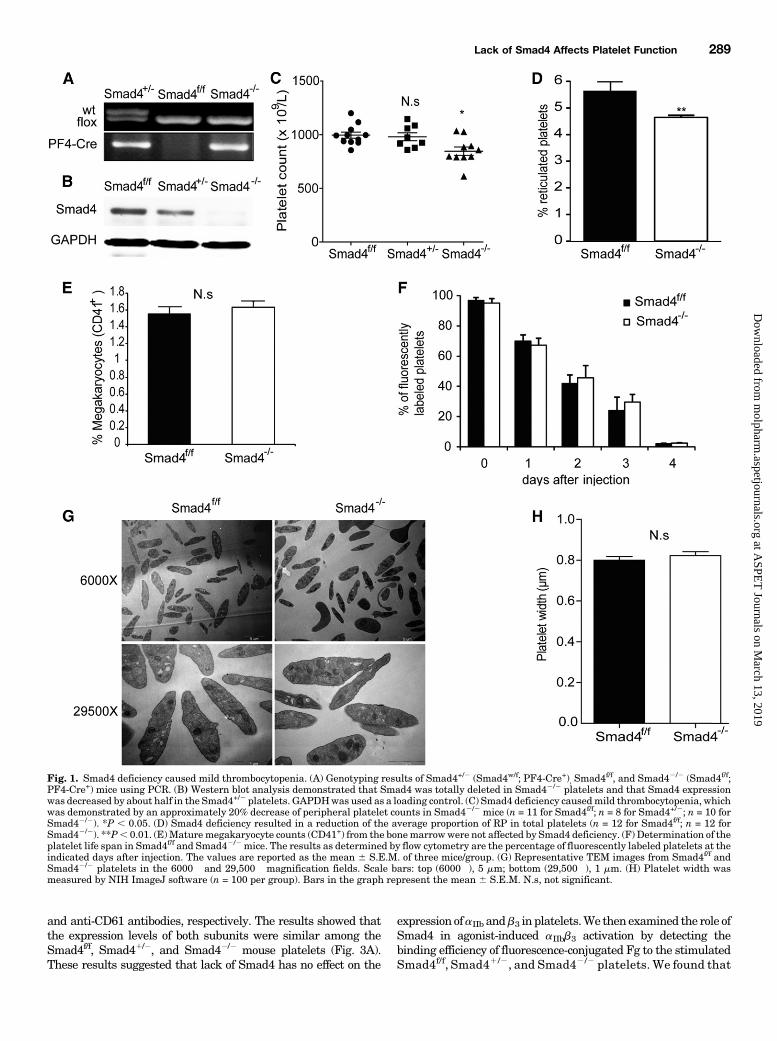
Thrombocytopenia. To study the role of Smad4 in plateletactivation and thrombus formation, the megakaryocyte/pla-telet-specific Smad4-deficient mice were examined. TheSmad4f/f, Smad41/2, (Smad4w/f, PF4-Cre1) and Smad42/2
(Smad4f/f, PF4-Cre1) mice were genotyped by PCR (Fig. 1A),and the platelet-specific Smad4 deficiency was confirmed byWestern blot analysis (Fig. 1B). The results showed that theSmad4 expression was significantly reduced in Smad41/2
platelets and totally ablated in Smad42/2 platelets. Becauselittle is known about the function of Smad4 in platelets, theplatelet number in these mouse was enumerated. The num-bers of peripheral platelets were 9966 29� 109/l, 9806 37�109/l, and 846 6 39 � 109/l in Smad4f/f mice (n 511 mice/group), Smad41 /2 mice (n 5 8 mice/group), andSmad42/2 mice (n 5 10 mice/group), respectively. Althoughthere was no visible difference in platelet numbers betweenthe Smad4f/f and Smad41/2 groups, the blood of Smad42/2
mice contained ∼20% fewer platelets than that of Smad4f/f
mice (P , 0.05) (Fig. 1C), indicating that the completedepletion of Smad4 in megakaryocytes/platelets causes mildthrombocytopenia. The proportion of RPs (%RP) in totalplatelets has been shown to reflect the production of plateletsfrom megakaryocytes. Therefore, the %RP was evaluated inSmad4f/f and Smad42/2 mice. The average %RP of Smad42/2
mice was 4.64% (n 5 12 mice/group), compared with 5.65%(n 5 12 mice/group) in Smad4f/f mice. The complete depletionof Smad4 resulted in an ∼17.7% reduction of the %RP (P ,
TABLE 1Primers used in RT-qPCR
Name Sequence
GAPDH forward 59CAGTATGACTCCACTCACGGC39GAPDH reverse 59GAGGGGCCATCCACAGTCTTC39ROCK-1 forward 59CTTGGGAAACGCTCCGAGAC39ROCK-1 reverse 59TCTCACTGGCATTTGCTGAAGG39ROCK-2 forward 59GGATGGTTGTCATTGCCTGTG39ROCK-2 reverse 59AAGGGTTGGACTGCTCTTTATC39Syk forward 59TACGCCCCC GAATGCATCAACTAC39Syk reverse 59GCACCCCATCCGCTCTCCTTTCT39Integrin-b1 forward 59TGTGACCCATTGCAAGGAGAAGG39Integrin-b1 reverse 59GGATGATGTCGGGACCAGTAGG39
Lack of Smad4 Affects Platelet Function 287
at ASPE
T Journals on M
arch 13, 2019m
olpharm.aspetjournals.org
Dow
nloaded from
0.01) (Fig. 1D). An analysis of the bone marrow revealed thatmature megakaryocytes (CD411) of Smad42/2 mice werenormal in number (Fig. 1E) and morphology (SupplementalFig. 1A). To determine whether the thrombocytopenia inSmad42/2 mice was caused by increased platelet turnover,the life span of Smad42/2 platelets in vivowas determined.Wefound no significant difference in life span between Smad4f/f
and Smad42 /2 platelets (Fig. 1F). Annexin V binding toplatelets was assayed to evaluate the role, if any, of apoptosisin the thrombocytopenia correlated with Smad4 deficiency.The results showed that Smad4 deficiency did not enhanceannexin V binding to platelets (Supplemental Fig. 1B). Thisresult suggested that it is impaired megakaryocyte differen-tiation, rather than enhanced platelet turnover, that causedthe thrombocytopenia in megakaryocyte/platelet-specificSmad4-deficient mice. In addition, the platelet size inSmad42/2 mice did not differ from that of Smad4f/f mice, asrevealed by transmission electronmicroscopy (TEM) (Fig. 1G).The average width of Smad42/2 platelets was 0.826 0.02 mmcompared with 0.8 6 0.02 mm for the Smad4f/f controls (n 5100 platelets; P . 0.05) (Fig. 1H). Moreover, the TEMexamination revealed that the intracellular structures ofresting Smad4f/f and Smad42/2 platelets, including thea-granules and dense granules, were indistinguishable (Fig.1G). Thus, instead of platelet turnover, Smad4 deficiency ismore likely to affect the differentiation of megakaryocytes intoplatelets, causing thrombocytopenia.Diminished Aggregation of Smad4-Deficient Platelets
in Response to Agonists. The role of Smad4 in agonist-induced platelet aggregation was investigated by stimulatingSmad4f/f, Smad41/2, and Smad42/2 platelets with thrombin,ADP, or CRP. As shown in Fig. 2A and Supplemental Fig. 2A,the aggregation of Smad41/2 and Smad42/2 platelets wasdiminished in response to different concentrations of thrombin(0.05 U/ml), ADP (40, 20, and 10 mM) or CRP (0.5, 0.35, and 0.2mg/ml). Moreover, Smad42/2 platelets showed more severelyimpaired aggregation in response to these agonists. Notably,markedly decreased aggregations of Smad42/2 and Smad41/2
platelets were seen upon activation with CRP. As CRP is aglycoprotein VI (GPVI)–specific agonist (Asselin et al., 1997;Kehrel et al., 1998), these results suggested that Smad4deficiency mainly affects GPVI-associated downstream signaltransduction in platelet activation.Decreased Secretion of a-Granules but Normal
Dense Granule Release in Smad4-Deficient Platelets.P-selectin resides in the a-granule membrane of unstimulatedplatelets, which is translocated to the activated plateletsurface via a secretory pathway (Polgar et al., 2005). Theexpression of P-selectin on the platelet surface as a readout fora-granule secretion was investigated by flow cytometry in
response to 0.1 U/ml thrombin, 2 mg/ml CRP, or 40 mM ADPwith 0.4 mg/ml EP. The secretion of P-selectin was signifi-cantly decreased in Smad42/2 platelets in response to CRP orADP but not thrombin (Fig. 2B). These results indicated thatthe secretion of a-granules was impaired due to the homozy-gous deletion of Smad4 in platelets (Fig. 2B; SupplementalFig. 2B). To test whether the release of dense granules wasalso affected, the amount of ATP released by stimulatedplatelets was measured. Surprisingly, no significant differ-ences in ATP secretion were found between these threegenotypes of platelets stimulated by either 0.35 mg/ml CRP,20 mM ADP, or 0.05 U/ml thrombin (Fig. 2C). Therefore, thelack of Smad4 affects platelet a-granule secretion but has noimpact on dense granule release.Delayed Arterial Occlusive Thrombosis and
Prolonged Bleeding Time in Smad4-Deficient Mice. AnFeCl3-induced arterial injury model and tail-bleeding timeassays were used to evaluate the physiologic role of targetedmolecules in thrombosis and hemostasis in vivo (Liu et al.,2006). Since lack of Smad4 results in defective agonist-induced platelet activation, thrombus formation in Smad4f/f,Smad41 /2 , and Smad42 /2 mice was examined using theFeCl3-induced carotid artery thrombosis model. The averagetime to first occlusion was 20.53 6 2.56 minutes in Smad42/2
mice (n 5 6 mice/group), compared with 9.11 6 0. 29 minutesin Smad4f/f mice (n 5 6 mice/group; P , 0.01) and 17.13 61.97 minutes in Smad41/2 mice (n 5 6 mice/group; P , 0.05)(Fig. 2D). These results indicated that Smad4 deficiency inplatelets inhibited arterial thrombus formation in vivo. Theeffect of Smad4 deficiency on hemostasis was evaluated bymeasuring tail-bleeding time. The average bleeding time ofSmad42/2 mice was 16.626 1.74 minutes (n5 7 mice/group),which is significantly longer than that of the Smad4f/f mice,which was 3.936 0.43 minutes (n5 8 mice/group; P, 0.001).In addition, the average bleeding time of the Smad41/2 groupwas 8.46 0.88 minutes (n5 6 mice/group; P, 0.05) (Fig. 2E).Thus, Smad4 deficiency in megakaryocytes/platelets caused aprolonged bleeding time, suggesting that Smad4may regulatethrombus formation and hemostasis in vivo.Lack of Smad4 Affects Platelet Integrin aIIbb3-Mediated
Inside-Out Signaling. Integrin aIIbb3-mediated bidirectionalsignaling plays a critical role in thrombosis and hemostasis(Moser et al., 2008). Stimulation of plateletswith various agonistswill activate integrin aIIbb3 (“inside-out” signaling), which is thenable to bind soluble Fg, resulting in stable platelet adhesion,platelet aggregation, and thrombus formation (Li et al., 2010).However, the function of Smad4 in the platelet integrin aIIbb3-related signaling is poorly understood. First, the expression levelsof the aIIb and b3 subunits in platelets from Smad4-deficientmicewere analyzed by flow cytometry using PE-conjugated anti-CD41
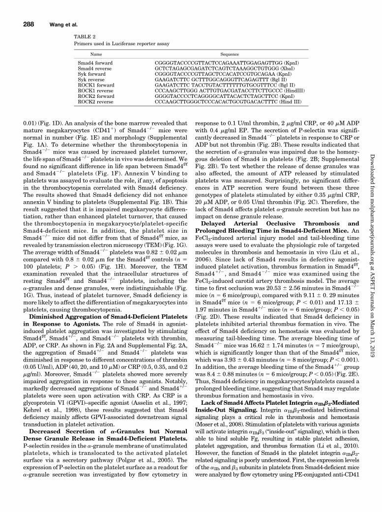
TABLE 2Primers used in Luciferase reporter assay
Name Sequence
Smad4 forward CGGGGTACCCCGTTACTCCAGAAATTGGAGAGTTGG (KpnI)Smad4 reverse GCTCTAGAGCGAGATCTCAGTCTAAAGGCTGTGGG (XbaI)Syk forward CGGGGTACCCCGTTAGCTCCACATCCGTGCAGAA (KpnI)Syk reverse GAAGATCTTC GCTTTGGCAGGGTTCAGAGTTT (Bgl II)ROCK1 forward GAAGATCTTC TACCTGTACTTTTTTGTGCGTTTCC (Bgl II)ROCK1 reverse CCCAAGCTTGGG ACTTGTGACGATACCTTCTTGCCC (HindIII)ROCK2 forward GGGGTACCCCTCAGGGGCATTACACTCTAGCTTCC (KpnI)ROCK2 reverse CCCAAGCTTGGGCTCCCACACTGCGTGACACTTTC (Hind III)
288 Wang et al.
at ASPE
T Journals on M
arch 13, 2019m
olpharm.aspetjournals.org
Dow
nloaded from
and anti-CD61 antibodies, respectively. The results showed thatthe expression levels of both subunits were similar among theSmad4f/f, Smad41/2, and Smad42/2 mouse platelets (Fig. 3A).These results suggested that lack of Smad4 has no effect on the
expression ofaIIb andb3 in platelets.We thenexamined the role ofSmad4 in agonist-induced aIIbb3 activation by detecting thebinding efficiency of fluorescence-conjugated Fg to the stimulatedSmad4f/f, Smad41/2, and Smad42/2 platelets. We found that
Fig. 1. Smad4 deficiency caused mild thrombocytopenia. (A) Genotyping results of Smad4+/2 (Smad4w/f; PF4-Cre+), Smad4f/f, and Smad42/2 (Smad4f/f;PF4-Cre+) mice using PCR. (B) Western blot analysis demonstrated that Smad4 was totally deleted in Smad42/2 platelets and that Smad4 expressionwas decreased by about half in the Smad4+/2 platelets. GAPDHwas used as a loading control. (C) Smad4 deficiency causedmild thrombocytopenia, whichwas demonstrated by an approximately 20% decrease of peripheral platelet counts in Smad42/2 mice (n = 11 for Smad4f/f; n = 8 for Smad4+/2; n = 10 forSmad42/2). *P , 0.05. (D) Smad4 deficiency resulted in a reduction of the average proportion of RP in total platelets (n = 12 for Smad4f/f; n = 12 forSmad42/2). **P, 0.01. (E)Maturemegakaryocyte counts (CD41+) from the bonemarrowwere not affected by Smad4 deficiency. (F) Determination of theplatelet life span in Smad4f/f and Smad42/2mice. The results as determined by flow cytometry are the percentage of fluorescently labeled platelets at theindicated days after injection. The values are reported as the mean 6 S.E.M. of three mice/group. (G) Representative TEM images from Smad4f/f andSmad42/2 platelets in the 6000� and 29,500� magnification fields. Scale bars: top (6000�), 5 mm; bottom (29,500�), 1 mm. (H) Platelet width wasmeasured by NIH ImageJ software (n = 100 per group). Bars in the graph represent the mean 6 S.E.M. N.s, not significant.
Lack of Smad4 Affects Platelet Function 289
at ASPE
T Journals on M
arch 13, 2019m
olpharm.aspetjournals.org
Dow
nloaded from

Fig. 2. Smad4 deficiency caused the dysfunction of platelets. (A) The aggregation of Smad4f/f, Smad4+/2, and Smad42/2 platelets in response to agonistsas indicated. The traces are representative of three experiments. (B)Washed, unstirred Smad4f/f, Smad4+/2, and Smad42/2 platelets at a concentration of3� 107/ml were incubated with an FITC-conjugated P-selectin monoclonal antibody in the presence of 0.1 U/ml thrombin, 40 mMADP plus 0.4 mg/ml EP,or 2mg/ml CRP. (C)Measurements of theATP released in the supernatant of activated Smad4f/f, Smad4+/2, and Smad42/2 platelets induced by 0.35mg/mlCRP, 20 mM ADP, or 0.05 U/ml thrombin. The bars in the graph represent the mean 6 S.E.M. from three independent experiments. (D) Mouse carotidarteries were treated with 10% FeCl3 as described. The times to occlusion were measured (n = 6 for each group; *P , 0.05; **P , 0.01). (E) Smad4deficiency in platelets prolonged the mouse tail vein bleeding time (n = 8 for Smad4f/f; n = 6 for Smad4+/2; n = 7 for Smad42/2; *P , 0.05; ***P, 0.001).
290 Wang et al.
at ASPE
T Journals on M
arch 13, 2019m
olpharm.aspetjournals.org
Dow
nloaded from

Fig. 3. Smad4 deficiency impaired integrin aIIbb3-mediated inside-out and outside-in bidirectional signaling. (A) Washed, resting Smad4f/f, Smad4+/2,and Smad42/2 platelets at a concentration of 3 � 107/ml were incubated with PE-conjugated CD41 and CD61 monoclonal antibodies. The expressionlevels of the integrin subunits aIIb and b3 were detected using a flow cytometer. (B) Binding of Alexa Fluor 647-Fg to washed, unstirred Smad4f/f, Smad4+/2,and Smad42/2 platelets stimulated with 0.1 U/ml thrombin, 40 mM ADP plus 0.4 mg/ml EP, or 2 mg/ml CRP. (C) Spreading of Smad4f/f, Smad4+/2, andSmad42/2 platelets on immobilized Fg for 2 hours. (D) Quantification of the area (pixel number) of three random fields in a 100� objective field. Statisticalanalysis of the spreading size of platelets was performed using Tukey’s test after ANOVA (n = 3;mean6S.E.M., ***P, 0.001). (E) The clot retraction of PRPcontaining Smad4f/f, Smad4+/2, or Smad42/2 platelets. (F) The clot retraction was measured using NIH ImageJ software, and the data were expressed asretraction ratios (mean 6 S.E.M. from three separate experiments; **P , 0.01; ***P , 0.001).
Lack of Smad4 Affects Platelet Function 291
at ASPE
T Journals on M
arch 13, 2019m
olpharm.aspetjournals.org
Dow
nloaded from
Smad4 deficiency significantly lowered the Fg binding to plateletsinduced by 2 mg/ml CRP under nonstirring conditions. However,no obvious differences in Fg binding were found among the threegenotypes when platelets were stimulated by either 0.1 U/mlthrombin or 40 mMADP plus 0.4 mg/ml EP (Fig. 3B; Supplemen-tal Fig. 2C). These results suggest that Smad4 plays aconsiderable role in the CRP-stimulated integrin aIIbb3-mediated inside-out signaling.Smad4 Deficiency Affects Integrin aIIbb3-Mediated
Outside-In Signaling. Platelet spreading on immobilizedFg is dependent on cytoskeletal reorganization driven byintegrin aIIbb3-mediated outside-in signaling (Calderwoodet al., 2002). To examine the role of Smad4 in aIIbb3-mediatedoutside-in signaling, the spreading of Smad4f/f, Smad41/2, andSmad42/2 platelets on immobilized Fgwas assessed under thesame conditions for 2 hours. Smad42/2 platelets were barelyable to spread on immobilized Fg and only formed filopodia, incontrast to Smad4f/f platelets. Similarly, the spreading ofSmad41/2 platelets was also significantly affected, butSmad41/2 platelets displayed more obvious filopodia exten-sions than did Smad42/2 platelets (Fig. 3C). The statisticalanalysis showed that the average size of the platelets thatspread on the Fg was 1169 6 60.47 pixels for Smad42/2
platelets compared with 2439 6 110.3 pixels for Smad4f/f
platelets and 13506 70.37 pixels for Smad41/2 platelets (P,0.001) (Fig. 3D). These findings demonstrated that Smad4deficiency in platelets severely interferes with platelet spread-ing on immobilized Fg. Since platelet clot retraction is drivenby integrin aIIbb3-mediated outside-in signaling, the effect ofSmad4 deficiency on clot retraction was also assessed. Theresults presented in Fig. 3, E and F, demonstrated that theaverage ratio of clot retraction of PRP containing Smad4f/f
platelets was 0.75 6 0.018 in 2 hours versus 0.52 6 0.034 inPRP containing Smad41/2 platelets (P, 0.01) and 0.356 0.04in PRP containing Smad42/2 platelets (P , 0.001). Similarresults were also observed at 4 and 12 hours (SupplementalFig. 3, A and B). Therefore, Smad4 deficiency in plateletsseverely delayed clot retraction in PRP.
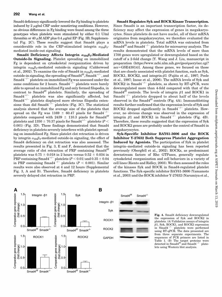
Smad4 Regulates Syk andROCKKinase Transcription.Since Smad4 is an important transcription factor, its de-ficiency may affect the expression of genes in megakaryo-cytes. Since platelets do not have nuclei, all of their mRNAoriginates from megakaryocytes, we therefore evaluated themRNA levels in platelets. Total mRNA was extracted fromSmad4f/f and Smad42/2 platelets for microarray analysis. Theresults demonstrated that the mRNA levels of more than1700 genes were upregulated or downregulated based on thecutoff of a 2-fold change [Y. Wang and J. Liu, manuscript inpreparation (https://www.ncbi.nlm.nih.gov/geo/query/acc.cgi?acc5GSE43814)]. Among these genes, we focused on thosethat are closely associated with platelet function, such as Syk,ROCK1, ROCK2, and integrin-b1 (Fujita et al., 1997; Pooleet al., 1997; Inoue et al., 2006). The mRNA levels of Syk andROCK2 in Smad42/2 platelets, as shown by RT-qPCR, weredownregulated more than 4-fold compared with that of theSmad4f/f controls. The levels of integrin b1 and ROCK1 inSmad42 /2 platelets dropped to about half of the levelsobserved in the Smad4f/f controls (Fig. 4A). Immunoblottingresults further confirmed that the expression levels of Syk andROCK2 dropped significantly in Smad42/2 platelets. How-ever, no obvious change was observed in the expression ofintegrin b1 and ROCK1 in Smad42/2 platelets (Fig. 4B).Therefore, these results suggested that the expression of Sykand ROCK2 genes are probably under the control of Smad4 inmegakaryocytes.Syk-Specific Inhibitor BAY61-3606 and the ROCK
Inhibitor Y-27632 Both Suppress Platelet AggregationInduced by Agonists. The participation of Syk in plateletintegrin–mediated outside-in signaling has been reportedpreviously (Obergfell et al., 2002). ROCKs, as predominantdownstream factors of Rho GTPases, generally regulatecytoskeletal reorganization and cell behaviors in a variety ofcell lines (Riento and Ridley, 2003).We then assessed the rolesof the kinases Syk and ROCK in Smad4-regulated plateletfunctions. The Syk-specific inhibitor BAY61-3606 (Yamamotoet al., 2003) and the ROCK inhibitor Y-27632 (Narumiya et al.,
Fig. 4. Smad4 deficiency downregulatedthe expression of Syk and ROCK2 inplatelets. (A) Validation assays of integrinb1, Syk, ROCK1, and ROCK2 expressionin Smad42/2 platelets were performedusing RT-qPCR. The data presented arefrom three separate experiments. Thesequences of PCR primers are listed inTable 1. (B) The target proteins weredetected in Smad4f/f and Smad42/2 plate-lets using Western blot analysis.
292 Wang et al.
at ASPE
T Journals on M
arch 13, 2019m
olpharm.aspetjournals.org
Dow
nloaded from

2000) were used for analyzing their effect on platelet aggre-gation. Smad4f/f platelets were preincubated with differentconcentrations of BAY61-3606 or 10 mM Y-27632 prior tobeing stimulated with different agonists. The results showedthat the Smad4f/f platelet aggregations induced by 0.05 U/mlthrombin or 20 mM ADP were hardly affected by treatmentwith 2.5 mMBAY61-3606. However, an obvious inhibition wasobserved when Smad4f/f platelets were treated with 5 mMBAY61-3606, which was similar to the suppressive effect of10 mM Y-27632 treatment (Fig. 5A). In addition, a moresignificant inhibitory effect was achieved when the plateletswere stimulated with 0.5 mg/ml CRP after treatment withthese inhibitors (Fig. 5A). The effect of BAY61-3606 andY-27632 on the platelet aggregation induced by these agonistswas consistent with that of Smad4 deficiency. The inhibitoryeffects of BAY61-3606 and Y-27632 were more obvious onCRP-induced platelet aggregation (Fig. 5A; Supplemental Fig.4). These results suggested that the kinases Syk and ROCKare significantly involved in CRP-associated GPVI signaltransduction in platelets.Both BAY61-3606 and Y-27632 Inhibit Platelet
Spreading on Immobilized Fg. Smad4f/f platelets werepreincubated with either 5 mM BAY61-3606 or 10 mMY-27632, and then the spreading of Smad4f/f platelets onimmobilized Fg was assessed under the same conditions for120 minutes. As shown in Fig. 5B, pretreatment of theplatelets with 5 mM BAY61-3606 or 10 mM Y-27632 obviouslydecreased the spreading of Smad4f/f platelets. The statisticalanalysis showed that the average size of the platelets thatspread on Fg was 1151 6 73.2 pixels in response to 5 mMBAY61-3606 treatment (P, 0.001) and 17056 87.46 pixels inresponse to 10 mM Y-27632 treatment (P , 0.001) comparedwith 23176 101.1 pixels for Smad4f/f platelets (Fig. 5C). Thus,the two inhibitors had an effect similar to that of Smad4deficiency. These results suggest that decreased Syk or ROCKexpression in Smad4-deficient platelets could be responsiblefor defective platelet spreading.Both BAY61-3606 and Y-27632 Inhibit Platelet Clot
Retraction. The role of Syk and ROCK in platelet clotretraction was also examined. Smad4f/f platelets were pre-treated with either 5mM BAY61-3606 or 10 mM Y-27632, andthe average ratios of clot retraction were analyzed after120 minutes. The results indicated, similar to the effect ofSmad4 deficiency, that both BAY61-3606 and Y-27632 signifi-cantly delayed platelet clot retraction in PRP (Fig. 5D). Theaverage ratio of clot retraction of the PRP containing Smad4f/f
platelets was 0.13 6 0.014 in response to 5 mM BAY61-3606treatment and 0.38 6 0.043 to 10 mM Y-27632 treatment,whereas the average ratio was 0.82 6 0.037 in the PRPcontaining Smad4f/f control platelets under the same conditions(P , 0.001) (Fig. 5E). The platelets treated with BAY61-3606and Y-27632 displayed similar defects in platelet clot retractionto that of Smad4 deficiency. These results demonstrated thatfunctional blockade of Syk or ROCK has the same suppressiveeffect on platelet clot retraction as that of Smad4 deficiency.Smad4 Directly Regulates ROCK2 Transcription but
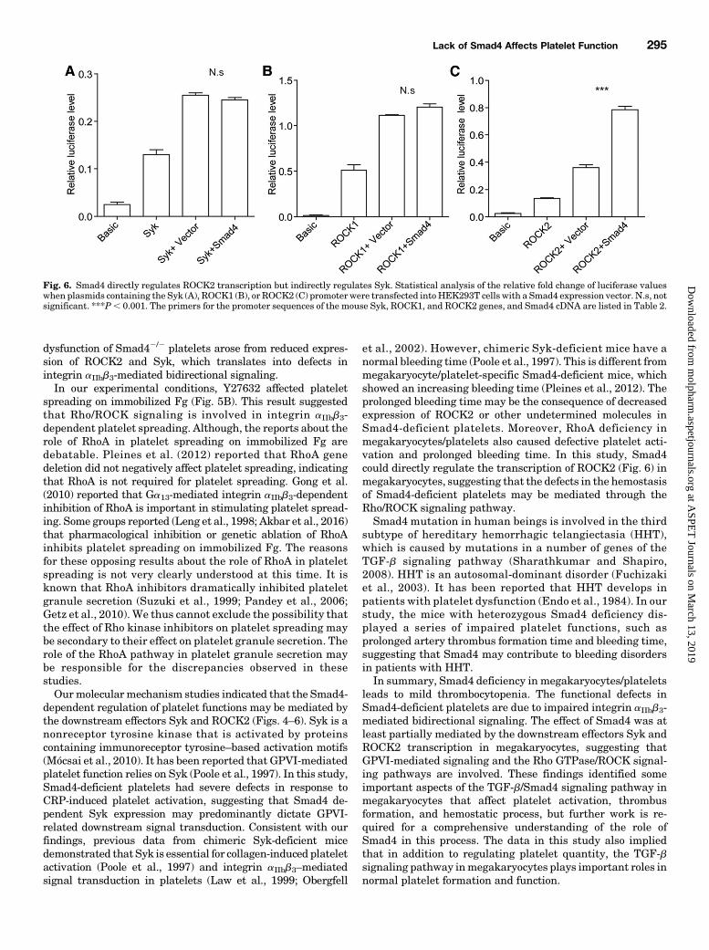
Indirectly Regulates Syk. Having shown that the defectiveplatelet function in Smad42/2 platelets is due to the reducedtranscription of Syk and ROCK2 genes in megakaryocytes,we speculated that the promoter of these genes may be underthe control of Smad4. A dual-luciferase reporter assay wasused to evaluate the mechanism of the Smad4-dependent
transcriptional regulation of Syk and ROCK genes. A Smad4expression plasmid was transfected into HEK293T cells withplasmids containing the Syk, ROCK1, or ROCK2 promoters.The results showed that the relative luciferase levels in-creased more than five times compared with cells transfectedwith the pGL3-basic control vector, suggesting that eachpromoter is transcriptionally activated in this system. Therelative luciferase activity of the ROCK2 promoter whencotransfected with the Smad4 expression plasmid was twotimes higher than that observed upon cotransfection with thepcDNA 3.1(1) vector (Fig. 6C). However, Smad4 had no sucheffect on the promoters of Syk and ROCK1. The mean relativeluciferase activity of the Syk promoter–containing andROCK1 promoter–containing plasmids exhibited no visibledifference between cells cotransfected with the Smad4 expres-sion plasmid or with the pcDNA 3.1(1) vector (Fig. 6, A and B).The results indicated that Smad4 regulates the promoteractivation of ROCK2 in a direct manner but indirectlyregulates Syk gene.
DiscussionIt has been reported that TGF-b1 plays an active role in
platelet aggregation and the maintenance of integrin function(Hoying et al., 1999). Smad4 is a key molecule in the TGF-bfamily signaling pathway (Shi and Massague, 2003), and theeffects of Smad4 on platelet function are unclear. Here, weobserved that megakaryocyte/platelet-specific inactivation ofSmad4 impaired the platelet aggregation induced by throm-bin, ADP, and CRP as well as thrombus formation andhemostasis. The results from Fg binding (Fig. 3B), plateletaggregation (Fig. 2A), platelet spreading, and clot retractionstudies (Fig. 3, C–F) revealed that lack of Smad4 results indefective platelet activation by affecting integrin aIIbb3-mediatedbidirectional signaling. The data shown in Figs. 4 and 5indicated that the platelet functions regulated by Smad4 aremediated through the transcriptional regulation of key plate-lets signaling proteins, Syk, and ROCK2 in megakaryocytes.The quantity of peripheral platelets is a balance between
platelet formation by megakaryocytes and platelet clearance.Previous studies have reported that TGF-b1 negativelyregulated murine megakaryocytopoiesis (Ishibashi et al.,1987; Jeanpierre et al., 2008) and that the numbers ofperipheral blood platelets are elevated in TGF-b1–deficientmice (Hoying et al., 1999). Recent studies showed that thehematopoietic (Vav-Cre) deficiency of Smad4 has no effect onplatelet count (Pan et al., 2007) but that Smad4 is critical forthe self-renewal of hematopoietic stem cells (Karlsson et al.,2007; Rörby et al., 2012). In this study, the platelet numberwas slightly reduced in megakaryocyte/platelet-specificSmad4-deficient mice. The results from a platelet turnoverstudy suggested that instead of enhanced platelet clearance, itmay be impaired megakaryocyte differentiation that led tothrombocytopenia (Fig. 1D). These findings suggested thatSmad4 may be involved in regulating megakaryocyte differ-entiation, thus affecting the number of platelets. This effect ofSmad4 may be achieved by transcriptional regulation ofmolecules in other signaling pathways. One example isROCK2, a downstream effector of RhoA that was demon-strated to be regulated by Smad4 in our study (Fig. 6) andmayalso be involved in platelet formation by megakaryocytes(Pleines et al., 2012).
Lack of Smad4 Affects Platelet Function 293
at ASPE
T Journals on M
arch 13, 2019m
olpharm.aspetjournals.org
Dow
nloaded from

TGF-b1 had no direct effect on thrombin- or collagen-induced aggregation of Smad4f/f and Smad42/2 platelets (datanot shown), indicating that Smad4 may not regulate plateletactivation directly. As the most abundant platelet surfacereceptor, integrin aIIbb3-mediated bidirectional signaling isrequired for various aspects of platelet functions (Li et al.,2010). Some of the functional deficits in the Smad4-deficient
platelets could arise from defects in the expression of theintegrin aIIbb3. However, we failed to find any effect of Smad4deficiency on the expression of either aIIb or b3 in platelets (Fig.3A). On the other hand, Smad4 deficiency significantly loweredFg binding to platelets after CRP-induced aIIbb3 activation andinhibited platelet spreading on immobilized Fg as well as clotretraction in PRP (Fig. 3, B–F). These results revealed that the
Fig. 5. The kinases Syk and ROCKwere both involved in platelet function.(A) The inhibitory effects of 2.5 and5mMBAY 61-3606 and 10mMY-27632on Smad4f/f platelet aggregation inresponse to either 0.05 U/ml thrombin,20 mM ADP, or 0.5 mg/ml CRP. (B)Spreading of Smad4f/f platelets onimmobilized Fg in the presence of5 mM BAY 61-3606 or 10 mMY-27632. (C) Quantification of the area(pixel number) of spreading plateletsin three random fields. The statisticalanalysis was performed using Tukey’stest after ANOVA (n = 3; mean 6 S.E.M., ***P , 0.001). (D) The clot re-traction of PRP containing Smad4f/f
in the presence of 5 mM BAY61-3606or 10 mM Y-27632. Both inhibitorssignificantly delayed the platelet-mediated clot retraction in PRP. (E)NIH ImageJ software was used for thestatistical analysis of clot retractionarea and the data were expressed asretraction ratios. ***P , 0.001. BAY,BAY 61-3606.
294 Wang et al.
at ASPE
T Journals on M
arch 13, 2019m
olpharm.aspetjournals.org
Dow
nloaded from
dysfunction of Smad42/2 platelets arose from reduced expres-sion of ROCK2 and Syk, which translates into defects inintegrin aIIbb3-mediated bidirectional signaling.In our experimental conditions, Y27632 affected platelet
spreading on immobilized Fg (Fig. 5B). This result suggestedthat Rho/ROCK signaling is involved in integrin aIIbb3-dependent platelet spreading. Although, the reports about therole of RhoA in platelet spreading on immobilized Fg aredebatable. Pleines et al. (2012) reported that RhoA genedeletion did not negatively affect platelet spreading, indicatingthat RhoA is not required for platelet spreading. Gong et al.(2010) reported that Ga13-mediated integrin aIIbb3-dependentinhibition of RhoA is important in stimulating platelet spread-ing. Some groups reported (Leng et al., 1998; Akbar et al., 2016)that pharmacological inhibition or genetic ablation of RhoAinhibits platelet spreading on immobilized Fg. The reasonsfor these opposing results about the role of RhoA in plateletspreading is not very clearly understood at this time. It isknown that RhoA inhibitors dramatically inhibited plateletgranule secretion (Suzuki et al., 1999; Pandey et al., 2006;Getz et al., 2010). We thus cannot exclude the possibility thatthe effect of Rho kinase inhibitors on platelet spreading maybe secondary to their effect on platelet granule secretion. Therole of the RhoA pathway in platelet granule secretion maybe responsible for the discrepancies observed in thesestudies.Ourmolecularmechanism studies indicated that the Smad4-
dependent regulation of platelet functions may be mediated bythe downstream effectors Syk and ROCK2 (Figs. 4–6). Syk is anonreceptor tyrosine kinase that is activated by proteinscontaining immunoreceptor tyrosine–based activation motifs(Mócsai et al., 2010). It has been reported that GPVI-mediatedplatelet function relies on Syk (Poole et al., 1997). In this study,Smad4-deficient platelets had severe defects in response toCRP-induced platelet activation, suggesting that Smad4 de-pendent Syk expression may predominantly dictate GPVI-related downstream signal transduction. Consistent with ourfindings, previous data from chimeric Syk-deficient micedemonstrated that Syk is essential for collagen-induced plateletactivation (Poole et al., 1997) and integrin aIIbb3–mediatedsignal transduction in platelets (Law et al., 1999; Obergfell
et al., 2002). However, chimeric Syk-deficient mice have anormal bleeding time (Poole et al., 1997). This is different frommegakaryocyte/platelet-specific Smad4-deficient mice, whichshowed an increasing bleeding time (Pleines et al., 2012). Theprolonged bleeding time may be the consequence of decreasedexpression of ROCK2 or other undetermined molecules inSmad4-deficient platelets. Moreover, RhoA deficiency inmegakaryocytes/platelets also caused defective platelet acti-vation and prolonged bleeding time. In this study, Smad4could directly regulate the transcription of ROCK2 (Fig. 6) inmegakaryocytes, suggesting that the defects in the hemostasisof Smad4-deficient platelets may be mediated through theRho/ROCK signaling pathway.Smad4 mutation in human beings is involved in the third
subtype of hereditary hemorrhagic telangiectasia (HHT),which is caused by mutations in a number of genes of theTGF-b signaling pathway (Sharathkumar and Shapiro,2008). HHT is an autosomal-dominant disorder (Fuchizakiet al., 2003). It has been reported that HHT develops inpatients with platelet dysfunction (Endo et al., 1984). In ourstudy, the mice with heterozygous Smad4 deficiency dis-played a series of impaired platelet functions, such asprolonged artery thrombus formation time and bleeding time,suggesting that Smad4 may contribute to bleeding disordersin patients with HHT.In summary, Smad4 deficiency in megakaryocytes/platelets
leads to mild thrombocytopenia. The functional defects inSmad4-deficient platelets are due to impaired integrin aIIbb3-mediated bidirectional signaling. The effect of Smad4 was atleast partially mediated by the downstream effectors Syk andROCK2 transcription in megakaryocytes, suggesting thatGPVI-mediated signaling and the Rho GTPase/ROCK signal-ing pathways are involved. These findings identified someimportant aspects of the TGF-b/Smad4 signaling pathway inmegakaryocytes that affect platelet activation, thrombusformation, and hemostatic process, but further work is re-quired for a comprehensive understanding of the role ofSmad4 in this process. The data in this study also impliedthat in addition to regulating platelet quantity, the TGF-bsignaling pathway inmegakaryocytes plays important roles innormal platelet formation and function.
Fig. 6. Smad4 directly regulates ROCK2 transcription but indirectly regulates Syk. Statistical analysis of the relative fold change of luciferase valueswhen plasmids containing the Syk (A), ROCK1 (B), or ROCK2 (C) promoter were transfected into HEK293T cells with a Smad4 expression vector. N.s, notsignificant. ***P, 0.001. The primers for the promoter sequences of the mouse Syk, ROCK1, and ROCK2 genes, and Smad4 cDNA are listed in Table 2.
Lack of Smad4 Affects Platelet Function 295
at ASPE
T Journals on M
arch 13, 2019m
olpharm.aspetjournals.org
Dow
nloaded from

Acknowledgments
The authors thank Li Zhu (Soochow University) for providing theCRP and Professor Xiao Yang (Institute of Biotechnology, Beijing) forproviding mice carrying the Smad4 gene flanked by loxP recognitionsites (Smad4f/f). The authors also thank Professor Ulhas P. Naik forassistance in polishing the final manuscript.
Authorship Contributions
Participated in research design: J. Liu and Wu.Conducted experiments: Wang, Jiang, Mo, Lan, X. Liu, and Wu.Performed data analysis: Yang, Zhang, Zhu, J. Liu, and Wu.Wrote or contributed to the writing of the manuscript: Wang, J. Liu,
and Wu.
References
Akbar H, Duan X, Saleem S, Davis AK, and Zheng Y (2016) RhoA and Rac1 GTPasesdifferentially regulate agonist-receptor mediated reactive oxygen species genera-tion in platelets. PLoS One 11:e0163227.
Asselin J, Gibbins JM, Achison M, Lee YH, Morton LF, Farndale RW, Barnes MJ,and Watson SP (1997) A collagen-like peptide stimulates tyrosine phosphorylationof syk and phospholipase C g2 in platelets independent of the integrin alpha2-beta1. Blood 89:1235–1242.
Calderwood DA, Yan B, de Pereda JM, Alvarez BG, Fujioka Y, Liddington RC,and Ginsberg MH (2002) The phosphotyrosine binding-like domain of talin acti-vates integrins. J Biol Chem 277:21749–21758.
Chen X, Zhang Y, Wang Y, Li D, Zhang L, Wang K, Luo X, Yang Z, Wu Y, and Liu J(2013) PDK1 regulates platelet activation and arterial thrombosis. Blood 121:3718–3726.
Endo Y, Mamiya S, Niitsu H, Miura A, and Nishimura S (1984) Reduced plateletaggregation induced by epinephrine and idiopathic thrombocytopenic purpura inhereditary haemorrhagic teleangiectasia. Thromb Haemost 52:369.
Flevaris P, Li Z, Zhang G, Zheng Y, Liu J, and Du X (2009) Two distinct roles ofmitogen-activated protein kinases in platelets and a novel Rac1-MAPK-dependentintegrin outside-in retractile signaling pathway. Blood 113:893–901.
Fuchizaki U, Miyamori H, Kitagawa S, Kaneko S, and Kobayashi K (2003) Heredi-tary haemorrhagic telangiectasia (Rendu-Osler-Weber disease). Lancet 362:1490–1494.
Fujita A, Saito Y, Ishizaki T, Maekawa M, Fujisawa K, Ushikubi F, and Narumiya S(1997) Integrin-dependent translocation of p160ROCK to cytoskeletal complex inthrombin-stimulated human platelets. Biochem J 328:769–775.
Gay LJ and Felding-Habermann B (2011) Contribution of platelets to tumour me-tastasis. Nat Rev Cancer 11:123–134.
George JN (2000) Platelets. Lancet 355:1531–1539.Getz TM, Dangelmaier CA, Jin J, Daniel JL, and Kunapuli SP (2010) Differentialphosphorylation of myosin light chain (Thr)18 and (Ser)19 and functional impli-cations in platelets. J Thromb Haemost 8:2283–2293.
Gong H, Shen B, Flevaris P, Chow C, Lam SC, Voyno-Yasenetskaya TA, Kozasa T,and Du X (2010) G protein subunit Galpha13 binds to integrin alphaIIbbeta3 andmediates integrin “outside-in” signaling. Science 327:340–343.
Hoying JB, Yin M, Diebold R, Ormsby I, Becker A, and Doetschman T (1999)Transforming growth factor beta1 enhances platelet aggregation through a non-transcriptional effect on the fibrinogen receptor. J Biol Chem 274:31008–31013.
Inoue O, Suzuki-Inoue K, McCarty OJ, Moroi M, Ruggeri ZM, Kunicki TJ, Ozaki Y,and Watson SP (2006) Laminin stimulates spreading of platelets through integrinalpha6beta1-dependent activation of GPVI. Blood 107:1405–1412.
Ishibashi T, Miller SL, and Burstein SA (1987) Type beta transforming growth factoris a potent inhibitor of murine megakaryocytopoiesis in vitro. Blood 69:1737–1741.
Jeanpierre S, Nicolini FE, Kaniewski B, Dumontet C, Rimokh R, Puisieux A,and Maguer-Satta V (2008) BMP4 regulation of human megakaryocytic differen-tiation is involved in thrombopoietin signaling. Blood 112:3154–3163.
Karlsson G, Blank U, Moody JL, Ehinger M, Singbrant S, Deng CX, and Karlsson S(2007) Smad4 is critical for self-renewal of hematopoietic stem cells. J Exp Med204:467–474.
Kehrel B, Wierwille S, Clemetson KJ, Anders O, Steiner M, Knight CG, FarndaleRW, Okuma M, and Barnes MJ (1998) Glycoprotein VI is a major collagen receptorfor platelet activation: it recognizes the platelet-activating quaternary structure ofcollagen, whereas CD36, glycoprotein IIb/IIIa, and von Willebrand factor do not.Blood 91:491–499.
Labelle M, Begum S, and Hynes RO (2011) Direct signaling between platelets andcancer cells induces an epithelial-mesenchymal-like transition and promotes me-tastasis. Cancer Cell 20:576–590.
Law DA, Nannizzi-Alaimo L, Ministri K, Hughes PE, Forsyth J, Turner M, ShattilSJ, Ginsberg MH, Tybulewicz VL, and Phillips DR (1999) Genetic and pharma-cological analyses of Syk function in alphaIIbbeta3 signaling in platelets. Blood 93:2645–2652.
Leng L, Kashiwagi H, Ren XD, and Shattil SJ (1998) RhoA and the function ofplatelet integrin alphaIIbbeta3. Blood 91:4206–4215.
Li Z, Delaney MK, O’Brien KA, and Du X (2010) Signaling during platelet adhesionand activation. Arterioscler Thromb Vasc Biol 30:2341–2349.
Liu J, Fitzgerald ME, Berndt MC, Jackson CW, and Gartner TK (2006) Brutontyrosine kinase is essential for botrocetin/VWF-induced signaling and GPIb-dependent thrombus formation in vivo. Blood 108:2596–2603.
Mahooti S, Graesser D, Patil S, Newman P, Duncan G, Mak T, and Madri JA (2000)PECAM-1 (CD31) expression modulates bleeding time in vivo. Am J Pathol 157:75–81.
Meyer A, Wang W, Qu J, Croft L, Degen JL, Coller BS, and Ahamed J (2012) PlateletTGF-b1 contributions to plasma TGF-b1, cardiac fibrosis, and systolic dysfunctionin a mouse model of pressure overload. Blood 119:1064–1074.
Mócsai A, Ruland J, and Tybulewicz VL (2010) The SYK tyrosine kinase: a crucialplayer in diverse biological functions. Nat Rev Immunol 10:387–402.
Moser M, Nieswandt B, Ussar S, Pozgajova M, and Fässler R (2008) Kindlin-3 isessential for integrin activation and platelet aggregation. Nat Med 14:325–330.
Narumiya S, Ishizaki T, and Uehata M (2000) Use and properties of ROCK-specificinhibitor Y-27632. Methods Enzymol 325:273–284.
NiuH, Chen X, Gruppo RA, Li D,Wang Y, Zhang L,Wang K, ChaiW, Sun Y, Ding Z, et al.(2012) Integrin aIIb-mediated PI3K/Akt activation in platelets. PLoS One 7:e47356.
Obergfell A, Eto K, Mocsai A, Buensuceso C, Moores SL, Brugge JS, Lowell CA,and Shattil SJ (2002) Coordinate interactions of Csk, Src, and Syk kinases with [al-pha]IIb[beta]3 initiate integrin signaling to the cytoskeleton. J Cell Biol 157:265–275.
Pan D, Schomber T, Kalberer CP, Terracciano LM, Hafen K, Krenger W, Hao-ShenH, Deng C, and Skoda RC (2007) Normal erythropoiesis but severe polyposis andbleeding anemia in Smad4-deficient mice. Blood 110:3049–3055.
Pandey D, Goyal P, Bamburg JR, and Siess W (2006) Regulation of LIM-kinase 1 andcofilin in thrombin-stimulated platelets. Blood 107:575–583.
Pleines I, Hagedorn I, Gupta S, May F, Chakarova L, van Hengel J, Offermanns S,Krohne G, Kleinschnitz C, Brakebusch C, et al. (2012) Megakaryocyte-specificRhoA deficiency causes macrothrombocytopenia and defective platelet activation inhemostasis and thrombosis. Blood 119:1054–1063.
Polgar J, Matuskova J, and Wagner DD (2005) The P-selectin, tissue factor, co-agulation triad. J Thromb Haemost 3:1590–1596.
Poole A, Gibbins JM, Turner M, van Vugt MJ, van de Winkel JG, Saito T, TybulewiczVL, and Watson SP (1997) The Fc receptor gamma-chain and the tyrosine kinaseSyk are essential for activation of mouse platelets by collagen. EMBO J 16:2333–2341.
Prislovsky A, Marathe B, Hosni A, Bolen AL, Nimmerjahn F, Jackson CW, Weiman D,and Strom TS (2008) Rapid platelet turnover in WASP(-) mice correlates with in-creased ex vivo phagocytosis of opsonized WASP(-) platelets. Exp Hematol 36:609–623.
Redondo S, Navarro-Dorado J, Ramajo M, Medina Ú, and Tejerina T (2012) Thecomplex regulation of TGF-b in cardiovascular disease. Vasc Health Risk Manag 8:533–539.
Riento K and Ridley AJ (2003) Rocks: multifunctional kinases in cell behaviour. NatRev Mol Cell Biol 4:446–456.
Rörby E, Hägerström MN, Blank U, Karlsson G, and Karlsson S (2012) Humanhematopoietic stem/progenitor cells overexpressing Smad4 exhibit impaired re-constitution potential in vivo. Blood 120:4343–4351.
Semple JW, Italiano, JrJE, and Freedman J (2011) Platelets and the immune con-tinuum. Nat Rev Immunol 11:264–274.
Sharathkumar AA and Shapiro A (2008) Hereditary haemorrhagic telangiectasia.Haemophilia 14:1269–1280.
Shi Y and Massagué J (2003) Mechanisms of TGF-beta signaling from cell membraneto the nucleus. Cell 113:685–700.
Sirard C, de la Pompa JL, Elia A, Itie A, Mirtsos C, Cheung A, Hahn S, Wakeham A,Schwartz L, Kern SE, et al. (1998) The tumor suppressor gene Smad4/Dpc4 isrequired for gastrulation and later for anterior development of the mouse embryo.Genes Dev 12:107–119.
Suzuki Y, Yamamoto M, Wada H, Ito M, Nakano T, Sasaki Y, Narumiya S, Shiku H,and Nishikawa M (1999) Agonist-induced regulation of myosin phosphatase ac-tivity in human platelets through activation of Rho-kinase. Blood 93:3408–3417.
Tiedt R, Schomber T, Hao-Shen H, and Skoda RC (2007) Pf4-Cre transgenic miceallow the generation of lineage-restricted gene knockouts for studying megakar-yocyte and platelet function in vivo. Blood 109:1503–1506.
Weng Z, Li D, Zhang L, Chen J, Ruan C, Chen G, Gartner TK, and Liu J (2010) PTENregulates collagen-induced platelet activation. Blood 116:2579–2581.
Yamamoto N, Takeshita K, Shichijo M, Kokubo T, Sato M, Nakashima K, Ishimori M,Nagai H, Li YF, Yura T, et al. (2003) The orally available spleen tyrosine kinaseinhibitor 2-[7-(3,4-dimethoxyphenyl)-imidazo[1,2-c]pyrimidin-5-ylamino]nicotinamidedihydrochloride (BAY 61-3606) blocks antigen-induced airway inflammation in ro-dents. J Pharmacol Exp Ther 306:1174–1181.
Yang X, Li C, Herrera PL, and Deng CX (2002) Generation of Smad4/Dpc4 condi-tional knockout mice. Genesis 32:80–81.
Yang X, Li C, Xu X, and Deng C (1998) The tumor suppressor SMAD4/DPC4 isessential for epiblast proliferation and mesoderm induction in mice. Proc Natl AcadSci USA 95:3667–3672.
Address correspondence to: Xiaolin Wu, The Eighth People’s hospital ofShanghai, Shanghai 200235, People’s Republic of China. E-mail: [email protected] or Junling Liu, Department of Biochemistry and Molecular Cell Biology,Shanghai Key Laboratory of Tumor Microenvironment and Inflammation, Instituteof Medical Science, Shanghai Jiao Tong University School of Medicine, Shanghai200025, People’s Republic of China. E-mail: [email protected]
296 Wang et al.
at ASPE
T Journals on M
arch 13, 2019m
olpharm.aspetjournals.org
Dow
nloaded from