Embed Size (px)
Citation preview

Medical Technology Department, Faculty of Science, Islamic University-Gaza
MMICROBBIOLOGY
Dr. Abdelraouf A. ElmanamaDr. Abdelraouf A. ElmanamaPh. D MicrobiologyPh. D Microbiology
2008
Chapter 25Chapter 25Microbial Diseases of the Digestive SystemMicrobial Diseases of the Digestive System
2008
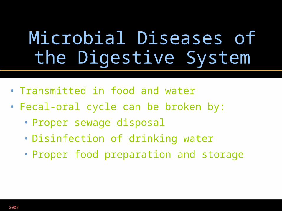
Microbial Diseases of the Digestive System
• Transmitted in food and water
• Fecal-oral cycle can be broken by:
• Proper sewage disposal
• Disinfection of drinking water
• Proper food preparation and storage
2008
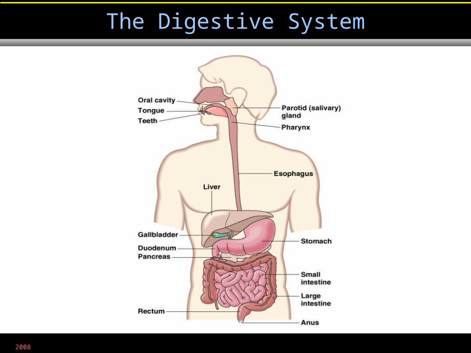
The Digestive System
Figure 25.1
2008
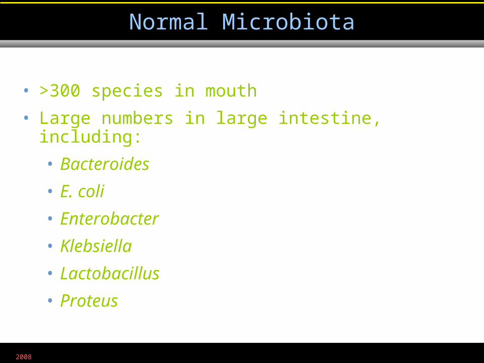
• >300 species in mouth
• Large numbers in large intestine, including:
• Bacteroides
• E. coli
• Enterobacter
• Klebsiella
• Lactobacillus
• Proteus
Normal Microbiota
2008
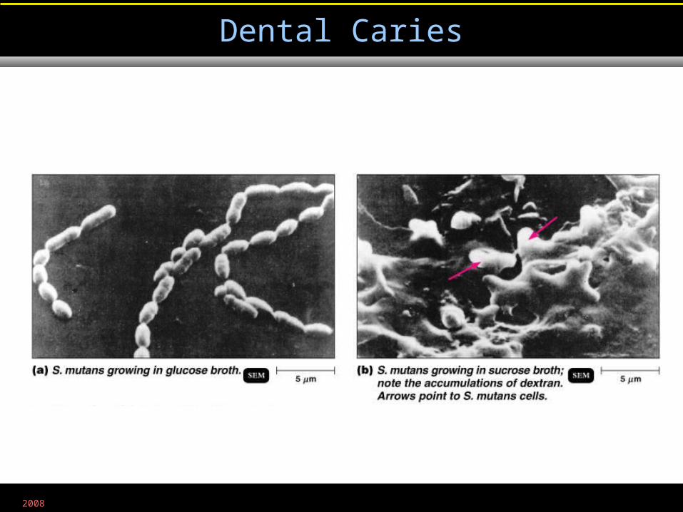
Dental Caries
Figure 25.3a, b
2008
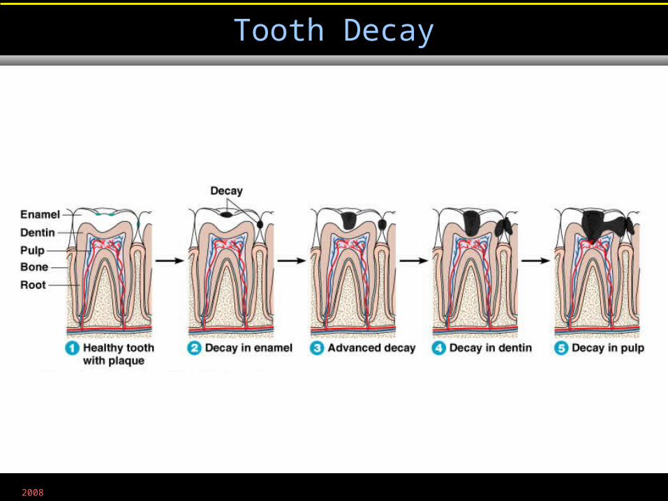
Tooth Decay
Figure 25.4
2008
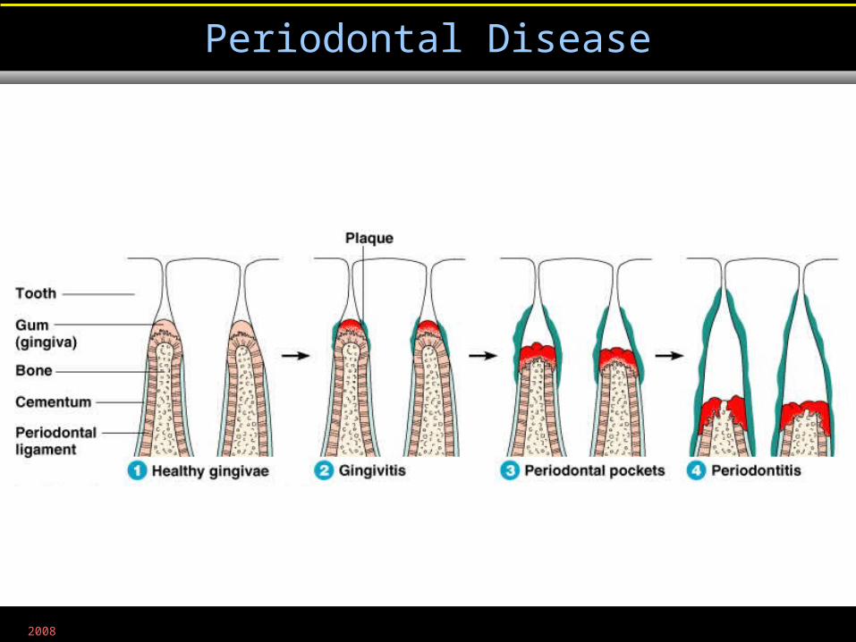
Periodontal Disease
Figure 25.5
2008
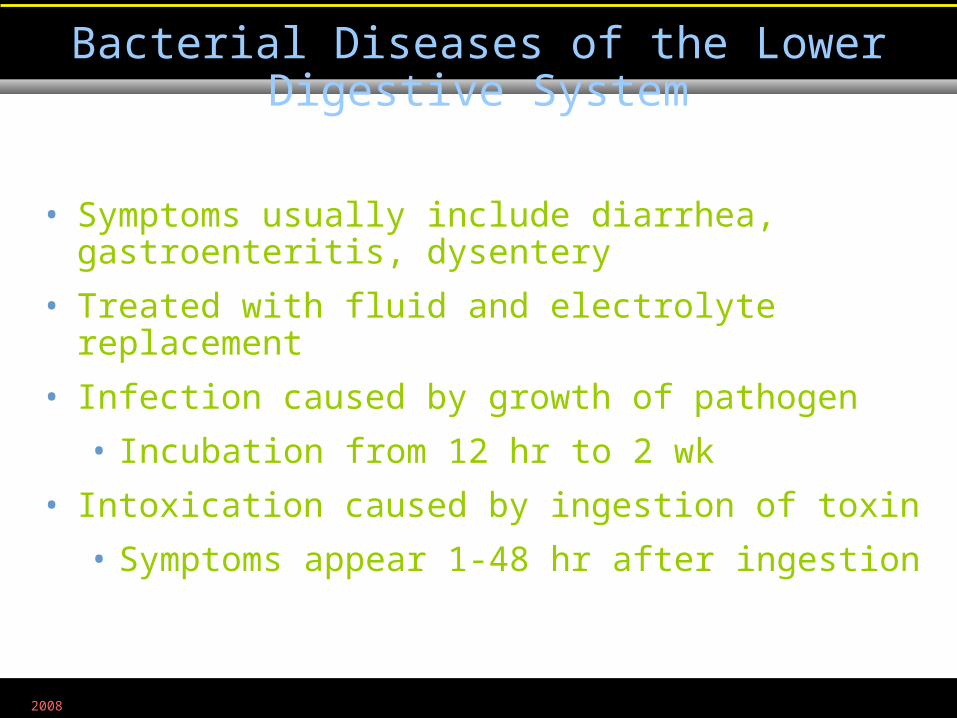
• Symptoms usually include diarrhea, gastroenteritis, dysentery
• Treated with fluid and electrolyte replacement
• Infection caused by growth of pathogen
• Incubation from 12 hr to 2 wk
• Intoxication caused by ingestion of toxin
• Symptoms appear 1-48 hr after ingestion
Bacterial Diseases of the Lower Digestive System
2008
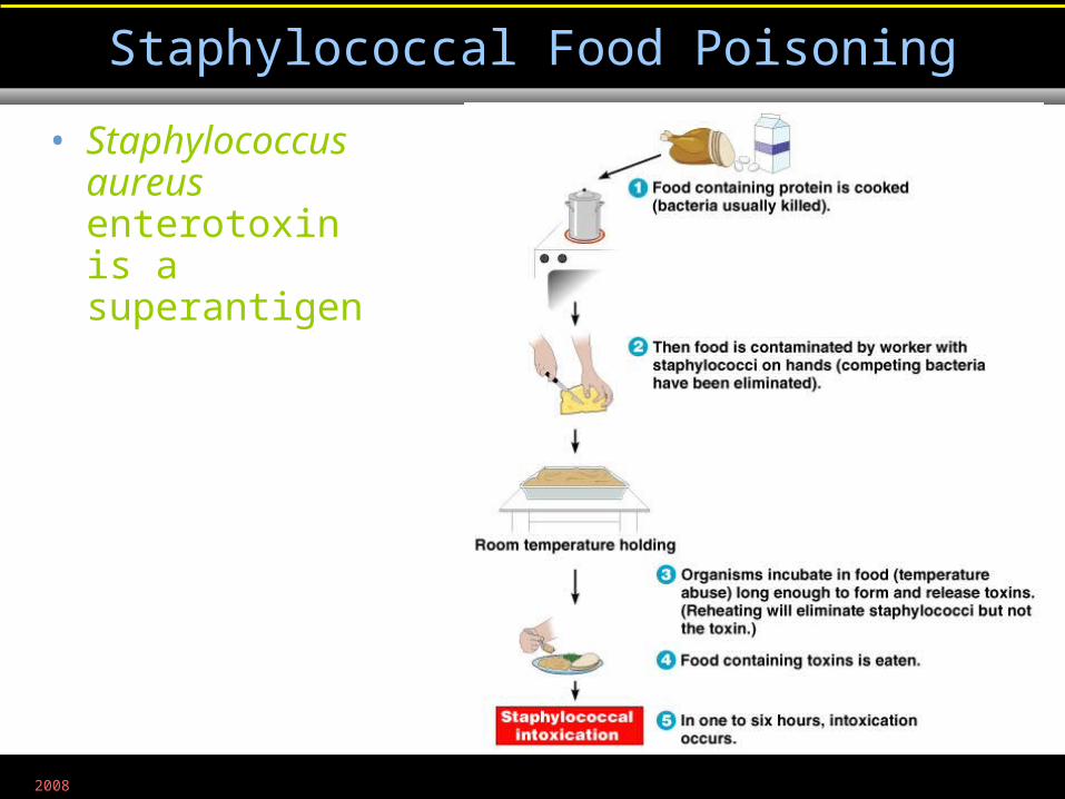
Staphylococcal Food Poisoning
Figure 25.6
• Staphylococcus aureus enterotoxin is a superantigen
2008
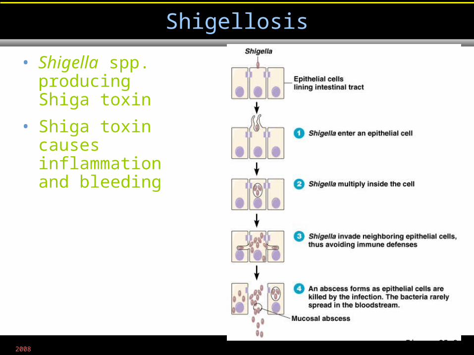
Shigellosis
Figure 25.8
• Shigella spp. producing Shiga toxin
• Shiga toxin causes inflammation and bleeding
2008
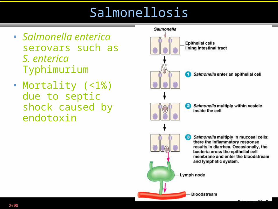
Salmonellosis
Figure 25.9
• Salmonella enterica serovars such as S. enterica Typhimurium
• Mortality (<1%) due to septic shock caused by endotoxin
2008
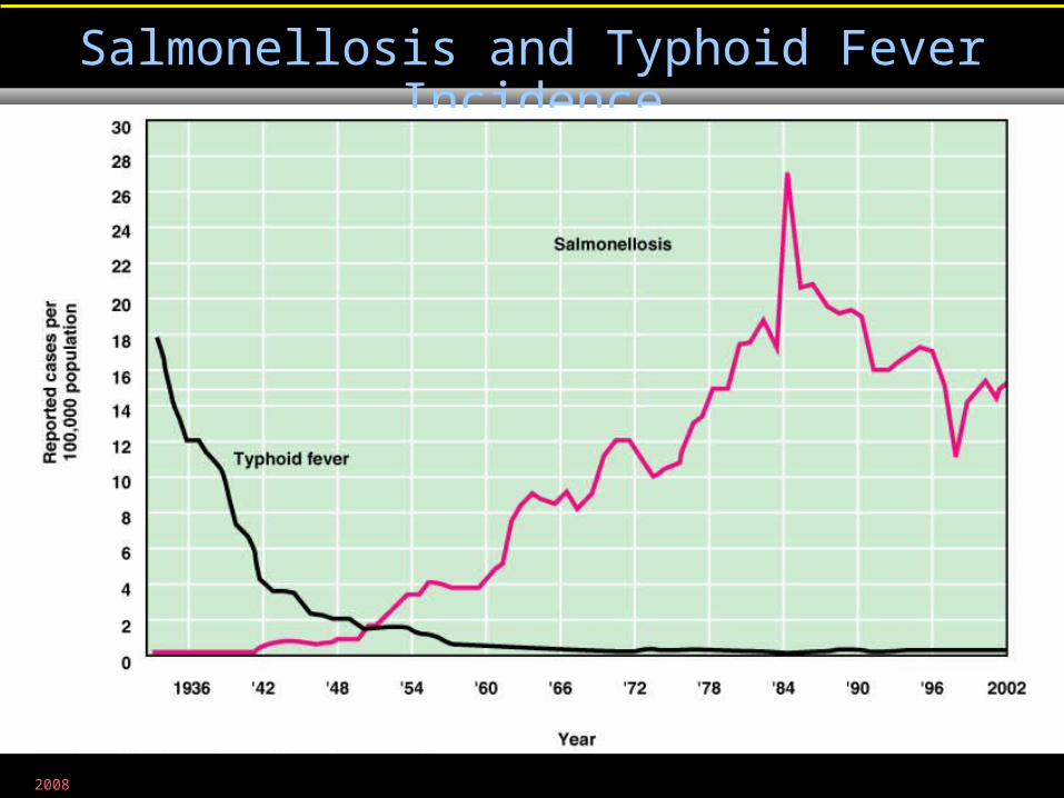
Salmonellosis and Typhoid Fever Incidence
Figure 25.10
2008
• Salmonella enterica Typhi
• Bacteria spread throughout body in phagocytes
• 1-3% recovered patients become carriers, harboring Salmonella in their gallbladder
Typhoid Fever
2008
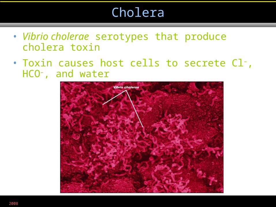
Cholera
Figure 25.12
• Vibrio cholerae serotypes that produce cholera toxin
• Toxin causes host cells to secrete Cl–, HCO–, and water
2008
• Usually from contaminated crustaceans or mollusks
• V. cholerae serotypes other than O:1, O:139, and eltor
• V. parahaemolyticus
• V. vulnificus
Noncholera Vibrios
2008
• Occurs as traveler's diarrhea and epidemic diarrhea in nurseries
• 50% of feedlot cattle may have enterohemorrhagic strains in their intestines
• Enterohemorrhagic strains such as E. coli O157:H7 produce Shiga toxin
• O = cell wall antigen
• H = flagellar antigen
Escherichia coli Gastroenteritis
2008
• Campylobacter jejuni
• Usually transmitted in cow's milk
Campylobacter Gastroenteritis
2008
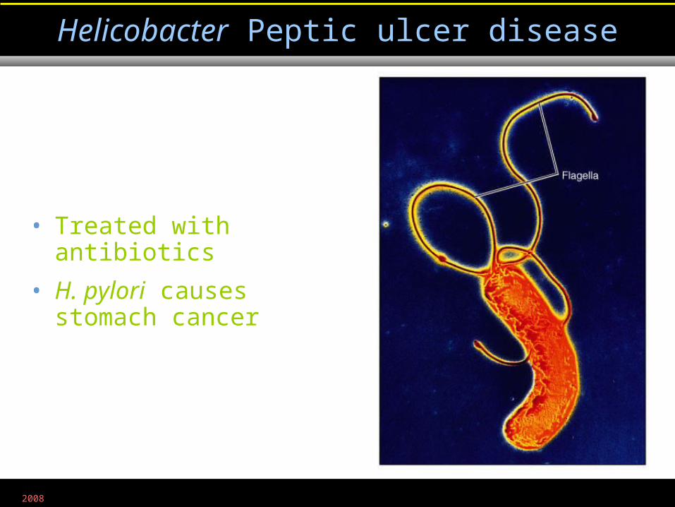
Helicobacter Peptic ulcer disease
• Treated with antibiotics
• H. pylori causes stomach cancer
Figure 11.11
2008
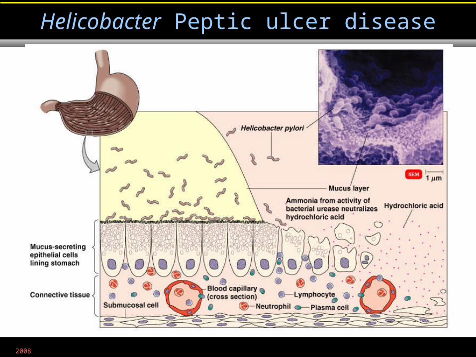
Helicobacter Peptic ulcer disease
Figure 25.13
2008
• Y. enterocolitica and Y. pseudotuberculosis
• Can reproduce at 4°C
• Usually transmitted in meat and milk
Yersinia Gastroenteritis
2008
• Grow in intestinal tract producing exotoxin
Clostridium perfringens Gastroenteritis
2008
• Ingestion of bacterial exotoxin produces mild symptoms
Bacillus cereus Gastroenteritis
2008
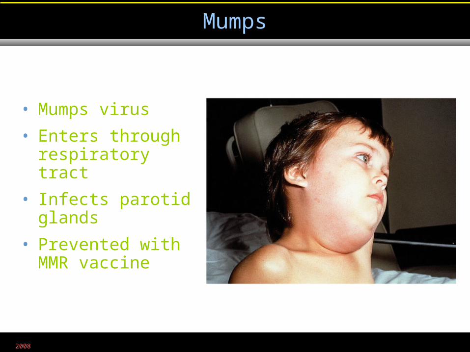
Mumps
Figure 25.14
• Mumps virus
• Enters through respiratory tract
• Infects parotid glands
• Prevented with MMR vaccine
2008
• Inflammation of the liver
• Hepatitis may result from drug or chemical toxicity, EB virus, CMV, or the Hepatitis viruses
Hepatitis
2008
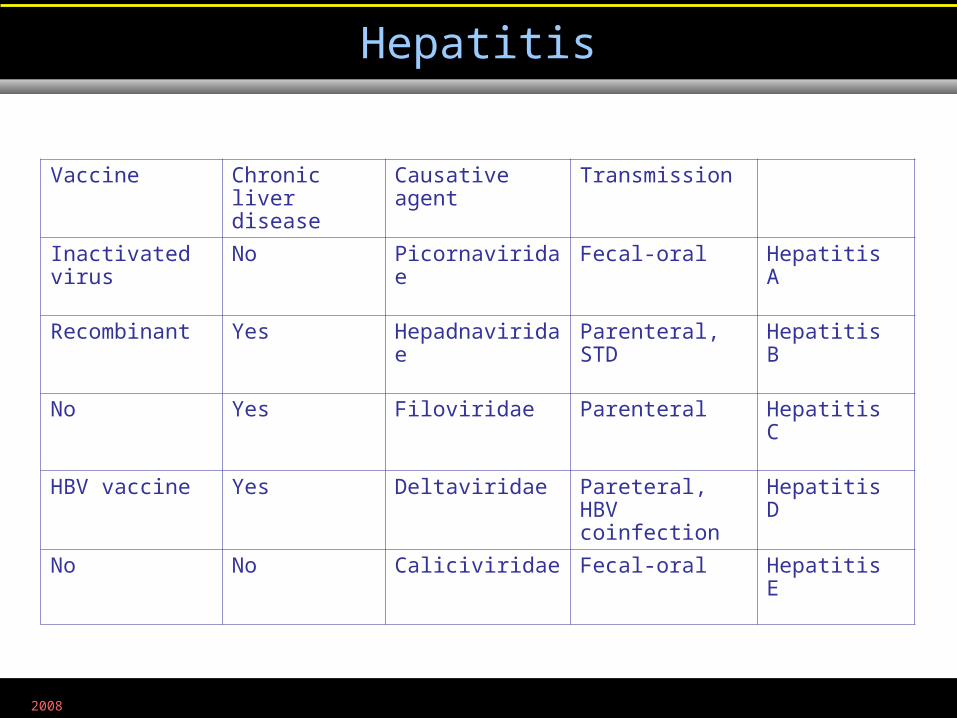
Hepatitis
Table 25.1
TransmissionCausative agentChronic liver disease
Vaccine
Hepatitis AFecal-oralPicornaviridaeNoInactivated virus
Hepatitis BParenteral, STDHepadnaviridaeYesRecombinant
Hepatitis CParenteralFiloviridaeYesNo
Hepatitis DPareteral, HBV coinfection
DeltaviridaeYesHBV vaccine
Hepatitis EFecal-oralCaliciviridaeNoNo
2008
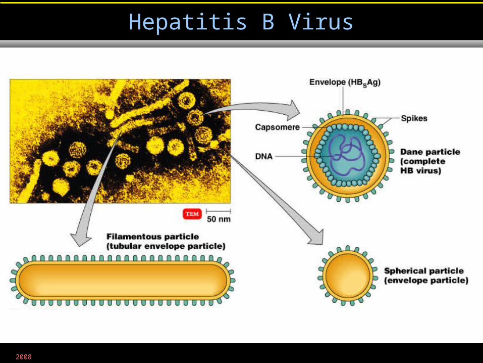
Hepatitis B Virus
Figure 25.15
2008
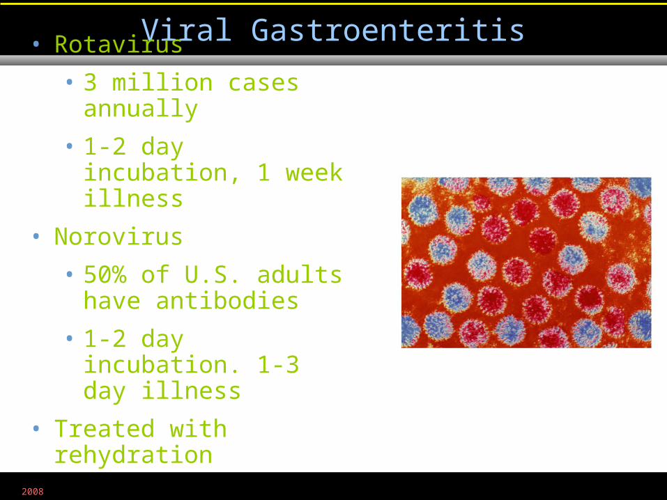
Viral Gastroenteritis
• Rotavirus
• 3 million cases annually
• 1-2 day incubation, 1 week illness
• Norovirus
• 50% of U.S. adults have antibodies
• 1-2 day incubation. 1-3 day illness
• Treated with rehydration
Figure 25.17
2008
• Mycotoxins are produced by some fungi:
• Claviceps purpurea
• Grows on grains
• Produces ergot
• Toxin restricts blood flow to limbs; causes hallucination
• Aspergillus flavus
• Grows on grains
• Produces aflatoxin
• Toxin causes liver damage; liver cancer
Mycotoxins
2008
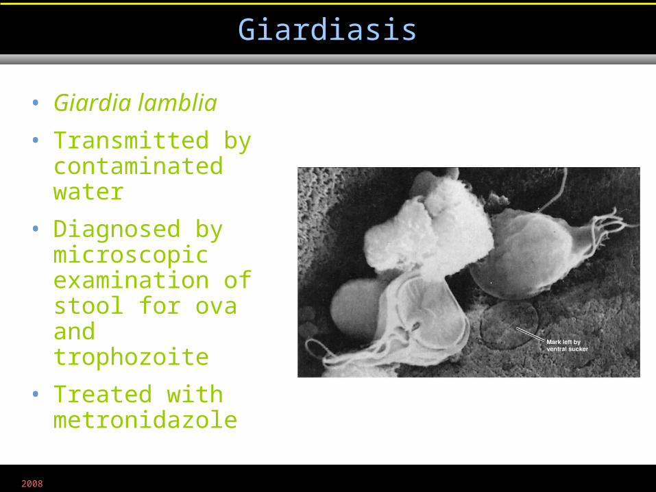
Giardiasis
Figure 25.18
• Giardia lamblia
• Transmitted by contaminated water
• Diagnosed by microscopic examination of stool for ova and trophozoite
• Treated with metronidazole
2008
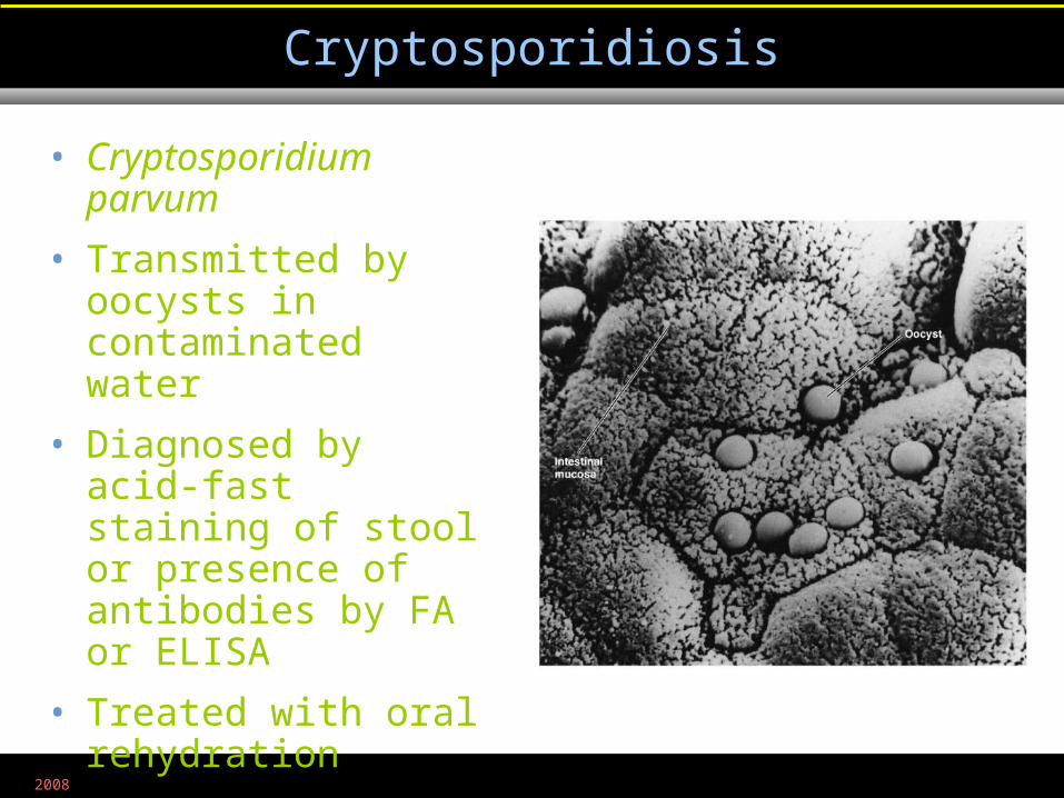
Cryptosporidiosis
Figure 25.19
• Cryptosporidium parvum
• Transmitted by oocysts in contaminated water
• Diagnosed by acid-fast staining of stool or presence of antibodies by FA or ELISA
• Treated with oral rehydration
2008
• Cyclospora cayetanensis
• Transmitted by oocysts in contaminated water
• Diagnosed by microscopic examination for oocysts
• Treated with trimethoprim and sulfamethoxazole
Cyclospora Diarrheal Infection
2008
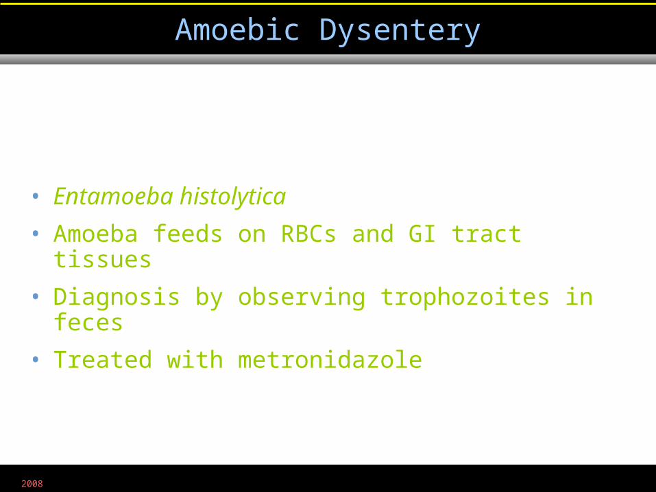
Amoebic Dysentery
• Entamoeba histolytica
• Amoeba feeds on RBCs and GI tract tissues
• Diagnosis by observing trophozoites in feces
• Treated with metronidazole
2008
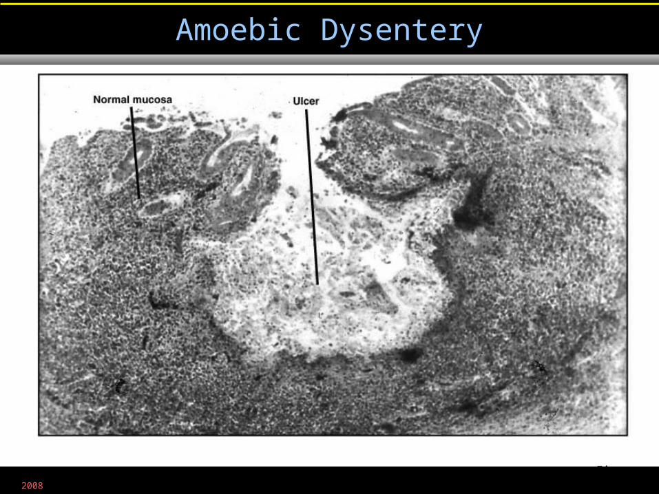
Amoebic Dysentery
Figure 25.20
2008
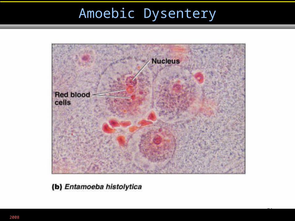
Amoebic Dysentery
Figure 12.18b
Medical Technology Department, Faculty of Science, Islamic University-Gaza
MMICROBBIOLOGY
Dr. Abdelraouf A. ElmanamaDr. Abdelraouf A. ElmanamaPh. D MicrobiologyPh. D Microbiology
2008
Chapter 25Chapter 25Microbial Diseases of the Digestive SystemMicrobial Diseases of the Digestive System
2008
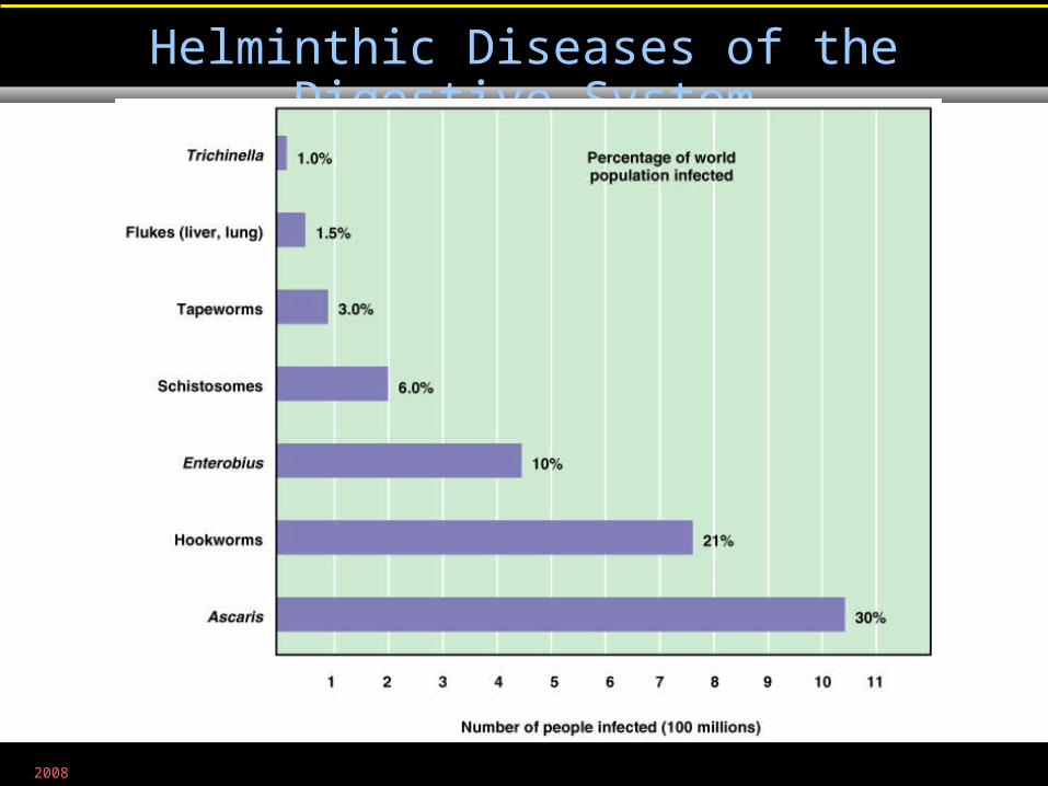
Helminthic Diseases of the Digestive System
Figure 25.21
2008
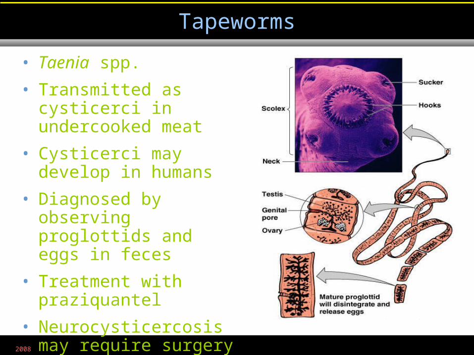
Tapeworms
Figure 12.27
• Taenia spp.
• Transmitted as cysticerci in undercooked meat
• Cysticerci may develop in humans
• Diagnosed by observing proglottids and eggs in feces
• Treatment with praziquantel
• Neurocysticercosis may require surgery
2008
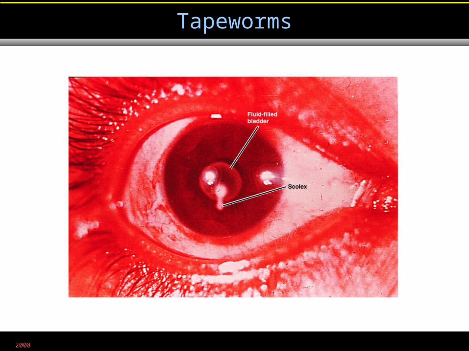
Tapeworms
Figure 25.22
2008
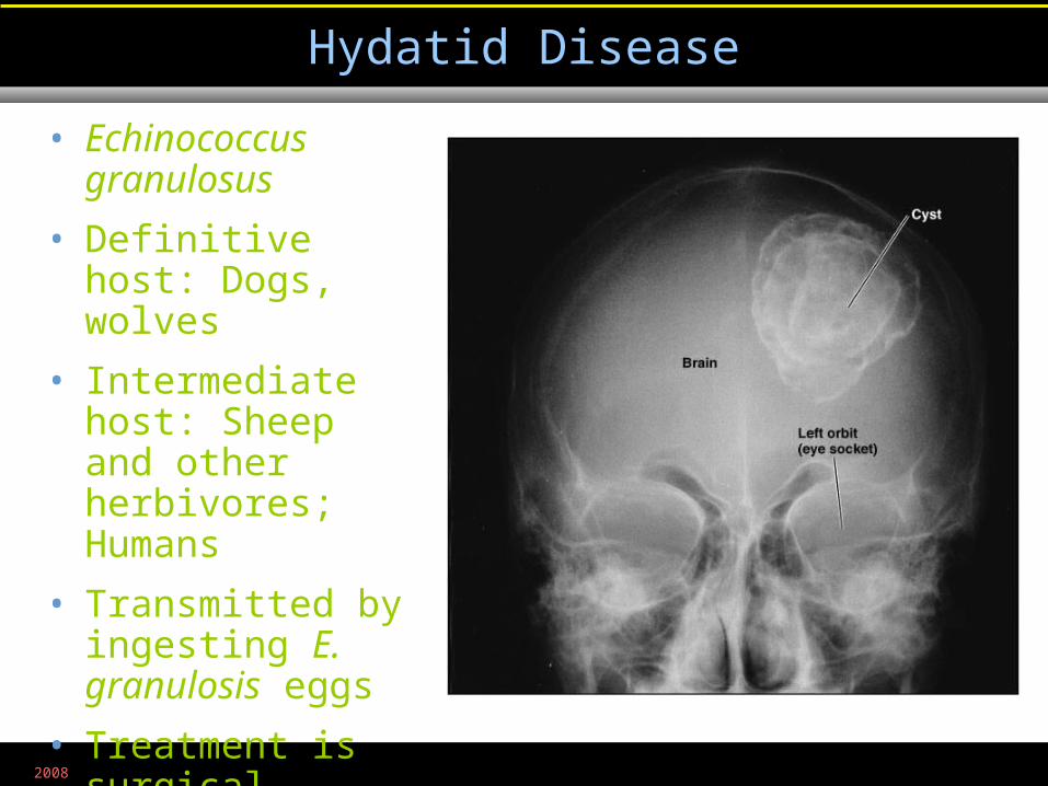
Hydatid Disease
Figure 25.23
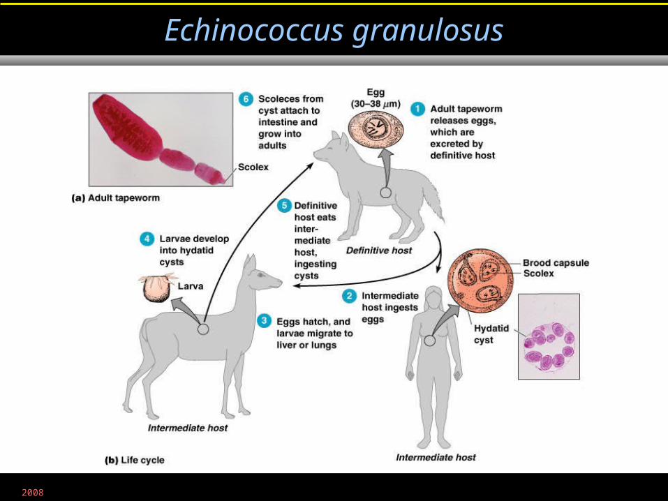
• Echinococcus granulosus
• Definitive host: Dogs, wolves
• Intermediate host: Sheep and other herbivores; Humans
• Transmitted by ingesting E. granulosis eggs
• Treatment is surgical
2008
Echinococcus granulosus
Figure 12.28
2008
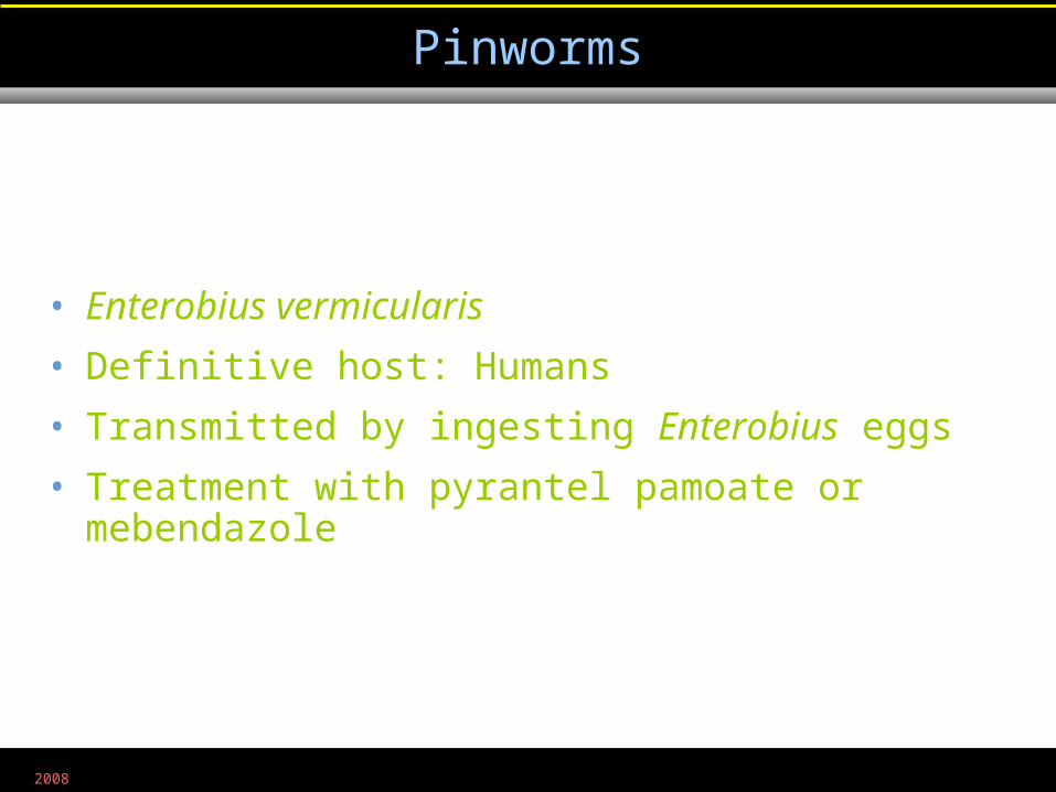
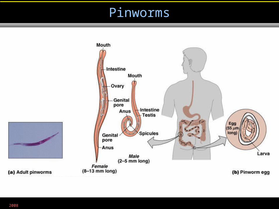
Pinworms
• Enterobius vermicularis
• Definitive host: Humans
• Transmitted by ingesting Enterobius eggs
• Treatment with pyrantel pamoate or mebendazole
2008
Pinworms
Figure 12.29
2008
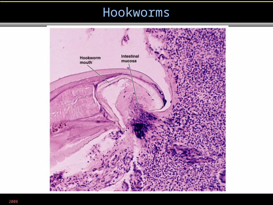
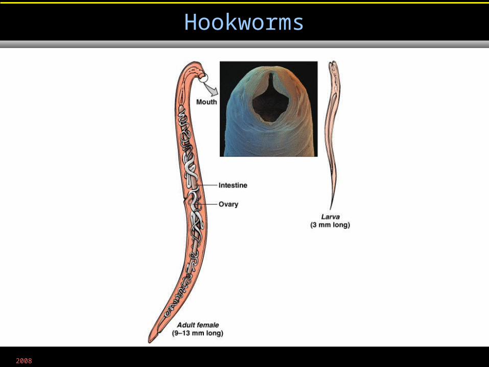
Hookworms
• Larvae in soil hatched from eggs shed in feces
• Larvae bore through skin; migrate to intestine
• Treated with mebendazole
2008
Hookworms
Figure 25.24
2008
Hookworms
Figure 12.30
2008
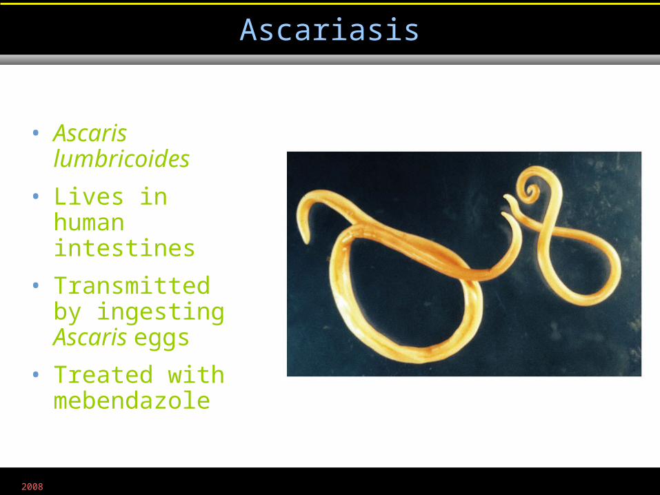
Ascariasis
Figure 25.25
• Ascaris lumbricoides
• Lives in human intestines
• Transmitted by ingesting Ascaris eggs
• Treated with mebendazole
2008
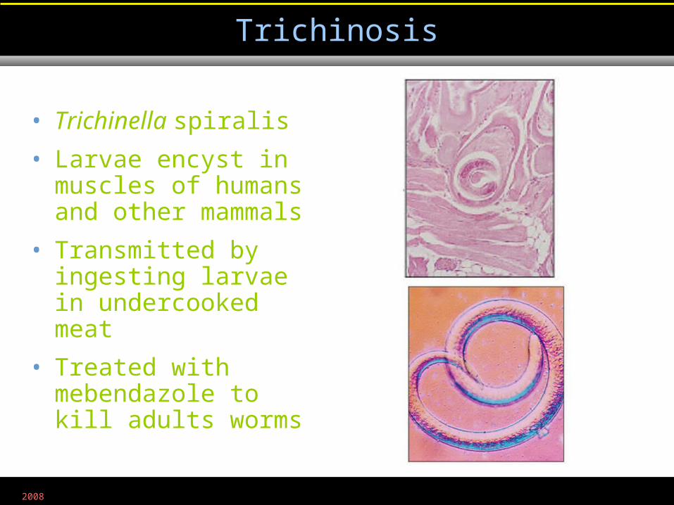
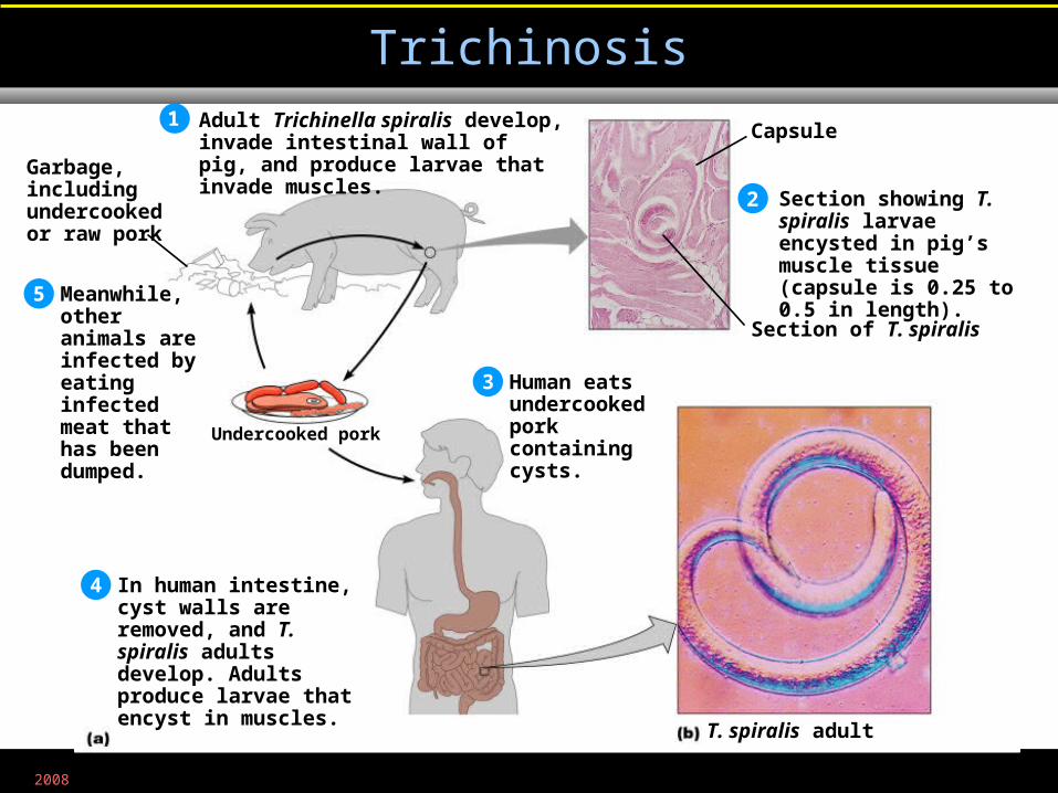
Trichinosis
Figure 25.26
• Trichinella spiralis
• Larvae encyst in muscles of humans and other mammals
• Transmitted by ingesting larvae in undercooked meat
• Treated with mebendazole to kill adults worms
2008
Trichinosis
Figure 25.26
Adult Trichinella spiralis develop, invade intestinal wall of pig, and produce larvae that invade muscles.
Section showing T. spiralis larvae encysted in pig’s muscle tissue (capsule is 0.25 to 0.5 in length).
Human eats undercooked pork containing cysts.
1
2
3
In human intestine, cyst walls are removed, and T. spiralis adults develop. Adults produce larvae that encyst in muscles.
4
Meanwhile, other animals are infected by eating infected meat that has been dumped.
5
Capsule
Section of T. spiralis
Undercooked pork
Garbage, including undercooked or raw pork
T. spiralis adult