Embed Size (px)
Citation preview

The Islamic University-Gaza
Deanship of Graduate Studies
Biological Sciences Master Program
Medical Technology
Facutly of Science
Risk Factors Associated with Helicobacter pylori Infection in
Gaza, Palestine
By
Rana M. Abu-Mugesieb
Supervisor
Dr. Abdelraouf A. Elmanama Dr. Mofeed M. Mokhallalati
Submitted in Partial Fulfillment for the Master Degree of Science in
Biological Science-Medical Technology
June 2007

i
Declaration
“I herby declare that this submission is my own work and that, to the best of
my knowledge and belief, it contains no material previously published or
written by another nor material which to a substantial extent has been
accepted for the award of any other higher degree or diploma of the
university or other institute of higher learning, except where due
acknowledgement is made in the text"
Author
Rana Mohammed Abu-Mughesieb
Signature: Rana Date: May- 2007
.
All Rights Reserved © 2007. No part of this work can be copied, translated
or stored in retrieval system, without prior permission of the author.
ii
Abstract
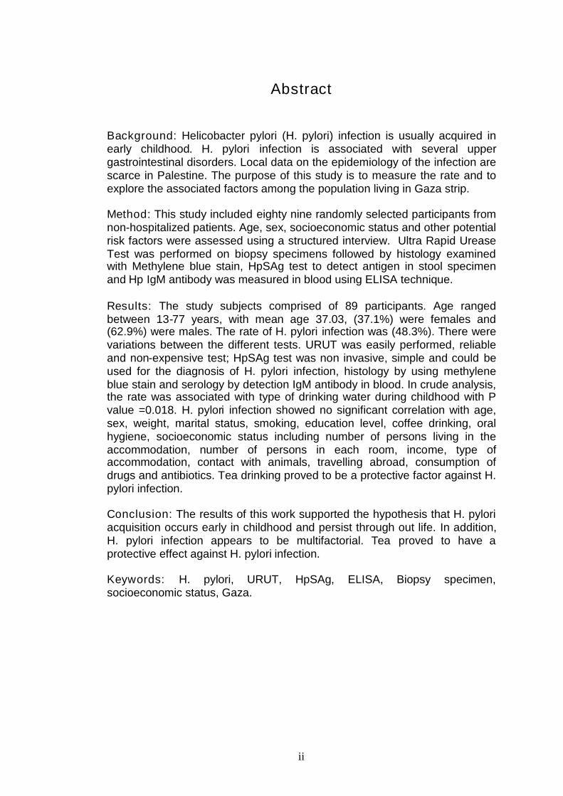
Background: Helicobacter pylori (H. pylori) infection is usually acquired inearly childhood. H. pylori infection is associated with several uppergastrointestinal disorders. Local data on the epidemiology of the infection arescarce in Palestine. The purpose of this study is to measure the rate and toexplore the associated factors among the population living in Gaza strip.
Method: This study included eighty nine randomly selected participants fromnon-hospitalized patients. Age, sex, socioeconomic status and other potentialrisk factors were assessed using a structured interview. Ultra Rapid UreaseTest was performed on biopsy specimens followed by histology examinedwith Methylene blue stain, HpSAg test to detect antigen in stool specimenand Hp IgM antibody was measured in blood using ELISA technique.
Results: The study subjects comprised of 89 participants. Age rangedbetween 13-77 years, with mean age 37.03, (37.1%) were females and(62.9%) were males. The rate of H. pylori infection was (48.3%). There werevariations between the different tests. URUT was easily performed, reliableand non-expensive test; HpSAg test was non invasive, simple and could beused for the diagnosis of H. pylori infection, histology by using methyleneblue stain and serology by detection IgM antibody in blood. In crude analysis,the rate was associated with type of drinking water during childhood with Pvalue =0.018. H. pylori infection showed no significant correlation with age,sex, weight, marital status, smoking, education level, coffee drinking, oralhygiene, socioeconomic status including number of persons living in theaccommodation, number of persons in each room, income, type ofaccommodation, contact with animals, travelling abroad, consumption ofdrugs and antibiotics. Tea drinking proved to be a protective factor against H.pylori infection.
Conclusion: The results of this work supported the hypothesis that H. pyloriacquisition occurs early in childhood and persist through out life. In addition,H. pylori infection appears to be multifactorial. Tea proved to have aprotective effect against H. pylori infection.
Keywords: H. pylori, URUT, HpSAg, ELISA, Biopsy specimen,socioeconomic status, Gaza.

iii
باللغة العربیةمستخلص
Helicobacter)(العدوى ببكتیریا :تمھید pyloriھذه ، ما تكتسب في الطفولة المبكرة عادًة
ال یوجد .العدوى یصاحبھا مجموعة من األمراض في الجزء العلوي من الجھاز الھضمي
ن ھذه الغرض م.معلومات وبائیة عن ھذه البكتریا واألمراض التي قد تصاحبھا في فلسطین
.الدراسة تحدید عوامل الخطر التي قد تزید من اإلصابة بھذه البكتیریا
ض عشوائیا من عدة مستشفیات في قطاع 89في ھذه الدراسة تم اختیار :البحثطریقة مری
تاریخھ المرضي باإلضافة لعدة ، نظام حیاتھ، عمربالتتعلق كل مریض خضع لعدة أسئلة .غزة
IgM عینات من كل مریض عینة دم لفحص 3تم أخذ .صاديأسئلة تشمل الوضع االقت
antibodyعینة براز لفحص ، في الدمAntigenوعینة خزعة من المعدة أخذت بواسطة
Methyleneوفحص نسیجي باستخدام صبغة URUTمختص بجھاز المنظار للقیام بفحص
Blue.
-13مرضى تتراوح ما بین مریض كانت أعمار ال89 في ھذه الدراسة التي شملت :النتائج
الرجال كانت نسبةبینما%37.1نسبة النساء كانت . سنة37 سنة ومعدل العمر 77
.Hنسبة انتشار ، 62.9% pylori)48.3(% ،من .ھناك اختالف ما بین الفحوصات األربع
اء أھم النتائج في ھذه الدراسة وجود عالقة مھمة بین اإلصابة بھذه البكتیریا ومصادر شرب الم
نوع ، لم یكن ھناك أي عالقة مھمة ما بین اإلصابة بالبكتیریا والعمر.في مرحلة الطفولة
التدخین ، أنواع األدویة التي یستخدمھا، عدد أفراد عائلتھ، الوضع االقتصادي للمریض، الجنس
كعامل حمایة من من أھم النتائج في ھذه الدراسة وجد أن شرب الشاي یعمل .و شرب القھوة
.تیریاالبك
.H(النتائج من ھذه الدراسة أثبتت أن اإلصابة ببكتریا :االستنتاج pylori( تكون في مرحلة
كما استنتج من ھذه الدراسة فائدة .الطفولة مرتبطة بعوامل خطر مثل مصادر شرب الماء
.لبكتیریاابھذهلالصابةالشاي كمضاد

iv
Dedication
This thesis is dedicated to my father Dr. Mohammed, my Mother and my
brothers, Ahmed, Sameh and my sister Sawsan who supported me all
the way since the beginning of my study and always encouraged me to
pursue my ambitions and my goals.
Finally, this thesis is dedicated to all those who believe in the richness of
learning.

v
Acknowledgment
First I would like to thank Allah for helping me to finish this thesis.
I would in particular like to thank:
Dr. Abdelraouf A. Elmanama and Dr. Mofeed M. Mokhallalati my
supervisors, for sharing their skills in the world of research and always
making time in their busy schedule when needed. I have been especially
fascinated by, and learnt from, their remarkable ability to always move
forward.
I also would like to thank Dr. Ahmed Shahwan, Dr. Mohamed Adwan, Dr.
Abdul-latif Alhaj, Dr. Mohammed Abu-humied, Dr. Abdul-Aziz Alfarra, Dr.
Eiad Al-jabri, Dr. Yousef Moa’amer, Dr. Samir Al-Ghazali , Dr. Majed
Hassona at Gaza hospitals for their cooperation. Kamal Huliel, Nahed Al-
Halabi, Rehab Al-Najar and Yaser for facilitating sample collection and their
advising during my work.
Thanks to Al-Amal lab supervisor; Yaser Hamdona and Mohamed for
helping and allowing me to perform some of my laboratory work in their lab.
Computer engineer Mahmood Al-azbat who helped me in my work on SPSS
software.
Thanks to the Deanship of science and research at the Islamic University-
Gaza for their support and generosity through its grants to the faculty
members in the University.
Thanks to the laboratory staff of the Islamic University-Gaza for allowing
me open access to the laboratory and facilities. Lastly but not least I would
like to thank my friends and family for all the help and support and for
keeping me aware of the fact that there is life out- side of work.
vi
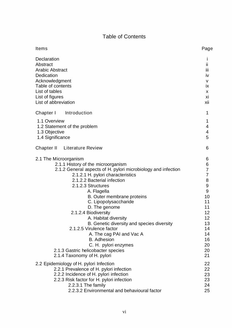
Table of Contents
Items Page
Declaration iAbstractArabic Abstract
iiiii
Dedication ivAcknowledgment vTable of contents ixList of tables xList of figures xiList of abbreviation xii
Chapter I Introduction 1
1.1 Overview 11.2 Statement of the problem 41.3 Objective 41.4 Significance 5
Chapter II Literature Review 6
2.1 The Microorganism 62.1.1 History of the microorganism 62.1.2 General aspects of H. pylori microbiology and infection 7
2.1.2.1 H. pylori characteristics 72.1.2.2 Bacterial infection 82.1.2.3 Structures 9
A. Flagella 9B. Outer membrane proteins 10C. Lipopolysaccharide 11D. The genome 11
2.1.2.4 Biodiversity 12A. Habitat diversity 12B. Genetic diversity and species diversity 13
2.1.2.5 Virulence factor 14A. The cag PAI and Vac A 14B. Adhesion 16C. H. pylori enzymes 20
2.1.3 Gastric helicobacter species2.1.4 Taxonomy of H. pylori
2021
2.2 Epidemiology of H. pylori Infection 222.2.1 Prevalence of H. pylori infection 222.2.2 Incidence of H. pylori infection 232.2.3 Risk factor for H. pylori infection 23
2.2.3.1 The family 242.2.3.2 Environmental and behavioural factor 25
vii
2.2.3.3 Host and bacterial factor 262.2.4 Transmission of H. pylori infection 28
2.2.4.1 Fecal oral and gastro-oral routs 282.2.4.2 Oral-oral rout 292.2.4.3 Environmental source: water and food 302.2.4.4 Animal reservoir 312.2.4.5 Intra familial transmission 32
2.3 Clinical Manifestation of H. pylori Infection 332.3.1 Natural course of H. pylori infection 332.3.2 Gastritis 342.3.3 Peptic ulcer disease 342.3.4 Gastric cancer 342.3.5 Other H. pylori associated condition 352.3.6 Extra gastric manifestation of H. pylori in children 36
2.3.6.1 Iron deficiency anemia 362.3.6.2 Atopic disease 362.3.6.3 Acute intestinal infection 372.3.6.4 Diminished growth 37
2.3.7 Barrette’s esophagus 382.3.8 Gastresophagal reflux2.3.9 Idiopathic thrombocytopenic purpura
3839
2.4 Diagnostic Tests for Detection of H. pylori Infection 392.4.1 Biopsy-based test 392.4.2 Tests based on detection of specific anti H. pylori antibodies 402.4.3 Urea breath test 422.4.4 Test based on detection of bacterial antigen or bacterial DNA
in feces42
2.5 Treatment of H. pylori Infection 432.5.1 Indication of treatment 432.5.2 Combined antibacterial regimens against H. pylori infection 452.5.3 Factor influencing treatment results 462.5.4 Alternative for treatment of H. pylori infection 472.5.5 Recurrence of H. pylori infection after eradication 482.5.6 Prevention of H. pylori associated disease 49
Chapter III Materials and Methods 52
3.1 Material and Reagents 523.2 Permission and Ethical Consideration 533.3 Patients 533.4 Sample Size 533.5 Sample Collection 53
3.5.1 Gastric biopsy 533.5.2 Blood sample for serological evaluation 553.5.3 Stool sample for antigen detection 55
3.6 Sample Processing 553.6.1 Blood sample 55
3.6.1.1 Principle of the ELISA test 55
viii
3.6.1.2 Assay procedure 563.6.1.3 Calculation of result 56
3.6.2 Biopsy specimen 573.6.2.1 Ultra rapid urease test (URUT) 573.6.2.2 Microscopy 57
3.6.3 Stool sample 583.6.3.1 Kit components 583.6.3.2 Pre assay control and operations 583.6.3.3 Assay procedure (Quantitative Assay ) 603.6.3.4 Calculation of result 60
3.7 Questionnaire 603.8 Analysis of Data 61
Chapter IV Results 62
4.1 Study Sample Description 624.2 Ultra Rapid Urease Test 634.3 Gastric biopsies Stained with Methylene Blue 634.4 H. pylori Serum IgM 644.5 H. pylori Antigen Detection From Stool 644.6 True Positive for H. pylori Infection. 654.7 Statistical Analysis of H. pylori Tests Results (Chi square) 664.8 H. pylori Infection among the Study Sample 694.9 Risk Factors For H. pylori Infection. 69
4.9.1 Personal variables 704.9.2 Life style variables 714.9.3 Drug history 734.9.4 Antibiotics intake during the last month 734.9.5 Socioeconomic status 74
Chapter V Discussion 76
5.1 H. pylori tests 775.1.1 Evaluation of H. pylori Tests5.1.2 The rate of H. pylori infection
7780
5.2 Risk Factors Associated with H. pylori infection 805.2.1 Age 805.2.2 Sex 815.2.3 Weight 815.2.4 Marital status 825.2.5 Smoking 825.2.6 Coffee drinking 835.2.7 Tea drinking 835.2.8 Type of drinking water 845.2.9 Oral hygiene 855.2.10 Antibiotic consumption 865.2.11 Drugs consumption 865.2.12 Education 87
ix
5.2.13 Income 885.2.14 Number of persons in the accommodation 885.2.15 Type of accommodation 89
Chapter VI Conclusion and Recommendations 90
References 92
Appendices 111
Appendix A: Questionnaire form in English 112Appendix B: Questionnaire form in Arabic 114
x
List of tables
Table (2.1)…………………………………………………………………………………….Human and land animal gastric Helicobacter
21
Table (4.1)…………………………………………………………………………………….Age and sex distribution of the study sample
62
Table (4.2)…………………………………………………………………………………….Distribution of positive and negative results in each of the four tests used
65
Table (4.3)…………………………………………………………………………………….True H. pylori positive
65
Table (4.4)…………………………………………………………………………………….Chi square test for statistical differences between the results of Methyleneblue stain and URUT
66
Table (4.5) ……………………………………………………………………………………Chi square test for statistical differences between the results ofMethylene blue stain and IgM in serum
66
Table (4.6)……………………………………………………............................................Chi square test for statistical differences between the results ofMethylene blue stain and HpSAg
67
Table (4.7) ……………………………………………………………………………………Chi square test for statistical differences between the results of URUTand HpSAg
67
Table (4.8) ……………………………………………………………………………………Chi square test for statistical differences between the results of IgM andURUT
67
Table (4.9) ……………………………………………………………………………………Chi square test for statistical differences between the results of IgM andHpSAg
68
Table (4.10)…………………………………………………………………………………..Pearson correlation between the different H. pylori tests
68
Table (4.11)…………………………………………………………………………………..Age distribution of H. pylori positive subjects
69
Table (4.12) ………………………………………………………………………………….Personal factor
70
Table (4.13)…………………………………………………………………………………..Life style variables
72
Table (4.14)…………………………………………………………………………………..Drug intake
73
Table (4.15)…………………………………………………………………………………..Antibiotics intake
73
Table (4.16)…………………………………………………………………………………..Effects of socioeconomic status
75
xi
List of Figure
Figure (2.1)…………………………………………………………………………………...H. pylori; The curved bacillus with unipolar flagella is visualized by ascanning electron microscope and depicted in a schematic drawing
8
Figure (2.2)…………………………………………………………………………………...Adhesion of Helicobacter pylori to gastric epithelial cells induces amultitude of changes to the host cell, the most common of which areshown in the diagram
17
Figure (3.1)…………………………………………………………………………………...Olympus Video trolley tv-z CLE-10 machine
54
Figure (3.2)…………………………………………………………………………………...Biopsy collection
54
Figure (3.3)…………………………………………………………………………………...Schematic presentation of the HpSAg detection procedures
59
Figure (4.1)…………………………………………………………………………………...Ultra rapid urease test
63
Figure (4.2)…………………………………………………………………………………...Methylene blue stained gastric biopsies showing the helical bacilli
64

xii
List of abbreviation
ALeb Blood group A derivative of Lewis b.BabA Blood group antigen binding A adhesion.BabB Blood group antigen binding B adhesionBE Barrett’s EsophagusBIC Bovine Immune ColostrumBLeb blood group B derivative of Lewis b.Cag A Cytotoxin-Associated GeneCag A Cytotoxin –Associated Protein.cag-PAI cag-Pathogenicity Island.CFU Colony Forming UnitCGA Cultured gastric adenocarcinomaDU Duodenal UlcerELISA Enzyme-linked immunosorbent assayESPCG European Society of Primary Care GastroenterologyG-C ratio Guanine-Cytosine ratioGERD Gastroesophageal Reflux DiseaseGI Gastrointestinal tractHpSAg Helicobacter pylori Stool Antigen detection kitH. pylori Helicobacter pyloriHLA-DQA1 Human Leukocyte Antigen LocusHP-NAP H. pylori Neutrophil-Activating ProteinIARCIgM, A,G
International Agency for Research on CancerImmunoglobulin M, A, G
ICAM Intercellular Adhesion MoleculeIL InterleukinLe b, x, y Lewis blood group antigen b, x and y.LPS LipopolysaccharideLT Labile ToxinMALT Mucosa Associated Lymphoid TissueMB Methylene BlueNADPH Nicotinamide Adenine Dinucleotide PhosphateNASPGN North American Society for Pediatric GastroenterologyNF_B Nuclear Factor_B.NIHNIS
National Institute of HealthNew Israel Shekel
NSAIDS Non Steroidal Anti Inflammatory DrugsOMP Outer Membrane ProteinsPAI Pathogenicity IslandPCR Polymerase Chain ReactionPMNs Polymorphic Nucleated CellsPPI Proton Pump Inhibitor

xiii
ROS Reactive Oxygen Species.SabA Sialic Acid Binding Adhesin.SES Socioeconomic StatusSib-SibsLea
Sibling-SiblingSialyl-Lewis a Antigen
sLex Sialyl-Lewis x Antigen.Th T helper cellTMB Tetra Methyl BenzidineTLR Toll-like receptorsUBT Urea Breath TestUrel Urea ChannelsURUT Ultra Rapid Urease TestUTIs Urinary Tract InfectionsVac A Vacuolating Cytotoxin GeneVac A Vacuolating Cytotoxin

Chapter IIntroduction

1
Chapter 1
Introduction
1.1 Overview
Barry Marshall and Robin Warren of Perth, Western Australia discovered H.
pylori in 1983. Originally, the organism was named Campylobacter pylori
because it was structurally similar to other Campylobacter species, such as
C. jejuni (1). Signs of H. pylori infection such as gram-negative gastric bacilli,
gastric urease and epidemics of hypochlorhydria have been described since
the late nineteenth century (2). These observations could be better explained
after Warren and Marshall in the early 1980’s managed to culture a
bacterium that was to be designated Campylobacter pyloridis (3). In 1989,
the genus Helicobacter was created and the bacterium received the name H.
pylori(4).
H. pylori is a small (0.5-1.0 μm in width and 2.5 to 5.0 μm in length), spiral
shaped, highly motile, gram negative rod with 4-6 unipolar sheathed flagella
(5). The microorganism grows slowly in vitro and requires special media and
a microaerophilic (5% O2, 10% CO2, and 85% N2) environment (6). The most
striking biochemical characteristic is the production of large quantities of
urease. This enzyme digests urea to produce carbon dioxide and ammonia.
In the presence of water this leads to the formation of ammonium hydroxide
(7). In this way, H. pylori is able to neutralize the acid in its direct
environment.
H. pylori, colonizes the human stomach, can cause gastritis, is strongly
associated with gastric and duodenal ulceration (DU) and has been
implicated in the causation of gastric carcinoma and mucosa-associated
lymphoid tissue (MALT) lymphomas. It has been reported that there is
relationship between H. pylori infection and children’s gastroenterological
diseases (8). However, only 10-20% of infected individuals manifests severe

2
complications and this selectivity in disease progression is inadequately
understood (9, 10). Epidemiological studies have shown that a weakly
positive correlation exists between chronic gastric infection with H. pylori and
coronary heart disease (11).
Urease, vacuolating cytotoxin Vac A, and the pathogenicity island (cag PAI)
gene products, are the main factors of virulence of this organism. H. pylori
LPS may have an important role in autoimmune-mediated damage in the
gastric mucosa (12).
Half of the world’s population is estimated to be infected with H. pylori, which
makes it one of the most common bacterial pathogens in humans (13). The
prevalence of H. pylori infection worldwide is approximately 50%, it reaches
as high as 80%–90% in developing countries, and about 35%–40% in the
United States (14). Approximately 20% of persons infected with H. pylori
develop related gastroduodenal disorders during their lifetime (14). The
annual incidence of H. pylori infection is about 4%–15% in developing
countries, compared with approximately 0.5% in industrialized countries (15).
Documented risk factors include low socioeconomic status, overcrowding,
poor sanitation or hygiene, and living in a developing country (16).
In "Israel", the prevalence of H. pylori infection is about 60%, and the annual
incidence of gastric cancer is about 15 per 100,000 populations (17). The
prevalence of H. pylori infection was 10% among children in Egypt (18). In
crude analyses, prevalence was associated with increasing age, non-white
skin color, lower current family income, lower education level, higher size of
the family, low socio-economic conditions in childhood, higher number of
siblings and attendance to day-care centers in childhood, and presence of
dyspeptic symptoms. Socioeconomic conditions in childhood besides
ethnicity and presence of dyspeptic symptoms were the factors significantly
associated with the infection (19).

3
There are several methods of diagnosing H. pylori infection including invasive
procedures using mucosa biopsies taken during endoscopy (mainly culture,
histology and the rapid urease test) and noninvasive procedures (20).
Non-invasive testing methods for detection of H. pylori or confirmation of
eradication include: 1) antibody tests (in serum, saliva or blood); 2) antigen
tests (in stools, saliva or urine); and 3) radioactive or non-radioactive urea
breath tests. The most interesting of the non-invasive tests is the detection of
antigens in stool samples by enzyme immunoassay technique. While this test
has good performance at a reasonable cost, doubts exist regarding patient
and clinician compliance and actual performance, particularly with regard to
inter laboratory variability. The urea breath test is based on analysis of
samples of exhaled air before and after ingestion of urea containing specially
labelled carbon (21).
PCR is a powerful technique for the detection of target DNA in various clinical
specimens, but its application to fecal specimens has been limited due to the
presence of substances inhibiting the reaction (22).
Eradication therapy of symptomatic H. pylori infection substantially reduces
the recurrence of associated gastroduodenal diseases. Therapy entails
complicated regimens of several antimicrobial agents for at least 2 weeks. In
general, triple therapy regimens usually entail two of the following
antimicrobial agents: metronidazole, amoxicillin, tetracycline, or
clarithromycin in combination with a proton pump inhibitor or bismuth salts
(23). The most common causes of treatment failure are patient
noncompliance and antimicrobial resistance of the infecting H. pylori strain
(24). Quadruple regimens are used as a salvage therapy when triple therapy
regimens have failed (23). However, the success of treatment is usually
dependent on early detection. Moreover, prevention of H. pylori infection
seems to be a wise strategy. Prevention strategies require deep
understanding of the transmission risks. H. pylori infection can be prevented

4
by interrupting the transmission of the infection by improving environment,
socioeconomic status and personal hygiene. It has been shown convincingly
that with the improvement of socioeconomic status in developed countries, H.
pylori infection has gone down by 50% over the last 3 decades (25).
Recently in a mouse model it has been demonstrated that oral immunization
with a crude lysate of H. felis induced protection against gastric infection by
H. pylori (26). Further studies have shown that superficial gastric ulcers in a
mouse model infected with H. pylori can be prevented by the administration
of a recombinant oral vaccine with E. coli heat labile toxin (LT) given as
adjuvant (27).
Despite the fact that Gaza is overcrowded, with poor sanitary conditions,
there are no previous studies or data concerning the prevalence of H. pylori
or the associated risk factors. Another vital issue regarding H. pylori
diagnosis, is the absence of many of the simple accurate tests in Gaza.
1.2 Statement of the Problem
H. pylori affects around 50% of the population in their lifetime, it is the
causative agent of serious disease and H. pylori classified as a class I
carcinogen. There is a need to examine the extent of the disease in Gaza city
and to study the risk factors associated with H. pylori infection and to
evaluate various diagnostic approaches.
1.3 Objectives
The general aim of this study is to evaluate risk factors associated with H.
pylori infection in Gaza, Palestine.
The following specific objectives were achieved:
1. The rate of H. pylori infection among the target population was estimated.
2. The performance of H. pylori detection techniques was evaluated.

5
1.4 Significance
The rate among middle-aged adults is over 80 percent in many developing
countries, as compared with 20 to 50 percent in industrialized countries (28).
H. pylori infection plays a role in the development of chronic gastritis, peptic
ulcer and gastric cancer (29).
The local epidemiological data on H. pylori infection are scarce and there is
no research done in Gaza on H. pylori. It is expected that data generated
from this work would provide an insight on the risk factors associated with H.
pylori infection. This may contribute to reducing the incidence of such
infections.

Chapter IILiterature Review

6
Chapter II
Literature Review
2.1 The Microorganism
2.1.1 History of H. pylori microorganism
The species Helicobacter was probably first observed by Bizzozero, an
Italian physiologist at the end of the 19th century. Bizzozero observed gram
negative, spiral shaped bacteria in the stomach of dogs. At around the same
time other medical professionals also reported presence of a spiral shaped
bacteria in upper gastrointestinal disorders. However these discoveries went
largely unnoticed. In 1938 an American pathologist discovered a spiral
shaped bacteria in 43 % of 242 subjects in an autopsy study, further it was
known that peptic ulcer disease responded well to treatment with bismuth
salts. Any further research into the findings was hindered by the lack of fresh
specimens of human gastric tissue as well as the fact that the newly
discovered bacteria could not be cultured. Thus, the findings soon fell into
oblivion (30).
In 1983, almost a century after it had first been discovered, two Australians,
Barry Marshal and Robin Warren noticed a gram negative, flagellated, spiral
shaped bacteria growing on agar plates containing human gastric biopsies,
that were accidentally left in an incubator over the Easter holidays. The close
physical resemblance of this new bacterium to that of the Campylobacter
species and the fact that peptic ulcers frequently occur in the pyloric gland
region of the stomach, led them to name it Campylobacter pyloridis. The
bacterium has undergone two name changes since, first to C. pylori and then
in 1989 the name was changed to the currently used Helicobacter pylori.
Early observations established a link between H. pylori and gastroduodenal

7
disease, such as peptic ulceration and gastritis. But it was not until Dr.
Marshall took it upon him self to fulfil Koch’s postulate by ingesting a liquid
culture of the bacteria that the link was proven. After ingesting the culture,
Marshall developed mild illness for 14 days; by day ten, gastritis had
developed which lasted for four days. The discovery of H. pylori and the
realization that it caused gastric ulcers earned the two Australians the 2005
Nobel Prize in physiology and medicine (30).
Helicobacter species belong to the epsilon sub-group of proteo-, or purple
bacteria. The group contains over thirty members of the Helicobacter species
colonizing a wide range of hosts (31). The species is diverse in morphology
but some features are shared among almost all members of the genus
Helicobacter, such as low (35-44 mol %) chromosomal guanine/ cytosine
(G/C) content, strong urease activity and the presence of sheathed flagella
(32). The fact that there is such a wide degree of morphological diversity and
host specificity suggest that the Helicobacter spp. is an old bacterium and
has co- evolved with its hosts for a long period of time (33).
2.1.2 General aspects of H. pylori microbiology and infection
2.1.2.1 H. pylori characteristic
H. pylori is a microaerophilic, gram negative, spiral shaped rod, between 2.5
and 4 μm in size, and under certain circumstances it can be U- shaped or
coccoid (34). H. pylori is actively motile using 4- 6 unipolar, sheathed flagella
(32). It resides naturally in the gastrointestinal tract of humans and non-
human primates.There is also evidence that it can infect pigs, cats, sheep
and pups (33, 35). In the stomach, the majority of H. pylori can be found in
the gastric mucosa; however a few are found adhered to the gastric mucosal
epithelium. The bacterium is highly adapted to survive in the hostile
environment of the stomach where few other organisms can survive.
Although H. pylori is considered to be a extra cellular bacteria, there is

8
evidence suggesting that the bacteria has a mechanism for intracellular
invasion (30).
H. pylori is the best known member of the Helicobacter genus, which
includes dozens of species that primarily colonize the gastrointestinal tract of
a variety of animals (33). H. pylori are a curved gram-negative bacillus with a
bundle of unipolar flagella (figure 2.1). Biochemical identification of H. pylori
relies on the activities of the urease, catalase and oxidase enzymes. The
bacterium is slow- growing and requires a rich medium and a microaerophilic
atmosphere for in vitro culture. After starvation through prolonged culturing, a
coccoid form can be found in the cultures and it has been debated whether
this form represents dormant or degenerated, non-viable bacteria (36).
Figure (2.1): H. pylori; The curved bacillus with unipolar flagella is visualized
by a scanning electron microscope (left) and depicted in a schematic drawing
(right)(37).
2.1.2.2 Bacterial infection
The human stomach is an inhospitable milieu and a fasting stomach is
normally devoid of bacterial species other than H. pylori and some
Lactobacilli. H. pylori is well adapted to its gastric niche and has developed a
broad spectrum of functions that enable colonization. For example, bacterial

9
urease hydrolyzes urea with the formation of carbon dioxide and ammonia,
providing protection against the highly acidic gastric environment. The
capability of H. pylori to maintain a chronic infection is of particular interest
and can be facilitated by i) protected localization and adherence, ii) evasion
and regulation of the immune response and iii) adaptation to changing
conditions (37).
The infection can be patchy and is primarily localized to the distal parts of the
stomach, but can spread proximally, especially in persons with low gastric
acid secretion (38). The majority of the bacteria are regarded to be free-living
in the gastric mucus layer, which can provide some protection against the
harsh environment (38). Part of the bacterial population adheres to the
gastric epithelial cells, which may benefit interactions with the host and
maintenance of the colonization. A significant role of adherence is indicated
by the array of products that contribute to this purpose in H. pylori (39, 40).
The bacterium is generally extracellular, but may invade host cells, although
the significance of internalization is uncertain (38).
Long-term persistence of the infection is likely to require evasion or
modulation of the immune response. However, the inflammation may benefit
the bacteria to some extent by disrupting the tissue integrity, thereby making
nutrients available. Thus, bacterial interactions with the immune system may
need to somewhat balance the risk of eradication against a more favorable
environment (41).
2.1.2.3 Structures
A. Flagella
The unipolar flagella of H. pylori enable motility, which is an important
bacterial feature (42). Two different flagellin proteins constitute the flagellar
filament, but about 40 additional genes are involved in the secretion and

10
assembly of the whole flagellar apparatus (37). Chemotactic systems offer
means for spatial orientation, for example towards the mucosal cell lining
where the pH is higher, nutrients can be more abundant and closer
interactions with host cells are possible (42).
B. Outer membrane proteins
A relatively large proportion (4%) of the coding capacity of the H. pylori
genome is devoted to outer membrane proteins (OMP) (37). Several of these
proteins have been suggested to possess adhesive properties. The receptors
include glycoconjugates expressed on host cells, such as the Lewis
carbohydrate blood group antigens, extra cellular matrix components are
unknowns (38). Two OMPs have received particular attention for their ability
to bind to host receptors. First, the Blood group antigen binding A (BabA)
adhesin binds to Lewis b that is expressed by gastric epithelial cells (37) and
the presence of this adhesin has been suggested to be associated with more
severe disease (43). Second, the Sialic acid binding Adhesion (SabA)
mediates a weaker and more intimate adherence by binding to sialyl-Lewis x,
which is upregulated by the inflammation (39). Accordingly, a model was
proposed where initial binding is mediated by Lewis b and BabA, which
results in inflammation, induction of sialyl- Lewis x and binding through SabA.
Altered adhesive properties may provide mechanisms for H. pylori to regulate
its interactions with the host if, for example, the immune response would
necessitate less tight adherence (39) or when the availability of receptors
differ between human populations (37). Such modulation of the binding
properties may occur by changed expression or evolution of functional
variants through frame-shift mutation or recombination between homologous
loci (39).

11
C. Lipopolysaccharide
Lipopolysaccharide (LPS) covers the surface of H. pylori and other gram-
negative bacteria and sustains membrane integrity and can mediate
interaction with the host. An unusual feature of H. pylori LPS is the
expression of Lewis antigens on the polymeric carbohydrate O- antigen
constituent, resembling blood group antigens expressed on various host
tissues. About 80-90% of H. pylori strains express Lewis antigens, where of
Lewis x and Lewis y are most common (44). Fucosyl transferases involved in
the synthesis of Lewis antigens can undergo slipped strand miss-pairing,
generating variability of Lewis antigen expression, which may aid adaptive
evolution (45). The molecular mimicry of H. pylori and human Lewis antigen
expression has been suggested to facilitate bacterial immune evasion and
give rise to autoimmunity (37). H. pylori Lewis antigens could further interact
with dendritic cells to balance the T helper cell (Th1/Th2) responses and
have been described to mediate adhesion (37). However, much of the
evidence regarding the biological roles of H. pylori Lewis antigen expression
is inconclusive and awaits confirmatory data (39, 44).
D. The genome
H. pylori has a small genome (1,667,876 base pair- that is, 1.7 Million base),
as compared with those of bacteria that can live in a wide range of habitats
such as Escherichia coli (4.6 million base), H. pylori has many fewer
regulatory genes of the type environment. Thus, this finding support
epidemiologic evidence that H. pylori lives only in the human stomach and
that the enzymatic pathway it needs for survival in this harsh milieu are the
continuous switched on (46). H. pylori is the first bacterial species for which
two genomes, those of strains 26695 and J99, were completely sequenced.
A third sequenced genome, AG7:8, is currently in the final stages of
annotation and analysis (37). The H. pylori genome is small and compact and
the 1.65 million base pairs accommodate about 1,500 genes. The limited
metabolic potential and the low number of regulatory networks have been
12
interpreted to reflect a restricted gastric niche of the bacterium (37). After a
revision of the annotation, 77% of the genes have been assigned a functional
category (47). Comparison of the two sequenced genomes revealed some
larger genomic rearrangements, but the gene order and metabolic potential
were relatively conserved (37). Strain-specific genes, 67% of the genes, were
concentrated in two genomic regions that hence were designated “plasticity
zones”. These zones have thereafter also been discernible by microarray-
based comparative genomics of other strains (37). Some gene functional
classes have been found to be especially variable, including genes related to
DNA metabolism and the cell envelope (45, 47).
2.1.2.4 Biodiversity
Here are three general kinds of biodiversity discussed: habitat diversity,
genetic diversity, and species diversity. The survival of each is linked to the
safety of the other two, and together they comprise the resources of bacterial
ecosystems.
A-Habitat diversity
Here, habitat diversity refers to the variety of organs and/or pathological
alterations caused by bacterial infection; i.e. Helicobacter species exist in the
stomach for H. pylori, the intestinal tract for H. canadensis, liver for. H.
hepaticus, and the gall bladder for H. bilis. The natural habitat of H. pylori
appears to be the gastric mucus and the mucus producing epithelium. In the
duodenum of H. pylori infected patients, the bacterium is always found
closely associated with gastric metaplastic cells, which is a precancerous
condition and relatively common in the upper Gastrointestinal tract (GI) tract
(39). The formation of gastric-type epithelium in the duodenum seems to be
related to increase gastric acid output. This new habitat could be essential for
colonization of H. pylori when gastric changes such as chronic active atrophic
gastritis or cancer induced by bacteria take place in the natural habitat (39).
13
B-Genetic diversity and species diversity
The genetic diversity within a species is mainly the variety of populations that
comprise it. The more variation within an H. pylori population in an infected
individual, the better the chance that some of the phenotypes will have an
allelic variant that is suited to the changing environment in the stomach, and
that it will produce offspring with variants that will in turn reproduce and
continue the population into subsequent generations (39). Compared to most
other organisms in the human biosphere, H. pylori is highly diverse. The
identification of genetically divergent sub-clones within individual hosts
indicates that H. pylori diversification continues during its decades-long
colonization of the host. This genetic diversity is generated through multiple
mechanisms, including spontaneous point mutations, recombination with
other bacterial cells, and intragenomic rearrangements involving mobile
genetic elements or repetitive DNA sequences (39). The babA and babB
genes code for a family of H. pylori OMPs, which have appreciable N and C-
terminal identity. Recently, Pride et al., (2002) analyzed the nucleotide
sequences of babA and babB and showed that geographic origin was the
major determinant of phylogenetic relationships. Simultaneous colonization of
the human stomach with more than one strain of H. pylori can be detected in
about 5-10% of patients in the United States, and this may occur more
commonly in other populations. Such mixed infections, even if transient,
provide an opportunity for genetic exchange between strains. Kersulyte et al.
(1999) could show different genetic exchanges between single cell clones
from a patient who was naturally infected with two different H. pylori strains
(39). One exchange resulted in deletion of the cag-PAI, while other
recombination involved a region encoding outer membrane proteins that
could be involved in adherence activities. Genetic exchange may play an
important role in the biology of H. pylori by generating new genotypes much
more rapidly than is possible by mutation alone, thereby allowing genera of
pathogenic bacteria to adapt to other organs (48).
14
2.1.2.5 Virulence factors
A virulence factor contributes some function that renders the microorganism
more pathogenic, that is, increases the likelihood for disease development.
H. pylori infection is usually lifelong and asymptomatic and disease may be
attributed to the host response towards the colonization. Thus, some of the
factors commonly designated as virulence factors in H. pylori, for instance
the flagella, may rather be regarded as “colonization factors” (38). These
factors primarily facilitate establishment and persistence of the infection,
which however, naturally also increases the risk for disease, blurring a
distinction from virulence factors. Studies of the contributions of individual
bacterial factors to infectivity and pathogenicity have to be cautiously
interpreted. Associations between different factors may give rise to
confounding (43) and the influences of unrecognized subtle mutations may
result in spurious findings (40).
A-The cag PAI and VacA
The cag- pathogenicity Islands (cag PAI) is one of the most studied loci in the
H. pylori genome and is present in the majority of strains worldwide (49). The
locus is associated with a more vigorous host response characterized by IL-8
induction (49) and an increased risk for ulceration and cancer (43, 49). The
cag PAI is an almost 40 kb stretch of DNA that encodes nearly 40 genes,
many of which are homologous to type IV secretion system components.
Type IV secretion systems assemble into a syringe-like structure that
mediates secretion of molecules extracellularly or into the cytosol of host
cells. The secretion system of H. pylori delivers the cag PAI-encoded and
immunodominant Cytotoxin Associated protein (CagA) protein into the gastric
epithelial cells. CagA gene codes for a relatively long (1186 amino acid)
protein, upon translocation, CagA is phosphorylated and initiates signal
transduction that results in cytoskeletal rearrangements and an inflammatory
response (37). The secretion system may also mediate transfer of H. pylori
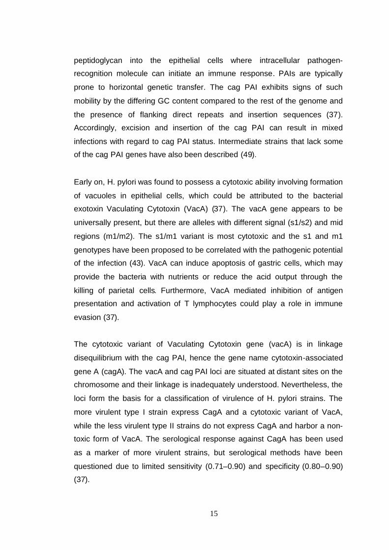
15
peptidoglycan into the epithelial cells where intracellular pathogen-
recognition molecule can initiate an immune response. PAIs are typically
prone to horizontal genetic transfer. The cag PAI exhibits signs of such
mobility by the differing GC content compared to the rest of the genome and
the presence of flanking direct repeats and insertion sequences (37).
Accordingly, excision and insertion of the cag PAI can result in mixed
infections with regard to cag PAI status. Intermediate strains that lack some
of the cag PAI genes have also been described (49).
Early on, H. pylori was found to possess a cytotoxic ability involving formation
of vacuoles in epithelial cells, which could be attributed to the bacterial
exotoxin Vaculating Cytotoxin (VacA) (37). The vacA gene appears to be
universally present, but there are alleles with different signal (s1/s2) and mid
regions (m1/m2). The s1/m1 variant is most cytotoxic and the s1 and m1
genotypes have been proposed to be correlated with the pathogenic potential
of the infection (43). VacA can induce apoptosis of gastric cells, which may
provide the bacteria with nutrients or reduce the acid output through the
killing of parietal cells. Furthermore, VacA mediated inhibition of antigen
presentation and activation of T lymphocytes could play a role in immune
evasion (37).
The cytotoxic variant of Vaculating Cytotoxin gene (vacA) is in linkage
disequilibrium with the cag PAI, hence the gene name cytotoxin-associated
gene A (cagA). The vacA and cag PAI loci are situated at distant sites on the
chromosome and their linkage is inadequately understood. Nevertheless, the
loci form the basis for a classification of virulence of H. pylori strains. The
more virulent type I strain express CagA and a cytotoxic variant of VacA,
while the less virulent type II strains do not express CagA and harbor a non-
toxic form of VacA. The serological response against CagA has been used
as a marker of more virulent strains, but serological methods have been
questioned due to limited sensitivity (0.71–0.90) and specificity (0.80–0.90)
(37).

16
B- Adhesions
A wide variety of molecules present on adherent structures of bacteria can
function as adhesions. In bacterial-eukaryotic interfaces, there are apparently
general systems for recognition of certain microbial cell surface carbohydrate
characteristics (39). Like other microorganisms, H. pylori require adhesive
molecules for colonization and persistence. H. pylori has a wide range of
adhesion properties and has been suggested to bind to many different
carbohydrates, mediated by various bacterial components(50). The Leb and
sLex antigens binding adhesins, BabA and SabA, respectively, are the best
described (51). Hemagglutination of RBCs by bacteria has been used to
study bacterial binding specificities and to identify the cognate bacterial
adhesions. The H. pylori sialic acid dependent hemagglutination was a
subject for researchers for more than a decade but recently, this sialic acid-
dependent binding activity has been shown to be mediated by SabA, since
the corresponding sabA deletion mutant lacks all hemagglutination properties
(39).
B-1. Consequences of bacterial adhesion
It is becoming increasingly obvious that the act of adhering to the host cells
induces intense changes in the adherent organism, regardless of the origin of
the host cells. It is also known that adhesion of an organism to a host
receptor greatly affects its rate of growth, carbohydrate utilization, protein
synthesis and energy generation, as well as its cell wall composition and
production of adhesive molecules. The host cell and its adherent bacterium
exchange signals (i.e. they engage in molecular “crosstalk”), which brings
about changes in both cell types (figure2.2) (52).
17
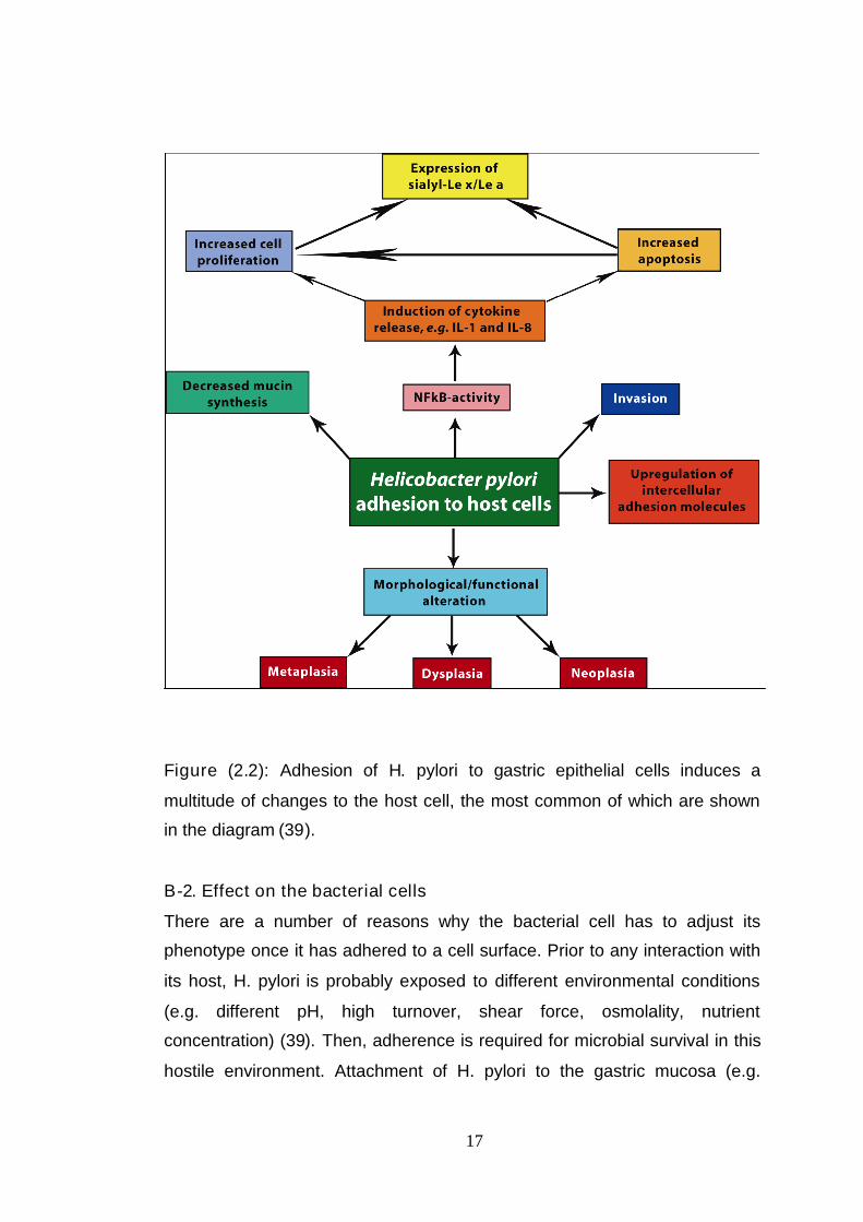
Figure (2.2): Adhesion of H. pylori to gastric epithelial cells induces a
multitude of changes to the host cell, the most common of which are shown
in the diagram (39).
B-2. Effect on the bacterial cells
There are a number of reasons why the bacterial cell has to adjust its
phenotype once it has adhered to a cell surface. Prior to any interaction with
its host, H. pylori is probably exposed to different environmental conditions
(e.g. different pH, high turnover, shear force, osmolality, nutrient
concentration) (39). Then, adherence is required for microbial survival in this
hostile environment. Attachment of H. pylori to the gastric mucosa (e.g.
18
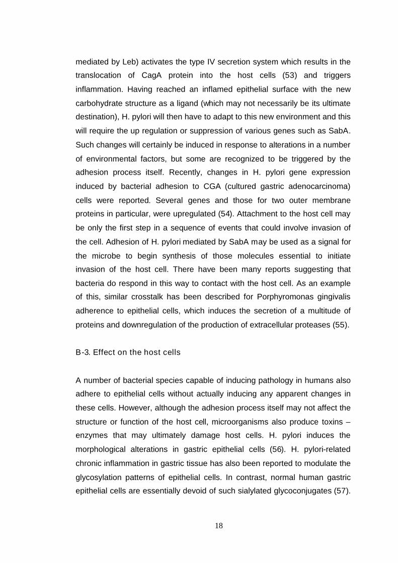
mediated by Leb) activates the type IV secretion system which results in the
translocation of CagA protein into the host cells (53) and triggers
inflammation. Having reached an inflamed epithelial surface with the new
carbohydrate structure as a ligand (which may not necessarily be its ultimate
destination), H. pylori will then have to adapt to this new environment and this
will require the up regulation or suppression of various genes such as SabA.
Such changes will certainly be induced in response to alterations in a number
of environmental factors, but some are recognized to be triggered by the
adhesion process itself. Recently, changes in H. pylori gene expression
induced by bacterial adhesion to CGA (cultured gastric adenocarcinoma)
cells were reported. Several genes and those for two outer membrane
proteins in particular, were upregulated (54). Attachment to the host cell may
be only the first step in a sequence of events that could involve invasion of
the cell. Adhesion of H. pylori mediated by SabA may be used as a signal for
the microbe to begin synthesis of those molecules essential to initiate
invasion of the host cell. There have been many reports suggesting that
bacteria do respond in this way to contact with the host cell. As an example
of this, similar crosstalk has been described for Porphyromonas gingivalis
adherence to epithelial cells, which induces the secretion of a multitude of
proteins and downregulation of the production of extracellular proteases (55).
B-3. Effect on the host cells
A number of bacterial species capable of inducing pathology in humans also
adhere to epithelial cells without actually inducing any apparent changes in
these cells. However, although the adhesion process itself may not affect the
structure or function of the host cell, microorganisms also produce toxins –
enzymes that may ultimately damage host cells. H. pylori induces the
morphological alterations in gastric epithelial cells (56). H. pylori-related
chronic inflammation in gastric tissue has also been reported to modulate the
glycosylation patterns of epithelial cells. In contrast, normal human gastric
epithelial cells are essentially devoid of such sialylated glycoconjugates (57).

19
In addition, recent studies have indicated that adherence of H. pylori induces
cell proliferation and apoptosis during the early phase of chronic inflammation
of the gastric mucosa (58). A characteristic feature of any bacterial infection
is the migration of PMNs towards the site colonized by the infecting
organism. Binding of H. pylori to the gastric epithelial cells induces the
expression and secretion of IL-8 (39). In response to a chemotactic gradient
from the site of infection, PMNs first adhere to, and then traverse, the
vascular endothelium (59). This process involves the interaction between E-
selectin, intercellular adhesion molecule (ICAM) 1 and 2 on the surface of the
endothelium cell, and sLex and 2-integrins on the PMN surface (60). PMNs
are rich in various sialylated glycoconjugates, which H. pylori can bind to. An
important consequence would be the bacterial interaction with, and activation
of PMNs (39). Nutrients released by the inflamed and damaged cells might
be used by H. pylori as an energy source (61).
B-4. Invasion of host cells
A characteristic feature of H. pylori-induced inflammation is massive
attraction of phagocytes (particularly PMNs) to the site of infection. This can
be achieved by the production of H. pylori neutrophil-activating protein (HP-
NAP). The protein was found to promote the adhesion of PMNs to endothelial
cells by upregulating adhesion receptors of the 2-integrin family (62). Satin
and colleagues showed that HP-NAP stimulates (NADPH) oxidase assembly
and production of reactive oxygen species (ROS). Also, as PMNs
consistently outnumber macrophages in H. pylori infected stomach; it induces
a state of chronic acute inflammation. Previous reports (63) have suggested
that H. pylori is capable of invading epithelial cells in the gastric mucosa;
however, the role of invasion in H. pylori pathogenesis remains unclear. The
following mechanisms have been postulated to explain how H. pylori evades
phagoctosis: 1- Strong binding between H. pylori and phagocytes correlates
with a rise in the level of urease on the surface of H. pylori, thus retarding
phagocytosis and strong respiratory burst in the phagocytes (64). 2-

20
Catalase, alkyl hydroperoxide reductase, and factors (cag pathogenicity
island type IV secretion apparatus) that are unique to type I strains allow
bacteria to resist phagocytic killing (65). 3- Delayed phagocytosis is linked to
intracellular survival, since type I H. pylori persists inside macrophages within
a novel vacuole called the megasome (66).
C- H. pylori enzymes
H. pylori produce catalase, oxidase and urease enzymes. Urease is an
abundantly produced enzyme. H. pylori has developed a unique mechanism
to control urease-dependent pH buffering. The urea channels (UreI) present
in the inner membrane are opened at pH below 6.5, to allow for delivery of
urea to intracellular urease. The NHзproduced can then buffer the periplasm
(67). Urease activity is therefore essential for maintenance of cytosolic pH
levels compatible with efficient metabolism and survival of H. pylori in the
acidic environment of the stomach (39).
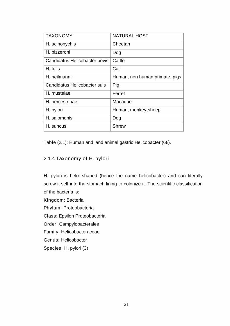
2.1.3 Gastric helicobacter species
To date nine Helicobacter species have been cultured from the stomach of
humans and land animals (table2.1), all are capable of hydrolyzing urea (68).
These can be further classified into Lockard types 1, 2 and 3: type 1 has a
fusiform to slightly spiral morphology with tapered ends; type 2 is spiral and
has sparsely distributed periplasmic fibers and can appear singly or in groups
of two to four; and type 3 is more tightly coiled and lacks periplasmic fibers.
In general the morphology of gastric Helicobacter species isolated from
animals other than cats and dogs can sometimes be distinctive and
sometimes resemble H. pylori. Phylogenetic analysis of current gastric,
enteric and hepatobiliary Helicobacter species, based on 16s rRNA similarity
has been performed (68).
21
TAXONOMY NATURAL HOST
H. acinonychis Cheetah
H. bizzeroni Dog
Candidatus Helicobacter bovis Cattle
H. felis Cat
H. heilmannii Human, non human primate, pigs
Candidatus Helicobacter suis Pig
H. mustelae Ferret
H. nemestrinae Macaque
H. pylori Human, monkey,sheep
H. salomonis Dog
H. suncus Shrew
Table (2.1): Human and land animal gastric Helicobacter (68).
2.1.4 Taxonomy of H. pylori
H. pylori is helix shaped (hence the name helicobacter) and can literally
screw it self into the stomach lining to colonize it. The scientific classification
of the bacteria is:
Kingdom: Bacteria
Phylum: Proteobacteria
Class: Epsilon Proteobacteria
Order: Campylobacterales
Family: Helicobacteraceae
Genus: Helicobacter
Species: H. pylori (3)

22
2.2 Epidemiology of H. pylori Infection
2.2.1 Prevalence of H. pylori infection
This gram-negative bacterium infects more than half the world’s population
and its prevalence has been shown to correlate with poor socio-economic
conditions. In many underdeveloped nations, more than 80% of the
population is infected with this pathogen (10).
H. pylori infection is one of the commonest infections worldwide, occurring in
all regions and infecting at least half of the world’s population (69). The
prevalence of H. pylori infection worldwide is approximately 50% (14), as
high as 80%–90% in developing countries, and ≈35%–40% in the United
States (14). While within countries, the prevalence is higher among groups
with lower socioeconomic status (70, 71). H. pylori prevalence is generally
found to increase with age, reaching 20-50% in adult populations in Europe
and North America (72). H. pylori-positivity in adults is more closely
associated with living conditions and with the parents’ socioeconomic status
in childhood than with current living conditions and socioeconomic status
(73).
The infection is also associated with low Socio Economic Status (SES) within
countries (74). In the United States, for instance, a significantly lower
prevalence was found in Caucasians (26%) compared to Hispanics (65%)
and Afro-Americans (66%) (74). This dissimilarity was interpreted to reflect
the different socioeconomic backgrounds of the groups. In a follow-up study,
it was found that the difference in prevalence between Afro- Americans and
Caucasians resulted from different seroconversion rates, although the rate of
seroreversion could also have played a role (75). H. pylori infection is usually
acquired before the age of five (75).
23
This pattern has been interpreted to partly reflect a birth-cohort phenomenon
caused by a higher incidence in the past due to poorer living conditions and
sanitation (37). Indeed, mathematical modeling has suggested that the
infection will eventually disappear in high-income countries even without
intervention (76).
2.2.2 Incidence of H. pylori infection
The annual incidence of H. pylori infection is ≈4%–15% in developing
countries, compared with approximately 0.5% in industrialized countries (14).
Documented risk factors include low socioeconomic status, overcrowding,
poor sanitation or hygiene, and living in a developing country (15).
In adults, the incidence rates are mostly derived from retrospective
longitudinal sero surveys, and these studies are mostly conducted in
industrialized countries. It appears that acquisition of the infection during
adulthood is a rare event: seroconversion (i.e. change of the serostatus from
seronegative to seropositive) occurs generally at a rate less than 1% per year
of follow-up. Seroreversion appears to occur approximately at the same or
even higher rate than seroconversion (77). Data about the incidence of H.
pylori infection among adults in developing countries are scarce. In Brazil, an
annual seroconversion rate of 1.1% and a seroreversion rate of 0.2% were
found during 56 months of follow-up (78). Annual incidence rates over 20%
have been reported in early childhood in low-income countries (37).
2.2.3 Risk factors for infection
An obvious necessary cause for acquisition of H. pylori infection is exposure
to the bacterium. The probability of exposure depends on the characteristics
of the infective source and contact, but factors of the recipient host and the
bacterium may also influence the probabilities of acquisition and persistence.
24
The bacterium has to overcome numerous barriers to successfully establish
an infection in a new individual (37).
1. Exit from an infected individual.
2. Transient survival outside the gastric niche.
3. Introduction into a new host.
4. Colonization of the new gastric mucosa.
5. Maintenance of the colonization.
A predominantly person-to-person transmission has been postulated. This
notion is based on the clustering of the infection in families (79) and in
institutionalized individuals while consistent and verified environmental
reservoirs are absent (80).
2.2.3.1 The family
The family stands out as the most important framework for transmission and
a child’s risk of being infected is associated with having infected family
members (79). Family size and residential crowding (persons per room or
m2) are frequently described as risk factors for H. pylori infection and may be
regarded as proxies for the number of infected family members (81).
Similarly, having familial connections to high-prevalence regions is
associated with infection in children living in low-prevalence areas and this
effect decreases in successive generations (82). The living conditions during
childhood can be predictive of infection in adulthood being in accordance with
H. pylori acquisition in childhood from household members (83).
Having an infected mother has been found to be a more prominent risk factor
for childhood infection than having an infected father, supporting primarily
mother-child transmission. Transmission among siblings has also been
indicated by clustering of the infection in sib ships (79).
25
Even though the infection is usually initiated in early childhood, some
epidemiological data point to the possibility of acquisition in adulthood from
infected family members. Having an infected spouse has been described as
a risk factor for infection (84). Furthermore, having more children has been
identified as a risk factor for infection in adults, possibly indicating that
children may serve as mediators of transmission within families (85).
2.2.3.2 Environmental and behavioral factors
Reasons for the association between H. pylori infection and low SES have
been sought among environmental and behavioral factors. A shared
environmental source of the infection could theoretically contribute to the
observed intrafamilial clustering. Possibly contaminated water has been
suggested as an infection source since using particular water sources, such
as wells, has been correlated to the infection (86). However, other studies
have not found the water source to be associated with infection (87). More
indirect environmental transmission has also been proposed following the
identification of the consumption of raw vegetables as a risk factor for
infection (88). Furthermore, H. pylori has been proposed to possess zoonotic
potential and suggested reservoirs include cats, houseflies and sheep, but
these theories are controversial (37).
Behavioral factors may influence the risk of H. pylori acquisition and
persistence. Residence in a high-prevalence country can facilitate
acquisition, as frequent close contact with infected individuals and poor
sanitary practices may enhance bacterial exposure (86). Intimate contact has
likewise been suggested to explain other observed risk factors, such as bed
sharing and breastfeeding. Breastfeeding has also been speculated to
possibly provide protection against early infection by passive immunization.
However, such protection, if any, should be of limited relevance after
weaning, as supported by negative findings (87). Moreover, H. pylori infection
has been negativelycorrelated with antibiotic consumption (37). In another

26
study, however, a similar negative association disappeared when the country
of origin was taken into account, which could be explained by the higher
antibiotic consumption in low-prevalence countries (81). Some behavioral
factors have been assessed as determinants for H. pylori infection
specifically in adults. Examples include a possible negative association with
alcohol intake (89) and perhaps a positive relationship with smoking, but data
are discrepant (83).
2.2.3.3 Host and bacterial factors
H. pylori strains differ in their ability to establish and maintain an infection in a
given host, which can be attributed to host and bacterial factors and their
compatibility (45). The transient infections in childhood may reflect instances
where the bacterium is not optimally suited for the new host and adaptation is
not feasible or rapid enough, leading to the host succeeding in clearing the
infection (75).
Host genetics have been indicated to be involved in susceptibility to H. pylori
infection, based on a higher concordance of infection in monozygotic (81%)
than in dizygotic (63%) twin pairs. The specific genetic components of this
suggested predisposition are unknown, but some host factors that may
contribute susceptibility to the infection have been proposed. Expression of
blood group antigens that mediate bacterial adherence to the gastric mucosa
has been suggested to be important for susceptibility to H. pylori infection
(37). Some research indicates that H. pylori strains have adapted their
binding affinities in accordance with the blood group antigen expression of
different human populations (90). Furthermore, individuals that excrete
receptors in body fluids, offering removable binding sites that can compete
with the tissue-bound receptors, have been reported to have a lower risk of
being infected (91). However, some studies have shed doubt on the theory
that blood group antigen-mediated adhesion contributes to susceptibility to
infection (40).

27
The immune system may also be involved in determining predisposition to H.
pylori infection. This notion is supported by studies that describe alleles
within the human leukocyte antigen locus HLA-DQA1 to be correlated with
the infection (92). An interleukin (IL)-1 receptor polymorphism of unknown
functional consequence has also been correlated with the infection (93).
Furthermore, the spread of the bacterium may be promoted by low gastric
acid secretion, which may be especially relevant in young children and during
infective gastroenteritis (94). Some studies have found a slightly higher
prevalence of H. pylori infection in males (83). The reasons for this tendency
are unclear and other studies have not been able to confirm this correlation
(75).
H. pylori has developed a repertoire of functions for survival in the harsh
gastric niche, including acid tolerance, motility, adherence, immune evasion
and mechanisms for adaptive evolution. These features are all involved in the
interplay between the host and the bacterium and may influence acquisition
and persistence of infection, as is typically studied in animal models.
Bacterial acid tolerance and motility were among the first factors found to
play a role in colonization (37). Global mutagenesis approaches have verified
these findings and have expanded the collection of putative essential genes
(95). However, the relevance of these observations in human populations is
largely unknown. In a Finnish population, strains with a bacterial virulence
factor, the cag pathogenicity island (PAI), have been indicated to disappear
more rapidly than strains without the cag PAI (96). This could speculatively
be explained by reduced transmissibility or persistence of cag PAI strains in
this population. However, cag PAI+ infections constitute the majority of H.
pylori infections worldwide (49).

28
2.2.4 Transmission of H. pylori infection
The exact routes of transmission are not definitely known due to the inability
to clinically detect acute H. pylori infection as well as due to technical
difficulties in isolating the microorganism from sources other than the gastric
mucosa (77).
Transmission of the infection probably occurs in multiple pathways, which
may differ in different societies and age groups. As childhood is a period of
high risk for H. pylori acquisition, a good understanding of the mode(s) of
transmission in children is required to identify how to break the chain of
transmission of the infection (77).
The minimum infectious dose of H. pylori for humans is not yet established.
In human volunteers, ingestion of 104–1010 of H. pylori after administration of
famotidine resulted in infection in 18 out of 20 subjects (77). For non-human
primates, the established minimum infectious dose of H. pylori is 104 cfu (68).
The most important reservoir of H. pylori is the human stomach; and
potentially, H. pylori may pass from the stomach into the external
environment by feces, vomitus or gastric regurgitation (77).
2.2.4.1 Fecal-oral and gastro-oral routes
For fecal-oral transmission, H. pylori must be excreted by feces, viable and at
sufficiently high concentrations. Culturing of H. pylori from normal feces has
been rarely successful, although higher isolation rates have obtained recently
by using modified culturing conditions (77). H. pylori can be more easily
isolated from diarrheal stool (97) indicating that H. pylori may preserve its
viability better if the transit time through the GI tract is shorter. The number of
bacteria isolated in diarrheal stool, though, is relatively small, 5–2125 cfu/ml.
The shedding of a large number of viable H. pylori, up to 30,000 cfu/ml has
29
found in artificially induced vomitus, and H. pylori has been isolated from the
air sampled in the vicinity of the vomitus. H. pylori has been successfully
cultured also from naturally secreting vomitus in a child with acute
gastroenteritis. This proves that the organism is potentially transmissible
during episodes of gastrointestinal tract illness, particularly with vomiting (97).
History of vomiting in siblings was found to be an independent risk factor for
H. pylori infection in children followed prospectively neurologically
handicapped children living in an institution, and found a chronological link
between outbreaks of gastroenteritis and new cases of H. pylori infection.
Therefore, the decreased incidence of diarrheal diseases, parallel with
socioeconomic development, is one of the possible explanations for the
decreased incidence of H. pylori infection (77).
The iatrogenic transmission of H. pylori from stomach to stomach via
contaminated endoscopic devices is possible but rare event. Proper
disinfection of endoscopes prevents iatrogenic spread. Hands may serve as
a potential vector of the infection. In rural Guatemala, carriage of H. pylori
under fingernail was detected in 58% of studied persons using PCR method
(77).
2.2.4.2 Oral-oral route
Human oral cavity has been implicated as a possible source of H. pylori
infection, although there exists controversy as to whether the oral cavity can
act as a permanent reservoir of H. pylori, or whether the microorganism can
be detected only occasionally in the saliva or on the oral mucosal surfaces as
result of gastroesophageal reflux or vomiting. The fastidious nature of H.
pylori and the complexity of the oral microflora make the isolation of the
microorganism from the oral cavity complicated. Most reports about presence
of H. pylori in the oral cavity are based on detection of a specific DNA, which
has been found in the dental plaque (98), in the periodontal pockets, and in
the saliva. The detection rate, though, has shown a great deal of variation,
30
from less than 10% among the subjects harboring the organism in the
stomach to 100% among the subjects under study, irrespective to their
gastric H. pylori-status (98).
A major weakness of PCR is its inability to distinguish between viable or
dead microorganisms, and therefore, detection of the DNA of the
microorganism in the oral cavity is not sufficient evidence for considering it a
reservoir of the infection (77). Although successful culture of H. pylori from
the oral cavity has also been reported, the success rate is low (97). The
number of organisms in the oral cavity, if present, is rather small: using a
competitive PCR assay the median number of H. pylori in dental plaque of
adults with gastric H. pylori infection was found to be 25 cells/mg ; in the
postemesis saliva collected half an hour after vomiting H. pylori cultures had
counts ranging from 50–500 cfu/ml (97). Some epidemiological results also
favor spread of H. pylori by an oral source. Pre-mastication of food and plate
sharing were found to be independent risk factors for H. pylori infection of
children (77).
2.2.4.3 Environmental sources: water and food
The analysis of the genome has shown that it is very unlikely that H. pylori
can multiply in environment. H. pylori can survive in water, milk and in
various foods under refrigerated storage for several days, suggesting that the
water or food contaminated with H. pylori could be potentially infectious to
humans (77). In water, H. pylori remained culturable for up to 24 hours at 20–
23 ºC and for 2–3 days at 16 ºC (99). H. pylori has not been cultured from
natural freshwater, but was isolated from wastewater in Mexico (77). Using
PCR method, H. pylori DNA has been detected in drinking water. H. pylori
DNA has been detected also in biofilms within water storage pots or water
distribution systems. Few quantitative data are available. Krumbiegel et al
(2004) found that the H. pylori DNA was present in approximately one tenth
of the private wells in rural counties in Germany, and that estimated average
31
infestation was approximately one H. pylori cell per ml. However, a positive
PCR result does not prove that the organism is viable and transmissible
(100).Under unfavourable conditions, H. pylori may transform from an
actively dividing spiral-shaped form into a non-culturable coccoid form, which
may represent an alternative survival system (99) or a morphologic
manifestation of cell death (77). These coccoid forms may persist for
extended periods in water (99). The question whether the coccoid form of H.
pylori is able to establish infection in humans is still unclear. In laboratory
conditions, coccoid H. pylori organisms given at high dosage to mice were
able to colonize their gastric mucosa and cause inflammation (101).
In epidemiological studies, generally no association has been found between
H. pylori infection and a water source in industrialized countries, possibly
because of high quality water treatment. Water-borne transmission may
occur in regions of the world where the quality of drinking water is low (77).
Stored household water, contaminated in home, may also serve as a vehicle
of the infection (87).
2.2.4.4 Animal reservoirs
Animals are unlikely to be an important reservoir of H. pylori infection,
although in specific settings, zoonotic transmission may occur. Sheep and
Monkey have been reported to harbor H. pylori in their stomachs. H. pylori
was isolated from sheep’s milk, suggesting that it might be a transmission
vehicle of H. pylori. Regular professional contact with sheep results in almost
100% H. pylori prevalence: 98% of studied shepherds in Italy and in Poland
were seropositive (77). The role of insects as a potential vector has been
studied as well. Houseflies and cockroaches, when fed pure cultures of H.
pylori, were able to harbor the microorganisms in their gut and excreta for
more than 24 hours after initial exposure. However, H. pylori was not
recovered from any of the houseflies fed human feces either naturally

32
infected or artificially infected with H. pylori, thereby not confirming that
houseflies are vectors for transmission (77).
2.2.4.5 Interfamilial transmission
H. pylori infection clusters within families: children living with infected parents
have higher prevalence of the infection than those living with uninfected
parent. Molecular studies have shown that family members often share the
same strain of H. pylori (77). Intrafamilial clustering of the infection suggests
either a person-to-person transmission within family or exposure to a
common environmental source of the infection. Close intrapersonal contact
appears to facilitate spread of the infection: domestic overcrowding in
childhood has been consistently found to be a significant risk factor for
H. pylori infection for children as well for adults, which supports the theory
of person-to-person transmission (102).
In developing countries, where there are large families with many siblings,
sibling-sibling transmission might be more important (87). In developed
countries, where the number of children in families is generally small,
mothers may play the key role (103). Studies have generally failed to identify
H. pylori–positive father as a risk factor of the infection in offspring (79).
Child-to-adult transmission seems to be an unlikely event. During a nine-year
follow-up of 46 families in Japan no case was identified where the infection
spread from an infected child to an uninfected parent (104). In Sweden, no
increased risk for H. pylori infection was seen among children who had
attended day-care centers in comparison with those who had been
exclusively looked after at home (77).

33
2.3 Clinical Manifestations of H. pylori Infection
2.3.1 Natural course of H. pylori infection
The ability of H. pylori to maintain persistent colonization is of key importance
for disease development (37). Acute H. pylori infection in adults is
accompanied by mild to moderate dyspeptic symptoms and occasional
vomiting, which appear few days after challenge, peak during the second
week and then resolve. The clinical course of chronic H. pylori infection is
highly variable and influenced by microbial, host and environmental factors.
In virtually all infected individuals H. pylori causes chronic inflammation in the
gastric mucosa (77). Gastritis develops rapidly after acquisition of H. pylori
infection and persists. Along with its persistence, through several years of the
infection, chronic gastritis may gradually progress to atrophic gastritis. The
annual incidence of atrophic gastritis among H. pylori-positive adults is
approximately 1–3% (105). Most people with H. pylori infection are
asymptomatic, but a proportion of infected individuals develop severe
gastroduodenal disease, including Duodenal Ulcer (DU), gastric ulcer and
rarely gastric adenocarcinoma and gastric Mucosa Associated Lymphoid
Tissue (MALT) lymphoma (77). H. pylori infected persons have
approximately a 4–6-fold increased risk of developing peptic ulcer disease
and approximately a 6-fold increased risk of non-cardia gastric
adenocarcinoma and gastric MALT lymphoma compared with uninfected
persons. In H. pylori-infected subjects, the estimated lifetime risk of peptic
ulcer is 6–20% ; the estimated lifetime risk of gastric cancer is 1–2% in
Western societies and 11–12% or even higher in Japan. It is hypothesized
that early life acquisition of H. pylori may increase the risk of subsequent
development of gastric cancer. In an animal experiment with Mongolian
gerbils, early acquisition of H. pylori significantly increased their susceptibility
to gastric chemical carcinogenesis as compared with the case of later
infection (77).

34
2.3.2 Gastritis
Acute H. pylori infection causes gastritis and hypochlorhydria and symptoms
such as vomiting and dyspepsia have been associated with acquisition (2).
Persistent infection causes chronic gastritis in virtually all infected individuals.
The gastric inflammation involves infiltration of immune cells, such as
neutrophils, lymphocytes, plasma cells and macrophages, and secretion of a
multitude of cytokines, of which IL-8 seems to have a central role (10). The
chronic gastritis is usually asymptomatic, but eradication of H. pylori in non-
ulcer dyspeptic patients alleviates symptoms in a fraction of the patients (37).
2.3.3 Peptic ulcer disease
The discovery of the role of H. pylori in the development of peptic ulcer
disease has lead to a paradigm shift in the treatment of ulcer patients (106).
The lifetime risk for peptic ulcer in infected individuals ranges from 3% in the
United States to 25% in Japan (10). It has been estimated that 95% of DU
and 70% of gastric ulcers can be attributed to H. pylori (107). Duodenal
ulcers are associated with H. pylori-induced antrum-predominant gastritis,
decreased somatostatin levels and augmented gastrin and acid secretion
(10). Development of gastric metaplasia in the duodenum can allow further
bacterial colonization, leading to duodenitis and epithelial damage. Gastric
ulcers are associated with corpus gastritis, which is believed to damage the
epithelium (9).
2.3.4 Gastric cancer
Gastric cancer ranks as the second most frequent cause of cancer deaths
worldwide despite a decreasing incidence in high-income countries (108).
The World Health Organization classified H. pylori as a class I carcinogen in
1994 due to its definite carcinogenic potential in humans (37). Subsequent
studies have corroborated the association to gastric cancer and about 70% of

35
non-cardia adenocarcinomas have been attributed to H. pylori infection (109).
However, only a few percent of infected individuals develop gastric cancer
(9).
Persons with low gastric acid secretion and corpus-predominant gastritis,
leading to atrophic gastritis, loss of parietal cells and further hypochlorhydria,
are at increased risk for gastric cancer (10). Postulated carcinogenic
mechanisms include the increased epithelial turnover caused by the
inflammation. Furthermore, the development of gastric atrophy and
hypochlorhydria can result in impaired antioxidant absorption, infection with
other carcinogenic microorganisms and formation of carcinogenic
compounds (9). H. pylori infection up regulates the proinflammatory cytokine
IL-1, which is also a potent inhibitor of acid secretion. Polymorphisms
considered to increase the activity of IL-1 have been proposed to be
associated with hypochlorhydria and cancer, supporting the central role of
gastric acidity in cancer development (110).
2.3.5 Other H. pylori-associated conditions
H. pylori has been assessed for its involvement in various additional
conditions. An association between the infection and gastric MALT
lymphoma belongs among the generally accepted findings and antibiotic
treatment alone leads to regression of the cancer in many cases (111).
Moreover, it has been debated whether there is a causal relationship
between the parallel decline of H. pylori prevalence and the increase of
gastroesophageal reflux disease (GERD) and esophageal adenocarcinoma
in high- income countries. There is accumulating evidence in favor of a
protective role of H. pylori infection against these conditions, but there are a
number of additional contributing factors (112). The high H. pylori prevalence
in many parts of the world accentuates the public health importance of any
associations between H. pylori and disease. This may be particularly
noteworthy for associations that are of modest strength or involve relatively

36
benign conditions, as these associations may otherwise tend to be neglected
(112).
2.3.6 Extragastric manifestations of H. pylori infection in children
2.3.6.1 Iron-deficiency anemia
H. pylori–infected subjects have lower mean serum ferritin levels compared
with the non-infected ones irrespective to their iron intake (113). H. pylori
infection is more prevalent among subjects with low serum ferritin level than
among subjects with normal serum ferritin level, both of adults and children
(113). A number of case reports and case series have described resolution of
refractory iron deficiency anemia only after eradication of H. pylori infection,
thereby supporting the hypothesis that H. pylori infection causes iron
deficiency and not vice versa (114). The mechanisms through which H. pylori
infection can cause iron deficiency and further lead to anemia have not been
fully elucidated. It has been suggested that H. pylori infection may increase
the iron demand as H. pylori itself uses iron for its growth and may capture
ingested iron and iron from human lactoferrin (115).
2.3.6.2 Atopic diseases
The composition of the microflora of the gastrointestinal tract may play a role
in development of and protection from allergy (116). The increase in the
occurrence of atopic diseases appears to have coincided with the decrease
in the prevalence of H. pylori infection (77).
In some studies, which have addressed predominantly respiratory allergy,
decreased prevalence of atopic diseases and/or allergen-specific IgE
antibodies has been found among H. pylori-positive adults. whereas some
pediatric case-control studies point out that H. pylori infection, especially the

37
infection with a CagA-positive strain may actually increase the risk of food
allergy (77).
Accordant with these results, the animal experiments and in vitro studies
have demonstrated that H. pylori infection increases the transcellular
passage of macromolecules and inhibits development of oral tolerance to
food antigens, thereby suggesting that H. pylori infection may predispose to
food allergy (117).
2.3.6.3 Acute intestinal infections
Some studies have suggested that H. pylori infection may predispose to
various gastrointestinal infections as cholera and shigellosis. Newly acquired
H. pylori infection in children was followed by increased occurrence of
diarrhea (118). Transient hypochlorhydria has thought to be the mechanism
underlying this phenomenon. Passaro et al. (2001) postulated that H. pylori-
promoted gastroenteritis some months after acute infection which is a
biologically plausible mechanism for successful spread of the H. pylori
infection (118). In contrast, in other studies, H. pylori infected children and
adults had significantly less acute diarrheal illnesses than non-infected
subjects (119). It cannot be excluded that some confounding factors may
also explain the conflicting results of these observational studies (77).
2.3.6.4 Diminished growth
Several population-based cross-sectional studies, conducted both in
developing as well as developed countries, have found an association
between diminished growth and H. pylori infection in children, whereas others
have not found such association (77).
38
2.3.7 Barrett’s esophagus
Barrett’s esophagus (BE) is the condition in which columnar epithelium
replaces the squamous epithelium. The condition develops when
gastroesophageal reflux disease damages the squamous esophageal
mucosa, through a metaplastic process in which columnar cells replace
squamous epithelium. The abnormal columnar epithelium that characterizes
BE is an incomplete form of intestinal metaplasia (called specialized intestinal
metaplasia) that predisposes patients to adenocarcinoma (120). Inflammation
of gastric cardiac mucosa decreases in prevalence from controls to patients
with GERD, and H. pylori and cagA-positive strains have been suggested to
protect against the development of BE (121). It is important to distinguish BE
from intestinal metaplasia related to carditis, because these conditions have
a different natural history, risk of malignancy, and treatment. BE is the most
important risk factor for the development of esophageal adenocarcinoma.
The risk of developing esophageal adenocarcinoma from BE is estimated to
be 0.5% per patient per year (39).
2.3.8 Gastroesophageal reflux (GERD)
Even though GERD is primarily a motility disorder, other pathophysiological
disturbances seem to play a role in its pathogenesis. Whether H. pylori may
be one of the players remains controversial and conflicting results have been
reported. Indications that the curing of H. pylori infection in DU patients may
provoke reflux esophagitis have been reported (122). Although not finding
any significant difference in the prevalence of H. pylori carriage in patients
with GERD and its sequel compared with the controls, Vicari found that
patients carrying CagA-positive strains might be at a decreased risk of
complications of GERD (123).

39
2.3.9 Idiopathic thrombocytopenic purpura
H. pylori causes an inflammatory response and provokes an immunologic
reaction. It has been proposed that other chronic immune disorder may be
caused by an immunologic reaction to H. pylori antigen in anti-bodies that
cross-react with human tissues. Uncontrolled studies have suggested a role
for H. pylori in chronic idiopathic thrombocytopenia (124).
2.4 Diagnostic Tests for Detection of H. pylori Infection
The diagnostic tests for H. pylori infection can be roughly divided into two
categories: biopsy-based tests which are invasive tests because as they
require gastroscopy, and non-invasive or minimally invasive tests where no
gastroscopy is required and the serum, whole blood, feces, expired air, saliva
or urine are used for testing. Selection of the test depends on the purpose of
testing, sensitivity and specificity of the test, cost-effectiveness of testing
strategy and availability of the test (77).
2.4.1 Biopsy-based tests
Biopsy-based tests include histological examination, culture, rapid urease
test and tests based on molecular methods. Histological examination: H.
pylori can be directly visualized by histological examination on the gastric
mucosa as characteristic spiral or curve-shaped bacteria. This allows
estimation of H. pylori colonization density as well as provides permanent
record (77). For optimal assessment and for minimizing sampling error, at
least two biopsy samples from both the gastric antrum and corpus mucosa
should be taken (125). The Giemsa stain appears to be the preferred stain
for evaluation of H. pylori infection on the basis of its good sensitivity,
excellent specificity, and lack of technical difficulty in preparation (126).
Besides diagnosing H. pylori infection, histology has the advantage of
allowing the excellent visualization of morphological changes in the gastric
mucosa; for the latter purpose, hematoxylin and eosin stain are generally

40
used (77). The results of histological examination are somewhat dependent
upon experience and on the accuracy of the pathologist, but generally,
histological examination has high sensitivity and specificity for detecting H.
pylori infection, with high inter observer agreement (127).
Culture is a very specific method for diagnosing H. pylori infection, however,
culturing is technically demanding, time-consuming, and its sensitivity varies
among laboratories (128). In experienced laboratories, sensitivity higher than
90% is achieved. The benefit of culture is the possibility to determine the
susceptibility of the strain to antimicrobial agents (77).
Rapid urease test is based on the capacity of H. pylori to secrete the enzyme
urease. When H. pylori infected biopsy specimen is placed in a urea-
containing medium, the bacterial urease splits urea into ammonia and Co2,
and pH of the medium rises. This is detectable by the color change of the pH
indicator dye (77). It is a quick and simple test with a sensitivity about 88–
90% and a specificity close to 100% (129). The sensitivity of the test is lower
in patients with active or recent upper gastrointestinal bleeding. The test
indicates only the presence or absence of the infection and gives no
additional information (77).
Tests based on molecular methods, mainly the PCR are used in order to
detect H. pylori from biopsy specimens with high sensitivity and specificity
(130), to detect point mutations encoding H. pylori resistance to antibiotics,
especially to clarithromycin and to detect virulence factors (131).
2.4.2 Tests based on detection of specific anti-H. pylori antibodies
H. pylori infection induces a humoral immune response in the host, resulting
in an early increase of specific IgM antibodies, and later a persistent increase
of specific IgG and IgA antibodies (132). Specific antibodies can be detected
in serum, whole blood, urine and saliva, both from fresh and freeze- stored

41
samples (133). The limitation of the tests based on specific antibody
detection is that acute infection can be missed due to the delay in antibody
production as IgG sero-conversion occurs approximately 7–14 weeks after
infection (134). On the other hand, false-positive results may occur, as
antibody titers may remain positive even for years after resolution of the
infection (135). However, the titer of specific antibodies has tendency to fall:
a 25–50% drop in the IgG antibody titer in paired sera 6 months after
treatment confirms eradication in most cases (136).
Enzyme-linked immunosorbent assay (ELISA) is commonly used for
detection of specific antibodies in the serum, as it is easy to perform,
inexpensive, widely available, and suitable also for large-scale screening
(137). The meta-analysis of 21 studies with commercially available ELISA
kits reported overall sensitivity of 85% and specificity of 79%. The
performance of different assays may vary between patient populations from
different geographic regions and ethnic backgrounds due to the differences
between antigenic properties of bacterial strains colonizing patients (138).
Therefore, the test should be locally validated. In young children, the
sensitivity of ELISA is low when the cut-off values validated for adults are
used (133).
Immunoblot is more costly and time-consuming than ELISA, but permits a
detailed analysis of antibody reactivity to the different H. pylori proteins. It has
been a useful tool that complement serology in children, particularly in cases
with doubtful ELISA results (139).
Several near-patient or office-based tests on the whole blood have been
developed, they are quick and easy to perform, however, the diagnostic
accuracy of these tests is inadequate for recommending them for widespread
usage (140). Tests on the saliva and urine are attractive because samples
can be easily obtained. Salivary tests have shown inconsistent results with

42
less than optimum sensitivity and specificity, urine antibody tests have
demonstrated accuracy comparable to that of serum-based ELISA (141).
2.4.3 Urea breath test
Urea breath test (UBT) is based on the capacity of H. pylori to secrete the
enzyme urease, which hydrolyses urea to ammonia and carbon dioxide. The
test substance used is urea, labelled with a carbon isotope, either with 14C
(a radioactive isotope) or 13C (a stable isotope). The 13C-UBT is most
frequently employed for its safety; however, the cost of 13C-UBT is higher
than of 14C- UBT. In non-infected individuals, ingested urea leaves the
stomach unchanged, while in infected subjects, urea is split into ammonia
and CO2, which can be detected in the expired air. Labeled urea is usually
given to the patient with a test meal to delay gastric emptying and to increase
the time of contact with the mucosa (77). The UBT detects current H. pylori
infection with a sensitivity and specificity of >95% (140). The test is indicated
for initial diagnosis of the infection and for follow-up of eradication therapy; in
the latter case, the UBT should not be performed before an interval of four
weeks has elapsed (142). False-negative results may occur during treatment
with proton pump inhibitors (143).
2.4.4 Tests based on detection of bacterial antigens or bacterial
DNA in feces
Identification of bacterial antigens in stool using enzyme immunoassays has
emerged in recent years, and tests based on polyclonal antibodies (144) and
monoclonal antibodies (145) have been developed. A systematic review of
89 studies including 10,585 patients concluded that stool antigen test is an
accurate method for diagnosing H. pylori infection: the mean sensitivity and
specificity were 91% and 93%, respectively (146). Overall results revealed
that tests based on monoclonal antibodies are slightly more sensitive and
specific and have greater reproducibility than tests based on polyclonal

43
antibodies (146). PCR-based methods of detecting H. pylori DNA in stool
samples have also been developed (147). They allow genotyping of strains,
detection of virulence factors and testing of antibiotic susceptibility (148).
2.5 Treatment of H. pylori Infection
The goal of treatment of H. pylori infection is complete eradication of the
organism. Eradication is currently defined as absence of H. pylori for a
minimum of 4 weeks after treatment, as confirmed by biopsy-based test, UBT
or stool antigen test (142). Clinically relevant eradication regimens must have
cure rates of at least 80%, according to intention-to-treat analysis, without
major side effects and with minimal induction of bacterial resistance (10).
2.5.1 Indications for treatment
Eradication of H. pylori infection is an effective way to cure H. pylori-
associated peptic ulcer disease, as it facilitates ulcer healing, prevents
recurrences in vast majority of patients (108), and significantly reduces the
risk of complications. Eradication of H. pylori infection leads to resolving of
gastric inflammation both in adults and in children, and it may lead to
reversion of existing atrophy in the gastric mucosa (149). Eradication of H.
pylori induces remission of gastric MALT lymphoma (150) and would likely
prevent development of gastric cancers if intervention comes early enough in
the sequence of carcinogenesis (151).
Whether the eradication of H. pylori results in improvement of symptoms in
patients with non-ulcer dyspepsia or not is still debatable. Meta-analyses of
randomized controlled trials have yielded equivocal results: one showed no
improvement of symptoms with H. pylori eradication, while another revealed
a statistically significant but small benefit (77). At the best, one case of non-
ulcer dyspepsia is being cured for every 10 persons whom the infection was
44
eradicated ; at the worst, eradication induces exacerbation of gastro-
esophageal reflux disease (152).
A number of guidelines on management of H. pylori infection have been
compiled, based on published clinical evidence and expert opinion, as the
Maastricht 2–2000 Consensus Report (142) and guidelines from European
Society of Primary Care Gastroenterology (ESPCG) (153).
All guidelines and consensus statements recommend testing for H. pylori
infection only when subsequent treatment of H. pylori infection is planned,
and recommend eradication of H. pylori in patients with peptic ulcer disease.
Other recommendations vary somewhat between different guidelines
according to the population and setting applied (77).
The patient should be investigated for H. pylori infection only when they
present with symptoms and signs suggestive of organic disease, such as
peptic ulcer, and that optimal approach to investigation for H. pylori infection
is upper gastrointestinal endoscopy with multiple biopsies. “Test-and-treat”
strategy, i.e. verifying presence of H. pylori infection by UBT or stool antigen
test and prescribing anti-H. pylori treatment (77). There is some
disagreement regarding whether H. pylori–positive children with abdominal
complaints but without peptic ulcer should receive eradication treatment. The
North America society for Pediatric Gastroenterology (NASPGN) guidelines
state that there is no compelling evidence to recommend eradication
treatment (154), whereas the proceedings of the European and Canadian
consensus conferences state that if symptoms are severe enough to justify
endoscopic evaluation, and if H. pylori infection is found, then the treatment
is indicated (155).

45
2.5.2 Combined antibacterial regimens against H. pylori infection
H. pylori is susceptible to many antibiotics in vitro, e.g. penicillin, ampicillin,
cefotaxime, erythromycin and tetracycline. In vivo, however, the effectiveness
of a treatment with a single agent is very low, less than 15% (154). For a
successful treatment, combined anti- bacterial therapy is needed, and
because low acidity influences the effectiveness of some antimicrobial
agents, combinations with proton-pump inhibitors (PPI) or ranitidine are often
used (77).
A large number of different medication and their combinations have been
evaluated to find optimal therapeutic regimen(s), and optimizing of treatment
is an ongoing challenge. Laheij et al. (1999) reviewed studies performed up
to 1998 and identified 132 different medication combinations used. Oderda et
al. (2000) reviewed studies done on pediatric population until 1999, and
found that a total of 22 different treatment combinations were used. The most
effective treatment combinations in adults comprise with three medicaments:
a so-called proton pump inhibitor based triple therapy which consists of a
proton pump inhibitor together with two antibiotics, usually clarithromycin with
either amoxicillin or metronidazole, or so-called classic triple therapy
consisting of a bismuth compound together with metronidazole and
tetracycline or amoxicillin. The cure rates of different triple regimens,
analyzed on intention-to-treat basis, are roughly 75–85% (156).
The recommended first-line therapy for adults according to the Maastricht 2–
2000 Consensus Report is a proton pump inhibitor (alternatively ranitidine
bismuth citrate) combined with clarithromycin and amoxicillin or
metronidazole. The recommended duration of the treatment ranges from 7 to
14 days (142).
In children, few randomized double-blinded trials have been performed on
eradication of H. pylori infection (157), and the best treatment strategies have
46
to be identified. Existing data indicate that dual therapies might be as
effective as triple therapies (158). whereas the European Pediatric Task
Force on H. pylori has not given any specific recommendations regarding
optimal treatment due to the insufficient data in relation to this issue (155).
2.5.3 Factors influencing treatment results
None of the regimens cure H. pylori infection in 100% of the patients; a
number of factors, as bacterial resistance to antibiotics, patient compliance,
high bacterial load, low gastric pH and others may affect outcome of
eradication therapy (159). The most important reason for treatment failure
appears to be drug resistance, especially clarithromycin-resistance, which
leads to about 70% decrease in efficacy of the clarithromycin-containing
regimen, whereas the impact of metronidazole resistance on clinical outcome
is relatively modest, leading to about 25% decrease in efficacy (160). The
controversy can be attributed to the methodological problems in
determination of metronidazole resistance of H. pylori (161). The pattern of
resistance to clarithromycin and metronidazole varies within wide limits in
different regions (162). The resistance of H. pylori to amoxicillin and
tetracycline is low, less than 1% of isolates worldwide (161). The primary or
acquired resistance of H. pylori to bismuth salts has never been reported
(163).
The other important determinant of successful eradication therapy is patient’s
compliance with the prescribed regimen (164). The common reason of non-
compliance is occurrence of side effects (164), while convenience also plays
a role: patients receiving a 2–3-times-a-day regimen were significantly more
compliant than those receiving a 4-times-a-day regimen (165). Eradication
treatment appears to be more effective in patients with peptic ulcer disease
than without it, irrespective of patient compliance (24).

47
2.5.4 Alternatives for treatment of H. pylori infection
The suboptimal success rate of current antibacterial regimens and emerging
antibiotic resistance has lead to the search of new modalities to treat H. pylori
infection (77). Investigation of the possible role of probiotics in the adjunctive
treatment and prophylaxis of H. pylori infection has yielded encouraging
results (166). The effects of medicinal plants on H. pylori have been studied
as well (167). Bovine lactoferrin in combination with antibiotic therapy has
improved the H. pylori eradication rate (168).
One of alternative approaches is passive immunization with orally
administered antibodies (77). One of the best accessible sources of
immunoglobulins is bovine colostrum, the milk secreted by cows during the
first four days after parturition. The bovine colostrum contains approximately
60 g immunoglobulins per liter (range 30–200 g/l); majority of it, over 75%
comprises IgG1 (169). The concentration of specific antibodies against
pathogens in the colostrum can be increased by immunizing cows with these
pathogens or their antigens (170). Such colostrum is referred to as immune
colostrum or hyperimmune colostrum. Orally administered bovine immune
colostrum (BIC) has been shown to provide effective protection against
various gastrointestinal infections such as rotaviral infection, shigellosis, and
infection with enterotoxigenic E. coli. Successful therapeutic interventions
have been reported in childhood rotaviral infections and cryptosporidial
infections in immunocompromized patients (171). In vitro, BIC exhibits high
bactericidal activity against H. pylori (172) and inhibits H. pylori adherence to
the gastric mucosa in a dose-dependent manner (170). Promising results
have been obtained in animal experiments: mice given monoclonal IgA anti-
H. felis antibodies at the time of initial challenge were protected from infection
(26). Marnila et al. (2003) demonstrated that the BIC derived from cows
immunized with H. felis but not control colostrum protected some mice
against infection: after an experimental challenge, 10 out of 17 mice in
immune preparation group remained H. felis negative versus none out of 18
48
mice in control group (p<0.005) (172). Casswall et al. (2002) reported
eradication of H. pylori infection in mice receiving BIC (170).
2.5.5 Recurrence of H. pylori infection after eradication
Recurrence refers to a situation where tests for H. pylori infection, which
were negative at four weeks after cessation of treatment, become positive at
a later stage (173). Recurrence is the result of either reinfection or
recrudescence. Recrudescence is a situation where the pre-treatment strain
of H. pylori, which was suppressed by treatment and was undetectable one
month after treatment, becomes detectable at a later stage, usually within
first three months after treatment (77).
Reinfection is acquisition of a new infection, either a new strain of H. pylori or
a genetically identical strain, after the original strain of H. pylori has been
completely eradicated (173). Due to the high grade of genetic diversity of H.
pylori strains among unrelated patients, distinguishing between
recrudescence and true reinfection by comparing pre- and post-treatment
isolates by genotyping methods is suggested (173). However, the results of
genetic analysis of the strains should be interpreted with caution, since an
individual patient may be colonized with multiple strains (174), also there may
occur reinfection by a strain identical to the one present prior to treatment
(77).
The recurrence rate during the first 6–12 months is inversely associated with
the efficacy of the original course of eradication therapy (175). This suggests
that in cases of low effective treatment, most recurrences that occur shortly
after treatment are actually recrudescence (77). The risk of reinfection
appears to be different in different regions of the world. In industrialized
countries, reinfection is a rare event: the reported recurrence rates in adults
are generally lower than 2% per patient per year. The lowest recurrence rate,

49
virtually no recurrences during 2 years after treatment has been reported in a
study conducted in Germany (176).
In developing countries, the reported recurrence rates are variable, being
generally higher than 5% per patient-year. The highest reinfection rates,
>15% per patient-year, have been reported from Peru and Bangladesh (177).
A recurrence rate as low as 1.1% per patient-year was found in a study
conducted in China (178).
Prevalence of the infection among family members seems not to predict
recurrence rate. In Germany, there were no recurrences detected in 96
successfully treated adult patients during a 2–year follow-up in spite of the
fact that 56% of the spouses were infected (176). The reinfection was an
unlikely event also among Irish children (reinfection rate 2.0% per patient-
year), even though 66% of the siblings, 78% of the mothers and 65% of the
fathers in this study were infected, and having infected parents and siblings
was not an independent risk factor for recurrences (179). Farrell et al. (2004)
conducted a prospective randomized study to investigate whether eradication
of H. pylori in the entire family reduces the risk of childhood reinfection, and
found no significant effect. These data suggest that H. pylori eradication
therapy should not be prescribed for infected spouses or siblings with the aim
to prevent reinfection of the patient (180).
2.5.6 Prevention of H. pylori-associated disease
Prevention of H. pylori-associated disease benefits from predictions of who
will become clinically ill (37). Accordingly, current treatment guidelines advise
prophylactic H. pylori eradication for some individuals at higher risk for
disease, for example patients with atrophic gastritis or taking Non Steroidal
Anti Inflammatory Drugs NSAIDs (10). Some studies have also targeted high-
risk population groups to study the effect of H. pylori eradication (37). Anti-H.
pylori treatment has been reported to increase regression of cancer
50
precursor lesions (181) and, despite low power and a lack of studies, there is
some evidence that H. pylori eradication may protect against gastric cancer
(151). Future prevention approaches may possibly benefit from a deeper
knowledge of the pathogenic mechanisms by allowing more precise
identification of individuals at high risk for disease (37).
Indiscriminate treatment of H. pylori infections has been proposed as an
approach to limit the burden of H. pylori-associated disease (37). The
appropriateness of such a large- scale and crude intervention has been
questioned due to the uncertain full spectrum of possible harmful
consequences, for example the development of antibiotic resistance (10).
Testing and treating large numbers of persons would also imply significant
costs and would therefore be unrealistic in many parts of the world (37).
An alternative approach could be to target the acquisition or persistence of
the infection, while limiting the use of antibiotics (37). The role of an H. pylori
vaccine is uncertain given the common failure of the immune system to clear
the infection and the apparently inadequate protective immunity against
reinfection (177). A protective vaccine would also have to be administered at
an early age before the infection is acquired. At this age, an immature
immune system may not respond sufficiently to immunization. Another
approach could perhaps be a therapeutic vaccine that would circumvent
problems with antibiotic resistance (37). There have been considerable
efforts to develop vaccines against H. pylori, but despite some encouraging
results further work is needed to bring about effective and safe candidates for
humans (182). Moreover, probiotics have been suggested to be capable of
contributing to control of H. pylori infection, but this area of research is in its
infancy (166). Any future prevention of H. pylori-associated disease should
likely be primarily aimed at high-risk populations and target both the infection
and other known risk factors. However, understanding and interfering with
the acquisition or persistence of the infection by other means may become
useful supplemental strategies. This is likely to be especially true in some

51
(low-income) populations, where effective antibiotic regimens may be
impaired by high cost, poor compliance, antibiotic resistance and high
reinfection rates (37).

Chapter IIIMaterials and Methods

52
CHAPTER III
Materials and Methods
The optimal diagnostic approach in patients with dyspepsia is still
controversial. Upper endoscopy is frequently performed as the primary
diagnostic test, but it is costly and in most patients no underlying disease can
be identified (183). It has been suggested that a strategy based on non-
invasive testing for H. pylori could be more cost-effective. Such a strategy
could either imply the referral of only H. pylori-positive patients for endoscopy
(“test and scope” strategy) (184) or subjecting H. pylori-positive patients to
anti-H. pylori treatment (“test and treat” strategy) (185). Whatever approach
is chosen, its efficacy is highly dependent on the accuracy of the test used to
diagnose H. pylori infection. This study aims at comparing between two
invasive tests and two non-invasive tests. In addition, risk factors associated
with H. pylori infection were evaluated.
3.1 Materials and Reagents
- Sterile syringes and needles.
-Alcohol.
-Red top tubes for serum.
-Plasters.
-Eppendorf tubes.
-Glass slides.
-Sterile containers for stool collection.
-Ice box for keeping the sample through transportation.
-Deep freezer for specimen storage.
- ELISA Kit for IgM antibodies detection in serum (DRG-Germany).
- HpSAg detection Kit to detect antigen in stool (DRG-Germany).
- Loeffler’s Methyelene blue for staining.
-Methyl Alcohol (absolute) for staining .
-Ultra Rapid Urease Test (prepared in house) for biopsy specimens.

53
3.2 Permissions and Ethical Consideration
The appropriate permissions were taken from the concerned authorities for
sample collection, and permissions were taken from the patients to make the
interview and the sample collection. Patients were informed about the
purpose of this research and a verbal agreement was given by the patient
before any sample collection or interview.
3.3 Patients
Eligible participants with possible H. pylori infection were defined as those
patients independently assessed by their attending physician based on
clinical symptoms (186). There was no specific age limitation for participant.
3.4 Sample Size
One hundred twenty randomly selected eligible patients were subjected to
endoscopy for exploration and gastric or duodenal biopsy collection by there
attending physicians. In addition, blood and stool samples were collected. All
patients were interviewed. However, only 89 participant submitted the three
types of samples.
3.5 Sample Collection
3.5.1 Gastric biopsies.
Gastric biopsies sampling was performed by a specialized physicians were
the patient underwent upper gastrointestinal endoscopy and a biopsy was
collected, placed in a sterile phosphate buffer for ultra rapid urease testing
(URUT) and for imprint smears (187). Biopsy tissue was collected by using
standard endoscopy biopsy forcipes, gastroscopies which were used
are(Olympus GIF Type Q40 – 2300903), Olympus Videotrolley tv-z CLE-10.
54
Figure (3.1): Olympus Videotrolley tv-z CLE-10 machine
Figure (3.2): A patient undergoing gastric biopsy collection.

55
3.5.2 Blood sample for serological evaluation
About 5 ml of venous blood were collected from patients in a plain tube with
no anticoagulant. The tube was labeled with the patient name, number and
date of collection. The tubes were placed in a water bath at 37 oC for 30
minutes or until clotted. The clotted blood was centrifuged and serum
aspirated into several (200 µl) Eppendorf tubes and stored at -70 oC until
tested.
3.5.3 Stool sample for antigen detection
About 20 grams of stool were collected into a sterile container. All stool
samples were frozen at -70 oC until tested for H. pylori antigen.
3.6 Sample processing
3.6.1 Blood sample
Serum was brought to room temperature for IgM detection by IgM enzyme-
linked immunosorbent assay (ELISA kit). Cat # EIA-2111, Tests: 96 wells,
DRG Instruments GmbH, Germany (188).
3.6.1.1 Principle of the ELISA test
Purified H. pylori antigen is coated on the surface of microwells. Diluted
patients serum is added to the wells and the H. pylori IgM- specific antibody,
if present, binds to the antigen. All unbound materials are washed away.
Enzyme conjugate is added, which binds to the antibody-antigen complex.
Excess enzyme conjugate is washed off and a solution of TMB Reagent is
added. The enzyme conjugate catalytic reaction is stopped at a specific time.
The intensity of the color generated is proportional to the amount of IgM-
specific antibody in the sample. The results are read by ELISA reader
compared in a parallel manner with calibrator and control.
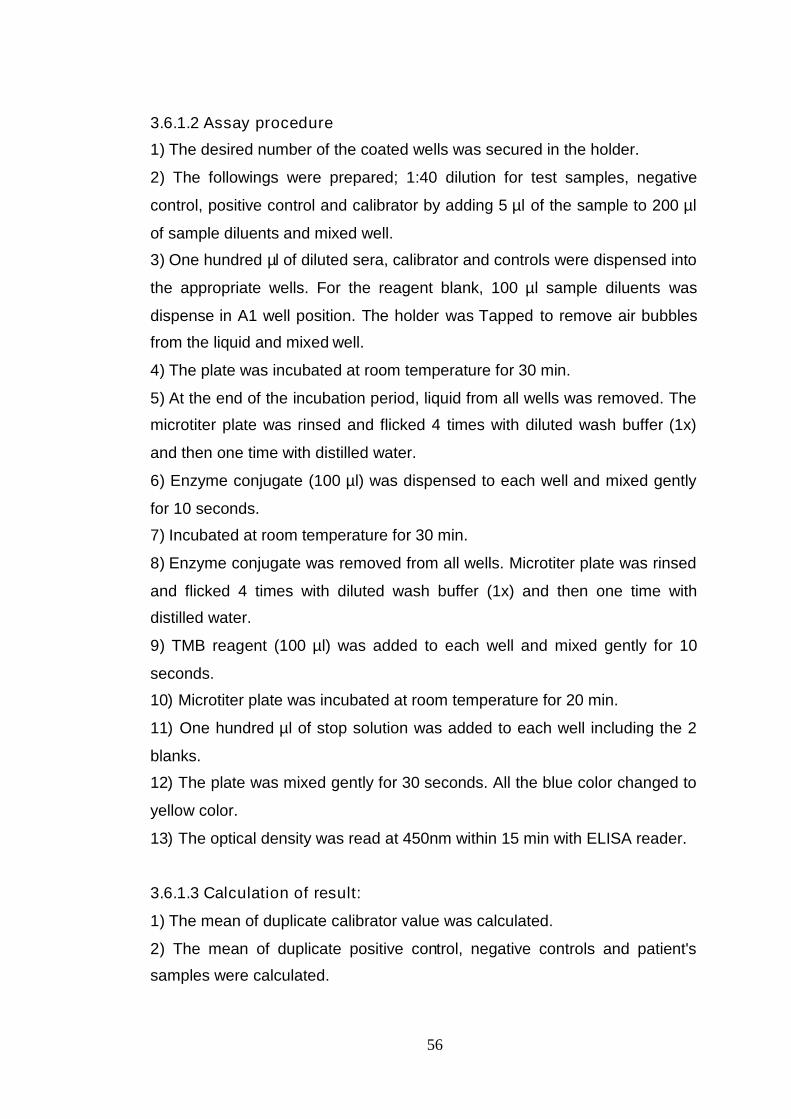
56
3.6.1.2 Assay procedure
1) The desired number of the coated wells was secured in the holder.
2) The followings were prepared; 1:40 dilution for test samples, negative
control, positive control and calibrator by adding 5 µl of the sample to 200 µl
of sample diluents and mixed well.
3) One hundred µl of diluted sera, calibrator and controls were dispensed into
the appropriate wells. For the reagent blank, 100 µl sample diluents was
dispense in A1 well position. The holder was Tapped to remove air bubbles
from the liquid and mixed well.
4) The plate was incubated at room temperature for 30 min.
5) At the end of the incubation period, liquid from all wells was removed. The
microtiter plate was rinsed and flicked 4 times with diluted wash buffer (1x)
and then one time with distilled water.
6) Enzyme conjugate (100 µl) was dispensed to each well and mixed gently
for 10 seconds.
7) Incubated at room temperature for 30 min.
8) Enzyme conjugate was removed from all wells. Microtiter plate was rinsed
and flicked 4 times with diluted wash buffer (1x) and then one time with
distilled water.
9) TMB reagent (100 µl) was added to each well and mixed gently for 10
seconds.
10) Microtiter plate was incubated at room temperature for 20 min.
11) One hundred µl of stop solution was added to each well including the 2
blanks.
12) The plate was mixed gently for 30 seconds. All the blue color changed to
yellow color.
13) The optical density was read at 450nm within 15 min with ELISA reader.
3.6.1.3 Calculation of result:
1) The mean of duplicate calibrator value was calculated.
2) The mean of duplicate positive control, negative controls and patient's
samples were calculated.
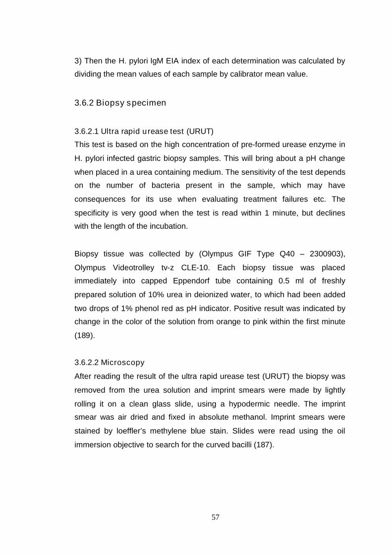
57
3) Then the H. pylori IgM EIA index of each determination was calculated by
dividing the mean values of each sample by calibrator mean value.
3.6.2 Biopsy specimen
3.6.2.1 Ultra rapid urease test (URUT)
This test is based on the high concentration of pre-formed urease enzyme in
H. pylori infected gastric biopsy samples. This will bring about a pH change
when placed in a urea containing medium. The sensitivity of the test depends
on the number of bacteria present in the sample, which may have
consequences for its use when evaluating treatment failures etc. The
specificity is very good when the test is read within 1 minute, but declines
with the length of the incubation.
Biopsy tissue was collected by (Olympus GIF Type Q40 – 2300903),
Olympus Videotrolley tv-z CLE-10. Each biopsy tissue was placed
immediately into capped Eppendorf tube containing 0.5 ml of freshly
prepared solution of 10% urea in deionized water, to which had been added
two drops of 1% phenol red as pH indicator. Positive result was indicated by
change in the color of the solution from orange to pink within the first minute
(189).
3.6.2.2 Microscopy
After reading the result of the ultra rapid urease test (URUT) the biopsy was
removed from the urea solution and imprint smears were made by lightly
rolling it on a clean glass slide, using a hypodermic needle. The imprint
smear was air dried and fixed in absolute methanol. Imprint smears were
stained by loeffler’s methylene blue stain. Slides were read using the oil
immersion objective to search for the curved bacilli (187).

58
3.6.3 Stool sample
3.6.3.1 Kit components
H. pylori Ag (stool) ELISA, Cat #: EIA-4354 Kit from DRG, Germany
Instruments GmbH. Tests: 48 wells. Two kits were used. Antibody coated
microwells, positive control, negative control, sample diluent, wash buffer,
enzyme conjugate, substrate, stop solution, transfer pipettes, strip holder,
strip sealer and wooden stick applicators.
3.6.3.2 Pre assay controls and operations
1) Stool sample was prepared as described in figure 3.3, which illustrate the
instructions for the proper use of H. pylori Ag extraction kit.
2) The liquid components were checked by the naked-eye for visible particles
or aggregates (to eliminate the possibility of contamination). The
chromogen/substrate was checked for color (colorless or pale blue) by
aspirating a small volume of it with a sterile transparent plastic pipette. All
other components were checked according to the manufacturer
recommendations.
3) The content of the 20x concentrated wash solution was diluted with the
proper diluent.
4) The calibrator set were dissolved.
5) All the other components were allowed to reach room temperature (about
1 hr) and then mixed by vortex.
6) The ELISA incubator was set at 37 oC.
7) The ELISA reader was turned on at least 20 minutes before reading.

59
Instruction for use
Open the collection device andintroduce the extraction brush deeplyinto the specimen. Rotate the brush 3-4times in order to collect the rightamount of sample (about 0.2 g).
Transfer the brush into a test tube.Add 1ml Extraction Buffer and then mixon vortex for 1 min in order to dissolvethe sample in solution.
Discard the brush and insert the filterpiston into the tube. Push the pistondown to the bottom in order to release asoluble filtered material with thePasteur pipette.
.
Figure (3.3): Schematic presentation of the HpSAg detection procedure
1
2
3
Extractionbrush
1
Collection deviceand sample
2
Test tube andextraction buffer
Vortex
3
Filtered sample
Well
Filtering piston
Pasteur pipette
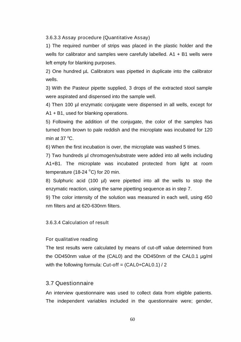
60
3.6.3.3 Assay procedure (Quantitative Assay)
1) The required number of strips was placed in the plastic holder and the
wells for calibrator and samples were carefully labelled. A1 + B1 wells were
left empty for blanking purposes.
2) One hundred µL Calibrators was pipetted in duplicate into the calibrator
wells.
3) With the Pasteur pipette supplied, 3 drops of the extracted stool sample
were aspirated and dispensed into the sample well.
4) Then 100 µl enzymatic conjugate were dispensed in all wells, except for
A1 + B1, used for blanking operations.
5) Following the addition of the conjugate, the color of the samples has
turned from brown to pale reddish and the microplate was incubated for 120
min at 37 oC.
6) When the first incubation is over, the microplate was washed 5 times.
7) Two hundreds µl chromogen/substrate were added into all wells including
A1+B1. The microplate was incubated protected from light at room
temperature (18-24 oC) for 20 min.
8) Sulphuric acid (100 µl) were pipetted into all the wells to stop the
enzymatic reaction, using the same pipetting sequence as in step 7.
9) The color intensity of the solution was measured in each well, using 450
nm filters and at 620-630nm filters.
3.6.3.4 Calculation of result
For qualitative reading
The test results were calculated by means of cut-off value determined from
the OD450nm value of the (CAL0) and the OD450nm of the CAL0.1 µg/ml
with the following formula: Cut-off = (CAL0+CAL0.1) / 2
3.7 Questionnaire
An interview questionnaire was used to collect data from eligible patients.
The independent variables included in the questionnaire were; gender,
61
weight, height, age, civil state, tea and coffee drinking, smoking, type of food
ingested (meat, fish, vegetables, othres), type of water drinking during
childhood and adulthood (filtered, municipality or well water), garbage
collection in the neighbourhood, indicators of socioeconomic status:
educational level, family income, type of accommodation (house, flat, villa),
water supply (well, municipality), sewage system, number of rooms and
number of persons residing in each dwelling, contact with animal, travelling
abroad, consuming antibiotics, consuming drugs and dental complain (See
annex for the Arabic questionnaire).
3.8 Analysis of Data
Data generated from the study was tabulated as Microsoft Excel sheets and
uploaded to Statistical Package for Social Sciences (SPSS version 11).
Cross tabulation of variables were generated. Chi square was used to detect
statistically significant correlation among variables.

Chapter IVResults
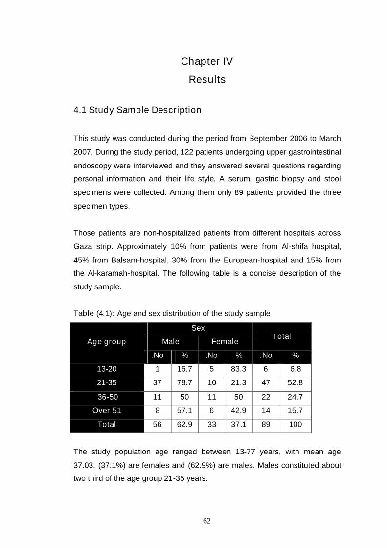
62
Chapter IV
Results
4.1 Study Sample Description
This study was conducted during the period from September 2006 to March
2007. During the study period, 122 patients undergoing upper gastrointestinal
endoscopy were interviewed and they answered several questions regarding
personal information and their life style. A serum, gastric biopsy and stool
specimens were collected. Among them only 89 patients provided the three
specimen types.
Those patients are non-hospitalized patients from different hospitals across
Gaza strip. Approximately 10% from patients were from Al-shifa hospital,
45% from Balsam-hospital, 30% from the European-hospital and 15% from
the Al-karamah-hospital. The following table is a concise description of the
study sample.
Table (4.1): Age and sex distribution of the study sample
Sex
Male FemaleTotal
Age group
No. % No. % No. %
13-20 1 16.7 5 83.3 6 6.8
21-35 37 78.7 10 21.3 47 52.8
36-50 11 50 11 50 22 24.7
Over 51 8 57.1 6 42.9 14 15.7
Total 56 62.9 33 37.1 89 100
The study population age ranged between 13-77 years, with mean age
37.03. (37.1%) are females and (62.9%) are males. Males constituted about
two third of the age group 21-35 years.

63
4.2 Ultra Rapid Urease Test
The test which was performed on biopsy collected from patients during upper
gastroscopy proved to be rapid and simple. The following table (4.2) shows
the results of rapid urease test which indicate 32.6% positivity. Figure (4.1)
shows positive (pink to red) and a negative (yellow to orange) URUT.
Figure (4.1): Ultra rapid urease test (left positive; right negative).
4.3 Gastric biopsies Stained with Methylene Blue
Although simple to perform and does not require special equipment, the
reading and interpreting of results was somewhat tedious and requires time.
From the 86 biopsies stained with Methylene blue, 40 showed H. pylori
constituting 46.5% (table 4.2). Three samples were lost during the course of
this work. Figure 4.2 illustrates a positive Methylene blue stained smear for
H. pylori.

64
Figure (4.2): Methylene blue stained gastric biopsies showing the helical
bacilli
4.4 H. pylori Serum IgM
The result of H. pylori Serum IgM performed using ELISA Kit is listed in table
(4.2). According to the manufacturer recommendations of interpreting
absorbances of serum samples, 40 (44.9%) of the samples were considered
positive.
4.5 H. pylori antigen Detection From Stool
H. pylori antigen detection from stool is receiving attention from service
laboratories as well as from researchers. According to the manufacturer
recommendation of interpreting absorbances of the extracted stool samples,
32 (36%) were considered positive (table 4.2).
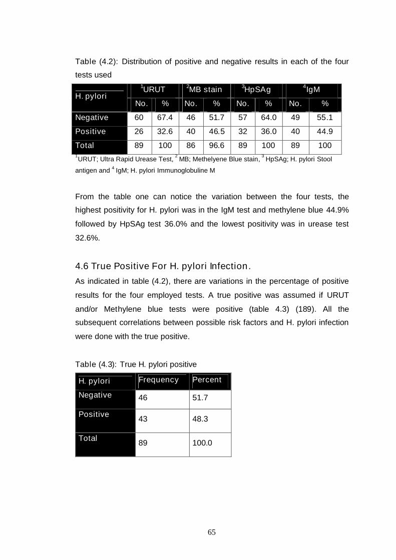
65
Table (4.2): Distribution of positive and negative results in each of the four
tests used1URUT 2MB stain 3HpSAg 4IgM
H. pyloriNo. % No. % No. % No. %
Negative 60 67.4 46 51.7 57 64.0 49 55.1
Positive 26 32.6 40 46.5 32 36.0 40 44.9
Total 89 100 86 96.6 89 100 89 1001URUT; Ultra Rapid Urease Test, 2 MB; Methelyene Blue stain, 3 HpSAg; H. pylori Stool
antigen and 4 IgM; H. pylori Immunoglobuline M
From the table one can notice the variation between the four tests, the
highest positivity for H. pylori was in the IgM test and methylene blue 44.9%
followed by HpSAg test 36.0% and the lowest positivity was in urease test
32.6%.
4.6 True Positive For H. pylori Infection.
As indicated in table (4.2), there are variations in the percentage of positive
results for the four employed tests. A true positive was assumed if URUT
and/or Methylene blue tests were positive (table 4.3) (189). All the
subsequent correlations between possible risk factors and H. pylori infection
were done with the true positive.
Table (4.3): True H. pylori positive
H. pylori Frequency Percent
Negative 46 51.7
Positive 43 48.3
Total 89 100.0

66
4.7 Statistical Analysis of H. pylori Tests Results (Chi square)
In table (4.4), (4.5) and (4.6) there were statistically significant differences
between MB and URUT, IgM and HpSAg test with P value<0.01. As shown in
table (4.4) the agreement in positive result between MB and URUT test was
92.9%. In table (4.5) the difference between IgM test and MB were significant
with P value<0.01. The agreement were 69.2% in positive result. In table
(4.6) the agreement between MB and HpSAg test were 96.8% in positive
result and in table (4.7) the agreement between URUT and HpSAg test were
89.7% in positive result, URUT and HpSAg gave statistical significant
differences with P=0.01. In table (4.8) the difference between IgM and URUT
was not significant (P=0.415), but the difference between HpSAg and IgM
that showed in table (4.9) was significant with P=0.034 and the agreement
between the positive result 47.5%.
Table (4.4): Chi square test for statistical differences between the results of
Methylene blue stain and URUT
MB test
Negative PositiveUltra rapid urease
testNo % No. %
P
value
Negative 44 75.9 14 24.1
positive 2 7.1 26 92.9<0.01
Total 46.0 53.5 40 46.5
Table (4.5): Chi square test for statistical differences between the results of
Methylene blue stain and IgM in serum
MB test
Negative PositiveIgM in serum
No % No. %
P
value
Negative 34 72.3 13 27.7
positive 12 30.8 27 69.2
Total 46 53.5 40 46.5
<0.01

67
Table (4.6): Chi square test for statistical differences between the results of
Methylene blue stain and HpSAg
MB test
Negative PositiveHpSAg
No % No. %
P
value
Negative 45.0 81.8 10.0 18.2
positive 1.0 3.2 30.0 96.8
Total 46.0 53.5 40.0 46.5
<0.01
Table (4.7): Chi square test for statistical differences between the results of
URUT and HpSAg
HpSAg
Negative PositiveURUT
No. % No. %
P
value
Negative 54.0 90.0 6.0 10.0
positive 3.0 10.3 26.0 89.7
Total 57.0 64.0 32.0 36.0
<0.01
Table (4.8): Chi square test for statistical differences between the results of
IgM and URUT
Ultra rapid urease test
Negative PositiveIgM in serum test
No % No. %
P value
Negative 34 69.4 15 30.6
positive 26 65.0 14 35.0
Total 60 67.4 29 32.6
0.415

68
Table (4.9): Chi square test for statistical differences between the results of
IgM and HpSAg
HpSAg
Negative PositiveIgM in serum test
No % No. %
P value
Negative 36 73.5 13 26.5
positive 21 52.5 19 47.5
Total 57 64.0 36 36.0
0.034
Table (4.10): Pearson correlation between the different H. pylori tests.
Test MB URUT IgM HpSAgTrue
positive
Correlation .646(**) .415(**) .757(**) .954(**)MB
Sig. (2-tailed) 0.0 0.0 0.0 0.0
Correlation .646(**) .047 .778(**) .719(**)URUT
Sig. (2-tailed) 0.0 0.7 0.0 0.0
Correlation .415(**) .047 .217(*) .347(**)IgM
Sig. (2-tailed) 0.0 0.7 0.0 0.0
Correlation .757(**) .778(**) .217(*) .728(**)HpSAg
Sig. (2-tailed) 0.0 0.0 0.0 0.0
Correlation .954(**) .719(**) .347(**) .728(**)True positive
Sig. (2-tailed) 0.0 0.0 0.0 0.0
** Correlation is significant at the 0.01 level (2-tailed).
* Correlation is significant at the 0.05 level (2-tailed).
Table (4.10) shows that there were correlation between the four tests, MB
shows a significant results with P value <0.01 with the other three test and
there is a high compatibility of negative and positive results with URUT, IgM,
HpSAg test and increased to 0.954 with the true positive infection.
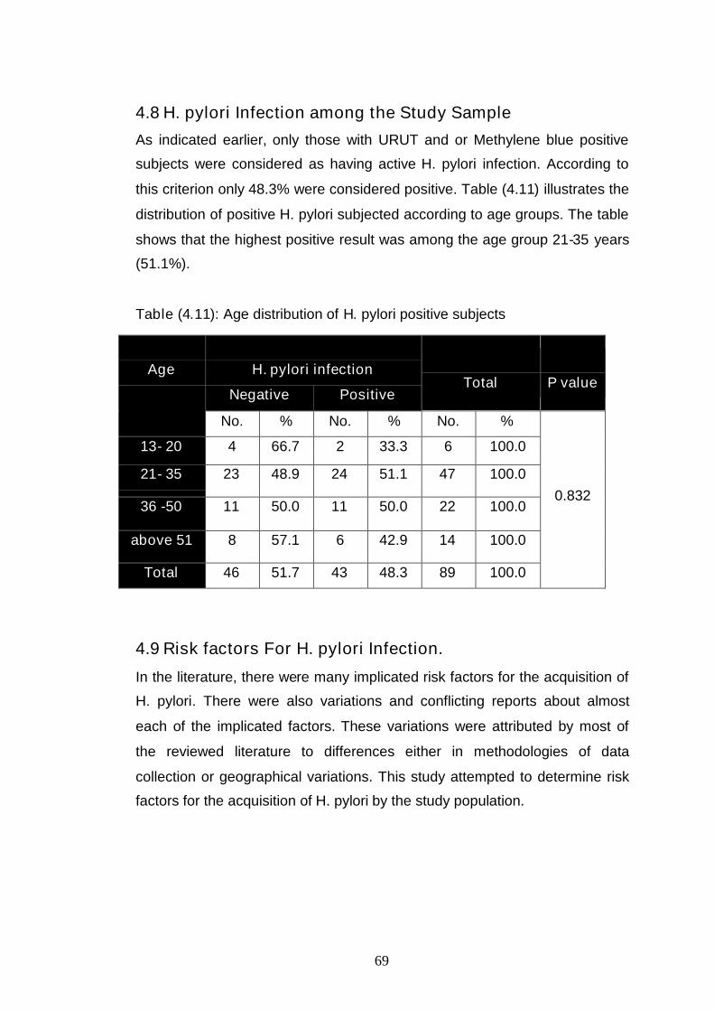
69
4.8 H. pylori Infection among the Study Sample
As indicated earlier, only those with URUT and or Methylene blue positive
subjects were considered as having active H. pylori infection. According to
this criterion only 48.3% were considered positive. Table (4.11) illustrates the
distribution of positive H. pylori subjected according to age groups. The table
shows that the highest positive result was among the age group 21-35 years
(51.1%).
Table (4.11): Age distribution of H. pylori positive subjects
4.9 Risk factors For H. pylori Infection.
In the literature, there were many implicated risk factors for the acquisition of
H. pylori. There were also variations and conflicting reports about almost
each of the implicated factors. These variations were attributed by most of
the reviewed literature to differences either in methodologies of data
collection or geographical variations. This study attempted to determine risk
factors for the acquisition of H. pylori by the study population.
H. pylori infection
Negative PositiveTotal P value
Age
No. % No. % No. %
13- 20 4 66.7 2 33.3 6 100.0
21- 35 23 48.9 24 51.1 47 100.0
36 -50 11 50.0 11 50.0 22 100.0
above 51 8 57.1 6 42.9 14 100.0
Total 46 51.7 43 48.3 89 100.0
0.832
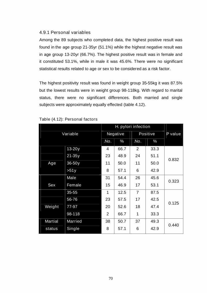
70
4.9.1 Personal variables
Among the 89 subjects who completed data, the highest positive result was
found in the age group 21-35yr (51.1%) while the highest negative result was
in age group 13-20yr (66.7%). The highest positive result was in female and
it constituted 53.1%, while in male it was 45.6%. There were no significant
statistical results related to age or sex to be considered as a risk factor.
The highest positivity result was found in weight group 35-55kg it was 87.5%
but the lowest results were in weight group 98-118kg. With regard to marital
status, there were no significant differences. Both married and single
subjects were approximately equally effected (table 4.12).
Table (4.12): Personal factors
H. pylori infection
Negative PositiveVariable
No.. % No.. %
P value
Age
13-20y
21-35y
36-50y
>51y
4
23
11
8
66.7
48.9
50.0
57.1
2
24
11
6
33.3
51.1
50.0
42.9
0.832
Sex
Male
Female
31
15
54.4
46.9
26
17
45.6
53.10.323
Weight
35-55
56-76
77-97
98-118
1
23
20
2
12.5
57.5
52.6
66.7
7
17
18
1
87.5
42.5
47.4
33.3
0.125
Martial
status
Married
Single
38
8
50.7
57.1
37
6
49.3
42.90.440

71
4.9.2 Life style variables
Life style varies from region to region and depends on many factors such as
socioeconomic factors, geographical distribution and religion. Such variations
are used in justifying conflicting results obtained by different investigators.
As shown in table (4.13), there is no significant difference between smokers
and non smokers with regard to H. pylori infection. The percentage of
positive results for smokers and non smokers were 46.9% and 49.1%
respectively. Even the number of cigarettes seems not to affect or increase
the possibility of infection. There is a statistically significant differences
among subjects who drinks tea and those who do not with P value = 0.045.
Subjects who drinks coffee are less likely to develop H. pylori infection
(41.0%) as compared to those who don’t drink coffee (54.0%). However, the
difference is not statistically significant. With regard to the number of cups
per day, lower percentage of H. pylori infection was observed among those
who consume more than 5 cups per day.
From statistical analysis of data, the type of water drunk during childhood
could be considered as a risk factor with P value=0.018. As shown in table
(4.13), the positive results were high in subjects who drunk municipality or
well water during childhood 53.2% while subjects who drunk filtered water
during childhood have 16.7% positive results. However, the type of drinking
water during adulthood did not influence the outcome of H. pylori infection.
Oral hygiene status of the subjects was assessed by asking the patient if
he/she had a dental complains. H. pylori infection was high in subjects with
dental complains 64.3%, but this result was not statistically significant.
Contact with animals was evaluated as a possible risk factor. The subjects
were classified as either those who handle animals or those who do not. It

72
was found that 50% was positive among those who have contact with
animals and 47.8% for those who don’t.
The subjects were asked if they traveled abroad or not and the positive result
in patients who traveled was 41.2% while the positive result in patients who
did not travel was 50% and there was no statistical significance difference
observed.
Table (4.13): Life style variables
H. pylori infection
Negative PositiveVariable
No. % No. %
Pvalue
SmokingNoYes
2917
50.953.1
2815
49.146.9
0.507
If smokingNo. of cigarettes per day
1-20>20
134
48.180.0
141
51.920.0
0.208
Drink tea NoYes
145
14.354.9
637
85.745.1
0.045
How many cups per day 1-5>5
369
54.556.3
307
45.543.8
0.564
Drink of coffee NoYes
2323
46.059.0
2716
54.041.0
0.158
How many cups per day 1-5>5
203
55.6100
16-
44.4-
0.194
Type of drinking waterduring childhood
Municipality orwell waterFiltered water
36
10
46.8
83.3
41
2
53.2
16.70.018
Type of drinking waterduring adulthood
Municipality orwell waterFiltered water
8
37
50.0
50.7
8
34
50.0
46.50.548
Dental complains YesNo
541
35.754.7
934
64.345.3
0.156
Consumed drugs No.Yes
2818
49.156.3
2914
50.943.8
0.336
Consumed antibiotics in thelast month
NoYes
2323
46.957.5
2617
53.142.5 0.218
Contact with animals NoYes
3610
52.250.0
3310
47.850.0 0.532
Traveling abroad NoYes
3610
50.058.8
367
50.041.2 0.351
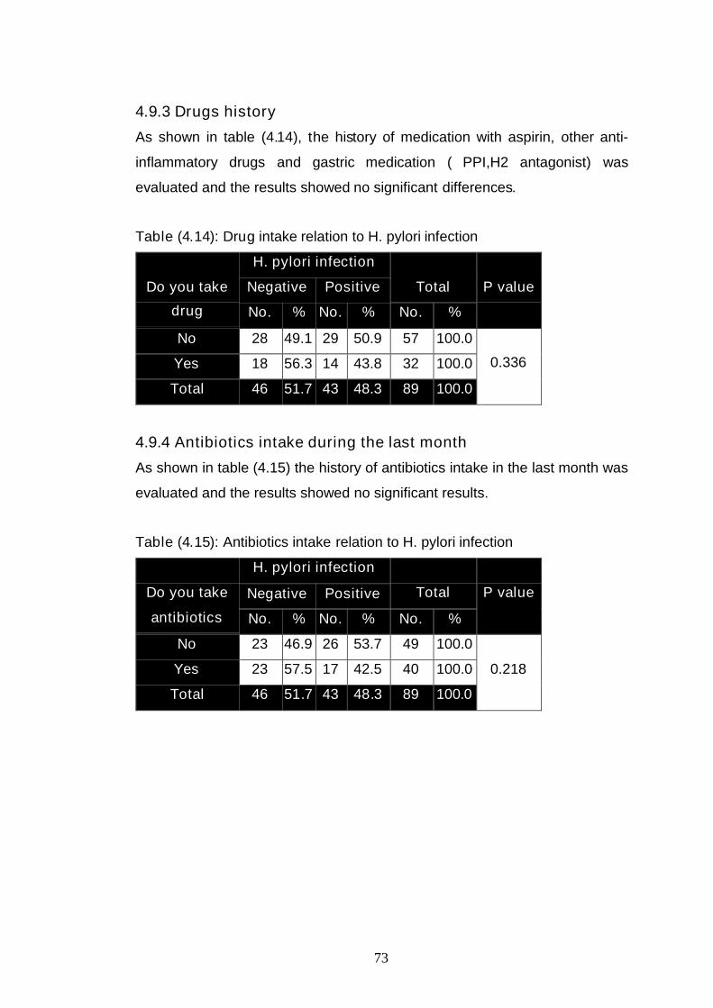
73
4.9.3 Drugs history
As shown in table (4.14), the history of medication with aspirin, other anti-
inflammatory drugs and gastric medication ( PPI,H2 antagonist) was
evaluated and the results showed no significant differences.
Table (4.14): Drug intake relation to H. pylori infection
H. pylori infection
Negative Positive TotalDo you take
drug No. % No. % No. %
P value
No 28 49.1 29 50.9 57 100.0
Yes 18 56.3 14 43.8 32 100.0
Total 46 51.7 43 48.3 89 100.0
0.336
4.9.4 Antibiotics intake during the last month
As shown in table (4.15) the history of antibiotics intake in the last month was
evaluated and the results showed no significant results.
Table (4.15): Antibiotics intake relation to H. pylori infection
H. pylori infection
Negative Positive TotalDo you take
antibiotics No. % No. % No. %
P value
No 23 46.9 26 53.7 49 100.0
Yes 23 57.5 17 42.5 40 100.0
Total 46 51.7 43 48.3 89 100.0
0.218
74
4.9.5 Socioeconomic status
For all 89 subjects who had participated in this study, the level of education,
monthly income, type of accommodation, number of rooms, number of
persons lived in each room, type of water supply and the sewage system
were recorded and correlated to H. pylori infection.
As shown in table (4.16), the level of education did not significantly influence
H. pylori infection. The highest positive result was found in those with
intermediate school level (66.7%) and the lowest positive result was in
university level (36.8%).
The monthly income of subjects was classified to those with 1000 NIS, 1000-
2000 NIS, 2000-3000 NIS and more than 3000 NIS. The lowest positive
result was observed in subjects with more than 3000 per month (22.0%).
However, this result was not statistically significant.
With regard to the type of accommodation, the number of persons lived in the
accommodation, type of water and type of the sewage system, no significant
differences were observed.
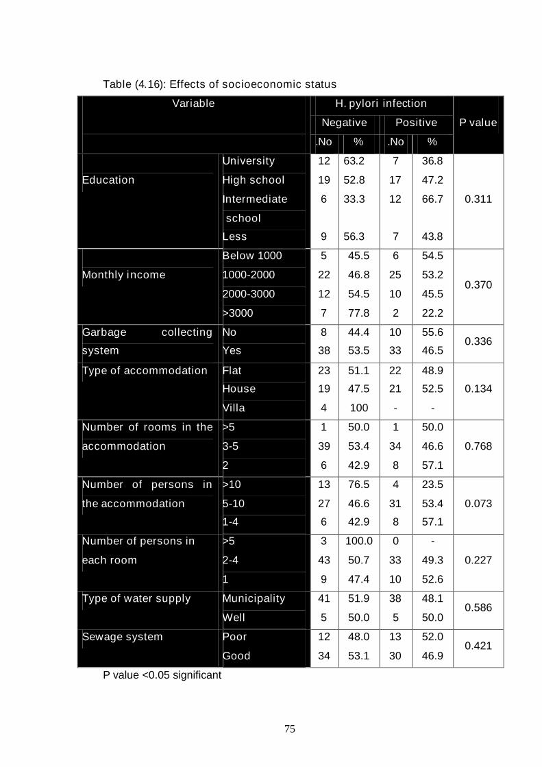
75
Table (4.16): Effects of socioeconomic status
H. pylori infection
Negative Positive
Variable
No. % No. %
P value
Education
University
High school
Intermediate
school
Less
12
19
6
9
63.2
52.8
33.3
56.3
7
17
12
7
36.8
47.2
66.7
43.8
0.311
Monthly income
Below 1000
1000-2000
2000-3000
>3000
5
22
12
7
45.5
46.8
54.5
77.8
6
25
10
2
54.5
53.2
45.5
22.2
0.370
Garbage collecting
system
No
Yes
8
38
44.4
53.5
10
33
55.6
46.50.336
Type of accommodation Flat
House
Villa
23
19
4
51.1
47.5
100
22
21
-
48.9
52.5
-
0.134
Number of rooms in the
accommodation
>5
3-5
2
1
39
6
50.0
53.4
42.9
1
34
8
50.0
46.6
57.1
0.768
Number of persons in
the accommodation
>10
5-10
1-4
13
27
6
76.5
46.6
42.9
4
31
8
23.5
53.4
57.1
0.073
Number of persons in
each room
>5
2-4
1
3
43
9
100.0
50.7
47.4
0
33
10
-
49.3
52.6
0.227
Type of water supply Municipality
Well
41
5
51.9
50.0
38
5
48.1
50.00.586
Sewage system Poor
Good
12
34
48.0
53.1
13
30
52.0
46.90.421
P value <0.05 significant

Chapter VDiscussion

76
Chapter (V)Discussion
H. pylori is a spiral-shaped microaerophilic Gram-negative bacterium that
colonizes the gastric mucosa of human beings. The microorganism is the
major agent of gastritis and plays an important role in the pathogenesis of
peptic ulcer and gastric cancer (190). H. pylori is believed to infect the host
by the fecal-oral route and home to the gastric mucosa. Although it is acid-
sensitive, H. pylori can survive in the stomach for short periods by
neutralizing the gastric acid. The current therapy for H. pylori infection is
efficacious but the treatment regimen is complex and demanding on the
patient and it does not provide resistance to future infections (191). Current
data suggest that the overall prevalence of H. pylori infection is higher both in
developing countries and in lower socioeconomic groups in the developed
world. Probably these populations are exposed to conditions that favor the
acquisition of the microorganism such as precarious hygiene, crowded
household conditions and deficient sanitation (192).
The major risk factor for acquiring H. pylori infection is poor socioeconomic
conditions during childhood. The rate of H. pylori infection in developed
countries ranges from 10% in children to 60% in 60 year olds (193).
Various methods are available for detecting H. pylori, but all have limitations.
H. pylori infection can be diagnosed by invasive test tests requiring
endoscopy (rapid urease test, histology, culture) and by noninvasive tests
(carbon13 urea breath test, serology, stool tests) (194).
The aim of this study is to determine the rate of H. pylori infection in the
population of Gaza strip, and to evaluate possible risk factors linked to
acquisition of the microorganism.
77
In this study 122 patients undergoing upper gastrointestinal endoscopy were
interviewed and serum, biopsy and stool specimens were collected. Among
them only 89 patients provided the three specimen types. Data concerning
age, sex, use of medication, water supply, sewage, number of rooms in the
household, number of persons residing in each dwelling and monthly family
income were obtained during interviews. Categorical variables were analyzed
using the Chi-square test. The study population age ranged between 13-77
years, with mean age of 37.03, (37.1%) are females and (62.9%) are males.
5.1 H. pylori tests
In our study four tests were used to evaluate H. pylori infection.
1- Invasive test required endoscopy for (URUT, MB staining).
2- Non invasive test including IgM detection in serum sample and HpSAg test
in stool sample.
5.1.1 Evaluation of H. pylori Tests
Unfortunately, at present, no single test can be relied upon to detect definitely
H. pylori infection (195) and a combination of tests is recommended as gold
standard (196). A patient was considered an H. pylori-positive if culture alone
or histology plus rapid urease test (RUT) were positive (197).
This fact was also evident in our study. As shown in table (4.2), there are
variations in the results between the four tests that we used in this study and
the highest positive results were observed with the IgM test (44.9%) then MB
test (46.5%), HpSAg test (36.0%) while the least positivity was observed in
the URUT (32.6%).
These variations could be attributed to several factors; one factor would be
the nature of the test it self; for instance, IgM test detects immunoglobulin M
which is the early responder. IgM is elevated during the early stages of an
exposure to H. pylori. There are two possible outcomes, either the immune
system will eradicate the bacteria or the bacteria will begin to populate the

78
mucosal lining of the upper GI tract. If IgM is the only elevated antibody to H.
pylori, it must be correlated with active symptoms to warrant treatment. If
asymptomatic, it is quite possible that the patient's immune system has won
the battle with H. pylori on its own. If treatment is not chosen at this time, it
would be prudent to retest in 2-3 months to make sure that the patient’s
immune system has in fact eradicated the infection (198).
The decision by the physician to select the proper part to collect a biopsy
during gastroscopy would greatly affect the outcome of the invasive tests
such as methylene blue staining and Ultra rapid urease tests. Patients on or
with a previous history of antibiotic therapy or proton pump inhibitors may
show negative results with tests that depend on the activity of H. pylori rather
than its mere presence. Another possible cause of such variations is the
cross-reactivity with other bacteria (e.g. campylobacter) by some tests such
as HpSAg. These differences between the four tests may in part be due to
patchy distribution in gastric mucosa, fastidious nature, mucosal atrophy,
intestinal metaplasia (in stomach), administration of antibiotics (due to some
other infection or protozoal infestation) and proton pump inhibitors. If patients
under medication or antibiotics against H. pylori infection, this may lead to the
death or inhibition of bacteria and as a consequence will not produce urease
enzyme and exhibit negative results in URUT and reduce the number in MB
stained smears. While IgM test may not be affected as well as HpSAg (199).
In a study done by Silvio K Ogata (2002) in Brazil, the results showed that
the highest agreement between two tests occurred in histology plus rapid
urease test combination (91.7%). Similar to results were observed in other
study (200).
In another study in Pakistan, (52/109) 48% of the study population used PPI
before presentation to the outpatient clinics, Pronto Dry (a commercial
urease test) was positive in 40% (44/109) and negative in 60% (65/109).
Histopathology was positive for H. pylori in 57% (62/109) and negative in

79
43%. In this study, treatment with a PPI before endoscopy reduced the
sensitivity of urease test from antral biopsies for H. pylori detection. Ideally
PPI should be discontinued before the endoscopy (201). In patients on PPI,
the biopsy specimen may contain low bacterial density of viable cells giving a
negative urease test. This also leads to lack of H. pylori identification on
histology. Of the various tests that are available for H. pylori detection,
histological examination of gastric biopsy is considered the most accurate
method of diagnosis (202). The migration of H. pylori from the antrum to the
fundus was also associated with a corresponding decrease in the activity of
antral gastritis and matched by a progressive fall in the excretion of 13C urea
breath test (203). If more than one gastric biopsy tissue is used to inoculate
the rapid urease test, a positive test might appear, thus improving the test
sensitivity without compromising its specificity. The diagnostic yield is said to
be increased by over 5% by taking more than a single biopsy (202).
Biopsy urease tests for H. pylori all have inherent inadequacies, notably in
patients with bleeding peptic ulcers, in which a false-negative result might be
found. Chan et al. found that in 480 H. pylori-associated ulcers, 49 H. pylori
infections were missed by a biopsy urease test, which was performed at
initial endoscopy during the bleeding episode. This result is probably caused
by the buffering effects of blood and plasma, which can interfere with the pH
indicator used in urease test (204). Gisbert et al. pointed out in his meta-
analysis that the diagnostic sensitivities of biopsy urease tests and histology
could be increased to 78% and 83%, respectively, if both antrum and corpus
biopsies were obtained. The specificity of biopsy-based H. pylori detection
per se is excellent (93%-100%) (205). Multiple H. pylori diagnostic tests, such
as the biopsy urease test, should be performed in the diagnosis of a bleeding
ulcer, and if negative, histology should be examined to increase the accuracy
of diagnosing H. pylori infection (206).

80
5.1.2 The rate of H. pylori infection
In our study there were variations in the percentage of positive results for the
four employed tests. A true positive was assumed if URUT and/or Methylene
blue tests were positive (table 4.3) (189). The rate of H. pylori infection
according the positive results was 48.3%. Our results do not agree with the
prevalence of H. pylori infection among patients in other countries in the
Middle East. These differences in the prevalence is might be due to the fact
that the number of patients included in our study was limited, different tests
used in other countries, different possible risk factor, source of drinking
water, eating habits among the people..
A study in Yemen (1998) showed that the prevalence of H. pylori infection
among 275 dyspeptic patients was 82.2%. In their study they depended on
URUT for detect the infection (207).
Another study in Saudi Arabia (2005) showed that the prevalence among 120
volunteer students was low (35%) in comparison to other study conducted
on a larger number of the Saudi population which showed prevalence of
80% (208).
5.2 Risk factors Associated with H. pylori infection
5.2.1 Age
This study demonstrated that the highest positive results were in the age
group of 21-35 yr (51.1%). There were no statistically significant results and
all age groups were equally exposed to infection. This finding does not agree
with similar studies in developed countries which showed that the infection
begin in younger age and increasing annually with age. This is may be due to
the reason that the number of participant of the older age in our study was
limited.
A study in Brazil, where the infection rate was of 84.7% in subjects 18 to 30
years of age, increasing to 92% in subjects 46-60 years old, while in subject

81
above 60 years old, the prevalence decreased slightly. As a whole, the
prevalence of infection did not increase significantly (P=0.147) with age.
There were no significant differences in the prevalence of H. pylori infection,
when patients were classified by age. This has been explained as being due
to a reduction in the specific serological response among older individuals
and/or to a decreased number of microorganisms as a consequence of
gastric atrophy (209).
In another study conducted in south of Brazil (2005), the author showed
totally different results. Among the 563 eligible individuals, the prevalence
rate of H. pylori infection was 63.4%. In crude analyses, prevalence was
associated with increasing age (19).
5.2.2 Sex
As shown in table (4.12) there is no significant difference in the overall
prevalence of H. pylori infection between males and females. And both of
them appear to be equally exposed. This result is in agreement with other
studies. In Brazil, two hundred and four individuals participated in the study,
49 males and 155 females, with ages ranging from 18 to 80 years. 165 of
204 participants (80%) were H. pylori positive, with no significant gender
differences (P= 0.49) (209).
This finding is also supported by those reported from south-eastern and
central south Brazil, and from Africa and India (210). As has also been found
in other studies made in developing countries, there were no gender
differences in the risk of acquisition of infection (211).
5.2.3 Weight
In table (4.12) the highest positive result was in weight group 35-55 Kg
(87.5%) and this percentage did not increase with increasing weight. This
could be explained by the fact that malnutrition could lower immunity and
therefore increase the susceptibility of those patients to be infected with H.
82
pylori organism. A study in UK (2005) demonstrated a relation between H.
pylori infection and weight lose. Ninety seven patients were H. pylori positive.
Their results suggest that children with dyspepsia and H. pylori infection are
shorter and lighter compared to children without the infection. It is possible
that H. pylori infection may have some detrimental effect on growth,
especially during the pubertal growth spurt (212).
5.2.4 Marital status
Marital status is not a risk factor as shown in table (4.12). Although a slightly
higher percentage of H. pylori infection was observed among married than
single subjects. But the increase was not statistically significant. This
increase may be attributed to age and personal contact. A Libyan study
showed a higher prevalence of H. pylori in married subjects (84%) in
comparison to single subjects (68%) (213).
5.2.5 Smoking
Smoking is considered as a risk factor for many diseases and is implicated
by several studies in the literature as a risk factor for H. pylori infection.
However, the results of this study showed no statistical differences between
smokers and non-smokers (15 (46.9%), 28 (49.1%) respectively) with regard
to H. pylori infection (table 4.13). A striking observation is that smoking of
more than 20 cigarettes reduced the incidence of H. pylori.
There are reasons why smoking might have little effect on, or even increase,
the hostility of the gastric environment to H. pylori. The acid gastric pH
prevents most organisms from thriving or even surviving in the stomach. H.
pylori, however, has an electropositive internal milieu; twice the number of
basic amino acids, arginine and lysine, as Haemophilus influenzae and
Escherichia coli; and powerful urease activity, with the ability to produce both
ammonia and factors that inhibit parietal cell acid production (214). All these
attributes make survival of H. pylori in the stomach less influenced by the
reduction in pH which may accompany with smoking consumption (215).
83
In another study which disagree with our result, El-Barrawy, demonstrated
that infection prevailed mostly (70%) in smokers (113 out of 161). According
to odds ratio, the risk of infection was 5.3 times higher for smokers than non-
smokers, which was significant. They attributed their finding to the destructive
effect of smoking on the immunity of gastric mucosa and lining layers and
hence increasing its susceptibility to infection by H. pylori. Communal Shisha
smoking might carry the risk of passing the infection from a diseased person
to an uninfected one, as oral-to-oral infection (216).
5.2.6 Coffee drinking
There was no statistically significant difference between those who drinks or
don’t drinks coffee. In fact a little bit lower percentage of positive H. pylori
was found in those who drinks coffee (41.0%) than those who do not
(54.0%). The number of cups consumed per day seems to affect the
outcome of H. pylori infection. Among those who drinks more than 5 cups,
none were positive.
A survey in Germany in 1997 on 447 patients with an overall prevalence of
21%., coffee consumption showed a positive dose-response relation with
active infection. The positive relation between coffee consumption and H.
pylori infection identified in that study is consistent with results from a cohort
study among epidemiologists in which the risk of seroconversion (change
from negative to positive results for antibodies to H. pylori in serum) was 4.6
times higher among those who drank more than 2 cups of caffeinated drinks
a day than among the others. The mechanisms underlying this association
require further research (217).
5.2.7 Tea drinking
A significant finding of this study is that (as shown in table 4.13) tea
consumption is a protective factor. Only 45.1% of those who drinks tea were
infected, a very much higher percentage was found among those who do not

84
drink tea (85.7%). This could be explained by the fact that tea has medicinal
effects. This finding is supported by a Japanese study (1999) on the benefits
of tea. Recent studies have presented data that show a variety of biological
activities of tea catechins, compounds which constitute about 15% (dry
weight) of tea. They investigated the antibacterial activity of catechins against
H. pylori in vitro and in vivo. Effect of these compounds wea investigated on
the gastric mucosal injury induced by this organism in Mongolian gerbils.
H. pylori was eradicated in about 10% of the gerbils in each of the catechin.
Although the mechanism of this action is still obscure, structure-activity
relationship studies indicated that the antibacterial activities of catechins were
predominantly related to the gallic acid moiety and the number of hydroxyl
groups. It has also been reported the catechins can damage the membrane
lipid bilayer. Catechins probably damage the membrane of H. pylori.
However, the exact mechanism is still unknown. Moreover, catechins inhibits
the urease activity and motility of H. pylori (218).
5.2.8 Type of drinking water
Type of water drunk during childhood proved to be a detrimental factor with a
statistically significant results (P value=0.018). H. pylori infection rate was
high in subjects who consumed municipality or well water during childhood
(53.2%) in comparison with subjects who consumed filtered water during
childhood (16.7%).
The type of drinking water during adulthood is not considered as a risk factor
for H. pylori infection. Both municipality or well water and filtered water
consumers showed similar infection rates (50.0%) and (47.9%) respectively.
This result is in agreement with other studies in developed and developing
countries. They implicated the type of water during childhood as the main risk
factor for H. pylori infection. The microorganism is transmitted by the fecal-
oral routs in the infected water to the child and persist all the life and as the
result showed that the type of water during adulthood does not affect the
infection rates.

85
In a study conducted in Leipzig, Germany, which consisted of a self-
administered or parent-completed questionnaire (age-dependent), eliciting
information on lifestyle habits and their use/drinking the well water as well as
the H. pylori infection. A total of 91 subjects (44 users of H. pylori positively
and 47 negatively tested wells) were screened for their H. pylori status. The
group was comprised of 42 males and 49 females, i.e., 73 adults and 19
children under the age of 18 (mean age 39.5 years with a range between 3-
80 years). Logistic regression analyses identified the drinking of well water as
the significant risk factor for a positive colonization status (P<0.001). Water
supplies have been identified as possible reservoirs to acquire the bacterium
(219).
5.2.9 Oral hygiene
Higher percentage of H. pylori infection was observed among those with
dental complains (64.3%) than those with out dental complains (45.3%).
Although, the difference is not statistically significant, the result is in
conformity with finding of many investigators who claimed that the bacteria
may persist in the oral cavity and in dental plaques of people with poor
hygiene.
In a study in Australia by Hedley G. Peach on 217 adults randomly selected
from the electoral roll, they found that the positive H. pylori status was
significantly associated with increasing number of tooth surfaces with a high
plaque, they confirmed that living in an overcrowded household during
childhood and visiting a dentist less than once a year are positively
associated with H. pylori infection in Australia. And they found the Negative
H. pylori status was significantly associated with increasing education, having
ever lived on a farm. H. pylori has been cultured from dental plaque, and
identical H. pylori ribotypes were found in the mouth and gastric antrum of
ulcer patients. Moreover, H. pylori has been detected by reverse transcription
polymerase chain reaction in only moderate to heavy accumulations of
86
plaque. If H. pylori inhabits plaque, scaling may lead to ingestion of the
organism and inoculation of the stomach (220).
5.2.10 Antibiotic consumption
Among the study subjects, 40 took antibiotics one day to one month prior
sample collection, 57.5% of them were shown to be negative while among
the 49 who did not consume antibiotics, only 46.9% of them were negative
for H. pylori. Although not statistically significant, the results showed a
reduction in positive cases of H. pylori among those who consumed
antibiotics (table 4.15).
This reduction could be explained by the fact that both URUT and Methylene
blue depends on the activity and the presence of H. pylori in the biopsy and
antibiotic consumption may reduce the number and activity of the organism,
therefore interfering with the result outcome producing false negative results.
5.2.11 Drug consumption
Among the study subjects, 32 took drugs, 56.3% of them were shown to be
negative while among the 57 who did not consume drugs, only 49.1% of
them were negative for H. pylori. Although not statistically significant, the
results showed a reduction in positive cases of H. pylori among those who
consumed drugs (table 4.14).
A rapid urease test is highly specific and simple, and can also be performed
on biopsy samples; however, it may have false-negative results, particularly if
the patient has recently taken a proton pump inhibitor (PPI) (221).
Proton pump inhibitors are known to decrease the activity of H. pylori
organisms within the stomach and to shift their distribution proximally. This
effect may reduce the sensitivity of histological examination and rapid urease
testing for H. pylori on biopsies taken from recommended sites. It is of

87
particular relevance if a proton pump inhibitor has been taken before the
patient has undergone diagnostic endoscopy (201).
Dickey and Kenny, investigated patients referred to open-access upper
gastrointestinal endoscopy service who had either been on no medication
(controls) or were already taking proton pump inhibitors. Biopsies taken from
the gastric antrum and corpus were used for rapid urease testing and for
histological examination. Sera taken from patients who had no evidence of H.
pylori in biopsies, they were tested for IgG H. pylori antibodies as an
alternative indicator of infection. H. pylori organisms were detected by
histological examination in 27 of 40 controls (68%) and in 13 of 25 patients
taking proton pump inhibitors (52%). Among patients with positive histology
(organisms detected in either antral or corpus biopsies, or both), only the
sensitivity of the antral urease test read at 1 h was significantly lower in
patients taking proton pump inhibitors than in controls, with no significant
difference in sensitivities of the antral urease test at 24 h, of the corpus
urease test at 1 or 24 h, or of histology from the antrum or corpus. Of patients
with negative histology, none of 13 controls compared with six of 12 patients
taking proton pump inhibitors (50%) had positive serology (P = 0.005). Five
(83%) of the six histology-negative, seropositive patients taking proton pump
inhibitors had histological changes consistent with H. pylori gastritis even
though no organisms were detected. Treatment with a proton pump inhibitor
before endoscopy reduces the sensitivity of antral and corpus biopsies for H.
pylori detection, both by urease testing and histological examination. If proton
pump inhibitors already prescribed cannot be discontinued for an adequate
period before endoscopy, patients should have biopsies taken from the
corpus as well as from the antrum, and serum should be tested for H. pylori
(201).
5.2.12 Education
Regardless of the educational level, the study population showed no
significance variation (table 4.16). The highest positive result were in
intermediate school level (66.7%) and the lowest positive result was in the
88
university level (36.8%). This finding is in agreement with a study conducted
in Brazil (2005). The study showed that in 165 out of 204 participants (80%)
were H. pylori positive; there were no significant differences in the prevalence
of H. pylori infection, when patients were classified by educational level
(209).
Another study (2005) in south of Brazil conducted by Ina and Jose, showed
different results. The prevalence rate of H. pylori infection was 63.4%. In
crude analyses, prevalence was associated with increasing lower education
level. This variation in findings could be explained by the fact that H. pylori
infection is multifactorial (19).
5.2.13 Income
The highest positive rate was observed in subjects with monthly income of
less than 1000 NIS (54.5%) and the lowest positive result were in patients
with income more than 3000 NIS per month (22.0%). However there is no
statistically significant variation. This contradicts the finding of Ina and Jose
(2005) who noted high prevalence rate in the low income group (19).
5.2.14 Number of persons in the accommodation
As shown in table (4.16), the number of persons living in the accommodation
did not show significant differences. This finding is similar to that of Maria and
Dulciene (2005), their study in poor urban community in Brazil, showed that
in 165 of 204 participants (80%) were H. pylori positive. The number of
persons per room, the number of children per household, the number of
adults per household and household pets, were not risk factors suggesting
that the infection, even in a poor population, may be acquired predominantly
during childhood; the relatively high prevalence that was observed may be
more due to a cohort effect than to acquisition of infection during adulthood
(209). Factors linked to household conditions, such as present family size,
were not relevant determinants of H. pylori infection status in this adult
population, a finding also observed by others, both in developed and in
89
developing countries. In most of these studies, a clear association was seen
between socioeconomic status and overcrowding in childhood, but not with
present household conditions (222). In fact, residential crowding is known to
facilitate transmission of infection within families (223).
Another study in south of Brazil (2005), showed different results. Of 563
eligible individuals, 363 agreed to perform the 13C-UBT (refusal rate of
35.5%). The prevalence rate of H. pylori infection was 63.4% (95%CI 59.3%–
69.3%). In crude analyses, prevalence was associated with increasing higher
size of the family, low socio-economic conditions in childhood, higher number
of siblings and the presence of dyspeptic symptoms. In their study past
socio-economic variables in childhood presented the strongest association
with the occurrence of the infection in adult life (19). This finding is in
agreement with the "birth cohort phenomenon", person-to-person contact
appears to be the most likely mode of transmission, number of siblings;
particularly the number of older siblings, domestic crowding and living in
orphanages are important determinants of the prevalence of H. pylori
infection (224).
5.2.15 Type of accommodation
Variations in housings and accommodations probably reflect variations in
socioeconomic conditions, however, in this study; H. pylori appear to ignore
such variations. As shown in table (4.16) the highest positive results were in
patients who lived in house (52.5%). Therefore, type of accommodation may
not be a risk factor. There was a similar finding by Fiedorek et al (1991) in a
study on children. They found no significant differences in H. pylori infection
related to gender, type of housing and location of housing (225).The type of
water in the accommodation whether municipality or private well was not
significant risk factor for H. pylori infection. Both groups exhibited similar
positive rates.

Chapter VIConclusion and Recommendation

90
Chapter VI
Conclusions and Recommendations
6.1 Conclusions
This study is the first in Gaza strip to detect H. pylori infection and attempted
to determine possible risk factors associated with the acquisition of the
microorganism. From the result of this work, the following conclusions were
drawn:
1. The rate of H. pylori infection among 89 subjects undergoing upper
endoscopy was 48.3%.
2. Ultra Rapid Urease test proved to be reliable, easy to prepare and to
perform, not expensive and the patient can get quick result while still in the
physician office. However, the exclusive use of the rapid urease test for the
diagnosis of H. pylori cannot be recommended in patients with prior
medication use
3. Drugs and antibiotics consumption appears to interfere with tests that
depend on either the presence or activity of H. pylori.
4. No single test among those used in this study could be used reliably to
diagnose active H. pylori Infection.
5. HpSAg test, none invasive, simple and accurate test for detecting H. pylori
in stool sample, however, its use for diagnosis should be evaluated.
6. Also it can be concluded that drinking and/or using H. pylori contaminated
municipality or well water is a risk in the acquisition of H. pylori. Given the
91
public health impact of H. pylori infection, this should be taken into account
when measures of prevention are considered.
7. Age, sex, weight, marital status, smoking, coffee consumption, oral
hygiene status, socioeconomic status include, education level, income, type
of accommodation, number of persons living in the accommodation, number
of persons in each room, type of water, the sewage system, contact with
animals, traveling abroad and drug consumption could not be considered as
a risk factor of H. pylori infection as demonstrated by the results of this study.
8. The statistical analysis of data obtained from this work demonstrated a
significance protective property of tea against H. pylori infection with a
significant P value.
9. Our results support the hypothesis that H. pylori infections in developing
countries is predominantly acquired during childhood.
6.2 Recommendations
1. As a result of our study we recommend that further investigation to detect
the possible sources of H. pylori infection especially drinking water
2. We recommend using urease test at the physician office as a simple, one
minute test.
3. More than one biopsy during gastroscopy is recommended to increase the
sensitivity of tests depending on the activity of the bacterium (URUT and
MB).
4. We recommend that tea leaves should be tested for their effect on H.
pylori both in vivo and vitro.

92
References
(1) Suporn T., Supujchara N., Aruchalean T., Kanit A., Paneeya P., et al,2006- A Rapid Serologic Test and Immunoblotting for the Detection ofHelicobacter pylori Infection in Children. J Trop Pediatr. 52(4):267-287
(2) Marshall B., 2001 - Helicobacter pylori physiology and genetics. Onehundred years of discovery and rediscovery of Helicobacter pylori and itsassociation with peptic ulcer disease. Mobley H., Mendz G., and HazellS.,(eds): ASM Press.: 19-24
(3) Marshall B., Warren J., 1984- Unidentified curved bacilli in the stomachof patients with gastritis and peptic ulceration. Lancet. 1(8390): 1311-1315.
(4) Goodwin C., Armstrong J., Chilvers T., Peters M., Collins D., et al,1989 - Transfer of Campylobacter pylori and Campylobacter mustelae toHelicobacter gen. nov. as Helicobacter pylori comb. nov. and Helicobactermustelae comb. nov., respectively. Int J Syst Bacteriol. 39(4): 397-405.
(5) Goodwin C., Worsley B.,1993- Microbiology of Helicobacter pylori.Gastroenterol. Clin North Am. 22(11) :5-19.
(6) Van Zwet A., Thijs J., Roosendaal R., et al,1996- Review: Practicaldiagnosis of Helicobacter pylori infection. Eur J Gastroenterol Hepatol. 8(5):501-507.
(7) Hu L., Mobley H., 1993-Expression of catalytically active recombinantHelicobacter pylori urease at wild type levels in Escherichia coli. InfectImmun. 61(6):2569-2569.
(8) Chun D., Shun N., Shi H., Jia Y., 2000- Seroepidemiology ofHelicobacter pylori infection among asymptomatic Chinese children. World J.Gastroenterol. 6(5) :759-761.
(9) Blaser M., Atherton J., 2004 -Helicobacter pylori persistence: Biologyand disease. J Clin Invest. 113(3) : 321-333.
(10) Suerbaum S., Michetti P., 2002 -Helicobacter pylori infection. N EnglJ Med. 347(15): 1175-1186
(11) Danesh J., Collins R., Peto R., 1997-Chronic infections and coronaryheart disease: is there a link?. Lancet. 350 ( 9075):430-436
(12) Germán R., Guadalupe A., Geny F., 2001- Helicobacter pylori: Recentadvances in the study of its pathogenicity and prevention. Salud pública deméxico. 43(3):237-247
93
(13) Torres J., Perez-Perez G., Goodman K., Atherton J., Gold B., et al ,2000- A comprehensive review of the natural history of Helicobacter pyloriinfection in children. Arch Med Res.31(5 ):431-469
(14) Lacy B., semore J., 2001- Helicobacter pylori: ulcers and more: thebeginning of an era. J Nutr. 131(27) :89-93
(15) Gold B., 2001- Helicobacter pylori infection in children. Curr ProblPediatr Adolesc Health Care. 31(8):247-266
(16) Brown L., 2000- Helicobacter pylori: epidemiology and routes oftransmission. Epidemiol Rev.22(2) :83-97
(17) Zmira S., Haim S., Yaron N., Gabriel D., Douglas J., et al, 2002-Resistance of Helicobacter pylori isolated in Israel to metronidazole,clarithromycin, tetracycline, amoxicillin and cefixime. J. Antimicrob.Chemother.49(6) :1023-1026.
(18) Abdollah B., Robert W., Remon A., Yongdai K., Malla R., et al, 2000-Seroepidemiology of Helicobacter pylori infection in a population of Egyptianchildren. Inter. J. Epidemiol. 29(5) :928-932.
(19) Ina S., Jose B., Ari S., Neiva C., Camila S., et al, 2005- Prevalence ofHelicobacter pylori infection and associated factors among adults in SouthernBrazil: a population-based cross-sectional Study. BMC Public Health. 5(118):1-10.
(20) Yi-Hui L., Hong G., Peng-Bin Z., Xiao-Yan Z., Si-Ping D., 2004-Clinical value of Helicobacter pylori stool antigen test, ImmunoCard STATHpSA, for detecting H pylori infection. World J Gastroenterol. 10(6) :913-914
(21) Agence d’évaluation des technologies et des modes d’interventionen santé ( AETMIS). 2005- The 13C-Urea Breath Test for Detection ofHelicobacter pylori: Potential Applications in Québec. Report prepared byLonny Erickson (AETMIS 05- 05). Montréal: AETMIS.
(22) Athanasios M., Eva P., Kurt S, Margit W., Manfred L., et al, 1998-Detection of Helicobacter pylori in Stool Specimens by PCR and AntigenEnzyme Immunoassay. J. Clin. Microbiol. 36(9): 2772–2774.
(23) Hoffman J., David R., 2001- Treatment of Helicobacter pylori. CurrOpin Gastroenterol. 17 (1):30–34
(24) Broutet N., Tchamgoue S., Pereira E., Lamouliatte H., Salamon R.,et al, 2003- Risk factors for failure of Helicobacter pylori therapy—results ofan individual data analysis of 2751 patients. Aliment Pharmacol Ther.17(1):99–109.

94
(25) Roosendaal R., Kuipers E., Buitenwerf J., Uffelen C., Meuwissen S.,et al. 1997- Helicobacter pylori and the birth cohort effect: Evidence of acontinuous decrease of infection rates in childhood. Am J Gastroenterol. 92(9):1480-1482.
(26) Czinn S., Cai A., Nedrud J., 1993-Protection of germ-free mice frominfection by Helicobacter felis after active oral or passive IgA immunization.Vaccine. 11(6) :637-642.
(27) Negrini R., Poiesi C., Savio A., Paterlini A., Buffoli F., et al. 1994-Molecular mimicry of gastric antigen by Helicobacter pylori: A role in thepathogenesis of atrophic gastritis? Am J Gastroenterol. (89): 1329.
(28) Sebastian S., Pierre M., 2002- Helicobacter pylori Infection. The NewEngland J. medic. 347(15):1175-1186.
(29) Bellack N., Koehoorn M., MacNab Y., Morshed M., 2006- Aconceptual model of water's role as a reservoir in Helicobacter pyloritransmission: a review of the evidence. Epidemiol Infect. 134(3) : 439-449
(30) Kusters J., Vanvilet A., Kuipers E., 2006- Pathogenesis of Helicobacterpylori infection. CMR. 19(3) : 449-490
(31) Gueneau P.,Loiseaux-De G., 2002- Helicobacter: molecular phylogenyand the origin of gastric colonization in the genus. Infect Genet Evol.1(3):215-223
(32) Owen R., 1998- Helicobacter--species classification and identification.Br Med Bull. 54(1): 17-30.
(33) Fox J., 2002- The non-H pylori helicobacters: their expanding role ingastrointestinal and systemic diseases. Gut. 50(2): 273-283
(34) Enroth H., K. Wreiber, et al. 1999- In vitro aging of Helicobacter pylori:changes in mor- phology, intracellular composition and surface properties.Helicobacter. 4(1): 7-16.
(35) Solnick J., K. Chang, et al. 2003- Natural acquisition of Helicobacterpylori infection in newborn rhesus macaques. J. C.M. 41(12), 5511-5516
(36) Andersen L., Dorland A., Karacan H., Colding H., Nilsson H., et al,2000- Possible clinical importance of the transformation of Helicobacter pyloriinto coccoid forms. Scand J Gastroenterol. 35 (9): 897-903.
(37) Kivi M., Johansson A., Reilly M., Tindberg Y., 2005- Helicobacterpylori status in family members as risk factors for infection in children.Epidemiol. & Infect. 133 (4): 645-652.

95
(38) Testerman T., McGee D., Mobley H., 2001- Helicobacter pyloriphysio logy and genetics. Adherence and colonization. (eds): ASM Press,Washington, D.C. USA.: 381-417.
(39) Mahdavi J., Sondén B., Hurtig M., Olfat F., Forsberg L., et. Al, 2002-Helicobacter pylori SabA adhesin in persistent infection and chronicinflammation. Sci. 297 (5581): 573-578.
(40) Yamaoka Y., Kita M., Kodama T., Imamura S., Ohno T., et al, 2002-Helicobacter pylori infection in mice: Role of outer membrane proteins incolonization and inflammation. Gastroenterol. 123 (6): 1992-2004.
(41) Blaser M., Berg D., 2001- Helicobacter pylori genetic diversity andrisk of human disease. J Clin Invest. 107 (7): 767-773.
(42) Terry K., Williams S., Connolly L., Ottemann K., 2005- Chemotaxisplays multiple roles during Helicobacter pylori animal infection. InfectImmun. 73 (2): 803-811.
(43) Zambon C., Navaglia F., Basso D., Rugge M., Plebani M., 2003-Helicobacter pylori babA2, cagA, and s1 vacA genes work synergistically incausing intestinal metaplasia. J Clin Pathol 56 (4): 287-291.
(44) Taylor D., Rasko D., Sherburne R., Ho C., Jewell L., 1998- Lackof correlation between Lewis antigen expression by Helicobacter pylori andgastric epithelial cells in infected patients. Gastroenterol. 115(5): 1113-1122.
(45) Salaün L., Ayraud S., Saunders N., 2005- Phase variation mediatedniche adaptation during prolonged experimental murine infection withHelicobacter pylori. Microbiol. 151(3): 917-923.
(46) Deitsch K., Moxon E., Wellems T., 1997- Shared themes of antigenicsvariation and virulence in bacteria, protozoa, and fungal infections. MicrobiolMol Biol Rev. 61(1): 281-293
(47) Boneca I., de Reuse H., Epinat J., Pupin M., Labigne A., et al, 2003- Arevised annotation and comparative analysis of Helicobacter pylorigenomes. Nucl. Acid. Res. 31(6): 1704-1714.
(48) Cover T., Berg D., Blaser M., Mobley H., 2001- Principles of BacterialPathogenesis, chapter 11; H. pylori patho. :509-558.
(49) Nilsson C., Sillén A., Eriksson L., Strand M., Enroth H., et al,2003- Correlation between cag pathogenicity island composition andHelicobacter pylori-associated gastroduodenal disease. Infect Immun.71(11): 6573-6581.
96
(50) Evans D., 2000- Helicobacter pylori adhesins: review andperspectives.. Helicobacter. 5 (4):183–195.
(51) Ilver D., Arnqvist A., Ogren J., Frick I., Kersulyte D., et al, 1998-Helicobacter pylori adhesin binding fucosylated histo-blood group antigensrevealed by retagging. Sci. 279 (5349):373- 377.
(52) Soto G., Hultgren S., 1999- Bacterial adhesins: common themes andvariations in architecture and assembly. J.B. 181 (4):1059-1071.
(53) Odenbreit S., Puls J., Sedlmaier B., Gerland E., Fischer W., et al,2000- Translocation of Helicobacter pylori CagA into gastric epithelial cells bytype IV secretion. Sci. 287 (5457):1497-1500.
(54) Kim N., Marcus E., Wen Y., Weeks D., Scott D., et al, 2004- Genes ofHelicobacter pylori Regulated by Attachment to AGS Cells. I.A.I. 72(4) :2358-2368.
(55) Park Y., Richard J.,1998- Contact-dependent protein secretion inPorphyromonas gingivalis. Infect. Immun. 66 (10):4777-4782.
(56) Segal E., Cha J., Lo J., Falkow S., Tompkins L., 1999- Altered states:involvement of phosphorylated CagA in the induction of host cellular growthchanges by Helicobacter pylori. Proc. Natl. Acad. Sci. USA. 96 (25):14559-14564.
(57) Madrid J., Ballesta J., Castells M., Hernandez F., 1990-Glycoconjugate distribution in the human fundic mucosa revealed by lectin-and glycoprotein-gold cytochemistry. Histochem. 95 (2):179-187.
(58) Ebert M., Schandl L., Malfertheiner P., 2002- Helicobacter pyloriinfection and molecular changes in gastric carcinogenesis. J. Gastroenterol.13 (1): 45-49.
(59) McCormick B., Parkos C., Colgan S., Carnes D., Madara J., 1998-Apical secretion of a pathogen-elicited epithelial chemoattractant activity inresponse to surface colonization of intestinal epithelia by Salmonellatyphimurium. J. Immunol. 160 (1):455-466.
(60) Slattery M., Dong C., 2003- Neutrophils influence melanoma adhesionand migration under flow conditions. Int. J. Cancer. 106 (5):713-722.
(61) Blaser M., 1996- The bacteria behind ulcers. Sci. Am. 274 (2):104-107.
(62) Satin B., Del Giudice G., Della Bianca V., Dusi S., Laudanna C., etal, 2000- The neutrophil-activating protein (HP-NAP) of Helicobacter pylori isa protective antigen and a major virulence factor. J. Exp. Med. 191 (9):1467-1476.

97
(63) Kwok T., Backert S., Schwarz H., Berger J., Meyer T., 2002- Specificentry of Helicobacter pylori into cultured gastric epithelial cells via a zipper-like mechanism. Infect. Immun. 70 (4):2108-2120.
(64) Telford J., Covacci A., Rappuoli R., Chiara P., 1997- Immunobiologyof Helicobacter pylori infection. Curr. Opin. Immunol. 9 (4):498-503.
(65) Allen L., 2001- The role of the neutrophil and phagocytosis in infectioncaused by Helicobacter pylori. Curr Opin Infect Dis. 14 (3): 273-277.
(66) Allen L., Schlesinger L., Kang B., 2000- Virulent strains ofHelicobacter pylori demonstrate delayed phagocytosis and stimulatehomotypic phagosome fusion in macrophages. J Exp Med. 191 (1):115-128.
(67) Weeks D., Eskandari S., Scott D., Sachs G., 2000- A H+-gated ureachannel: the link between Helicobacter pylori urease and gastric colonization.Sci. 287 (5452):482–485.
(68) Solnick J., Schauer D., 2001- Emergence of diverse Helicobacterspecies in the pathogenesis of gastric and enterohepatic diseases. ClinMicrobiol Rev. 14 (1):59-97.
(69) Parsonnet J., 1998- Helicobacter pylori: the size of the problem.Gut.;43 (1):6–9.
(70) Murray L., McCrum E., Evans A.,Bamford K., 1997- Epidemiology ofHelicobacter pylori infection among 4742 randomly selected subjects fromNorthern Ireland. Int J Epidemiol. 26(1) :880–887.
(71) Reshetnikov O., Denisova D., Zavyalova L., Haiva V., GranbergC., 2003- Helicobacter pylori seropositivity among adolescents inNovosibirsk, Russia: prevalence and associated factors. J PediatrGastroenterol Nutr. 36 (1):72–76
(72) Bergenzaun P., Kristinsson KG., Thjodleifsson B., SigvaldadottirE., Mölstad S., et al., 1996- Seroprevalence of Helicobacter pylori in southSweden and Iceland. Scand J Gastroenterol. 31(12):1157–1161
(73) Malaty H., Graham D., Isaksson I., Engstrand L., Pedersen N.,1998- Co-twin study of the effect of environment and dietary elements onacquisition of Helicobacter pylori infection. Am J Epidemiol. 148 (8):793–797
(74) Malaty H., Evans D., Evans D., Jr., Graham D., 1992- Helicobacterpylori in Hispanics: Comparison with blacks and whites of similar age andsocioeconomic class. Gastroenterol. 103 (3): 813-816.

98
(75) Malaty H., El-Kasabany A., Graham D., Miller C., Reddy S., et al,2002- Age at acquisition of Helicobacter pylori infection: A follow-up studyfrom infancy to adulthood. Lancet. 359 (9310): 931-935.
(76) Rupnow M., Shachter R., Owens D., Parsonnet J., 2000- A dynamictransmission model for predicting trends in Helicobacter pylori and associateddiseases in the United States. Emerg Infect Dis. 6 (3): 228-237.
(77) Oona M., Utt M., Nilsson I., Uibo O., Vorobjova T., et al, 2004-Helicobacter pylori infection in children in Estonia: decreasingseroprevalence during the 11-year period of profoundsocioeconomicchanges. Helicobacter. 9(3) :233–241
(78) de Oliveira A., Rocha G., Queiroz D., de Moura S., Rabello A.,1999a- Seroconversion for Helicobacter pylori in adults from Brazil. Trans RSoc Trop Med Hyg. 93 (3) :261–263
(79) Rocha G., Rocha A., Silva L., Santos A., Bocewicz A., et al, 2003-Transmission of Helicobacter pylori infection in families of preschool-agedchildren from Minas Gerais, Brazil. Trop Med Int Health. 8 (11) : 987-991
(80) Lambert J., Lin S., Sievert W., Nicholson L., Schembri M., et al,1995-High prevalence of Helicobacter pylori antibodies in aninstitutionalized population: Evidence for person-to-person transmission.AmJ Gastroenterol. 90 (12) : 2167-2171.
(81) Tindberg Y., Bengtsson C., Granath F., Blennow M., Nyrén O., eta l , 2001b- Helicobacter pylori infection in Swedish school children: Lack ofevidence of child-to-child transmission outside the family. Gastroenterol.121 (2) : 310-316.
(82) Tsai C., Perry S., Sanchez L., Parsonnet J., 2005- Helicobacter pyloriinfection in different generations of hispanics in the San Francisco bayarea. Am J Epidemiol. 162 (4) : 351-357.
(83) Woodward M., Morrison C., McColl K., 2000- An investigation intofactors associated with Helicobacter pylori infection. J Clin Epidemiol. 53 (2) :175-181.
(84) Brenner H., Rothenbacher D., Bode G., Dieudonne P., Adler G.,1999- Active infection with Helicobacter pylori in healthy couples. EpidemiolInfect. 122 (1) : 91-95
(85) Mendall M., Goggin P., Molineaux N., Levy J., Toosy T., et al, 1992-Childhood living conditions and Helicobacter pylori seropositivity in adult life.Lancet. 339 (8798) : 896-897

99
(86) Brown L., Thomas T., Ma J., Chang Y., You W., et al, 2002-Helicobacter pylori infection in rural China: Demographic, lifestyle andenvironmental factors. Int J Epidemiol 31.(3) : 638-645
(87) Glynn M., Friedman C., Gold B., Khanna B., Hutwagner L., et al,2002- Seroincidence of Helicobacter pylori infection in a cohort of ruralBolivian children: Acquisition and analysis of possible risk factors. Clin InfectDis. 35 (9) : 1059-1065
(88) Goodman K., Correa P., Tengana Aux H., Ramirez H., DeLany J.,et al 1996- Helicobacter pylori infection in the Colombian Andes: Apopulation-based study of transmission pathways. Am J Epidemiol. 144 (3) :290-299
(89) Kuepper-Nybelen J., Thefeld W., Rothenbacher D., Brenner H., 2005-Patterns of alcohol consumption and Helicobacter pylori infection: Results of apopulation-based study from Germany among 6545 adults. Aliment PharmacolTher. 21 (1): 57-64.
(90) Aspholm-Hurtig M., Dailide G., Lahmann M., Kalia A., Ilver D., et al,2004- Functional adaptation of BabA, the H. pylori ABO blood group antigenbinding adhesin. Sci. 305 (5683): 519-522.
(91) Rothenbacher D., Weyermann M., Bode G., Kulaksiz M., Stahl B.,et al, 2004- Role of Lewis A and Lewis B blood group antigens inHelicobacter pylori infection. Helicobacter. 9 (4): 324-329.
(92) Magnusson P., Enroth H., Eriksson I., Held M., Nyrén O., et al, 2001-Gastric cancer and human leukocyte antigen: Distinct DQ and DR allelesare associated with development of gastric cancer and infection byHelicobacter pylori. Cancer Res. 61 (6): 2684-2689
(93) Hartland S., Newton J., Griffin S., Donaldson P., 2004- A functionalpolymorphism in the interleukin-1 receptor-1 gene is associated withincreased risk of Helicobacter pylori infection but not with gastric cancer. DigDis Sci. 49 (9): 1545-1550.
(94) Björkholm B., Guruge J., Karlsson M., O'Donnell D., Engstrand L.,et al, 2004- Gnotobiotic transgenic mice reveal that transmission ofHelicobacter pylori is facilitated by loss of acid-producing parietal cells indonors and recipients. Microbes Infect. 6 (2): 213-220.
(95) Salama N., Shepherd B., Falkow S., 2004- Global transposonmutagenesis and essential gene analysis of Helicobacter pylori. JBacteriol. 186 (23): 7926- 7935
(96) Perez-Perez G., Salomaa A., Kosunen T., Daverman B., RautelinH., et al, 2002- Evidence that cagA+ Helicobacter pylori strains are
100
disappearing more rapidly than cagA- strains. Gut. 50 (1): 295- 298.
(97) Parsonnet J, Shmuely H, Haggerty T., 1999- Fecal and oralshedding of Helicobacter pylori from healthy infected adults. JAMA. 282(23):2240–2245
(98) Song Q., Spahr A., Schmid R., Adler G., Bode G., 2000a-Helicobacter pylori in the oral cavity: high prevalence and great DNA diversity.Dig Dis Sci. 45.(11):2162–2167
(99) Adams B., Bates T., Oliver J., 2003- Survival of Helicobacter pylori in anatural freshwater environment. Appl Environ Microbiol. 69 (12):7462–7466
(100) Krumbiegel P., Lehmann I., Alfreider A., Fritz G., Boeckler D., etal., 2004- Helicobacter pylori determination in non-municipal drinking waterand epidemio- logical findings. Isotopes Environ Health Stud. 40 (1):75–80.
(101) She F., Su D., Lin J., Zhou L., 2001- Virulence and potentialpathogenicity of coccoid Helicobacter pylori induced by antibiotics. World JGastroenterol.7 (2):254–258
(102) Rodrigues M., Queiroz D., Bezerra Filho J., Pontes.K, RodriguesR., et al, 2004- Prevalence of Helicobacter pylori infection in children from anurban community in north-east Brazil and risk factors for infection. Eur JGastroenterol Hepatol. 16 (2):201–205.
(103) Rothenbacher D., Winkler M., Gonser T., Adler G., Brenner H.,2002b- Role of infected parents in transmission of Helicobacter pylori to theirchildren. Pediatr Infect Dis J. 21 (7):674–679
(104) Malaty H., Kumagai T., Tanaka E., Ota H., Kiyosawa K., et al.,2000- Evidence from a nine-year birth cohort study in Japan of transmissionpathways of Helicobacter pylori infection. J Clin Microbiol. 38 (5) :1971–1973
(105) Maaroos H., Vorobjova T., Sipponen P., Tammur R., Uibo R., etal., 1999- An 18- year follow-up study of chronic gastritis and Helicobacterpylori association of CagA positivity with development of atrophy and activityof gastritis. Scand J Gastroenterol. 34 (9):864–869.
(106) NIH Consensus Conference, 1994- Helicobacter pylori in pepticulcer disease. NIH consensus development panel on Helicobacter pylori inpeptic ulcer disease. JAMA. 272 (1): 65-69.
(107) Rothenbacher D., Brenner H., 2003- Burden of Helicobacter pyloriand H. pylori-related diseases in developed countries: Recent developmentsand future implications. Microbes Infect. 5 (8): 693-703
101
(108) Correa P., Piazuelo M., Camargo M., 2004- The future of gastriccancer prevention. Gast. Cancer. 7 (1): 9-16.
(109) Uemura N., Okamoto S., Yamamoto S., Matsumura N., YamaguchiS., et al, 2001- Helicobacter pylori infection and the development of gastriccancer. N Engl J Med. 345 (11): 784-789.
(110) El-Omar E., Carrington M., Chow W., McColl K., Bream J., et al,2000- Interleukin-1 polymorphisms associated with increased risk of gastriccancer. Natur. 404 (6776): 398-402.
(111) Farinha P., Gascoyne R., 2005- Helicobacter pylori and MALTlymphoma. Gastroenterol. 128 (6): 1579-1605
(112) Malfertheiner P., Peitz U., 2005- The interplay between Helicobacterpylori, gastro-oesophageal reflux disease, and intestinal metaplasia. Gut 54.(1): 13-20
(113) Milman N., Rosenstock S., Andersen L., Jorgensen T., BonnevieO., 1998- Serum ferritin, hemoglobin, and Helicobacter pylori infection: Aseroepidemiologic survey comprising 2794 Danish adults. Gastroenterol.115 (2): 268-274.
(114) Russo-Mancuso G., Branciforte F., Licciardello M., La Spina M.,2003- Iron deficiency anemia as the only sign of infection with Helicobacterpylori: a report of 9 pediatric cases. Int J Hematol. 78(5):429–431
(115) Ciacci C., Sabbatini F., Cavallaro R., Castiglione F., Di Bella S., etal, 2004- Helicobacter pylori impairs iron absorption in infected individuals.Dig Liver Dis. 36(7):455–460
(116) Björksten B., Sepp E., Julge K., Voor T., Mikelsaar M., 2001- Allergydevelopment and the intestinal microflora during the first year of life. J AllergyClin Immunol. 108(4):516–520
(117) Matysiak-Budnik T., van Niel G., Mégraud F., Mayo K., BevilacquaC., et al., 2003- Gastric Helicobacter infection inhibits development of oraltolerance to food antigens in mice. Infect Immun. 71(9):5219–5224.
(118) Passaro D., Taylor D., Meza R., Cabrera L., Gilman R., et al, 2001-Acute Helicobacter pylori infection is followed by an increase in diarrhealdisease among Peruvian children. Pediatric. 108(5):87.
(119) Perry S., Sanchez L., Yang S., Haggerty T., Hurst P., et al, 2004-Helicobacter pyloriand risk of gastroenteritis. J Infect Dis. 90.(2):303–310

102
(120) Spechler S., 2002- Barrett's esophagus and esophagealadenocarcinoma: pathogenesis, diagnosis, and therapy. Med. Clin. North.Am. 86 (6):1423-1445.
(121) Ackermark P., Kuipers E., Wolf C., Breumelhof R., Seldenrijk C., etal, 2003- Colonization with cagA-positive Helicobacter pylori strains inintestinal metaplasia of the esophagus and the esophagogastric junction.Am. J. Gastroenterol. 98 (8):1719-1724.
(122) Labenz J., Blum A., Bayerdörffer E., et al., 1997- OccuringHelicobacter pylori infection in patients with duodenal ulcer may provokereflux esophagitis. Gastroenterol. 112 (5) :1442-1447.
(123) Vicari J., Peek R., Falk G., et al., 1998- The seroprevalence of cagA-positive Helicobacter pylori strains in the spectrum of gastroesophagealreflux disease. Gastroenterol. 115 (1):50-57
(124) Gasbarrini A., Marignani M., Tartaglione R., et al., 1998- Regressionof autoimmune thrombocytopenia after eradication of Helicobacter pylori.Lancet. 352 (1) : 878
(125) Dixon M., Genta R., Yardley J., Correa P., 1996- Classification andgrading of gastritis. The updated Sydney System. International Workshop onthe Histopathology of Gastritis, Houston. Am J Surg Pathol. 20 (10):1161–1181.
(126) Rotimi O., Cairns A., Gray S., Moayyedi P., Dixon M., 2000-Histological identification of Helicobacter pylori: comparison of stainingmethods. J Clin Pathol. 53(10):756–759.
(127) Chen X., van der Hulst R., Bruno M., van der Ende A., Xiao S., etal., 1999- Interobserver variation in the histopathological scoring ofHelicobacter pylori related gastritis. J Clin Pathol. 52 (8) :612–5.
(128) Zullo A., Hassan C., Lorenzetti R., Winn S., Morini S., 2003- Aclinical practice viewpoint: to culture or not to culture Helicobacter pylori?.Dig Liver Dis. 35(5):357–361.
(129) Malfertheiner P., Enrique Dominguez-Munoz J., Heckenmüller H.,Neubrand M., Fischer H., et al, 1996- Modified rapid urease test fordetection of Helicobacter pylori infection. Eur J Gastroenterol Hepatol. 8(1):53–6
(130) Weiss J., Mecca J., da Silva E., Gassner D., 1994- Comparison ofPCR and other diagnostic techniques for detection of Helicobacter pyloriinfection in dyspeptic patients. J Clin Microbiol. 32 (7) :1663–1668.
103
(131) Menard A., Santos A., Mégraud F., Oleastro M., 2002- PCR-restrictionfragment length polymorphism can also detect point mutation A2142C in the23S rRNA gene, associated with Helicobacter pylori resistance toclarithromycin. Antimicrob Agents Chemother. 46 (4) :1156–1157.
(132) Gold B., Khanna B., Huang L., Lee C., Banatvala N., 1997-Helicobacter pylori acquisition in infancy after decline of maternal passiveimmunity. Pediatr Res. 41 (5) :641–646.
(133) Kindermann A., Konstantopoulos N., Lehn N., Demmelmair H.,Koletzko S., 2001- Evaluation of two commercial enzyme immunoassays,testing immunoglobulin G (IgG) and IgA responses, for diagnosis ofHelicobacter pylori infection in children. J Clin Microbiol. 39 (10) :3591–3596
(134) Sobala G., Crabtree J., Dixon M., Schorah C., Taylor J., et al,1991- Acute Helicobacter pylori infection: clinical features, local andsystemic immune response, gastric mucosal histology, and gastric juiceascorbic acid concentrations. Gut. 32 (11) :1415–1418
(135) Cutler A., Prasad V., Santogade P., 1998a- Four-year trends inHelicobacter pylori IgG serology following successful eradication. Am J Med.105 (1):18–20
(136) Bergey B., Marchildon P., Peacock J., Mégraud F., 2003- What isthe role of serology in assessing Helicobacter pylori eradication?. AlimentPharmacol Ther. 18 (6) :635–639.
(137) Cutler A., 1998b- Accuracy and economics of Helicobacter pyloridiagnosis.Yale J Biol Med. 71 (2) :75–79.
(138) Hoang T., Wheeldon T., Bengtsson C., Phung D., Sörberg M., etal, 2004- Enzyme-linked immunosorbent assay for Helicobacter pylori needsadjustment for the population investigated. J Clin Microbiol. 42 (2) :627–630.
(139) Rocha G., Oliveira A., Queiroz D., Carvalho A., Nogueira A., 2000-Immunoblot analysis of humoral immune response to Helicobacter pylori inchildren with and without duodenal ulcer. J Clin Microbiol. 38 (5) :1777–1781.
(140) Vaira D., Vakil N., 2001- Blood, urine, stool, breath, money, andHelicobacter pylori. Gut. 48 (3) :287–289.
(141) Kabir S., 2003- Review article: clinic-based testing for Helicobacterpylori infection by enzyme immunoassay of faeces, urine and saliva. AlimentPharmacol Ther. 17 (11):1345–1354.
(142) Malfertheiner P., Mégraud F., O'Morain C., Hungin A., Jones R.,et al, 2002- European Helicobacter pylori Study Group (EHPSG). Currentconcepts in the management of Helicobacter pylori infection – the Maastricht

104
2–2000 Consensus Report. Aliment Pharmacol Ther. 16 (2):167–180.
(143) Laine L., Estrada R., Trujillo M., Knigge K., Fennerty M.,1998-Effect of proton-pump inhibitor therapy on diagnostic testing for Helicobacterpylori. Ann Intern Med. 129 (7) :547–550
(144) Vaira D., Malfertheiner P., Mégraud F., Axon A., 1999- Diagnosis ofHelicobacter pylori infection by HpSA test. European Helicobacter pyloriHpSA Study Group. Lancet. 354 (9191):1732.
(145) Makristathis A., Barousch W., Pasching E., Binder C., KudernaC., et al, 2000- Two enzyme immunoassays and PCR for detection ofHelicobacter pylori in stool specimens from pediatric patients before andafter eradication therapy. J Clin Microbiol. 38 (10) :3710–3714
(146) Gisbert J., Pajares J., 2004a- Stool antigen test for the diagnosis ofHelicobacter pylori infection: a systematic review. Helicobacter. 9 (4) :347–368
(147) Casswall T., Nilsson H., Bergström M., Aleljung P., WadströmT., et al., 1999- Evaluation of serology, 13C-urea breath test, andpolymerase chain reaction of stool samples to detect Helicobacter pylori inBangladeshi children. J Pediatr Gastroenterol Nutr. 28 (1) :31-36
(148) Fontana C., Favaro M., Pietroiusti A., Pistoia E., Galante A., et al,2003- Detection of clarithromycin-resistant Helicobacter pylori in stoolsamples. J Clin Microbiol. 41 (8) :3636–3640.
(149) Hojo M., Miwa H., Ohkusa T., Ohkura R., Kurosawa A., et al, 2002-Alteration of histological gastritis after cure of Helicobacter pylori infection.Aliment Pharmacol Ther. 16 (11) :1923–1932
(150) Wotherspoon A., Doglioni C., Diss T., Pan L., Moschini A., et al.,1993- Regression of primary low-grade B-cell gastric lymphoma of mucosa-associated lymphoid tissue type after eradication of Helicobacter pylori.Lancet. 342 (8871) :575–577.
(151) Wong B., Lam S., Wong W., Chen J., Zheng T., et al, 2004- ChinaGastric Cancer Study Group. Helicobacter pylori eradication to preventgastric cancer in a high-risk region of China: a randomized controlled trial.JAMA. 291 (2) :187–194
(152) Wu J., Chan F., Ching J., Leung W., Hui Y., Leong R., et al.,2004- Effect of Helicobacter pylori eradication on treatment of gastro-oesophageal reflux disease: a double blind, placebo controlled, randomisedtrial. Gut. 53 (2) :174–179.
105
(153) Rubin G., Meineche-Schmidt., Roberts A., Childs S., de Wit N.,1999- The management of Helicobacter pylori infection in primary care. Eur JGen Pract. 5 (1) :98–104.
(154) Gold B., Colletti R., Abbott M., Czinn S., Elitsur Y., et al, 2000-North American Society for Pediatric Gastroenterology and Nutrition.Helicobacter pylori infection in children: recommendations for diagnosis andtreatment. J Pediatr Gastroenterol Nutr. 31 (5) :490–497
(155) Drumm B.,Koletzko S.,Oderda G., 2000- Helicobacter pylori infectionin children: a consensus statement. European Paediatric Task Force onHelicobacter pylori. J Pediatr Gastroenterol Nutr. 30 (2) :207–213
(156) Laheij R., Rossum L., Jansen J., Straatman H., Verbeek A., 1999-Evaluation of treatment regimens to cure Helicobacter pylori infection – ameta-analysis. Aliment Pharma- col Ther. 13 (7) :857–864.
(157) Tindberg Y., Casswall T., Blennow M., Bengtsson C., GranströmM., et al, 2004- Helicobacter pylori eradication in children and adolescentsby a once daily 6-day treatment with or without a proton pump inhibitor in adouble-blind randomized trial. Aliment Pharmacol Ther. 20 (3) :295–302.
(158) Sherman P., Hassall E., Hunt R., Fallone C., Veldhuyzen VanZanten S., et al, 1999- Canadian Helicobacter Study Group ConsensusConference on the approach to Helicobacter pylori infection in children andadolescents. Can J Gastroenterol. 13 (7) :553–559
(159) Mégraud F., Lamouliatte H., 2003- Review article: the treatment ofrefractory Helicobacter pylori infection. Aliment Pharmacol Ther. 17 (11):1333–1343
(160) Mégraud F. 2004- Basis for the management of drug-resistantHelicobacter pylori infection. Drugs. 64 (17) :1893–1904
(161) Mégraud F., Lehn N., Lind T., Bayerdorffer E., O'Morain C., etal., 1999- Antimicrobial susceptibility testing of Helicobacter pylori in a largemulticenter trial: the MACH 2 study. Antimicrob Agents Chemother. 43 (11):2747–2752
(162) Wheeldon T., Granström M., Hoang T., Phuncarg D., Nilsson L.,et al, 2004- The importance of the level of metronidazole resistance for thesuccess of Helicobacter pylori eradication. Aliment Pharmacol Ther. 19 (12):1315–1312
(163) Lambert J., Midolo P., 1997- The actions of bismuth in thetreatment of Helicobacter pylori infection. Aliment Pharmacol Ther. 11(1):27–33.

106
(164) Wermeille J.,Cunningham M., Dederding J., Girard L., Baumann R.,et al., 2002- Failure of Helicobacter pylori eradication: is poor compliance themain cause?. Gastroenterol Clin Biol. 26 (3) :216–219
(165) Fennerty M., Lieberman D., Vakil N., Magaret N., Faigel D., et al,1999- Effectiveness of Helicobacter pylori therapies in a clinical practicesetting. Arch Intern Med. 159 (14) :1562–1566
(166) Hamilton-Miller J., 2003- The role of probiotics in the treatment andprevention of Helicobacter pylori infection. Int J Antimicrob Agents. 22 (4):360–366
(167) Annuk H., Hirmo S., Türi E., Mikelsaar M., Arak E., et al, 1999-Effect on cell surface hydrophobicity and susceptibility of Helicobacterpylori to medicinal plant extracts. FEMS Microbiol Lett. 172 (1) :41–45
(168) Di Mario F., Aragona G., Dal Bo N., Cavestro G., Cavallaro L., etal., 2003- Use of bovine lactoferrin for Helicobacter pylori eradication. DigLiver Dis. 35 (10) :706– 710
(169) Korhonen H., Marnila P., Gill H., 2000a- Milk immunoglobulins andcomplement factors. Br J Nutr. 84 (1) :75–80.
(170) Casswall T., Nilsson H., Björck L., Sjöstedt S., Xu L., et al.,2002- Bovine antiHelicobacter pylori antibodies for oral immunotherapy.Scand J Gastroenterol. 37 (12) :1380–1385
(171) Korhonen H., Marnila P., Gill H., 2000b- Bovine milk antibodies forhealth. Br J Nutr. 84 (1) :135–146
(172) Marnila P., Rokka S., Rehnberg-Laiho L., Kärkkäinen P.,Kosunen T., et al., 2003- Prevention and suppression of Helicobacter felisinfection in mice using colostral preparation with specific antibodies.Helicobacter. 8 (3) :192–201.
(173) Xia H., Talley N., Keane C., O'Morain C., 1997- Recurrence ofHelicobacter pylori infection after successful eradication: nature and possiblecauses. Dig Dis Sci. 42 (9) :1821–1834
(174) Leal-Herrera Y., Torres J., Monath T., Ramos I., Gomez A., etal, 2003-High rates of recurrence and of transient reinfections of Helicobacter pylori ina population with high prevalence of infection. Am J Gastroenterol. 98 (11):2395–2402
(175) Seo M., Okada M., Shirotani T., Nishimura H., Maeda K., et al.,2002b- Recurrence of Helicobacter pylori infection and the long-termoutcome of peptic ulcer after successful eradication in Japan. J ClinGastroenterol. 34 (2) :129–134

107
(176) Knippig C., Arand F., Leodolter A., Nilius M., Bayerdorffer E., etal., 2002- Prevalence of H. pylori-infection in family members of H. pyloripositive and its influence on the reinfection rate after successful eradicationtherapy: a two-year follow-up. Z Gastroenterol. 40 (6) :383–387
(177) Soto G., Bautista C., Roth D., Gilman R., Velapatino B., et al., 2003-Helicobacter pylori reinfection is common in Peruvian adults after antibioticeradication therapy. J Infect Dis. 188 (9) :1263–1275
(178) Mitchell H., Hu P., Chi Y., Chen M., Li Y., Hazell S., 1998- A low rate ofreinfection following effective therapy against Helicobacter pylori in adeveloping nation (China). Gastroenterol. 114 (2) :256–261
(179) Rowland M., Kumar D., Daly L., O'Connor P., Vaughan D., et al,1999- Low rates of Helicobacter pylori reinfection in children. Gastroenterol.117 (2) :336–341
(180) Farrell S., Milliken I., Doherty G., Murphy J., Wootton S., et al.,2004- Total family unit Helicobacter pylori eradication and pediatric re-infection rates. Helicobacter. 9 (4) :285–288
(181) Kuipers E., Nelis G., Klinkenberg-Knol E., Snel P., Goldfain D., etal, 2004- Cure of Helicobacter pylori infection in patients with refluxoesophagitis treated with long term omeprazole reverses gastritis withoutexacerbation of reflux disease: Results of a randomised controlled trial. Gut.53 (1) : 12-20.
(182) Ruggiero P., Peppoloni S., Rappuoli R., Del Giudice G., 2003- Thequest for a vaccine against Helicobacter pylori: How to move from mouse toman?. Microbes Infect.5 (8): 749-756.
(183) Talley N., Silverstein M., Agreus L., Nyren O., Sonnenberg A., etal, 1998- AGA Technical review: evaluation of dyspepsia. Gastroenterol.114(3) : 582-595.
(184) Patel P., Khulusi S., Mendall M., Lloyd R., Jazrawi R., et al., 1995-Prospective screening of dyspeptic patients by Helicobacter pylori serology.Lancet. 346(8986) : 1315-1318.
(185) Ofman J., Etchason J., Fullerton S., Kahn K., Soll A., 1997-Management strategies for Helicobacter pylori - seropositive patients withdyspepsia: clinical and economic consequences. Annals of Internal Medicine;126(4) : 280-291.
(186) Destura R., Labio E., Barrett L,, Alcantara C., Gloria V., et al, 2004-Laboratory diagnosis and susceptibility profile of Helicobacter pylori infection
108
in the Philippines. Annals of Clinical Microbiology and Antimicrobials. 16(3):25.
(187) Misra S., Misra V., Dwivedi M., singh P., Bhargava V., et al, 1999-Evaluation of the one minute ultra rapid urease test diagnosing helicobacterpylori. Postgrad. Med. J. 75 (881):154-156
(188) Porsch-Özcürümez M., Doppl W., Hardt P., Schnell-Kretschmer H.,Tuncay M., et al, 2003- Impact of migration on helicobacter pyloriseroprevalence in the offspring of Turkish immigrants in Germany) - TheTurk. J. of Pediatr. 45(3): 203-208
(189) Ogata S., Kawakami E., Reis F., 2002 –Evaluation of invasivemethod to diagnosis Helicobacter pylori infection in children and adolescentswith dyspepsia invasive method to diagnose Hp infection. Medicina, RibeirãoPreto, 35(1) : 24-29.
(190) McGuigan J., 1996- Helicobacter pylori: the versatile pathogen. Dig.Dis., 14(5) :284-303,.
(191) Soll A., 1996- Consensus conference. Medical treatment of pepticulcer disease.Practice guidelines. Practice Parameters Committee of theAmerican College of Gastroenterology. Jama. 275(8):622-629
(192) de Oliveira A., Rocha G., Queiroz D., Barbosa M., Silva S., 1999-prevalence of H.pylori infection in a population from the rural area ofaracua,MG,Brazil. Revista de Microbiologia., 30(1) :59-61.
(193) Imrie C., FRACP, Rowland M., Billy Bourke M., Brendan DrummM., 2001- Is Helicobacter pylori Infection in Childhood a Risk Factor forGastric Cancer?. Pediatrics. 107(2): 373-380
(194) Lehmann F., Drewe J., Terracciano L., Stuber R., Frei R., et al,1999- Comparison of stool immunoassay with standard methods fordetecting Helicobacter pylori infection. BMJ. 319(7222);1409
(195) Vaira D., Holton J., Menegatti M., Ricci C., Gattal L., et al, 2000-Invasive and non-invasive tests for Helicobacter pylori infection. AlimentPharmacol Ther, 14(3) :13-22.
(196) Thijs J., Vanzewit A., Thijs W., Oey H., Karrenbild A., et al, 1996-Diagnostic tests for Helicobacter pylori: A prospective evaluation of theiraccuracy, without selecting a single test as the gold standard. Am JGastroenterol. 91(10) :2125-2129,.
(197) Falsafi T., Valizadeh N., Sepehr S., Mehri Najafi., 2005- Applicationof a Stool Antigen Test To Evaluate the Incidence of Helicobacter pylori

109
Infection in Children and Adolescents from Tehran, Iran. Clinic. and diag.laborat immun. 12(9) : 1094–1097
(198) http://www.biodia.com/test383.html (Last accessed 25/5/2007)
(199) Caravalho A., Queiroz D., Mendes E., Rocha G., Penna F., 1991-Diagnosis and distribution of Helicobacter pylori in the gastric mucosa ofsymptomatic children. Braz J Med Biol Res 24(2) :163-166,.
(200) Wong B., Wong W., Wang W., Tang V., Young J., et al, 2001- Anevaluation of invasive and non-invasive tests for the diagnosis ofHelicobacter pylori infection in Chinese. Aliment Pharmacol Ther. 15(4) :505-511,.
(201) Dickey W, Kenny B., McConnell J., 1996- Effect of proton pumpinhibitors on the detection of Helicobacter pylori in gastric biopsies. AlimentPharmacol Ther, 10(3):289-293.
(202) Lam S., Talley N, 1998- Consensus conference on the managementof Helicobacter pylori infection. J Gastroenterol Hepatol, 13(1):1-12
(203) Logan R., Walker M., Misiewicz J., Gummett P., Karim Q., et al,1995- Changes in the intragastric distribution of Helicobacter pylori duringtreatment with omeprazole. Gut, 36(1):12-6.
(204) Leung W., et al, 1998- False-negative biopsy urease test in bleedingulcers is due to buffering effects of blood. Am J Gastroenterol. 93(10): 1914–1918
(205) Gisbert J., et al, 2006- Accuracy of Helicobacter pylori diagnostictests in patients with bleeding peptic ulcer: a systematic review and meta-analysis. Am J Gastroenterol 101(4) : 848–863
(206) Chow D., Sung J., 2007- Is the prevalence of idiopathic ulcers reallyon the increase? Nature Clinical Practice Gastroenterology & Hepatology.4(4) : 176-177
(207) Gunaid A., Hassan N., Murray-Lyon I., 2003- Prevalence and riskfactors for Helicobacter pylori infection among Yemeni dyspeptic patients.Saud. Med. J. 24(5) :512-517
(208) Almadi M., Aljebreen A., Tounesi F., Abdo A., 2007- Helicobacterpylori prevalence among medical students in a high endemic area. Saud.Med. J. 28(6) :896-898
(209) Rodrigues M., Queiroz D., Rodrigues R., Rocha A., Braga Neto M.,et al, 2005- Helicobacter pylori infection in adults from a poor urban
110
community in northeastern Brazil: demographic, lifestyle and environmentalfactors. Braz J Infect Dis 9(5):405-410
(210) Graham D., Malaty H., Dolores G., et al, 1991- Epidemiology ofHelicobacter pylori in an Asymptomatic Population in the United States.Effect of Age, Race and Socioeconomic Status. Gastroenterol; 100(6):1495-1501.
(211) Sathar M., Gouws E., Simjeea E., Mayat A., 1997-Seroepidemiological study of H. pylori infection in South African. Trans R SocTrop Med Hyg; 91(4):393-395
(212) Sood M., Joshi S., Akobeng A., Mitchell J., Thomas G., 2005-Growth in children with Helicobacter pylori infection and dyspepsia. Archiv. ofDisea in Child. 90(10):1025-1028
(213) Bakka A.,,Salih B., 2002-. Prevalence of Helicobacter pylori infectionin asymptomatic subjects in Libya. Diagn Microbiol Infect Dis. 43(4):265-268.
(214) Tomb J., White O., Kerlavage A., Clayton R., Sutton G., et al., 1997-The complete genome sequence of the gastric pathogen Helicobacter pylori.Nature. 388(6642):539-47
(215) Endoh K, Leung F., 1994- Effects of smoking and nicotine on thegastric mucosa: A review of clinical and experimental evidence.Gastroenterol. 107(3):864-878.
(216) El-Barrawy M., Morad M., Gaber M., 1997-Role of Helicobacter pyloriin the genesis of gastric ulcerations among smokers and nonsmokers. East.Medit. Health J. 3(2): 316-321.
(217) Brenner H., Rothenbacher D., Bode G., Adler G.,1997- Relation ofsmoking and alcohol and coffee consumption to active Helicobacter pyloriinfection: cross sectional study. BMJ. 315(7121):1489-1492.
(218) Mabe K., Yamada M., Oguni I., Takahashi T., 1999- In Vitro and InVivo Activities of Tea Catechins against Helicobacter pylori. Antimicrob.Agent. and Chemother.. 43(7):1788-1791.
(219) Rolle-Kampczyk U., Fritz G., Diez U., Lehmann I., Richter M., et al,2004- Contaminated well water: a risk factor for Helicobacter pylori infectionInt J Hyg Environ Health. 207(4):363-368
(220) Peach H., Pearce D., Farish S., 1997- Helicobacter pylori infection inan Australian regional city: prevalence and risk factors MJA. 167(6):310-313.
(221) Megraud F., 1997- How should Helicobacter pylori infection bediagnosed? Gastroenterol. 113 (6): 93-98.

111
(222) Rothenbacher D., Bode G., Adler G., Brenner H., 1997- Use ofcommonly prescribed antibiotics is not associated with prevalence ofHelicobacter pylori infection in adults. Scand J Gastroenterol. 32(11):1096-1099.
(223) McCallion W., Murray L., Bailie A., et al., 1997- Helicobacter pyloriinfection in children: relation with current household living conditions. Gut.39(1) :18-21.
(224) Fox J., Batchelde M., Marini R., et al., 1995- Helicobacter pyloriinduced gastritis in the domestic cats. Infect. Immun. 63(7):2674-81.
(225) Fiedorek S., Malaty H., MP., Dolores L. Evans P., et al, 1991-Factors Influencing the Epidemiology of Helicobacter pylori Infection inChildren. Pediatrics. 88(3). 578-582

Appendices

112
Islamic university – Gaza
Medical Technology
Department
غزة–الجامعة اإلسالمیة
قسم التحالیل الطبیة
The aim of this questionnaire is to determine the risk factor associated with H.pylori
infection.
All personal information will be confidential
Thanks for your corporation ……
Sample number…………….. Patients name……………….
1-Age……………
2- Sex ………… Male Female
3-Weight …………..
4-Martial Status…… Married Single Divorced
5-Do you smoke? Yes No
................6-How many cigarettes per day?
7- Do you drink tea? Yes No
8- How many cups of tea do you drink per day?………………
9-Do you drink coffee? Yes No
10- How many cups of coffee do you drink per day? ……..11- What type of water did you drink during childhood?
Municipality or well water Filtered
12-What type of water did you drink during adulthood ?
Municipality or well water Filtered
13-Have you any dental complains?
No Yes

113
14-What is your education level?
University High school Intermediate school Less
15-What is your family income?………………….
16-What is the socio-economics level of your neighborhood?
Good Fair Poor
17-Is there a garbage collecting system in your area?
Yes No
18-What is the type of your accommodation?
Villa House Flat
19-How many rooms are in your accommodation? ……….
20-How many members are in your family? …………….
21-How many persons are living in each room?...........
22-What’s the source of water in your house?
Municipality Wells
23- Evaluation of sewage system in your house
Good Fair Poor
24- Do you take drugs? Yes
25- Did you take any antibiotics in the last month? Yes
26- Have you any animals in your house? Yes
27- Did you ever travel abroad?
Yes No
No
No
No
Medical Tests
1-(Ultra Rapid Urease Test)…………..
2-(Methylene Blue stains)……………….
3-(IgM in serum)…………..
4-(HpSAg)………………

114
Islamic university – Gaza
Medical Technology
Department
غزة–الجامعة اإلسالمیة
قسم التحالیل الطبیة
.Hیھدف ھذا االستبیان إلى معرفة عوامل الخطر المرتبطة ببكتیریا pylori المسببة للقرحة المعدة :
......كل البیانات ستبقى قید السریة التامة
جاز ھذه الدراسة نرجو مساعدتكم في أن
شاكرین لكم حسن تعاونكم
................. اسم المریض.....................العینةرقم
.......................... العمر -1
الجنس -2 أنثى ذكر
.......................... الوزن-3
مطلق أو منفصل أعزب متزوج
ال نعم
الحالة االجتماعیة -4
ھل تدخن السجائر أو الشیشة؟-5
………………….. 6 إذا كنت تدخن فكم مرة بالیوم؟-
ال ال نعم
.......................
ال نعم
........................
7ھل تشرب الشاي؟-
من إذا كنت تشرب الشاي كم كوبا -8
الشاي تشرب في الیوم؟
ھل تشرب القھوة؟-9كم فنجان في،القھوة إذا كنت تشرب -10
الیوم تشرب؟تشربھ في ما ھو نوع الماء الذي كنت-11 من اآلبارأو بلدیة مفلتر
الطفولة؟
ما ھو نوع الماء الذي كنت تشربھ حالیا؟12 أو من اآلبار بلدیة مفلتر
من مشاكل صحیة في أسنانك؟ھل تعاني13 نعم ال
حصلت علیھ؟ ھو مستوى التعلیم الذي 14 دون ذالك متوسط ثانوي جامعي

115
..................... ما ھو الدخل المادي للعائلة؟15
الحي الذي تسكن بھ كیف تصنفھ؟16 دون ذالك متوسط جید
ھل یتم جمع القمامة باستمرار؟17 ال نعم
ما ھو نوع السكن؟18 شقةوسط سكن مت فیال
..................... ما ھو عدد الغرف في المنزل؟19
................... كم عدد أفراد العائلة ؟20
.................... كم فرد یسكن كل غرفة؟21
ما ھو مصدر الماء في المنزل؟22 میاه البلدیة میاه اآلبار
كیف ھو نظام الصرف الصحي لدیكم؟23 دون ذالك متوسط جید
ال نعم
ال نعم
ھل تتناول أي مسكن باستمرار؟24
ھل تناولت أي مضاد حیوي خالل 25
الشھر الماضي؟
ھل یوجد حیوانات بالمنزل أو بالجوار؟26 ال نعم
ھل سافرت إلى الخارج؟27 ال نعم
:أجراؤھاالفحوصات التي تم
1-Ultra Rapid Urease Test.............................
2-Methylene Blue stain..............................
3-)IgM in serum(..............................
4-)HpSAg.(.............................