Embed Size (px)
Citation preview
The Space Congress® Proceedings 1979 (16th) Space: The Best Is Yet To Come
Apr 1st, 8:00 AM
Medical Applications of Aerospace Technology Medical Applications of Aerospace Technology
Donald C. Harrison M.D. Chief, Cardiology Div., Director, Biomedical
Eugene V. Schmidt M.D. Assistant Director, Biomedical
Luke F. Brennan M.S.B.M.E., Biomedical Engineer
Follow this and additional works at: https://commons.erau.edu/space-congress-proceedings
Scholarly Commons Citation Scholarly Commons Citation Harrison, Donald C.; Schmidt, Eugene V.; and Brennan, Luke F., "Medical Applications of Aerospace Technology" (1979). The Space Congress® Proceedings. 2. https://commons.erau.edu/space-congress-proceedings/proceedings-1979-16th/session-6/2
This Event is brought to you for free and open access by the Conferences at Scholarly Commons. It has been accepted for inclusion in The Space Congress® Proceedings by an authorized administrator of Scholarly Commons. For more information, please contact [email protected].
MEDICAL APPLICATIONSOF
AEROSPACE TECHNOLOGY
Donald C. Harrison, M.D.Chief, Cardiology Div.Director, BiomedicalApplications Team*
Eugene V. Schmidt, M.D. Assistant Director
Biomedical Applications Team*
Luke F. Brennan, M.S.B.M.E.Biomedical Engineer
Biomedical Applications Team*
ABSTRACT
Biomedical Application Teams are funded by the National Aeronautics and Space Administration for the purpose of applying aerospace technology to the solution of significant problems in biomedical research and clinical medicine. The Team at Stanford University Medical School is part of the Division of Cardiology and participates in all phases of the technology transfer process including: identification of significant biomedical problems, matching them with appro priate aerospace solutions, testing the new technology in the laboratory and clinical environment and finally, being the link to the medical device industry for commercially pro ducing the technology. The underlying philosophy and gen eral approach by which aerospace engineering and scientific expertise can be used to solve biomedical instrumentation problems is discussed. The methods by which the Teams accelerate the diffusion of new technology from aerospace- related research to medical applications are reviewed. Speci fic examples of successful transfers are presented to illustrate the many phases of the technology transfer process and the need for a multidisciplinary, team approach. Innovative technology derived from aerospace-related research is pro viding the physician with new and better instrumentation for medical research and patient care.
INTRODUCTION
In the Space Act of 1958 Congress directed NASA to "pro vide for the widest practical and appropriate dissemination of information concerning its activity and the results thereof." ' Following this mandate, NASA established a Technology Utilization Program to apply its technology to problems in such diverse areas as industry, transportation, communica tions, housing, and energy. Because of the special need "to bridge" the gap between engineering and medicine, NASA subsequently established Biomedical Applications Teams (BATeams) affiliated with major medical institutions. There are BATeams located at the following institutions:
1. Stanford University School of Medicine, Cardiology Division
701 Welch Road, Suite 3301 Palo Alto, CA 94304
2. Research Triangle Institute P.O. Box 12194 Research Triangle Park, NC 27709
3. University of Wisconsin 1500 Johnson Drive Madison, Wl 53706
The Stanford BATeam began in June, 1971 and is part of the Division of Cardiology at the Stanford University School of Medicine, Stanford, CA. The Team consists of scientists, physicians, and engineers who work in close cooperation with the NASA Field Centers and in particular, with the bio medical and engineering personnel at the NASA-Ames Research Center, Moffett Field, CA. Initially, emphasis was placed on the solution of important problems in the diagno sis and treatment of cardiovascular diseases. Consequently, applications addressed instrumentation problems in electro- cardiography, cardiac catheterization, biomedical electrodes, and ultrasonic cardiac imaging. More recently, the Team has found applications for space technology in a wide range of medical specialties including: neurosurgery, orthopedics, communicative disorders, pediatrics, radiology, ophthalmol ogy, and neurology.
THE TECHNOLOGY TRANSFER PROCESS
The overall goal of the Biomedical Technology Transfer Program is the solution of significant technological problems which are impeding progress in medical research and clinical practice. To reach this goal, the following intermediate objectives must be met:
1. Identification of significant medical problems amenable to technological solution.
* Stanford University School of Medicine Stanford, CA 94305
6-15
2. Selection of the appropriate NASA technology which may have been developed for other purposes.
3. Adaptive engineering to meet the medical requirements.
4. Demonstration of technical and medical feasibility.
5. Commercial transfer to the medical device industry to permit widespread utilization.
PROBLEM IDENTIFICATION AND SELECTION
The term "technology/' as used here, is broadly defined to include the hardware, software, and engineering expertise which has been an integral part of aerospace research and development. Technology includes scientific instruments, unique test facilities, computer programs, technical reports, research results, and new materials developed for special aerospace applications. Of greater importance, however, are the human resources (the scientific and engineering expertise, and spirit of cooperation) which permit the free exchange of ideas so necessary to the identification and implementation of solutions.
There are many ways in which technology developed through the space program can be applied or transferred to solving biomedical problems. No single approach to transferring technology is applicable in all situations; however, the following is the general approach most frequently used by BATeams.
A team representative (physician, engineer or biomedical engineer) confers with the medical scientist or clinician who has requested assistance with a specific technical problem. At this initial meeting a Problem Statement is developed which includes the following: the general medical background; specific technical needs; engineering constraints and specifi cations; facilities, staff, and funding available for imple mentation; existing solutions and their deficiencies; and selected literature references. The Problem Statements are then evaluated by the Team to ensure that they meet the following criteria:
A. There is no satisfactory solution currently available from industry.
B. The problem can be specified in terms which made it amenable to a solution derived from aerospace technology.
C. Solution of the problem would, make a significant contribution to medical research or clinical practice.
D. There is sufficient interest on the part of the medical device industry to anticipate their early involvement and cost-sharing in either the feasibility demonstration or later developmental stages.
If a problem meets these criteria, the next step is to identify NASA hardware, software, or engineering expertise relevant to the problem. First, a computer search of the aerospace data banks, which may be accessed through one of NASAs six Industrial Applications Centers, is performed. These on line computer searches utilize the NASA Scientific and Technical Information Facility in Maryland which contains more than a million documents, articles, and translations ab stracted from the scientific and technical aerospace reports, the international aerospace abstracts, computer program abstracts, tech briefs, and many other publications. Next, the Team contacts NASA personnel who have done research or project work related to the specific problem requirements. These NASA engineers are contacted through the Field Centers' Technology Utilization Offices. The problem state ment, formulated during the initial interview, is circulated among scientists and engineers who have been selected by the BATeam, the Technology Utilization Officers at the individ ual field centers, or by program managers at NASA Head quarters in Washington^D.C.
All potentially relevant technology that is identified by these searches is evaluated by the BATeam to determine whether a promising solution has been found. These potential solutions are then presented to the problem originator along with the supporting NASA data and documents.
Yet another approach to transferring NASA technology to medicine is to identify an instrument already developed for an aerospace application and to find a new use for it in the medical field. Usually significant modification and human factors engineering are needed to satisfy the requirements for use in the medical application. Assuming that an appropriate application can be found, this approach has the following ad vantages: technical feasibility has already been demonstrated and only the medical feasibility need be evaluated; since the instrument already exists, much time has been saved in pro totype development; and it is possible that the manufacturer of the instrument for the specialized aerospace application will also be interested in making it commercially available for medical applications.
ADAPTIVE ENGINEERING
Instrumentation originally designed for the space program usually needs to be modified or re-engineered to optimize it for its new application in the research laboratory or clinical setting. The problem originator often requires engineering support to make these modifications. The Team assists the medical investigator in obtaining funding from either NASA, a potential manufacturer, or one of the federally funded health agencies.
DEMONSTRATION OF FEASIBILITY
Demonstration of technical and medical feasibility for any new instrument is costly and time-consuming. The NASA Technical Utilization (TU) program alone cannot fund
6-16
clinical or preclinical trials; and therefore, the Team and the problem originator must solicit support from the National Institutes of Health, the Rehabilitation Services Administra tion, the Veterans Administration, or the medical device industry. For example, if NASA technology is being utilized in a new instrument for diagnosing a cardiac abnormality, then the medical investigators would seek support from the National Heart, Lung, and Blood Institute within the Nation al Institute of Health (NIH) for laboratory studies and clinical trials. In addition to broadening the source of fund ing for a given project, the requirement for support by a non- NASA governmental agency helps to ensure that the tech nology being transferred will meet a widely perceived need. After successful clinical trials, presentations are made at major medical and engineering symposia and published in biomedical journals in order to gain acceptance of the new technology by the medical community. Even when each of these objectives has been met, the new biomedical device cannot achieve widespread use until it becomes commercially available.
COMMERCIALIZATION
Widespread use of a device resulting from the Technology Transfer Program requires the assistance of the medical device industry. Before a new technology can achieve com mercial success, it must meet the following criteria:
1. The NASA technological solution must provide an improvement in medical treatment or diagnosis.
2. The solution must provide a reduction in health care cost or a significant improvement in the quality of care.
3. The medical market must be large enough to warrant the necessary capital risk by industry.
4. The device must be manufacturable at a cost which will permit penetration into the market.
5. The identified manufacturer must be able to bear most of the future developmental costs.
The initial step in the commercialization process is to de termine the potential market for the new device. Market data is accumulated by contacts with potential manufacturers and through a formal market survey conducted by the Illinois Institute of Technology Research Institute, which is under contract to NASA for this purpose. If the market survey is favorable, proposals are solicited from manufacturers to pro ceed with further development and commercialization. The Team then selects the most suitable commercial manufac turer based on evidence of understanding of the technical problems, previous experience with the requisite technologies, availability of resources for quality control, and large-scale manufacturing and marketing resources.
EXAMPLES OF COMPLETED MEDICAL TECHNOLOGY TRANSFERS
The following three exafnples illustrate both the scope of the Stanford BATeam program, and the evolution of technology transfer from the problem definition stage to the complete commercial transfer. In each example the medical back ground and need, the NASA technological solution, and the current status of the transfer are described.
1. Biomedical Electrodes
A biomedical electrode is an electrically conductive interface for the recording of physiological signals. The most familiar examples of biomedical electrodes are the plates and suction cups used in recording a routine electrocardiogram. Similarly, adhesive-backed surface electrodes are used extensively in monitoring the cardiac and respiratory signals of patients in hospital intensive care units. The seemingly simple task of obtaining dependable long-term electrical signals from patients has proven to be a difficult bioengineering problem. Haslam and Bruner have reported that half of the monitoring system failures in the typical hospital are the result of difficulties with leads and electrodes.^
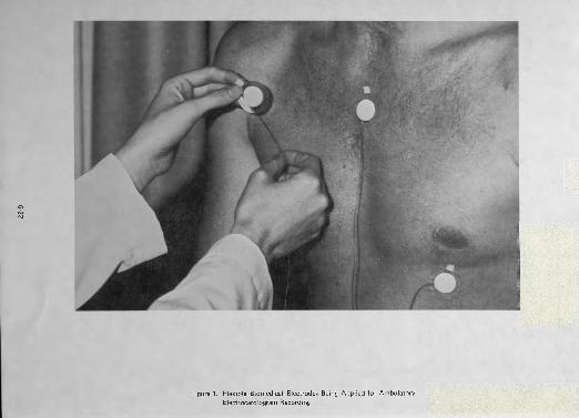
In order to stimulate advances in electrode technology, the BATeam held a conference in 1973 attended by 200 special ists in representing academia, government, industry, and medicine. The proceedings of this conference were edited and published^ and provided a comprehensive review of the subject, clarifying the needs and deficiencies in present technology, as well as suggesting improvements and stimulat ing further research to advance the state of electrode tech nology. The Conference stimulated changes in the types of materials and designs used in electrode manufacture. One electrode manufacturer subsequently developed and market ed a low-profile, soft, conformable surface electrode based on a presentation made by a NASA engineer. The elec trode is made by impregnating an elasticized nylon mesh cloth with conductive silver particles and is backed with an adhesive silicon sponge rubber disc (Figure 1). A tab of the conductive nylon is then attached to a lead wire with a conductive epoxy. The manufacturer who attended the NASA-sponsored conference has been marketing the elec trode since 1975. Because these electrodes are soft and easily conform to the body surface, they permit continuous moni toring with minimal patient discomfort. This type of elec trode has also been applied in rehabilitative medicine.
2. Biotelemetry for Pediatric Gait Analysis
Cerebral palsy is a crippling neurological disease which manifests itself in early childhood. It results in varying degrees of muscular spasticity and loss of coordination. The inappropriate muscular contractions and loss of coordination frequently result in severe walking impairment. Although there is no cure for the disease, accuate diagnosis of the in dividual patient's gait abnormality is essential to the per formance of corrective orthopedic surgery.
6-17
Normal ambulation is a complex activity requiring the co ordinated contraction and relaxation of many individual muscle groups in the legs. In analyzing walking abnormalities, physical therapists make use of the electromyographic (EMG) signals associated with muscle contraction. In adults and large children it is feasible to attach surface electrodes over the muscles to be examined and run wires from those electrodes to a recording device. Children with cerebral palsy, who are candidates for corrective surgery, are fre quently only 3-4 years old and a bundle of wires trailing behind the child may, itself, affect his already unstable gait pattern. In addition, since the child is aware of being moni tored by the physician or therapist, he may modify his usual walking pattern and give a false impression of his gait ab normality. The problem posed to NASA, therefore, was to devise a monitoring system which would allow the recording of the EMG signal in small children with cerebral palsy with out interfering, either physically or psychologically, with their natural ambulatory pattern.
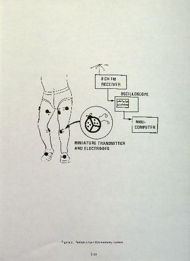
The NASA solution is represented diagrammatically in Figure 2. Multiple individual radio transmitters (decorated as lady-bugs) are utilized. The transmitter circuit was designed by a NASA engineer to minimize external electromagnetic interference, maximize battery life, and provide a wide band width for both research and clinical applications. This approach provided the following advantages over earlier instrumentation:
1 . The size and weight of the transmitters were more suitable for evaluating younger children.
2. Use of individual transmitters simplified instrumenting the patient, thus reducing the time required for the examination.
3. The individual, self-contained transmitters eliminated earlier problems with cables and connectors and permitted adding or subtracting units as needed for the individual patient.
An 8-channel system is now in use at the Children's Hospital at Stanford, Palo Alto, CA. Up to six channels of EMG data can be recorded simultaneously. The other two channels are used for foot switch information, indicating either the swing or weight-bearing phase of gait. The system is now being manufactured commercially and a design which makes in creased use of integrated circuitry to further reduce size and weight is under consideration.
3. Intracranial Pressure Monitorying.
In cases of head -trauma, brain tumor, cerebral infection, and hydrocephalus, the pressure Inside the head, or intracranial pressure (ICPLcan become dangerously elevated. Elevation in ICP can lead to reduced blood flow to the brain, resulting in irreversible damage or death within a few minutes. Opti
mal management of the brain-injured patient, therefore,
necessitates continuous and accurate monitoring of ICP. The measurement of ICP is particularly useful in deciding which medical and surgical therapies are appropriate. The most commonly used technique for monitoring ICP utilizes a fluid- filled catheter in direct communication with the cerebro- spinal fluid and coupled externally to a pressure transducer. One of the problems associated with this technique is that the catheter provides a direct pathway for brain infection, limiting the monitoring period to at most a few days.
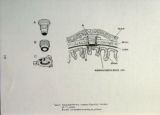
In cooperation with the Departments of Neurosurgery and Anesthesiology at Stanford University Medical School, NASA-Ames Research Center engineers have designed an epidural pressure transducer/transmitter for continuous monitoring of ICP. The pressure transducer is adapted from a design originally used by NASA for the measurement of air pressure over the wings of experimental aircraft during wind tunnel tests. Likewise, the inductively-powered biotelemetry
electronics are based on developments within the NASA Life Sciences Program for monitoring man and animals in space.
As shown in Figure 3, the neurosurgeon inserts the ICP transducer/transmitter through a small hole in the skull and secures it in a mounting fixture. The transmitter is locked in place so that its transducer diaphragm is in direct contact with the dura, a protective membrane which surrounds the brain. The scalp is sutured closed over the unit so that there is no direct pathway for infection. Changes in ICP are con verted into a frequency and transmitted to an external receiver (not shown in the figure).
Clinical trials are in progress at the Stanford University. The ICP monitor is inserted at the end of a neurosurgical pro cedure and patients are monitored continuously in the Intensive Care Unit. The approach has proven to be safe and reliable gnd a medical device manufacturer has been licensed by NASA to produce the next generation system for wide spread clinical use.
ADDITIONAL BIOMEDICAL EXAMPLES OF TECHNOLOGY TRANSFER
Each of the preceding examples are completed technology transfers which have already reached the commercialization stage. The following are short descriptions of additional Stanford BATeam projects which are in the earlier stages of transfer.
1. Versatile Portable Speech Prosthesis
This is a communications device for use in rehabilitative medicine by nonvocal, handicapped patients. The system consists of a user interface module, a microprocessor-based
computer system, and a synthesized speech output device. Extensive use is made of aeronautical communications technology developed by NASA during investigations of synthesized speech in advanced aircraft. The device has been
designed to minimize training time, to permit the user to
6-18
develop his own vocabulary, to adapt to a wide range of user physical disabilities, to permit rapid sentence construction, and to provide improved speech intelligibility. The system will be used initially with wheelchair-confined cerebral palsy patients at the Children's Hospital at Stanford; however, the versatility of its design makes it potentially applicable to patients with multiple sclerosis, Parkinson's disease, muscular dystrophy, and stroke.
2. Radiographic Spatial Frequency Multiplexing
This is an x-ray filtering and decoding technique for enhanc ing the contrast of selected body tissues and organs. The technique holds promise for imaging soft tissue tumors otherwise hidden by overlying bone as well as permitting ex amination of internal organs using peripheral injections of low doses of contrast materials. The x-ray decoding system is an application of NASA technology developed for the Earth Resources Technology Satellite. At present, only laboratory animal studies have been performed; it is anticipated that patient studies will commence this year.
3. Pediatric Roentgen Densitometry
The Roentgen Densitometer is a diagnostic x-ray device de signed for screening newborn infants for intracardiac shunts. The system consists of a standard x-ray source coupled to an array of solid-state gamma detector diodes which were developed during NASA-sponsored solar cell research for powering satellites. When an infant is born with a heart murmur or develops cyanosis, it may be difficult to tell whether he has a surgically correctable congenital heart defect or some other nonsurgical problem. To resolve this diagnostic dilemma, cardiac catheterization may be necess ary. Cardiac catheterization not only requires elaborate instru mentation and technical personnel, but involves some risk to the infant. In contrast, roentgen densitometry, which re quires only the peripheral injection of a small amount of radiopaque material, can be performed in the newborn nursery with far less risk and expense. Used as a screening device, it could reduce the number of catheterizations per formed. Studies in over 50 infants using a prototype device were successfully completed and three second-generation densitometers are being fabricated for evaluation at major university cardiology departments.
4. Nanophor
This is a laboratory instrument which uses an electric field for separating and identifying medically important serum proteins. This instrument's initial development was funded in part by NASA for use in analyzing the serum proteins of Apollo astronauts. It is an improvement over existing electro- phoresis instruments in that it permits analysis of nanogram quantities of serum and incorporates a multiple-sample appli cator to speed up the analysis and improve the accuracy of the test. NASA has recently granted a license for the com mercial manufacture of this new instrument.
5. Liquid Circulating Garments
Temperature-regulated garments were developed for astro nauts to permit them to withstand the temperature extremes associated with extravehicular activity during spaceflight. The following medical applications have been proposed and are currently under evaluation:
1. Elevation of body temperature in the treatment of metastatic cancer, both alone and in conjunction with chemotherapy.
2. Surface cooling prior to cooling by cardiopulmonary bypass for certain types of heart and brain surgery.
3. Lowering of body temperature in patients with diffuse brain injury to reduce metabolic rate and oxygen requirements.
4. Surface cooling of the breasts prior to thermography to aid in the detection of breast cancer.
5. Applications in such rare diseases as hyperkeratosis and "burning limb" syndrome.
6. Regulation of infant body temperature during surgery.
The NASA garments contain flexible heat transfer panels made of a polymer inscribed with tiny channels through which cooled or heated water is circulated.
6. Noninvasive Determination of Bone Properties
Stanford University engineers in collaboration with NASA scientists have developed a mechanical device for measuring the driving point impedance of bone. The development of this new instrument grew out of NASAs interest in deter mining bone demineralization caused by the zero gravity of space flight. In performing the test, a vibrating probe is placed in contact with the subject's forearm and the force and displacement of the bone are analyzed and used to calcu late bending ridigity and axial buckling load index, that is, the load or weight which the bone can support without fracturing. The device incorporates a microprocessor which permits performance of five tests complete with computer print-out in approximately 10 seconds. Preliminary tests in the Orthopedic Clinic have shown that the instrument can provide a useful measure of bone strength, and can readily detect differences between an injured and uninjured limb. Although further data are being obtained on the repro- ducibility and sensitivity of the test, the instrument promises to be useful in diagnosing fractures, and following the course of fracture healing, as well as in evaluating osteoporosis, a common bone disease of older patients.
CONFERENCES AND PUBLICATIONS
In addition to its daily activities in transferring technology to
6-19
the solution of particular biomedical problems, the Stanford BATeam, sponsored by NASA, has conducted both national and international conferences addressing broader areas of biomedical instrumentation. The first of these conferences, held in 1973, was entitled "Biomedical Electrode Tech nology: Theory and Practice/' This was followed in 1975 by "Cardiovascular Imaging and Image Processing - Ultrasound, Angiography, and Isotopes/ 7 In 1976 the 'Third Inter national Symposium on Biotelemetry" was held on the West Coast, and most recently, the BATeam hosted the "Noninvasive Cardiovascular Measurements Conference" in September 1978. The proceedings of each of these confer ences have been compiled, edited, and published.3' 8, 9, 10 These conferences highlight NASA technological develop ments and provide a review of related advances in biomedical instrumentation, as well as a forum for evaluating com petitive technologies, and identifying those areas where improvements are needed. In general, such conferences provide a means by which ". . . the technologist and the user can educate each other as to what each might be able to do and ... to reach a point where some technology can be transferred."^
IMPEDIMENTS TO BIOMEDICAL TECHNOLOGY TRANSFER
Although each of the previously described biomedical tech nology transfer projects has achieved some degree of success, there are many unsolved problems which tend to hinder the development and introduction of new biomedical tech nology. The following is a list of some of the current prob lems:
— Insufficient federal funding for biomedical technolog ical research and development
— Lack of cooperation among the various federal health agencies in accepting responsibility for new technolog ical developments
— Lack of a central clearing house of information about existing biomedical technology
— The long lag-time (1-2 years) between the identifica tion of technological break-throughs and the granting of funds for preliminary concept validation
— Lack of appreciation of the time span (5-10 years) re quired in taking a concept from the prototype phase to the point of being generally accepted by the medical profession
— An increasing tendency to use technology as a scape goat for rapidly rising medical costs without carefully weighing potential medical and cost benefits
— The potential for PDA regulations, with their attend ant documentation requirements, to over-control and stifle technological innovation
— The tendency on the part of federal patenting and licensing regulations to impede rather than promote commercialization
— A lack of appreciation of the technical difficulties and financial risks involved in commercially developing new technology
Though each of these problems is complex and involves diverse groups with their own special interests, collaborative solutions must be found to achieve the common goal of more efficient use of medical resources.
CONCLUDING REMARKS
Although technology from one field can become transferred or reapplied to a new discipline and for a new application by the slow, natural process of diffusion, successful transfer is frequently hindered by language, economic, and regulatory barriers. The transfer of technology, however, can be acceler ated through multidisciplinary and systematic efforts by Bio medical Applications Teams which act as catalysts in the application of NASA technology to the solution of biomed ical problems.
ACKNOWLEDGMENTS
The authors gratefully acknowledge the assistance provided by the numerous NASA scientists and engineers who have provided workable solutions to the various medical problems discussed in this paper.
Manuscript preparation by Ms. Marilyn Anderson.
REFERENCES
1. Public Law No. 85-568, National Aeronautics and Space Act of 1958.
2. Haslam, Kenneth D, MD and Bruner, John MR, MD: 'The Epidemiology of Failure in Cardiac Monitoring Systems" Medical Instrumentation vol 7, no. 5, Nov.-Dec. 1973, pp. 393-396.
3. "Biomedical Electrode Technology — Theory and Practice" Eds. Harry A. Miller and Donald C. Harrison, MD; Academic Press, Inc., 1974.
4. "Flexible Electrodes — For the Patient's Sake" S.A. Rositano, Biomedical Electrode Technology — Theory and Practice, Academic Press, 1974.
5. "Electromyographic Applications of NASA Electrodes, "Katherine B. Robertson; Proceeding of the San Diego Biomedical Symposium, vol 12, pp. 105-108, 1973.
6. Ford, F; Rositano, SA; Schmidt, EV: "Clinical Pediatric Gait Biotelemetry": Biotelemetry III pp. 131-134, Academic Press, Inc., 1976-
7. Westbrook, RM; Fryer, TB, Rositano, SA: "A Wideband EMG Telemetry System" Biotelemetry III pp. 329-332, Academic Press, Inc., 1976.
6-20

8. "Cardiovascular Imaging and Image Processing — Theory and Practice - 1975" Eds. Donald C. Harrison, MD; Harold Sandier, MD; and Harry A. Miller; Society of Photo-optical Instrumentation Engineers, 1975.
9. Biotelemetry III, Eds. Thomas B. Fryer; Harry A. Miller; and Harold Sandier, MD.
10. "Noinvasive Cardiovascular Measurements." Eds. Harry A. Miller; Eugene V. Schmidt, MD; and Donald C. Harrison, MD; Society of Photo- optical Instrumentation Engineers, 1978.
11. "Technology Transfer Policy Considerations'/ Robert A. Frosh; Aerospace Technology Transfer to the Public Sector, Eds. Jerry Gray and Martin Newman, American Institute of Aeronautics and Astronautics, 1978.
6-21
PO N)
Figure 1. Flexible Biomedical Electrodes Being Applied for Ambulatory Electrocardiogram Recording
OSCILLOSCOPE
8CH FM RECEIVER
MINI COMPUTER
MINIATURE TRANSMITTER AND ELECTRODES
Figure 2. Pediatric Gait Biotelemetry System
6-23
B
SCALP
SKULL
DURA
BRAIN
SUBARACHNOID SPACE (CSF)
FigureS. Intracranial Pressure Transducer/Transmitter Assembly.
A) Transmitter B and C) Components of the Mounting Fixture