Embed Size (px)
Citation preview
Mechanisms of Sound Localization in Mammals
BENEDIKT GROTHE, MICHAEL PECKA, AND DAVID McALPINE
Division of Neurobiology, Department of Biology II, Ludwig-Maximilians-Universitaet, Munich, Germany;
and UCL Ear Institute, University College London, United Kingdom
I. Introduction 983II. Acoustic Cues for Sound Localization in Mammals 986
A. Cues for sound localization 986B. Human sound localization: resolutions and limits 987
III. Neuronal Mechanisms for Spatial Processing in the Mammalian Auditory System 987A. The neuronal basis for spectral integration: the basis for sound localization in the vertical plane 988B. The binaural system: the basis for sound localization in the horizontal plane 989C. Neuronal representation of auditory space 999
IV. Conclusions and Open Questions 1004Appendix A: Do Humans Lack the MNTB? 1004Appendix B: ITD Processing: Important Definitions 1005Appendix C: Role of Glycinergic Inhibition in ITD Processing in the MSO 1005
Grothe B, Pecka M, McAlpine D. Mechanisms of Sound Localization in Mammals. Physiol Rev 90: 983–1012, 2010;doi:10.1152/physrev.00026.2009.—The ability to determine the location of a sound source is fundamental to hearing.However, auditory space is not represented in any systematic manner on the basilar membrane of the cochlea, thesensory surface of the receptor organ for hearing. Understanding the means by which sensitivity to spatial cues iscomputed in central neurons can therefore contribute to our understanding of the basic nature of complex neuralrepresentations. We review recent evidence concerning the nature of the neural representation of auditory space inthe mammalian brain and elaborate on recent advances in the understanding of mammalian subcortical processingof auditory spatial cues that challenge the “textbook” version of sound localization, in particular brain mechanismscontributing to binaural hearing.
I. INTRODUCTION
The ability to locate the source of a sound is criticalto the survival of a wide range of species. From theirappearance as primarily nocturnal animals more than 200million years ago, mammals relied heavily on sound lo-calization abilities to achieve this, and to this day, locatingthe source of a sound remains an important sensory abil-ity for prey and predator alike. Spatial hearing also con-tributes to human communication, for example, by pro-viding cues as to the relative number and location ofsources and objects in the environment, helping deter-mine the dimensions and characteristics of rooms andenclosed spaces, and contributing to the “cocktail partyeffect,” whereby listeners are able to hear out speakersagainst other, interfering, voices in crowded listening en-vironments1 (14).
In terms of its neural processing, sound localizationis highly complex but, nevertheless, represents a well-
established model by which the principles of neuronalcomputation might be explored. Here, we review recentevidence concerning the nature of the neural representa-tion of auditory space in the mammalian brain, concen-trating on subcortical structures generally considered tobe specialized for processing auditory spatial cues, andelaborating on recent advances in the understanding ofthe mammalian auditory system that challenge the “text-book” version of sound localization, particularly brainmechanisms contributing to binaural hearing. We focuson several advances that have altered our understandingof how sound localization is achieved in mammals.
1) The neural representation of auditory space isapparently not confined to the form of a topographic mapgenerated by a labeled line system, the so-called “space-map”; rather, source location appears to be representedby a population of relatively broadly tuned spatial chan-nels in both brain hemispheres.
2) While coincidence detection of binaural inputs in thesubmillisecond range remains a basic feature of sound lo-calization mechanisms, the historic view that neural tuning1In concert with other cues, mainly the spectrum of a voice.
Physiol Rev 90: 983–1012, 2010;doi:10.1152/physrev.00026.2009.
www.prv.org 9830031-9333/10 Copyright © 2010 the American Physiological Society
on March 14, 2012
physrev.physiology.orgD
ownloaded from
for preferred spatial locations based on interaural time dif-ferences (ITD) arises by means of purely excitatory axonal“delay lines” does not appear to hold in mammals.
3) The binaural auditory system in mammals is less “hard-wired” than has been imagined and appears instead to be highlydynamic, able to adjust rapidly its tuning properties to takeaccount of the context in which sounds are heard.
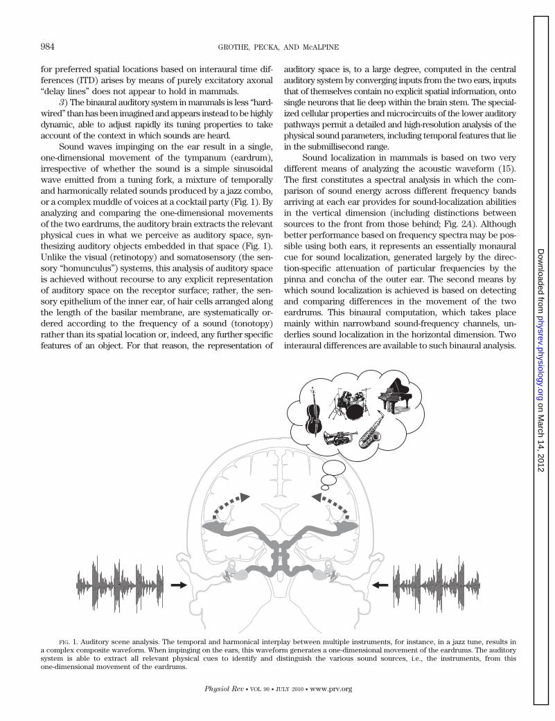
Sound waves impinging on the ear result in a single,one-dimensional movement of the tympanum (eardrum),irrespective of whether the sound is a simple sinusoidalwave emitted from a tuning fork, a mixture of temporallyand harmonically related sounds produced by a jazz combo,or a complex muddle of voices at a cocktail party (Fig. 1). Byanalyzing and comparing the one-dimensional movementsof the two eardrums, the auditory brain extracts the relevantphysical cues in what we perceive as auditory space, syn-thesizing auditory objects embedded in that space (Fig. 1).Unlike the visual (retinotopy) and somatosensory (the sen-sory “homunculus”) systems, this analysis of auditory spaceis achieved without recourse to any explicit representationof auditory space on the receptor surface; rather, the sen-sory epithelium of the inner ear, of hair cells arranged alongthe length of the basilar membrane, are systematically or-dered according to the frequency of a sound (tonotopy)rather than its spatial location or, indeed, any further specificfeatures of an object. For that reason, the representation of
auditory space is, to a large degree, computed in the centralauditory system by converging inputs from the two ears, inputsthat of themselves contain no explicit spatial information, ontosingle neurons that lie deep within the brain stem. The special-ized cellular properties and microcircuits of the lower auditorypathways permit a detailed and high-resolution analysis of thephysical sound parameters, including temporal features that liein the submillisecond range.
Sound localization in mammals is based on two verydifferent means of analyzing the acoustic waveform (15).The first constitutes a spectral analysis in which the com-parison of sound energy across different frequency bandsarriving at each ear provides for sound-localization abilitiesin the vertical dimension (including distinctions betweensources to the front from those behind; Fig. 2A). Althoughbetter performance based on frequency spectra may be pos-sible using both ears, it represents an essentially monauralcue for sound localization, generated largely by the direc-tion-specific attenuation of particular frequencies by thepinna and concha of the outer ear. The second means bywhich sound localization is achieved is based on detectingand comparing differences in the movement of the twoeardrums. This binaural computation, which takes placemainly within narrowband sound-frequency channels, un-derlies sound localization in the horizontal dimension. Twointeraural differences are available to such binaural analysis.
FIG. 1. Auditory scene analysis. The temporal and harmonical interplay between multiple instruments, for instance, in a jazz tune, results ina complex composite waveform. When impinging on the ears, this waveform generates a one-dimensional movement of the eardrums. The auditorysystem is able to extract all relevant physical cues to identify and distinguish the various sound sources, i.e., the instruments, from thisone-dimensional movement of the eardrums.
984 GROTHE, PECKA, AND McALPINE
Physiol Rev • VOL 90 • JULY 2010 • www.prv.org
on March 14, 2012
physrev.physiology.orgD
ownloaded from
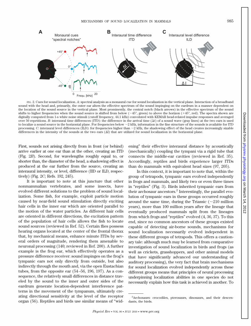
First, sounds not arising directly from in front (or behind)arrive earlier at one ear than at the other, creating an ITD(Fig. 2B). Second, for wavelengths roughly equal to, orshorter than, the diameter of the head, a shadowing effect isproduced at the ear further from the source, creating aninteraural intensity, or level, difference (IID or ILD, respec-tively) (Fig. 2C; Refs. 192, 245).
It is important to note at this juncture that othernonmammalian vertebrates, and some insects, haveevolved different solutions to the problem of sound local-ization. Some fish, for example, exploit particle motioncaused by near-field sound stimulation directly excitinghair cells in the inner ear which are oriented parallel tothe motion of the water particles. As different hair cellsare oriented in different directions, the excitation patternof the population of hair cells allows the fish to locatesound sources (reviewed in Ref. 52). Certain flies possesshearing organs located at the center of the frontal thoraxthat, by mechanical means, enhance minute ITDs by sev-eral orders of magnitude, rendering them amenable toneuronal processing (140; reviewed in Ref. 200). A furtherexample is the frog ear, which effectively operates as apressure difference receiver: sound impinges on the frog’stympanic ears not only directly from outside, but alsoindirectly through the mouth and, via the open Eustachiantubes, from the opposite ear (54–56, 196, 197). As a con-sequence, the relatively small differences in distance trav-eled by the sound to the inner and outer sides of theeardrum generate location-dependent interference pat-terns in the movement of the tympanum, ultimately cre-ating directional sensitivity at the level of the receptororgan (56). Reptiles and birds use similar means of “wid-
ening” their effective interaural distance by acoustically(mechanically) coupling the tympani via a rigid tube thatconnects the middle-ear cavities (reviewed in Ref. 35).Accordingly, reptiles and birds experience larger ITDsthan do mammals with equivalent head sizes (97, 205).
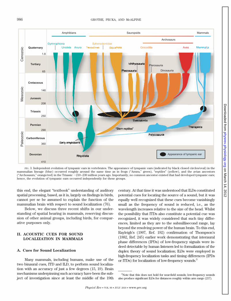
In this context, it is important to note that, within thegroup of tetrapods, tympanic ears evolved independentlyin frogs and mammals, and likely two or even three timesin “reptiles” (Fig. 3). Birds inherited tympanic ears fromtheir archosaur ancestors.2 Interestingly, the parallel evo-lution of tympanic ears in these groups occurred roughlyaround the same time, during the Triassic (�210 millionyears), more than 100 million years after the lineage thateventually produced mammals split from the lineagesfrom which frogs and “reptiles” evolved (4, 36, 37). To thisend, since no common ancestors of these groups existedcapable of detecting air-borne sounds, mechanisms forsound localization necessarily evolved independent inthese different groups of tetrapods. This offers a caution-ary tale: although much may be learned from comparativeinvestigation of sound localization in birds and frogs (aswell as crickets, grasshoppers, and other animal modelsthat have significantly advanced our understanding ofauditory processing), the very fact that brain mechanismsfor sound localization evolved independently across thesedifferent groups means that principles of neural processingunderpinning localization abilities in one species do notnecessarily explain how this task is achieved in another. To
2Archosaurs: crocodiles, pterosaurs, dinosaurs, and their descen-dants, the birds.
FIG. 2. Cues for sound localization. A: spectral analysis as a monaural cue for sound localization in the vertical plane. Interaction of a broadbandsound with the head and, primarily, the outer ear alters the effective spectrum of the sound impinging on the eardrum in a manner dependent onthe location of the sound source in the vertical plane. Most prominently, the central notch (black arrows) in the effective spectrum of the soundshifts to higher frequencies when the sound source is shifted from below (�45°, green) to above the horizon (�45°, red). The spectra shown aredigitally computed from 1-s white noise stimuli (cutoff frequency, 44.1 kHz) convoluted with KEMAR head-related impulse responses and averagedover 50 repetitions. B: interaural time differences (ITD): the difference in the arrival time (�t) of a sound wave (gray lines) at the two ears is usedto localize a sound source in the horizontal plane. For frequencies below �2 kHz, information in the fine structure of the sounds is available for ITDprocessing. C: interaural level differences (ILD): for frequencies higher than �2 kHz, the shadowing effect of the head creates increasingly sizabledifferences in the intensity of the sounds at the two ears (�I) that are utilized for sound localization in the horizontal plane.
MECHANISMS OF SOUND LOCALIZATION IN MAMMALS 985
Physiol Rev • VOL 90 • JULY 2010 • www.prv.org
on March 14, 2012
physrev.physiology.orgD
ownloaded from
this end, the elegant “textbook” understanding of auditoryspatial processing, based, as it is, largely on findings in birds,cannot per se be assumed to explain the function of themammalian brain with respect to sound localization (76).
Below, we discuss three recent shifts in our under-standing of spatial hearing in mammals, reserving discus-sion of other animal groups, including birds, for compar-ative purposes only.
II. ACOUSTIC CUES FOR SOUND
LOCALIZATION IN MAMMALS
A. Cues for Sound Localization
Many mammals, including humans, make use of thetwo binaural cues, ITD and ILD, to perform sound localiza-tion with an accuracy of just a few degrees (15, 19). Brainmechanisms underpinning such accuracy have been the sub-ject of investigation since at least the middle of the 19th
century. At that time it was understood that ILDs constitutedpotential cues for locating the source of a sound, but it wasequally well recognized that these cues become vanishinglysmall as the frequency of sound is reduced, i.e., as thewavelength increases relative to the size of the head. Whilstthe possibility that ITDs also constitute a potential cue wasrecognized, it was widely considered that such tiny differ-ences, limited as they are to the submillisecond range, laybeyond the resolving power of the human brain. To this end,Rayleigh’s (1907, Ref. 192) confirmation of Thompson’s(1882, Ref. 245) earlier work demonstrating that interauralphase differences (IPDs) of low-frequency signals were in-deed detectable by human listeners led to formalization of theduplex theory of sound localization; ILDs were employed inhigh-frequency localization tasks and timing differences (IPDsor ITDs) for localization of low-frequency sounds.3
3Note that this does not hold for near-field sounds; low-frequency soundsalso produce significant ILDs for distances roughly within arm range (217).
FIG. 3. Independent evolution of tympanic ears in vertebrates. The appearance of tympanic ears (indicated by black closed circles/oval) in themammalian lineage (blue) occurred roughly around the same time as in frogs (“Anura,” green), “reptiles” (yellow), and the avian ancestors(“Archosaurs,” orange/red) in the Triassic �210–230 million years ago. Importantly, no common ancestor existed that had developed tympanic ears;hence, the evolution of tympanic ears occurred independently for these groups.
986 GROTHE, PECKA, AND McALPINE
Physiol Rev • VOL 90 • JULY 2010 • www.prv.org
on March 14, 2012
physrev.physiology.orgD
ownloaded from
In addition to binaural cues, the auditory systemexploits frequency-specific modifications in the magni-tude and phase of the sound reaching the eardrum thatarise from the interaction of the sound with the head andthe ears, to determine source location in the verticalplane. These spectral cues for localization underpin theability to disambiguate the so-called cone of confusion,resolving sources in front from those behind as well asdetermining their elevation, a task not possible usingbinaural cues alone (15). The function describing thesespectral modifications, which are generated largely by thepinna and concha of the outer ear, is referred to as thehead-related transfer function, or HRTF (colored traces inFig. 2A). Spectral cues for localization consist of notches(or changes) in the sound spectrum at specific frequen-cies; the exact frequency and magnitude of the notchchanging as the location of the source shifts in elevation(19). Spectral cues can be manipulated by modificationsto the external ear, and the extent to which birds andmammals, including humans, can adapt to the alteredcues that arise from these manipulations demonstratestheir importance in localization tasks. For example, im-mediately following insertion of ear molds that permit thepassage of sound to the tympanic membrane, but alter theshape of the concha, human listeners’ performance isdegraded to the extent that they have trouble distinguish-ing the location of sounds in the vertical plane (althoughleft-right discriminations appear unaltered). Over thecourse of several weeks, however, performance improvessuch that by the end of �1 mo, listeners have adaptedalmost completely to the altered cues. Interestingly, sub-sequent removal of the ear molds leaves no residual def-icit in performance, suggesting that sensitivity to spectralcues represents a learned behavior (96). This is perhapsnot surprising given the multitude of complex sounds thatmight arrive from many different directions, with variousfeatures of the source, the environment, and the headitself all contributing to the final sound impinging on thetympanum.
B. Human Sound Localization: Resolutions
and Limits
Humans show remarkable abilities in sound local-ization, discriminating changes of just 1–2 degrees inthe angular location of a sound source (13, 118). Stud-ies under listening conditions using headphones (whichenables the isolation of specific cues) confirm the re-markable accuracy of human spatial hearing, withthresholds as low as 10 �s for ITDs (119) or 1–2 deci-bels for ILDs for presentations of binaural clicks (254).For the ITD cue at least, this represents a level ofresolution that must be considered with respect to themillisecond duration of nerve action potentials trans-
mitting information in the brain. Threshold sensitivityto ILD, whilst impressive, is no better than sensitivity tosound level per se (151).
Rayleigh’s classical distinction of the duplex the-ory (1907, Ref. 192), separating cues for auditory spatialprocessing into low- and high-frequency processes, wasseemingly confirmed by Stevens’ and Newman’s (1934,Ref. 233) investigations of absolute localization perfor-mance in humans, in which blindfolded listeners wererequired to indicate the location of a speaker deliveringpure tones of various frequencies. Discounting sourcesto the front and back, where ITD information is ambig-uous, performance was best for frequencies below �2kHz, and above 5 kHz, with intermediate frequencieseliciting the highest number of localization errors, sug-gesting an incomplete representation of either cue overthis range.
Psychophysical studies using headphone stimulationhave sought to determine the range of ITD detectorsrepresented in the human brain. Based on measurementof ITD discrimination thresholds and binaural unmasking,the range of ITDs encoded is considered to be 1) roughlyconstant across the range of sound frequencies at whichsensitivity to ITDs in the fine-structure of sounds is ob-served (�1,500 Hz) and 2) set largely by the physiologicalrange (�700 �s in humans) but with ITDs up to at least�3,000 �s explicitly encoded in each sound-frequencyband (251). ITDs longer than the physiological range canbe experienced over stereo headphones, arising naturallywhen multiple sound sources interact, and when acousticreflections are present (although at high fluctuation rates;compare Ref. 150).
III. NEURONAL MECHANISMS FOR SPATIAL
PROCESSING IN THE MAMMALIAN
AUDITORY SYSTEM
Mechanical deflections of the eardrum in response tosound stimulation are transmitted to the cochlea via thethree middle ear bones, the ossicles, which act as animpedance-matching device effectively coupling air-bornesound to the fluids of the inner ear. Hair cells in the organof Corti, the sensory epithelium that runs the length of thebasilar membrane of the cochlea, transduce mechanicalenergy into bioelectric activity, and this activity, in theform of nerve action potentials, is transmitted via theauditory nerve to the central auditory nervous system(Fig. 4). The central auditory nervous system can beconsidered as consisting of different ascending streams,or functionally segregated pathways. For instance, threedistinct pathways project from different subdivisions ofthe cochlear nucleus, the first synaptic station in theauditory brain stem (206). The subdivisions that make upthe cochlear nucleus comprise several neural subtypes
MECHANISMS OF SOUND LOCALIZATION IN MAMMALS 987
Physiol Rev • VOL 90 • JULY 2010 • www.prv.org
on March 14, 2012
physrev.physiology.orgD
ownloaded from
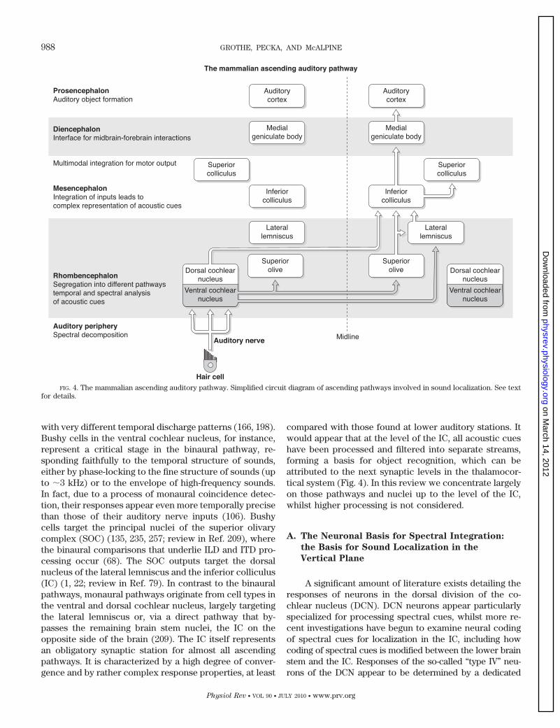
with very different temporal discharge patterns (166, 198).Bushy cells in the ventral cochlear nucleus, for instance,represent a critical stage in the binaural pathway, re-sponding faithfully to the temporal structure of sounds,either by phase-locking to the fine structure of sounds (upto �3 kHz) or to the envelope of high-frequency sounds.In fact, due to a process of monaural coincidence detec-tion, their responses appear even more temporally precisethan those of their auditory nerve inputs (106). Bushycells target the principal nuclei of the superior olivarycomplex (SOC) (135, 235, 257; review in Ref. 209), wherethe binaural comparisons that underlie ILD and ITD pro-cessing occur (68). The SOC outputs target the dorsalnucleus of the lateral lemniscus and the inferior colliculus(IC) (1, 22; review in Ref. 79). In contrast to the binauralpathways, monaural pathways originate from cell types inthe ventral and dorsal cochlear nucleus, largely targetingthe lateral lemniscus or, via a direct pathway that by-passes the remaining brain stem nuclei, the IC on theopposite side of the brain (209). The IC itself representsan obligatory synaptic station for almost all ascendingpathways. It is characterized by a high degree of conver-gence and by rather complex response properties, at least
compared with those found at lower auditory stations. Itwould appear that at the level of the IC, all acoustic cueshave been processed and filtered into separate streams,forming a basis for object recognition, which can beattributed to the next synaptic levels in the thalamocor-tical system (Fig. 4). In this review we concentrate largelyon those pathways and nuclei up to the level of the IC,whilst higher processing is not considered.
A. The Neuronal Basis for Spectral Integration:
the Basis for Sound Localization in the
Vertical Plane
A significant amount of literature exists detailing theresponses of neurons in the dorsal division of the co-chlear nucleus (DCN). DCN neurons appear particularlyspecialized for processing spectral cues, whilst more re-cent investigations have begun to examine neural codingof spectral cues for localization in the IC, including howcoding of spectral cues is modified between the lower brainstem and the IC. Responses of the so-called “type IV” neu-rons of the DCN appear to be determined by a dedicated
FIG. 4. The mammalian ascending auditory pathway. Simplified circuit diagram of ascending pathways involved in sound localization. See textfor details.
988 GROTHE, PECKA, AND McALPINE
Physiol Rev • VOL 90 • JULY 2010 • www.prv.org
on March 14, 2012
physrev.physiology.orgD
ownloaded from
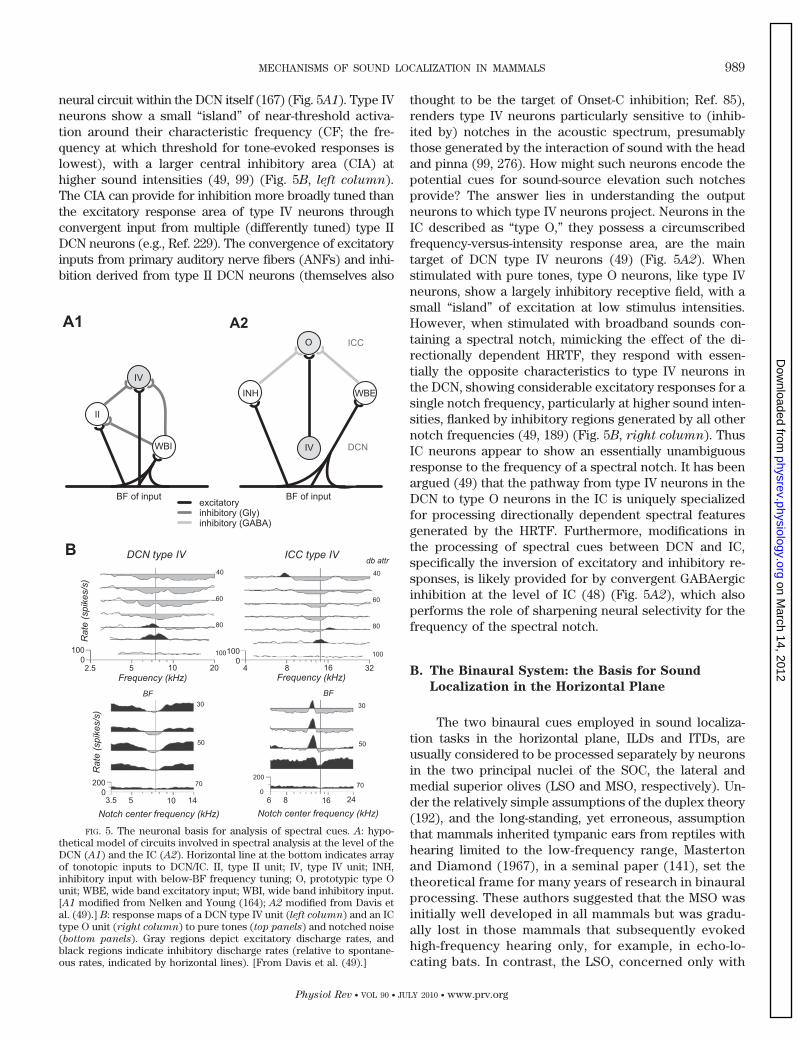
neural circuit within the DCN itself (167) (Fig. 5A1). Type IVneurons show a small “island” of near-threshold activa-tion around their characteristic frequency (CF; the fre-quency at which threshold for tone-evoked responses islowest), with a larger central inhibitory area (CIA) athigher sound intensities (49, 99) (Fig. 5B, left column).The CIA can provide for inhibition more broadly tuned thanthe excitatory response area of type IV neurons throughconvergent input from multiple (differently tuned) type IIDCN neurons (e.g., Ref. 229). The convergence of excitatoryinputs from primary auditory nerve fibers (ANFs) and inhi-bition derived from type II DCN neurons (themselves also
thought to be the target of Onset-C inhibition; Ref. 85),renders type IV neurons particularly sensitive to (inhib-ited by) notches in the acoustic spectrum, presumablythose generated by the interaction of sound with the headand pinna (99, 276). How might such neurons encode thepotential cues for sound-source elevation such notchesprovide? The answer lies in understanding the outputneurons to which type IV neurons project. Neurons in theIC described as “type O,” they possess a circumscribedfrequency-versus-intensity response area, are the maintarget of DCN type IV neurons (49) (Fig. 5A2). Whenstimulated with pure tones, type O neurons, like type IVneurons, show a largely inhibitory receptive field, with asmall “island” of excitation at low stimulus intensities.However, when stimulated with broadband sounds con-taining a spectral notch, mimicking the effect of the di-rectionally dependent HRTF, they respond with essen-tially the opposite characteristics to type IV neurons inthe DCN, showing considerable excitatory responses for asingle notch frequency, particularly at higher sound inten-sities, flanked by inhibitory regions generated by all othernotch frequencies (49, 189) (Fig. 5B, right column). ThusIC neurons appear to show an essentially unambiguousresponse to the frequency of a spectral notch. It has beenargued (49) that the pathway from type IV neurons in theDCN to type O neurons in the IC is uniquely specializedfor processing directionally dependent spectral featuresgenerated by the HRTF. Furthermore, modifications inthe processing of spectral cues between DCN and IC,specifically the inversion of excitatory and inhibitory re-sponses, is likely provided for by convergent GABAergicinhibition at the level of IC (48) (Fig. 5A2), which alsoperforms the role of sharpening neural selectivity for thefrequency of the spectral notch.
B. The Binaural System: the Basis for Sound
Localization in the Horizontal Plane
The two binaural cues employed in sound localiza-tion tasks in the horizontal plane, ILDs and ITDs, areusually considered to be processed separately by neuronsin the two principal nuclei of the SOC, the lateral andmedial superior olives (LSO and MSO, respectively). Un-der the relatively simple assumptions of the duplex theory(192), and the long-standing, yet erroneous, assumptionthat mammals inherited tympanic ears from reptiles withhearing limited to the low-frequency range, Mastertonand Diamond (1967), in a seminal paper (141), set thetheoretical frame for many years of research in binauralprocessing. These authors suggested that the MSO wasinitially well developed in all mammals but was gradu-ally lost in those mammals that subsequently evokedhigh-frequency hearing only, for example, in echo-lo-cating bats. In contrast, the LSO, concerned only with
FIG. 5. The neuronal basis for analysis of spectral cues. A: hypo-thetical model of circuits involved in spectral analysis at the level of theDCN (A1) and the IC (A2). Horizontal line at the bottom indicates arrayof tonotopic inputs to DCN/IC. II, type II unit; IV, type IV unit; INH,inhibitory input with below-BF frequency tuning; O, prototypic type Ounit; WBE, wide band excitatory input; WBI, wide band inhibitory input.[A1 modified from Nelken and Young (164); A2 modified from Davis etal. (49).] B: response maps of a DCN type IV unit (left column) and an ICtype O unit (right column) to pure tones (top panels) and notched noise(bottom panels). Gray regions depict excitatory discharge rates, andblack regions indicate inhibitory discharge rates (relative to spontane-ous rates, indicated by horizontal lines). [From Davis et al. (49).]
MECHANISMS OF SOUND LOCALIZATION IN MAMMALS 989
Physiol Rev • VOL 90 • JULY 2010 • www.prv.org
on March 14, 2012
physrev.physiology.orgD
ownloaded from
the processing of ILDs at high frequencies, was under-developed in mammals with largely low-frequency hear-ing, and in humans may even be absent altogether (fordetails, see sect. IIIB1). Despite this attractively simpleperspective, however, evidence from the fossil recorddoes not support the assumption of an originally rep-tilian-like ear in early mammals (4, 36, 37), (see Fig. 3and sect. I). Furthermore, despite general acceptance,the dichotomy suggested by the duplex theory is notstrict. First, significant ILDs can occur for low-fre-quency sounds located in the near field (21, 217). Sec-ond, extensive psychophysical evidence indicates thatsensitivity to ITDs is conveyed by the envelopes ofhigh-frequency complex sounds (11, 47, 94, 119, 148,273). In this regard, recent studies have shown thatwhen provision is made for temporal information in theenvelopes of high-frequency modulated tones to matchas closely as possible temporal information normallypresent in the output of low-frequency channels, ITDdiscrimination thresholds can be as good as, and insome cases surpass, those for low-frequency tones (12).
Taken together, these findings support the notionthat the duplex theory describes more the frequency de-pendence of the two binaural cues rather than an absolutesegregation into two distinct, nonoverlapping channels. Infact, brain mechanisms processing the binaural cues ap-pear to be present across the entire frequency range ofhuman hearing. To this end, the need for a refined theoryof binaural processing, one that also encompasses itsevolutionary development, appears timely. Such a theorymust also account for emerging data sets describing thephysiological nature of binaural processing, as well as themeans by which spatial cues are represented in the hu-man brain (compare Refs. 73 and 76).
1. Neuronal mechanisms of ILD processing
From several lines of evidence, it is reasonable toassume that ILDs were the only binaural cue used by earlymammals. 1) It appears that the mammalian tympanic ear,when it first appeared some 200 million years ago, wasmechanically tuned to frequencies between 4 and 18 kHz(152, 204) and subsequently evolved rapidly to incorpo-rate higher frequencies during the early evolution of mam-mals (70, 203). Hence, it is likely that these animals ex-perienced prominent ILDs suited to sound localizationacross their entire hearing range. 2) All terrestrial mam-mals investigated thus far, including echo-locating bats,possess well-developed LSOs that seem to correlate insize with the range of frequencies an animal can hear(156). 3) All mammals appear to share one common neu-ral mechanism for processing ILDs (267),4 4) requiring
considerably less temporally precise cellular and circuitproperties than does the processing of ITDs (see below).5) The earliest mammals were very small and experiencedITDs produced by sound arriving directly from a source ofup to �50 �s only (73).5 As such, the ability to processITDs evolved either by means of a sudden improvement intemporal resolution of the auditory brain stem (by twoorders of magnitude) or, more likely, gradually, and con-sequently much later, during evolutionary development(73, 76). 6) ITD processing, with few exceptions (64),appears to be realized only in low-frequency hearingmammals and, hence, in a minority of mammalian spe-cies: those that employ low-frequency communicationcalls (all larger terrestrial mammals) and/or those thatreside in open environments and benefit from hearingover larger distances, for example, to detect predators[e.g., some desert rodents (132, 133, 258–260)]. Thesegroups must localize sounds for which no appreciable ILDis generated.
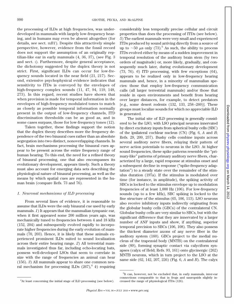
The initial site of ILD processing is generally consid-ered to be the LSO, with LSO principal neurons innervatedby direct excitatory inputs from spherical bushy cells (SBC)of the ipsilateral cochlear nucleus (CN) (Fig. 6, A and B;Refs. 29, 209, 257). Bushy cells combine inputs acrossseveral auditory nerve fibers, relaying their pattern ofnerve action potentials to neurons in the LSO. At highersound frequencies, these spike patterns resemble the “pri-mary-like” patterns of primary auditory nerve fibers, char-acterized by a large, rapid response at stimulus onset anda subsequent decline in response (“spike-frequency adap-tation”) to a steady state over the remainder of the stim-ulus duration (197a). If the stimulus is modulated overtime (for instance, in amplitude), the spiking activity ofSBCs is locked to the stimulus envelope up to modulationfrequencies of at least 1,000 Hz (106). For low-frequencysounds (up to a few kHz), SBC spiking is locked to thefine structure of the stimulus (65, 106, 113). LSO neuronsalso receive inhibitory inputs indirectly originating fromthe globular bushy cells (GBCs) of the contralateral CN.Globular bushy cells are very similar to SBCs, but with thesignificant difference that they are innervated by a largernumber of ANF inputs and show, if anything, superiortemporal precision to SBCs (106, 108). They also possessthe thickest diameter axons of any nerve fiber in theauditory system (160). GBCs project to the medial nu-cleus of the trapezoid body (MNTB) on the contralateralside (89), forming synaptic contact via calyciform syn-apses (calyx of Held; Refs. 93, 161) onto glycinergic (262)MNTB neurons, which in turn project to the LSO at thesame side (62, 142, 207, 226) (Fig. 6, A and B). The calyx
4At least concerning the initial stage of ILD processing (see below).
5It can, however, not be excluded that, in early mammals, inter-earconnections comparable to that in frogs and sauropsids slightly in-creased the range of physiological ITDs (126).
990 GROTHE, PECKA, AND McALPINE
Physiol Rev • VOL 90 • JULY 2010 • www.prv.org
on March 14, 2012
physrev.physiology.orgD
ownloaded from
of Held, in combination with a battery of potassium con-ductances that prevent temporal summation of inputs inMNTB cells (reviewed in Ref. 255), renders the MNTB ahigh-fidelity station in the ascending auditory pathway(95, 125, 149, 224), converting well-timed excitatory in-puts into well-timed inhibitory outputs. MNTB cells alsoproject to a range of target nuclei other than the LSO,including the ipsilateral MSO (see below).
The convergence of excitatory inputs from the ipsi-lateral CN and inhibitory inputs from the opposite CN viathe MNTB resembles a relatively simple subtraction pro-cess (159) (Fig. 6B), creating the well-described ILD sen-sitivity of LSO neurons (16, 26, 83, 176). These functionsare usually sigmoid in form, with neurons completelyinhibited when the sound at the contralateral, inhibi-tory, ear is more intense (“negative ILDs”) and maxi-mally responsive when the sound at the ipsilateral,excitatory, ear is more intense (“positive ILDs”) (re-view in Ref. 267; Fig. 6C).
Although temporally precise inputs have been dem-onstrated not to be required for ILD sensitivity of thesustained response of neurons in the LSO, timing does infact appear to be important for generating ILD sensitivityat stimulus onset and, generally, to LSO neurons tuned to
low-frequency sounds (77, 101, 176). In particular, in-creasing evidence exists to suggest that, based on theirknown excitatory-inhibitory (EI) interaction, low-fre-quency LSO neurons are sensitive to ITDs, with a resolu-tion almost comparable to that of neurons in the MSO(247). In contrast to MSO neurons, however, coincidenceof ipsilateral excitatory input with the contralateral, in-hibitory input generates response minima (“troughs” inthe rate versus ITD functions); peak activity occurs wheninputs are noncoincident (or maximally out of phase inresponse functions) (159, 247, 272). This issue is dis-cussed in more detail in section III, B and C3.
As suggested above, the two major inputs to theLSO are specialized for high-fidelity temporal transmis-sion. Nevertheless, exquisite timing of inhibitory influ-ences appears not to be a prerequisite for the subtrac-tion mechanism underpinning ILD sensitivity, at leastfor high frequencies where phase-locking is not anissue. Interestingly, the contralateral input to LSO neu-rons must traverse a greater axonal distance to reachthe LSO including, in the process, an additional synap-tic stage via the MNTB. One might therefore expect thatcontralaterally derived inhibition should arrive later inthe LSO (a similar argument has traditionally been used
FIG. 6. The neuronal basis underlying ILD processing. A: schematic of the mammalian ILD circuit. ILD sensitivity is first created in the LSO byconvergence of excitatory inputs from SBCs located in the ipsilateral AVCN and inhibitory inputs from ipsilateral MNTB, which is itself innervatedby GBCs of the contralateral AVCN. Projections of the LSO to the contralateral DNLL and IC are excitatory and inhibitory to the ipsilateral DNLLand IC. In the IC, ILD sensitivity is created de novo by the convergence of monaural contralateral excitatory input from the AVCN and binauralinhibitory input from the DNLL. B: schematic of the distribution of excitatory (red) and inhibitory (blue) input onto a LSO neuron (orange). Depictedare somatic and dendritic areas of a LSO principal cell. C: schematic of the typical sigmoid ILD function of a LSO neuron is shown.
MECHANISMS OF SOUND LOCALIZATION IN MAMMALS 991
Physiol Rev • VOL 90 • JULY 2010 • www.prv.org
on March 14, 2012
physrev.physiology.orgD
ownloaded from
to explain why MSO neurons prefer contralaterallyleading sounds, potentially compensating for a longeraxonal delay). Nevertheless, Tsuchitani (1988, Ref. 249)described the inhibitory effect in cat LSO neurons to bestrongest at onset, and to adapt during ongoing stimu-lation. Hence, by some means, the GBC-MNTB pathwaycompensates for the longer distance contralateral ax-ons must span before innervating their target neuronsin the LSO. Possible explanations could be the shorterlatency and larger axon diameter of GBCs comparedwith SBCs (review in Ref. 209), and a minimized syn-aptic delay to the MNTB due to the giant calyx of Held(review in Ref. 255). Delaying the contralateral stimu-lus in the range of a few hundreds of microsecondsresults in the ipsilaterally generated excitation preced-ing the contralaterally generated synaptic inhibitionsufficiently to evoke at least a single action potential(101, 174, 176, 178). This may be related to the well-known phenomenon of “time-intensity trading,”whereby ITDs leading at one ear can be used to com-pensate for ILDs favoring the other, and vice versa (50,264, 274). Time-intensity trading can also be observedneuronally (20, 77, 187, 271) and, in a significant propor-tion of LSO neurons, can produce different latency shiftsfor the two binaural inputs, depending on the overallintensity (101, 176; see also Ref. 271 for similar responsesin the IC). In this context, it is important to note that thiseffect most likely depends on the stimulus envelope, i.e.,largely on the overall energy accumulated, correspondingto the integral of the envelope of the waveform ratherthan the absolute sound level (92).
The LSO, which has no homolog in other verte-brates including birds, is the primary site for processingILDs in the mammalian auditory system. Nevertheless,there is no reason a priori why neurons in any braincenter in which inputs from the two ears could con-ceivably converge onto individual neurons, should notalso constitute sites at which neural sensitivity to ILDsis generated (Fig. 6A). Consistent with this notion, ILDsensitivity generated de novo has been demonstrated inexperiments in which the ILD sensitivity of IC neuronswas abolished by blocking the action of GABAergicinhibition locally (179). A likely source of GABAergicinhibition to the IC is the cross projection from thedorsal nucleus of the lateral lemniscus (DNLL), andremoving this connection by sectioning its projectingaxons or by pharmacological inactivation of the DNLLitself, leads to a release from binaural inhibition in ICneurons (24, 102, 134). Furthermore, a greater propor-tion of IC than LSO neurons shows ILD sensitivity thatis stable with average sound intensity (177). WhereasILD sensitivity in LSO neurons is characterized by sig-moidal ILD functions that shift in a systematic mannerwith increasing intensity to the excitatory ear, those inIC are less affected. This is consistent with at least
some of the inhibitory effect in IC being derived from asource other than the inhibitory input to the LSO (177).Nevertheless, given the sufficiency of the LSO in pro-ducing neural sensitivity to ILDs, it seems likely thatILD sensitivity in the majority of IC neurons reflectsLSO input, with modifications to this input provided bymechanisms local to the IC.
Current evidence, anatomical and physiologicaldata from experiments conducted in a wide variety ofmammals and psychophysics of ILD processing inmammals including humans, is consistent with the viewthat brain mechanisms for processing ILDs are highlyconserved, possibly for almost 200 million years. Nev-ertheless, an ongoing controversy concerning underly-ing brain mechanisms in humans must be addressed ifthis consistency is to be accepted. This controversyrelates to a number of anatomical studies seeminglydemonstrating the lack of a MNTB in the human brain(see Appendix A). If this were true, and we argue belowthat it is unlikely, humans would had to have developedfundamentally different brain mechanisms, comparedwith all other mammals, by which ILDs are processedparticularly as this important source of inhibitory inputto the LSO would be absent. That the LSO itself repre-sents a prominent structure even in the human brainhas been confirmed by a number of anatomical studies(9, 128, 236) and, since chimpanzees are reported topossess an MNTB (237), the presumed loss of theMNTB in the human lineage would have had to occurwithin the last six million years. Given that every singlemammalian species examined to date possesses aprominent MNTB, as well as an LSO, this appears un-likely indeed. Neurons in the human LSO would eitherhave a completely different function, or a differentsource of inhibitory input compared with other mam-mals, a difficult proposition to sustain given that theMNTB is the major source of contralaterally evokedinhibition in the entire auditory brain stem. This issuebecomes even more problematic if one considers thatthe MNTB is also the major source of well-timed inhi-bition not only for binaural circuits (including alsothose involved in ITD processing, see below) but alsofor monaural processing. MNTB projections to monau-ral neurons in the SOC, for example, are responsible forcreating transient “on” and “off” responses (72, 129).Axons from the MNTB branch multiple times, targetingmultiple sources within the SOC and beyond (e.g., thedorsal and ventral nuclei of the lateral lemniscus; Refs.82, 219, 224, 225). Hence, without the MNTB, the humanauditory system would have to have evolved completelydifferent strategies not only for processing binaural,but also monaural acoustic cues. In the light of thecurrent direct (see Appendix A) and indirect (above)evidence, there is no reason to assume that brain mech-
992 GROTHE, PECKA, AND McALPINE
Physiol Rev • VOL 90 • JULY 2010 • www.prv.org
on March 14, 2012
physrev.physiology.orgD
ownloaded from
anisms underlying sound localization in humans devi-ate from the general pattern observed in mammals.
2. Neuronal mechanisms of ITD processing
Neural coding of ITDs demands the highest preci-sion of any temporal process known to exist within themammalian, reptilian, or avian brain. In essence, neu-rons must resolve differences in the time of arrival ofthe sound at each ear that are almost two orders ofmagnitude shorter than the duration of action poten-tials bearing that information. Nevertheless, despite theapparently insurmountable challenges posed by thistask, different groups of animals have, independently,evolved the ability to use ITDs in sound localizationtasks (see sect. I). Sauropsids (“reptiles” and birds), forexample, have evolved a mechanical coupling of thetwo ears that increases the magnitude of ITDs prior tothe sensory transduction process in the inner ear (97,205). Nonetheless, these modifications aside, which ap-pear fitted to enhance the magnitude of ITDs generatedby the size of the head, it is the exquisite temporalprecision of coincidence detection by the mammalianMSO, and its analogous structure in birds, the nucleuslaminaris (NL) (Fig. 7A) that makes possible sensitivityto ITDs in the submillisecond range. For importantdefinitions concerning ITD processing, see Appendix B.
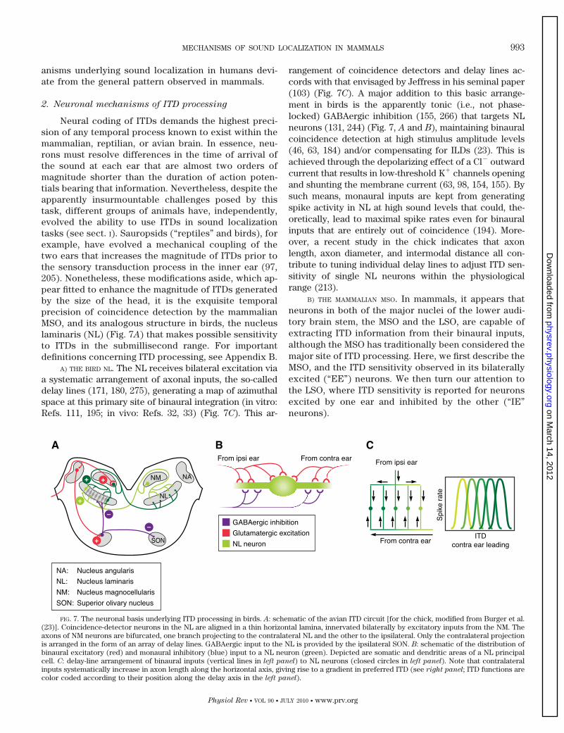
A) THE BIRD NL. The NL receives bilateral excitation viaa systematic arrangement of axonal inputs, the so-calleddelay lines (171, 180, 275), generating a map of azimuthalspace at this primary site of binaural integration (in vitro:Refs. 111, 195; in vivo: Refs. 32, 33) (Fig. 7C). This ar-
rangement of coincidence detectors and delay lines ac-cords with that envisaged by Jeffress in his seminal paper(103) (Fig. 7C). A major addition to this basic arrange-ment in birds is the apparently tonic (i.e., not phase-locked) GABAergic inhibition (155, 266) that targets NLneurons (131, 244) (Fig. 7, A and B), maintaining binauralcoincidence detection at high stimulus amplitude levels(46, 63, 184) and/or compensating for ILDs (23). This isachieved through the depolarizing effect of a Cl� outwardcurrent that results in low-threshold K� channels openingand shunting the membrane current (63, 98, 154, 155). Bysuch means, monaural inputs are kept from generatingspike activity in NL at high sound levels that could, the-oretically, lead to maximal spike rates even for binauralinputs that are entirely out of coincidence (194). More-over, a recent study in the chick indicates that axonlength, axon diameter, and intermodal distance all con-tribute to tuning individual delay lines to adjust ITD sen-sitivity of single NL neurons within the physiologicalrange (213).
B) THE MAMMALIAN MSO. In mammals, it appears thatneurons in both of the major nuclei of the lower audi-tory brain stem, the MSO and the LSO, are capable ofextracting ITD information from their binaural inputs,although the MSO has traditionally been considered themajor site of ITD processing. Here, we first describe theMSO, and the ITD sensitivity observed in its bilaterallyexcited (“EE”) neurons. We then turn our attention tothe LSO, where ITD sensitivity is reported for neuronsexcited by one ear and inhibited by the other (“IE”neurons).
FIG. 7. The neuronal basis underlying ITD processing in birds. A: schematic of the avian ITD circuit [for the chick, modified from Burger et al.(23)]. Coincidence-detector neurons in the NL are aligned in a thin horizontal lamina, innervated bilaterally by excitatory inputs from the NM. Theaxons of NM neurons are bifurcated, one branch projecting to the contralateral NL and the other to the ipsilateral. Only the contralateral projectionis arranged in the form of an array of delay lines. GABAergic input to the NL is provided by the ipsilateral SON. B: schematic of the distribution ofbinaural excitatory (red) and monaural inhibitory (blue) input to a NL neuron (green). Depicted are somatic and dendritic areas of a NL principalcell. C: delay-line arrangement of binaural inputs (vertical lines in left panel) to NL neurons (closed circles in left panel). Note that contralateralinputs systematically increase in axon length along the horizontal axis, giving rise to a gradient in preferred ITD (see right panel; ITD functions arecolor coded according to their position along the delay axis in the left panel).
MECHANISMS OF SOUND LOCALIZATION IN MAMMALS 993
Physiol Rev • VOL 90 • JULY 2010 • www.prv.org
on March 14, 2012
physrev.physiology.orgD
ownloaded from
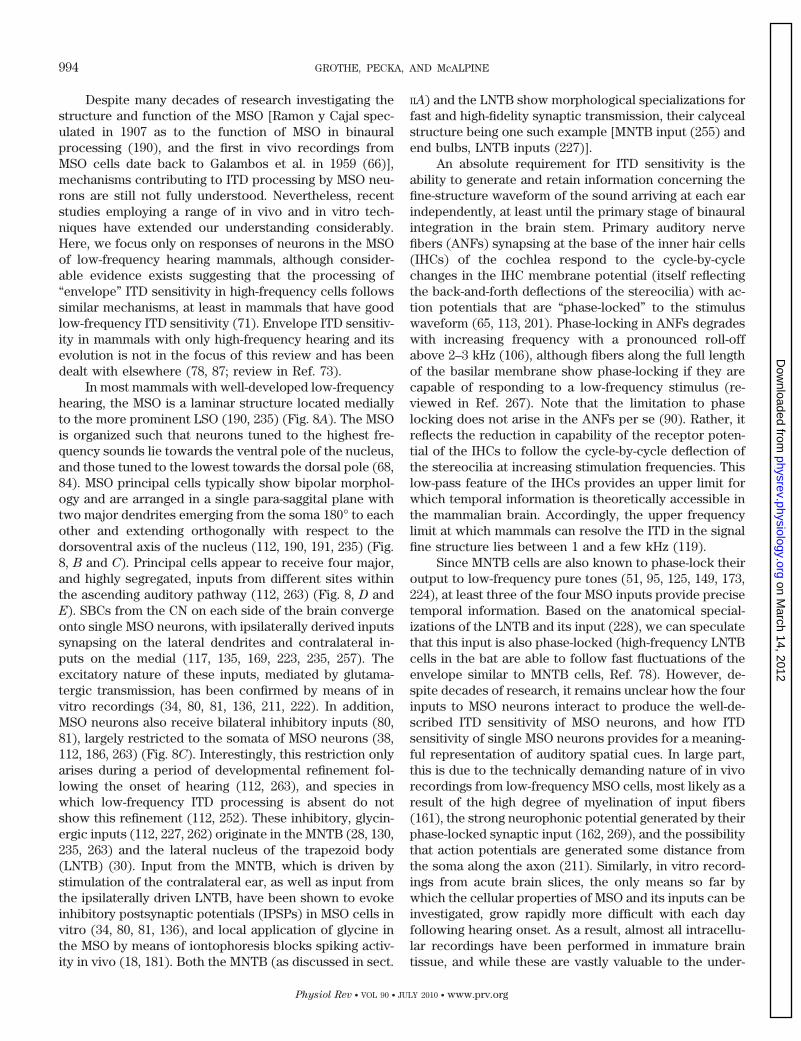
Despite many decades of research investigating thestructure and function of the MSO [Ramon y Cajal spec-ulated in 1907 as to the function of MSO in binauralprocessing (190), and the first in vivo recordings fromMSO cells date back to Galambos et al. in 1959 (66)],mechanisms contributing to ITD processing by MSO neu-rons are still not fully understood. Nevertheless, recentstudies employing a range of in vivo and in vitro tech-niques have extended our understanding considerably.Here, we focus only on responses of neurons in the MSOof low-frequency hearing mammals, although consider-able evidence exists suggesting that the processing of“envelope” ITD sensitivity in high-frequency cells followssimilar mechanisms, at least in mammals that have goodlow-frequency ITD sensitivity (71). Envelope ITD sensitiv-ity in mammals with only high-frequency hearing and itsevolution is not in the focus of this review and has beendealt with elsewhere (78, 87; review in Ref. 73).
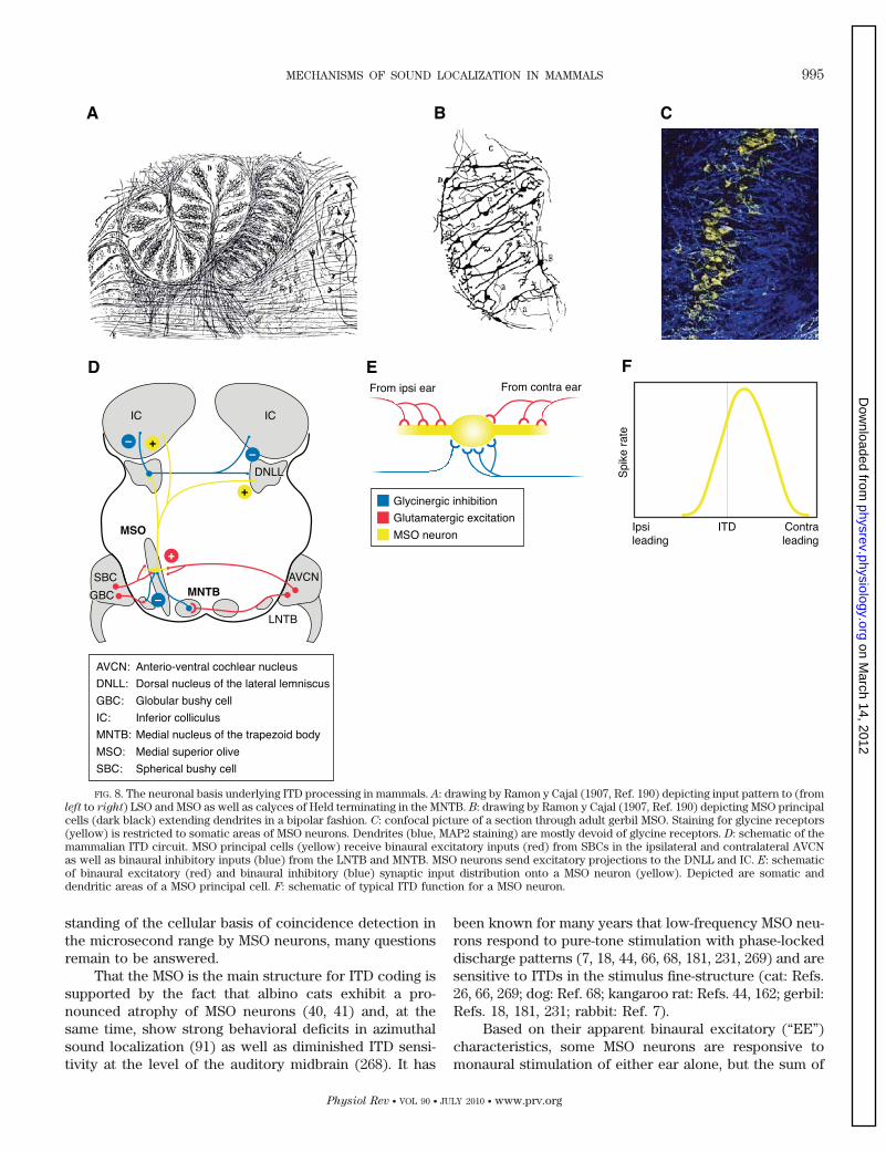
In most mammals with well-developed low-frequencyhearing, the MSO is a laminar structure located mediallyto the more prominent LSO (190, 235) (Fig. 8A). The MSOis organized such that neurons tuned to the highest fre-quency sounds lie towards the ventral pole of the nucleus,and those tuned to the lowest towards the dorsal pole (68,84). MSO principal cells typically show bipolar morphol-ogy and are arranged in a single para-saggital plane withtwo major dendrites emerging from the soma 180° to eachother and extending orthogonally with respect to thedorsoventral axis of the nucleus (112, 190, 191, 235) (Fig.8, B and C). Principal cells appear to receive four major,and highly segregated, inputs from different sites withinthe ascending auditory pathway (112, 263) (Fig. 8, D andE). SBCs from the CN on each side of the brain convergeonto single MSO neurons, with ipsilaterally derived inputssynapsing on the lateral dendrites and contralateral in-puts on the medial (117, 135, 169, 223, 235, 257). Theexcitatory nature of these inputs, mediated by glutama-tergic transmission, has been confirmed by means of invitro recordings (34, 80, 81, 136, 211, 222). In addition,MSO neurons also receive bilateral inhibitory inputs (80,81), largely restricted to the somata of MSO neurons (38,112, 186, 263) (Fig. 8C). Interestingly, this restriction onlyarises during a period of developmental refinement fol-lowing the onset of hearing (112, 263), and species inwhich low-frequency ITD processing is absent do notshow this refinement (112, 252). These inhibitory, glycin-ergic inputs (112, 227, 262) originate in the MNTB (28, 130,235, 263) and the lateral nucleus of the trapezoid body(LNTB) (30). Input from the MNTB, which is driven bystimulation of the contralateral ear, as well as input fromthe ipsilaterally driven LNTB, have been shown to evokeinhibitory postsynaptic potentials (IPSPs) in MSO cells invitro (34, 80, 81, 136), and local application of glycine inthe MSO by means of iontophoresis blocks spiking activ-ity in vivo (18, 181). Both the MNTB (as discussed in sect.
IIA) and the LNTB show morphological specializations forfast and high-fidelity synaptic transmission, their calycealstructure being one such example [MNTB input (255) andend bulbs, LNTB inputs (227)].
An absolute requirement for ITD sensitivity is theability to generate and retain information concerning thefine-structure waveform of the sound arriving at each earindependently, at least until the primary stage of binauralintegration in the brain stem. Primary auditory nervefibers (ANFs) synapsing at the base of the inner hair cells(IHCs) of the cochlea respond to the cycle-by-cyclechanges in the IHC membrane potential (itself reflectingthe back-and-forth deflections of the stereocilia) with ac-tion potentials that are “phase-locked” to the stimuluswaveform (65, 113, 201). Phase-locking in ANFs degradeswith increasing frequency with a pronounced roll-offabove 2–3 kHz (106), although fibers along the full lengthof the basilar membrane show phase-locking if they arecapable of responding to a low-frequency stimulus (re-viewed in Ref. 267). Note that the limitation to phaselocking does not arise in the ANFs per se (90). Rather, itreflects the reduction in capability of the receptor poten-tial of the IHCs to follow the cycle-by-cycle deflection ofthe stereocilia at increasing stimulation frequencies. Thislow-pass feature of the IHCs provides an upper limit forwhich temporal information is theoretically accessible inthe mammalian brain. Accordingly, the upper frequencylimit at which mammals can resolve the ITD in the signalfine structure lies between 1 and a few kHz (119).
Since MNTB cells are also known to phase-lock theiroutput to low-frequency pure tones (51, 95, 125, 149, 173,224), at least three of the four MSO inputs provide precisetemporal information. Based on the anatomical special-izations of the LNTB and its input (228), we can speculatethat this input is also phase-locked (high-frequency LNTBcells in the bat are able to follow fast fluctuations of theenvelope similar to MNTB cells, Ref. 78). However, de-spite decades of research, it remains unclear how the fourinputs to MSO neurons interact to produce the well-de-scribed ITD sensitivity of MSO neurons, and how ITDsensitivity of single MSO neurons provides for a meaning-ful representation of auditory spatial cues. In large part,this is due to the technically demanding nature of in vivorecordings from low-frequency MSO cells, most likely as aresult of the high degree of myelination of input fibers(161), the strong neurophonic potential generated by theirphase-locked synaptic input (162, 269), and the possibilitythat action potentials are generated some distance fromthe soma along the axon (211). Similarly, in vitro record-ings from acute brain slices, the only means so far bywhich the cellular properties of MSO and its inputs can beinvestigated, grow rapidly more difficult with each dayfollowing hearing onset. As a result, almost all intracellu-lar recordings have been performed in immature braintissue, and while these are vastly valuable to the under-
994 GROTHE, PECKA, AND McALPINE
Physiol Rev • VOL 90 • JULY 2010 • www.prv.org
on March 14, 2012
physrev.physiology.orgD
ownloaded from
standing of the cellular basis of coincidence detection inthe microsecond range by MSO neurons, many questionsremain to be answered.
That the MSO is the main structure for ITD coding issupported by the fact that albino cats exhibit a pro-nounced atrophy of MSO neurons (40, 41) and, at thesame time, show strong behavioral deficits in azimuthalsound localization (91) as well as diminished ITD sensi-tivity at the level of the auditory midbrain (268). It has
been known for many years that low-frequency MSO neu-rons respond to pure-tone stimulation with phase-lockeddischarge patterns (7, 18, 44, 66, 68, 181, 231, 269) and aresensitive to ITDs in the stimulus fine-structure (cat: Refs.26, 66, 269; dog: Ref. 68; kangaroo rat: Refs. 44, 162; gerbil:Refs. 18, 181, 231; rabbit: Ref. 7).
Based on their apparent binaural excitatory (“EE”)characteristics, some MSO neurons are responsive tomonaural stimulation of either ear alone, but the sum of
FIG. 8. The neuronal basis underlying ITD processing in mammals. A: drawing by Ramon y Cajal (1907, Ref. 190) depicting input pattern to (fromleft to right) LSO and MSO as well as calyces of Held terminating in the MNTB. B: drawing by Ramon y Cajal (1907, Ref. 190) depicting MSO principalcells (dark black) extending dendrites in a bipolar fashion. C: confocal picture of a section through adult gerbil MSO. Staining for glycine receptors(yellow) is restricted to somatic areas of MSO neurons. Dendrites (blue, MAP2 staining) are mostly devoid of glycine receptors. D: schematic of themammalian ITD circuit. MSO principal cells (yellow) receive binaural excitatory inputs (red) from SBCs in the ipsilateral and contralateral AVCNas well as binaural inhibitory inputs (blue) from the LNTB and MNTB. MSO neurons send excitatory projections to the DNLL and IC. E: schematicof binaural excitatory (red) and binaural inhibitory (blue) synaptic input distribution onto a MSO neuron (yellow). Depicted are somatic anddendritic areas of a MSO principal cell. F: schematic of typical ITD function for a MSO neuron.
MECHANISMS OF SOUND LOCALIZATION IN MAMMALS 995
Physiol Rev • VOL 90 • JULY 2010 • www.prv.org
on March 14, 2012
physrev.physiology.orgD
ownloaded from
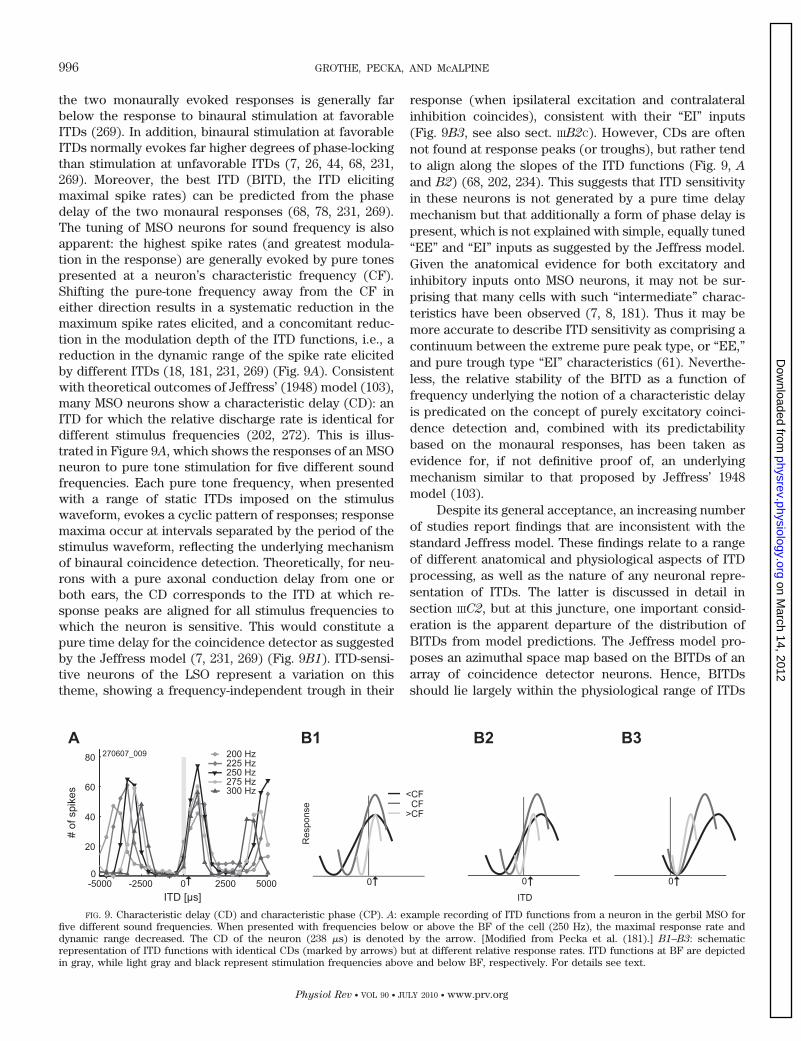
the two monaurally evoked responses is generally farbelow the response to binaural stimulation at favorableITDs (269). In addition, binaural stimulation at favorableITDs normally evokes far higher degrees of phase-lockingthan stimulation at unfavorable ITDs (7, 26, 44, 68, 231,269). Moreover, the best ITD (BITD, the ITD elicitingmaximal spike rates) can be predicted from the phasedelay of the two monaural responses (68, 78, 231, 269).The tuning of MSO neurons for sound frequency is alsoapparent: the highest spike rates (and greatest modula-tion in the response) are generally evoked by pure tonespresented at a neuron’s characteristic frequency (CF).Shifting the pure-tone frequency away from the CF ineither direction results in a systematic reduction in themaximum spike rates elicited, and a concomitant reduc-tion in the modulation depth of the ITD functions, i.e., areduction in the dynamic range of the spike rate elicitedby different ITDs (18, 181, 231, 269) (Fig. 9A). Consistentwith theoretical outcomes of Jeffress’ (1948) model (103),many MSO neurons show a characteristic delay (CD): anITD for which the relative discharge rate is identical fordifferent stimulus frequencies (202, 272). This is illus-trated in Figure 9A, which shows the responses of an MSOneuron to pure tone stimulation for five different soundfrequencies. Each pure tone frequency, when presentedwith a range of static ITDs imposed on the stimuluswaveform, evokes a cyclic pattern of responses; responsemaxima occur at intervals separated by the period of thestimulus waveform, reflecting the underlying mechanismof binaural coincidence detection. Theoretically, for neu-rons with a pure axonal conduction delay from one orboth ears, the CD corresponds to the ITD at which re-sponse peaks are aligned for all stimulus frequencies towhich the neuron is sensitive. This would constitute apure time delay for the coincidence detector as suggestedby the Jeffress model (7, 231, 269) (Fig. 9B1). ITD-sensi-tive neurons of the LSO represent a variation on thistheme, showing a frequency-independent trough in their
response (when ipsilateral excitation and contralateralinhibition coincides), consistent with their “EI” inputs(Fig. 9B3, see also sect. IIIB2C). However, CDs are oftennot found at response peaks (or troughs), but rather tendto align along the slopes of the ITD functions (Fig. 9, A
and B2) (68, 202, 234). This suggests that ITD sensitivityin these neurons is not generated by a pure time delaymechanism but that additionally a form of phase delay ispresent, which is not explained with simple, equally tuned“EE” and “EI” inputs as suggested by the Jeffress model.Given the anatomical evidence for both excitatory andinhibitory inputs onto MSO neurons, it may not be sur-prising that many cells with such “intermediate” charac-teristics have been observed (7, 8, 181). Thus it may bemore accurate to describe ITD sensitivity as comprising acontinuum between the extreme pure peak type, or “EE,”and pure trough type “EI” characteristics (61). Neverthe-less, the relative stability of the BITD as a function offrequency underlying the notion of a characteristic delayis predicated on the concept of purely excitatory coinci-dence detection and, combined with its predictabilitybased on the monaural responses, has been taken asevidence for, if not definitive proof of, an underlyingmechanism similar to that proposed by Jeffress’ 1948model (103).
Despite its general acceptance, an increasing numberof studies report findings that are inconsistent with thestandard Jeffress model. These findings relate to a rangeof different anatomical and physiological aspects of ITDprocessing, as well as the nature of any neuronal repre-sentation of ITDs. The latter is discussed in detail insection IIIC2, but at this juncture, one important consid-eration is the apparent departure of the distribution ofBITDs from model predictions. The Jeffress model pro-poses an azimuthal space map based on the BITDs of anarray of coincidence detector neurons. Hence, BITDsshould lie largely within the physiological range of ITDs
FIG. 9. Characteristic delay (CD) and characteristic phase (CP). A: example recording of ITD functions from a neuron in the gerbil MSO forfive different sound frequencies. When presented with frequencies below or above the BF of the cell (250 Hz), the maximal response rate anddynamic range decreased. The CD of the neuron (238 �s) is denoted by the arrow. [Modified from Pecka et al. (181).] B1–B3: schematicrepresentation of ITD functions with identical CDs (marked by arrows) but at different relative response rates. ITD functions at BF are depictedin gray, while light gray and black represent stimulation frequencies above and below BF, respectively. For details see text.
996 GROTHE, PECKA, AND McALPINE
Physiol Rev • VOL 90 • JULY 2010 • www.prv.org
on March 14, 2012
physrev.physiology.orgD
ownloaded from
(humans: �690 �s, Ref. 157; gerbils: �135 �s, Ref. 138),6
and potentially with greatest density in the region ofhighest psychophysical resolution (and ethological rele-vance) around 0 ITD. Such a pattern conforms to descrip-tions of ITD coding in the avian brain (see sect. IIIC1) buthas not been confirmed for mammals. Indeed, BITDsappear not to be restricted to the physiological range (orat least predominantly within it, again as found in birds),but often lie beyond it, at ITDs greater than would becreated by the interaural distance. This is based on solidpopulation statistics at different levels of the ITD codingpathway such as the IC of the guinea pig (146), cat (86),and chinchilla (246); the MSO of the kangaroo rat (44)(although not discussed there) and gerbil (18, 181; see Fig.9A1); as well as the dorsal nucleus of the lateral lemnis-cus in the latter (219). Furthermore, there appears to belittle difference in the distribution of BITDs between spe-cies with different interaural distances (cf. cat and guineapig, Refs. 172, 246). Moreover, neurons in the MSO of thedog, described in some detail by Goldberg and Brown(68), show BITDs far beyond the range of ITDs predictedby the animal’s head width, and Kuwada and colleagues(60, 61) reported many, if not the large majority (judgingfrom their Fig. 8 in Ref. 61, for example), of ITD-sensitive“peak-type” cells in the lateral lemniscus having BITDsoutside the rabbit’s physiological range. In relation to theJeffress model, this creates two conceptual problems.First, the large number of cells with BITDs beyond thatpredicted by the size of the head requires explanation.Second, in the absence of any other delay mechanism, thecoding of these exceptionally long BITDs would requiresubstantial differences in axonal path-lengths from eachear to individual MSO neurons.
Related to the first conceptual problem, the existenceof BITDs outside the range predicted by the head sizecould be explained by the need to process reverberationsthat result in reduced correlations of the phase-lockedinputs between the two ears (59). In fact, the mixing ofmultiple direct and indirect sound sources that results inreduced interaural coherence (15) produces rapidly fluc-tuating ITDs and ILDs that can greatly exceed the “phys-iological range” (150). In such cases, neural mechanismsfor detecting reduced interaural coherence could exist inthe form of coincidence detectors wired for longer delays.Nevertheless, such long ITDs are relatively rare in mostnatural environments and their importance lessened byrelatively long integration times at higher auditory sta-tions. There appears no particular reason why the mam-malian auditory system might devote the majority of itsITD-sensitive neurons to the processing of interaurallydecorrelated sounds or fluctuating ITDs that do not
indicate a specific source location, as well as the ques-tions as to why this does not appear to be the case inbirds. Interestingly, and as mentioned above, there isno obvious difference in the distribution of BITDs insmall mammals like gerbils or guinea pigs and the muchlarger cat (172, 246). Rather, BITDs depend on the fre-quency tuning of the ITD-sensitive cells, i.e., the CF (thefrequency with the lowest tone-evoked threshold). Basedon a large population of guinea pig IC neurons, McAlpineet al. (146) first pointed out that the average BITD in-creases with decreasing CF and, in fact, shows an almostconstant best IPD. Such a constant BIPD has also beenshown for the gerbil MSO (18, 181) and dorsal nucleus ofthe lateral lemniscus (219), although only with tonal stim-ulation, and for the cat IC (86). The conceptual implica-tions on coding strategies of ITD in mammals are dis-cussed in section IV. But it should be noted here that theobserved dependency of BITD on CF is hardly compatiblewith the idea of a labeled line coding of auditory spaceassumed in Jeffress’ model.
The second conceptual problem in the context of theJeffress model that arises from the existence of longBITDs concerns the existence of delay lines per se and therequirement for exceptionally long delay lines originatingfrom the contralateral ear (60). What mechanism couldaccount for the actual delays observed for contralateralinputs responsible for generating neural BITDs, almost allof which correspond to stimulus locations in the con-tralateral hemisphere (sound leading at the contralateralear)? The traditional textbook notion is that the internaldelay mechanism is realized by means of specific combi-nations of the axonal length of the binaural excitatoryinputs. As obvious as it is in the bird NL, the anatomicalarrangement of MSO inputs on the contrary is difficult tointerpret. Some inputs may resemble delay-line arrange-ments as proposed by Jeffress (223), but the interpreta-tion of these single projections is problematic (compareRef. 74). A detailed anatomical analysis comes from Ol-iver and colleagues (10) who performed a complex recon-struction of small tracer injections into the CN. Theirresults include an analysis of the axonal diameter ofindividual fibers and suggest some form of graded inputfrom the contralateral side where, in some reconstruc-tions, shorter collaterals tended to innervate more rostralparts of MSO and longer collaterals more caudal. How-ever, other injections revealed very restricted terminalfields or even gradients running in the “wrong” directions.Accordingly, the authors were very careful in their con-clusions and suggested that “other factors may be in-volved in the computation of ITDs.” Most exactly, asstated above, the calculated delays would, in some cases,fit the Jeffress model with ITDs only within the physio-logical range, but definitely would not account for theobserved long delays. Hence, axonal length, althoughlikely an important factor in generating delays, appears
6Note that the actually occurring ITD at a given head to some extentdepends on sound frequency (246).
MECHANISMS OF SOUND LOCALIZATION IN MAMMALS 997
Physiol Rev • VOL 90 • JULY 2010 • www.prv.org
on March 14, 2012
physrev.physiology.orgD
ownloaded from
not to be systematically arranged and cannot account forthe long delays observed physiologically. In this sense, thesituation is fundamentally different from that in birds.Interestingly, a factor that has received little attentionuntil lately is the distance of nodes of Ranvier and theinfluence of axon diameter, which could, if varied fromaxon to axon or at different axonal branches, easily con-tribute to a substantial delay (42); accordingly, data fromthe chick brain stem indicate a significant contribution ofinternode distance (213).
Another form of physical delay that has been pro-posed to contribute to ITD processing is the cochleardelay (208). Due to the limited propagation speed of thetraveling wave within the cochlea, moving from basal(high frequencies) to apical (low frequencies), the re-sponse latency to an acoustic event, for instance, a verybrief click that activates inner hair cell transmitter re-lease, occurs at increasingly longer delays relative to thestimulus onset for progressively lower frequency regionsalong the basilar membrane. Schroeder’s notion that aninteraural mismatch in the frequency tuning of the inputsto MSO neurons could be responsible for generating ITDdelay tuning (Schroeder 1977, Ref. 208) was popularizedby Shamma (216) and has been revisited by Joris andcolleagues in recent years (104, 109). However, it must bestated that no convincing evidence for the employment ofsuch cochlear delays in the tuning of neurons to a pre-ferred ITD has been provided.
Yet another means by which internal delays could begenerated, and by which the experimentally observedphase delays could be explained, is synaptic inhibition. Asdiscussed above, the MSO receives bilateral inhibitoryinputs that employ glycine as their neurotransmitter.These inputs are highly specialized for high-fidelity andhigh-precision (i.e., phase-locked) temporal transmission.In vivo experiments combined with pharmacology dem-onstrate that blocking glycinergic inhibition with strych-nine results in a broadening of the ITD tuning functiontowards ipsilaterally leading ITDs resulting in a shift ofthe best ITD towards 0 (to the “left”; for details, seeAppendix C) (18, 181). This suggests that inhibition playsa crucial role in the ITD tuning of MSO neurons. More-over, a “leftward” shift of the best ITD is also observedwhen endogenous inhibition, rather than being blocked, issupplemented through iontophoretic application of gly-cine onto MSO neurons. However, this shift, most likely,arose due to disruption of the normal timing of the gly-cinergic inputs (for details, see Appendix C). Together,these results suggest that it is not only the inhibition assuch that tunes the ITD function, but its timing relative tothe timing of the excitatory inputs (181). A detailed ex-planation of the findings and the suggested underlyingmechanism is given in Appendix C.
An important consideration for the role inhibitoryinputs in tuning neurons for their preferred ITD is the
possibility of adjusting or refining the contralateral delayduring ontogeny. Anatomical refinement of the glyciner-gic inhibition has been shown to be dependent, at leastpartially, on auditory experience during early develop-ment. Raising gerbils in omni-directional noise, whichmasks most spatial cues (265), reduces the degree ofsynaptic selection of glycinergic MSO inputs during whatappears to be a critical period shortly following the onsetof hearing (112, 263). In animals subject to such depriva-tion, the distribution of BITDs deviates significantly fromthat in control animals; BITDs are, on average, muchcloser to 0 ITD than in normally reared animals (212).These findings are consistent with the interpretation thatother mechanisms (axonal length, cochlear delays, etc.)establish an approximate BITD but, following the onset ofhearing, an experience-dependent process selectively en-hances inhibitory inputs to further tune BITDs to thedesired population mean. Moreover, by changing the bal-ance of excitatory and inhibitory inputs (compare Ref.137), it is possible that BITDs might also be adjustedunder dynamic control as needed (e.g., in noisy environ-ments with many concurrent sounds).
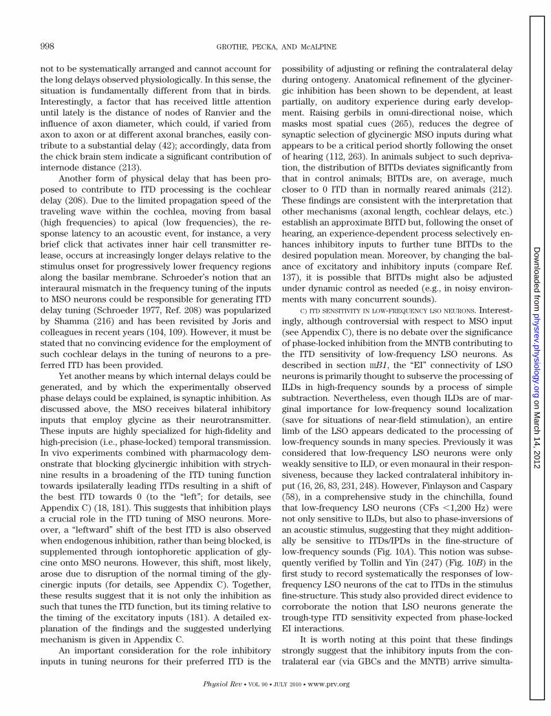
C) ITD SENSITIVITY IN LOW-FREQUENCY LSO NEURONS. Interest-ingly, although controversial with respect to MSO input(see Appendix C), there is no debate over the significanceof phase-locked inhibition from the MNTB contributing tothe ITD sensitivity of low-frequency LSO neurons. Asdescribed in section IIIB1, the “EI” connectivity of LSOneurons is primarily thought to subserve the processing ofILDs in high-frequency sounds by a process of simplesubtraction. Nevertheless, even though ILDs are of mar-ginal importance for low-frequency sound localization(save for situations of near-field stimulation), an entirelimb of the LSO appears dedicated to the processing oflow-frequency sounds in many species. Previously it wasconsidered that low-frequency LSO neurons were onlyweakly sensitive to ILD, or even monaural in their respon-siveness, because they lacked contralateral inhibitory in-put (16, 26, 83, 231, 248). However, Finlayson and Caspary(58), in a comprehensive study in the chinchilla, foundthat low-frequency LSO neurons (CFs �1,200 Hz) werenot only sensitive to ILDs, but also to phase-inversions ofan acoustic stimulus, suggesting that they might addition-ally be sensitive to ITDs/IPDs in the fine-structure oflow-frequency sounds (Fig. 10A). This notion was subse-quently verified by Tollin and Yin (247) (Fig. 10B) in thefirst study to record systematically the responses of low-frequency LSO neurons of the cat to ITDs in the stimulusfine-structure. This study also provided direct evidence tocorroborate the notion that LSO neurons generate thetrough-type ITD sensitivity expected from phase-lockedEI interactions.
It is worth noting at this point that these findingsstrongly suggest that the inhibitory inputs from the con-tralateral ear (via GBCs and the MNTB) arrive simulta-
998 GROTHE, PECKA, AND McALPINE
Physiol Rev • VOL 90 • JULY 2010 • www.prv.org
on March 14, 2012
physrev.physiology.orgD
ownloaded from
neously with the ipsilateral excitatory inputs (via SBCs) atthe LSO to suppress any responses, although the inhibi-tory inputs feature a longer pathway and an additionalsynapse. Possible explanations for this faster contralat-eral conduction are discussed in section IIIB1.
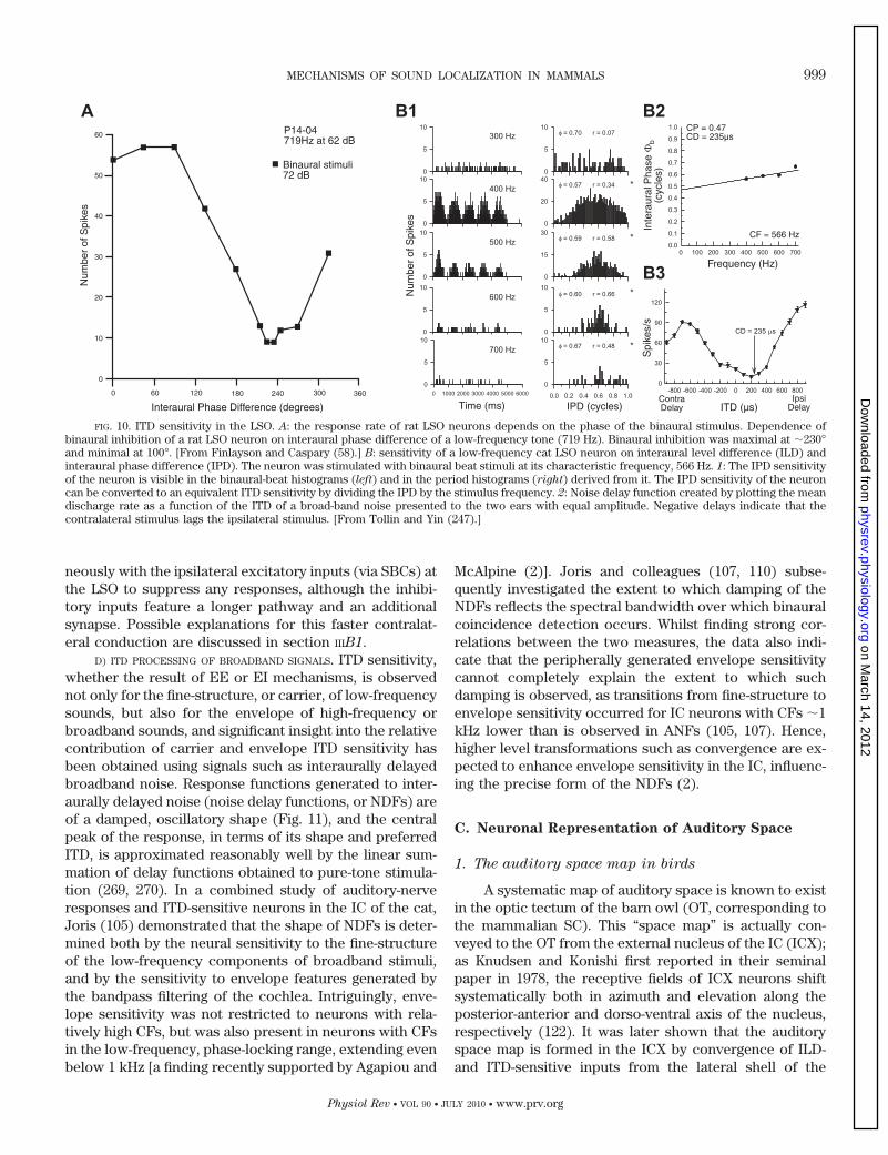
D) ITD PROCESSING OF BROADBAND SIGNALS. ITD sensitivity,whether the result of EE or EI mechanisms, is observednot only for the fine-structure, or carrier, of low-frequencysounds, but also for the envelope of high-frequency orbroadband sounds, and significant insight into the relativecontribution of carrier and envelope ITD sensitivity hasbeen obtained using signals such as interaurally delayedbroadband noise. Response functions generated to inter-aurally delayed noise (noise delay functions, or NDFs) areof a damped, oscillatory shape (Fig. 11), and the centralpeak of the response, in terms of its shape and preferredITD, is approximated reasonably well by the linear sum-mation of delay functions obtained to pure-tone stimula-tion (269, 270). In a combined study of auditory-nerveresponses and ITD-sensitive neurons in the IC of the cat,Joris (105) demonstrated that the shape of NDFs is deter-mined both by the neural sensitivity to the fine-structureof the low-frequency components of broadband stimuli,and by the sensitivity to envelope features generated bythe bandpass filtering of the cochlea. Intriguingly, enve-lope sensitivity was not restricted to neurons with rela-tively high CFs, but was also present in neurons with CFsin the low-frequency, phase-locking range, extending evenbelow 1 kHz [a finding recently supported by Agapiou and
McAlpine (2)]. Joris and colleagues (107, 110) subse-quently investigated the extent to which damping of theNDFs reflects the spectral bandwidth over which binauralcoincidence detection occurs. Whilst finding strong cor-relations between the two measures, the data also indi-cate that the peripherally generated envelope sensitivitycannot completely explain the extent to which suchdamping is observed, as transitions from fine-structure toenvelope sensitivity occurred for IC neurons with CFs �1kHz lower than is observed in ANFs (105, 107). Hence,higher level transformations such as convergence are ex-pected to enhance envelope sensitivity in the IC, influenc-ing the precise form of the NDFs (2).
C. Neuronal Representation of Auditory Space
1. The auditory space map in birds
A systematic map of auditory space is known to existin the optic tectum of the barn owl (OT, corresponding tothe mammalian SC). This “space map” is actually con-veyed to the OT from the external nucleus of the IC (ICX);as Knudsen and Konishi first reported in their seminalpaper in 1978, the receptive fields of ICX neurons shiftsystematically both in azimuth and elevation along theposterior-anterior and dorso-ventral axis of the nucleus,respectively (122). It was later shown that the auditoryspace map is formed in the ICX by convergence of ILD-and ITD-sensitive inputs from the lateral shell of the
FIG. 10. ITD sensitivity in the LSO. A: the response rate of rat LSO neurons depends on the phase of the binaural stimulus. Dependence ofbinaural inhibition of a rat LSO neuron on interaural phase difference of a low-frequency tone (719 Hz). Binaural inhibition was maximal at �230°and minimal at 100°. [From Finlayson and Caspary (58).] B: sensitivity of a low-frequency cat LSO neuron on interaural level difference (ILD) andinteraural phase difference (IPD). The neuron was stimulated with binaural beat stimuli at its characteristic frequency, 566 Hz. 1: The IPD sensitivityof the neuron is visible in the binaural-beat histograms (left) and in the period histograms (right) derived from it. The IPD sensitivity of the neuroncan be converted to an equivalent ITD sensitivity by dividing the IPD by the stimulus frequency. 2: Noise delay function created by plotting the meandischarge rate as a function of the ITD of a broad-band noise presented to the two ears with equal amplitude. Negative delays indicate that thecontralateral stimulus lags the ipsilateral stimulus. [From Tollin and Yin (247).]
MECHANISMS OF SOUND LOCALIZATION IN MAMMALS 999
Physiol Rev • VOL 90 • JULY 2010 • www.prv.org
on March 14, 2012
physrev.physiology.orgD
ownloaded from
central nucleus of the IC coding for the same region ofauditory space. Moreover, these converging projectionscombine their input across a wide range of CFs (39, 240),pooling that is crucial in overcoming phase ambiguitiesthat arise for narrow-band signals in the relatively high-frequency range employed (uniquely) by the owl in sound-localization tasks using ITDs (122, 143, 240). The head-centered auditory space map in the OT is then mergedwith that of the visual space map7 (120), ultimately pro-viding the anatomical substrate for the pronounced audio-visually driven head-saccade reflex of the barn owl. Knud-sen and colleagues went on to show that the visual inputprovides instructive signals for the calibration of the au-ditory map during maturation (for review, see Ref. 121).Hence, the formation of the auditory space map isstrongly related to that of the visual system, and it maynot be surprising, therefore, that the neural representa-tion of the auditory world resembles that of the visualworld. It is well established that in owls the topographi-cally ordered arrangement of auditory space is present atthe level of the NL, the initial site of ITD processing (33,
185, 239)), and recent data (127) suggest a similar arrange-ment in the chicken (Fig. 12, A–C).8
As in other birds, ILD sensitivity in barn owls isinitially encoded in the lateral lemniscus via a reciprocalinhibition between the lemniscal nuclei on opposite sidesof the brain. Already at this level of processing thereexists a systematic gradient of ILD sensitivity and, hence,a rough topographic map of ILDs (31, 139, 242, 243).However, barn owls are clearly an exception in that theybelong to the group of so-called “asymmetrical owls,”having the two outer ears and ear canals located atslightly different positions and pointing into different ver-tical directions (153, 165, 253). This asymmetry results iniso-ILD contours in the frontal field being tilted by almost45 degrees compared with the vertical alignment found inanimals with symmetrical ears. Thus, in barn owls, ILDsserve to encode the elevation, rather than the azimuth, ofa source location. To this end, the representation of ILDsin these birds is almost orthogonal to the representationof ITDs, which are unaffected by the asymmetry. Bymeans of a multiplicative interaction of ITD and ILDsensitivity at the level of the auditory midbrain (182),“classical” two-dimensional auditory-spatial receptivefields are created (183). These are then projected, andsystematically arranged, in a two-dimensional map of au-ditory space in the barn owl tectum (17, 67, 122).
An interesting feature of the auditory space maps inthe owl OT is the fact that individual cells are relativelycoarsely tuned (�20° on average) compared with thebehavioral acuity of �2–3° (6, 120). Takahashi and col-leagues (5, 241) pointed out that this apparent discrep-ancy can be overcome by the analysis of spike ratechanges (scaled to their variance) in individual neuronswith changes in sound location, i.e., changes in spike ratealong the slope of the ITD functions. This idea is in linewith the theoretical framework of optimal coding positedin recent years (25, 88). Hence, the systematic map ofITDs in the owl brain stem and midbrain is undisputed,although the nature of the associated neural code is stillopen to debate.9
2. The representation of binaural cues in mammals
The topographic arrangement of auditory space rep-resented in the avian OT has a counterpart in the mam-malian SC, with a systematic arrangement of ILD sensi-tivity extending along the mediolateral extent of the nu-cleus, creating a space map (review in Ref. 114). However,
7Note that the barn owl is not capable of moving its eyes, hence doesnot perform eye saccades.
8Recent evidence indicates that in the barn owl forebrain ITD-sensi-tive neurons are not arranged in an orderly map (39), and the distribu-tion of BITDs is similar to that in small mammals, with the steepestslopes clustering around the midline (256).
9Actually, broader tuning of neurons relative to behavior is not aspecial feature of the sound localization system but rather a ubiquitousfeature of sensory systems.
FIG. 11. Noise delay functions to correlated and anticorrelatedstimuli. Example recordings of delay functions in response to correlated(solid line) and anticorrelated noise (dotted line) for two DNLL neurons.Gray shaded area depicts physiological range. Characteristic frequen-cies (CFs) of the neurons are shown at the top left of each panel. Notethe differences in damping of the side peaks.
1000 GROTHE, PECKA, AND McALPINE
Physiol Rev • VOL 90 • JULY 2010 • www.prv.org
on March 14, 2012
physrev.physiology.orgD
ownloaded from
FIG. 12. The representation of auditory space in birds and mammals. A: in birds, the location of sound sources in the horizontal plane is encodedby labeled lines originating in the NL. Neurons in isofrequency laminae of each NL (closed circles) vary systematically in their preferred ITD, withdifferent neurons encoding different locations ranging from midline to contralateral 90° (as illustrated by the color code). Locations near midlineare represented by neurons in either NL, giving rise to a higher resolution for medial locations compared with lateral locations. Schematic includesonly one isofrequency lamina. B: example of systematic shift of best ITDs along a iso-frequency axis of the chick NL. C: distribution of best ITDsin the chick NL as a function of BF. D: in mammals, locations in the horizontal plane are encoded by two hemispheric channels with MSO neuronsresponding maximally to stimuli positioned far in the contralateral hemifield. Nevertheless, spatial acuity is best near midline due to the positionof the steepest slope of the function being positioned near midline. E and F, top panels: example recordings of neurons with varying BFs from thegerbil MSO and guinea pig IC, respectively. Shaded areas denote physiological range of ITDs. Note that best ITDs increased with decreasing neuronalBF. Bottom panels: distribution of best ITD as a function of BF in the MSO and IC, respectively. Best ITDs of functions shown in top panels aredepicted in respective colors. Shown in black solid line is the function [best ITD � 0.125/BF]. Shaded areas denote physiological range of ITDs forthe gerbil and guinea pig, respectively. [A and C from Koppl and Carr (127); E modified from Pecka et al. (181); F from McAlpine (144).]
MECHANISMS OF SOUND LOCALIZATION IN MAMMALS 1001
Physiol Rev • VOL 90 • JULY 2010 • www.prv.org
on March 14, 2012
physrev.physiology.orgD
ownloaded from
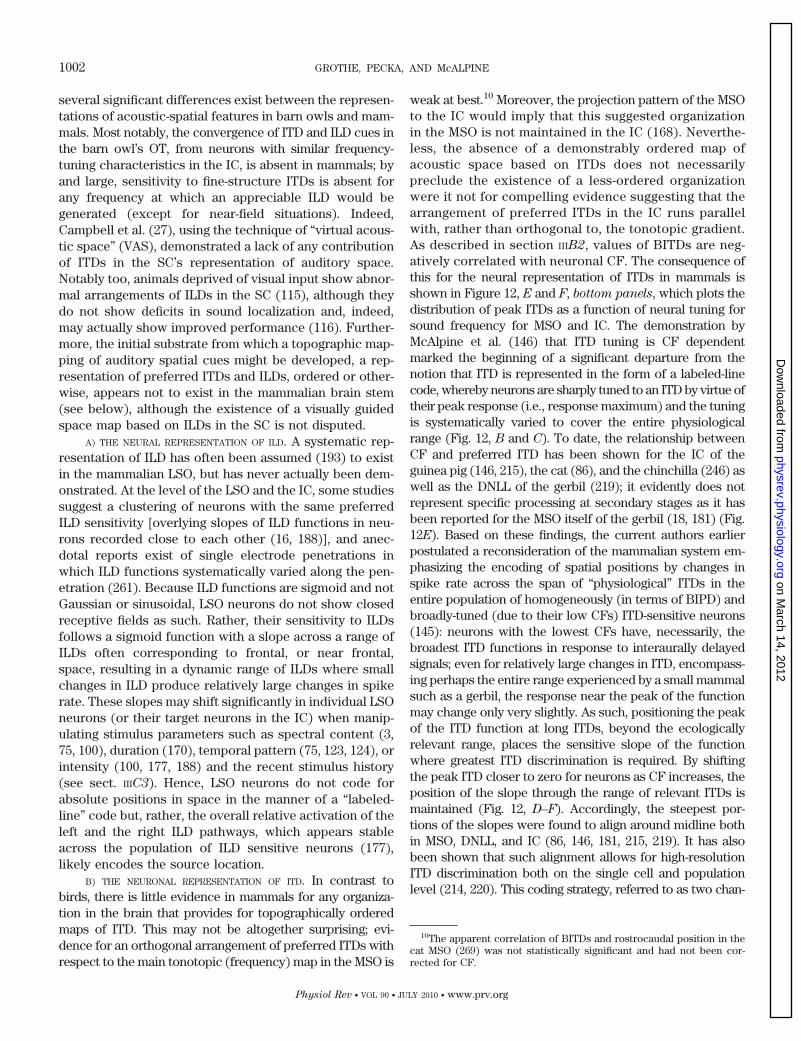
several significant differences exist between the represen-tations of acoustic-spatial features in barn owls and mam-mals. Most notably, the convergence of ITD and ILD cues inthe barn owl’s OT, from neurons with similar frequency-tuning characteristics in the IC, is absent in mammals; byand large, sensitivity to fine-structure ITDs is absent forany frequency at which an appreciable ILD would begenerated (except for near-field situations). Indeed,Campbell et al. (27), using the technique of “virtual acous-tic space” (VAS), demonstrated a lack of any contributionof ITDs in the SC’s representation of auditory space.Notably too, animals deprived of visual input show abnor-mal arrangements of ILDs in the SC (115), although theydo not show deficits in sound localization and, indeed,may actually show improved performance (116). Further-more, the initial substrate from which a topographic map-ping of auditory spatial cues might be developed, a rep-resentation of preferred ITDs and ILDs, ordered or other-wise, appears not to exist in the mammalian brain stem(see below), although the existence of a visually guidedspace map based on ILDs in the SC is not disputed.
A) THE NEURAL REPRESENTATION OF ILD. A systematic rep-resentation of ILD has often been assumed (193) to existin the mammalian LSO, but has never actually been dem-onstrated. At the level of the LSO and the IC, some studiessuggest a clustering of neurons with the same preferredILD sensitivity [overlying slopes of ILD functions in neu-rons recorded close to each other (16, 188)], and anec-dotal reports exist of single electrode penetrations inwhich ILD functions systematically varied along the pen-etration (261). Because ILD functions are sigmoid and notGaussian or sinusoidal, LSO neurons do not show closedreceptive fields as such. Rather, their sensitivity to ILDsfollows a sigmoid function with a slope across a range ofILDs often corresponding to frontal, or near frontal,space, resulting in a dynamic range of ILDs where smallchanges in ILD produce relatively large changes in spikerate. These slopes may shift significantly in individual LSOneurons (or their target neurons in the IC) when manip-ulating stimulus parameters such as spectral content (3,75, 100), duration (170), temporal pattern (75, 123, 124), orintensity (100, 177, 188) and the recent stimulus history(see sect. IIIC3). Hence, LSO neurons do not code forabsolute positions in space in the manner of a “labeled-line” code but, rather, the overall relative activation of theleft and the right ILD pathways, which appears stableacross the population of ILD sensitive neurons (177),likely encodes the source location.
B) THE NEURONAL REPRESENTATION OF ITD. In contrast tobirds, there is little evidence in mammals for any organiza-tion in the brain that provides for topographically orderedmaps of ITD. This may not be altogether surprising; evi-dence for an orthogonal arrangement of preferred ITDs withrespect to the main tonotopic (frequency) map in the MSO is
weak at best.10 Moreover, the projection pattern of the MSOto the IC would imply that this suggested organizationin the MSO is not maintained in the IC (168). Neverthe-less, the absence of a demonstrably ordered map ofacoustic space based on ITDs does not necessarilypreclude the existence of a less-ordered organizationwere it not for compelling evidence suggesting that thearrangement of preferred ITDs in the IC runs parallelwith, rather than orthogonal to, the tonotopic gradient.As described in section IIIB2, values of BITDs are neg-atively correlated with neuronal CF. The consequence ofthis for the neural representation of ITDs in mammals isshown in Figure 12, E and F, bottom panels, which plots thedistribution of peak ITDs as a function of neural tuning forsound frequency for MSO and IC. The demonstration byMcAlpine et al. (146) that ITD tuning is CF dependentmarked the beginning of a significant departure from thenotion that ITD is represented in the form of a labeled-linecode, whereby neurons are sharply tuned to an ITD by virtue oftheir peak response (i.e., response maximum) and the tuningis systematically varied to cover the entire physiologicalrange (Fig. 12, B and C). To date, the relationship betweenCF and preferred ITD has been shown for the IC of theguinea pig (146, 215), the cat (86), and the chinchilla (246) aswell as the DNLL of the gerbil (219); it evidently does notrepresent specific processing at secondary stages as it hasbeen reported for the MSO itself of the gerbil (18, 181) (Fig.12E). Based on these findings, the current authors earlierpostulated a reconsideration of the mammalian system em-phasizing the encoding of spatial positions by changes inspike rate across the span of “physiological” ITDs in theentire population of homogeneously (in terms of BIPD) andbroadly-tuned (due to their low CFs) ITD-sensitive neurons(145): neurons with the lowest CFs have, necessarily, thebroadest ITD functions in response to interaurally delayedsignals; even for relatively large changes in ITD, encompass-ing perhaps the entire range experienced by a small mammalsuch as a gerbil, the response near the peak of the functionmay change only very slightly. As such, positioning the peakof the ITD function at long ITDs, beyond the ecologicallyrelevant range, places the sensitive slope of the functionwhere greatest ITD discrimination is required. By shiftingthe peak ITD closer to zero for neurons as CF increases, theposition of the slope through the range of relevant ITDs ismaintained (Fig. 12, D–F). Accordingly, the steepest por-tions of the slopes were found to align around midline bothin MSO, DNLL, and IC (86, 146, 181, 215, 219). It has alsobeen shown that such alignment allows for high-resolutionITD discrimination both on the single cell and populationlevel (214, 220). This coding strategy, referred to as two chan-
10The apparent correlation of BITDs and rostrocaudal position in thecat MSO (269) was not statistically significant and had not been cor-rected for CF.
1002 GROTHE, PECKA, AND McALPINE
Physiol Rev • VOL 90 • JULY 2010 • www.prv.org
on March 14, 2012
physrev.physiology.orgD
ownloaded from
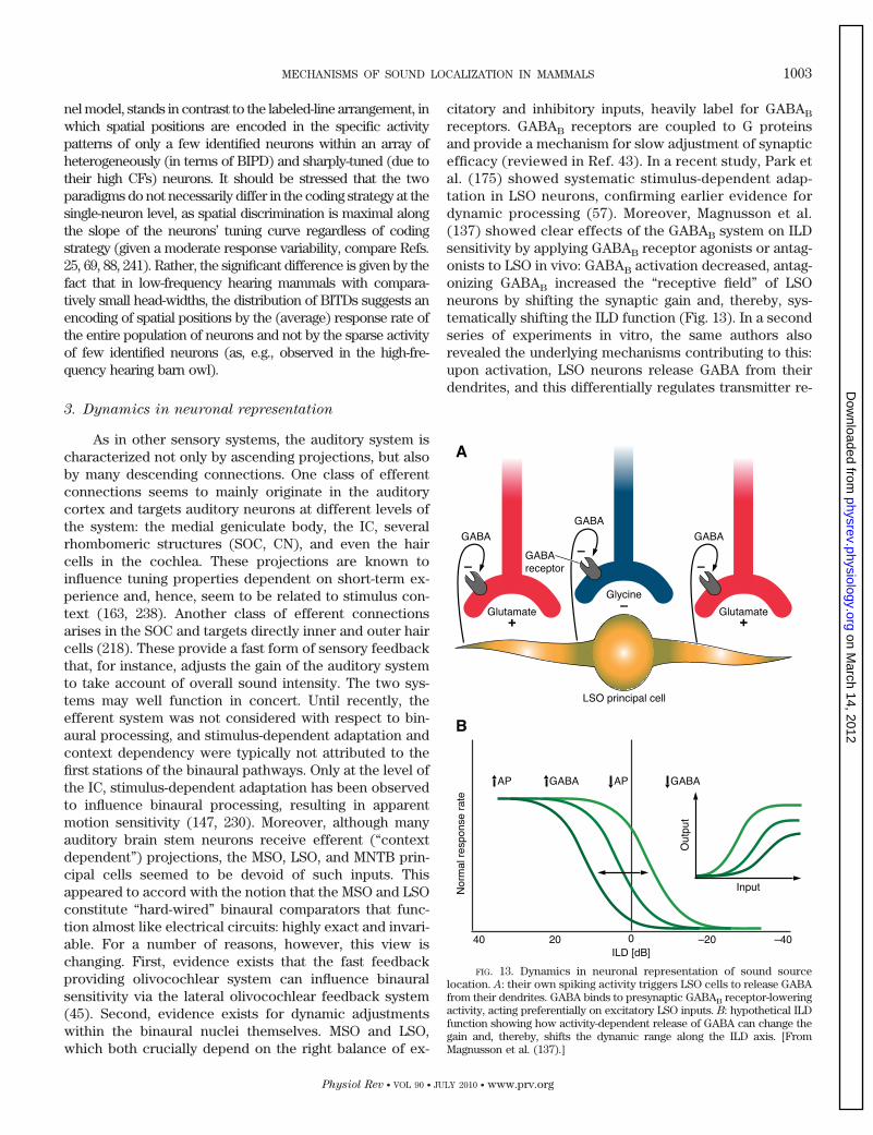
nel model, stands in contrast to the labeled-line arrangement, inwhich spatial positions are encoded in the specific activitypatterns of only a few identified neurons within an array ofheterogeneously (in terms of BIPD) and sharply-tuned (due totheir high CFs) neurons. It should be stressed that the twoparadigms do not necessarily differ in the coding strategy at thesingle-neuron level, as spatial discrimination is maximal alongthe slope of the neurons’ tuning curve regardless of codingstrategy (given a moderate response variability, compare Refs.25, 69, 88, 241). Rather, the significant difference is given by thefact that in low-frequency hearing mammals with compara-tively small head-widths, the distribution of BITDs suggests anencoding of spatial positions by the (average) response rate ofthe entire population of neurons and not by the sparse activityof few identified neurons (as, e.g., observed in the high-fre-quency hearing barn owl).
3. Dynamics in neuronal representation
As in other sensory systems, the auditory system ischaracterized not only by ascending projections, but alsoby many descending connections. One class of efferentconnections seems to mainly originate in the auditorycortex and targets auditory neurons at different levels ofthe system: the medial geniculate body, the IC, severalrhombomeric structures (SOC, CN), and even the haircells in the cochlea. These projections are known toinfluence tuning properties dependent on short-term ex-perience and, hence, seem to be related to stimulus con-text (163, 238). Another class of efferent connectionsarises in the SOC and targets directly inner and outer haircells (218). These provide a fast form of sensory feedbackthat, for instance, adjusts the gain of the auditory systemto take account of overall sound intensity. The two sys-tems may well function in concert. Until recently, theefferent system was not considered with respect to bin-aural processing, and stimulus-dependent adaptation andcontext dependency were typically not attributed to thefirst stations of the binaural pathways. Only at the level ofthe IC, stimulus-dependent adaptation has been observedto influence binaural processing, resulting in apparentmotion sensitivity (147, 230). Moreover, although manyauditory brain stem neurons receive efferent (“contextdependent”) projections, the MSO, LSO, and MNTB prin-cipal cells seemed to be devoid of such inputs. Thisappeared to accord with the notion that the MSO and LSOconstitute “hard-wired” binaural comparators that func-tion almost like electrical circuits: highly exact and invari-able. For a number of reasons, however, this view ischanging. First, evidence exists that the fast feedbackproviding olivocochlear system can influence binauralsensitivity via the lateral olivocochlear feedback system(45). Second, evidence exists for dynamic adjustmentswithin the binaural nuclei themselves. MSO and LSO,which both crucially depend on the right balance of ex-
citatory and inhibitory inputs, heavily label for GABAB
receptors. GABAB receptors are coupled to G proteinsand provide a mechanism for slow adjustment of synapticefficacy (reviewed in Ref. 43). In a recent study, Park etal. (175) showed systematic stimulus-dependent adap-tation in LSO neurons, confirming earlier evidence fordynamic processing (57). Moreover, Magnusson et al.(137) showed clear effects of the GABAB system on ILDsensitivity by applying GABAB receptor agonists or antag-onists to LSO in vivo: GABAB activation decreased, antag-onizing GABAB increased the “receptive field” of LSOneurons by shifting the synaptic gain and, thereby, sys-tematically shifting the ILD function (Fig. 13). In a secondseries of experiments in vitro, the same authors alsorevealed the underlying mechanisms contributing to this:upon activation, LSO neurons release GABA from theirdendrites, and this differentially regulates transmitter re-
FIG. 13. Dynamics in neuronal representation of sound sourcelocation. A: their own spiking activity triggers LSO cells to release GABAfrom their dendrites. GABA binds to presynaptic GABAB receptor-loweringactivity, acting preferentially on excitatory LSO inputs. B: hypothetical ILDfunction showing how activity-dependent release of GABA can change thegain and, thereby, shifts the dynamic range along the ILD axis. [FromMagnusson et al. (137).]
MECHANISMS OF SOUND LOCALIZATION IN MAMMALS 1003
Physiol Rev • VOL 90 • JULY 2010 • www.prv.org
on March 14, 2012
physrev.physiology.orgD
ownloaded from
lease from the excitatory and inhibitory input terminalsvia presynaptic GABAB receptors. Hence, at the veryleast, LSO neurons are able to adjust rapidly the efficacyof their excitatory and inhibitory inputs, altering theirbinaural sensitivity in the process. The function and exactlocation of GABAB receptors in the MSO are not knownyet; however, it seems likely here also that stimulus-dependent dynamics might act on binaural propertieseven at the first level of binaural interaction.
IV. CONCLUSIONS AND OPEN QUESTIONS
When Lloyd Jeffress published his concept on binauralsound localization in 1948, his elegant model fundamentallyinfluenced for generations the way researchers consideredneural mechanisms contributing to the brain’s representa-tion of sound-source location (103). The model was there-after consolidated by data obtained from ITD-sensitive neu-rons in the brain stem and midbrain, revealing that severalfeatures of the neural code matched Jeffress’ predictions(e.g., submillisecond coincidence detection and the exis-tence of characteristic delays). Moreover, the finding of analmost exact manifestation of the delay-line concept in theavian brain appeared to confirm the general validity of thismodel.
However, it is fundamentally important to bear in mindthat the evolution of the mammalian auditory system oc-curred independently from that of birds. Consequently,brain mechanisms responsible for processing binaural cuesdeveloped under different evolutionary pressures, leading todistinct constraints for sound localization mechanisms, e.g.,different hearing ranges, and head and ear morphology. TheJeffress-like labeled-line mechanism of ITD processing andthe related topographic representation of space (as reportedfor the owl) are related to a number of specializations notpresent in the mammalian system. An important aspect ofthe differences between birds and mammals is that, in themammalian auditory system, the two binaural localizationcues are utilized in distinct frequency ranges (ITD for lowfrequency, ILD for high frequency) with the analysis of mon-aural, spectral cues employed to resolve the cone of confu-sion along the vertical axis. Both spectral cues and ILDprocessing show no evidence of a representation based onlabeled lines, bringing into question the necessity of such arepresentation for ITDs. Finally, recent evidence concerningmechanisms of neural tuning for ITD, including the role ofinhibition, as well as the form of the representation of ITDsin the MSO and IC, are not well described by the Jeffressmodel, but rather favors an alternative theory of binauralsound localization first mooted by von Bekesy in the 1930s(254) and later developed by van Bergeijk (1962, Ref. 250), inwhich the neural representation of auditory space is ar-ranged in the form of broadly hemispheric channels, withhorizontal space encoded in the form of an average popula-
tion response. This is consistent with recent evidence fromITD and ILD recordings. Indeed, the appearance of neuronswith broadly tuned response functions is characteristic ofthe majority of studies examining auditory spatial tuning inmammals and is evident across a wide range of species andstages of brain processing from brain stem to cortex [brainstem: gerbil (18, 181); midbrain: guinea pig (146, 215), cat(86), gerbil (219), chinchilla (246); A1: cat (232)].
To this end, the representation of auditory space inthe mammalian brain might best be described as a relativerate code, comparing activity across brain hemispheresrather than within individual brain nuclei, a more distrib-uted and therefore more plastic code compared with the“hard-wired” local code suggested in the Jeffress model,and evident in the avian brain.
We trust that it has become apparent to the readerthat brain mechanisms underpinning sound localization invertebrates are not only interesting of themselves, thecurrent state of the field being characterized by evolvingviews and concepts, but also that they represent a generalmodel system by which a broader understanding of brainprocessing may be obtained. First, the almost uniquestructure-function relationship of the binaural system,combined with the few physical cues involved in soundlocalization, allow for very specific and testable hypothesesof how information is processed in neuronal circuits. Sec-ond, the different implementations of binaural systems, inparticular in birds and mammals, are informative as to therange of possible strategies of computing and coding infor-mation in the brain and how these strategies might dependon phylogeny and evolutionary precedents of a nervoussystem. Third, new insights concerning the exquisite bal-ance of excitatory and inhibitory inputs, down to the sub-millisecond range, and how the recent history of activationinfluences this balance, renders the auditory pathway a po-tential model system for studying age-related changes thataffect neurotransmission in a variety of ways.
APPENDIX A: DO HUMANS LACK THE MNTB?
Besides the logical impossibility of having to provide evidence ofabsence, one must consider that the MNTB is sited within one ofthe most prominent fiber bundles in the entire human brain: thetrapezoid body, with its highly myelinated fibers crossing the mid-line from both cochlear nuclei. Many of these fibers traverse theMNTB itself, and hence, MNTB neurons are literally spread apartfrom each other, making it difficult to delineate the nucleus bytraditional histological means. In many species, particularly thosewith good low-frequency hearing, high-quality histological prepara-tions are required to define the MNTB as a distinct structure, and inhumans, this represents a particular problem. Nevertheless, Ba-zwinsky et al. (9) managed to demonstrate the existence of neu-rons residing within the trapezoid body fibers that receive parval-bumin-positive synaptic terminals. Because MNTB inputs in othermammals contain parvalbumin (53), it is therefore likely that theMNTB is simply difficult to delineate in humans and that this hasled to the misunderstanding, perpetuated to this day, that it isabsent altogether. Interestingly, the first study claiming that hu-mans do not possess an MNTB (158) described a structure the
1004 GROTHE, PECKA, AND McALPINE
Physiol Rev • VOL 90 • JULY 2010 • www.prv.org
on March 14, 2012
physrev.physiology.orgD
ownloaded from
authors referred to as “nucleus of the trapezoid body” (NTB, intheir paper), which is sited in some distance from the location ofthe MNTB. This study ignored the Nissl-stained cell bodies pre-cisely at the position where MNTB would be in other mammals,and where Richter et al. (199) described loosely arranged singlecells with morphological characteristics resembling MNTB neu-rons in other species. What Moore and Moore (158) referred to asNTB resembles, and was most likely mistaken for, a group ofperi-olivary neurons, or possibly the ventral nucleus of the trape-zoid body (VNTB). This particular region of the brain stem, in fact,appears somewhat different in other primates, perhaps not surpris-ing given these neuronal clusters are known to be arranged slightlydifferently in almost every species (for discussions of this problem,compare Refs. 79, 82, 209, 210). Hence, the view that the MNTB isabsent from the human brain was initiated with a publication thatwas, almost certainly, referring to a different neural population.Furthermore, Strominger and Hurwitz (236) pointed out that thehuman MNTB was not easily delineated as a clear nucleus, leadingsome authors to deny its existence in humans.
Together, the problematic “evidence” against the existence ofan MNTB in the human brain stem with the, admittedly weak(for the same reasons), positive evidence for its existence, raisesconceptual problems for the existence of a prominent LSO inhumans; its absence would bring into question the evolutionaryunderstanding and the important general role of the MNTB, anda strong case exists for both the existence and the importance ofthis nucleus in the human brain.
APPENDIX B: ITD PROCESSING: IMPORTANT DEFINITIONS
The following are expressions frequently used in discussionson ITD processing.
Jeffress Model
Model of ITD processing put forward by L. Jeffress (1948), basedon three crucial assumptions: 1) ITD detection via neuronal coin-cidence detection of binaural excitatory inputs, 2) “delay lines”(systematically varying axonal length from both ears) adjust thecoincidence detectors to different best ITDs by compensating forall possible ITDs, and 3) creation of a topographic representationof ITDs in each frequency channel via evenly distributed and sys-tematically varying tuning functions.
Labeled Line Code
Coding strategy in which each ITD is encoded by a specificsubset of neurons within the population of neurons. It followsthat the identity of each neuron is preserved in order to allowupstream evaluation. This is a direct consequence of Jeffressmodel-like ITD processing.
Peak Coding Strategy
Coding of a specific ITD via the peak activity of single or fewneurons within a heterogeneously tuned population. Typicallylinked to a labeled line code, although not obligatory.
Average Population Coding Strategy
Coding of ITDs via response rate modulation along the slopeof the tuning function of a neuron. Individual neurons in apopulation are tuned similarly and do not require to be identifiedfor upstream evaluation.
Space Map
Topographic representation of external space (azimuth andelevation) via an ordered arrangement of systematically varyingneuronal receptive fields along multiple axes of a nucleus.
Two-Channel Model
Model of ITD processing on the basis of the average populationcoding strategy in which two broadly and inversely tuned channelsare created by the populations of neurons in the two brain hemi-spheres. The location of a sound source is encoded in the inverselyproportional relative firing rates in the two channels.
APPENDIX C: ROLE OF GLYCINERGIC INHIBITION IN ITD
PROCESSING IN THE MSO
Indirect evidence for an important role of the inhibitory inputsto the MSO for tuning ITD sensitivity comes, on one hand, from invitro experiments in which contralaterally driven inhibitory inputswere found to dominate binaurally coincident excitation (80). Thisoccurs only over a very short time window (a few hundreds ofmicroseconds) when examined in acute brain slices excised some10 days following hearing onset (81). In fact, these inhibitory ef-fects are extremely strong, generated by some of the fastest- andshortest-acting inhibitory currents measured to date. When mea-sured during blockade of other currents, which normally increasesinput resistance, potentially slowing membrane kinetics, time con-stants of isolated glycine currents (measured in even youngertissue; 4–5 days following hearing onset) remain in the range of 1–2ms (136, 221). More direct evidence for the role of inhibition ingenerating internal delays comes from pharmacological studies ofthe gerbil MSO in vivo, which indicate that glycinergic inhibitionplays a major role in determining the ITD sensitivity of single MSOneurons. During blockade of glycinergic inhibition by means oflocal iontophoretic application of its antagonist, strychnine, neuraldischarge rates are elevated, as expected. More interesting, how-ever, the inhibitory block asserts an asymmetric effect on ITDfunctions of the neurons tested: notably, the slope of the ITDfunction closest to zero (the slope “facing” ipsilateral ITDs) shiftsto beyond the physiological range due to increased responsivenessto ipsilateral leading ITDs. As a result, the BITD shifts toward 0 ITD(indicating less effective contralateral-leading stimulation; Fig. 14).In contrast, the contralateral-facing slope is almost unaffected byblockade of inhibition. These effects are reversible, with the BITDreturning to its original value a few minutes following terminationof the iontophoretic application of strychnine (18, 181). The effectof blocking glycinergic inhibition with strychnine shifts both theonset as well as the ongoing component of each neuron’s ITDfunction.11 A likely explanation for this effect is that glycinergicinhibition provides for a net delay in the effective contralateralexcitation. Two potential scenarios had been posited for this effect.One assumes the action of tonic synaptic inhibition, and the otherthe action of phase-locked synaptic inhibition. To differentiatebetween these two possibilities, Pecka et al. (181) applied glycineiontophoretically to MSO neurons. The rationale of this experimentis that in the event that glycinergic inhibition is tonic, further tonicapplication would enhance the contralateral delay (277), but wouldmask the effect of inhibition if endogenous glycinergic inhibition isphase-locked. BITDs should shift to even longer ITDs if the endog-enous inhibition is tonic but should cause a similar shift to thatobserved during inhibitory block (although at a highly reduceddischarge rate) if the timing of the inhibitory input is important.The results favored the latter scenario: tonic glycine application
11There was a recent debate (104) about the example cell displayed inthe original publication showing the strychnine effect (18), which was,unfortunately for an example, heavily dominated by the onset effect. Theauthors therefore raised the possibility that the observed effects reflectmainly an “onset artifact.” A careful statistical analysis and the examplesshown in Pecka et al. (181), however, reveal that the glycine affects bothonset and ongoing component roughly equally.
MECHANISMS OF SOUND LOCALIZATION IN MAMMALS 1005
Physiol Rev • VOL 90 • JULY 2010 • www.prv.org
on March 14, 2012
physrev.physiology.orgD
ownloaded from
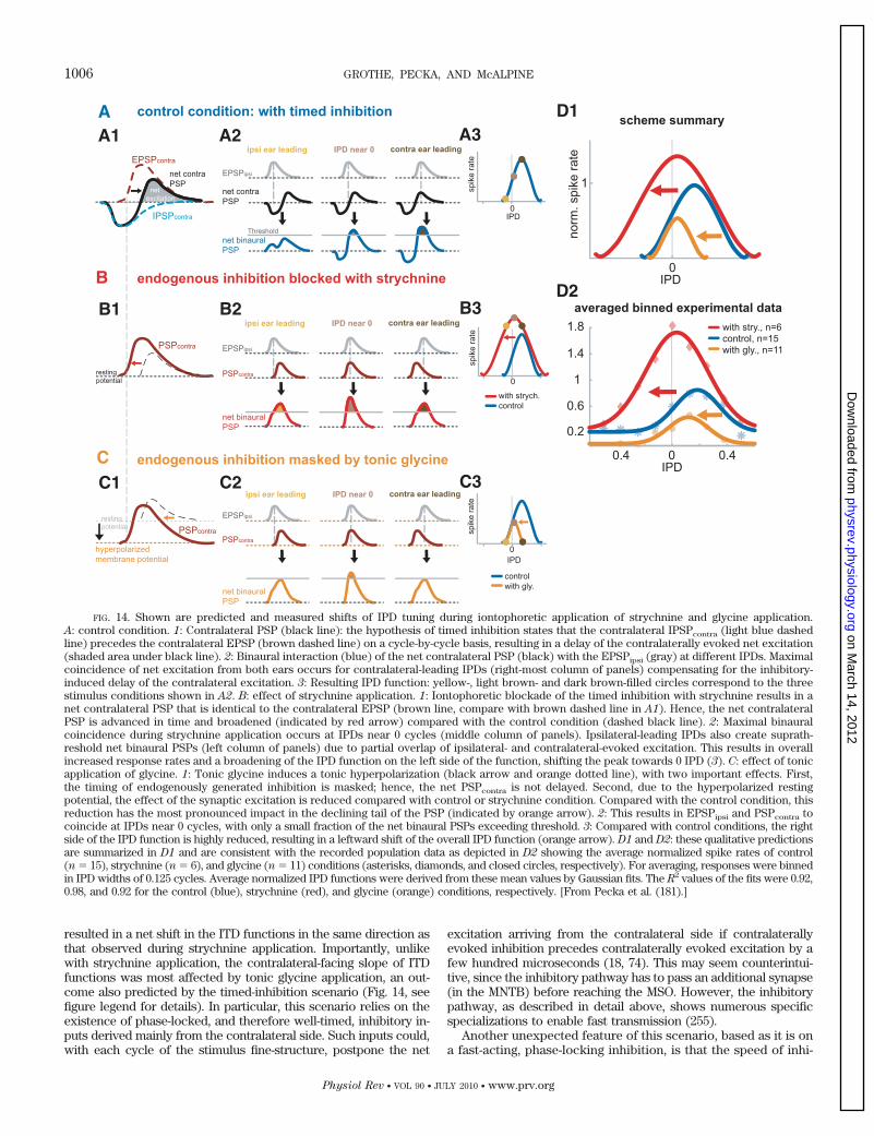
resulted in a net shift in the ITD functions in the same direction asthat observed during strychnine application. Importantly, unlikewith strychnine application, the contralateral-facing slope of ITDfunctions was most affected by tonic glycine application, an out-come also predicted by the timed-inhibition scenario (Fig. 14, seefigure legend for details). In particular, this scenario relies on theexistence of phase-locked, and therefore well-timed, inhibitory in-puts derived mainly from the contralateral side. Such inputs could,with each cycle of the stimulus fine-structure, postpone the net
excitation arriving from the contralateral side if contralaterallyevoked inhibition precedes contralaterally evoked excitation by afew hundred microseconds (18, 74). This may seem counterintui-tive, since the inhibitory pathway has to pass an additional synapse(in the MNTB) before reaching the MSO. However, the inhibitorypathway, as described in detail above, shows numerous specificspecializations to enable fast transmission (255).
Another unexpected feature of this scenario, based as it is ona fast-acting, phase-locking inhibition, is that the speed of inhi-
FIG. 14. Shown are predicted and measured shifts of IPD tuning during iontophoretic application of strychnine and glycine application.A: control condition. 1: Contralateral PSP (black line): the hypothesis of timed inhibition states that the contralateral IPSPcontra (light blue dashedline) precedes the contralateral EPSP (brown dashed line) on a cycle-by-cycle basis, resulting in a delay of the contralaterally evoked net excitation(shaded area under black line). 2: Binaural interaction (blue) of the net contralateral PSP (black) with the EPSPipsi (gray) at different IPDs. Maximalcoincidence of net excitation from both ears occurs for contralateral-leading IPDs (right-most column of panels) compensating for the inhibitory-induced delay of the contralateral excitation. 3: Resulting IPD function: yellow-, light brown- and dark brown-filled circles correspond to the threestimulus conditions shown in A2. B: effect of strychnine application. 1: Iontophoretic blockade of the timed inhibition with strychnine results in anet contralateral PSP that is identical to the contralateral EPSP (brown line, compare with brown dashed line in A1). Hence, the net contralateralPSP is advanced in time and broadened (indicated by red arrow) compared with the control condition (dashed black line). 2: Maximal binauralcoincidence during strychnine application occurs at IPDs near 0 cycles (middle column of panels). Ipsilateral-leading IPDs also create suprath-reshold net binaural PSPs (left column of panels) due to partial overlap of ipsilateral- and contralateral-evoked excitation. This results in overallincreased response rates and a broadening of the IPD function on the left side of the function, shifting the peak towards 0 IPD (3). C: effect of tonicapplication of glycine. 1: Tonic glycine induces a tonic hyperpolarization (black arrow and orange dotted line), with two important effects. First,the timing of endogenously generated inhibition is masked; hence, the net PSPcontra is not delayed. Second, due to the hyperpolarized restingpotential, the effect of the synaptic excitation is reduced compared with control or strychnine condition. Compared with the control condition, thisreduction has the most pronounced impact in the declining tail of the PSP (indicated by orange arrow). 2: This results in EPSPipsi and PSPcontra tocoincide at IPDs near 0 cycles, with only a small fraction of the net binaural PSPs exceeding threshold. 3: Compared with control conditions, the rightside of the IPD function is highly reduced, resulting in a leftward shift of the overall IPD function (orange arrow). D1 and D2: these qualitative predictionsare summarized in D1 and are consistent with the recorded population data as depicted in D2 showing the average normalized spike rates of control(n � 15), strychnine (n � 6), and glycine (n � 11) conditions (asterisks, diamonds, and closed circles, respectively). For averaging, responses were binnedin IPD widths of 0.125 cycles. Average normalized IPD functions were derived from these mean values by Gaussian fits. The R2 values of the fits were 0.92,0.98, and 0.92 for the control (blue), strychnine (red), and glycine (orange) conditions, respectively. [From Pecka et al. (181).]
1006 GROTHE, PECKA, AND McALPINE
Physiol Rev • VOL 90 • JULY 2010 • www.prv.org
on March 14, 2012
physrev.physiology.orgD
ownloaded from
bition is generally considered too slow to allow for any suchphase-locked action. Thus the question might arise, Is inhibitionfast enough? As outlined in the main body of the text, repolar-ization time constants for the inhibition in the gerbil MSO as-sessed in vitro are in the range of 1–2 ms (136) and effective insuppressing subsequent firing for �1 ms (81), which would besufficiently fast for the proposed mechanism of ITD processing forfrequencies up to at least 1 kHz. The question remains then, Whathappens for higher frequencies? Interestingly, recent calculationsbased on optimal coding theory suggested a dichotomy for ITDcoding strategies dependent on frequency with the cutoff fre-quency being �1 kHz for the gerbil (88) (for details, see sect.IIIC2B).
ACKNOWLEDGMENTS
Present address of D. McAlpine: UCL Ear Institute, Univer-sity College, London, UK.
Address for reprint requests and other correspondence: B.Grothe, Div. of Neurobiology, Ludwig-Maximilians-UniversitaetMunich, Grosshaderner Strasse 2, D-82152 Martinsried, Ger-many (e-mail: [email protected]).
REFERENCES
1. Adams JC. Ascending projections to the inferior colliculus. J
Comp Neurol 183: 519–538, 1979.2. Agapiou JP, McAlpine D. Low-frequency envelope sensitivity
produces asymmetric binaural tuning curves. J Neurophysiol 100:2381–2396, 2008.
3. Aitkin LM, Martin RI. The representation of stimulus azimuth byhigh best-frequency azimuth-selective neurons in the central nu-cleus of the inferior colliculus of the cat. J Neurophysiol 57:1185–1200, 1987.
4. Allin EF. Evolution of the mammalian middle ear. J Morphol 147:403–437, 1975.
5. Bala AD, Spitzer MW, Takahashi TT. Prediction of auditoryspatial acuity from neural images on the owl’s auditory space map.Nature 424: 771–774, 2003.
6. Bala AD, Takahashi TT. Pupillary dilation response as an indica-tor of auditory discrimination in the barn owl. J Comp Physiol A
Sens Neural Behav Physiol 186: 425–434, 2000.7. Batra R, Kuwada S, Fitzpatrick DC. Sensitivity to interaural
temporal disparities of low- and high-frequency neurons in thesuperior olivary complex. I. Heterogeneity of responses. J Neuro-
physiol 78: 1222–1236, 1997.8. Batra R, Kuwada S, Fitzpatrick DC. Sensitivity to interaural
temporal disparities of low- and high-frequency neurons in thesuperior olivary complex. II. Coincidence detection. J Neuro-
physiol 78: 1237–1247, 1997.9. Bazwinsky I, Hilbig H, Bidmon HJ, Rubsamen R. Characteriza-
tion of the human superior olivary complex by calcium bindingproteins and neurofilament H (SMI-32). J Comp Neurol 456: 292–303, 2003.
10. Beckius GE, Batra R, Oliver DL. Axons from anteroventral co-chlear nucleus that terminate in medial superior olive of cat: ob-servations related to delay lines. J Neurosci 19: 3146–3161, 1999.
11. Bernstein LR. Auditory processing of interaural timing informa-tion: new insights. J Neurosci Res 66: 1035–1046, 2001.
12. Bernstein LR, Trahiotis C. Enhancing sensitivity to interauraldelays at high frequencies by using “transposed stimuli.” J Acoust
Soc Am 112: 1026–1036, 2002.13. Blauert J. Ein Versuch zum Richtungshören bei gleichzeitiger
optischer Stimulation. Acustica 226–229, 1970.14. Blauert J. Spatial hearing with multiple sound sources and in
enclosed spaces. In: Spatial Hearing: The Psychophysics of Hu-
man Sound Localization. Cambridge, MA: MIT Press, 1997, p.201–287.
15. Blauert J. Spatial hearing with one sound source. In: Spatial
Hearing: The Psychophysics of Human Sound Localization. Cam-bridge, MA: MIT Press, 1997, p. 36–200.
16. Boudreau JC, Tsuchitani C. Binaural interaction in the cat su-perior olive S segment. J Neurophysiol 31: 442–454, 1968.
17. Brainard MS, Knudsen EI. Experience-dependent plasticity inthe inferior colliculus: a site for visual calibration of the neuralrepresentation of auditory space in the barn owl. J Neurosci 13:4589–4608, 1993.
18. Brand A, Behrend O, Marquardt T, McAlpine D, Grothe B.
Precise inhibition is essential for microsecond interaural time dif-ference coding. Nature 417: 543–547, 2002.
19. Brown CH, May BJ. Comparative mammalian sound localization.In: Sound Source Localization, edited by Popper AN and Fay RR.New York: Springer, 2005, p. 124–178.
20. Brugge JF, Anderson DJ, Aitkin LM. Responses of neurons inthe dorsal nucleus of the lateral lemniscus of cat to binaural tonalstimulation. J Neurophysiol 33: 441–458, 1970.
21. Brungart DS, Rabinowitz WM. Auditory localization of nearbysources. Head-related transfer functions. J Acoust Soc Am 106:1465–1479, 1999.
22. Brunso-Bechtold JK, Thompson GC, Masterton RB. HRP studyof the organization of auditory afferents ascending to central nu-cleus of inferior colliculus in cat. J Comp Neurol 197: 705–722,1981.
23. Burger RM, Cramer KS, Pfeiffer JD, Rubel EW. Avian superiorolivary nucleus provides divergent inhibitory input to parallel au-ditory pathways. J Comp Neurol 481: 6–18, 2005.
24. Burger RM, Pollak GD. Reversible inactivation of the dorsalnucleus of the lateral lemniscus reveals its role in the processing ofmultiple sound sources in the inferior colliculus of bats. J Neurosci
21: 4830–4843, 2001.25. Butts DA, Goldman MS. Tuning curves, neuronal variability, and
sensory coding. PLoS Biol 4: e92, 2006.26. Caird D, Klinke R. Processing of binaural stimuli by cat superior
olivary complex neurons. Exp Brain Res 52: 385–399, 1983.27. Campbell RA, Doubell TP, Nodal FR, Schnupp JW, King AJ.
Interaural timing cues do not contribute to the map of space in theferret superior colliculus: a virtual acoustic space study. J Neuro-
physiol 95: 242–254, 2006.28. Cant NB. Projections to the lateral and medial superior olivary
nuclei from the spherical and globular bushy cells of the antero-ventral cochlear nucleus. In: Neurobiology of Hearing: The Central
Auditory System, edited by Altschuler RA, Bobbin RP, CloptonBM, and Hoffman DW. New York: Raven, 1991, p. 99–119.
29. Cant NB, Casseday JH. Projections from the anteroventral co-chlear nucleus to the lateral and medial superior olivary nuclei. J
Comp Neurol 247: 457–476, 1986.30. Cant NB, Hyson RL. Projections from the lateral nucleus of the
trapezoid body to the medial superior olivary nucleus in the gerbil.Hear Res 58: 26–34, 1992.
31. Carr CE, Fujita I, Konishi M. Distribution of GABAergic neuronsand terminals in the auditory system of the barn owl. J Comp
Neurol 286: 190–207, 1989.32. Carr CE, Konishi M. Axonal delay lines for time measurement in
the owl’s brainstem. Proc Natl Acad Sci USA 85: 8311–8315, 1988.33. Carr CE, Konishi M. A circuit for detection of interaural time
differences in the brain stem of the barn owl. J Neurosci 10:3227–3246, 1990.
34. Chirila FV, Rowland KC, Thompson JM, Spirou GA. Develop-ment of gerbil medial superior olive: integration of temporallydelayed excitation and inhibition at physiological temperature. J
Physiol 584: 167–190, 2007.35. Christensen-Dalsgaard J. Directional hearing in nonmammalian
tetrapods. In: Sound Source Localization, edited by Popper AN andFay RR. New York: Springer, 2005, p. 67–123.
36. Clack JA. The evolution of tetrapod ears and the fossil record.Brain Behav Evol 50: 198–212, 1997.
37. Clack JA. Patterns and processes in the early evolution of thetetrapod ear. J Neurobiol 53: 251–264, 2002.
38. Clark GM. Vesicle shape versus type of synapse in the nerveendings of the cat medial superior olive. Brain Res 15: 548–551,1969.
39. Cohen YE, Knudsen EI. Maps versus clusters: different represen-tations of auditory space in the midbrain and forebrain. Trends
Neurosci 22: 128–135, 1999.
MECHANISMS OF SOUND LOCALIZATION IN MAMMALS 1007
Physiol Rev • VOL 90 • JULY 2010 • www.prv.org
on March 14, 2012
physrev.physiology.orgD
ownloaded from
40. Conlee JW, Parks TN, Creel DJ. Reduced neuronal size anddendritic length in the medial superior olivary nucleus of albinorabbits. Brain Res 363: 28–37, 1986.
41. Conlee JW, Parks TN, Romero C, Creel DJ. Auditory brainstemanomalies in albino cats. II. Neuronal atrophy in the superior olive.J Comp Neurol 225: 141–148, 1984.
42. Court FA, Sherman DL, Pratt T, Garry EM, Ribchester RR,
Cottrell DF, Fleetwood-Walker SM, Brophy PJ. Restrictedgrowth of Schwann cells lacking Cajal bands slows conduction inmyelinated nerves. Nature 431: 191–195, 2004.
43. Couve A, Moss SJ, Pangalos MN. GABAB receptors: a newparadigm in G protein signaling. Mol Cell Neurosci 16: 296–312,2000.
44. Crow G, Rupert AL, Moushegian G. Phase locking in monauraland binaural medullary neurons: implications for binaural phenom-ena. J Acoust Soc Am 64: 493–501, 1978.
45. Darrow KN, Maison SF, Liberman MC. Cochlear efferent feed-back balances interaural sensitivity. Nat Neurosci 9: 1474–1476,2006.
46. Dasika VK, White JA, Carney LH, Colburn HS. Effects ofinhibitory feedback in a network model of avian brain stem. J
Neurophysiol 94: 400–414, 2005.47. David EE, Guttman N, van Bergeijk AW. Binaural interaction of
high-frequency complex stimuli. J Acoust Soc Am 31: 774–782,1959.
48. Davis KA. Evidence of a functionally segregated pathway fromdorsal cochlear nucleus to inferior colliculus. J Neurophysiol 87:1824–1835, 2002.
49. Davis KA, Ramachandran R, May BJ. Auditory processing ofspectral cues for sound localization in the inferior colliculus. J
Assoc Res Otolaryngol 4: 148–163, 2003.50. Deatherage BH, Eldrege DH, Hallowell D. Latency of action
potentials in the cochlea of the guinea pig. J Acoust Soc Am 31:479–486, 1959.
51. Englitz B, Tolnai S, Typlt M, Jost J, Rubsamen R. Reliability ofsynaptic transmission at the synapses of Held in vivo under acous-tic stimulation. PLoS One 4: e7014, 2009.
52. Fay RR. Sound source localization by fishes. In: Sound Source
Localization, edited by Popper AN and Fay RR. New York:Springer, 2005, p. 36–66.
53. Felmy F, Schneggenburger R. Developmental expression of theCa2�-binding proteins calretinin and parvalbumin at the calyx ofHeld of rats and mice. Eur J Neurosci 20: 1473–1482, 2004.
54. Feng AS, Capranica RR. Sound localization in anurans. I. Evi-dence of binaural interaction in dorsal medullary nucleus of bull-frogs (Rana catesbeiana). J Neurophysiol 39: 871–881, 1976.
55. Feng AS, Capranica RR. Sound localization in anurans. II. Bin-aural interaction in superior olivary nucleus of the green tree frog(Hyla cinerea). J Neurophysiol 41: 43–54, 1978.
56. Feng AS, Gerhardt HC, Capranica RR. Sound localization be-havior of green treefrog (Hyla cinerea) and barking treefrog (Hyla
gratiosa). J Comp Physiol 107: 241–252, 1976.57. Finlayson PG, Adam TJ. Excitatory and inhibitory response ad-
aptation in the superior olive complex affects binaural acousticprocessing. Hear Res 103: 1–18, 1997.
58. Finlayson PG, Caspary DM. Low-frequency neurons in the lateralsuperior olive exhibit phase-sensitive binaural inhibition. J Neuro-
physiol 65: 598–605, 1991.59. Fitzpatrick DC, Kuwada S. Tuning to interaural time differences
across frequency. J Neurosci 21: 4844–4851, 2001.60. Fitzpatrick DC, Kuwada S, Batra R. Neural sensitivity to inter-
aural time differences: beyond the Jeffress model. J Neurosci 20:1605–1615, 2000.
61. Fitzpatrick DC, Kuwada S, Batra R. Transformations in process-ing interaural time differences between the superior olivary com-plex and inferior colliculus: beyond the Jeffress model. Hear Res
168: 79–89, 2002.62. Friauf E, Ostwald J. Divergent projections of physiologically
characterized rat ventral cochlear nucleus neurons as shown byintra-axonal injection of horseradish peroxidase. Exp Brain Res
73: 263–284, 1988.
63. Funabiki K, Koyano K, Ohmori H. The role of GABAergic inputsfor coincidence detection in the neurones of nucleus laminaris ofthe chick. J Physiol 508: 851–869, 1998.
64. Fuzessery ZM. Acute sensitivity to interaural time differences inthe inferior colliculus of a bat that relies on passive sound local-ization. Hearing Res 109: 46–62, 1997.
65. Galambos R, Davis H. The response of single auditory-nervefibres to acoustic stimulation. J Neurophysiol 6: 39–57, 1943.
66. Galambos R, Schwartzkopff J, Rupert A. Microelectrode studyof superior olivary nuclei. Am J Physiol 197: 527–536, 1959.
67. Gold JI, Knudsen EI. A site of auditory experience-dependentplasticity in the neural representation of auditory space in the barnowl’s inferior colliculus. J Neurosci 20: 3469–3486, 2000.
68. Goldberg JM, Brown PB. Response of binaural neurons of dogsuperior olivary complex to dichotic tonal stimuli: some physiolog-ical mechanisms of sound localization. J Neurophysiol 32: 613–636,1969.
69. Gordon N, Shackleton TM, Palmer AR, Nelken I. Responses ofneurons in the inferior colliculus to binaural disparities: insightsfrom the use of Fisher information and mutual information. J
Neurosci Methods 169: 391–404, 2008.70. Graybeal A, Rosowski JJ, Ketten DR, Crompton AW. Inner-ear
structure in morganucodon, an early Jurassic mammal. Zool J
Linnean Soc 96: 107–118, 1989.71. Griffin SJ, Bernstein LR, Ingham NJ, McAlpine D. Neural
sensitivity to interaural envelope delays in the inferior colliculus ofthe guinea pig. J Neurophysiol 93: 3463–3478, 2005.
72. Grothe B. Interaction of excitation and inhibition in processing ofpure tone and amplitude-modulated stimuli in the medial superiorolive of the mustached bat. J Neurophysiol 71: 706–721, 1994.
73. Grothe B. The evolution of temporal processing in the medialsuperior olive, an auditory brainstem structure. Prog Neurobiol 61:581–610, 2000.
74. Grothe B. New roles for synaptic inhibition in sound localization.Nature Rev Neurosci 4: 540–550, 2003.
75. Grothe B, Covey E, Casseday JH. Spatial tuning of neurons inthe inferior colliculus of the big brown bat: effects of sound level,stimulus type and multiple sound sources. J Comp Physiol A Sens
Neural Behav Physiol 179: 89–102, 1996.76. Grothe B, Fritzsch B, Köppl C, Casseday JH, Carr CE. The
evolution of central pathways and their neural processing patterns.In: Evolution of the Vertebrate Auditory System, edited by ManleyGA, Popper AN, and Fay RR. New York: Springer, 2004, p. 289–359.
77. Grothe B, Park TJ. Time can be traded for intensity in the lowerauditory system. Naturwissenschaften 82: 521–523, 1995.
78. Grothe B, Park TJ. Sensitivity to interaural time differences in themedial superior olive of a small mammal, the Mexican free-tailedbat. J Neurosci 18: 6608–6622, 1998.
79. Grothe B, Park TJ. Structure and function of the bat superiorolivary complex. Microsc Res Tech 51: 382–402, 2000.
80. Grothe B, Sanes DH. Bilateral inhibition by glycinergic afferentsin the medial superior olive. J Neurophysiol 69: 1192–1196, 1993.
81. Grothe B, Sanes DH. Synaptic inhibition influences the temporalcoding properties of medial superior olivary neurons: an in vitrostudy. J Neurosci 14: 1701–1709, 1994.
82. Grothe B, Schweizer H, Pollak GD, Schuller G, Rosemann C.
Anatomy and projection patterns of the superior olivary complex inthe Mexican free-tailed bat, Tadarida brasiliensis mexicana. J
Comp Neurol 343: 630–646, 1994.83. Guinan JJ Jr, Guinan SS, Norris BE. Single auditory units in the
superior olivary complex. I: Responses to sounds and classifica-tions based on physiological properties. Int J Neurosci 4: 101–120,1972.
84. Guinan JJ Jr, Norris BE, Guinan SS. Single auditory units in thesuperior olivary complex. II: Locations of unit categories and tono-topic organization. Int J Neurosci 4: 147–166, 1972.
85. Hancock KE, Davis KA, Voigt HF. Modeling inhibition of type IIunits in the dorsal cochlear nucleus. Biol Cybern 76: 419–428,1997.
86. Hancock KE, Delgutte B. A physiologically based model of inter-aural time difference discrimination. J Neurosci 24: 7110–7117,2004.
1008 GROTHE, PECKA, AND McALPINE
Physiol Rev • VOL 90 • JULY 2010 • www.prv.org
on March 14, 2012
physrev.physiology.orgD
ownloaded from
87. Harnischfeger G, Neuweiler G, Schlegel P. Interaural time andintensity coding in superior olivary complex and inferior colliculusof the echolocating bat Molossus ater. J Neurophysiol 53: 89–109,1985.
88. Harper NS, McAlpine D. Optimal neural population coding of anauditory spatial cue. Nature 430: 682–686, 2004.
89. Harrison JM, Warr WB. A study of the cochlear nuclei andascending auditory pathways of the medulla. J Comp Neurol 119:341–380, 1962.
90. Hartmann R, Klinke R. Impulse patterns of auditory nerve fibresto extra- and intracochlear electrical stimulation. Acta Otolaryngol
Suppl 469: 128–134, 1990.91. Heffner RS, Heffner HE. Auditory function in albino cats. Assoc
Res Otolaryngol Abstr 1987.92. Heil P. First-spike latency of auditory neurons revisited. Curr
Opin Neurobiol 14: 461–467, 2004.93. Held H. Die zentrale Gehörleitung. Arch Anat Physiol Anat Abtheil
17: 201–248, 1893.94. Henning GB. Detectability of interaural delay in high-frequency
complex waveforms. J Acoust Soc Am 55: 84–90, 1974.95. Hermann J, Pecka M, von GH, Grothe B, Klug A. Synaptic
transmission at the calyx of Held under in vivo like activity levels.J Neurophysiol 98: 807–820, 2007.
96. Hofman PM, VanRiswick JA, VanOpstal AJ. Relearning soundlocalization with new ears. Nature Neurosci 1: 417–421, 1998.
97. Hyson RL, Overholt EM, Lippe WR. Cochlear microphonic mea-surements of interaural time differences in the chick. Hear Res 81:109–118, 1994.
98. Hyson RL, Reyes AD, Rubel EW. A depolarizing inhibitory re-sponse to GABA in brainstem auditory neurons of the chick. Brain
Res 677: 117–126, 1995.99. Imig TJ, Bibikov NG, Poirier P, Samson FK. Directionality
derived from pinna-cue spectral notches in cat dorsal cochlearnucleus. J Neurophysiol 83: 907–925, 2000.
100. Irvine DR, Gago G. Binaural interaction in high-frequency neu-rons in inferior colliculus of the cat: effects of variations in soundpressure level on sensitivity to interaural intensity differences. J
Neurophysiol 63: 570–591, 1990.101. Irvine DR, Park VN, McCormick L. Mechanisms underlying the
sensitivity of neurons in the lateral superior olive to interauralintensity differences. J Neurophysiol 86: 2647–2666, 2001.
102. Ito M, van Adel B, Kelly JB. Sound localization after transectionof the commissure of probst in the albino rat. J Neurophysiol 76:3493–3502, 1996.
103. Jeffress LA. A place theory of sound localization. J Comp Physiol
Psychol 41: 35–39, 1948.104. Joris P, Yin TCT. A matter of time: internal delays in binaural
processing. Trends Neurosci 30: 70–78, 2007.105. Joris PX. Interaural time sensitivity dominated by cochlea-induced
envelope patterns. J Neurosci 23: 6345–6350, 2003.106. Joris PX, Carney LH, Smith PH, Yin TC. Enhancement of neural
synchronization in the anteroventral cochlear nucleus. I. Re-sponses to tones at the characteristic frequency. J Neurophysiol
71: 1022–1036, 1994.107. Joris PX, Louage DH, van der Heijden M. Temporal damping in
response to broadband noise. II. Auditory nerve. J Neurophysiol
99: 1942–1952, 2008.108. Joris PX, Smith PH, Yin TC. Enhancement of neural synchroni-
zation in the anteroventral cochlear nucleus. II. Responses in thetuning curve tail. J Neurophysiol 71: 1037–1051, 1994.
109. Joris PX, Van de Sande B, Louage DH, van der Heijden M.
Binaural and cochlear disparities. Proc Natl Acad Sci USA 103:12917–12922, 2006.
110. Joris PX, Van De Sande B, van der Heijden M. Temporaldamping in response to broadband noise. I. Inferior colliculus. J
Neurophysiol 93: 1857–1870, 2005.111. Joseph AW, Hyson RL. Coincidence detection by binaural neu-
rons in the chick brain stem. J Neurophysiol 69: 1197–1211, 1993.112. Kapfer C, Seidl AH, Schweizer H, Grothe B. Experience-depen-
dent refinement of inhibitory inputs to auditory coincidence-detec-tor neurons. Nat Neurosci 5: 247–253, 2002.
113. Kiang NY, Watanabe T, Thomas C, Clark LF. Discharge Pat-
terns of Single Fibres in the Cat’s Auditory Nerve. Cambridge, MA:MIT Press, 1965.
114. King AJ. The superior colliculus. Curr Biol 14: R335–R338, 2004.115. King AJ, Carlile S. Changes induced in the representation of
auditory space in the superior colliculus by rearing ferrets withbinocular eyelid suture. Exp Brain Res 94: 444–455, 1993.
116. King AJ, Parsons CH. Improved auditory spatial acuity in visuallydeprived ferrets. Eur J Neurosci 11: 3945–3956, 1999.
117. Kitzes LM, Kageyama Gh Semple MN, Kil J. Development ofectopic projections from the ventral cochlear nucleus to the supe-rior olivary complex induced by neonatal ablation of the contralat-eral cochlea. J Comp Neurol 353: 341–363, 1995.
118. Klemm O. Untersuchungen über die Lokalisation von SchallreizenIV: über den Einfluss des binauralen Zeitunterschieds auf die Lokal-isation. Arch ges Psychol 40: 117–145, 1920.
119. Klumpp R, Eady H. Some measurements of interaural time differ-ence thresholds. J Acoust Soc Am 28: 215–232, 1956.
120. Knudsen EI. Auditory and visual maps of space in the optictectum of the owl. J Neurosci 2: 1177–1194, 1982.
121. Knudsen EI. Instructed learning in the auditory localization path-way of the barn owl. Nature 417: 322–328, 2002.
122. Knudsen EI, Konishi M. A neural map of auditory space in theowl. Science 200: 795–797, 1978.
123. Koch U, Grothe B. Azimuthal position affects analysis of complexsounds in the mammalian auditory system. Naturwissenschaften
84: 160–162, 1997.124. Koch U, Grothe B. Interdependence of spatial and temporal cod-
ing in the auditory midbrain. J Neurophysiol 83: 2300–2314, 2000.125. Kopp-Scheinpflug C, Lippe WR, Dorrscheidt GJ, Rubsamen R.
The medial nucleus of the trapezoid body in the gerbil is more thana relay: comparison of pre- and postsynaptic activity. J Assoc Res
Otolaryngol 4: 1–23, 2003.126. Koppl C. Evolution of sound localisation in land vertebrates. Curr
Biol 19: R635–R639, 2009.127. Koppl C, Carr CE. Maps of interaural time difference in the
chicken’s brainstem nucleus laminaris. Biol Cybern 98: 541–559,2008.
128. Kulesza RJ Jr. Cytoarchitecture of the human superior olivarycomplex: medial and lateral superior olive. Hear Res 225: 80–90,2007.
129. Kulesza RJ Jr, Spirou GA, Berrebi AS. Physiological responseproperties of neurons in the superior paraolivary nucleus of the rat.J Neurophysiol 89: 2299–2312, 2003.
130. Kuwabara N, Zook JM. Projections to the medial superior olivefrom the medial and lateral nuclei of the trapezoid body in rodentsand bats. J Comp Neurol 324: 522–538, 1992.
131. Lachica EA, Rubsamen R, Rubel EW. GABAergic terminals innucleus magnocellularis and laminaris originate from the superiorolivary nucleus. J Comp Neurol 348: 403–418, 1994.
132. Lay DM. The anatomy, physiology, and functional significance andevolution of specialized hearing organs of gerbilline rodents. J
Morphol 138: 41–120, 1972.133. Lay DM. Differential predation on gerbils (Meriones) by the little
owl, Athene brama. J Mammal 55: 608–614, 1974.134. Li L, Kelly JB. Inhibitory influence of the dorsal nucleus of the
lateral lemniscus on binaural responses in the rat’s inferior collicu-lus. J Neurosci 12: 4530–4539, 1992.
135. Lindsey BG. Fine structure and distribution of axon terminalsfrom the cochlear nucleus on neurons in the medial superiorolivary nucleus of the cat. J Comp Neurol 160: 81–103, 1975.
136. Magnusson AK, Kapfer C, Grothe B, Koch U. Maturation ofglycinergic inhibition in the gerbil medial superior olive after hear-ing onset. J Physiol 568: 497–512, 2005.
137. Magnusson AK, Park TJ, Pecka M, Grothe B, Koch U. Retro-grade GABA signaling adjusts sound localization by balancing ex-citation and inhibition in the brainstem. Neuron 59: 125–137, 2008.
138. Maki K, Furukawa S. Acoustical cues for sound localization bythe Mongolian gerbil, Meriones unguiculatus. J Acoust Soc Am
118: 872–886, 2005.139. Manley GA, Koppl C, Konishi M. A neural map of interaural
intensity differences in the brain stem of the barn owl. J Neurosci
8: 2665–2676, 1988.
MECHANISMS OF SOUND LOCALIZATION IN MAMMALS 1009
Physiol Rev • VOL 90 • JULY 2010 • www.prv.org
on March 14, 2012
physrev.physiology.orgD
ownloaded from
140. Mason AC, Oshinsky ML, Hoy RR. Hyperacute directional hear-ing in a microscale auditory system. Nature 410: 686–690, 2001.
141. Masterton RB, Diamond IT. Medial superior olive and soundlocalization. Science 155: 1696–1697, 1967.
142. Matsubara JA. Calbindin D-28K immunoreactivity in the cat’ssuperior olivary complex. Brain Res 508: 353–357, 1990.
143. Mazer JA. How the owl resolves auditory coding ambiguity. Proc
Natl Acad Sci USA 95: 10932–10937, 1998.144. McAlpine D. Creating a sense of auditory space. J Physiol 566:
21–28, 2005.145. McAlpine D, Grothe B. Sound localization and delay lines: do
mammals fit the model? Trends Neurosci 26: 347–350, 2003.146. McAlpine D, Jiang D, Palmer AR. A neural code for low-fre-
quency sound localization in mammals. Nat Neurosci 4: 396–401,2001.
147. McAlpine D, Jiang D, Shackleton TM, Palmer AR. Responses ofneurons in the inferior colliculus to dynamic interaural phase cues:evidence for a mechanism of binaural adaptation. J Neurophysiol
83: 1356–1365, 2000.148. McFadden D, Pasanen EG. Lateralization of high frequencies
based on interaural time differences. J Acoust Soc Am 59: 634–639,1976.
149. McLaughlin M, van der Heijden M, Joris PX. How secure is invivo synaptic transmission at the calyx of Held? J Neurosci 28:10206–10219, 2008.
150. Meffin H, Grothe B. Selective filtering to spurious localizationcues in the mammalian auditory brainstem. J Acoust Soc Am 126:2437–2454, 2009.
151. Miller GA. Sensitivity to changes in the intensity of white noiseand its relation to masking and loudness. J Acoust Soc Am 609–619, 1947.
152. Mills DM, Shepherd RK. Distortion product otoacoustic emissionand auditory brainstem responses in the echidna (Tachyglossus
aculeatus). J Assoc Res Otolaryngol 2: 130–146, 2001.153. Moiseff A. Binaural disparity cues available to the barn owl for
sound localization. J Comp Physiol A Sens Neural Behav Physiol
164: 629–636, 1989.154. Monsivais P, Rubel EW. Accommodation enhances depolarizing
inhibition in central neurons. J Neurosci 21: 7823–7830, 2001.155. Monsivais P, Yang L, Rubel EW. GABAergic inhibition in nucleus
magnocellularis: implications for phase locking in the avian audi-tory brainstem. J Neurosci 20: 2954–2963, 2000.
156. Moore JK. Organization of the human superior olivary complex.Microsc Res Tech 51: 403–412, 2000.
157. Moore BCJ. An Introduction to the Psychology of Hearing. Lon-don: Academic, 2004, p. 235–237.
158. Moore JK, Moore RY. A comparative study of the superior olivarycomplex in the primate brain. Folia Primatol 16: 35–51, 1971.
159. Moore MJ, Caspary DM. Strychnine blocks binaural inhibition inlateral superior olivary neurons. J Neurosci 3: 237–242, 1983.
160. Morest DK. The collateral system of the medial nucleus of thetrapezoid body of the cat, its neuronal architecture and relation tothe olivo-cochlear bundle. Brain Res 9: 288–311, 1968.
161. Morest DK. The growth of synaptic endings in the mammalianbrain: a study of the calyces of the trapezoid body. Z Anat En-
twicklungsgesch 127: 201–220, 1968.162. Moushegian G, Rupert AL, Gidda JS. Functional characteristics
of superior olivary neurons to binaural stimuli. J Neurophysiol 38:1037–1048, 1975.
163. Nakamoto KT, Jones SJ, Palmer AR. Descending projectionsfrom auditory cortex modulate sensitivity in the midbrain to cuesfor spatial position. J Neurophysiol 99: 2347–2356, 2008.
164. Nelken I, Young ED. Two separate inhibitory mechanisms shapethe responses of dorsal cochlear nucleus type IV units to narrow-band and wideband stimuli. J Neurophysiol 71: 2446–2462, 1994.
165. Norberg RA. Occurrence and independent evolution of bilateralear asymmetry in owls and implications on owl taxonomy. Philos
Trans Royal Soc Lond B Biol Sci 280: 375–408, 1978.166. Oertel D. The role of timing in the brain stem auditory nuclei of
vertebrates. Annu Rev Physiol 61: 497–519, 1999.167. Oertel D, Young ED. What’s a cerebellar circuit doing in the
auditory system? Trends Neurosci 27: 104–110, 2004.
168. Oliver DL. Ascending efferent projections of the superior olivarycomplex. Microsc Res Tech 51: 355–363, 2000.
169. Osen KK. Cytoarchitecture of the cochlear nuclei in the cat. J
Comp Neurol 136: 453–484, 1969.170. Oswald JP, Klug A, Park TJ. Interaural intensity difference pro-
cessing in auditory midbrain neurons: effects of a transient earlyinhibitory input. J Neurosci 19: 1149–1163, 1999.
171. Overholt E, Rubel EW, Hyson RL. A circuit for coding interauraltime differences in the chick brainstem. J Neurosci 12: 1698–1708,1992.
172. Palmer AR. Reassessing mechanisms of low-frequency sound lo-calisation. Curr Opin Neurobiol 14: 457–460, 2004.
173. Paolini AG, FitzGerald JV, Burkitt AN, Clark GM. Temporalprocessing from the auditory nerve to the medial nucleus of thetrapezoid body in the rat. Hear Res 159: 101–116, 2001.
174. Park TJ. IID sensitivity differs between two principal centers inthe interaural intensity difference pathway: the LSO and the IC. J
Neurophysiol 79: 2416–2431, 1998.175. Park TJ, Brand A, Koch U, Ikebuchi M, Grothe B. Dynamic
changes in level influence spatial coding in the lateral superiorolive. Hear Res 238: 58–67, 2008.
176. Park TJ, Grothe B, Pollak GD, Schuller G, Koch U. Neuraldelays shape selectivity to interaural intensity differences in thelateral superior olive. J Neurosci 16: 6554–6566, 1996.
177. Park TJ, Klug A, Holinstat M, Grothe B. Interaural level differ-ence processing in the lateral superior olive and the inferior col-liculus. J Neurophysiol 92: 289–301, 2004.
178. Park TJ, Monsivais P, Pollak GD. Processing of interaural inten-sity differences in the LSO: role of interaural threshold differences.J Neurophysiol 77: 2863–2878, 1997.
179. Park TJ, Pollak GD. Azimuthal receptive fields are shaped byGABAergic inhibition in the inferior colliculus of the mustache bat.J Neurophysiol 72: 1080–1102, 1994.
180. Parks TN, Rubel EW. Organization and development of brainstem auditory nuclei of the chicken: organization of projectionsfrom n. magnocellularis to n laminaris. J Comp Neurol 164: 435–448, 1975.
181. Pecka M, Brand A, Behrend O, Grothe B. Interaural time differ-ence processing in the mammalian medial superior olive: the roleof glycinergic inhibition. J Neurosci 28: 6914–6925, 2008.
182. Pena JL, Konishi M. Auditory spatial receptive fields created bymultiplication. Science 292: 249–252, 2001.
183. Pena JL, Konishi M. From postsynaptic potentials to spikes in thegenesis of auditory spatial receptive fields. J Neurosci 22: 5652–5658, 2002.
184. Pena JL, Viete S, Albeck Y, Konishi M. Tolerance to soundintensity of binaural coincidence detection in the nucleus laminarisof the owl. J Neurosci 16: 7046–7054, 1996.
185. Pena JL, Viete S, Funabiki K, Saberi K, Konishi M. Cochlearand neural delays for coincidence detection in owls. J Neurosci 21:9455–9459, 2001.
186. Perkins RE. An electron microscopic study of synaptic organiza-tion in the medial superior olive of normal and experimental chin-chillas. J Comp Neurol 148: 387–415, 1973.
187. Pollak GD. Time is traded for intensity in the bat’s auditorysystem. Hear Res 36: 107–124, 1988.
188. Rajan R, Aitkin LM, Irvine DR. Azimuthal sensitivity of neuronsin primary auditory cortex of cats. II. Organization along frequency-band strips. J Neurophysiol 64: 888–902, 1990.
189. Ramachandran R, May BJ. Functional segregation of ITD sensi-tivity in the inferior colliculus of decerebrate cats. J Neurophysiol
88: 2251–2261, 2002.190. Ramon y Cajal S. Histologie du systeme nerveux de l’homme et
des vertebrates. Paris: Malonie, 1907.191. Rautenberg PL, Grothe B, Felmy F. Quantification of the three-
dimensional morphology of coincidence detector neurons in themedial superior olive of gerbils during late postnatal development.J Comp Neurol 517: 385–396, 2009.
192. Rayleigh L. On our perception of sound direction. Philos Mag 13:214–232, 1907.
193. Reed MC, Blum JJ. A model for the computation and encoding ofazimuthal information by the lateral superior olive. J Acoust Soc
Am 88: 1442–1453, 1990.
1010 GROTHE, PECKA, AND McALPINE
Physiol Rev • VOL 90 • JULY 2010 • www.prv.org
on March 14, 2012
physrev.physiology.orgD
ownloaded from
194. Reed MC, Durbeck L. Delay lines and auditory processing. Com-
ments Theoret Biol 3: 441–461, 1995.195. Reyes AD, Rubel EW, Spain WJ. In vitro analysis of optimal
stimuli for phase-locking and time-delayed modulation of firing inavian nucleus laminaris neurons. J Neurosci 16: 993–1007, 1996.
196. Rheinländer J, Gerhardt HC, Yager DD, Capranica RR. Accu-racy of phono taxis by the green tree frog (Hyla cinerea). J Comp
Physiol 133: 247–256, 1979.197. Rheinländer J, Walkowiak W, Gerhardt HC. Directional hearing
in the green tree frog: a variable mechanism? Naturwissenschaften
68: 430–431, 1981.197a.Rhode WS, Oertel D, Smith PH. Physiological response proper-
ties of cells labeled intracellularly with horseradish peroxidase incat ventral cochlear nucleus. J Comp Neurol 213: 448–463, 1983.
198. Rhode WS, Smith PH. Physiological studies on neurons in thedorsal cochlear nucleus of cat. J Neurophysiol 56: 287–307, 1986.
199. Richter EA, Norris BE, Fullerton BC, Levine RA, Kiang NY. Isthere a medial nucleus of the trapezoid body in humans? Am J Anat
168: 157–166, 1983.200. Robert D. Directional hearing in insects. In: Sound Source Local-
ization, edited by Popper AN and Fay RR. New York: Springer,2005, p. 6–35.
201. Rose JE, Brugge JF, Anderson DJ, Hind JE. Phase-lockedresponse to low-frequency tones in single auditory nerve fibers ofthe squirrel monkey. J Neurophysiol 30: 769–793, 1967.
202. Rose JE, Gross NB, Geisler CD, Hind JE. Some neural mecha-nisms in the inferior colliculus of the cat which may be relevant tolocalization of a sound source. J Neurophysiol 29: 288–314, 1966.
203. Rosowski JJ. Hearing in transitional mammals. Predictions fromthe middle ear anatomy and hearing capabilities of extant mam-mals. In: The Evolutionary Biology of Hearing, edited by WebsterDB, Fay RR, and Popper AN. New York: Springer-Verlag, 1992, p.615–632.
204. Rosowski JJ, Graybeal A. What did Morganucodon hear. Zool J
Linnean Soc 101: 131–168, 1991.205. Rosowski JJ, Saunders JC. Sound transmission through the
avian interaural pathways. J Comp Physiol A Sens Neural Behav
Physiol 136: 183–190, 1980.206. Ryugo DK, Parks TN. Primary innervation of the avian and mam-
malian cochlear nucleus. Brain Res Bull 60: 435–456, 2003.207. Sanes DH. An in vitro analysis of sound localization mechanisms
in the gerbil lateral superior olive. J Neurosci 10: 3494–3506, 1990.208. Schroeder MR. New viewpoints in binaural interactions. In: Psy-
chophysics and Physiology of Hearing, edited by Evans EF andWilson JP. New York: Academic, 1977, p. 455–467.
209. Schwartz IR. The superior olivary complex and lateral lemniscalnuclei. In: The Mammalian Auditory Pathway: Neuroanatomy,edited by Webster DB, Popper AN, and Fay RR. New York:Springer, 1992, p. 117–167.
210. Schweizer H. The connections of the inferior colliculus and theorganization of the brainstem auditory system in the greater horse-shoe bat (Rhinolophus ferrumequinum). J Comp Neurol 201: 25–49, 1981.
211. Scott LL, Mathews PJ, Golding NL. Posthearing developmentalrefinement of temporal processing in principal neurons of themedial superior olive. J Neurosci 25: 7887–7895, 2005.
212. Seidl AH, Grothe B. Development of sound localization mecha-nisms in the mongolian gerbil is shaped by early acoustic experi-ence. J Neurophysiol 94: 1028–1036, 2005.
213. Seidl AH, Rubel EW, Harris DM. Mechanisms for adjusting in-teraural time differences to achieve binaural coincidence detec-tion. J Neurosci 30: 70–80, 2010.
214. Shackleton TM, Arnott RH, Palmer AR. Sensitivity to interauralcorrelation of single neurons in the inferior colliculus of guineapigs. J Assoc Res Otolaryngol 6: 244–259, 2005.
215. Shackleton TM, Skottun BC, Arnott RH, Palmer AR. Interauraltime difference discrimination thresholds for single neurons in theinferior colliculus of guinea pigs. J Neurosci 23: 716–724, 2003.
216. Shamma SA, Shen NM, Gopalaswamy P. Stereausis: binauralprocessing without neural delays. J Acoust Soc Am 86: 989–1006,1989.
217. Shinn-Cunningham BG, Santarelli S, Kopco N. Tori of confu-sion: binaural localization cues for sources within reach of a lis-tener. J Acoust Soc Am 107: 1627–1636, 2000.
218. Simmons DD. Development of the inner ear efferent system acrossvertebrate species. J Neurobiol 53: 228–250, 2002.
219. Siveke I, Pecka M, Seidl AH, Baudoux S, Grothe B. Binauralresponse properties of low-frequency neurons in the gerbil dorsalnucleus of the lateral lemniscus. J Neurophysiol 96: 1425–1440,2006.
220. Skottun BC, Shackleton TM, Arnott RH, Palmer AR. Theability of inferior colliculus neurons to signal differences in inter-aural delay. Proc Natl Acad Sci USA 98: 14050–14054, 2001.
221. Smith AJ, Owens S, Forsythe ID. Characterisation of inhibitoryand excitatory postsynaptic currents of the rat medial superiorolive. J Physiol 529: 681–698, 2000.
222. Smith PH. Structural and functional differences distinguish prin-cipal from nonprincipal cells in the guinea pig MSO slice. J Neu-
rophysiol 73: 1653–1667, 1995.223. Smith PH, Joris PX, Yin TC. Projections of physiologically char-
acterized spherical bushy cell axons from the cochlear nucleus ofthe cat: evidence for delay lines to the medial superior olive. J
Comp Neurol 331: 245–260, 1993.224. Smith PH, Joris PX, Yin TC. Anatomy and physiology of principal
cells of the medial nucleus of the trapezoid body (MNTB) of thecat. J Neurophysiol 79: 3127–3142, 1998.
225. Sommer I, Lingenhohl K, Friauf E. Principal cells of the ratmedial nucleus of the trapezoid body: an intracellular in vivo studyof their physiology and morphology. Exp Brain Res 95: 223–239,1993.
226. Spangler KM, Warr WB, Henkel CK. The projections of principalcells of the medial nucleus of the trapezoid body in the cat. J Comp
Neurol 238: 249–262, 1985.227. Spirou GA, Berrebi AS. Organization of ventrolateral periolivary
cells of the cat superior olive as revealed by pep-19 immunocyto-chemistry and nissl stain. J Comp Neurol 368: 100–120, 1996.
228. Spirou GA, Rowland KC, Berrebi AS. Ultrastructure of neuronsand large synaptic terminals in the lateral nucleus of the trapezoidbody of the cat. J Comp Neurol 398: 257–272, 1998.
229. Spirou GA, Young ED. Organization of dorsal cochlear nucleustype IV unit response maps and their relationship to activation bybandlimited noise. J Neurophysiol 66: 1750–1768, 1991.
230. Spitzer MW, Semple MN. Responses of inferior colliculus neu-rons to time-varying interaural phase disparity: effects of shiftingthe locus of virtual motion. J Neurophysiol 69: 1245–1263, 1993.
231. Spitzer MW, Semple MN. Neurons sensitive to interaural phasedisparity in gerbil superior olive: diverse monaural and temporalresponse properties. J Neurophysiol 73: 1668–1690, 1995.
232. Stecker GC, Harrington IA, Middlebrooks JC. Location codingby opponent neural populations in the auditory cortex. Plos Biol 3:520–528, 2005.
233. Stevens SS, Newman BB. The localization of pure tones. Proc
Natl Acad Sci USA 593–596, 1934.234. Stillman RD. Characteristic delay neurons in the inferior collicu-
lus of the kangaroo rat. Exp Neurol 32: 404–412, 1971.235. Stotler WA. An experimental study of the cells and connections of
the superior olivary complex of the cat. J Comp Neurol 98: 401–432, 1953.
236. Strominger NL, Hurwitz JL. Anatomical aspects of the superiorolivary complex. J Comp Neurol 170: 485–497, 1976.
237. Strominger NL, Nelson LR, Dougherty WJ. Second order audi-tory pathways in the chimpanzee. J Comp Neurol 172: 349–365,1977.
238. Suga N. Role of corticofugal feedback in hearing. J Comp Physiol
A Sens Neural Behav Physiol 194: 169–183, 2008.239. Sullivan WE, Konishi M. Neural map of interaural phase differ-
ence in the owl’s brainstem. Proc Natl Acad Sci USA 83: 8400–8404, 1986.
240. Takahashi T, Konishi M. Selectivity for interaural time differencein the owl’s midbrain. J Neurosci 6: 3413–3422, 1986.
241. Takahashi TT, Bala AD, Spitzer MW, Euston DR, Spezio ML,
Keller CH. The synthesis and use of the owl’s auditory space map.Biol Cybern 89: 378–387, 2003.
MECHANISMS OF SOUND LOCALIZATION IN MAMMALS 1011
Physiol Rev • VOL 90 • JULY 2010 • www.prv.org
on March 14, 2012
physrev.physiology.orgD
ownloaded from
242. Takahashi TT, Barberini CL, Keller CH. An anatomical sub-strate for the inhibitory gradient in the VLVp of the owl. J Comp
Neurol 358: 294–304, 1995.243. Takahashi TT, Keller CH. Commissural connections mediate
inhibition for the computation of interaural level difference in thebarn owl. J Comp Physiol A Sens Neural Behav Physiol 170:161–169, 1992.
244. Takahashi TT, Konishi M. Projections of the cochlear nuclei andnucleus laminaris to the inferior colliculus of the barn owl. J Comp
Neurol 274: 190–211, 1988.245. Thompson SP. On the function of the two ears in the perception
of space. Philos Mag 13: 406–416, 1882.246. Thornton JL, Koka K, Jones HG, Tollin DJ. A frequency-depen-
dence in both the acoustic interaural time difference cues to soundlocation and their encoding by neurons in the inferior colliculus.ARO Abstr 529, 2009.
247. Tollin DJ, Yin TCT. Interaural phase and level difference sensi-tivity in low-frequency neurons in the lateral superior olive. J
Neurosci 25: 10648–10657, 2005.248. Tsuchitani C. Functional organization of lateral cell groups of cat
superior olivary complex. J Neurophysiol 40: 296–318, 1977.249. Tsuchitani C. The inhibition of cat lateral superior olive unit
excitatory responses to binaural tone bursts. II. The sustaineddischarges. J Neurophysiol 59: 184–211, 1988.
250. Van Bergeijk AW. Variation on a theme of Bekesy: a model ofbinaural interaction. J Acoust Soc Am 34: 1431–1437, 1962.
251. Van der Heijden M, Trahiotis C. Masking with interaurally de-layed stimuli: the use of “internal” delays in binaural detection. J
Acoust Soc Am 105: 388–399, 1999.252. Vater M. Ultrastructural and immunocytochemical observations
on the superior olivary complex of the mustached bat. J Comp
Neurol 358: 155–180, 1995.253. Volman SF, Konishi M. Comparative physiology of sound local-
ization in 4 species of owls. Brain Behav Evol 36: 196–215, 1990.254. Von Bekesy G. Zur Theorie des Hörens: Über das Richtungshören
bei einer Zeitdifferenz oder Lautstärkeungleichheit der beidseitigenSchalleinwirkungen. Phys Z 824–838, 1930.
255. Von Gersdorff H, Borst JG. Short-term plasticity at the calyx ofheld. Nat Rev Neurosci 3: 53–64, 2002.
256. Vonderschen K, Wagner H. Tuning to interaural time differenceand frequency differs between the auditory arcopallium and theexternal nucleus of the inferior colliculus. J Neurophysiol 101:2348–2361, 2009.
257. Warr WB. Fiber degeneration following lesions in the anteriorventral cochlear nucleus of the cat. Exp Neurol 14: 453–474, 1966.
258. Webster DB. A function of the enlarged middle ear cavities of theKangaroo rat, Dipodomys. Am J Anat 108: 123–148, 1962.
259. Webster DB, Plassmann W. Parallel evolution of low-frequencysensitivity in old world and new world desert rodents. In: The
Evolutionary Biology of Hearing, edited by Webster DB, Fay RR,and Popper AN. New York: Springer-Verlag, 1992.
260. Webster DB, Webster M. Adaptive value of hearing and vision inkangaroo rat predator avoidance. Brain Behav Evol 4: 310–322,1971.
261. Wenstrup JJ, Ross LS, Pollak GD. Binaural response organiza-tion within a frequency-band representation of the inferior collicu-lus: implications for sound localization. J Neurosci 6: 962–973,1986.
262. Wenthold RJ, Huie D, Altschuler RA, Reeks KA. Glycine im-munoreactivity localized in the cochlear nucleus and superior oli-vary complex. Neuroscience 22: 897–912, 1987.
263. Werthat F, Alexandrova O, Grothe B, Koch U. Experience-dependent refinement of the inhibitory axons projecting to themedial superior olive. Dev Neurobiol 68: 1454–1462, 2008.
264. Whitworth RH, Jeffress LA. Time vs. intensity in the localisationof tones. J Acoust Soc Am 33: 925–929, 1961.
265. Withington-Wray DJ, Binns KE, Dhanjal SS, Brickley SG,
Keating MJ. The maturation of the superior collicular map ofauditory space in the guinea pig is disrupted by developmentalauditory deprivation. Eur J Neurosci 2: 693–703, 1990.
266. Yang L, Monsivais P, Rubel EW. The superior olivary nucleusand its influence on nucleus laminaris: a source of inhibitory feed-back for coincidence detection in the avian auditory brainstem. J
Neurosci 19: 2313–2325, 1999.267. Yin TC. Neural mechanisms of encoding binaural localization cues.
In: Integrative Functions in the Mammalian Auditory Pathway,edited by Oertel D, Fay RR, and Popper AN. New York: Springer-Verlag, 2002, p. 99–159.
268. Yin TC, Carney LH, Joris PX. Interaural time sensitivity in theinferior colliculus of the albino cat. J Comp Neurol 295: 438–448,1990.
269. Yin TC, Chan JC. Interaural time sensitivity in medial superiorolive of cat. J Neurophysiol 64: 465–488, 1990.
270. Yin TC, Chan JC, Irvine DR. Effects of interaural time delays ofnoise stimuli on low-frequency cells in the cat’s inferior colliculus.I. Responses to wideband noise. J Neurophysiol 55: 280–300, 1986.
271. Yin TC, Hirsch JA, Chan JC. Responses of neurons in the cat’ssuperior colliculus to acoustic stimuli. II. A model of interauralintensity sensitivity. J Neurophysiol 53: 746–758, 1985.
272. Yin TC, Kuwada S. Binaural interaction in low-frequency neuronsin inferior colliculus of the cat. III. Effects of changing frequency.J Neurophys 50: 1020–1042, 1983.
273. Yost WA, Wightman FL, Green DM. Lateralization of filteredclicks. J Acoust Soc Am 50: 1526–1531, 1971.
274. Young LL Jr, Carhart R. Time-intensity trading functions for puretones and a high-frequency am signal. J Acoust Soc Am 56: 605–609,1974.
275. Young ED, Rubel EW. Frequency-specific projections of individ-ual neurons in chick brainstem auditory nuclei. J Neuroscience 3:1373–1378, 1983.
276. Young ED, Spirou GA, Rice JJ, Voigt HF. Neural organizationand responses to complex stimuli in the dorsal cochlear nucleus.Philos Trans R Soc Lond B Biol Sci 336: 407–413, 1992.
277. Zhou Y, Carney LH, Colburn HS. A model for interaural timedifference sensitivity in the medial superior olive: interaction ofexcitatory and inhibitory synaptic inputs, channel dynamics, andcellular morphology. J Neurosci 25: 3046–3058, 2005.
1012 GROTHE, PECKA, AND McALPINE
Physiol Rev • VOL 90 • JULY 2010 • www.prv.org
on March 14, 2012
physrev.physiology.orgD
ownloaded from
doi:10.1152/physrev.00026.2009 90:983-1012, 2010.Physiol RevBenedikt Grothe, Michael Pecka and David McAlpineMechanisms of Sound Localization in Mammals
You might find this additional info useful...
256 articles, 108 of which can be accessed free at:This article cites http://physrev.physiology.org/content/90/3/983.full.html#ref-list-1
12 other HighWire hosted articles, the first 5 are:This article has been cited by
[PDF] [Full Text] [Abstract]
, August 31, 2011; 31 (35): 12566-12578.J. Neurosci.Richard A. Felix II, Anders Fridberger, Sara Leijon, Albert S. Berrebi and Anna K. MagnussonParaolivary NucleusSound Rhythms Are Encoded by Postinhibitory Rebound Spiking in the Superior
[PDF] [Full Text] [Abstract], September 21, 2011; 31 (38): 13386-13399.J. Neurosci.
Giovanbattista Grande and Lu-Yang WangIn VitroFidelity at the Calyx of Held Synapse
Morphological and Functional Continuum Underlying Heterogeneity in the Spiking
[PDF] [Full Text] [Abstract], October 26, 2011; 31 (43): 15245-15256.J. Neurosci.
Kazuo Funabiki, Go Ashida and Masakazu KonishiComputation of Interaural Time Difference in the Owl's Coincidence Detector Neurons
[PDF] [Full Text] [Abstract], January 11, 2012; 32 (2): 462-473.J. Neurosci.
Ida Siveke, Christian Leibold, Evelyn Schiller and Benedikt GrotheAdaptation of Binaural Processing in the Adult Brainstem Induced by Ambient Noise
[PDF] [Full Text] [Abstract], February 15, 2012; 107 (4): 1186-1198.J Neurophysiol
Kiri Couchman, Benedikt Grothe and Felix Felmyneurons of the medial superior oliveFunctional localization of neurotransmitter receptors and synaptic inputs to mature
including high resolution figures, can be found at:Updated information and services http://physrev.physiology.org/content/90/3/983.full.html
can be found at:Physiological Reviewsabout Additional material and information http://www.the-aps.org/publications/prv
This infomation is current as of March 14, 2012.
website at http://www.the-aps.org/.MD 20814-3991. Copyright © 2010 by the American Physiological Society. ISSN: 0031-9333, ESSN: 1522-1210. Visit ourpublished quarterly in January, April, July, and October by the American Physiological Society, 9650 Rockville Pike, Bethesda
provides state of the art coverage of timely issues in the physiological and biomedical sciences. It isPhysiological Reviews
on March 14, 2012
physrev.physiology.orgD
ownloaded from