Embed Size (px)
Citation preview
RESEARCH ARTICLE Open Access
Mechanisms of Heshouwuyin in regulatingapoptosis of testicular cells in aging ratsthrough mitochondrial pathwayJingbo Chen1, Yujuan Wang1, Chenhong Hui1, Yao Xi1, Xiang Liu1, Feng Qi3, Haokun Liu4, Zhenshan Wang2*
and Siyun Niu1*
Abstract
Background: Polygonum multiflorum has important effects on anti-aging and immunity enhancement. Manytraditional Chinese medicine preparations based on Polygonum multiflorum are widely used for the clinicalprevention and treatment of aging. However the mechanisms of these herb mixtures are often unknown. Thisstudy investigates the effect of Heshouwuyin, a Chinese herbal compound for invigorating the kidney, on theregulation of testicular cells apoptosis in aging rats.
Methods: In this study, 18-month-old Wistar rats served as a model of natural aging and 12-month-old rats servedas a young control group. Heshouwuyin group 1 and group 2 were comprised 18-month-old rats givenHeshouwuyin intragastrically for 60 days and 30 days respectively. Then testes of the young control group wereisolated in the age of 12 months, the other three groups were in the age of 18 months.
Results: TUNEL assay showed that the rate of testicular cell apoptosis was obviously higher and Flow cytometryanalysis showed that the rate of cell proliferation was significantly lower in the natural aging group than in theyoung control group and that intervention with Heshouwuyin could reverse this phenomenon. Therefore, wefurther applied microarray analysis to screen out differentially expressed genes regulated by Heshouwuyin andrelated to cell apoptosis. The expression of these genes was observed by quantitative fluorescence PCR,immunofluorescence staining, and western blot. The results showed that the expression of 14-3-3σ was significantlylower and that the expression of DR6, BAX, caspase-3 and Cytc were significantly higher in the natural aging groupthan in the young control group, but intervention with Heshouwuyin significantly reversed this phenomenon.Moreover, the curative efficacy of Heshouwuyin after 60 days was better than that of Heshouwuyin after 30 days.
Conclusion: Our study suggests that Heshouwuyin has anti-aging effects on the testis by means of inhibiting theoccurrence of apoptosis in spermatogenic cells, thus improving the spermatogenic function of the testis. This ismainly achieved by regulating the expression of key genes in the mitochondrial apoptosis pathway.
Keywords: Heshouwuyin, Cell apoptosis, Mitochondrial apoptosis pathway
Abbreviations: PBS, Phosphate-buffered saline; DAPI, 4′,6-diamidino-2-phenylindole; DR6, Death receptor 6;Caspase, Cysteinyl aspartate specific proteinase; Cytc, Cytochrome c
* Correspondence: [email protected]; [email protected] of Life Science, Hebei University, Baoding 071002, Hebei Province,China1School of Medicine, Hebei University, Baoding 071002, Hebei Province,ChinaFull list of author information is available at the end of the article
© 2016 The Author(s). Open Access This article is distributed under the terms of the Creative Commons Attribution 4.0International License (http://creativecommons.org/licenses/by/4.0/), which permits unrestricted use, distribution, andreproduction in any medium, provided you give appropriate credit to the original author(s) and the source, provide a link tothe Creative Commons license, and indicate if changes were made. The Creative Commons Public Domain Dedication waiver(http://creativecommons.org/publicdomain/zero/1.0/) applies to the data made available in this article, unless otherwise stated.
Chen et al. BMC Complementary and Alternative Medicine (2016) 16:337 DOI 10.1186/s12906-016-1323-6
BackgroundPolygonum multiflorum, considered as one of Chinesefour great panaceas, was first recorded in Kai Yuan BenCao (English title, Kaiyuan Medical), a book on Chineseherbal medicine. Prepared Polygonum multiflorum isbeneficial to the liver and kidney as well as the humanphysique; it is able to strengthen the muscles and bones,and blacken the hair [1]. Recent studies have confirmedthat Polygonum multiflorum improves immunity, lowersthe blood fat concentration, and has obvious anti-agingeffects such as anti-atherosclerosis and neuroprotectiveeffects [2, 3]. Additionally, it is associated with little tox-icity and few side effects. Many traditional Chinesemedicine preparations based on Polygonum multiflorumare widely used for the clinical prevention and treatmentof aging; such preparations include Heshouwu pills,Qidanbaomeisong pills and Shouwu yanshou. Previousstudies have shown that Heshouwuyin up-regulates thelevel of serum testosterone and down-regulates the expres-sion of Cox7a2 in testis tissue of exercised-induced fatiguerats [4], moreover, Heshouwuyin regulates hypothalamic-pituitary-testicular secretion of gonadotropin-releasinghormone, gonadotropin and insulin-like growth factor-1[5]. Recent studies have found that Heshouwuyin improvesthe expression of testosterone synthesis enzyme in testicu-lar Leydig cells, promotes the secretion of testosterone,and improves the sperm quality of natural aging rats [6].Heshouwuyin may also up-regulate Bcl-2 protein anddown-regulate Bax protein in the testicular Leydig cells ofover-training rats as well as reduce to damage Leydig cells[7]. However, the mechanism by which Heshouwuyin regu-lates the apoptosis of testicular cells in aging rats remainsunclear. In this study, microarray analysis technology wasused to screen out differentially expressed genes that areassociated with apoptosis and regulated by Heshouwuyin.Next, quantitative real-time polymerase chain reaction(qRT-PCR), immunofluorescence, and western blot wereused to observe the expression of several genes in themitochondrial apoptosis pathway. The purpose of thisstudy was to further explore the mechanisms ofHeshouwuyin in delaying testes aging and regulatingspermatogenesis.
MethodsDesignThis study was a randomized controlled animal experiment.
Time and settingThe experiment was completed at the School of LifeScience, Hebei University, from April 2013 to March 2014.
MaterialsFifty five clean-grade male Wistar rats weighing 350 to390 g were provided by the Experimental Animal
Laboratory, Quality Inspection Center of Shandong LukangPharmaceutical Group Co., Ltd., P.R. China (licenseNo. 20080001). Disposal of experimental animals was per-formed in accordance with the Guidance Suggestions forthe Care and Use of Laboratory Animals, formulated bythe Ministry of Science and Technology of China. The ex-perimental procedures were conducted according to theguidelines by the Animal Care and Ethics Committee ofHebei University, P.R. China.
Chinese herbal compound preparationHeshouwuyin prescriptionThe Heshouwuyin prescription used in this study com-prised Polygonum multiflorum, Cistanche deserticola,Radix Achyranthis Bidentatae, Epimedium spp., Salviamiltiorrhiza, and Poria cocos. All herbs were purchasedfrom the Hebei Hospital of Traditional Chinese Medicine.The herbs were cut into pieces and mixed in a mass ratioof 3:2:3:2:5:3, respectively. The mixture was immersed indistilled water that was eight times the mass of the mix-ture for 1 h, decocted with water twice (once for 30 min),and then filtered and concentrated. The mixture (finalconcentration of 4.8 g/mL) was stored at 4 °C until use.The herbal compound was rewarmed to 25 to 30 °Cbefore administration.
Drug doseAccording to the adult dose conversion, 100 g of decoctedHeshouwuyin containing 2.4 g of crude drug is equivalentto the adult dose. Our preliminary findings implicated thattwice the adult dose produced the best effects. Therefore,100 g of Heshouwuyin containing 4.8 g of crude drug wasconsidered the administration dose in the presentstudy. Heshouwuyin group 1 was intragastrically adminis-tered a dose of 4.8 g/100 g body weight for 60 days andHeshouwuyin group 2 was intragastrically administered adose of 4.8 g/100 g body weight for 30 days.
β-galactosidase enzyme assayA β-galactosidase staining kit was used (GMS10012.3;Genmed Scientific Inc., Wilmington, DE, USA). Accord-ing to the experimental requirements, 8-μm-thick sec-tions of testis tissue were washed in β-galactosidasecleansing solution for 5 min and fixed using β-galactosidase fixative for 10 min. The fixative was thenaspirated, and the sections were treated with acid solu-tion three times for 5 min. The sections were incubatedwith β-galactosidase dye working solution (19:1 dilu-tion:staining solutions) at 37 °C overnight, then washedin β-galactosidase cleansing solution for 5 min. Underan optical microscope (Olympus E53; Olympus, Tokyo,Japan), the cytoplasm of β-galactosidase-positive cellswas blue. The number of blue cells among 500 cells was
Chen et al. BMC Complementary and Alternative Medicine (2016) 16:337 Page 2 of 15
observed and recorded. The experiments were repeatedthree times for robust statistical analysis.
In situ germ cell apoptosis detection and quantificationTo determine the percentage of apoptotic cells ineach sample, we performed terminal deoxynucleotidyltransferase-mediated dUTP nick-end labeling assay(TUNEL) staining using the DeadEnd™ FluorometricTUNEL System (Promega, Madison, WI, USA). First, 8-μm-thick frozen sections of testis tissue were washed threetimes in phosphate-buffered saline (PBS) for 10 min. Theslides were then permeabilized with 0.5 % Triton X-100 in0.1 % sodium citrate for 20 min. The permeabilizedsections were washed in PBS, pre-balanced by bufferfor 10 min, and then covered with TUNEL reactionmixture in a dark room at 37 °C for 1 h. The sec-tions were treated with termination by 2 × SCC con-verter at room temperature for 15 min. After fourwashes in PBS, the sections were incubated with 4′,6-diamidino-2-phenylindole (DAPI) for 20 min at roomtemperature, washed, and mounted in a fluorescenceprotector medium. The sections were evaluated usingfluorescence microscopy. The number of green cellsamong 500 cells was observed and recorded. Threesamples from each group were analyzed.
Flow cytometry to detect amount of DNA in testicular cellsThe appropriate amount of frozen sections from thetestis tissue was rewarmed at 42 °C, the white film andfat pad were removed, and the sections were placed in apetri dish. Next, 1 ml of 0.01 M precooling PBS(pH 7.2–7.4) was added to the petri dish, the sectionswere cut into pieces, and the suspension was placed intothe loading slot of a tissue sample preparation instru-ment (BD Medimachine, TY4123; BD Biosciences,Franklin Lakes, NJ, USA) and broken for 1 min. The cellsuspension was suctioned out and centrifuged at 1500 ×g for 10 min (Eppendorf 5424 Microcentrifuge; FisherScientific, Waltham, MA, USA), and the supernatantwas discarded. Next, 1 ml of 70 % precooling ethanolwas added to the precipitation, and the sample was pi-petted up and down and stored at 4 °C overnight. Thecell suspension was removed and centrifuged at 1500 × gfor 8 min, and the ethanol supernatant was discarded.The suspension was resuspended in 1 ml of PBS, centri-fuged at 1500 × g for 8 min, and resuspended; this wasrepeated twice. Next, 500 μl of propidium iodide wasadded to the precipitation, incubated for 30 min at 4 °C,and filtered with a 200-mesh sieve. Flow cytometry wasperformed using a FACS420 (Becton Dickinson, SanJose, CA, USA). For quantitative analysis, data were in-put into a Consort-30 computer for processing. The pro-liferation index, which indicates the cell proliferationactivity, was calculated as follows:
PI %ð Þ ¼ S þ G2=MG0=G1þ S þ G2=M
� 100%
Gene microarray hybridization and data analysisRandomly selected testis tissue from rats of the youngcontrol group, natural aging group, and Heshouwuyingroup 1 were placed into the RNAlater, and gene micro-array hybridization was carried out by Shanghai Biotech-nology Corporation (SBC). After obtaining the originaldata, the fluorescence signal intensity was normalized bythe cubic spline method, and the SBC online systemprovided by the company (http://www.shbiochip.bioon.com.cn/) was applied. Differentially expressed genes wereselected by the fold-change method (FC method), inwhich the principle of choosing differential genes is FC ≥2.0 or FC ≤ 0.5.
RNA isolation and qRT-PCRTo verify the accuracy of microarray data, differentiallyexpressed genes were chosen to quantify their mRNAexpression levels by qRT-PCR. First, we prepared totalRNA from testis tissue using Trizol reagent (Invitrogen,Carlsbad, CA, USA) according to the manufacturer’sinstructions. After reverse transcription, the resultingmaterials were used for qRT-PCR amplification usinggene-specific primer pairs (Table 1) and SYBR Green PCRMaster Mix (Applied Biosystems, Foster City, CA, USA).
ImmunofluorescenceFrozen 8-μm-thick sections of testis tissue were washedtwice in PBS for 10 min. The slides were then perme-abilized with 0.5 % Triton X-100 for 10 min. Nonspecificsites were blocked by incubation with 5 % serum albu-min for 3 h at room temperature before incubating thesection with the following primary antibodies: BAX,DR6, caspase-3, 14-3-3σ and Cytc (BAX, DR6, 14-3-3σ,1:100; Santa Cruz Biotechnology, Santa Cruz, CA, USA;caspase-3, 1:100; Cell Signaling Technology, Beverly,MA, USA; Cytc, 1:100; Abcam, Cambridge, UK) at 4 °Covernight. The next day, the sections were washed withPBS and incubated for 1 h at 37 °C with secondary anti-bodies. After four washes in PBS, the sections were incu-bated with DAPI for 20 min at room temperature,washed, and mounted in a fluorescence protectormedium (Electron Microscopy Sciences, Hatfield, PA,USA). The number of positive cells among 500 cells wasobserved and recorded by confocal microscopy.
Western blottingWestern blotting was performed using testicular lysates.Cytoplasmic proteins were isolated from the testes ac-cording to the protocols of the Tissue MitochondriaIsolation Kit (Beyotime Institute of Biotechnology, Jiangsu,
Chen et al. BMC Complementary and Alternative Medicine (2016) 16:337 Page 3 of 15
China). The protein concentration was quantified using abicinchoninic acid protein assay kit (Pierce BCA ProteinAssay Kit; Thermo Scientific, Waltham, MA, USA). Inbrief, protein extracts from each sample were added to a5× gel loading buffer and boiled for 3 min. Equal amountsof protein (100 μg) were separated by SDS-PAGE andtransferred onto PVDE membranes. The membranes wereblocked in 5 % nonfat powdered milk in Tris-buffered sa-line for 1 h. The membranes were then incubated over-night at 4 °C with the appropriate primary antibodies
(BAX, DR6, 14-3-3σ, 1:200, Santa Cruz Biotechnology;caspase-3, 1:400; Cell Signaling Technology; Cytc, 1:400;Abcam). After three washes in Tris-buffered saline/Tween20 for 10 min each, the membranes were incubatedfor 1 h in the dark with the appropriate IRDye 800-conjugated secondary antibodies (1:1,0000; LI-CORBiosciences, Inc. Lincoln, NE, USA). The signals weredetected using the Odyssey Imaging System (LI-CORBiosciences, Inc.).
Statistical analysisData are expressed as mean ± standard deviation (SD)and were analyzed using SPSS 16.0 statistical software(SPSS, Inc., Chicago, IL, USA). Normal distribution wasassessed by Q–Q plots and variance homogeneity by ro-bust variance tests. The data showed a normal distribu-tion and homogeneity of variance. Differences betweengroups were compared using one-way analysis of vari-ance. Intergroup comparison was performed using theStudent–Newman–Keuls q test. A value of P < 0.05 wasconsidered statistically significant.
ResultsAnimals and tissue collectionAnimalsFifty five 12-month-old male Wistar rats were randomlydivided into four groups after 1 week of adaptive feeding.The young control group (YCG) (n = 10, 12 months of age)and the natural aging group (NAG) (n = 15, 16 months ofage) were both intragastrically administered normal salinefor 60 days. Rats in the Heshouwuyin group 1 (SWY1G)(n = 15, 16 months of age) were given Heshouwuyin(4.8 g/100 g body weight) intragastrically for 60 days.Rats in the Heshouwuyin group 2 (SWY2G) (n = 15,17 months of age) were given Heshouwuyin (4.8 g/100 gbody weight) intragastrically for 30 days. After exclusionof rats with cancer or other diseases, 10 rats in each groupwere included in this study.
Collection of testicular tissuesAfter weighing, five rats in each group were anesthetizedusing 6 % chloral hydrate (0.5 mL/100 g body weight).With 75 % ethanol disinfection, the abdominal cavitywas opened to remove the bilateral testicular tissuesquickly. Randomly selected testicular tissues from rats ofthe YCG, NAG, and SWY1G were placed into the RNA-later for gene microarray hybridization, the rest of thetesticular tissues were put into liquid nitrogen for 5 min,and then were transferred to −80 °C for later use; the otherrats in each group were anesthetized using 6 % chloral hy-drate and were perfused using 4 % paraformaldehyde. After75 % ethanol disinfection, the abdominal cavity wasopened, and the bilateral testicular tissues were quickly re-moved, tissues were fixed in 4 % paraformaldehyde and
Table 1 Primer sequences and annealing temperature of eachgene (Table 1 should be listed at the end of 2.9 RNA isolationand qRT-PCR, on the Page 8)
Gene Primer sequences Annealing Product
Tm (°C) size (bp)
Ccng1 F:CCTTCCAATTTCTGCAGCTC 60 °C 281
R:CTTGGAAACAAGCTCTTGCC
GHR F:ATCTTTGGCGGGTGTTCTTA 60 °C 78
R:TGTTGGCTATCTCGTAGTGGA
Cabin1 F:AGTCCAGCAGAGCCAAGTCC 60 °C 313
R:TGAACCCGTCATACGTCCAT
Capn8 F:ACGCTGTCTACCAGATTCCC 64 °C 342
R:TGCCCACAAACTCCTCAAAC
AK1 F:GTGGACGATAACGAGGAG 60 °C 166
R:TCAGGGAGTCAAGATAGGTG
Cybrd1 F:CTTCGTACCATTCATTCCCACC 58 °C 171
R:CCATTCCGTCTGCGTTGC
Kdm2b F:TGCCGAGATGAAATACCC 60 °C 170
R:CATACAGAGCCAAGTTGTGC
Glrx3 F:AGCACCCAAGTTAGAGGA 60 °C 292
R:TAGCAATTCACCGTTGTC
ADAM5 F:CCGTTGAAATCTGGTCG 55 °C 107
R:AATGTGCTGCGGTCTAT
BAX F:GCGATGAACTGGACAACAACAT 62 °C 153
R:TAGCAAAGTAGAAAAGGGCAACC
caspase-3 F:GACTGCGGTATTGAGACAGA 60 °C 209
R:CGAGTGAGGATGTGCATGAA
DR6 F:CAGACCATGAACGAGCCT 60 °C 142
R:GTATCTTCCATCAGCCCAC
14-3-3σ R:TAGCTGGTGTAGCCCCACTT 55 °C 95
F:CATGGACATCAGCAAGAAGGA
GSY2 F:CCTCGATGGCTGTGATTTCTGACAC 60 °C 172
R:CTTGGGCGTTATCTCTGTGCAGCAA
SOCS3 F:CTGGACCCATTCGGGAGTTC 62 °C 105
R:AACTGGGAGCTACCGACCATTG
β-actin F:GACGTTGACATCCGTAAAGACC 60 °C 115
R:TGCTAGGAGCCAGGGCAGTA
Chen et al. BMC Complementary and Alternative Medicine (2016) 16:337 Page 4 of 15
dehydrated in 30 % sucrose solutions. The fixed tissueswere prepared for immunofluorescence.
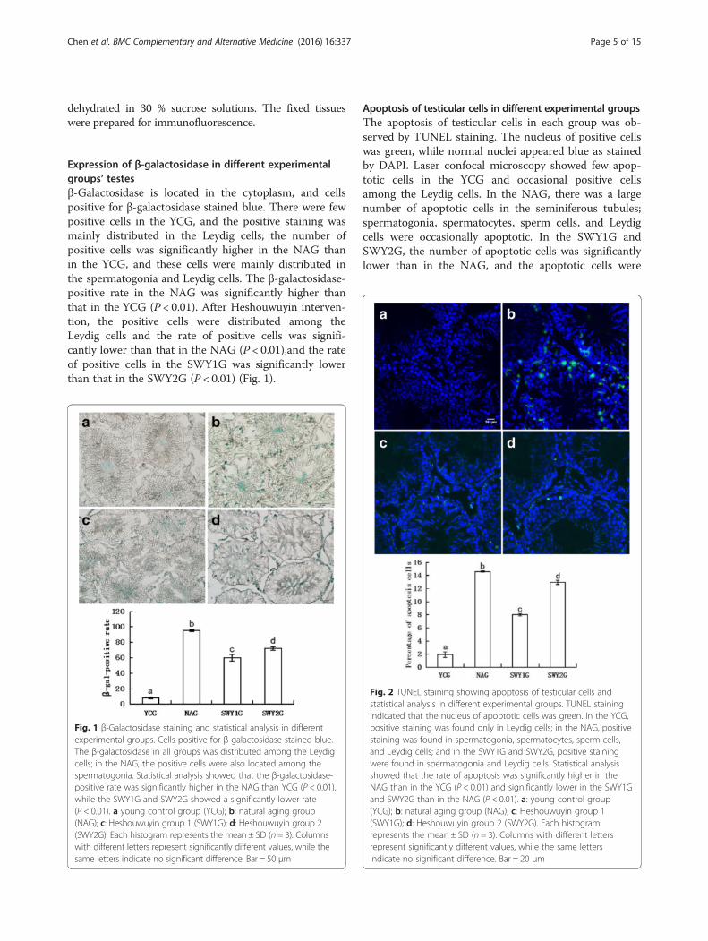
Expression of β-galactosidase in different experimentalgroups’ testesβ-Galactosidase is located in the cytoplasm, and cellspositive for β-galactosidase stained blue. There were fewpositive cells in the YCG, and the positive staining wasmainly distributed in the Leydig cells; the number ofpositive cells was significantly higher in the NAG thanin the YCG, and these cells were mainly distributed inthe spermatogonia and Leydig cells. The β-galactosidase-positive rate in the NAG was significantly higher thanthat in the YCG (P < 0.01). After Heshouwuyin interven-tion, the positive cells were distributed among theLeydig cells and the rate of positive cells was signifi-cantly lower than that in the NAG (P < 0.01),and the rateof positive cells in the SWY1G was significantly lowerthan that in the SWY2G (P < 0.01) (Fig. 1).
Apoptosis of testicular cells in different experimental groupsThe apoptosis of testicular cells in each group was ob-served by TUNEL staining. The nucleus of positive cellswas green, while normal nuclei appeared blue as stainedby DAPI. Laser confocal microscopy showed few apop-totic cells in the YCG and occasional positive cellsamong the Leydig cells. In the NAG, there was a largenumber of apoptotic cells in the seminiferous tubules;spermatogonia, spermatocytes, sperm cells, and Leydigcells were occasionally apoptotic. In the SWY1G andSWY2G, the number of apoptotic cells was significantlylower than in the NAG, and the apoptotic cells were
Fig. 1 β-Galactosidase staining and statistical analysis in differentexperimental groups. Cells positive for β-galactosidase stained blue.The β-galactosidase in all groups was distributed among the Leydigcells; in the NAG, the positive cells were also located among thespermatogonia. Statistical analysis showed that the β-galactosidase-positive rate was significantly higher in the NAG than YCG (P < 0.01),while the SWY1G and SWY2G showed a significantly lower rate(P < 0.01). a young control group (YCG); b: natural aging group(NAG); c: Heshouwuyin group 1 (SWY1G); d: Heshouwuyin group 2(SWY2G). Each histogram represents the mean ± SD (n = 3). Columnswith different letters represent significantly different values, while thesame letters indicate no significant difference. Bar = 50 μm
Fig. 2 TUNEL staining showing apoptosis of testicular cells andstatistical analysis in different experimental groups. TUNEL stainingindicated that the nucleus of apoptotic cells was green. In the YCG,positive staining was found only in Leydig cells; in the NAG, positivestaining was found in spermatogonia, spermatocytes, sperm cells,and Leydig cells; and in the SWY1G and SWY2G, positive stainingwere found in spermatogonia and Leydig cells. Statistical analysisshowed that the rate of apoptosis was significantly higher in theNAG than in the YCG (P < 0.01) and significantly lower in the SWY1Gand SWY2G than in the NAG (P < 0.01). a: young control group(YCG); b: natural aging group (NAG); c: Heshouwuyin group 1(SWY1G); d: Heshouwuyin group 2 (SWY2G). Each histogramrepresents the mean ± SD (n = 3). Columns with different lettersrepresent significantly different values, while the same lettersindicate no significant difference. Bar = 20 μm
Chen et al. BMC Complementary and Alternative Medicine (2016) 16:337 Page 5 of 15
mainly distributed among the spermatogonia and Leydigcells. Statistical analysis showed that the rate of apop-tosis was significantly higher in the NAG than in theYCG (P < 0.01) and significantly lower in the SWY1Gand SWY2G than in the NAG (P < 0.01) and the rate ofapoptosis in SWY1G was significantly lower than in theSWY2G (P < 0.01) (Fig. 2).
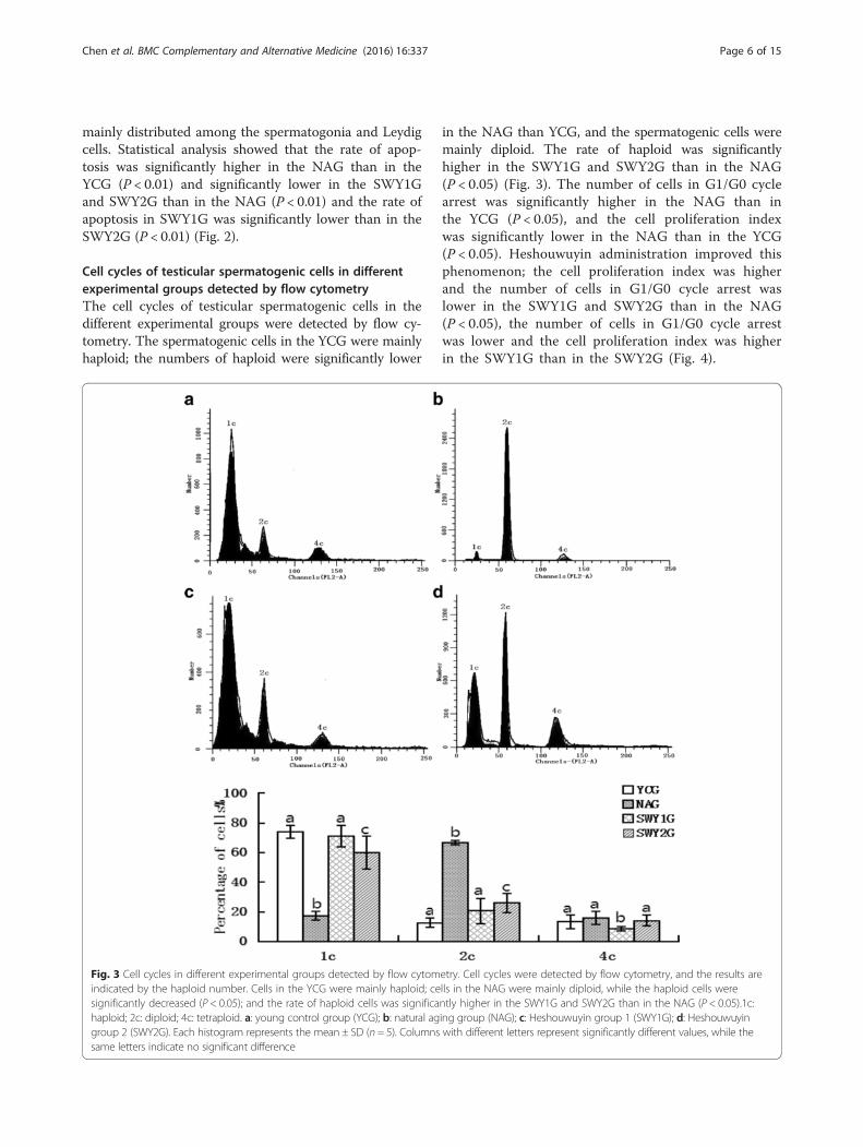
Cell cycles of testicular spermatogenic cells in differentexperimental groups detected by flow cytometryThe cell cycles of testicular spermatogenic cells in thedifferent experimental groups were detected by flow cy-tometry. The spermatogenic cells in the YCG were mainlyhaploid; the numbers of haploid were significantly lower
in the NAG than YCG, and the spermatogenic cells weremainly diploid. The rate of haploid was significantlyhigher in the SWY1G and SWY2G than in the NAG(P < 0.05) (Fig. 3). The number of cells in G1/G0 cyclearrest was significantly higher in the NAG than inthe YCG (P < 0.05), and the cell proliferation indexwas significantly lower in the NAG than in the YCG(P < 0.05). Heshouwuyin administration improved thisphenomenon; the cell proliferation index was higherand the number of cells in G1/G0 cycle arrest waslower in the SWY1G and SWY2G than in the NAG(P < 0.05), the number of cells in G1/G0 cycle arrestwas lower and the cell proliferation index was higherin the SWY1G than in the SWY2G (Fig. 4).
Fig. 3 Cell cycles in different experimental groups detected by flow cytometry. Cell cycles were detected by flow cytometry, and the results areindicated by the haploid number. Cells in the YCG were mainly haploid; cells in the NAG were mainly diploid, while the haploid cells weresignificantly decreased (P < 0.05); and the rate of haploid cells was significantly higher in the SWY1G and SWY2G than in the NAG (P < 0.05).1c:haploid; 2c: diploid; 4c: tetraploid. a: young control group (YCG); b: natural aging group (NAG); c: Heshouwuyin group 1 (SWY1G); d: Heshouwuyingroup 2 (SWY2G). Each histogram represents the mean ± SD (n = 5). Columns with different letters represent significantly different values, while thesame letters indicate no significant difference
Chen et al. BMC Complementary and Alternative Medicine (2016) 16:337 Page 6 of 15
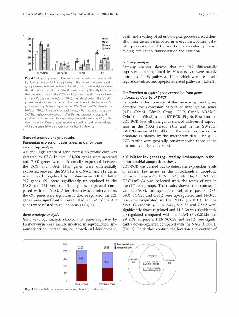
Gene microarray analysis resultsDifferential expression genes screened out by genemicroarray analysisAgilent single standard gene expression profile chip wasdetected by SBC. In total, 21,260 genes were screenedout, 2100 genes were differentially expressed betweenthe YCG and NAG, 1948 genes were differentiallyexpressed between the SWY1G and NAG, and 912 geneswere directly regulated by Heshouwuyin. Of the latter912 genes, 691 were significantly up-regulated in theNAG and 221 were significantly down-regulated com-pared with the YCG. After Heshouwuyin intervention,the 691 genes were significantly down-regulated, the 221genes were significantly up-regulated, and 65 of the 912genes were related to cell apoptosis (Fig. 5).
Gene ontology analysisGene ontology analysis showed that genes regulated byHeshouwuyin were mainly involved in reproduction, im-mune function, metabolism, cell growth and development,
death and a variety of other biological processes. Addition-ally, these genes participated in energy metabolism, cata-lytic processes, signal transduction, molecular synthesis,folding, circulation, transportation and nutrition.
Pathway analysisPathway analysis showed that the 912 differentiallyexpressed genes regulated by Heshouwuyin were mainlydistributed in 59 pathways, 12 of which were cell cycleregulation-related and apoptosis-related pathways (Table 2).
Confirmation of typical gene expression from genemicroarray data by qRT-PCRTo confirm the accuracy of the microarray results, wedetected the expression pattern of nine typical genes(AK1, Cabin1, Kdm2b, Ccng1, GHR, Capn8, ADAM5,Cybrd1 and Glrx3) using qRT-PCR (Fig. 6). Based on theqRT-PCR data, all nine genes showed differential expres-sion in the NAG versus YCG and in the SWY1G,SWY2G versus NAG, although the variation was not asdramatic as shown by the microarray data. The qRT-PCR results were generally consistent with those of themicroarray analysis (Table 3).
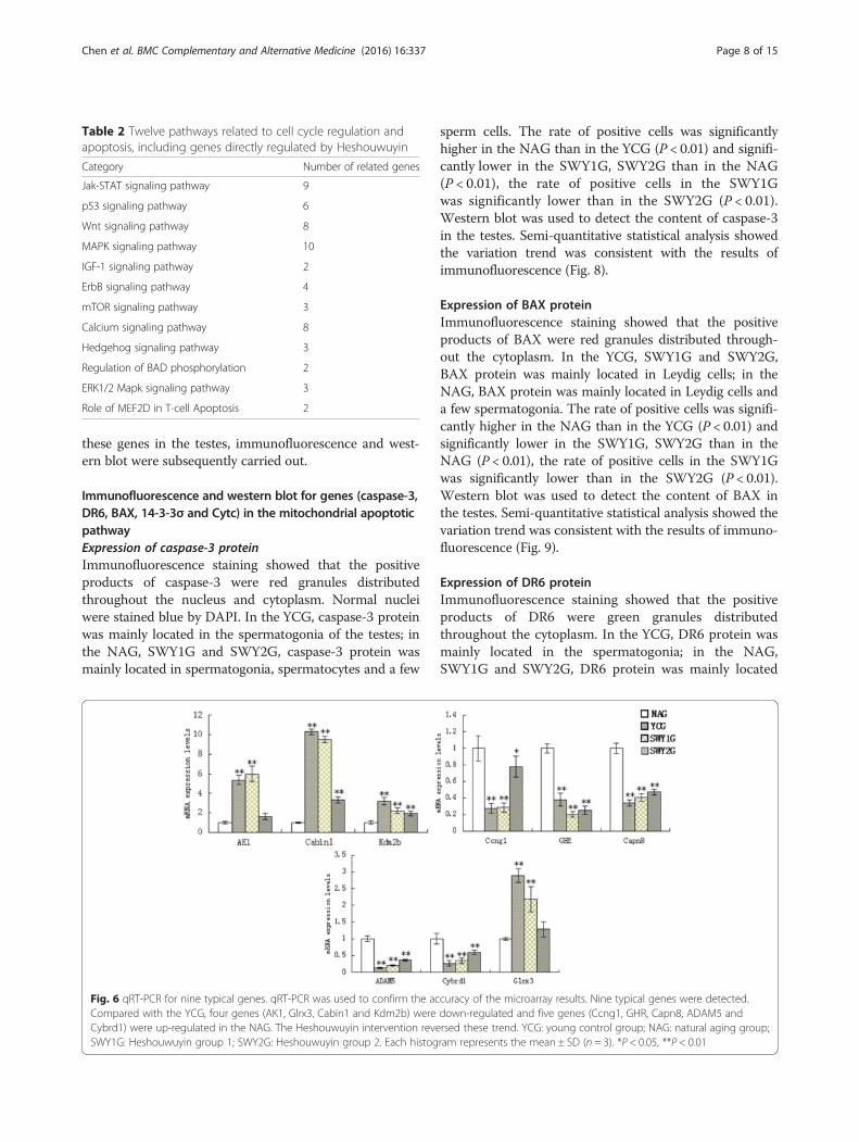
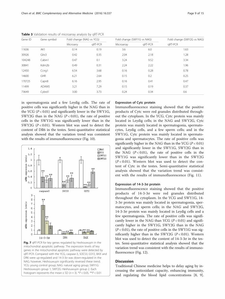
qRT-PCR for key genes regulated by Heshouwuyin in themitochondrial apoptotic pathwayqRT-PCR was carried out to detect the expression levelsof several key genes in the mitochondrial apoptoticpathway (caspase-3, DR6, BAX, 14-3-3σ, SOCS3 andGSY2).mRNA was collected from the testes of rats inthe different groups. The results showed that comparedwith the YCG, the expression levels of caspase-3, DR6,BAX, SOCS3 and GSY2 were up-regulated and 14-3-3σwas down-regulated in the NAG (P < 0.01). In theSWY1G, caspase-3, DR6, BAX, SOCS3 and GSY2 weresignificantly down-regulated and 14-3-3σ was significantlyup-regulated compared with the NAG (P < 0.01).In theSWY2G, caspase-3, DR6, SOCS3 and GSY2 were signifi-cantly down-regulated compared with the NAG (P < 0.01)(Fig. 7). To further confirm the location and content of
Fig. 4 Cell cycles phase in different experimental groups detectedby flow cytometry. Cell cycle phases in the different experimentalgroups were detected by flow cytometry. Statistical analysis showedthat the rate of cells in the G1/G0 phase was significantly higher andthat the rate of cells in the G2/M and S phases was significantly lowerin the NAG than in the YCG (P < 0.05). The rate of cells in the G1/G0phase was significantly lower and the rate of cells in the G2/M and Sphases was significantly higher in the SWY1G and SWY2G than in theNAG (P < 0.05). YCG: young control group; NAG: natural aging group;SWY1G: Heshouwuyin group 1; SWY2G: Heshouwuyin group 2. PI,proliferation index. Each histogram represents the mean ± SD (n = 5).Columns with different letters represent significantly different values,while the same letters indicate no significant difference
Fig. 5 Differentially expressed genes regulated by Heshouwuyin
Chen et al. BMC Complementary and Alternative Medicine (2016) 16:337 Page 7 of 15
these genes in the testes, immunofluorescence and west-ern blot were subsequently carried out.
Immunofluorescence and western blot for genes (caspase-3,DR6, BAX, 14-3-3σ and Cytc) in the mitochondrial apoptoticpathwayExpression of caspase-3 proteinImmunofluorescence staining showed that the positiveproducts of caspase-3 were red granules distributedthroughout the nucleus and cytoplasm. Normal nucleiwere stained blue by DAPI. In the YCG, caspase-3 proteinwas mainly located in the spermatogonia of the testes; inthe NAG, SWY1G and SWY2G, caspase-3 protein wasmainly located in spermatogonia, spermatocytes and a few
sperm cells. The rate of positive cells was significantlyhigher in the NAG than in the YCG (P < 0.01) and signifi-cantly lower in the SWY1G, SWY2G than in the NAG(P < 0.01), the rate of positive cells in the SWY1Gwas significantly lower than in the SWY2G (P < 0.01).Western blot was used to detect the content of caspase-3in the testes. Semi-quantitative statistical analysis showedthe variation trend was consistent with the results ofimmunofluorescence (Fig. 8).
Expression of BAX proteinImmunofluorescence staining showed that the positiveproducts of BAX were red granules distributed through-out the cytoplasm. In the YCG, SWY1G and SWY2G,BAX protein was mainly located in Leydig cells; in theNAG, BAX protein was mainly located in Leydig cells anda few spermatogonia. The rate of positive cells was signifi-cantly higher in the NAG than in the YCG (P < 0.01) andsignificantly lower in the SWY1G, SWY2G than in theNAG (P < 0.01), the rate of positive cells in the SWY1Gwas significantly lower than in the SWY2G (P < 0.01).Western blot was used to detect the content of BAX inthe testes. Semi-quantitative statistical analysis showed thevariation trend was consistent with the results of immuno-fluorescence (Fig. 9).
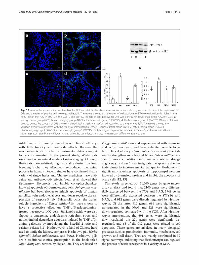
Expression of DR6 proteinImmunofluorescence staining showed that the positiveproducts of DR6 were green granules distributedthroughout the cytoplasm. In the YCG, DR6 protein wasmainly located in the spermatogonia; in the NAG,SWY1G and SWY2G, DR6 protein was mainly located
Table 2 Twelve pathways related to cell cycle regulation andapoptosis, including genes directly regulated by Heshouwuyin
Category Number of related genes
Jak-STAT signaling pathway 9
p53 signaling pathway 6
Wnt signaling pathway 8
MAPK signaling pathway 10
IGF-1 signaling pathway 2
ErbB signaling pathway 4
mTOR signaling pathway 3
Calcium signaling pathway 8
Hedgehog signaling pathway 3
Regulation of BAD phosphorylation 2
ERK1/2 Mapk signaling pathway 3
Role of MEF2D in T-cell Apoptosis 2
Fig. 6 qRT-PCR for nine typical genes. qRT-PCR was used to confirm the accuracy of the microarray results. Nine typical genes were detected.Compared with the YCG, four genes (AK1, Glrx3, Cabin1 and Kdm2b) were down-regulated and five genes (Ccng1, GHR, Capn8, ADAM5 andCybrd1) were up-regulated in the NAG. The Heshouwuyin intervention reversed these trend. YCG: young control group; NAG: natural aging group;SWY1G: Heshouwuyin group 1; SWY2G: Heshouwuyin group 2. Each histogram represents the mean ± SD (n = 3). *P < 0.05, **P < 0.01
Chen et al. BMC Complementary and Alternative Medicine (2016) 16:337 Page 8 of 15
in spermatogonia and a few Leydig cells. The rate ofpositive cells was significantly higher in the NAG than inthe YCG (P < 0.01) and significantly lower in the SWY1G,SWY2G than in the NAG (P < 0.01), the rate of positivecells in the SWY1G was significantly lower than in theSWY2G (P < 0.01). Western blot was used to detect thecontent of DR6 in the testes. Semi-quantitative statisticalanalysis showed that the variation trend was consistentwith the results of immunofluorescence (Fig. 10).
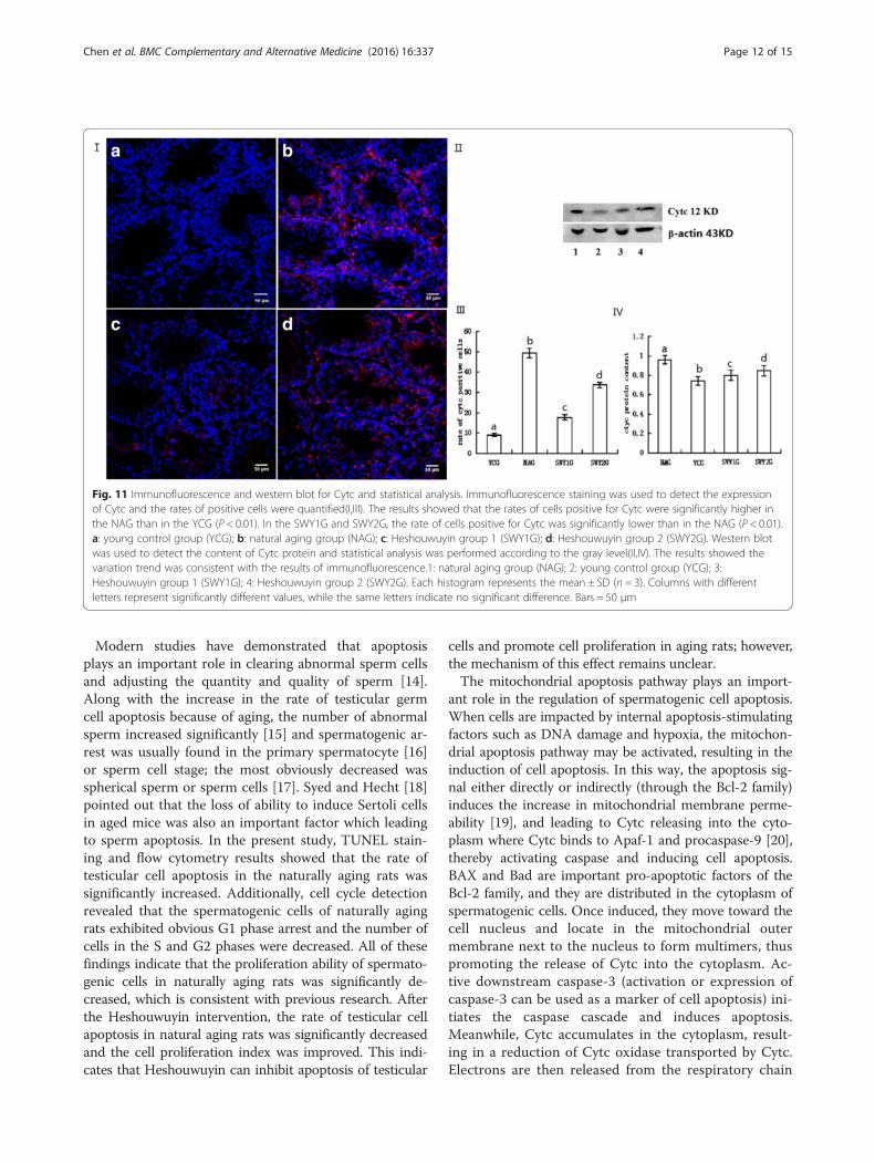
Expression of Cytc proteinImmunofluorescence staining showed that the positiveproducts of Cytc were red granules distributed through-out the cytoplasm. In the YCG, Cytc protein was mainlylocated in Leydig cells; in the NAG and SWY2G, Cytcprotein was mainly located in spermatogonia, spermato-cytes, Leydig cells, and a few sperm cells; and in theSWY1G, Cytc protein was mainly located in spermato-gonia and spermatocytes. The rate of positive cells wassignificantly higher in the NAG than in the YCG (P < 0.01)and significantly lower in the SWY1G, SWY2G than inthe NAG (P < 0.01), the rate of positive cells in theSWY1G was significantly lower than in the SWY2G(P < 0.01). Western blot was used to detect the con-tent of Cytc in the testes. Semi-quantitative statisticalanalysis showed that the variation trend was consist-ent with the results of immunofluorescence (Fig. 11).
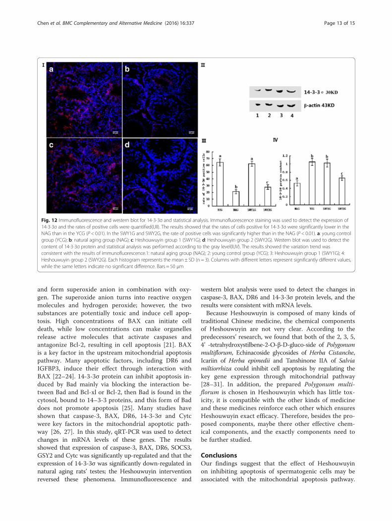
Expression of 14-3-3σ proteinImmunofluorescence staining showed that the positiveproducts of 14-3-3σ were red granules distributedthroughout the cytoplasm. In the YCG and SWY1G, 14-3-3σ protein was mainly located in spermatogonia, sper-matocytes, and sperm cells; in the NAG and SWY2G,14-3-3σ protein was mainly located in Leydig cells and afew spermatogonia. The rate of positive cells was signifi-cantly lower in the NAG than YCG (P < 0.01) and signifi-cantly higher in the SWY1G, SWY2G than in the NAG(P < 0.01), the rate of positive cells in the SWY1G was sig-nificantly higher than in the SWY2G (P < 0.01). Westernblot was used to detect the content of 14-3-3σ in the tes-tes. Semi-quantitative statistical analysis showed that thevariation trend was consistent with the results of immuno-fluorescence (Fig. 12).
DiscussionTraditional Chinese medicine helps to delay aging by in-creasing the antioxidant capacity, enhancing immunity,and regulating the blood lipid concentrations [8, 9].
Table 3 Validation results of microarray analysis by qRT-PCR
Gene ID Gene symbol Fold change (NAG vs YCG) Fold change (SWY1G vs NAG) Fold change (SWY2G vs NAG)
Microarry qRT-PCR Microarray qRT-PCR qRT-PCR
11636 AK1 0.14 0.19 3.6 6.0 1.63
30926 Glrx3 0.42 0.35 2.04 2.18 1.28
104248 Cabin1 0.47 0.1 3.24 9.52 3.34
30841 Kdm2b 0.49 0.31 2.24 2.22 1.96
12450 Ccng1 6.54 3.68 0.16 0.28 0.78
14600 GHR 6.21 2.64 0.15 0.2 0.25
170725 Capn8 6.16 2.95 0.16 0.41 0.47
11499 ADAM5 3.21 7.29 0.15 0.19 0.37
73649 Cybrd1 3.00 3.73 0.24 0.34 0.6
Fig. 7 qRT-PCR for key genes regulated by Heshouwuyin in themitochondrial apoptotic pathway. The expression levels of keygenes in the mitochondrial apoptotic pathway were detected byqRT-PCR. Compared with the YCG, caspase-3, SOCS3, GSY2, BAX andDR6 were up-regulated and 14-3-3σ was down-regulated in theNAG; however, Heshouwuyin significantly reversed these trend.YCG: young control group; NAG: natural aging group; SWY1G:Heshouwuyin group 1; SWY2G: Heshouwuyin group 2. Eachhistogram represents the mean ± SD (n = 3). *P < 0.05, **P < 0.01
Chen et al. BMC Complementary and Alternative Medicine (2016) 16:337 Page 9 of 15
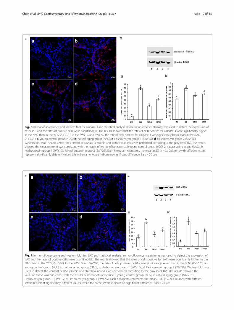
Fig. 8 Immunofluorescence and western blot for caspase-3 and statistical analysis. Immunofluorescence staining was used to detect the expression ofcaspase-3 and the rates of positive cells were quantified(I,III). The results showed that the rates of cells positive for caspase-3 were significantly higherin the NAG than in the YCG (P < 0.01). In the SWY1G and SWY2G, the rate of cells positive for caspase-3 was significantly lower than in the NAG(P < 0.01). a: young control group (YCG); b: natural aging group (NAG); c: Heshouwuyin group 1 (SWY1G); d: Heshouwuyin group 2 (SWY2G).Western blot was used to detect the content of caspase-3 protein and statistical analysis was performed according to the gray level(II,IV). The resultsshowed the variation trend was consistent with the results of immunofluorescence.1: young control group (YCG); 2: natural aging group (NAG); 3:Heshouwuyin group 1 (SWY1G); 4: Heshouwuyin group 2 (SWY2G). Each histogram represents the mean ± SD (n = 3). Columns with different lettersrepresent significantly different values, while the same letters indicate no significant difference. Bars = 20 μm
Fig. 9 Immunofluorescence and western blot for BAX and statistical analysis. Immunofluorescence staining was used to detect the expression ofBAX and the rates of positive cells were quantified(I,III). The results showed that the rates of cells positive for BAX were significantly higher in theNAG than in the YCG (P < 0.01). In the SWY1G and SWY2G, the rate of cells positive for BAX was significantly lower than in the NAG (P < 0.01). a:young control group (YCG); b: natural aging group (NAG); c: Heshouwuyin group 1 (SWY1G); d: Heshouwuyin group 2 (SWY2G). Western blot wasused to detect the content of BAX protein and statistical analysis was performed according to the gray level(II,IV). The results showed thevariation trend was consistent with the results of immunofluorescence.1: young control group (YCG); 2: natural aging group (NAG); 3:Heshouwuyin group 1 (SWY1G); 4: Heshouwuyin group 2 (SWY2G). Each histogram represents the mean ± SD (n = 3). Columns with differentletters represent significantly different values, while the same letters indicate no significant difference. Bars = 20 μm
Chen et al. BMC Complementary and Alternative Medicine (2016) 16:337 Page 10 of 15
Additionally, it have produced good clinical efficacy,with little toxicity and few side effects. Because themechanism is still unclear, experimental datas were yetto be consummated. In the present study, Wistar ratswere used as an animal model of natural aging. Althoughthese rats have relatively high mortality during the longbreeding cycle, they effectively reproduced the agingprocess in humans. Recent studies have confirmed that avariety of single herbs and Chinese medicines have anti-aging and anti-apoptotic effects. Yuan et al. showed thatEpimedium flavonoids can inhibit cyclophosphamide-induced apoptosis of spermatogenic cells. Polygonum mul-tiflorum has been shown to inhibit apoptosis of humanumbilical vein endothelial cells and down-regulate the ex-pression of caspase-3 [10]. Salvianolic acids, the water-soluble ingredient of Salvia miltiorrhiza, were shown tohave a protective effect on TNF-α/D-galactosamine-treated hepatocyte LO2 cells. Salvianolic acids were alsoshown to antagonize endoplasmic reticulum stress andmitochondrial-dependent apoptosis induced by TNF-α/D-amino galactose by modulating the Bax/Bcl-2 ratio andcalcium release [11]. Heshouwuyin, a kind of Chinese herbused to tonify the kidney, comprises Heshouwu pill, Herbaepimedii, Salvia miltiorrhiza, and Poria. Heshouwu pillsare a traditional clinical prescription in the book titledXuan Ming Lun, written by Hejian Liu. They are based on
Polygonum multiflorum and supplemented with cistancheand achyranthes root, and have exhibited reliable long-term clinical efficacy. Herba epimedii can tonify the kid-ney to strengthen muscles and bones, Salvia miltiorrhizacan promote circulation and remove stasis to dredgeangiocarpy, and Poria can invigorate the spleen and elim-inate damp to increase mental tranquility. Heshouwuyinsignificantly alleviates apoptosis of hippocampal neuronsinduced by β-amyloid protein and inhibit the apoptosis ofovary cells [12, 13].This study screened out 21,260 genes by gene micro-
array analysis and found that 2100 genes were differen-tially expressed between the YCG and NAG, 1948 geneswere differentially expressed between the SWY1G andNAG, and 912 genes were directly regulated by Heshou-wuyin. Of the latter 912 genes, 691 were significantlyup-regulated in the NAG and 221 were significantlydown-regulated compared with the YCG. After Heshou-wuyin intervention, the 691 genes were significantlydown-regulated, the 221 genes were significantly up-regulated, and 65 of the 912 genes were related to cellapoptosis. These genes are involved in many biologicalprocesses such as proliferation, immunity, metabolism, cellgrowth, and cell death. They are mainly distributed in 59signal pathways, indicating that Heshouwuyin can regulatethe process of testis senescence in a variety of ways.
Fig. 10 Immunofluorescence and western blot for DR6 and statistical analysis. Immunofluorescence staining was used to detect the expression ofDR6 and the rates of positive cells were quantified(I,III). The results showed that the rates of cells positive for DR6 were significantly higher in theNAG than in the YCG (P < 0.01). In the SWY1G and SWY2G, the rate of cells positive for DR6 was significantly lower than in the NAG (P < 0.01). a:young control group (YCG); b: natural aging group (NAG); c: Heshouwuyin group 1 (SWY1G); d: Heshouwuyin group 2 (SWY2G). Western blot wasused to detect the content of DR6 protein and statistical analysis was performed according to the gray level(II,IV). The results showed thevariation trend was consistent with the results of immunofluorescence.1: young control group (YCG); 2: natural aging group (NAG); 3:Heshouwuyin group 1 (SWY1G); 4: Heshouwuyin group 2 (SWY2G). Each histogram represents the mean ± SD (n = 3). Columns with differentletters represent significantly different values, while the same letters indicate no significant difference. Bars = 20 μm
Chen et al. BMC Complementary and Alternative Medicine (2016) 16:337 Page 11 of 15
Modern studies have demonstrated that apoptosisplays an important role in clearing abnormal sperm cellsand adjusting the quantity and quality of sperm [14].Along with the increase in the rate of testicular germcell apoptosis because of aging, the number of abnormalsperm increased significantly [15] and spermatogenic ar-rest was usually found in the primary spermatocyte [16]or sperm cell stage; the most obviously decreased wasspherical sperm or sperm cells [17]. Syed and Hecht [18]pointed out that the loss of ability to induce Sertoli cellsin aged mice was also an important factor which leadingto sperm apoptosis. In the present study, TUNEL stain-ing and flow cytometry results showed that the rate oftesticular cell apoptosis in the naturally aging rats wassignificantly increased. Additionally, cell cycle detectionrevealed that the spermatogenic cells of naturally agingrats exhibited obvious G1 phase arrest and the number ofcells in the S and G2 phases were decreased. All of thesefindings indicate that the proliferation ability of spermato-genic cells in naturally aging rats was significantly de-creased, which is consistent with previous research. Afterthe Heshouwuyin intervention, the rate of testicular cellapoptosis in natural aging rats was significantly decreasedand the cell proliferation index was improved. This indi-cates that Heshouwuyin can inhibit apoptosis of testicular
cells and promote cell proliferation in aging rats; however,the mechanism of this effect remains unclear.The mitochondrial apoptosis pathway plays an import-
ant role in the regulation of spermatogenic cell apoptosis.When cells are impacted by internal apoptosis-stimulatingfactors such as DNA damage and hypoxia, the mitochon-drial apoptosis pathway may be activated, resulting in theinduction of cell apoptosis. In this way, the apoptosis sig-nal either directly or indirectly (through the Bcl-2 family)induces the increase in mitochondrial membrane perme-ability [19], and leading to Cytc releasing into the cyto-plasm where Cytc binds to Apaf-1 and procaspase-9 [20],thereby activating caspase and inducing cell apoptosis.BAX and Bad are important pro-apoptotic factors of theBcl-2 family, and they are distributed in the cytoplasm ofspermatogenic cells. Once induced, they move toward thecell nucleus and locate in the mitochondrial outermembrane next to the nucleus to form multimers, thuspromoting the release of Cytc into the cytoplasm. Ac-tive downstream caspase-3 (activation or expression ofcaspase-3 can be used as a marker of cell apoptosis) ini-tiates the caspase cascade and induces apoptosis.Meanwhile, Cytc accumulates in the cytoplasm, result-ing in a reduction of Cytc oxidase transported by Cytc.Electrons are then released from the respiratory chain
Fig. 11 Immunofluorescence and western blot for Cytc and statistical analysis. Immunofluorescence staining was used to detect the expressionof Cytc and the rates of positive cells were quantified(I,III). The results showed that the rates of cells positive for Cytc were significantly higher inthe NAG than in the YCG (P < 0.01). In the SWY1G and SWY2G, the rate of cells positive for Cytc was significantly lower than in the NAG (P < 0.01).a: young control group (YCG); b: natural aging group (NAG); c: Heshouwuyin group 1 (SWY1G); d: Heshouwuyin group 2 (SWY2G). Western blotwas used to detect the content of Cytc protein and statistical analysis was performed according to the gray level(II,IV). The results showed thevariation trend was consistent with the results of immunofluorescence.1: natural aging group (NAG); 2: young control group (YCG); 3:Heshouwuyin group 1 (SWY1G); 4: Heshouwuyin group 2 (SWY2G). Each histogram represents the mean ± SD (n = 3). Columns with differentletters represent significantly different values, while the same letters indicate no significant difference. Bars = 50 μm
Chen et al. BMC Complementary and Alternative Medicine (2016) 16:337 Page 12 of 15
and form superoxide anion in combination with oxy-gen. The superoxide anion turns into reactive oxygenmolecules and hydrogen peroxide; however, the twosubstances are potentially toxic and induce cell apop-tosis. High concentrations of BAX can initiate celldeath, while low concentrations can make organellesrelease active molecules that activate caspases andantagonize Bcl-2, resulting in cell apoptosis [21]. BAXis a key factor in the upstream mitochondrial apoptosispathway. Many apoptotic factors, including DR6 andIGFBP3, induce their effect through interaction withBAX [22–24]. 14-3-3σ protein can inhibit apoptosis in-duced by Bad mainly via blocking the interaction be-tween Bad and Bcl-xl or Bcl-2, then Bad is found in thecytosol, bound to 14–3-3 proteins, and this form of Baddoes not promote apoptosis [25]. Many studies haveshown that caspase-3, BAX, DR6, 14-3-3σ and Cytcwere key factors in the mitochondrial apoptotic path-way [26, 27]. In this study, qRT-PCR was used to detectchanges in mRNA levels of these genes. The resultsshowed that expression of caspase-3, BAX, DR6, SOCS3,GSY2 and Cytc was significantly up-regulated and that theexpression of 14-3-3σ was significantly down-regulated innatural aging rats’ testes; the Heshouwuyin interventionreversed these phenomena. Immunofluorescence and
western blot analysis were used to detect the changes incaspase-3, BAX, DR6 and 14-3-3σ protein levels, and theresults were consistent with mRNA levels.Because Heshouwuyin is composed of many kinds of
traditional Chinese medicine, the chemical componentsof Heshouwuyin are not very clear. According to thepredecessors’ research, we found that both of the 2, 3, 5,4’ -tetrahydroxystilbene-2-O-β-D-gluco-side of Polygonummultiflorum, Echinacoside glycosides of Herba Cistanche,Icariin of Herba epimedii and Tanshinone IIA of Salviamiltiorrhiza could inhibit cell apoptosis by regulating thekey gene expression through mitochondrial pathway[28–31]. In addition, the prepared Polygonum multi-florum is chosen in Heshouwuyin which has little tox-icity, it is compatible with the other kinds of medicineand these medicines reinforce each other which ensuresHeshouwuyin exact efficacy. Therefore, besides the pro-posed components, maybe there other effective chem-ical components, and the exactly components need tobe further studied.
ConclusionsOur findings suggest that the effect of Heshouwuyinon inhibiting apoptosis of spermatogenic cells may beassociated with the mitochondrial apoptosis pathway.
Fig. 12 Immunofluorescence and western blot for 14-3-3σ and statistical analysis. Immunofluorescence staining was used to detect the expression of14-3-3σ and the rates of positive cells were quantified(I,III). The results showed that the rates of cells positive for 14-3-3σ were significantly lower in theNAG than in the YCG (P < 0.01). In the SWY1G and SWY2G, the rate of positive cells was significantly higher than in the NAG (P < 0.01). a: young controlgroup (YCG); b: natural aging group (NAG); c: Heshouwuyin group 1 (SWY1G); d: Heshouwuyin group 2 (SWY2G). Western blot was used to detect thecontent of 14-3-3σ protein and statistical analysis was performed according to the gray level(II,IV). The results showed the variation trend wasconsistent with the results of immunofluorescence.1: natural aging group (NAG); 2: young control group (YCG); 3: Heshouwuyin group 1 (SWY1G); 4:Heshouwuyin group 2 (SWY2G). Each histogram represents the mean ± SD (n = 3). Columns with different letters represent significantly different values,while the same letters indicate no significant difference. Bars = 50 μm
Chen et al. BMC Complementary and Alternative Medicine (2016) 16:337 Page 13 of 15
Heshouwuyin reduces the release of Cytc by up-regulatingthe anti-apoptotic factor 14-3-3σ, down-regulating DR6and BAX, and preventing interaction of Bad with Bcl-xl orBcl-2, thus inhibits the mitochondrial apoptosis pathwayand reduces apoptosis of spermatogenic cells. Heshouwuyintherefore plays a role in anti-apoptosis, although the mech-anisms need to be further studied.
AcknowledgementsNot applicable.
FundingThis work was supported by the Natural Science Foundation of Hebei province(’The regulation of Heshouwuyin on differential expression genes related toapoptosis of spermatogenic cells in aging rats’ [H2013201139]) and the NaturalScience Foundation of China (’Based on the signal transduction pathway toresearch the molecular mechanism of Heshouwuyin on testes degenerativediseases” [81373787]).
Availability of data and materialsThe datasets supporting the conclusions of this article are included withinthis paper.
Authors’ contributionsSYN designed the study, modified the paper, and was responsible for thefunding. ZSW instructed the experiment and examined the paper. JBC, YJW,CHH, YX, XL, FQ and HKL performed the research, analyzed the data. JBC andYJW wrote the paper. All authors read and approved the final manuscript.
Competing interestsThe authors declare that they have no competing interests.
Consent for publicationNot applicable.
Ethics approval and consent to participateThe experiment was approved by the Animal Ethics Committee of HebeiUniversity.
Author details1School of Medicine, Hebei University, Baoding 071002, Hebei Province,China. 2College of Life Science, Hebei University, Baoding 071002, HebeiProvince, China. 3Baoding NO.1 Hospital Of TCM, NO.530 Yuhua West Road,Baoding 071000, Hebei Province, China. 4Military Transportation University,NO.1 Dongjuzi, Chenglin RoadHedong District, Tianjin 300161, China.
Received: 7 May 2016 Accepted: 25 August 2016
References1. Park HJ, Zhang NN, Park DK. Topical application of Polygonum multiflorum
extract induces hair growth of resting hair follicles through upregulatingShh and β-catenin expression in C57BL/6 mice. J Ethnopharmacol. 2011;135:369–75. doi:10.1016/j.jep.2011.03.028.
2. Um MY, Choi WH, Aan JY, Kim SR, Ha TY. Protective effect of Polygonummultiflorum Thunb. on amyloid β-peptide 25–35 induced cognitive deficitsin mice. J Ethnopharmacol. 2006;104:144–8.
3. Yin JH, Zhou XY, Zhu XQ. Pharmacological and clinical studies on theprocessed products of radix Polygoni multiflori. Zhongguo Zhong Yao ZaZhi. 1992;17(12):722–4.
4. Zhao XJ, Guo Y, Song QL, Li L, Jiang Y, Sun YC, Qu YE, Gao FL. Effect ofHeshouwuyin on the expression of Cox7a2 protein in testis tissue of exercised-induced fatigue rat. Sichuan Da Xue Xue Bao Yi Xue Ban. 2013;44(2):205–8.
5. Niu SY, Kou SR, Zhou XC, Ding L. Heshouwu decoction, a Chinese herb fortonifying kidney, ameliorates hypothalamic-pituitary-testicular axis secretionin aging rats. Neural Regenerat Res. 2012;7(21):1611–7. doi:10.3969/j.issn.1673-5374.2012.21.002.
6. Niu SY, Chen JB, Duan F, Song QL, Qin MY, Wang ZS, Liu JZ. Possiblemechanism underlying the effect of Heshou wuyin, a tonifying kidney herb,
on sperm quality in aging rats. BMC Complement Altern Med. 2014;14:250.doi:10.1186/1472-6882-14-250.
7. Guo Y, Niu SY, Gao FL, Zhao XJ, Sun YC, Qu YE, Chen X, Guo KH. Effect ofHeshowuyin on Bax and Bcl-2 expression of testicular Leydig cells in theovertraining rat testis. Lishizhen Med Mater Med Res. 2012; 23(3):589-90.doi:10. 3969/j. issn. 1008–0805. 2012. 03.032. (in Chinese)
8. Chik WI, Zhu L, Fan LL, Yi T, Zhu GY, Gou XJ, Tang YN, Xu J, Yeung WP,Zhao ZZ, Yu ZL, Chen HB. Saussurea involucrata: a review of the botany,phytochemistry and ethnopharmacology of a rare traditional herbalmedicine. J Ethnopharmacol. 2015;172:44–60. doi:10.1016/j.jep.2015.06.033.
9. Chen HF, Chen YH, Liu CH, Wang L, Chen X, Yu BY, Qi J. Integratedchemometric fingerprints of antioxidant activities and HPLC-DAD-CL forassessing the quality of the processed roots of Polygonum multiflorumThunb. Chin Med. 2016;11:18. doi:10.1186/s13020-016-0087-8.
10. Yuan D, Wang H, He H, Jia L, He Y, Wang T, Zeng X, Li Y, Li S, Zhang C.Protective effects of total flavonoids from Epimedium on the male mousereproductive system against cyclophosphamide-induced oxidative injury byup-regulating the expressions of SOD3 and GPX1. Phytother Res. 2014;28(1):88–97. doi:10.1002/ptr.4956.
11. Yan X, Jiang Z, Bi L, Yang Y, Chen W. Salvianolic acid A attenuates TNF-αand D-GalN-induced ER stress-mediated and mitochondrial-dependentapoptosis by modulating Bax/Bcl-2 ratio and calcium release in hepatocyteLO2 cells. Naunyn Schmiedebergs Arch Pharmacol. 2015;388(8):817–30.doi:10.1007/s00210-015-1116-3.
12. Chen JB, Wang YJ, Guo SN, Niu SY, Duan F. Effect and mechanism ofHeshowuyin on alleviating the hippocampal neurons injury inducedby β-amyloid protein. Acta Anatomica Sinica. 2015; 46(2):177–183.doi:10. 16098/j. issn. 0529–1356. 2015. 02. 006. (in Chinese)
13. Zhang MM, Jia XM, Wang C, Gao W, Peng KN, Zhao M, Zhang CG, Li YL. Theeffects of heshowuyin on the expression of apoptotic proteins FOX、SIRT1and c-Myc of ovary tissues in the aging rats. J Med Pest Control. 2013;12:1368 (in Chinese).
14. Shaha C, Tripathi R, Mishra DP. Male germ cell apoptosis: regulation andbiology. Philos Trans R Soc Lond B Biol Sci. 2010;365(1546):1501–15.doi:10.1098/rstb.2009.0124.
15. Jiang H, Zhu WJ, Li J, Chen QJ, Liang WB, Gu YQ. Quantitative histologicalanalysis and ultrastructure of the aging human testis. Int Urol Nephrol. 2014;46(5):879–85. doi:10.1007/s11255-013-0610-0.
16. Chen Y, Wang H, Qi N, Wu H, Xiong W, Ma J, Lu Q, Han D. Functions ofTAM RTKs in regulating spermatogenesis and male fertility in mice.Reproduction. 2009;138(4):655–66. doi:10.1530/REP-09-0101.
17. Nakata H, Wakayama T, Takai Y, Iseki S. Quantitative analysis of the cellularcomposition in seminiferous tubules in normal and genetically modified infertilemice. J Histochem Cytochem. 2015;63(2):99–113. doi:10.1369/0022155414562045.
18. Syed V, Hecht NB. Selective loss of Sertoli cell and germ cell function leadsto a disruption in sertoli cell-germ cell communication during aging in theBrown Norway rat. Biol Reprod. 2001;64(1):107–12.
19. Yana W, Suominena J, Samsonb M, Je goub B, Toppari J. Involvement ofBcl-2 family proteins in germ cell apoptosis during testicular developmentin the rat and pro-survival effect of stem cell factor on germ cells in vitro.Mol Cell Endocrino. 2000;165(1–2):115–29.
20. Chana CK, Supriadya H, Gohb BH, Kadir HA. Elephantopus scaber inducesapoptosis through ROS-dependent mitochondrial signaling pathway in HCT116human colorectal carcinoma cells. J Ethnopharmacol. 2015;168:291–304.doi:10.1016/j.jep.2015.03.072.
21. Zhang CC, Zhou AF, Zhang ML, Cao JG. Effects of experimental varicoceleon the apoptosis of spermatogenic cells in rats. Zhonghua Nankeuxe. 2003;9(7):507–11.
22. Pan G, Bauer JH, Haridas V, Wang S, Liu D, Yu G, Vincenz C, Aggarwal BB, NiJ, Dixit VM. Identification and functional characterization of DR6, a noveldeath domain -containing TNF receptor. FEBS Lett. 1998;431(3):351–6.
23. Zeng L, Li T, Xu DC, Liu J, Mao G, Cui MZ, Fu X, Xu X. Death receptor 6induces apoptosis not through type I or type II pathways, but via a uniquemitochondria-dependent pathway by interacting with Bax protein. J BiolChem. 2012;287(34):29125–33. doi:10.1074/jbc.M112.362038.
24. Jia Y, Lee KW, Swerdloff R, Hwang D, Cobb LJ, Sinha Hikim A, Lue YH,Cohen P, Wang C. Interaction of insulin-like growth factor-binding protein-3and BAX in mitochondria promotes male germ cell apoptosis. J Biol Chem.2010;285:1726–32. doi:10.1074/jbc.M109.046847.
25. Nagappana A, Il Parka K, Parka HS, Kimb JA, Honga GE, Kangc SR, Leea DH,Kima GS. Vitamin C induces apoptosis in AGS cells by down-regulation of
Chen et al. BMC Complementary and Alternative Medicine (2016) 16:337 Page 14 of 15
14–3-3σ via a mitochondrial dependent pathway. Food Chem. 2012;135(3):1920–8. doi:10.1016/j.foodchem.2012.06.050.
26. Harris MH, Thompson CB. The role of the Bcl-2 family in the regulation of outermitochondrial membrane permeability. Cell Death Differ. 2000;7(12):1182–91.
27. Ricci JE, Munoz-Pinedo C, Fitzgerald P, Bailly-Maitre B, Perkins GA, Yadava N,Scheffler IE, Ellisman MH, Green DR. Disruption of mitochondrial functionduring apoptosis is mediated by caspase cleavage of the p75 subunit ofcomplex I of the electron transport chain. Cell. 2004;117(6):773–86.
28. Lin L, Ni B, Lin H, Zhang M, Li X, Yin X, Qu C, Ni J. Traditional usages,botany, phytochemistry, pharmacology and toxicology of Polygonummultiflorum Thunb.: A review. J Ethnopharmacol. 2015;159:158–83.
29. Kuang R, Sun Y, Yuan W, Lei L, Zheng X. Protective effects of echinacoside,one of the phenylethanoid glycosides, on H2O2-induced cytotoxicity inPC12 cells. Planta Med. 2009;75(14):1499–504. doi:10.1055/s-0029-1185806.
30. Zhang D, Wang Z, Sheng C, Peng W, Hui S, Gong W, Chen S. Icariinprevents amyloid beta-induced apoptosis via the PI3K/Akt pathway in PC-12cells. Evid Based Complement Alternat Med. 2015. doi:10.1155/2015/235265.
31. Li J, He C, Tong W, Zou Y, Li D, Zhang C, Xu W. Tanshinone IIA blocksdexamethasone-induced apoptosis in osteoblasts through inhibitingNox4-derived ROS production. Int J Clin Exp Pathol. 2015;8(10):13695–706.
• We accept pre-submission inquiries
• Our selector tool helps you to find the most relevant journal
• We provide round the clock customer support
• Convenient online submission
• Thorough peer review
• Inclusion in PubMed and all major indexing services
• Maximum visibility for your research
Submit your manuscript atwww.biomedcentral.com/submit
Submit your next manuscript to BioMed Central and we will help you at every step:
Chen et al. BMC Complementary and Alternative Medicine (2016) 16:337 Page 15 of 15