Embed Size (px)
Citation preview
Apoptosis: Mechanisms and Relevance in Liver Diseases
Harmeet Malhi
Gregory J. Gores Mayo Clinic College of Medicine, Rochester, MN Address for correspondence: G. J. Gores
Professor of Medicine Miles and Shirley Fiterman Center for Digestive Diseases Mayo Clinic 200 First Street SW Rochester, Minnesota 55905 Tel: 507 284 0686
Fax: 507 284 0762 E-mail: [email protected]
Support for the work is provided by NIH grant DK 41876 and the Mayo Foundation. Abbreviations: ATF6 activating transcription factor 6, ATF4 activating transcription factor-4, CHOP C/EBP-homologous protein, ER endoplasmic reticulum, eIF2α eukaryotic translation initiation factor-2α, FADD Fas associated death domain, IRE1 inositol-requiring protein-1, JNK c-jun N-terminal kinase, NK natural killer, NFκB nuclear factor kappa B, PERK protein kinase RNA-like ER kinase, TNF-α tumor necrosis factor alpha, TRAIL tumor necrosis factor-related apoptosis inducing ligand, TNFR1 tumor necrosis factor receptor 1, TNFR2 tumor necrosis factor receptor 2, TRAIL-R1 tumor necrosis factor-related apoptosis inducing ligand receptor 1, TRAIL-R2 tumor necrosis factor-related apoptosis inducing ligand receptor 2, TRAF-2 tumor necrosis factor receptor associated protein, XBP1 X-box binding protein-1, Conflict of interest: The authors have no conflict of interest. Words: 5198 References: 144 Figures: 4
Apoptosis is a ubiquitous form of cell death occurring in human liver diseases. Apoptosis has
historically been defined morphologically by the presence of cytoplasmic shrinkage (pyknosis),
chromatin condensation, nuclear fragmentation (karyorhexis), the presence of plasma membrane
blebbing and the maintenance of an intact plasma membrane which retains its integrity as the cell
fragments into apoptotic bodies(1). Indeed, apoptotic bodies were first described in the liver in
patients with Yellow Fever where they were referred to as councilman bodies(2). Apoptosis is
classically defined morphologically, and therefore the detection and confirmation of apoptosis has
previously required tissue or cellular analysis. Caspases are a group of intracellular enzymes that
mediate the cellular demolition characterized phenotypically as apoptosis. Circulating markers of
caspase activity can now be measured in serum and serve as surrogate markers of caspase-mediated
cell death. In particular, serum levels of specific caspase-mediated cleavage products of cytokeratin
18 are indicators of epithelial cell apoptosis, including hepatocyte apoptosis. Apoptosis is a highly
regulated form of cell death, with multiple check points and molecular mediators. Also, apoptosis
occurring during development and ageing is genetically regulated and therefore the term
programmed cell death is used to describe apoptosis in this context.
Hepatocyte apoptosis can be initiated via the death receptor or extrinsic pathway of apoptosis,
or by cellular perturbations that together comprise the intrinsic pathway of apoptosis(3) (Figure 1).
Figure 1 The extrinsic and intrinsic pathways of hepatocyte apoptosis. Mitochondrial permeabilization is required for
hepatocyte apoptosis. The extrinsic pathway is mediated by death receptors. Fas or TRAIL, upon ligation with their cognate
receptors, activate events leading to mitochondrial permeabilization. The death inducing signaling complex is formed on the
intracellular domain of ligated homotrimerized receptors in conjunction with adaptor proteins, leading to caspase 8
activation, Bid cleavage, and activation of Bax and Bak. TNF-α signaling pathway can promote apoptosis by Bid induced
lysosomal permeabilization. Intracellular perturbations such as ER stress, lysosomal permeabilization, or JNK activate the
intrinsic pathway of cell death. ER stress induced apoptosis is partly mediated by the transcription factor CHOP, which can
upregulate TRAIL-R2 or Bim expression. JNK activation can be induced by TNF-α, ER stress, or reactive oxygen species.
These pathways are regulated by the proapoptotic and antiapoptotic proteins of the Bcl-2 family.
In hepatocytes, both pathways converge on mitochondria. Multiple intracellular molecules both
mediate and regulate the apoptotic signalling cascades, upstream and downstream of mitochondria(4).
Mitochondrial permeabilization is not only requisite but also sufficient for hepatocyte apoptosis;
therefore, regulators downstream of mitochondrial permeabilization cannot prevent cell death. Unlike
developmental apoptosis which is carefully regulated in a spacio-temporal pattern and does not
involve secondary events, pathologic apoptosis is unregulated and can be massive. This pathologic
apoptosis can evoke tissue injury, inflammation and fibrosis. Thus, in acute liver injury apoptosis is
massive and correlates with outcome, i.e. liver transplantation or death(5). In chronic liver injury
apoptosis is continuous, modulates the inflammatory response and promotes fibrogenesis, resulting in
cirrhosis(6). Hepatocyte apoptosis is evident in liver injury related to viral hepatitis, metabolic
diseases, alcoholic steatohepatitis, autoimmune hepatitis and drug induced liver injury, (7-11),
emphasizing the shared pathogenic role of hepatocyte cell death in liver injury from multiple, varied,
acute and chronic insults. Apoptosis of other cellular compartments, such as sinusoidal endothelial
cells and stellate cells, also plays a role in liver injury. Apoptotic signalling concepts, mediators and
regulators of apoptosis are discussed further, with information from both hepatocyte and select non-
hepatocyte cellular paradigms, with inclusion of injury stimulus-specific information within each
mechanism.
THE EXTRINSIC PATHWAY
Death receptors are cell surface transmembrane proteins that belong to the tumor necrosis factor/nerve
growth factor (TNF/NGF) receptor superfamily, and are defined on the basis of ligand specificity, i.e.,
their affinity for tumor necrosis factor alpha (TNF-α), Fas ligand (FasL), or tumor necrosis factor-
related apoptosis inducing ligand (TRAIL)(12). The extracellular N-terminal domain binds their
respective ligands; there is a membrane spanning region and then the intracellular C-terminal domain,
which contains a conserved sequence known as the death domain (DD). The ligand-bound
trimerized receptor complex brings together the DD allowing recruitment of other adaptor proteins.
For death signaling, Fas-associated protein with death domain (FADD), must be recruited to the
receptor stimulated protein complex(13). FADD contains a death effector domain (DED) through
which it binds inactive initiator caspases 8 and 10, in their procaspase form. The procaspases form
homodimers and undergo autoproteolytic cleavage with formation of active caspase 8 or 10(14). The
complex consisting of trimerized receptor death domains, adaptor proteins and procaspases 8 or 10 is
referred to as the death-inducing signaling complex (DISC).
In hepatocytes, mitochondrial permeabilization with amplification of the apoptotic cascade
occurs in death receptor initiated apoptosis. This involves release of mitochondrial mediators of
apoptosis, eventual activation of caspase 3 and 7, with positive feedback amplification of caspase 8
activation. This requirement for mitochondrial amplification categorizes hepatocytes as type-II cells,
in distinction to type-I cells in which caspase 8 or 10 can directly activate caspase 3 and 7, without
mitochondrial involvement(15). Caspase 8 proteolytically cleaves the proapoptotic BH3 only protein
of the Bcl-2 family Bid to tBid (truncated Bid), which leads to activation of Bax and Bak
(proapoptotic multidomain members of the Bcl-2 family), and pore formation in the outer
mitochondrial membrane(16). Multiple levels of signal transduction and amplification present
opportunities for regulation of death receptor mediated apoptosis at many levels. Availability of cell
surface receptor and ligand is one level, e.g., the hepatocyte growth factor (HGF) receptor met,
associates with and regulates the availability of Fas for binding its ligand(17). Cellular caspase-8
(FLICE)- like inhibitory protein (cFLIP) can inhibit cytotoxic signaling by death receptors(18).
cFLIP is an enzymatically inactive homolog of caspase 8 with conserved structural homology in the
DED that allows binding to FADD. This binding precludes maximal cellular activation of caspase 8.
Pro- and antiapoptotic members of the Bcl-2 family regulate the extrinsic pathway, by modulating the
ability of tBid to activate Bax and Bak (vide infra)(19).
Tumor necrosis factor-α: TNF-α is a circulating cytokine, primarily produced by the
macrophage component of the innate immune system, represented by Kupffer cells in the liver, and
can also be produced by other cells types, such as hepatocytes. Hepatocytes express both tumor
necrosis factor receptor 1 (TNFR1), a 55 kDa protein and tumor necrosis factor receptor 2 (TNFR2), a
75 kDa protein, though their functional significance differs(20). TNFR1 is thought to mediate most of
the biologic effects of TNF-α; it expresses a cytoplasmic death domain and executes the apoptotic
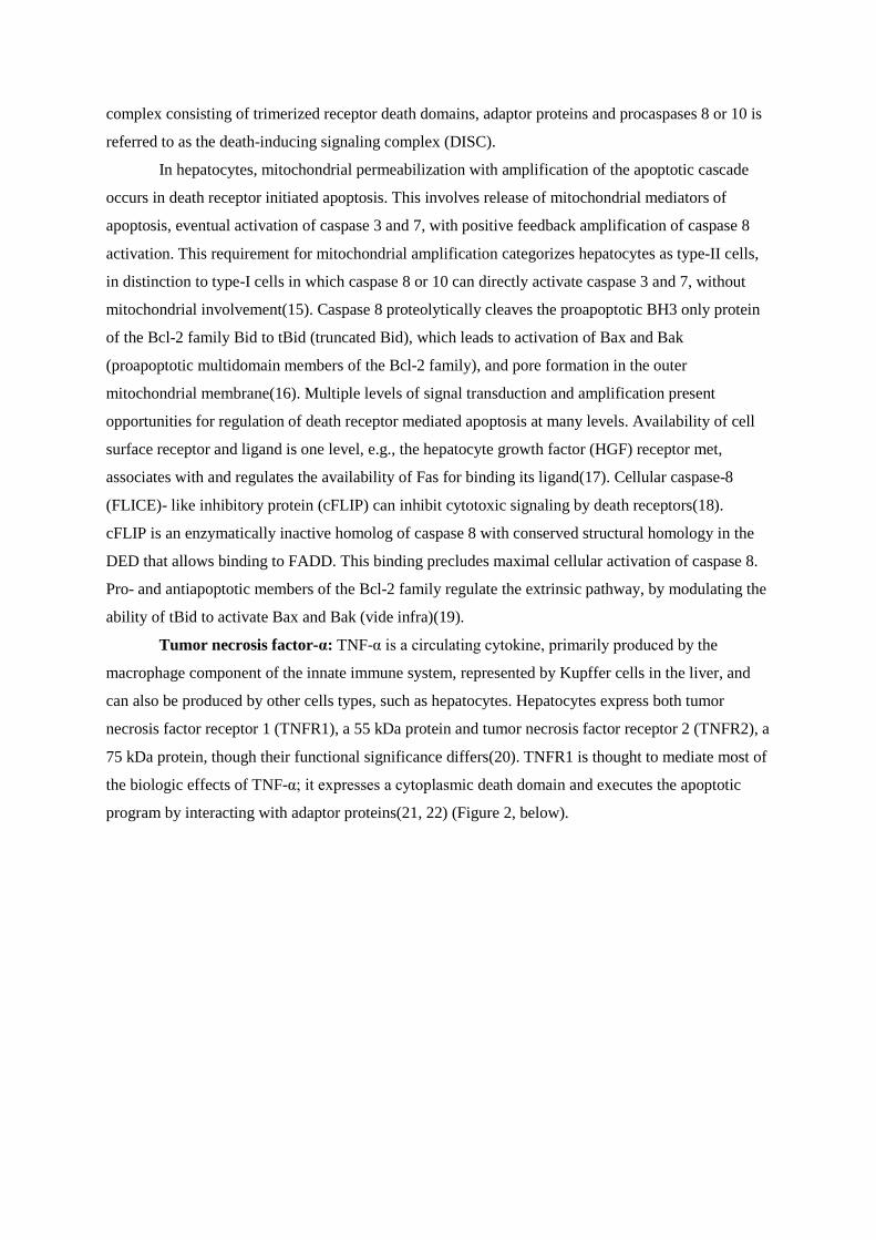
program by interacting with adaptor proteins(21, 22) (Figure 2, below).
Figure 2 Complex I and complex II of Tumor necrosis factor-alpha: Tumor necrosis factor receptor 1 (TNFR1), upon
binding TNF-α on its extracellular domain activates complex I and complex II. Complex I is formed by the adaptor proteins,
TNFR1- associated death domain protein (TRADD) and receptor interacting protein (RIP), which recognize and bind via
their death domains (DD) and TNF receptor associated factor (TRAF2) via its kinase domain or an intermediate domain.
Complex I mediates the activation of nuclear factor κ B (NF-κB) and transient c-jun N-terminal kinase (JNK) activation. NF-
κB translocates to the nucleus transcriptionally activating antiapoptotic and inflammatory genes, such as, cellular FLICE like
inhibitory protein (cFLIP), Bcl-xL, Mcl-1, A1 and XIAP, which regulate apoptosis at multiple levels. Sustained JNK
activation requires the adaptor protein RIP and is mediated in part by oxidative stress. Complex II is formed by receptor
dissociation of TRADD, RIP and TRAF2 and ligand independent recruitment of Fas associated death domain (FADD) via its
DD. FADD contains a death effector domain (DED) leading to recruitment and activation of procaspase 8.
On binding TNF-α, TNFR1 recruits the adaptor protein tumor necrosis factor receptor-associated
death domain (TRADD)(23). Signaling then proceeds in two steps, the first step or complex I involves
recruitment of tumor necrosis factor receptor associated protein (TRAF-2) and receptor interacting
protein 1 (RIP1) leading to activation of nuclear factor κB (NFκB) (24, 25). TRADD then dissociates
from the ligated receptor, recruits FADD and procaspase 8 to initiate apoptotic signaling; this
signaling pathway is referred to as complex-II. TRADD does not interact with TNFR2, nor does
FADD directly interact with TNFR1. Therefore, TNF-α-TNFR1 signaling first leads to NFκB
mediated transcriptional activation of prosurvival (e.g. Bcl-xL, A1, XIAP and cFLIP) and
proinflammatory genes (e.g. interleukin 6). In cells resistant to NFκB, or in the presence of a
transcriptional inhibitor (actinomycin D), the apoptotic effect of TNF-α is unmasked.
TNF-α has pleiotropic effects in vivo, including hepatocyte proliferation, liver inflammation
and modulation of hepatocyte apoptosis. In a murine model of TNF-α induced liver injury (TNF-α +
D-galactosamine), liver injury is Bax-dependent(26). TNF-α associated caspase 8 activation can also
cause lysosomal permeabilization with release of intralysosomal cathepsin B into the cytosol which
causes mitochondrial dysfunction(27). Mice deficient in cathepsin B are protected from the injurious
effects of TNF-α(28). C-jun N terminal kinase (JNK), a stress activated kinase, is activated by TNF-α.
Sustained activation of JNK can lead to apoptosis by modulation of the Bcl-2 family of proteins. JNK
can also transcriptionally activate death receptor expression, i.e. TRAIL-receptor 2/death receptor 5.
Furthermore, JNK can promote TNF-α induced apoptotic signaling at complex-II by facilitating
degradation of cFLIP, thus antagonizing an antiapoptotic TNF-α induced NFκB target gene(29).
Similarly, loss of cellular inhibitors of apoptosis proteins 1 and 2, also antiapoptotic NFκB target
genes, sensitizes carcinoma cells to TNF-α mediated cytotoxicity(30). TNF-α can lead to superoxide
formation and caspase-independent cell death, by TRADD and RIP1 mediated activation of Nox1
NADPH oxidase leading to reactive oxygen species formation(31). This process is independent of
FADD, and caspase 8 activation. Thus, a multitude of complex processes contributes to TNF-α
cytotoxicity.
In experimental models of liver injury, a role for TNF-α cell death has been elucidated.
Following partial hepatectomy, massive hepatocyte cell death occurs after completion of cell cycle
progression, due to sustained TNF-α signaling, in mice lacking tissue inhibitor of metalloproteinase 3
(Timp3), a model characterized by abnormal chronically elevated TNF-α activity(32). In ethanol fed
mice, TNFR1 deficiency results in decreased hepatocyte apoptosis, serum alanine aminotransferase
levels (ALT) and inflammatory foci as compared to wild type ethanol fed mice(33); TNFR2 deficient
mice developed liver injury and apoptosis comparable to wild type controls(34). In ischemia
reperfusion injury mice lacking TNFR1 and treated with a pentoxyfylline, a pharmacologic TNF-α
inhibitor, liver injury and apoptosis are significantly reduced(35) Liver samples from patients with
alcoholic steatohepatitis or nonalcoholic steatohepatitis demonstrate enhanced TNFR1 expression(36).
Serum levels of TNFR1 in patients with alcoholic hepatitis are predictive of 3month survival(37).
Thus, the TNF-α cascade is activated in patients with many liver diseases, including fulminant hepatic
failure, alcoholic steatohepatitis, nonalcoholic steatohepatitis, chronic hepatitis C, and chronic
hepatitis B(36, 38-40); it is indeed a hallmark of inflammatory changes in these conditions and likely
contributes to hepatocyte apoptosis in vivo. Our understanding of why the TNF-R1 initiated NFκB
cell survival pathways fail in these diseases remain rudimentary.
Fas: Fas (also known as Apo-1, CD95) is ubiquitously expressed in the liver(41- 43).
Hepatocytes are exquisitely sensitive to Fas induced apoptosis, and exogenously administered Fas
agonistic antibody results in fulminant hepatic failure in mice (44). Fas signaling usually results in
hepatocytes apoptosis; although there are reports of Fas induced proliferation of T cells and
fibroblasts and a report describing Fas-mediated acceleration of liver regeneration after partial
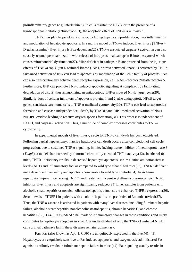
hepatectomy in mice(45, 46). Fas-Fas ligand (FasL) binding leads to receptor oligomerization,
bringing together the intracellular DD, recruitment of FADD and procaspase 8 or 10 at the DISC
(Figure 3, below).
Figure 3 Fas and TRAIL receptor signaling: Fas and TRAIL receptors are activated by ligand binding, which leads to receptor oligomerization, bringing together their conserved death domains (DD). The adaptor protein Fas associated death domain (FADD) binds to the trimerized intracellular death domain (DD) and via its death effector domain (DED) leads to activation of procaspase 8. Active caspase 8 leads to proteolytic cleavage of Bid to tBid and downstream mitochondrial permeabilization via activation of Bax and Bak. Mitochondrial permeabilization leads to release of the contents of the intermembrane space including cytochrome c, smac/DIABLO, Apaf 1 and endonuclease G culminating in the activation of caspase 3/7 and cleavage of cellular proteins.
This leads to activation and autoproteolytic activation of procaspase 8 or 10, generation of tBid,
activation of Bax and Bak, mitochondrial permeabilization with eventual activation of caspase 3 and
7. Fas can be activated by soluble or circulating as well as membrane bound FasL. Fas ligand is
expressed by cells of the immune system, such as cytotoxic T lymphocytes (CTL) and Natural Killer
(NK) cells(47). The liver is enriched in both these cell populations, therefore under constant “Fas-
attack”. However Fas induced signaling is regulated at many levels. Cell surface expression of Fas,
levels of FasL, and cFLIP inhibition of caspase 8 activation at the DISC are potential regulatory
sites. Of interest in hepatocytes is the sequestration of Fas by the hepatocyte growth factor receptor
(HGF), Met(17). Met-Fas complexes prevent binding of FasL to Fas; however, Fas does not affect
HGF binding to its receptor Met. Pretreatment of cells with HGF releases Fas from this complex, and
enhances FasL binding and toxicity at lower concentrations of FasL; High concentrations of FasL are
maximally toxic even in the absence of HGF. Thus, the Met-Fas complex fine tunes and regulates the
biologic availability of Fas in hepatocytes. In embryonic hepatocytes, Met prevents Fas induced
cFLIP degradation, thus preventing apoptosis(48).
In adult mice, genetic deficiency of Fas leads to hepatic hyperplasia, in addition to
enlargement of lymphnodes and spleen (49). The induction of fulminant hepatic failure in mice by
exogenous administration of Fas agonistic antibody is further regulated by the Bcl-2 family of
proteins. It can be abrogated by overexpression of Bcl-2 and enhanced by genetic inhibition of Bcl-xL
(50, 51). Genetic inhibition of Fas itself or Bid mitigates liver injury by Fas agonists(51, 52).
Circulating levels of serum Fas are elevated in patients with fulminant hepatic failure(5, 53).
Levels of serum Fas vary by etiology, and the highest levels occur in patients with drug induced liver
injury. Fas expression and apoptosis are enhanced in liver samples from patients with chronic
hepatitis C(54). Circulating levels of soluble Fas correlate with histologic activity, and along with
levels of caspase 3 activity, are predictive of response to therapy(55-57). Similarly in patients with
chronic hepatitis B hepatocyte Fas levels and circulating levels of sFas are elevated(54, 58, 59). Fas
expression is enhanced in liver samples from patients with nonalcoholic fatty liver disease(7). In
experimental models of dietary and genetic fatty liver, steatotic livers are sensitized to exogenous Fas
administration. Indeed, in patients with nonalcoholic fatty liver disease, the inhibition of Fas by Met is
diminished, providing another mechanism to explain the enhanced sensitivity to Fas induced
hepatocyte apoptosis(60). Furthermore, free fatty acid treatment can increase Fas expression in vitro,
in cell culture models of hepatocyte steatosis, sensitizing cells to Fas-induced apoptosis. In the bile
duct ligated mouse model of cholestatic liver injury hepatocyte apoptosis is mediated by Fas, and Fas
induced apoptosis promotes hepatic fibrosis(61, 62). Toxic bile acids promote cell surface expression
of Fas, and can lead to ligand-independent Fas oligomerization and induction of hepatocyte
apoptosis(63, 64). In bile salt mediated ligand-independent hepatocyte apoptosis Fas phosphorylation
is required for its translocation to the cell surface; this can occur in a Yes kinase, epidermal growth
factor receptor-dependent, and JNK-dependent manner(65, 66).
Tumor necrosis factor-related apoptosis inducing ligand (TRAIL): The role of tumor
necrosis factor-related apoptosis inducing ligand (TRAIL, also known as Apo-2 Ligand) and its
receptors in liver disease is an area with remarkable recent advances. TRAIL binds with several
receptors(67). TRAIL receptor 1 (TRAIL-R1/ Death receptor (DR) 4) and TRAIL receptor 2 (TRAIL-
R2/ DR 5/ Killer/ TRICK2) are complete receptors and can induce apoptosis via caspase activation,

similar to Fas(68). This occurs via the adaptor protein FADD, recruitment of procaspase 8 and 10 to
the TRAIL receptor DISC, in a cFLIP-regulated manner (Figure 3). TRAIL receptor 3 (TRAIL-R3/
Apo-3/ TRAMP/ WSL-1/LARD, Decoy receptor 1(DcR1)) and TRAIL receptor 4 (TRAIL-R4,
DR6, Decoy receptor 2(DcR2)) are incomplete cell surface receptors and cannot stimulate apoptotic
signaling. Normal human hepatocytes, in situ and in vivo, are considered resistant to TRAIL-induced
apoptosis, though there are occasional reports of in vitro TRAIL-induced hepatocyte apoptosis (69-
71). This resistance to cell death may be secondary to cFLIP induced inhibition of caspase 8
activation at the DISC or cell surface expression/availability of TRAIL-R1 or TRAIL-R2. However,
diseased hepatocytes are sensitized to TRAIL-induced apoptosis(72-75). TRAIL also sensitizes to Fas
induced hepatocyte apoptosis by activating JNK and the proapoptotic BH3 only protein Bim(76).
TRAIL-induced hepatocyte apoptosis has been demonstrated in cholestatic, viral and
metabolic liver diseases. Toxic bile acids transcriptionally regulate hepatocyte cell surface TRAIL-R2
expression in Fas deficient cells, and inactivate cFLIP by phosphorylation, thus dually sensitizing
cells to TRAIL-induced apoptosis(77, 78). In the bile duct ligated mouse model of cholestasis,
hepatocyte TRAIL-R2 expression is enhanced and hepatocytes are sensitized to exogenously
administered TRAIL(79). By corollary, liver injury and hepatocyte apoptosis are significantly reduced
in TRAIL deficient mice following bile duct ligation(80). Steatosis is also associated with increased
hepatocyte expression of TRAIL-R2 and TRAIL-R1 which imparts sensitivity to TRAIL toxicity(69).
Free fatty acids, which are elevated in the metabolic syndrome, transcriptionally enhance TRAIL-R2
expression in cell culture and render steatotic cells sensitive to TRAIL toxicity(75). In acute hepatitis
B-induced liver failure in humans and experimental adenoviral acute hepatitis in mice, TRAIL-R2
expression is enhanced, as is sensitivity to TRAIL. This occurs independently of Kupffer cells and
NK cells, suggesting a hepatocyte generated paracrine loop for elimination of virally infected
cells(72). Circulating soluble TRAIL levels are elevated in patients with chronic viral hepatitis B.
Hepatitis B x antigen increases TRAIL-R1 expression in cell culture experiments, conferring
sensitivity to TRAIL(81). In liver samples from patients with chronic hepatitis C, TRAIL-R1 and
TRAIL-R2 expression and TRAIL induced apoptosis were enhanced(69). Hepatitis C virus core
protein also selectively modulates cellular responsiveness to TRAIL by promoting TRAIL induced
Bid cleavage(82).
THE INTRINSIC PATHWAY
Intracellular stress leads to the activation of the intrinsic pathway of apoptosis. Stress can be
perceived and transduced by any membrane defined organelle in the cell. For example, lysosomes can
mediate steatotic liver cell death, as can the endoplasmic reticulum. DNA damage and steatosis can
activate c-jun N terminal kinase, also a mediator of the intrinsic pathway of apoptosis. These
processes converge on mitochondria and are transduced by the Bcl-2 family of proteins, therefore, are
usually referred to as the Bcl-2-regulated or mitochondrial pathway of apoptosis. The Bcl-2 family
consists of proapoptotic and antiapoptotic proteins. The proapoptotic proteins are structurally divided
based on the number of shared Bcl-2 homology (BH) domains, into multidomain (Bak and Bax,
display BH1,2, and 3 domains) and BH3 only proteins (Bid, Noxa, Puma, Bim, Bmf, Bik, Hrk and
Bad). The antiapoptotic proteins include Bcl-2, Bcl-xL, Bcl-w, A1, Mcl-1 and Boo, share 3 (Mcl-1) or
4 BH domains. The liver expresses Bcl-xL and Mcl-1; Bcl-2 is not expressed by hepatocytes. Bax and
Bak are both expressed by hepatocytes. The large number of BH-3 domain only proteins, while may
impart redundancy, primarily imparts stimulus specificity. For example free fatty acids activate
Bim(83); Puma and Noxa are target genes of the tumor suppressor p53(84). The antiapoptotic
members of this family are located on the cytoplasmic aspect of membrane bound organelles. They
protect cells from death, and may be necessary for survival of certain cell types. Bax and Bak are
required from mitochondrial permeabilization, while Bax is located in the cytosol and translocates to
mitochondria upon activation; Bak is a resident mitochondrial membrane protein. The activation of
Bax and Bak is regulated by interactions between the antiapoptotic Bcl-2 proteins and the BH-3
domain only proapoptotic proteins. Several models have been proposed to explain the biochemical
activation of Bax or Bak by proapoptotic BH-3 only proteins. Using Bim as an example, upon
activation, Bim is released from the dynein motor complex, and can directly engage and activate Bax
and Bak. Alternatively, Bim can bind and negate the inhibitory effect of Bcl-2 or Bcl-xL, releasing
Bax and Bak from inhibition by these proteins (the derepression model).
Mitochondria: In addition to the metabolic functions of mitochondria, hepatocytes require
mitochondria to die. The mitochondrial intermembrane space sequesters a number of proapoptotic
proteins including cytochrome c, SMAC/DIABLO (second mitochondrial activator of caspase/direct
IAP binding protein with low pI), HtrA2/Omi, AIF (apoptosis inducing factor), and endonuclease
G(4, 19). Active Bax or Bak form pores in the outer mitochondrial membrane leading to
mitochondrial outer membrane permeabilization (MOMP) and release of these mediators into the
cytosol. MOMP can also occur secondary to the permeability transition pore, a complex of adenine
nucleotide transporter (ANT) on the inner mitochondrial membrane, voltage dependent anion channel
(VDAC) on the outer mitochondrial membrane, and cyclophilin D located within the mitochondrial
matrix. Opening of the permeability transition pore leads to rapid fluxes of ions and water, dissipation
of the mitochondrial inner transmembrane potential, swelling of the mitochondria, outer
mitochondrial membrane rupture leading to the release of the contents on the intermembrane space.
Recent studies have demonstrated that stimuli leading to mitochondrial permeability transition require
cyclophilin D and that this can occur independently of ANT(85, 86). However, in mice and isolated
liver mitochondria lacking cyclophilin D, stimulus-specific MOMP occurs via engagement and
activation of Bax or tBid(86), which could also be the case in intact hepatocytes, given the richness of
death receptor expression and sensitivity to death ligands.
MOMP releases intermembrane contents into the cytosol and commits the cell to apoptosis.
SMAC inactivates post-mitochondrial inhibitors of apoptosis proteins (IAP). Cytosolic cytochrome c,
apoptotic protease activating factor-1 (Apaf) and ATP form a complex called the apoptosome, leading

to activation of procaspase 9 and effector caspases 3 and 7(87). These effector caspases cleave over
500 substrates resulting in cellular demolition. Cytokeratin 18 is a structural protein expressed in most
epithelial cells that is cleaved by caspase 3 at aspartate positions 238 and 396. The fragment generated
by this cleavage, cytokeratin 18-aspartate 396 (CK18-asp396) forms a neoepitope that is recognized
by the M30 antibody. This neoepitope can be detected in apoptotic tissues as well as serum by a
commercially available ELISA. Indeed circulating levels of CK18-asp396 are elevated in patients
with liver injury and can correlate with outcome(5). Thus this biomarker presents a noninvasive,
simple and mechanistic tool to monitor progress, and response to therapy in liver injury.
Lysosomes: Lysosomes are intracellular organelles with acid intravesicular pH that contain
lysosomal proteases, known as cathepsins(88). Cathepsin B and D, two of 11 known human
cathepsins, are stable and active at neutral pH. Methodical dissection of pathways that mediate
intracellular death signals, demonstrates that lysosomes can be involved in the intrinsic pathway of
cell death. Typically lysosomal permeabilization, when it mediates apoptosis, is selective and partial
and is observed upstream of mitochondrial permeabilization. Cathepsin B induced mitochondrial
permeabilization can occur via caspase 2 (in mice) and via proteolytic cleavage of Bid similar to death
receptor induced activation of Bid(89, 90). Indeed Bid also links death receptors to lysosomal
permeabilization; providing cross talk between death receptors and their engagement of the lysosomal
and mitochondrial pathways(89, 91). Bax activation by intracellular stress can also result in lysosomal
permeabilization(92). Cathepsin D levels were elevated in serum from patients with fulminant hepatic
failure as well as chronic hepatitis(93, 94). Cathepsin B deficient mice are resistant to TNF-α induced
hepatocyte apoptosis(28). In models of cellular steatosis, cathepsin B inhibition prevents
mitochondrial permeabilization and apoptosis(95). In cathepsin B deficient mice liver apoptosis,
injury and fibrosis are diminished following bile duct ligation(6); liver apoptosis and injury are
abrogated in ischemia reperfusion injury as well(96).
Endoplasmic Reticulum: The endoplasmic reticulum (ER) has an inbuilt mechanism to cope
with excess or altered unfolded proteins that serves to correct the inciting imbalance. This process is
termed the unfolded protein response (UPR). The UPR can also be activated by stimuli that affect the
function of the ER, such as calcium depletion, glycosylation inhibition (tunicamycin), ultraviolet
radiation and insulin resistance. The ER stress response consists of a series of compensatory processes
to correct both the excess and the stress of the unfolded proteins. Global translation is attenuated to
reduce the functional protein load of the ER. There is also selective translation of UPR target genes
aimed at protecting the ER(97, 98). The transducers of ER stress are membrane proteins that have an
ER lumenal domain and a cytosolic domain. Inositol-requiring protein-1 (IRE1) and protein kinase
RNA-like ER kinase (PERK) auto-transphosphorylate, when released from the ER chaperone
BiP/Grp78. IRE1 possesses endoribonucleolytic activity leading to excision of an intron within Xbox
binding protein-1 (XBP1) mRNA to generate spliced XBP1 (sXBP1), a transcription factor that
activates a subset of UPR target genes. IRE1 also recruits TRAF2 leading to JNK activation. PERK

phosphorylates and inactivates the eukaryotic translation initiation factor-2α (eIF2α), resulting in
global translation attenuation with selective translation of activating transcription factor-4 (ATF4)
which leads to transcription of C/EBP-homologous protein (CHOP), and the ER chaperone
BiP/Grp78. Activating transcription factor-6 (ATF6) is cleaved within the ER membrane, generating
an ATF6 fragment that translocates to the nucleus and activates a subset of UPR target genes. It is not
known if ATF6 also regulates apoptotic signaling.
ER stress also activates a negative feedback regulatory loop that terminates the UPR; however
in the setting of sustained ER stress pro-apoptotic signaling occurs(99). Bax and Bak, both bind to the
cytoplasmic domain of IRE1, and in cells lacking Bax and Bak, IRE1 stress generated JNK activation
and XBP1 splicing are reduced(100), thus linking the core apoptotic machinery to ER stress response.
Bax and Bak localize on the ER membrane, in addition to mitochondrial membranes. In cells lacking
both Bax and Bak, the ER is depleted of calcium and unable to respond to certain death stimuli(101).
The proapoptotic transcription factor CHOP can increase Bim expression, transcriptionally and by
inhibiting its proteasomal degradation, leading to Bim-dependent ER stress induced apoptosis(102).
CHOP can also upregulate TRAIL-R2 expression, sensitizing cancer cells to TRAIL induced
apoptosis(103).
The involvement of the ER stress-induced apoptotic pathway in liver diseases is an area of
emerging research. In the bile duct ligated mouse model of cholestasis, an early and transient
induction of CHOP expression is observed(104). Mice deficient in CHOP are protected from
hepatocyte apoptosis, liver injury and liver fibrosis. In cell culture, the toxic bile acid,
glycochenodeoxycholic acid, also induces ER stress and CHOP expression in isolated rat
hepatocytes(105). In transgenic mice expressing hepatitis C viral core and E2 proteins hepatocyte
apoptosis is associated with CHOP expression(106). Cycloheximide, an inhibitor of protein synthesis,
induces ER stress, induction of CHOP expression and apoptotic hepatocyte cell death, in rat
livers(107). In nonalcoholic fatty liver disease, markers of ER stress were variably activated(108).
Toxic saturated fatty acids also induce ER stress and apoptosis in liver cell lines(109, 110) In a mouse
model of alcohol induced liver injury CHOP deficient mice are protected from hepatocyte apoptosis,
though able to mount an ER stress response(111).
C-jun N-terminal Kinase: Given the role of c-jun N-terminal kinase (JNK) in multiple
models of cell death, it warrants a separate discussion as a final common cell death mediator. JNK 1
and 2 are ubiquitously expressed, including liver, whereas JNK 3 is not expressed in the liver(112).
JNK activation occurs downstream of kinase cascades that can be activated by multiple stimuli
including TNF-α, IRE1, reactive oxygen species, free fatty acids, bile acids(113-115). JNK
involvement in apoptosis is temporally regulated and stimulus specific(116). The same inciting
stimulus, e.g. TNF-α, can induce biphasic JNK activation mediated by distinct intracellular pathways.
Transient and early JNK activation promotes survival; and sustained and late activation of JNK
promotes apoptosis(117). In the case of TNF-α, production of reactive oxygen species mediates the
delayed and sustained activation of JNK. Other stimuli, e.g. toxic free fatty acids, result in early and
sustained JNK activation, culminating in apoptotic signaling(118). JNK stimulated proapoptotic
signaling converges on mitochondria via the activation of Bax and Bak. In the absence of Bax and
Bak, JNK induced cell death is mitigated(119). Furthermore, mitochondrial permeabilization and
release of cytochrome c are abolished in cells derived from mice lacking JNK 1 and 2 genes, in
response to stimuli that cause intracellular stress(116). JNK mediated phosphorylation of pro- and
anti-apoptotic proteins upstream of mitochondria also regulates apoptotic sensitivity. JNK can
phosphorylate and activate the BH-3 only proteins, e.g. Bim phosphorylation releases it
from binding to the dynein motor complex and promotes apoptosis(76, 120). Sustained JNK
activation promotes caspase 8 formation at the DISC by activation of the E3 ubiquitin ligase Itch,
which ubiquinates and degrades cFLIP promoting liver cell death(29). JNK can phosphorylate the
antiapoptotic proteins, Bcl-2, Bxl-xL and Mcl-1, and the proapoptotic proteins Bmf and Bad(120-
123).
JNK 1 or JNK 2 can both mediate liver injury in a stimulus specific manner. In a murine
model of steatohepatitis induced by methionine and choline deficient diet JNK1 plays a predominant
role(124). In high fat diet-induced obesity and genetic obesity in mice, JNK was activated, and was
predominantly JNK 1; though JNK 2 plays a role, that is unmasked in the absence of JNK 1(125,
126). In free fatty acid based cellular models of hepatocyte steatosis JNK2 is the predominant isoform
that mediates apoptosis(118). Oleic acid, a minimally toxic free fatty acids also sensitizes steatotic
hepatocytes to TRAIL-induced apoptosis by JNK dependent transcriptional upregulation of the death
receptor TRAIL-R2(75). This mechanism is shared by toxic bile acids, which too sensitize to TRAIL
induced apoptosis by transcriptionally activating TRAIL-R2 expression in a JNK-dependent
manner(77, 79). Liver injury induced by ischemia reperfusion is also mediated by JNK, and
pharmacologic inhibition of JNK in the donor livers improved graft survival and decreased apoptosis
after orthotopic liver transplantation (127, 128). In acetaminophen (APAP) induced acute liver injury
JNK activation was robust and sustained, led to Bax translocation to mitochondria and poor animal
survival. Pharmacologic inhibition of JNK decreased liver injury, hepatocyte cell death and improved
survival; utilizing genetically deficient models of JNK 1 or JNK 2, it was demonstrated that both
mediate liver injury, though JNK 2 was predominant(129). JNK activation was observed in
hepatocytes in human liver samples from patients with acetaminophen induced acute liver
failure(130). JNK inhibition was more effective in decreasing hepatocyte cell death than N-
acetylcysteine in a murine model of acetaminophen induced liver injury(130). In a murine model of
TNF-α induced liver injury utilizing galactosamine and lipopolysaccharide, JNK 2 mediated caspase 8
activation and mitochondrial permeabilization(131).
The consequences of hepatocyte apoptosis: Apoptosis, inflammation and injury are in some
ways inseparable, and it is difficult sometimes to dissect the primary event. However, based on the
inciting stimulus apoptosis or inflammatory signaling may be the primary event; each stimulating the
other (Figure 4, below).
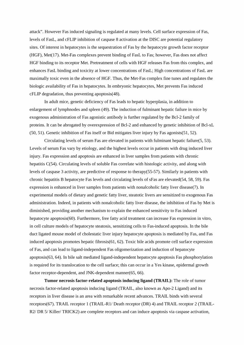
Figure 4 Apoptosis and its consequences: Hepatocyte apoptosis and liver inflammation are interconnected. Apoptosis of vulnerable hepatocytes results in apoptotic bodies that can be engulfed by Kupffer cells and stellate cells. This engulfment leads to Kupffer cell activation and secretion of TNF-α, interleukins and interferon, all of which promote the inflammatory response. With ongoing hepatocyte apoptosis, activated Kupffer cells also facilitate the activation of stellate cells by secreting transforming growth factor beta (TGFβ). Activated stellate cells lead to liver fibrosis by secreting collagen type 1. Inhibition of hepatocyte apoptosis or Kupffer cell depletion, both mitigate liver injury, inflammation and fibrosis. Activated stellate cells are sensitized to apoptosis, such as with TRAIL, and this leads to resolution of fibrosis.
The liver has a large population of Kupffer cells, NK cells and NK T cells(132). These cells
are a ready source of TNF-α and other cytokines that mediate inflammation, Fas, TRAIL and TNF-α
that mediate hepatocyte apoptosis, and transforming growth factor-beta (TGF β) that activates stellate
cells. Apoptotic hepatocytes can be engulfed by Kupffer cells leading to generation of cytokines;
pharmacologic inhibition of apoptosis prevents Kupffer cell activation. Also, in the bile duct ligated
mouse, Kupffer cell depletion decreases hepatocyte apoptosis, liver injury and liver
inflammation(133). In addition, stressed hepatocytes increase expression of NKG2D ligands; thus
inviting NK and NKT cell mediated destruction(134).
Fibrosis is the hallmark of ongoing liver injury. Hepatic stellate cells mediate hepatic fibrosis.
In the normal liver, stellate cells maintain a quiescent phenotype. On activation, they undergo a
metamorphosis, to become myofibroblasts, secreting collagen which leads to liver fibrosis. Stellate

cells in vitro can engulf apoptotic hepatocytes, leading to their activation, and increased expression of
TGF β, alpha smooth muscle actin and collagen alpha1(135). Similarly, in vivo hepatocyte apoptosis
is a fibrogenic stimulus. Several experimental studies have demonstrated that the inhibition of
hepatocyte apoptosis abrogates liver fibrosis(6, 62, 136). By corollary, apoptosis of activated stellate
cells should decrease liver fibrosis and dissociate ongoing hepatocyte apoptosis from the ensuing
fibrogenic response. Indeed, activated stellate cells are sensitized to apoptotic signaling. This can be
achieved by inhibition of NFκB, TRAIL mediated stellate cell apoptosis, and NK cell mediated
stellate cell apoptosis(137-139). Indeed, the resolution phase of fibrosis requires apoptosis of
activated hepatic stellate cells(140).
The clinical applications of apoptosis are discussed in the conclusion of this chapter. The
cytokeratin 18 derived M30 neoantigen reflects epithelial cell apoptosis, is abundant in hepatocytes,
can easily be measured in serum by a commercially available ELISA and correlates with hepatocyte
apoptosis in diverse liver diseases(141). In a study with a small number of patients with chronic
hepatitis C, pre-treatment M30 levels were predictive of response to therapy(57), inferring from this
that patients with an apoptotic response to virally infected hepatocytes are more likely to have a
treatment response. In another study with chronic hepatitis C patients with normal transaminases,
serum M30 levels correlated with fibrosis(57). In patients with nonalcoholic fatty liver disease, serum
M30 levels offer reliable discrimination of patients with steatohepatitis from simple steatosis, and
increasing levels are predictive of a higher likelihood if inflammation(142). Caspase inhibitors have
demonstrated efficacy in preventing hepatocyte apoptosis and injury in experimental models of liver
injury(136, 143). In patients with chronic hepatitis C, orally administered caspase inhibitor was found
to be safe, and lowered transaminases(144).
In conclusion, hepatocyte apoptosis is a key mediator of liver injury and inflammation in most
forms of liver disease. Multiple apoptotic pathways are activated by a given injurious stimulus in a
vulnerable hepatocyte. The predominant signalling pathway that results in mitochondrial dysfunction
in a given cell is difficult to discern; however, multiple pathways could potentially cooperate or
oppose each other, to eventually result in mitochondrial permeabilization. Once mitochondrial
permeabilization occurs, the hepatocyte is committed to cell death. Evidence of hepatocyte apoptosis
can be demonstrated by serum markers and early studies demonstrate prognostic significance of
apoptosis markers. Lastly, therapeutic manipulation of apoptosis is of benefit, by preventing liver
injury and fibrosis.
Acknowledgement: The authors would like to acknowledge the excellent secretarial support of Ms. Erin Nystuen-Bungum. References: 1. Kroemer G, El-Deiry WS, Golstein P, et al. Classification of cell death: recommendations of the Nomenclature Committee on Cell Death. Cell Death Differ. 2005;12 Suppl 2:1463-1467.
2. Vieira WT, Gayotto LC, de Lima CP, et al. Histopathology of the human liver in yellow fever with special emphasis on the diagnostic role of the Councilman body. Histopathology. 1983;7:195-208. 3. Malhi H, Gores GJ, Lemasters JJ. Apoptosis and necrosis in the liver: a tale of two deaths? Hepatology. 2006;43:S31-44. 4. Green DR, Kroemer G. The pathophysiology of mitochondrial cell death. Science 2004;305:626-629. 5. Rutherford AE, Hynan LS, Borges CB, et al. Serum Apoptosis Markers in Acute Liver Failure: A Pilot Study. Clin Gastroenterol Hepatol. 2007. 6. Canbay A, Guicciardi ME, Higuchi H, et al. Cathepsin B inactivation attenuates hepatic injury and fibrosis during cholestasis. J Clin Invest. 2003;112:152-159. 7. Feldstein AE, Canbay A, Angulo P, et al. Hepatocyte apoptosis and fas expression are prominent features of human nonalcoholic steatohepatitis. Gastroenterology. 2003;125:437-443. 8. Natori S, Rust C, Stadheim LM, et al. Hepatocyte apoptosis is a pathologic feature of human alcoholic hepatitis. J Hepatol. 2001;34:248-253. 9. Papakyriakou P, Tzardi M, Valatas V, et al. Apoptosis and apoptosis related proteins in chronic viral liver disease. Apoptosis. 2002;7:133-141. 10. Natori S, Selzner M, Valentino KL, et al. Apoptosis of sinusoidal endothelial cells occurs during liver preservation injury by a caspase-dependent mechanism. Transplantation. 1999;68:89-96. 11. Kohli V, Selzner M, Madden JF, et al. Endothelial cell and hepatocyte deaths occur by apoptosis after ischemia-reperfusion injury in the rat liver. Transplantation. 1999;67:1099-1105. 12. Guicciardi ME, Gores GJ. The Death Receptor Family and the Extrinsic Pathway. In: Yin X-M, Dong Z, eds. Essentials of Apoptosis: A guide for basic and clinical research. Totowa: Humana Press Inc; 2003:67-84. 13. Strasser A, Newton K. FADD/MORT1, a signal transducer that can promote cell death or cell growth. Int J Biochem Cell Biol. 1999;31:533-537. 14. Boatright KM, Salvesen GS. Mechanisms of caspase activation. Curr Opin Cell Biol. 2003;15:725-731. 15. Scaffidi C, Fulda S, Srinivasan A, et al. Two CD95 (APO-1/Fas) signaling pathways. Embo J. 1998;17:1675-1687. 16. Yin XM. Bid, a critical mediator for apoptosis induced by the activation of Fas/TNF-R1 death receptors in hepatocytes. J Mol Med. 2000;78:203-211. 17. Wang X, DeFrances MC, Dai Y, et al. A mechanism of cell survival: sequestration of Fas by the HGF receptor Met. Molecular Cell. 2002;9:411-421. 18. Budd RC, Yeh WC, Tschopp J. cFLIP regulation of lymphocyte activation and development. Nat Rev Immunol. 2006;6:196-204. 19. Danial NN, Korsmeyer SJ. Cell death: critical control points. Cell. 2004;116:205-219. 20. Yamada Y, Webber EM, Kirillova I, et al. Analysis of liver regeneration in mice lacking type 1 or type 2 tumor necrosis factor receptor: requirement for type 1 but not type 2 receptor. Hepatology 1998;28:959-970. 21. Tartaglia LA, Ayres TM, Wong GH, et al. A novel domain within the 55 kd TNF receptor signals cell death. Cell. 1993;74:845-853. 22. Hsu H, Xiong J, Goeddel DV. The TNF receptor 1-associated protein TRADD signals cell death and NF-kappa B activation. Cell. 1995;81:495-504. 23. Hsu H, Shu HB, Pan MG, et al. TRADD-TRAF2 and TRADD-FADD interactions define two distinct TNF receptor 1 signal transduction pathways. Cell. 1996;84:299-308. 24. Micheau O, Tschopp J. Induction of TNF receptor I-mediated apoptosis via two sequential signaling complexes. Cell. 2003;114:181-190. 25. Barnhart BC, Peter ME. The TNF receptor 1: a split personality complex. Cell. 2003;114:148-150. 26. Sass G, Shembade ND, Haimerl F, et al. TNF pretreatment interferes with mitochondrial apoptosis in the mouse liver by A20-mediated down-regulation of Bax. J Immunol. 2007;179:7042-7049. 27. Guicciardi ME, Deussing J, Miyoshi H, et al. Cathepsin B contributes to TNF-alpha-mediated hepatocyte apoptosis by promoting mitochondrial release of cytochrome c. J Clin Invest. 2000;106:1127-1137. 28. Guicciardi ME, Miyoshi H, Bronk SF, et al. Cathepsin B knockout mice are resistant to tumor necrosis factor-alpha-mediated hepatocyte apoptosis and liver injury: implications for therapeutic applications. Am J Pathol. 2001;159:2045-2054. 29. Chang L, Kamata H, Solinas G, et al. The E3 ubiquitin ligase itch couples JNK activation to TNF-alpha-induced cell death by inducing c-FLIP(L) turnover. Cell. 2006;124:601-613. 30. Varfolomeev E, Blankenship JW, Wayson SM, et al. IAP Antagonists Induce Autoubiquitination of c-IAPs, NF-kappaB Activation, and TNFalpha-Dependent Apoptosis. Cell. 2007;131:669-681. 31. Kim YS, Morgan MJ, Choksi S, et al. TNF-induced activation of the Nox1 NADPH oxidase and its role in the induction of necrotic cell death. Molecular cell. 2007;26:675-687. 32. Mohammed FF, Smookler DS, Taylor SE, et al. Abnormal TNF activity in Timp3-/- mice leads to chronic hepatic inflammation and failure of liver regeneration. Nature genetics. 2004;36:969-977.

33. Ji C, Deng Q, Kaplowitz N. Role of TNF-alpha in ethanol-induced hyperhomocysteinemia and murine alcoholic liver injury. Hepatology 2004;40:442-451. 34. Yin M, Wheeler MD, Kono H, et al. Essential role of tumor necrosis factor alpha in alcohol induced liver injury in mice. Gastroenterology. 1999;117:942-952. 35. Rudiger HA, Clavien PA. Tumor necrosis factor alpha, but not Fas, mediates hepatocellular apoptosis in the murine ischemic liver. Gastroenterology. 2002;122:202-210. 36. Ribeiro PS, Cortez-Pinto H, Sola S, et al. Hepatocyte apoptosis, expression of death receptors, and activation of NF-kappaB in the liver of nonalcoholic and alcoholic steatohepatitis patients. Am J Gastroenterol. 2004;99:1708-1717. 37. Spahr L, Giostra E, Frossard JL, et al. Soluble TNF-R1, but not tumor necrosis factor alpha, predicts the 3-month mortality in patients with alcoholic hepatitis. J Hepatol. 2004;41:229-234. 38. Muto Y, Nouri-Aria KT, Meager A, et al. Enhanced tumour necrosis factor and interleukin-1 in fulminant hepatic failure. Lancet. 1988;2:72-74. 39. Torre F, Rossol S, Pelli N, et al. Kinetics of soluble tumour necrosis factor (TNF)-alpha receptors and cytokines in the early phase of treatment for chronic hepatitis C: comparison between interferon (IFN)-alpha alone, IFN-alpha plus amantadine or plus ribavirin. Clin Exp Immunol. 2004;136:507-512. 40. Fang JW, Shen WW, Meager A, et al. Activation of the tumor necrosis factor-alpha system in the liver in chronic hepatitis B virus infection. Am J Gastroenterol. 1996;91:748-753. 41. Muschen M, Warskulat U, Douillard P, et al. Regulation of CD95 (APO-1/Fas) receptor and ligand expression by lipopolysaccharide and dexamethasone in parenchymal and nonparenchymal rat liver cells. Hepatology 1998;27:200-208. 42. Cardier JE, Schulte T, Kammer H, et al. Fas (CD95, APO-1) antigen expression and function in murine liver endothelial cells: implications for the regulation of apoptosis in liver endothelial cells. Faseb J. 1999;13:1950-1960. 43. Ueno Y, Ishii M, Yahagi K, et al. Fas-mediated cholangiopathy in the murine model of graft versus host disease. Hepatology 2000;31:966-974. 44. Ogasawara J, Watanabe-Fukunaga R, Adachi M, et al. Lethal effect of the anti-Fas antibody in mice. Nature. 1993;364:806-809. 45. Budd RC. Death receptors couple to both cell proliferation and apoptosis. J Clin Invest. 2002;109:437-441. 46. Desbarats J, Newell MK. Fas engagement accelerates liver regeneration after partial hepatectomy. Nature Medicine. 2000;6:920-923. 47. Berke G. The CTL's kiss of death. Cell. 1995;81:9-12. 48. Moumen A, Ieraci A, Patane S, et al. Met signals hepatocyte survival by preventing Fas-triggered FLIP degradation in a PI3k-Akt-dependent manner. Hepatology 2007;45:1210- 1217. 49. Adachi M, Suematsu S, Kondo T, et al. Targeted mutation in the Fas gene causes hyperplasia in peripheral lymphoid organs and liver. Nature genetics. 1995;11:294-300. 50. Lacronique V, Mignon A, Fabre M, et al. Bcl-2 protects from lethal hepatic apoptosis induced by an anti-Fas antibody in mice. Nature Medicine. 1996;2:80-86. 51. Zhang H, Taylor J, Luther D, et al. Antisense oligonucleotide inhibition of Bcl-xL and Bid expression in liver regulates responses in a mouse model of Fas-induced fulminant hepatitis. The Journal of Pharmacology and Experimental Therapeutics. 2003;307:24-33. 52. Yin XM, Wang K, Gross A, et al. Bid-deficient mice are resistant to Fas-induced hepatocellular apoptosis. Nature. 1999;400:886-891. 53. Ryo K, Kamogawa Y, Ikeda I, et al. Significance of Fas antigen-mediated apoptosis in human fulminant hepatic failure. Am J Gastroenterol. 2000;95:2047-2055. 54. Kiyici M, Gurel S, Budak F, et al. Fas antigen (CD95) expression and apoptosis in hepatocytes of patients with chronic viral hepatitis. Eur J Gastroenterol Hepatol. 2003;15:1079-1084. 55. Hiramatsu N, Hayashi N, Katayama K, et al. Immunohistochemical detection of Fas antigen in liver tissue of patients with chronic hepatitis C. Hepatology 1994;19:1354-1359. 56. Toyoda M, Kakizaki S, Horiguchi N, et al. Role of serum soluble Fas/soluble Fas ligand and TNFalpha on response to interferon-alpha therapy in chronic hepatitis C. Liver. 2000;20:305-311. 57. Volkmann X, Cornberg M, Wedemeyer H, et al. Caspase activation is required for antiviral treatment response in chronic hepatitis C virus infection. Hepatology (Baltimore, MD) 2006;43:1311-1316. 58. Mochizuki K, Hayashi N, Hiramatsu N, et al. Fas antigen expression in liver tissues of patients with chronic hepatitis B. J Hepatol. 1996;24:1-7. 59. Song le H, Binh VQ, Duy DN, et al. Variations in the serum concentrations of soluble Fas and soluble Fas ligand in Vietnamese patients infected with hepatitis B virus. J Med Virol. 2004;73:244-249. 60. Zou C, Ma J, Wang X, et al. Lack of Fas antagonism by Met in human fatty liver disease. Nature medicine. 2007;13:1078-1085.
61. Miyoshi H, Rust C, Roberts PJ, et al. Hepatocyte apoptosis after bile duct ligation in the mouse involves Fas. Gastroenterology. 1999;117:669-677. 62. Canbay A, Higuchi H, Bronk SF, et al. Fas enhances fibrogenesis in the bile duct ligated mouse: a link between apoptosis and fibrosis. Gastroenterology. 2002;123:1323-1330. 63. Sodeman T, Bronk SF, Roberts PJ, et al. Bile salts mediate hepatocyte apoptosis by increasing cell surface trafficking of Fas. Am J Physiol Gastrointest Liver Physiol. 2000;278:G992-999. 64. Faubion WA, Guicciardi ME, Miyoshi H, et al. Toxic bile salts induce rodent hepatocyte apoptosis via direct activation of Fas. J Clin Invest. 1999;103:137-145. 65. Eberle A, Reinehr R, Becker S, et al. CD95 tyrosine phosphorylation is required for CD95 oligomerization. Apoptosis. 2007;12:719-729. 66. Reinehr R, Becker S, Wettstein M, et al. Involvement of the Src family kinase yes in bile salt induced apoptosis. Gastroenterology. 2004;127:1540-1557. 67. Kimberley FC, Screaton GR. Following a TRAIL: update on a ligand and its five receptors. Cell Res. 2004;14:359-372. 68. Schneider P, Thome M, Burns K, et al. TRAIL receptors 1 (DR4) and 2 (DR5) signal FADD dependent apoptosis and activate NF-kappaB. Immunity. 1997;7:831-836. 69. Volkmann X, Fischer U, Bahr MJ, et al. Increased hepatotoxicity of tumor necrosis factor-related apoptosis-inducing ligand in diseased human liver. Hepatology 2007;46:1498- 1508. 70. Malhi H. TRAILs and tribulation. Hepatology 2007;46:1320-1322. 71. Mori E, Thomas M, Motoki K, et al. Human normal hepatocytes are susceptible to apoptosis signal mediated by both TRAIL-R1 and TRAIL-R2. Cell Death Differ. 2004;11:203-207. 72. Mundt B, Kuhnel F, Zender L, et al. Involvement of TRAIL and its receptors in viral hepatitis. Faseb J. 2003;17:94-96. 73. Zheng SJ, Wang P, Tsabary G, et al. Critical roles of TRAIL in hepatic cell death and hepatic inflammation. J Clin Invest. 2004;113:58-64. 74. Dunn C, Brunetto M, Reynolds G, et al. Cytokines induced during chronic hepatitis B virus infection promote a pathway for NK cell-mediated liver damage. J Exp Med. 2007;204:667-680. 75. Malhi H, Barreyro FJ, Isomoto H, et al. Free fatty acids sensitise hepatocytes to TRAIL mediated cytotoxicity. Gut. 2007;56:1124-1131. 76. Corazza N, Jakob S, Schaer C, et al. TRAIL receptor-mediated JNK activation and Bim phosphorylation critically regulate Fas-mediated liver damage and lethality. J Clin Invest. 2006;116:2493-2499. 77. Higuchi H, Bronk SF, Takikawa Y, et al. The bile acid glycochenodeoxycholate induces trailreceptor 2/DR5 expression and apoptosis. J Biol Chem. 2001;276:38610-38618. 78. Higuchi H, Yoon JH, Grambihler A, et al. Bile acids stimulate cFLIP phosphorylation enhancing TRAIL-mediated apoptosis. J Biol Chem. 2003;278:454-461. 79. Higuchi H, Bronk SF, Taniai M, et al. Cholestasis increases tumor necrosis factor-related apoptotis-inducing ligand (TRAIL)-R2/DR5 expression and sensitizes the liver to TRAIL mediated cytotoxicity. The Journal of Pharmacology and Experimental Therapeutics. 2002;303:461-467. 80. Kahraman A, Barreyro FJ, Bronk SF, et al. TRAIL mediates liver injury by the innate immune system in the bile duct-ligated mouse. Hepatology 2008. 81. Janssen HL, Higuchi H, Abdulkarim A, et al. Hepatitis B virus enhances tumor necrosis factor related apoptosis-inducing ligand (TRAIL) cytotoxicity by increasing TRAIL-R1/death receptor 4 expression. J Hepatol. 2003;39:414-420. 82. Chou AH, Tsai HF, Wu YY, et al. Hepatitis C virus core protein modulates TRAIL-mediated apoptosis by enhancing Bid cleavage and activation of mitochondria apoptosis signaling pathway. J Immunol. 2005;174:2160-2166. 83. Barreyro FJ, Kobayashi S, Bronk SF, et al. Transcriptional regulation of Bim by FoxO3A mediates hepatocyte lipoapoptosis. J Biol Chem. 2007;282:27141-27154. 84. Yu J, Zhang L. The transcriptional targets of p53 in apoptosis control. Biochemical and biophysical research communications. 2005;331:851-858. 85. Kokoszka JE, Waymire KG, Levy SE, et al. The ADP/ATP translocator is not essential for the mitochondrial permeability transition pore. Nature. 2004;427:461-465. 86. Baines CP, Kaiser RA, Purcell NH, et al. Loss of cyclophilin D reveals a critical role for mitochondrial permeability transition in cell death. Nature. 2005;434:658-662. 87. Riedl SJ, Salvesen GS. The apoptosome: signalling platform of cell death. Nat Rev Mol Cell Biol. 2007;8:405-413. 88. Guicciardi ME, Leist M, Gores GJ. Lysosomes in cell death. Oncogene. 2004;23:2881-2890. 89. Guicciardi ME, Bronk SF, Werneburg NW, et al. Bid is upstream of lysosome-mediated caspase 2 activation in tumor necrosis factor alpha-induced hepatocyte apoptosis. Gastroenterology. 2005;129:269-284.

90. Stoka V, Turk B, Schendel SL, et al. Lysosomal protease pathways to apoptosis. Cleavage of bid, not pro-caspases, is the most likely route. J Biol Chem. 2001;276:3149-3157. 91. Werneburg NW, Guicciardi ME, Bronk SF, et al. Tumor necrosis factor-related apoptosis inducing ligand activates a lysosomal pathway of apoptosis that is regulated by Bcl-2 proteins. J Biol Chem. 2007;282:28960-28970. 92. Feldstein AE, Werneburg NW, Li Z, et al. Bax inhibition protects against free fatty acid-induced lysosomal permeabilization. Am J Physiol Gastrointest Liver Physiol. 2006;290:G1339-1346. 93. Gove CD, Wardle EN, Williams R. Circulating lysosomal enzymes and acute hepatic necrosis. J Clin Pathol. 1981;34:13-16. 94. Kyaw A, Aung T, Htut T, et al. Lysosomal enzyme activities in normals and in patients with chronic liver diseases. Clin Chim Acta. 1983;131:317-323. 95. Li Z, Berk M, McIntyre TM, et al. The lysosomal-mitochondrial axis in free fatty acid-induced hepatic lipotoxicity. Hepatology 2007. 96. Baskin-Bey ES, Canbay A, Bronk SF, et al. Cathepsin B inactivation attenuates hepatocyte apoptosis and liver damage in steatotic livers after cold ischemia-warm reperfusion injury. Am J Physiol Gastrointest Liver Physiol. 2005;288:G396-402. 97. Ron D, Walter P. Signal integration in the endoplasmic reticulum unfolded protein response. Nat Rev Mol Cell Biol. 2007;8:519-529. 98. Ji C, Kaplowitz N. ER stress: can the liver cope? J Hepatol. 2006;45:321-333. 99. Lin JH, Li H, Yasumura D, et al. IRE1 signaling affects cell fate during the unfolded protein response. Science New York, N.Y. 2007;318:944-949. 100. Hetz C, Bernasconi P, Fisher J, et al. Proapoptotic BAX and BAK modulate the unfolded protein response by a direct interaction with IRE1alpha. Science New York, N.Y. 2006;312:572-576. 101. Scorrano L, Oakes SA, Opferman JT, et al. BAX and BAK regulation of endoplasmic reticulum Ca2+: a control point for apoptosis. Science 2003;300:135-139. 102. Puthalakath H, O'Reilly LA, Gunn P, et al. ER stress triggers apoptosis by activating BH3-only protein Bim. Cell. 2007;129:1337-1349. 103. He Q, Luo X, Jin W, et al. Celecoxib and a novel COX-2 inhibitor ON09310 upregulate death receptor 5 expression via GADD153/CHOP. Oncogene. 2007. 104. Tamaki N, Hatano E, Taura K, et al. CHOP-deficiency attenuates cholestasis-induced liver fibrosis by reduction of hepatocyte injury. Am J Physiol Gastrointest Liver Physiol. 2008. 105. Tsuchiya S, Tsuji M, Morio Y, et al. Involvement of endoplasmic reticulum in glycochenodeoxycholic acid-induced apoptosis in rat hepatocytes. Toxicology letters. 2006;166:140-149. 106. Tumurbaatar B, Sun Y, Chan T, et al. Cre-estrogen receptor-mediated hepatitis C virus structural protein expression in mice. Journal of Virological Methods. 2007;146:5-13. 107. Ito K, Kiyosawa N, Kumagai K, et al. Molecular mechanism investigation of cycloheximide induced hepatocyte apoptosis in rat livers by morphological and microarray analysis. Toxicology. 2006;219:175-186. 108. Puri P, Mirshahi F, Cheung O, et al. Activation and dysregulation of the unfolded protein response in nonalcoholic fatty liver disease. Gastroenterology. 2008;134:568-576. 109. Wei Y, Wang D, Topczewski F, et al. Saturated fatty acids induce endoplasmic reticulum stress and apoptosis independently of ceramide in liver cells. American Journal of Physiology. 2006;291:E275-281. 110. Wei Y, Wang D, Pagliassotti MJ. Saturated fatty acid-mediated endoplasmic reticulum stress and apoptosis are augmented by trans-10, cis-12-conjugated linoleic acid in liver cells. Molecular and Cellular Biochemistry. 2007;303:105-113. 111. Ji C, Mehrian-Shai R, Chan C, et al. Role of CHOP in hepatic apoptosis in the murine model of intragastric ethanol feeding. Alcohol Clin Exp Res. 2005;29:1496-1503. 112. Czaja MJ. The future of GI and liver research: editorial perspectives. III. JNK/AP-1 regulation of hepatocyte death. Am J Physiol Gastrointest Liver Physiol. 2003;284:G875-879. 113. Urano F, Wang X, Bertolotti A, et al. Coupling of stress in the ER to activation of JNK protein kinases by transmembrane protein kinase IRE1. Science (New York, N.Y. 2000;287:664-666. 114. Ueda S, Masutani H, Nakamura H, et al. Redox control of cell death. Antioxid Redox Signal. 2002;4:405-414. 115. Higuchi H, Grambihler A, Canbay A, et al. Bile acids up-regulate death receptor 5/TRAIL receptor 2 expression via a c-Jun N-terminal kinase-dependent pathway involving Sp1. J Biol Chem. 2004;279:51-60. 116. Tournier C, Hess P, Yang DD, et al. Requirement of JNK for stress-induced activation of the cytochrome c-mediated death pathway. Science (New York, N.Y. 2000;288:870-874. 117. Ventura JJ, Hubner A, Zhang C, et al. Chemical genetic analysis of the time course of signal transduction by JNK. Molecular Cell. 2006;21:701-710. 118. Malhi H, Bronk SF, Werneburg NW, et al. Free fatty acids induce JNK-dependent hepatocyte lipoapoptosis. J Biol Chem. 2006;281:12093-12101.
119. Lei K, Nimnual A, Zong WX, et al. The Bax subfamily of Bcl2-related proteins is essential for apoptotic signal transduction by c-Jun NH(2)-terminal kinase. Molecular and cellular biology. 2002;22:4929-4942. 120. Lei K, Davis RJ. JNK phosphorylation of Bim-related members of the Bcl2 family induces Bax dependent apoptosis. Proceedings of the National Academy of Sciences of the United States of America. 2003;100:2432-2437. 121. Fan M, Goodwin M, Vu T, et al. Vinblastine-induced phosphorylation of Bcl-2 and Bcl-XL is mediated by JNK and occurs in parallel with inactivation of the Raf-1/MEK/ERK cascade. J Biol Chem. 2000;275:29980-29985. 122. Deng X, Xiao L, Lang W, et al. Novel role for JNK as a stress-activated Bcl2 kinase. J Biol Chem. 2001;276:23681-23688. 123. Yu C, Minemoto Y, Zhang J, et al. JNK suppresses apoptosis via phosphorylation of the proapoptotic Bcl-2 family protein BAD. Molecular Cell. 2004;13:329-340. 124. Schattenberg JM, Singh R, Wang Y, et al. JNK1 but not JNK2 promotes the development of steatohepatitis in mice. Hepatology 2006;43:163-172. 125. Hirosumi J, Tuncman G, Chang L, et al. A central role for JNK in obesity and insulin resistance. Nature. 2002;420:333-336. 126. Tuncman G, Hirosumi J, Solinas G, et al. Functional in vivo interactions between JNK1 and JNK2 isoforms in obesity and insulin resistance. Proceedings of the National Academy of Sciences of the United States of America. 2006;103:10741-10746. 127. Uehara T, Bennett B, Sakata ST, et al. JNK mediates hepatic ischemia reperfusion injury. J Hepatol. 2005;42:850-859. 128. Uehara T, Xi Peng X, Bennett B, et al. c-Jun N-terminal kinase mediates hepatic injury after rat liver transplantation. Transplantation. 2004;78:324-332. 129. Gunawan BK, Liu ZX, Han D, et al. c-Jun N-terminal kinase plays a major role in murine acetaminophen hepatotoxicity. Gastroenterology. 2006;131:165-178. 130. Henderson NC, Pollock KJ, Frew J, et al. Critical role of c-jun (NH2) terminal kinase in paracetamol- induced acute liver failure. Gut. 2007;56:982-990. 131. Wang Y, Singh R, Lefkowitch JH, et al. Tumor necrosis factor-induced toxic liver injury results from JNK2-dependent activation of caspase-8 and the mitochondrial death pathway. J Biol Chem. 2006;281:15258-15267. 132. Szabo G, Mandrekar P, Dolganiuc A. Innate immune response and hepatic inflammation. Semin Liver Dis. 2007;27:339-350. 133. Canbay A, Feldstein AE, Higuchi H, et al. Kupffer cell engulfment of apoptotic bodies stimulates death ligand and cytokine expression. Hepatology 2003;38:1188-1198. 134. Chen Y, Wei H, Sun R, et al. Increased susceptibility to liver injury in hepatitis B virus transgenic mice involves NKG2D-ligand interaction and natural killer cells. Hepatology 2007;46:706-715. 135. Canbay A, Taimr P, Torok N, et al. Apoptotic body engulfment by a human stellate cell line is profibrogenic. Lab Invest. 2003;83:655-663. 136. Canbay A, Feldstein A, Baskin-Bey E, et al. The caspase inhibitor IDN-6556 attenuates hepatic injury and fibrosis in the bile duct ligated mouse. The Journal of Pharmacology and Experimental Therapeutics. 2004;308:1191-1196. 137. Anan A, Baskin-Bey ES, Bronk SF, et al. Proteasome inhibition induces hepatic stellate cell apoptosis. Hepatology (Baltimore, Md. 2006;43:335-344. 138. Taimr P, Higuchi H, Kocova E, et al. Activated stellate cells express the TRAIL receptor-2/death receptor-5 and undergo TRAIL-mediated apoptosis. Hepatology Baltimore, MD. 2003;37:87-95. 139. Radaeva S, Sun R, Jaruga B, et al. Natural killer cells ameliorate liver fibrosis by killing activated stellate cells in NKG2D-dependent and tumor necrosis factor-related apoptosis-inducing ligand dependent manners. Gastroenterology. 2006;130:435-452. 140. Iredale JP, Benyon RC, Pickering J, et al. Mechanisms of spontaneous resolution of rat liver fibrosis. Hepatic stellate cell apoptosis and reduced hepatic expression of metalloproteinase inhibitors. J Clin Invest. 1998;102:538-549. 141. Yagmur E, Trautwein C, Leers MP, et al. Elevated apoptosis-associated cytokeratin 18 fragments (CK18Asp386) in serum of patients with chronic liver diseases indicate hepatic and biliary inflammation. Clin Biochem. 2007;40:651-655. 142. Wieckowska A, Zein NN, Yerian LM, et al. In vivo assessment of liver cell apoptosis as a novel biomarker of disease severity in nonalcoholic fatty liver disease. Hepatology 2006;44:27-33. 143. Natori S, Higuchi H, Contreras P, et al. The caspase inhibitor IDN-6556 prevents caspase activation and apoptosis in sinusoidal endothelial cells during liver preservation injury. Liver Transpl. 2003;9:278-284. 144. Pockros PJ, Schiff ER, Shiffman ML, et al. Oral IDN-6556, an antiapoptotic caspase inhibitor, may lower aminotransferase activity in patients with chronic hepatitis C. Hepatology 2007;46:324-329.