Embed Size (px)
Citation preview
Apoptosis: Molecularmechanisms, regulation and
role in pathogenesis
RYAN WEI YAN HUNG BSc, ANTHONY W CHOW MD FRCPC FACP
REVIEW
Division of Infectious Diseases, Department of Medicine, University of British Columbia, Vancouver Hospital Health Sciences Center,
Vancouver, British Columbia
Correspondence and reprints: Dr Anthony W Chow, Division of Infectious Diseases, GF Strong Research Laboratories, Vancouver Hospital
Health Sciences Center, 2733 Heather Street, Vancouver, British Columbia V5Z 3J5. Telephone 604-875-4148, fax 604-875-4013,
e-mail [email protected]
RWY HUNG, AW CHOW. Apoptosis: Molecular mechanisms, regulation and role in pathogenesis. Can J Infect Dis1997;8(2):103-109.
OBJECTIVE: To review the current state of knowledge of apoptosis, with an emphasis on identifying potential and
established roles for apoptosis in the pathogenesis of infectious diseases.
DATA SOURCES: MEDLINE and the University of British Columbia library system were searched using the search
subject, “apoptosis”, for the years 1992 to 1996. Further search terms (eg, “pathogenesis”) were used to narrow the
results. These review articles and reference books were used as the basis for locating original articles on particular
studies.DATA SELECTION: Approximately 40 studies were reviewed, with the criterion for selection being the relevance to
either the molecular mechanisms behind apoptosis or roles for apoptosis in the pathogenesis of infectious diseases.
DATA EXTRACTION: Relevant information from each study was collated into categories specific to morphological and
biochemical characterization, and the regulation and molecular mechanisms of apoptosis and its role in the pathogenesis
of infectious diseases.DATA SYNTHESIS AND CONCLUSIONS: Apoptosis is characterized by distinct morphological and biochemical changes
that distinguish it from cell necrosis. Different signal transduction events and transcription factors can promote or
inhibit apoptosis, although where and how these tie into the cell death pathway is still poorly understood. Apoptosishas been implicated in the pathogenesis of infectious diseases in two distinct ways: first, multicellular organisms use
apoptosis to combat viral infections; and second, pathogens can alter the normal process of apoptosis in host cells by
abnormal upregulation or downregulation. Many diseases have been shown to implicate apoptosis in their pathogenesis,
raising the possibility of novel treatments for some disorders by therapeutically altering the occurrence and course of
apoptosis. Therefore, further study of apoptosis in both health and disease needs to be rigorously pursued.
Key Words: Apoptosis, Cell death, Pathogenesis, Regulation
Apoptose : mécanismes moléculaires, régulation et rôle dans la pathogenèse
OBJECTIF : Passer en revue l’état actuel des connaissances sur l’apoptose en insistant sur l’identification des rôles
potentiels et confirmés de l’apoptose dans la pathogenèse des maladies infectieuses.SOURCE DES DONNÉES : On a effectué une recherche auprès du Réseau MEDLINE et du système des bibliothèques de
l’Université de la Colombie-Britannique à partir du thème apoptose pour les années 1992 à 1996. D’autres mots clés
(p. ex., pathogenèse) ont été utilisés pour réduire le champ de l’interrogation. Des articles de synthèse et des ouvragesde référence ont été utilisés comme base pour localiser les articles originaux portant sur des études particulières.
SÉLECTION DES DONNÉES : Environ 40 études ont été passées en revue à partir du critère de sélection suivant :pertinence des mécanismes moléculaires sous-jacents dans l’apoptose ou rôles de l’apoptose dans la pathogenèse des
maladies infectieuses.
EXTRACTION DES DONNÉES : Les renseignements pertinents de chaque étude ont été classés par type de caractères
voir page suivante
CAN J INFECT DIS VOL 8 NO 2 MARCH/APRIL 1997 103
hung.chpMon Mar 31 15:46:28 1997
Color profile: DisabledComposite Default screen
Five years ago, few biologists could have defined the term
apoptosis. Today apoptosis, or programmed cell death, is
recognized as so fundamentally important to almost all organ-
isms that most researchers in the life sciences are familiar
with it. Every week, new information is added to the mountain
of research already published on apoptosis, with implications
for a great number of fields: immunology, embryogenesis,
oncology, pathogenesis and others (1). It is evident that a
review of this scale cannot possibly cover in any depth the
enormous body of data on apoptosis; nor is this review in-
tended to be all inclusive. Rather, only succinct summaries of
the most important and relevant topics of apoptosis are pre-
sented, with a particular emphasis on its role in the patho-
genesis of infectious diseases.
Apoptosis is not a new discovery – it can be traced to the
1970s when pathologists first described apoptosis as being a
morphologically distinct process from necrosis, the other ma-
jor form of cell death. However, after the initial charac-
terization in 1972 (2), the scientific community set apoptosis
on the backburner for almost 10 years. There were several
reasons for this apparent lack of interest. First, while most
biologists knew that cells had to die naturally for a multicel-
lular organism to maintain homeostasis, few could fathom
why learning the exact mechanisms of cell death would be
important. Second, cell death in itself seemed somewhat
counter-intuitive: why should multicellular organisms expend
the luxury of wasting cells? Many believed cell death to be a
relatively uncommon process, associated more with unusual
or abnormal circumstances than with normalcy. Finally, apop-
tosis itself, by virtue of its morphology and quickness of
completion, was difficult to observe directly. Hence, patholo-
gists studying cells in the light microscope for half a century
failed to notice apoptotic cells.
In the 10 year hiatus from the initial characterization to the
next major developments in apoptosis research, many scien-
tists considered it to be a relatively rare process in organisms.
In fact, for another 10 years after this, the scientific validity of
apoptosis, in terms of whether it existed and what its impor-
tance was, engendered considerable controversy.
CHARACTERIZATIONThe first characteristics of apoptosis identified were mor-
phological (3). The term apoptosis is derived from Greek to
describe its morphological features, meaning ‘falling off’, in
reference to leaves falling off a tree. Apoptosis usually affects
individual, scattered cells, making it difficult to observe by
light microscopy, even though a large number of cells may be
dying as in embryogenesis.
Under the light microscope, one observes two phases of
apoptosis. First, an apoptotic cell forms apoptotic bodies,
small membrane-bound fragments of the original cell. Second,
the apoptotic bodies are rapidly phagocytized by surrounding
cells, either neighbouring cells or by macrophages (4). In the
initial stage, the first visible indication of apoptosis is cell
shrinkage. This is attributed to water loss, and hence impli-
cates the membrane ion channels. The nuclear chromatin
becomes aggregated in well-defined crescent-shaped clumps
on the nuclear membrane. The cell membrane loses microvilli
and becomes convoluted from both cell shrinkage around
organelles and cytoskeletal reorganization (5). The convolu-
tion causes membrane blebbing that eventually leads to the
budding off of apoptotic bodies. The apoptotic bodies usually
contain one or a few organelles or condensed nuclear chroma-
tin. Changes to the cell membrane of apoptotic cells apparently
play a role in promoting phagocytosis of the apoptotic bodies
by neighbouring cells. As well, these changes allow macro-
phages to phagocytose without apparent activation.
In mammalian cells, the molecular hallmark used today for
the characterization of apoptosis is the nucleosomal ladder.
Chromosomal DNA is degraded during apoptosis into single
and multiple oligonucleosomes by cleavage in the internu-
cleosomal linker region of the DNA. A calcium-dependent
endonuclease is responsible for this specific cleavage. Electro-
phoretic separation of the DNA extracted from apoptotic cells
show a characteristic banding pattern corresponding to frag-
ments which are multiples of 180 to 200 base pairs, the length
of the nucleosomes (6). However, caution must be exercised
with this approach because apoptosis has been described in
the absence of DNA laddering (7-9). Therefore, the nucleoso-
mal ladder cannot be used as the sole criterion for apoptosis.
The entire process of apoptosis is very quick, with only
minutes passing from the first visible signs to the formation
of apoptotic bodies. However, phagocytosis of the apoptotic
bodies may take up to 24 h. The entire process, when occurring
in vivo, leads to no release of inflammatory mediators and no
induction of an inflammatory response.
Both apoptosis and necrosis lead to cell death. However,
there are significant differences between these two types of
morphologiques et biochimiques et selon leur qu’ils concernaient la régulation et le mécanisme moléculaire de l’apoptose
et son rôle dans la pathogenèse des maladies infectieuses.
SYNTHÈSE DES DONNÉES ET CONCLUSIONS : L’apoptose est caractérisée par des modifications morphologiques etbiochimiques distinctes de celles de la nécrose cellulaire. Différentes étapes de la transduction des signaux et des facteurs
de la transcription peuvent favoriser ou inhiber l’apoptose. Même si l’on comprend encore mal la localisation et le
mécanisme de ces liens dans la mort cellulaire. L’apoptose a été associée à la pathogenèse des maladies infectieusesde deux façons : premièrement, les organismes multicellulaires utilisent l’apoptose pour combattre les infections virales
et deuxièmement, les organismes pathogènes peuvent altérer le processus normal d’apoptose chez les cellules hôtes en
influant à la hausse ou à la baisse sur la régulation. De nombreuses maladies se sont révélées associées à l’apoptose
sur le plan de leur pathogenèse, soulevant la possibilité de nouveaux traitements pour certaines maladies par le
truchement d’une modification thérapeutique de l’installation et de l’évolution de l’apoptose. Par conséquent, il faudrapoursuivre énergiquement la recherche sur l’apoptose dans les états normaux et pathologiques.
104 CAN J INFECT DIS VOL 8 NO 2 MARCH/APRIL 1997
Hung and Chow
hung.chpMon Mar 31 15:46:29 1997
Color profile: DisabledComposite Default screen
cell death in terms of context, morphology and biochemistry.
While apoptosis appears to be an innate process of virtually all
cells of multicellular organisms and necessary for maintaining
homeostasis, necrosis is a pathological form of cell death
associated with an insult or injury to the cell. Necrosis is
characterized by an initial cell swelling, indicating a loss of
cell membrane function that results in increased permeability
and intracellular edema. Nuclear chromatin breaks down into
ill-defined clumps. Finally, organelle and lysosomal degrada-
tion cause cell lysis, releasing cytoplasmic and nuclear macro-
molecules and leading to an inflammatory response. In
contrast, cells undergoing apoptosis retain membrane integ-
rity, and inflammation is prevented. At the molecular level, the
apoptotic process consists of the orderly implementation of the
cell death program of biochemical events. It is, therefore, a
biologically active process requiring protein synthesis (6,10).
An important characteristic of this is the specific DNA cleavage
that occurs between nucleosomes. Necrosis, by contrast, is
more an effect of injury than an actual orderly biochemical
process. In necrosis, DNA cleavage and degradation is random.
MOLECULAR MECHANISMS AND REGULATIONWhile apoptosis is increasingly recognized as complemen-
tary to mitosis, used by organisms to remove unwanted, un-
desirable or redundant cells, very little is known about the
basic biochemical processes and mechanisms of this form of
cell death or its effect on the overall well-being of the organ-
ism. Much of the current knowledge of the molecular mecha-
nisms and biochemistry of apoptosis stems from studies of the
nematode Caenorhabditis elegans. C elegans is a very useful
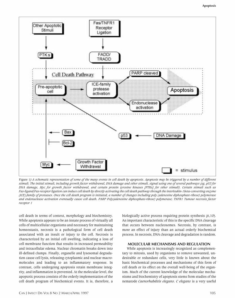
Figure 1) A schematic representation of some of the many events in cell death by apoptosis. Apoptosis may be triggered by a number of different
stimuli. The initial stimuli, including growth factor withdrawal, DNA damage and other stimuli, signal using one of several pathways (eg, p53 for
DNA damage, Myc for growth factor withdrawal, and certain protein tyrosine kinases [PTKs] for other stimuli). Certain stimuli such as
Fas-ligand/Fas-receptor ligation can induce cell death by directly activating the cell death pathway through the interleukin-1beta-converting enzyme
(ICE)-family of proteases. Once the cell death program is initiated, a number of changes including poly (adenosine diphosphate-ribose) polymerase
and endonuclease activation eventually cause cell death. PARP Poly(adenosine diphosphate-ribose) polymerase; TNFR1 Tumour necrosis factor
receptor 1
CAN J INFECT DIS VOL 8 NO 2 MARCH/APRIL 1997 105
Apoptosis
hung.chpMon Mar 31 15:46:34 1997
Color profile: DisabledComposite Default screen
model system for the study of apoptosis because it has a very
precise developmental pattern, with 131 of 1090 total cells
undergoing apoptosis. Mutant nematodes have allowed the
cell death pathways and the role of various genes important to
apoptosis to be pieced together. These genes can be divided
into several classes: those that are required for the induction
of apoptosis, those required for carrying out the cell death
program and those involved in the disintegration of the dead
cells. This model of cell death has been found to translate well
into mammalian systems. While the actual genes differ, a
great number of the cell death genes in C elegans have paral-
lels in mammalian cells. In particular, the Ced-9 suppressor of
apoptosis in C elegans is homologous to the Bcl-2 family of
proteins in mammalian cells, and the Ced-3 protein is homolo-
gous to the interleukin-1beta-converting enzyme (ICE) family
of proteases. There are, as expected, some deficiencies in the
model because certain parallels are absent (11).
A vastly simplified scheme of the current understanding of
the molecular mechanisms, some triggering factors and regu-
latory components of the cell death pathway are depicted in
Figure 1. It is now recognized that the cells of multicellular
organisms require signals to remain alive. In the absence of
these factors, most cells seem to initiate apoptosis by default.
The factors involved and their modes of action are extremely
diverse and differ among cell types. The induction of apoptosis
can be brought about by either the removal of growth factors
or the addition of some triggering factor. A combination of the
two allows for precise control of the populations of cell types
and allows for targeting of particular cells. Some fairly ubiq-
uitous triggering factors include DNA damage, release of tu-
mour necrosis factor (TNF) or glucocorticoids, and Fas/Apo-1
activation (12). Other triggering factors are more specific to
the cell types involved, such as the deletion of immature
thymocytes by binding of the T cell receptor (TCR) and death
of macrophages following certain bacterial infections (13). Fas
(also called CD95) is a receptor of the TNF receptor family.
Fas-mediated apoptosis can be used by cytotoxic T lympho-
cytes and is also important for negative selection in T cell
development. Another interesting triggering factor allows cells
to detect changes in its extracellular structure and environ-
ment. For example, endothelial cells that lose contact with
their extracellular matrix detect this via integrins and are
induced to die by apoptosis.
A number of signal transduction pathways are used to
communicate to the cell either a positive or negative signal for
apoptosis. The pathways may be activated either through
cell-surface receptors or through direct stimulation by agents
which can traverse the cell membrane. Such signaling is usu-
ally context-dependent: intracellular signals involved in apop-
tosis are often also involved in differentiation and
proliferation under different situations or conditions. In the
case of the Fas/TNF receptor 1 (TNFR1) family of cell-surface
receptors, ligation of certain members will promote cell sur-
vival, while ligation of others such as Fas and TNFR1 tends to
cause apoptosis. The cell death signal in the case of Fas and
TNFR1 is transduced by the activation of proteins associated
with these receptors called FADD and TRADD, respectively.
These, in turn, function as adapter molecules linking the death
signal downstream by causing the activation of various ICE-
family proteases, which activate other ICE-family proteases
and finally result in apoptosis (14,15).
The protease apopain and its proenzyme CPP32 have been
recently identified and shown to be necessary for apoptosis,
but are not sufficient to induce apoptosis (16). CPP32, unlike
many proteases, is unable autocatalytically to cleave and
activate itself into apopain. It requires the proteolytic activity
of other proteases. Possible proteases include ICE, granzyme B
(secreted by cytotoxic T cells to induce apoptosis in a target
cell) and fragmentin-2 (secreted by natural killer cells. As
mentioned earlier, ICE-family proteases are involved in
Fas/TNFR1 family receptor-mediated apoptosis. After activa-
tion of CPP32 to apopain by other proteases, apopain cleaves
several other cellular substrates. One that has been identified
is poly(adenosine diphosphate-ribose) polymerase (PARP).
PARP is involved in DNA repair and maintenance and its
proteolytic inactivation by apopain has been shown to be an
initial event in apoptosis.
One of the earliest changes observed after a cell is commit-
ted to death is an increase in the intracellular calcium levels
(17,18). Many cell-surface receptors signal through the Ca2+
pathway by activating phospholipase C (PLC). PLC hydrolyzes
phosphatidylinositol 4,5-bisphosphate into inositol trisphos-
phate and diacylglycerol. Inositol trisphosphate causes an
increase in calcium levels by opening calcium regulating chan-
nels in the endoplasmic reticulum and plasma membrane.
Diacylglycerol exerts its effect by activating protein kinase C
(PKC). However, because of crosstalk between different signal-
ing pathways, PKC activation has been reported to both induce
apoptosis and proliferation. Furthermore, many studies of PKC
activation and apoptosis use inhibitors of PKC that may them-
selves induce apoptosis, thus complicating the interpretation
of the studies’ results.
Protein tyrosine kinases (PTK) are often the intermediate
molecules involved in transducing either positive or negative
signals for apoptosis from cell-surface receptors and cell-sur-
face associated proteins to the nucleus. Certain cell-surface
receptors possess intrinsic PTK activity, while others are asso-
ciated with other proteins that are PTKs. A number of signal-
ing pathways promoting cell survival involve PTKs; for
example, activation of certain PTKs is required for growth
factor promoted survival, and cells that overproduce some of
the PTKs may become resistant to apoptosis induced by
growth factor withdrawal (19).
Another important signalling pathway in apoptosis in-
volves Ras, the GTPase that links upstream receptor and non-
receptor associated PTKs to downstream serine/threonine
kinases. Ras is switched off by GTPase-activating proteins and
is switched on by guanine nucleotide-releasing proteins. Acti-
vated Ras binds to and activates Raf, a serine/threonine kinase
that activates other serine/threonine kinases. This cascade
continues down to the mitogen-activated protein (MAP) ki-
nases. The Ras/Raf/MAP kinase cascade appears to be involved
in the inhibition of apoptosis, either directly or through the
regulation of other apoptosis inhibitors (eg, Bcl-2) (20).
106 CAN J INFECT DIS VOL 8 NO 2 MARCH/APRIL 1997
Hung and Chow
hung.chpMon Mar 31 15:46:36 1997
Color profile: DisabledComposite Default screen
The tumour repressor gene p53 is known to promote differ-
entiation or apoptosis depending on the context and is one of
the most common genes inactivated in human cancers. p53 is
important, allowing cells to respond to DNA damage (eg, by
irradiation) by causing growth arrest at the G1 phase. Cells
with mutant p53 lack this checkpoint and arrest in G2 (21).
This system allows the cell to assess the damage and to repair
itself, if possible, or to undergo apoptosis if the DNA damage
is too great. The ability of p53 to induce G1 arrest following
DNA damage lies in its function as a transcription factor.
Increased levels of p53 following DNA damage upregulate a
number of proteins that, in turn, inhibit cyclin-dependent
kinases, causing cell cycle arrest. p53 appears to be necessary
for the induction of apoptosis following DNA damage (22).
However, p53-independent apoptosis also exists because cer-
tain cells in p53-deficient mice are resistant to radiation-in-
duced apoptosis, but still undergo apoptosis in response to
other stimuli.
A large number of oncogenes mediate and regulate apop-
tosis. A typical example of a transcription factor that is an
oncogene affecting apoptosis is Myc. Myc is important in both
cell proliferation and apoptosis. It interacts with the retino-
blastoma protein (pRB), so that in the presence of certain
growth-promoting cytokines, Myc can override the pRB-in-
duced cell cycle arrest. However, during growth arrest (eg,
during the absence of a growth factor), the presence of Myc
induces p53-dependent apoptosis, which is suppressed by
Bcl-2 (see below). Thus, overexpression of Myc acts as a
two-way controlling mechanism that forces cells to either
proliferate or die, depending on the immediate environment
and presence of signals (23). Many viruses also produce onco-
proteins that can act by binding to proteins that normally
restrict cell growth, such as pRB (which controls cell cycle
progression and suppresses apoptosis) or p53 (described be-
low), or by suppressing apoptosis of the host cell, thus pre-
venting the important immune defense mechanism of
inducing apoptosis in virally infected cells.
Bcl-2 (first isolated from a B-cell lymphoma) has an inhibi-
tory effect on apoptosis and was the first gene to be shown to
regulate apoptosis (24). In adult organisms, Bcl-2 is primarily
expressed in tissues that are constantly renewed, are prolifera-
tive or are long-lived. Bcl-2 derives its oncogenic potential not
from any inherent ability to induce proliferation, but rather by
its ability to prevent apoptosis in cycling cells, and to promote
the survival of noncycling cells, thus acting in conjunction
with oncogenes like c-Myc, which can disregulate the cell
cycle. Bcl-2 accomplishes these effects, in part, by forming
high affinity heterodimers with Bax. Because Bax homodimers
promote apoptosis (overexpression speeds apoptosis during
growth factor withdrawal for some cells, but does not induce
apoptosis in the presence of growth factor), either raising the
transcription level of the Bax gene or decreasing the amount
of Bcl-2 has the effect of increasing the susceptibility of the cell
to apoptosis. The tumour suppressor p53 also plays a role
because it is both a negative regulator of Bcl-2 expression and
a transcription factor for the Bax gene (the Bax promotor has
four p53 binding regions) (25, 26). The scope of Bcl-2 protec-
tion against apoptosis appears quite large, including protec-
tion of many factor-dependent cells in G0 until growth factor
is restored; delay of p53-dependent apoptosis; resistance to
killing by various chemotherapeutic agents; and blocking of
apoptosis induced by glucocorticoids in immature lympho-
cytes. While much speculation about the exact mechanism of
Bcl-2 action exists, little is known about how it actually
prevents the induction of apoptosis.
SIGNIFICANCE OF APOPTOSISThe significance of apoptosis is two-fold: first, it is essen-
tial for the normal health of multicellular organisms; and
second, it has been implicated in both the pathogenesis and
battle against many diseases.
From the earliest stages of the development of an organism,
apoptosis plays a role in the maturation of tissues by altering
their structure and function. During embryogenesis of hu-
mans, for example, the interdigital webs and other vestigial
organs are removed by apoptosis. In the fetal lung, specific
cells are programmed to die in order to enlarge the alveoli.
Throughout a human’s life, cell turnover occurs in many
locations in the body. The hemopoietic system constantly
produces an enormous number of cells per day, which must be
eliminated to maintain a constant level of blood cells. This is
accomplished by apoptosis. Even neutrophils, which are
packed with highly toxic chemicals, die by apoptosis. This
indicates how inconspicuously apoptosis is integrated into the
everyday existence of organisms. Tissue turnover also occurs
on a variety of epithelial surfaces, including the skin and the
intestinal lining (27). During the menstrual cycle, many types
of cells proliferate and then must be eliminated. The process
includes the shedding of the uterine lining and the turnover of
mammary epithelial cells.
Apoptosis also plays a vital role in developmental immu-
nology and host defense against certain viral infections. Both
B cell and T cell development and selection involve apoptosis.
Cells that recognize self-antigens must be eliminated so that
immune responses are not targeted to the body’s own cells.
During T cell maturation, cells differentiate into different types
of T cells. One type, the activated effector T killer cells, uses
apoptosis to kill virally infected cells. Cytotoxic T cells recog-
nize viral antigens presented on the cell surface and signal to
them (either by degranulation or by Fas activation) to undergo
apoptosis.
The improper activation or the deactivation of apoptosis
has been implicated in many disorders. It has been associated
with the etiology of several neurodegenerative disorders, in-
cluding Alzheimer’s, Parkinson’s and Huntington’s diseases.
As well, increased apoptosis has been associated with is-
chemic injury (28).
As one might expect, a sizable number of cancers have been
shown to be caused not so much by an increase in growth and
response to growth signals, but by a decrease in responsive-
ness to apoptotic signals, making cells less likely to die. For
example, the B cell lymphoma from which Bcl-2 was first
isolated, overexpressed Bcl-2, thus inhibiting apoptosis. Other
tumours modify or suppress p53, making them resistant to
CAN J INFECT DIS VOL 8 NO 2 MARCH/APRIL 1997 107
Apoptosis
hung.chpMon Mar 31 15:46:37 1997
Color profile: DisabledComposite Default screen
death from DNA damage. This is an initial step allowing an
otherwise normal cell to accumulate further mutations, and
thus adapt readily for metastasis to other tissues.
A variety of viruses have been found that can modify the
host cells’ behaviour by expressing viral copies of some of the
genes involved in apoptosis. For example, viruses such as the
herpes viruses, pox viruses and adenoviruses encode apop-
tosis-inhibitors in their genomes (29, 30), increasing the prob-
ability that the host cells they infect will survive to produce
more viral particles. It is by this mechanism that some viruses
ultimately induce the formation of tumour cells in the infected
host cells. A virus that acts not by inhibiting apoptosis, but by
inducing it, is human immunodeficiency virus (HIV). Virally
infected CD4+ T cells undergo activation-induced cell death (31).
This contributes to the depletion of CD4+ T cells during AIDS.
Apoptosis has also been implicated in many superantigen
(sAg)-mediated diseases, such as staphylococcal and strepto-
coccal toxic shock, certain autoimmune and neoplastic dis-
eases, and both murine and human retroviral infections.
While conventional antigens bind within the antigen-present-
ing groove of the major histocompatibility complex molecules
and are recognized by TCR in that context, sAgs bind outside
of the antigen-presenting groove and interact with the V-beta
region of the TCR. sAgs bypass the normal selectivity of TCR
and can stimulate entire V-beta subsets of the T cell repertoire
present within the host organism. In the case of staphylococ-
cal and group A streptococcal toxic shock syndrome, this
causes a massive proliferation of the sAg-reactive T cells,
along with a pronounced release of various pro-inflammatory
cytokines. For staphylococcal toxic shock syndrome, several
sAgs produced by Staphylococcus aureus, including toxic
shock syndrome toxin-1 and staphylococcal enterotoxins A, B,
C1, C2, D and E, have been implicated as the primary cause of
this illness. For streptococcal toxic shock syndrome, the strep-
tococcal pyrogenic exotoxins A and C as well as several other
sAgs produced by Streptocococcus pyrogenes are the major
causes (32). Some other known sAg-producing microorgan-
isms include Yersinia enterocolitica, Mycoplasma arthritidis
and the mouse mammary tumour virus. While the most obvi-
ous effect of sAgs is the polyclonal proliferation of T cells, the
other important effect of sAgs is apoptosis that appears to
occur simultaneously, along with a downregulation of the
TCR that results in anergy (33). The net result of these two
contrary effects is to produce an initial rapid proliferation of
sAg-reactive T cells, followed by either a profound deletion or
anergy of these T cells. Therefore, while patients might sur-
vive staphylococcal or streptococcal toxic shock, they may
still be in danger because of immunosuppression from the
loss of up to 20% of their T cells. With viral sAgs, the picture
is somewhat different. Instead, some viruses use sAgs to
compel the host cells to proliferate, thereby promoting their
own survival, replication and spread. In regard to HIV, there
have been reports of Vbeta specific deletion of CD4+ T cells
(34). This suggests that sAgs may be a cause of CD4+ T cell
depletion in AIDS patients, with the sAg in question being
either an unidentified HIV product, or more likely, a sAg from
another pathogen that opportunistically coinfects the immu-
nocompromised AIDS patient. Recently, evidence has also
emerged to suggest that coinfection of host cells with cy-
tomegalovirus may result in the expression of a herpes viral
sAg that drives HIV replication preferentially in selected sAg-
responsive CD4+ V-beta12 T cell subsets (35). Hence, in AIDS
retroviral sAgs may exert their effect primarily through induc-
ing apoptosis and depleting HIV-infected CD4+ T cells, while
sAgs from coexisting infections may actively promote HIV
replication.
CONCLUSIONWithin the past few years, apoptosis has been recognized
to be fundamentally important in both the health and disease
of multicellular organisms. Despite the enormous explosion of
investigation in this area, little is known about the underlying
mechanisms and pathways of apoptosis, and much of its
regulation still remains a mystery. Future applications from
an improved knowledge base of apoptosis include the possibil-
ity of treating many disorders and diseases by therapeutically
altering the occurrence of apoptosis. As well, greater knowl-
edge of apoptosis will improve the understanding of the com-
plex balance that exists between life and death in multicellular
organisms.
REFERENCES1. Gregory CD. Apoptosis and the Immune Response. New York:
Wiley-Liss, 1995.2. Kerr JFR, Wyllie AH, Currie AR. Apoptosis: A basic biological
phenomenon with wide-ranging implications in tissue kinetics.Br J Cancer 1972;26:239-57.
3. Wyllie AH, Kerr JFR, Currie AR. Cell death: The significance ofapoptosis. Int Rev Cytol 1980;68:251-305.
4. Savill J. The innate immune system: Recognition of apoptoticcells. In: Gregory CD, ed. Apoptosis and the Immune Response.New York: Wiley-Liss, 1995:341.
5. Martin SJ, Green GR, Cotter TG. Dicing with death: dissecting thecomponents of apoptosis machinery. Trends Biol Sciences1994;19:26-30.
6. Wyllie AH, Morris RG, Smith AL, Dunlop D. Chromatin cleavagein apoptosis: Association with condensed chromatin morphologyand dependence on macromolecular synthesis. J Pathol1984;142:67.
7. Cohen GM, Sun XM, Snowden RT, Dinsdale D, Skilleter DN. Keymorphological features of apoptosis may occur in the absence of
internucleosomal DNA fragmentation. Biochem J1992;286:331-4.
8. Tomei LD, Shapiro JP, Cope FO. Apoptosis in C3H/10T1/2 mouseembryonic cells: Evidence for internucleosomal DNAmodification in the absence of double-strand cleavage. Proc NatlAcad Sci USA 1993;90:853-7.
9. Zakeri ZF, Quaglino D, Latham T, Lockshin RA. Delayedinternucleosomal DNA fragmentation in programmed cell death.FASEB J 1993;7:470-8.
10. Schwartzman RA. Cidlowski JA. Apoptosis: the biochemistry andmolecular biology of programmed cell death. Endocrine Reviews1993;14:133-51.
11. Yuan J. Molecular control of life and death. Curr Opin Cell Biol1995;7:211-4.
12. Cohen PL, Eisenberg RA. Fas/APO-1: A cell surface receptor thatsignals apoptosis. In: Gregory CD, ed. Apoptosis and the ImmuneResponse. New York: Wiley-Liss, 1995:169.
13. Collins MKL, Lopez-Rivas A. The control of apoptosis inmammalian cells. Trends in Biochemical Sciences 1993;18:307-9.
108 CAN J INFECT DIS VOL 8 NO 2 MARCH/APRIL 1997
Hung and Chow
hung.chpMon Mar 31 15:46:39 1997
Color profile: DisabledComposite Default screen
14. Chinnaiyan AM, O’Rouke K, Tewari M, Dixit VM. FADD, a noveldeath domain-containing protein, interacts with the deathdoamin of Fas and initiates apoptosis. Cell 1995;81:505-12.
15. Hsu HL, Xiong J, Goeddel DV. The TNF receptor 1-associatedprotein TRADD signals cell death and NFκB activation. Cell1995;81:495-504.
16. Nicholson DW, Ali A, Thornberry NA, et al. Identification andinhibition of the ICE/Ced-3 protease necessary for mammalianapoptosis. Nature 1995;76:37-43.
17. Martin SJ, Green GR, Cotter TG. Dicing with death: dissecting thecomponents of apoptosis machinery. Trends Biol Sci1994;19:26-30.
18. Schwartzman RA, Cidlowski JA. Apoptosis: The biochemistry andmolecular biology of programmed cell death. Endocr Rev1993;14:133-51.
19. Evans CA, Owen-Lynch JP, Whetton AD, Dive C. Activation of theAbelson tyrosine kinase-activity is associated with suppressionof apoptosis in hemopoetic cells. Cancer Res1993;53:1735-8.
20. Wang HG, Miyashita T, Takayama S, et al. Apoptosis regulationby interaction of Bcl-2 protein and Raf-1 kinase. Oncogene1994;9:2751-6.
21. Kuerbitz SJ, Plunkett BS, Walsh WV, Kastan MB. Wild-type p53is a cell cycle checkpoint determinant following irradiation. ProcNatl Acad Sci USA 1992;89:7491-5.
22. Furuya Y, Berges R, Lundmo P, Isaacs, JT. Cell proliferation, p53gene expression, and intracellular calcium in programmed death:Prostate Model. In: Tomei LD, Cope FO, eds. Apoptosis II: TheMolecular Basis of Apoptosis in Disease. New York: Cold SpringHarbor Laboratory Press, 1994:231.
23. Bissonnette RP, Shi Y, Mahboubi A, Glynn JM, Green DR. c-mycand Apoptosis. In: Tomei LD, Cope FO, eds. Apoptosis II: TheMolecular Basis of Apoptosis in Disease. New York: Cold SpringHarbor Laboratory Press, 1994:327.
24. Smith CA, Grimes EA, McCarthy NJ, Williams GT. Multiple generegulation of apoptosis: Significance in immunology andoncology. In: Tomei LD, Cope FO, eds. Apoptosis II: TheMolecular Basis of Apoptosis in Disease. New York: Cold SpringHarbor Laboratory Press, 1994:43.
25. Miyashita T, Harigai M, Hanada M, Reed JC. Identification of a
p53-dependent negative response element in the Bcl-2 gene.Cancer Res 1994;54:3131-5.
26. Miyashita T, Reed JC. Tumor-suppressor p53 is a directtranscriptional activator of the human Bax gene. Cell1995;80:293-9.
27. Budtz PE. Epidermal homeostasis: A new model that includesapoptosis. In: Tomei LD, Cope FO, eds. Apoptosis II: TheMolecular Basis of Apoptosis in Disease. New York: Cold SpringHarbor Laboratory Press,1994:165.
28. Thompson CB. Apoptosis in the pathogenesis and treatment ofdisease. Science 1995;267:1456-62.
29. Smith CA, Grimes EA, McCarthy NJ, Williams GT. Multiple generegulation of apoptosis: significance in immunology andoncology. In: Tomei LD, Cope FO, eds. Apoptosis II: TheMolecular Basis of Apoptosis in Disease. New York: Cold SpringHarbor Laboratory Press, 1994:43.
30. White E, Gooding LR. Regulation of apoptosis by humanadenoviruses. In: Tomei LD, Cope FO, eds. Apoptosis II: TheMolecular Basis of Apoptosis in Disease. New York: Cold SpringHarbor Laboratory Press, 1994:111.
31. Gougeon ML, Montagnier L. Apoptosis in peripheral Tlymphocytes during HIV infection: Influence of superantigensand correlation with AIDS pathogenesis. In: Tomei LD, Cope FO,eds. Apoptosis II: The Molecular Basis of Apoptosis in Disease.New York: Cold Spring Harbor Laboratory Press, 1994:5.
32. Watanabe-Ohnishi R, Low DE, McGeer A, et al. Selectivedepletion of Vβ-bearing T cells in patients with severe invasiveGroup A streptococcal infections and Streptococcal Toxic ShockSyndrome. J Infect Dis 1995;171:74-84.
33. Miethke T, Wahl C, Heeg K, Wagner H. Superantigens: Theparadox of T-cell activation versus inactivation. Int Arch AllergyImmunol 1995;106:3-7.
34. Posnett DN, Kabak S, Asch A, Hodtsev AS. HIV-1 replication in Tcells dependent on TCR Vβ expression. In: Huber BT, Palmer E,eds, Superantigens: A Pathogen’s View of the Immune System.Plainview: Cold Spring Harbor Laboratory Press, 1993:139.
35. Posnett DN. CMV drives HIV replication in selected Vb subsets:Evidence for a herpes virus superantigen. National Symposiumof Basic Aspects of Immunology. Bethesda, MD, May 1-3, 1996.(Abst)
CAN J INFECT DIS VOL 8 NO 2 MARCH/APRIL 1997 109
Apoptosis
hung.chpMon Mar 31 15:46:40 1997
Color profile: DisabledComposite Default screen

Submit your manuscripts athttp://www.hindawi.com
Stem CellsInternational
Hindawi Publishing Corporationhttp://www.hindawi.com Volume 2014
Hindawi Publishing Corporationhttp://www.hindawi.com Volume 2014
MEDIATORSINFLAMMATION
of
Hindawi Publishing Corporationhttp://www.hindawi.com Volume 2014
Behavioural Neurology
EndocrinologyInternational Journal of
Hindawi Publishing Corporationhttp://www.hindawi.com Volume 2014
Hindawi Publishing Corporationhttp://www.hindawi.com Volume 2014
Disease Markers
Hindawi Publishing Corporationhttp://www.hindawi.com Volume 2014
BioMed Research International
OncologyJournal of
Hindawi Publishing Corporationhttp://www.hindawi.com Volume 2014
Hindawi Publishing Corporationhttp://www.hindawi.com Volume 2014
Oxidative Medicine and Cellular Longevity
Hindawi Publishing Corporationhttp://www.hindawi.com Volume 2014
PPAR Research
The Scientific World JournalHindawi Publishing Corporation http://www.hindawi.com Volume 2014
Immunology ResearchHindawi Publishing Corporationhttp://www.hindawi.com Volume 2014
Journal of
ObesityJournal of
Hindawi Publishing Corporationhttp://www.hindawi.com Volume 2014
Hindawi Publishing Corporationhttp://www.hindawi.com Volume 2014
Computational and Mathematical Methods in Medicine
OphthalmologyJournal of
Hindawi Publishing Corporationhttp://www.hindawi.com Volume 2014
Diabetes ResearchJournal of
Hindawi Publishing Corporationhttp://www.hindawi.com Volume 2014
Hindawi Publishing Corporationhttp://www.hindawi.com Volume 2014
Research and TreatmentAIDS
Hindawi Publishing Corporationhttp://www.hindawi.com Volume 2014
Gastroenterology Research and Practice
Hindawi Publishing Corporationhttp://www.hindawi.com Volume 2014
Parkinson’s Disease
Evidence-Based Complementary and Alternative Medicine
Volume 2014Hindawi Publishing Corporationhttp://www.hindawi.com


![Review Survivin and Tumorigenesis: Molecular Mechanisms ... · Fas-induced apoptosis [28], regulation of cytokinesis and cell cycle progression [29], and participation in a variety](https://img.pdfslide.us/doc/110x75/5cc83c0388c993a6188d5399/review-survivin-and-tumorigenesis-molecular-mechanisms-fas-induced-apoptosis.jpg)