Embed Size (px)
Citation preview

Mechanical and Metabolic Injury to the SkinBarrier Leads to Increased Expression of Murineb-Defensin-1, -3, and -14Kerstin Ahrens1, Michael Schunck1, Graziella-Francesca Podda1, Josef Meingassner2, Anton Stuetz2,Jens-Michael Schroder1, Jurgen Harder1 and Ehrhardt Proksch1
Protection of the skin against microbiological infection is provided by the permeability barrier and byantimicrobial proteins. We asked whether the expression of murine b-defensins (mBDs)-1, -3, and -14—orthologs of human b-defensins hBD-1, -2, and -3, respectively—is stimulated by mechanically/physicochemi-cally (tape stripping or acetone treatment) or metabolically (essential fatty acid–deficient (EFAD) diet) inducedskin barrier dysfunction. Both methods led to a moderate induction of mBD-1 and mBD-14 and a pronouncedinduction of mBD-3 mRNA. Protein expression of the mBDs was increased as shown by immunohistology andby western blotting. Artificial barrier repair by occlusion significantly reduced the increased expression of mBD-14 after mechanical injury and of all three mBDs in EFAD mice, supporting an interrelationship betweenpermeability and the antimicrobial barrier. mBD-3 expression was stimulated in vitro by tumor necrosis factor-a(TNF-a), and a neutralizing anti-TNF-a antibody significantly reduced increased mBD-3 expression after barrierinjury in mouse skin, indicating that induction of mBD-3 expression is mediated by cytokines. The expression ofmBD-14 was stimulated by transforming growth factor-a and not by TNF-a. In summary, we demonstratedupregulation of mBD1, -3, and -14 after mechanically and metabolically induced skin barrier disruption, whichmay be an attempt to increase defense in the case of permeability barrier dysfunction.
Journal of Investigative Dermatology (2011) 131, 443–452; doi:10.1038/jid.2010.289; published online 14 October 2010
INTRODUCTIONThe skin is constantly exposed to a variety of microbialchallenges. The permeability barrier, localized in the stratumcorneum, is a mechanical protective barrier against bacterialinfection. Impairment of the physical barrier by injury is aprerequisite for experimental skin infection by application ofa bacterial suspension (Singh et al., 1971). In eczema orpsoriasis—skin diseases accompanied by disturbed perme-ability barrier function—the skin is colonized with potentiallypathogenic microorganisms (Aly et al., 1976, 1977). It is wellknown in clinical dermatology that occlusive conditions inthe skin body folds and the use of occlusive latex gloves andshoes can cause hyperhydration and skin irritation (Dendaet al., 1998). Irritation results in impaired barrier function,which in turn predisposes to skin infections such as intertrigo,impetigo, and tinea (Warner et al., 2003; Fluhr et al., 2005).
Despite an impaired permeability barrier and significantbacterial colonization, psoriatic skin is usually free of infection.In atopic dermatitis, clinical signs of bacterial infection arepredominantly found in severe cases or after extensivescratching (Lubbe, 2003). Therefore, additional defense me-chanisms besides the physical barrier have been suggested,leading to the discovery of antimicrobial peptides produced byepithelial cells (reviewed by Ganz and Lehrer, 1994; Gallo andHuttner, 1998; Schroder, 1999; Schroder and Harder, 1999;Harder and Schroder, 2005a, b; Izadpanah and Gallo, 2005).Several types of antimicrobial peptides, including b-defensins,cathelicidin (Gallo et al., 1997), catestatin (Radek et al., 2008),RNase-7 (Harder and Schroder, 2002), and psoriasin (Glaseret al., 2005), have been identified in the skin. In addition, threehuman b-defensins (hBD-1, -2, and -3) with a specific spectrumof antimicrobial activity have been characterized in humanskin and are expressed in keratinocytes (Harder et al., 1997,2001). Constitutive expression of hBD-1 has been detected inhuman skin (Fulton et al., 1997; Ali et al., 2001), but its role incutaneous defense is still emerging because of a lack of therespective studies. In contrast, hBD-2 and hBD-3 wereoriginally isolated in native form from psoriatic scale extracts(Harder et al., 1997, 2001), and both peptides are induciblyexpressed in keratinocytes. The expression of hBD-2 and hBD-3 can be upregulated by bacteria as well as endogenousmediators such as cytokines (e.g., IL-1b and IL-17 for hBD-2)and growth factors (e.g., transforming growth factor-a (TGF-a)
See related commentary on pg 285
& 2011 The Society for Investigative Dermatology www.jidonline.org 443
ORIGINAL ARTICLE
Received 2 December 2008; revised 2 July 2010; accepted 23 July 2010;published online 14 October 2010
1Department of Dermatology, University of Kiel, Kiel, Germany and 2NovartisInstitute for BioMedical Research, Vienna, Austria
Correspondence: Ehrhardt Proksch, Department of Dermatology, UniversityHospitals of Schleswig-Holstein, Campus Kiel, Schittenhelmstrasse 7,Kiel 24105, Germany.E-mail: [email protected]
Abbreviations: EFAD, essential fatty acid–deficient; hBD, human b-defensin;mBD, murine b-defensin

for hBD-3) (Schroder and Harder, 1999; Sørensen et al., 2003;Pazgier et al., 2006; Yin et al., 2006). In addition, it has beenreported that hBD-3 is induced upon epidermal injury,suggesting a role for hBD-3 in wound healing (Sørensenet al., 2006). Moreover, it has been documented that hBD-3 aswell as hBD-2 stimulates keratinocyte migration, proliferation,and production of proinflammatory cytokines and chemokines(Niyonsaba et al., 2007).
To further elucidate the role of b-defensins after epidermalbarrier disruption, we aimed to study the role of b-defensins ina mouse model using mechanical/physicochemical (via tapestripping or acetone treatment) as well as a metabolic (viaessential fatty acid–deficient (EFAD) diet) disruption of the skinbarrier. We focused on the expression of mouse orthologs ofthe human b-defensins hBD-1, -2, and -3. Mouse b-defensin-1(mBD-1) is the ortholog of hBD-1 (Huttner et al., 1997;Morrison et al., 1998), but its expression in mouse keratino-cytes has not yet been investigated. Mouse b-defensin-3(mBD-3) is considered an ortholog of hBD-2 (Morrison et al.,2003). mBD-3 is expressed in epithelia, especially in the lungand gastrointestinal tract, and its expression is induced byPseudomonas aeruginosa (Bals et al., 1999). Expression andinduction of mBD-3 in mouse skin after barrier disruption andby UVB exposure have recently been described (Aberg et al.,2007, 2008; Hong et al., 2008). Synthetic mBD-3 inhibitedthe growth of Escherichia coli, P. aeruginosa, Staphylococcusaureus, and Candida albicans at concentrations from 25 to50mg ml�1 (Burd et al., 2002). Recently, mouse b-defensin-14(mBD-14) has been identified as the ortholog of hBD-3(Hinrichsen et al., 2008; Rohrl et al., 2008; Taylor et al.,2008). mBD-14 exhibited a broad spectrum of potentantimicrobial activity against various microorganisms, includ-ing Gram-positive and Gram-negative bacteria and the yeastC. albicans (Hinrichsen et al., 2008).
Both the permeability barrier and the antimicrobial barrierare regulated by specific signaling mechanisms, includingcytokines and growth factors (Sørensen et al., 2003, 2005).Oren et al. (2003) showed that hBD-2 is localized in epidermallamellar bodies and that IL-1a stimulates movement from theendoplasmatic reticulum to lamellar bodies (post-transcrip-tional). We and others previously described an important rolefor proinflammatory cytokines in the regeneration of thephysical barrier (Wood et al., 1992; Jensen et al., 1999). Also,a role for tumor necrosis factor-a (TNF-a) in the induction ofhBD-2 has been described (Harder et al., 1997; Varoga et al.,2004). Cytokines and growth factors, which are necessary forthe potential induction of b-defensins during barrier repair inmouse skin, are only partly known. In the present study, weobserved that mBD-1, -3, and -14 are upregulated bymechanical/physicochemical as well as metabolic barrierinjury in mouse skin in vivo, and we present evidence thatcytokines and growth factors are involved in this process.
RESULTSMechanical/physicochemical barrier injury induces expressionof mBD-1, -3, and -14
The major goal of our study was to assess a potentialinteraction between the permeability barrier and the anti-
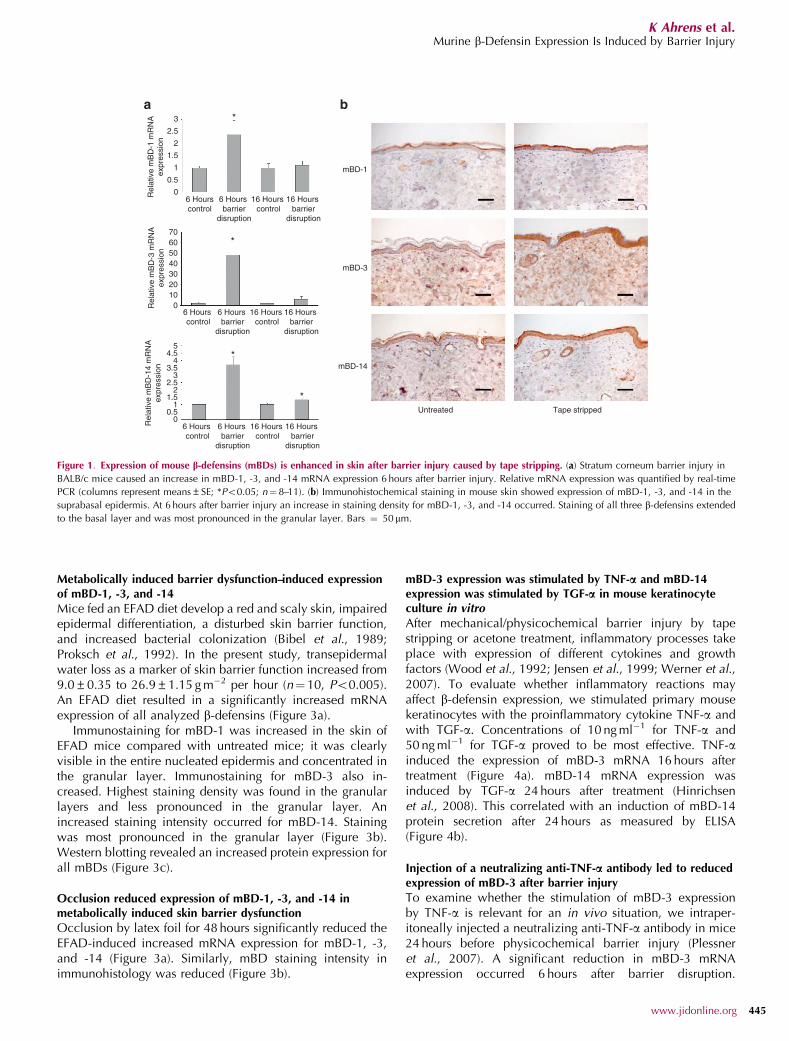
microbial barrier and whether acute and chronic disruptionof the physical skin barrier induces changes in theantimicrobial barrier. We performed mechanical injury tothe skin barrier through repeated tape stripping of theskin of shaved BALB/c mice, which potentially leads to aninvasion of environmental germs into the living epidermallayers. Subsequently we examined mRNA and proteinexpression of mBD-1, -3, and -14 at different time pointsafter barrier disruption. mRNA levels revealed a significant,slight increase in mBD-1 and mBD-14 expression 6 hoursafter barrier disruption as well as a significant, pronouncedincrease of mBD-3 expression. At 16 hours after barrierdisruption, a very slight but significant increase in themRNA expression could be detected only for mBD-14(Figure 1a).
mBD-1 protein immunostaining in untreated skin wasvisible in granular and spinous layers (Figure 1b). A moderateincrease in staining density occurred at 4–48 hours afterbarrier injury, especially in granular layers, and staining alsoinvolved spinous and basal layers. For mBD-3, only faintstaining was detected in untreated skin, which was concen-trated in basal and spinous layers (Figure 1b). Stainingstrongly increased at 4–48 hours after skin barrier disruption.Immunoreactivity for mBD-3 was visible in the entirenucleated epidermis with slightly higher staining density ingranular layers. Staining for mBD-14 was already moderate inuntreated skin, concentrated in granular layers, and alsovisible in spinous and basal layers (Figure 1b). At 4–48 hoursafter injury of the skin barrier, the staining of mBD-14 proteinincreased as seen for mBD-1 and -3.
Occlusion partially reduced expression of mBD-1, -3, and -14after barrier injury
In previous studies, artificial skin barrier repair reduced theexpression of several, but not all, metabolic events in the skininduced by skin barrier disruption (Proksch et al., 1990;Wood et al., 1994). For technical reasons, we used hairlessSKH-1 mice—a tight fit of the occlusive foil (with the thumbof a latex glove used as a tube) was more easily obtained thanwith shaved hairy mice. We used acetone treatment as wellas tape stripping for barrier disruption. However, we showocclusion data only for acetone-induced barrier disruptionbecause tape stripping yielded a high variance in mRNAexpression (data not shown). After acetone-induced barrierinjury, we found a significant but moderate increase in mBD-1 and -14 expression and a significant, pronounced increasein mBD-3 mRNA expression (Figure 2a). However, theincrease in mRNA expression, especially for mBD-1 andmBD-14, was more pronounced after acetone treatmentcompared with tape stripping. Occlusion for 24 hours afteracetone treatment reduced mRNA expression of the investi-gated b-defensins significantly for mBD-14—however, not tonormal levels (Figure 2a).
Immunohistology revealed increased staining. A reducedincrease in staining intensity, in particular for mBD-14,occurred with occlusion (Figure 2b). Western blottingrevealed an increased protein expression for mBD-1, -3,and -14 after barrier injury in SKH-1 mice (Figure 2c).
444 Journal of Investigative Dermatology (2011), Volume 131
K Ahrens et al.Murine b-Defensin Expression Is Induced by Barrier Injury
Metabolically induced barrier dysfunction–induced expressionof mBD-1, -3, and -14
Mice fed an EFAD diet develop a red and scaly skin, impairedepidermal differentiation, a disturbed skin barrier function,and increased bacterial colonization (Bibel et al., 1989;Proksch et al., 1992). In the present study, transepidermalwater loss as a marker of skin barrier function increased from9.0±0.35 to 26.9±1.15 g m�2 per hour (n¼ 10, Po0.005).An EFAD diet resulted in a significantly increased mRNAexpression of all analyzed b-defensins (Figure 3a).
Immunostaining for mBD-1 was increased in the skin ofEFAD mice compared with untreated mice; it was clearlyvisible in the entire nucleated epidermis and concentrated inthe granular layer. Immunostaining for mBD-3 also in-creased. Highest staining density was found in the granularlayers and less pronounced in the granular layer. Anincreased staining intensity occurred for mBD-14. Stainingwas most pronounced in the granular layer (Figure 3b).Western blotting revealed an increased protein expression forall mBDs (Figure 3c).
Occlusion reduced expression of mBD-1, -3, and -14 inmetabolically induced skin barrier dysfunction
Occlusion by latex foil for 48 hours significantly reduced theEFAD-induced increased mRNA expression for mBD-1, -3,and -14 (Figure 3a). Similarly, mBD staining intensity inimmunohistology was reduced (Figure 3b).
mBD-3 expression was stimulated by TNF-a and mBD-14expression was stimulated by TGF-a in mouse keratinocyteculture in vitro
After mechanical/physicochemical barrier injury by tapestripping or acetone treatment, inflammatory processes takeplace with expression of different cytokines and growthfactors (Wood et al., 1992; Jensen et al., 1999; Werner et al.,2007). To evaluate whether inflammatory reactions mayaffect b-defensin expression, we stimulated primary mousekeratinocytes with the proinflammatory cytokine TNF-a andwith TGF-a. Concentrations of 10 ng ml�1 for TNF-a and50 ng ml�1 for TGF-a proved to be most effective. TNF-ainduced the expression of mBD-3 mRNA 16 hours aftertreatment (Figure 4a). mBD-14 mRNA expression wasinduced by TGF-a 24 hours after treatment (Hinrichsenet al., 2008). This correlated with an induction of mBD-14protein secretion after 24 hours as measured by ELISA(Figure 4b).
Injection of a neutralizing anti-TNF-a antibody led to reducedexpression of mBD-3 after barrier injury
To examine whether the stimulation of mBD-3 expressionby TNF-a is relevant for an in vivo situation, we intraper-itoneally injected a neutralizing anti-TNF-a antibody in mice24 hours before physicochemical barrier injury (Plessneret al., 2007). A significant reduction in mBD-3 mRNAexpression occurred 6 hours after barrier disruption.
*
*
*
*
3
2.5
2
1.5
1
0.5
0
706050403020100
54.5
43.5
32.5
21.5
10.5
0
6 Hourscontrol
6 Hours barrier
disruption
16 Hours control
16 Hours barrier
disruption
mBD-1
mBD-3
mBD-14
Untreated Tape stripped
6 Hours control
6 Hours barrier
disruption
16 Hours control
16 Hours barrier
disruption
6 Hours control
6 Hours barrier
disruption
16 Hours control
16 Hours barrier
disruption
Rel
ativ
e m
BD
-1 m
RN
Aex
pres
sion
Rel
ativ
e m
BD
-3 m
RN
Aex
pres
sion
Rel
ativ
e m
BD
-14
mR
NA
expr
essi
on
Figure 1. Expression of mouse b-defensins (mBDs) is enhanced in skin after barrier injury caused by tape stripping. (a) Stratum corneum barrier injury in
BALB/c mice caused an increase in mBD-1, -3, and -14 mRNA expression 6 hours after barrier injury. Relative mRNA expression was quantified by real-time
PCR (columns represent means±SE; *Po0.05; n¼ 8–11). (b) Immunohistochemical staining in mouse skin showed expression of mBD-1, -3, and -14 in the
suprabasal epidermis. At 6 hours after barrier injury an increase in staining density for mBD-1, -3, and -14 occurred. Staining of all three b-defensins extended
to the basal layer and was most pronounced in the granular layer. Bars ¼ 50 mm.
www.jidonline.org 445
K Ahrens et al.Murine b-Defensin Expression Is Induced by Barrier Injury

A similar trend was noted for mBD-1 and mBD-14 mRNAexpression (Figure 5a).
Similar results were observed at the protein levelinvestigated by immunohistochemistry (Figure 5b).
DISCUSSIONThe skin is permanently exposed to a variety of potentiallyharmful microorganisms but usually remains free ofinfection. An intact permeability barrier prevents infection.
* *
Anti-mBD-1
B
17 kDa
17 kDa
17 kDa
42 kDa
A
Anti-mBD-3
Anti-mBD-14
Anti-β-actin
A = 10 μg SKH-1 skin lysate (untreated)
B = 10 μg SKH-1 skin lysate (acute barrier disruption, acetone treatment)
9876543210
9876543210
Barrier disruptionControl Barrier disruptionControl
Without occlusion
9080706050403020100
With occlusion
Barrier disruptionControl Barrier disruptionControl
Without occlusion With occlusion
Barrier disruptionControl Barrier disruptionControl
Without occlusion
mBD-1
mBD-3
mBD-14
Untreated Acetone treated Acetone treated + occlusion
With occlusion
*
*
*
*
*
Rel
ativ
e m
BD
-1 m
RN
Aex
pres
sion
Rel
ativ
e m
BD
-3 m
RN
Aex
pres
sion
Rel
ativ
e m
BD
-14
mR
NA
expr
essi
on
c
b
Figure 2. Expression of mouse b-defensins (mBDs) is induced after barrier injury by acetone treatment and can be reduced by occlusion. (a) Stratum
corneum barrier injury by acetone treatment led to an increased expression of mBD-1, -3, and -14 in SKH-1 mice 6 hours after barrier injury. The increase was
reduced by application of an occlusive foil immediately after barrier disruption, though no basal levels were obtained (significant for mBD-14 only).
Relative mRNA expression was quantified by real-time PCR (columns represent means±S.E. *Po0.05; n¼10–16). (b) Staining for mBD-1, -3, and -14 in
immunohistochemistry increased 6 hours after barrier injury by acetone treatment in SKH-1 mice. Staining density was reduced after artificial barrier repair
by latex occlusion. Bars¼ 50 mm. (c) Six hours after barrier disruption by acetone treatment, SKH-1 mice showed an increased protein staining for mBD-1, -3,
and -14 in western blot compared to untreated controls. b-Actin served as loading control.
446 Journal of Investigative Dermatology (2011), Volume 131
K Ahrens et al.Murine b-Defensin Expression Is Induced by Barrier Injury

However, impairment of the permeability barrier byscratches and other minor injuries is common and does notnecessarily result in skin infection. This indicates that
the skin has additional defense mechanisms. Severalstudies have suggested that a major cutaneous defensemechanism may be the inducible release of antimicrobial
*
* *
* *
*40
35
30
25
20
15
10
5
0
600
500
400
300
200
100
0
9876543210
Normal diet EFA diet EFA diet+ occlusion A B
17 kDa
17 kDa
17 kDa
42 kDa
A = 10 μg SKH-1 skin lysate (untreated)
B = 10 μg EFAD skin lysate (chronic barrier disruption)
Anti-mBD-1
Anti-mBD-3
Anti-mBD-14
Anti-β-actinNormal diet EFA diet EFA diet
+ occlusion
Normal diet EFA diet EFA diet+ occlusion
mBD-1
mBD-3
mBD-14
Control EFA diet EFA diet + occlusion
Rel
ativ
e m
BD
-1 m
RN
Aex
pres
sion
Rel
ativ
e m
BD
-3 m
RN
Aex
pres
sion
Rel
ativ
e m
BD
-14
mR
NA
expr
essi
on
Figure 3. Expression of murine b-defensins (mBDs) is enhanced in metabolically barrier-disrupted skin and can be reduced by occlusion. (a) In metabolically
barrier-disrupted skin of mice fed an essential fatty acid–deficient (EFAD) diet, an increased mRNA expression was noted for mBD-1, -3, and -14 compared with
control mice fed a normal diet. Occlusion of EFAD mouse skin with a latex foil for 48 hours did significantly reduce the mRNA expression for all three mBDs
(columns represent means±S.E. *Po0.05; n¼11–12). (b) In EFAD mice, immunohistochemical staining density markedly increased for all three defensins.
Occlusion by a latex foil for 48 hours in EFAD mice caused reduction in staining intensity. Bars¼ 50 mm. (c) In EFAD mice all three investigated mBDs showed
an increase in protein staining in western blot. b-Actin served as loading control.
www.jidonline.org 447
K Ahrens et al.Murine b-Defensin Expression Is Induced by Barrier Injury

peptides (Schroder and Harder, 2006; Schauber and Gallo,2008).
To gain more insight into the role of antimicrobial peptidesupon barrier disruption, we used mouse models to investigatethe expression of mouse b-defensins mBD-1, -3, and -14—orthologs of human defensins hBD-1, hBD-2, and hBD-3,respectively—following skin barrier disruption. Acute stratumcorneum barrier injury can easily be caused by tape strippingor acetone treatment, and the EFAD mouse is a model formetabolically induced permeability barrier disruption(Proksch et al., 1992). We found that barrier injury andmetabolically induced barrier dysfunction led to a slight tomoderate induction of mBD-1 and mBD-14 and a pro-nounced increase in mBD-3 mRNA expression. An inductionof protein expression was confirmed by immunohistologicalstaining and western blotting, with highest induction formBD-3. Immunoreactivity for mBD-1, -3, and -14 inunstimulated mouse epidermis was localized in the supraba-sal layers with increased staining intensity toward the outerlayers of the epidermis. In injury and metabolically inducedbarrier-disrupted skin, staining was extended to the entireepidermis, although staining intensity was still most
pronounced in the outer layers. This is in agreement withthe proposed function of b-defensins as defense moleculesagainst invading microorganisms and is in concordance withfindings for human orthologs (Liu et al., 1998, 2002; Sørensenet al., 2006; Jensen et al., 2007; Harder et al., 2010). Theseresults are also in line with a recent study showingupregulation of mBD-3 and CRAMP (cathelin-related anti-microbial peptide) expression after acute skin barrier injury(Aberg et al., 2008).
Although there is a global increase in b-defensin expres-sion after barrier injury, there are differences in the level ofinduction. In contrast to the slight to moderate induction ofmBD-1 and mBD-14, we observed a pronounced inductionof mBD-3 after barrier injury. The upregulation of mBD-1upon barrier injury was unexpected, because mBD-1 and thehuman ortholog hBD-1 are generally considered constitu-tively expressed b-defensins that are not induced by infectionor inflammation (Zhao et al., 1996; Morrison et al., 1998;Mathews et al., 1999). The mechanism of mBD-1 (andhBD-1) induction is unknown. In primary mouse keratino-cytes stimulated with various cytokines and growth factorsupregulation of mBD-1 could not be observed (datanot shown).
Previous publications showed that the expression ofmBD-3 is induced by inflammation and infection (Balset al., 1999; Burd et al., 2002). Thus, one can speculate thatthe strong induction of mBD-3 after skin barrier disruptionmay be mediated by an increased expression of proinflam-matory cytokines stimulated by skin injury. The hypothesisthat the induction of b-defensins in barrier dysfunction maybe mediated via proinflammatory cytokines and growthfactors is supported by several studies reporting that proin-flammatory cytokines and growth factors are of crucialimportance for the repair of the permeability barrier inmouse skin in vivo (Wood et al., 1992; Liou et al., 1997;Jensen et al., 1999). To further investigate this hypothesis, weintraperitonally injected mice with a neutralizing anti-TNF-aantibody before performing barrier disruption. This treatmentsignificantly reduced the increase in mBD-3 expression. Thisindicates that TNF-a contributes to the induction of mBD-3upon barrier injury.
It has been reported that sterile wounding inducedexpression of hBD-3, the human ortholog of mBD-14, aprocess that is mediated via activation of the EGFR (Sørensenet al., 2006). The observation that expression of mBD-14mRNA (Hinrichsen et al., 2008) and protein can be inducedby the EGFR ligand TGF-a in mouse keratinocytes in vitroprompts the interesting hypothesis that increased levels ofTGF-a and other growth factors may mediate the induction ofmBD-14 after barrier disruption.
Artificial barrier restoration by occlusion with a latex foilreduced the increase in b-defensin mRNA expressionalthough significantly only for mBD-14. It has been shownthat several biochemical mechanisms involved in barrierrepair can be reduced by artificial barrier repair (Prokschet al., 1990). However, not all biochemical effects arereduced; in particular, cytokine expression is reduced onlyunder certain circumstances. Wood et al. (1994) reported that
6 Hoursuntreated
6 HoursTNF
16 Hoursuntreated
16 HoursTNF
*
*
24 Hoursuntreated
24 HoursTGF-α
6
5
4
3
2
1
0
mB
D-1
4 pr
otei
n le
vel
(ng
ml–1
)
Rel
ativ
e m
BD
-3 m
RN
Aex
pres
sion
5a
b
3
2
1
0
4
Figure 4. TNF-a and TGF-a influenced the expression of murine b-defensins
(mBDs) in primary mouse keratinocytes in vitro. (a) Primary mouse
keratinocytes were stimulated with 10 ng ml�1 TNF-a for 6 and 16 hours, and
mBD-3 mRNA expression was analyzed by real-time PCR. The mRNA
expression of mBD-3 was increased. Bars represent the relative mBD-3
transcript levels normalized to GAPDH transcript levels. Results are presented
as means ± standard errors (*Po0.05; n¼ 9). (b) Expression of mBD-14
protein in supernatants of primary mouse keratinocytes stimulated for
24 hours with TGF-a (50 ng ml�1) was measured by ELISA. The protein release
of mBD-14 was significantly increased after stimulation with TGF-a compared
with medium controls (*Po0.05; n¼ 9).
448 Journal of Investigative Dermatology (2011), Volume 131
K Ahrens et al.Murine b-Defensin Expression Is Induced by Barrier Injury

occlusion does not lower cytokine mRNA levels after acutebarrier disruption. Similarly, we found a reduction only inmBD-14 expression, and not for mBD-3, because theexpression of mBD-3 is induced by proinflammatory stimulisuch as TNF-a, whereas the expression of mBD-14 is inducedby TGF-a. Growth factor expression is partially blocked inoccluded skin barrier disruption (Liou et al., 1997). Wood
et al. showed that cytokine expression was reduced in EFADmice after occlusion. Consistent with this, we foundreduction of all three mBDs after occlusion in these mice.
In summary, we found increased expression of mBD-1, -3,and -14 after mechanical and metabolic skin barrierdisruption in mouse skin. The increased expression mayreflect a defense response to protect the skin against harmful
25
20
15
10
5
0
0
160
140
120
100
80
60
40
20
**
**
*
*
*
Control Barrier disruption Control
Without TNF blocking With TNF blocking
Barrier disruption
With TNF blocking With TNF blocking
Control Barrier disruption Control Barrier disruption
Without TNF blocking
mBD-1
mBD-3
mBD-14
Control Barrier disruptedwithout TNF blocking
Barrier disruptedwith TNF blocking
With TNF blocking
Control Barrier disruption Control Barrier disruption
Rel
ativ
e m
BD
-1 m
RN
Aex
pres
sion
Rel
ativ
e m
BD
-3 m
RN
Aex
pres
sion
Rel
ativ
e m
BD
-14
mR
NA
expr
essi
on
14
12
10
8
4
2
0
6
Figure 5. Treatment of mice with a tumor necrosis factor-a (TNF-a) neutralizing antibody before barrier disruption reduced induction of murine b-defensin-3
(mBD-3) expression. (a) To block the activity of TNF-a, a neutralizing anti-TNF-a antibody was intraperitoneally injected into SKH-1 mice 24 hours before
barrier disruption by acetone treatment. Six hours later, the mRNA expression of mBD-3 was significantly reduced in mice treated with anti-TNF-a antibody
compared with mice treated with phosphate-buffered saline. No significant difference was recognized in the expression of mBD-1 and mBD-14 mRNA. Relative
mRNA expression was quantified by real-time PCR (columns represent means±SE. *Po0.05; n¼ 6). (b) Immunohistochemical analyses of skin samples from
mice treated with anti-TNF-a antibody showed a reduction of b-defensin protein staining, most pronounced for mBD-3. Bars¼ 50 mm.
www.jidonline.org 449
K Ahrens et al.Murine b-Defensin Expression Is Induced by Barrier Injury

microorganisms that can invade skin with a dysfunction ofthe permeability barrier.
MATERIALS AND METHODSMice
SKH-1 mice (Crl:(hr/hr) BR) were supplied by Charles River (Sulzfeld,
Germany). BALB/c mice were bred in the central animal facility at
the University of Kiel. For purposes of investigating barrier
disruption, male 6- to 8-week-old mice were used. EFAD mice
were obtained by feeding 3-week-old SKH-1 mice an EFAD diet
(Bavandi et al., 1992), modified by adding MgSO4 �7H2O. Mice
were individually and conventionally maintained in plastic cages
under standardized conditions (room temperature 25 1C, relative
humidity 45–55%, circadian rhythm 12 hours, and standard labora-
tory animal chow (V1534, SSNIFF Spezialdiaten, Soest, Germany)
and water supplied ad libitum. The University of Kiel Committee for
Animal Care approved the study.
Barrier injury of mouse skin and artificial barrier repair
The barrier of one flank of mouse skin was disrupted by repeated
tape stripping (cellophane tape) or by application of acetone with a
small cotton stick until a six- to eightfold increase in transepidermal
water loss (Tewameter TM 210, Courage & Khazaka, Cologne,
Germany) occurred. In one set of animals, the disrupted skin barrier
was immediately occluded with a latex foil after treatment. Hairy
mice were shaved with a razor blade 1 day before treatment. EFAD
mice were compared with a normally fed control group and
occluded for 48 hours.
Injection of neutralizing anti-TNF-a antibody in mice
To evaluate a possible role for TNF-a in induction of b-defensins, a
neutralizing anti-TNF-a antibody (0.5 mg in phosphate-buffered
saline (PBS); eBioscience, Frankfurt, Germany, MP6-XT3) was
intraperitoneally injected into SKH-1 mice 24 hours before acetone
treatment. Control mice were injected with PBS.
Cell culture and stimulation
Primary keratinocytes were isolated from the skin of 1- to 2-day-old
BALB/c mice and cultivated in EpiLife-Medium (Sigma, Taufkirchen,
Germany) in collagenized six-well culture plates (9.6 cm2 per well;
Sarstedt, Nurnbrecht, Germany) under standardized conditions
(37 1C, 0.5% CO2). Cells were stimulated at a confluence of
90–100%.
mRNA isolation, reverse transcription, and real-time PCR
mRNA isolation from cultured primary keratinocytes was performed
using the NucleoSpin RNA II Kit (Macherey-Nagel, Duren,
Germany). Skin samples were minced in liquid nitrogen, and RNA
was isolated using the TRIzol reagent (Invitrogen, Karlsruhe,
Germany). Reverse-transcription reagents were obtained from
Applied Biosystems (HighCap cDNA RT Kit; Darmstadt, Germany).
cDNA was analyzed by real-time PCR (LightCycler 2.0; Roche
Diagnostics GmbH, Mannheim, Germany) using the SYBR Advan-
tage qPCR Premix (TaKaRa Bio Europe, Saint-Germain-en-Laye,
France). cDNA corresponding to 10 ng RNA was used as a template.
Samples were incubated for an initial denaturation at 95 1C for
10 minutes followed by 45 cycles, each cycle consisting of 95 1C for
10 seconds, 60 1C (touchdown of �1 1C per cycle from 66 1C to
60 1C) for 5 seconds, and 72 1C for 15 seconds. To confirm
amplification of specific transcripts, melting curve profiles were
produced. (See Supplementary Table 1 online for a list of real-time
PCR primers (Eurofins MWG Operon, Ebersberg, Germany).)
Quantifications were normalized to the housekeeping gene
glyceraldehyde 3-phosphate dehydrogenase (GAPDH). Relative
expression is given as the ratio between target gene expression
and GAPDH expression.
Generation of mBD-14-specific antibodies
Expression of recombinant mBD-14 protein was described pre-
viously (Hinrichsen et al., 2008). In the present study, a fusion
protein (2.2 mg) was used for immunization. This (1.5 mg) was
conjugated to keyhole limpet hemocyanine (Sigma) using glutar-
aldehyde. Keyhole limpet hemocyanine (1 mg) in 1 ml PBS was
mixed with 1ml 25% glutaraldehyde (Serva, Heidelberg, Germany)
and incubated for 1 hour at room temperature with gentle shaking.
The reaction mixture was diafiltrated and concentrated in 400 ml
PBS using a Vivaspin 0.5 ml concentrator column (30 kDa cutoff,
Vivascience, Hannover, Germany). The concentrate was incubated
with 600ml (1.5 mg) mBD-14 fusion protein in PBS for 1 hour at
room temperature with gentle rotation. The reaction was stopped
by the addition of 5 ml 1 M Tris (pH 8.0). mBD-14 fusion protein
(0.7 mg) in 500 ml of PBS was added. The preparation was divided
into one 450ml aliquot for initial immunization and three 350ml
aliquots for booster immunization of a goat. Immunization was
carried out by ZIKA-Kaninchenbetrieb (Gottin, Germany). We
generated an mBD-14 affinity column (1 ml HiTrap NHS-activated
columns, Amersham Pharmacia Biotech, Vienna, Austria) using
500mg recombinant mBD-14 to selectively isolate mBD-14-specific
antibodies from the serum. Also, a pET-32 affinity column using
650mg pET-32 was generated to remove pET-32-specific antibodies.
The mBD-14 affinity column was loaded with 750 ml goat anti-mBD-
14 serum and washed with 10 mM sodium phosphate buffer, pH 7.4.
Low-affinity antibodies were eluted using 1 M NaCl (pH 7), and high-
affinity mBD-14 antibodies were eluted using 200 mM glycine (pH
3), immediately neutralized with 1 M Tris (pH 7.5) and dialyzed
against PBS. Antibodies with affinity to pET-32 were removed
by pET-32 affinity column. Specificity of mBD-14 antibodies was
verified by immunohistochemistry and western blotting.
Western blot analysis
Peptides were extracted from mouse skin using a lysis buffer
containing 62.5 M Tris, 5% SDS, and 10 mM DTT. Proteins were
separated by 16.5% SDS-tricine polyacrylamide gel containing 6 M
urea (Schagger and von Jagow, 1987). Proteins were transferred to a
nitrocellulose membrane (0.2 mm, Amersham Hybond-ECL, GE
Healthcare, Freiburg, Germany), blocked for 1 hour in blocking
buffer (5% (wt/vol) BSA in PBSþ 0.05% Tween), then incubated
overnight at 4 1C in 3% (wt/vol) BSA in PBSþ 0.05% Tween
containing 1:250 anti-mBD-1 antibodies, 1:250 anti-mBD-3 anti-
bodies (both Santa Cruz Biotechnology, Santa Cruz, CA), or
1:10,000 mBD-14 affinity-purified antibodies (1.85 mg/ml). The
membrane was washed with PBSþ 0.05% Tween six times for
5 minutes each and incubated for 1 hour in 3% (wt/vol) BSA in
PBSþ 0.05% Tween containing 1:20,000 dilution of goat anti-rabbit
or rabbit anti-goat IgG HRP conjugate (Dianova, Hamburg,
Germany). After another six washes, the membrane was incubated
450 Journal of Investigative Dermatology (2011), Volume 131
K Ahrens et al.Murine b-Defensin Expression Is Induced by Barrier Injury

for 5 minutes with chemiluminescent peroxidase substrate
(Lumi-Light PLUS Western Blotting Substrate, Roche Diagnostics,
Mannheim, Germany) and visualized using a Diana III cooled
CCD-camera imaging system (Raytest, Straubenhardt, Germany).
b-Actin served as loading control in all western blots.
Immunohistochemistry
For immunohistochemistry, 5mm vertical paraffin sections were
deparaffinized and rehydrated followed by heat-induced antigen
retrieval in citrate acid buffer (10 mM, pH 6.0). Slides were incubated
with 3% aqueous H2O2 for 5 minutes at room temperature to block
endogenous peroxidase activity. After blocking nonspecific antibody
binding by incubation with normal rabbit or swine serum (1:5 in
Tris-buffered saline, Dako Diagnostics, Hamburg, Germany), sec-
tions were incubated with antibodies for mBD-1, mBD-3 (1:100,
both Santa Cruz Biotechnology), and mBD-14 (1.85 mg ml�1, 1:100)
diluted in Tris-buffered saline (0.15 M NaCl, 0.05 M Tris, pH 7.6)
overnight at 4 1C. Sections were incubated for 30 minutes with
secondary biotinylated antibodies (Dianova, Hamburg, Germany)
followed by incubation for 30 minutes with StreptABComplex/HRP
(Dako Diagnostics), developed with Liquid DAB (Biogenex, San
Ramon, CA), and counterstained with Mayer’s hemalaun (Merck,
Darmstadt, Germany).
ELISA
Ninety-six-well immunoplates (MaxiSorp, Nunc, Roskilde, Denmark)
were coated at 4 1C overnight with 50 ml anti-mBD-14 antibody
diluted 1:2,000 to 1mg ml�1 in 0.05 M carbonate buffer, pH 9.6.
Wells were blocked with 200ml 1% BSA in PBS for 10 minutes
at 37 1C. After being washed three times with 200ml PBSþ 0.1%
Tween 20, 50ml cell culture supernatants and serial dilutions
of recombinant mBD-14 protein were incubated for 30 minutes
at 37 1C. Plates were washed three times and incubated for
30 minutes at 37 1C with 50ml biotinylated anti-mBD-14 antibody
diluted 1:4,000 to 0.23mg ml�1 in PBSþ 0.1% Tween 20. Plates
were washed again three times, filled with 50ml Streptavidin-POD
(Roche Diagnostics; 1:10,000 in PBSþ 0.1% Tween 20) and
incubated for 30 minutes at 37 1C, washed six times, and incubated
with 2,29-azino-bis-3-ethylbenzthiazoline-6-sulfonic acid (Roche
Diagnostics) for 30–45 minutes at 37 1C. Absorbance was measured
at 405 nm with a multichannel photometer (Sunrise; Tecan,
Crailsheim, Germany).
Statistical analysis
All calculations were performed using the unpaired Student’s
t-test.
CONFLICT OF INTERESTThe authors state no conflict of interest.
ACKNOWLEDGMENTSWe thank Nadja Linke for invaluable technical assistance. The work wassupported by grants from the Deutsche Forschungsgemeinschaft (SFB 617,Pr272/6-1) to JM Schroder, J Harder, and E Proksch. J Harder is supported bythe Heisenberg program of the Deutsche Forschungsgemeinschaft.
SUPPLEMENTARY MATERIAL
Supplementary material is linked to the online version of the paper at http://www.nature.com/jid
REFERENCES
Aberg KM, Man MQ, Gallo RL et al. (2008) Co-regulation and interdepen-dence of the mammalian epidermal permeability and antimicrobialbarriers. J Invest Dermatol 128:917–25
Aberg KM, Radek KA, Choi EH et al. (2007) Psychological stress down-regulates epidermal antimicrobial peptide expression and increasesseverity of cutaneous infections in mice. J Clin Invest 117:3339–49
Ali RS, Falconer A, Ikram M et al. (2001) Expression of the peptide antibioticshuman beta defensin-1 and human beta defensin-2 in normal humanskin. J Invest Dermatol 117:106–11
Aly R, Maibach HE, Mandel A (1976) Bacterial flora in psoriasis. Br JDermatol 95:603–6
Aly R, Maibach HI, Shinefield HR (1977) Microbial flora of atopic dermatitis.Arch Dermatol 113:780–2
Bals R, Wang X, Meegalla RL et al. (1999) Mouse beta-defensin 3 is aninducible antimicrobial peptide expressed in the epithelia of multipleorgans. Infect Immun 67:3542–7
Bavandi A, Meingassner JG, Becker S (1992) Diet-induced dermatitis responseof hairless rats to systemic treatment with cyclosporin A (Sandimmun),cyclosporin H and FK506. Exp Dermatol 1:199–205
Bibel DJ, Miller SJ, Brown BE et al. (1989) Antimicrobial activity of stratum-corneum lipids from normal and essential fatty acid-deficient mice.J Invest Dermatol 92:632–8
Burd RS, Furrer JL, Sullivan J et al. (2002) Murine beta-defensin-3 is aninducible peptide with limited tissue expression and broad-spectrumantimicrobial activity. Shock 18:461–4
Denda M, Sato J, Masuda Y et al. (1998) Exposure to a dry environmentenhances epidermal permeability barrier function. J Invest Dermatol111:858–63
Fluhr JW, Akengin A, Bornkessel A et al. (2005) Additive impairment of thebarrier function by mechanical irritation, occlusion and sodium laurylsulphate in vivo. Br J Dermatol 153:125–31
Fulton C, Anderson GM, Zasloff M et al. (1997) Expression of natural peptideantibiotics in human skin. Lancet 350:1750–1
Gallo RL, Huttner KM (1998) Antimicrobial peptides: an emerging concept incutaneous biology. J Invest Dermatol 111:739–43
Gallo RL, Kim KJ, Bernfield M et al. (1997) Identification of CRAMP, acathelin-related antimicrobial peptide expressed in the embryonic andadult mouse. J Biol Chem 272:13088–93
Ganz T, Lehrer RI (1994) Defensins. Curr Opin Immunol 6:584–9
Glaser R, Harder J, Lange H et al. (2005) Antimicrobial psoriasin (S100A7)protects human skin from Escherichia coli infection. Nat Immunol6:57–64
Harder J, Bartels J, Christophers E et al. (1997) A peptide antibiotic fromhuman skin. Nature 387:861
Harder J, Bartels J, Christophers E et al. (2001) Isolation and characterizationof human beta -defensin-3, a novel human inducible peptide antibiotic.J Biol Chem 276:5707–13
Harder J, Dressel S, Wittersheim M et al. (2010) Enhanced expression andsecretion of antimicrobial peptides in atopic dermatitis and aftersuperficial skin injury. J Invest Dermatol 130:1355–64
Harder J, Schroder JM (2002) RNase 7, a novel innate immune defenseantimicrobial protein of healthy human skin. J Biol Chem 277:46779–84
Harder J, Schroder JM (2005a) Antimicrobial peptides in human skin. ChemImmunol Allergy 86:22–41
Harder J, Schroder JM (2005b) Psoriatic scales: a promising source for theisolation of human skin-derived antimicrobial proteins. J Leukoc Biol77:476–86
Hinrichsen K, Podschun R, Schubert S et al. (2008) Mouse beta-defensin-14,an antimicrobial ortholog of human beta-defensin-3. Antimicrob AgentsChemother 52:1876–9
Hong SP, Kim MJ, Jung MY et al. (2008) Biopositive effects of low-dose UVBon epidermis: coordinate upregulation of antimicrobial peptides andpermeability barrier reinforcement. J Invest Dermatol 128:2880–7
www.jidonline.org 451
K Ahrens et al.Murine b-Defensin Expression Is Induced by Barrier Injury

Huttner KM, Kozak CA, Bevins CL (1997) The mouse genome encodes asingle homolog of the antimicrobial peptide human beta-defensin 1.FEBS Lett 413:45–9
Izadpanah A, Gallo RL (2005) Antimicrobial peptides. J Am Acad Dermatol52:381–90
Jensen JM, Pfeiffer S, Akaki T et al. (2007) Barrier function, epidermaldifferentiation, and human beta-defensin 2 expression in tinea corporis.J Invest Dermatol 127:1720–7
Jensen JM, Schutze S, Forl M et al. (1999) Roles for tumor necrosis factorreceptor p55 and sphingomyelinase in repairing the cutaneous perme-ability barrier. J Clin Invest 104:1761–70
Liou A, Elias PM, Grunfeld C et al. (1997) Amphiregulin and nerve growthfactor expression are regulated by barrier status in murine epidermis.J Invest Dermatol 108:73–7
Liu AY, Destoumieux D, Wong AV et al. (2002) Human beta-defensin-2production in keratinocytes is regulated by interleukin-1, bacteria, andthe state of differentiation. J Invest Dermatol 118:275–81
Liu L, Wang L, Jia HP et al. (1998) Structure and mapping of the human beta-defensin HBD-2 gene and its expression at sites of inflammation. Gene222:237–44
Lubbe J (2003) Secondary infections in patients with atopic dermatitis. Am JClin Dermatol 4:641–54
Mathews M, Jia HP, Guthmiller JM et al. (1999) Production of beta-defensinantimicrobial peptides by the oral mucosa and salivary glands. InfectImmun 67:2740–5
Morrison GM, Davidson DJ, Kilanowski FM et al. (1998) Mouse betadefensin-1 is a functional homolog of human beta defensin-1. MammGenome 9:453–7
Morrison GM, Semple CA, Kilanowski FM et al. (2003) Signal sequenceconservation and mature peptide divergence within subgroups of themurine beta-defensin gene family. Mol Biol Evol 20:460–70
Niyonsaba F, Ushio H, Nakano N et al. (2007) Antimicrobial peptides humanbeta-defensins stimulate epidermal keratinocyte migration, proliferationand production of proinflammatory cytokines and chemokines. J InvestDermatol 127:594–604
Oren A, Ganz T, Liu L et al. (2003) In human epidermis, beta-defensin 2 ispackaged in lamellar bodies. Exp Mol Pathol 74:180–2
Pazgier M, Hoover DM, Yang D et al. (2006) Human beta-defensins. Cell MolLife Sci 63:1294–313
Plessner HL, Lin PL, Kohno T et al. (2007) Neutralization of tumor necrosisfactor (TNF) by antibody but not TNF receptor fusion moleculeexacerbates chronic murine tuberculosis. J Infect Dis 195:1643–50
Proksch E, Elias PM, Feingold KR (1990) Regulation of 3-hydroxy-3-methylglutaryl-coenzyme-A reductase-activity in murine epidermis—modulation of enzyme content and activation state by barrier require-ments. J Clin Invest 85:874–82
Proksch E, Feingold KR, Elias PM (1992) Epidermal Hmg CoA reductase-activity in essential fatty-acid deficiency—barrier requirementsrather than eicosanoid generation regulate cholesterol-synthesis. J InvestDermatol 99:216–20
Radek KA, Lopez-Garcia B, Hupe M et al. (2008) The neuroendocrine peptidecatestatin is a cutaneous antimicrobial and induced in the skin afterinjury. J Invest Dermatol 128:1525–34
Rohrl J, Yang D, Oppenheim JJ et al. (2008) Identification and biological
characterization of mouse beta-defensin 14, the orthologue of human
beta-defensin 3. J Biol Chem 283:5414–9
Schagger H, von Jagow G (1987) Tricine sodium dodecyl-sulfate polyacry-
lamide-gel electrophoresis for the separation of proteins in the range
from 1- to 100-Kda. Anal Biochem 166:368–79
Schauber J, Gallo RL (2008) Antimicrobial peptides and the skin immune
defense system. J Allergy Clin Immunol 122:261–6
Schroder JM (1999) Epithelial antimicrobial peptides: innate local host
response elements. Cell Mol Life Sci 56:32–46
Schroder JM, Harder J (1999) Human beta-defensin-2. Int J Biochem Cell Biol
31:645–51
Schroder JM, Harder J (2006) Antimicrobial skin peptides and proteins. Cell
Mol Life Sci 63:469–86
Singh G, Marples RR, Kligman AM (1971) Experimental staphylococcus
aureus infections in humans. J Invest Dermatol 57:149–62
Sørensen OE, Cowland JB, Theilgaard-Monch K et al. (2003) Wound healing
and expression of antimicrobial peptides/polypeptides in human
keratinocytes, a consequence of common growth factors. J Immunol
170:5583–9
Sørensen OE, Thapa DR, Rosenthal A et al. (2005) Differential regulation of
beta-defensin expression in human skin by microbial stimuli. J Immunol
174:4870–9
Sørensen OE, Thapa DR, Roupe KM et al. (2006) Injury-induced
innate immune response in human skin mediated by transactivation
of the epidermal growth factor receptor. J Clin Invest 116:
1878–85
Taylor K, Clarke DJ, McCullough B et al. (2008) Analysis and separation of
residues important for the chemoattractant and antimicrobial activities of
beta-defensin 3. J Biol Chem 283:6631–9
Varoga D, Pufe T, Harder J et al. (2004) Production of endogenous antibiotics
in articular cartilage. Arthritis Rheum 50:3526–34
Warner RR, Stone KJ, Boissy YL (2003) Hydration disrupts human stratum
corneum ultrastructure. J Invest Dermatol 120:275–84
Werner S, Krieg T, Smola H (2007) Keratinocyte-fibroblast interactions in
wound healing. J Invest Dermatol 127:998–1008
Wood LC, Elias PM, Sequeiramartin SM et al. (1994) Occlusion lowers
cytokine messenger-RNA levels in essential fatty acid-deficient and
normal mouse epidermis, but not after acute barrier disruption. J Invest
Dermatol 103:834–8
Wood LC, Jackson SM, Elias PM et al. (1992) Cutaneous barrier perturbation
stimulates cytokine production in the epidermis of mice. J Clin Invest
90:482–7
Yin C, Dang HN, Zhang HB et al. (2006) Capacity of human beta-defensin
expression in gene-transduced and cytokine-induced cells. Biochem
Biophys Res Commun 339:344–54
Zhao C, Wang I, Lehrer RI (1996) Widespread expression of beta-defensin
hBD-1 in human secretory glands and epithelial cells. FEBS Lett
396:319–22
452 Journal of Investigative Dermatology (2011), Volume 131
K Ahrens et al.Murine b-Defensin Expression Is Induced by Barrier Injury