Embed Size (px)
Citation preview
TitleTargeted over-expression of endothelin-1 in astrocytes leads tomore severe brain damage and vasospasm after subarachnoidhemorrhage
Author(s) Yeung, PKK; Shen, J; Chung, SS; Chung, SK
Citation B M C Neuroscience, 2013, v. 14, p. article no. 131
Issued Date 2013
URL http://hdl.handle.net/10722/195658
RightsB M C Neuroscience. Copyright © BioMed Central Ltd.; Thiswork is licensed under a Creative Commons Attribution-NonCommercial-NoDerivatives 4.0 International License.
Yeung et al. BMC Neuroscience 2013, 14:131http://www.biomedcentral.com/1471-2202/14/131
RESEARCH ARTICLE Open Access
Targeted over-expression of endothelin-1 inastrocytes leads to more severe brain damageand vasospasm after subarachnoid hemorrhagePatrick KK Yeung1, Jiangang Shen2, Stephen SM Chung4 and Sookja K Chung1,3,5*
Abstract
Background: Endothelin-1 (ET-1) is a potent vasoconstrictor, and astrocytic ET-1 is reported to play a role in thepathogenesis of cerebral ischemic injury and cytotoxic edema. However, it is still unknown whether astrocytic ET-1also contributes to vasogenic edema and vasospasm during subarachnoid hemorrhage (SAH). In the present study,transgenic mice with astrocytic endothelin-1 over-expression (GET-1 mice) were used to investigate thepathophysiological role of ET-1 in SAH pathogenesis.
Results: The GET-1 mice experienced a higher mortality rate and significantly more severe neurological deficits,blood–brain barrier breakdown and vasogenic edema compared to the non-transgenic (Ntg) mice following SAH.Oral administration of vasopressin V1a receptor antagonist, SR 49059, significantly reduced the cerebral watercontent in the GET-1 mice. Furthermore, the GET-1 mice showed significantly more pronounced middle cerebralarterial (MCA) constriction after SAH. Immunocytochemical analysis showed that the calcium-activated potassiumchannels and the phospho-eNOS were significantly downregulated, whereas PKC-α expression was significantlyupregulated in the MCA of the GET-1 mice when compared to Ntg mice after SAH. Administration of ABT-627(ETA receptor antagonist) significantly down-regulated PKC-α expression in the MCA of the GET-1 mice following SAH.
Conclusions: The present study suggests that astrocytic ET-1 involves in SAH-induced cerebral injury, edema andvasospasm, through ETA receptor and PKC-mediated potassium channel dysfunction. Administration of ABT-627(ETA receptor antagonist) and SR 49059 (vasopressin V1a receptor antagonist) resulted in amelioration of edema andvasospasm in mice following SAH. These data provide a strong rationale to investigate SR 49059 and ABT-627 astherapeutic drugs for the treatment of SAH patients.
Keywords: Subarachnoid hemorrhage, Vasospasm, Endothelium, Astrocytes, Brain edema
BackgroundSubarachnoid hemorrhage (SAH) is a subset of stroke,which occurs due to bleeding between the brain and themeninges in the subarachnoid space. Patients with SAHsuffer from fatal complications, such as rapid develop-ment of cerebral vasogenic brain edema, and rise ofintracranial pressure, mainly due to hematoma [1]. Onethird of patients with SAH also suffer from secondaryischemia due to the delayed narrowing of intracranial
* Correspondence: [email protected] of Anatomy, Li Ka Shing Faculty of Medicine, The University ofHong Kong, Hong Kong SAR, China3Research Center of Heart, Brain, Hormone and Healthy Aging, The Universityof Hong Kong, Hong Kong, SAR, ChinaFull list of author information is available at the end of the article
© 2013 Yeung et al.; licensee BioMed CentralCommons Attribution License (http://creativecreproduction in any medium, provided the or
arteries (vasospasm). Vasospasm results in the interrup-tion of blood flow to the vital parts of the brain causingmorbidity and mortality in up to 30% patients [2]. Vaso-pressin receptor (V1 or V2) antagonists have been evalu-ated in numerous studies using different brain injuryparadigms to investigate their effects on attenuatingedema development after brain injury in animal models[3-5]. Until now, the pathophysiological aspects andmechanisms of SAH-induced vasospasm have not beencompletely elucidated, and the treatment for SAH-induced vasospasm remains one of the major challengesin neurosurgery.Break down of red blood cells and release of oxyhemo-
globin is the putative cause of delayed vasospasm afterSAH [6-9]. One of the oxyhemoglobin-mediated cerebral
Ltd. This is an open access article distributed under the terms of the Creativeommons.org/licenses/by/2.0), which permits unrestricted use, distribution, andiginal work is properly cited.

Yeung et al. BMC Neuroscience 2013, 14:131 Page 2 of 14http://www.biomedcentral.com/1471-2202/14/131
vasospasm mechanisms is the release of a potent vaso-constricting agent, endothelin-1 (ET-1) [10,11]. ET-1 is apotent vasoconstrictor originally isolated from aorticendothelial cells. However, ET-1 has also been detectedin several other types of brain cells, including endothe-lial cells, neurons and astrocytes [12-14]. It has beendemonstrated that activated mononuclear leukocytes areinvolved in ET-1 production under SAH [15]. Addition-ally, the release of ET-1 can be stimulated by oxyhemo-globlin or thrombin in endothelial and smooth musclecells [16]. However, it is still unclear whether astrocyticET-1 and the mechanisms of its release are responsiblefor cerebral vasospasm development. Several clinicalstudies have established a correlation between elevatedET-1 levels in plasma and cerebral spinal fluid, and cere-bral vasospasm-mediated ischemic damage after SAH[17-19], indicating that ET-1 may also be produced dur-ing delayed ischemia after SAH.Astrocytic ET-1 has been shown to play a role in the
pathogenesis of cerebral ischemic injury. Previously, wehave demonstrated that transgenic mice (GET-1 mice)that over-express endothelin-1 (ET-1) specifically in theastrocytes are more susceptible to brain damage, includ-ing increased infarct volume, hemispheric swelling aswell as cerebral water content, upon transient focal is-chemia induced by middle cerebral artery occlusion(MCAO). We have also demonstrated that GET-1 micedevelop more severe cytotoxic edema when induced bywater intoxication [20,21]. However, it is still unclearwhether astrocytic ET-1 also plays an important role invasogenic edema formation.In the present study, transgenic mice with astrocytic
endothelin-1 (GET-1 mice) over-expression were used toinvestigate the pathophysiological role(s) of ET-1 in SAHpathogenesis with the aim of dissecting the mechanismsinvolved in the formation of secondary ischemia in SAH.A better understanding of the molecular mechanisms in-volved in SAH formation may help in formulation oftherapeutic strategies for the treatment and managementof the disease.
MethodsMouse subarachnoid hemorrhage modelBoth Ntg and GET-1 mice were housed under controlleddiurnal lighting conditions and allowed free access tofood and water. The protocol of this study was reviewedand approved by the Committee on the Use of LiveAnimals in Teaching and Research in the University ofHong Kong.SAH was induced in mice by the artery puncture
method as previously described [22]. In brief, age-matched Ntg or GET-1 mice were anesthetized with gas(2% halothane in 70% N2O/30% O2 for induction and1% halothane in 70% N2O/30% O2 for maintenance)
[23]. Regional cerebral blood flow (rCBF) was monitoredand recorded with a Laser-Doppler system (PeriFlux5001 and Perisoft software) during the whole surgicalprocedure to confirm successful SAH induction. Theaverage value of rCBF was recorded one minute beforeinduction of SAH served as the control value. SAH wasinduced, and rCBF was recorded for 20 minutes afterSAH induction. The rectal temperature was maintainedat 37 ± 0.5°C with a temperature control system (FHC,Brunswick, ME, USA). The right carotid artery was iden-tified along with all its extracranial branches, and the ex-ternal carotid artery was dissected. A sharpened 5–0monofilament suture was advanced into the external ca-rotid artery past the common carotid bifurcation andinto the internal carotid artery. The suture was advanceddistally into the intracranial internal carotid artery untilresistance was felt and then pushed 3 mm furtherthrough the right anterior cerebral artery (ACA) nearits intracranial bifurcation. The suture was then with-drawn into the external carotid artery immediately,which allowed reperfusion of the right internal carotidartery (ICA) leading to SAH. After the surgery, the micewere returned to the Intensive Care Unit (ICU) to facili-tate recovery from anesthesia.
Neurological evaluation following SAHA subset of animals was used for neurological evaluationin the SAH experiment. A 100-point neurological scor-ing system (scoring scale 0 to 100), which assessed thegeneral behavioral deficit, cranial nerve reflexes deficit,motor deficit, sensory deficit and coordination deficit,was used to evaluate the neurological deficit of mice24 hours after SAH and continued for three days [24]. Ageneral behavioral score (0–40) was derived from spon-taneous activity and respirations. A cranial nerve reflexscore (0–20) was derived from examination of olfactory,vision, blinding reflex, whisker movement and hearingresponse to stimulus. A motor score (0–10) was derivedfrom examination of symmetry of limb movement. Asensory score (0–10) was derived from the response ofmice while their tails were being pinched. A coordin-ation score (0–20) was derived from examination oftraveling ledge balance, righting reflex, front paws reach-ing and retreatment response. Mice were sacrificedafterwards and brain tissues were collected for furtheranalysis. The behavioral tests were performed blinded tothe genotypes.
Perfusion-fixation and vascular diameter measurementAt 72 hours after SAH induction, when vasospasm hasbeen reported to peak in mice models [25], cerebralvascular perfusion and vascular diameter measurementwere performed as described previously [26]. Afterneurological deficit evaluation, mice were anesthetized.
Yeung et al. BMC Neuroscience 2013, 14:131 Page 3 of 14http://www.biomedcentral.com/1471-2202/14/131
The chest was opened, and the aorta was cannulatedwith a blunted 20-gauge needle. Flexible plastic tubingconnected to the needle was used to deliver infusion so-lutions by manual pulsatile syringe pressure, and thetubing was connected to a 30-ml syringe. An incisionwas made in the right atrium to allow outflow of perfu-sion solutions. 20 ml of 0.9% NaCl was infused followedby 10% formalin and gelatin-india ink solution. The deadmice were refrigerated for 24 hours to allow gelatin so-lidification, and thereafter the brains were harvestedand stored in 4% neutral buffered formaldehyde. Thecerebral vasculature was photographed by using avideo-linked microscope (Fluorescence Stereo Micro-scope, MZ FLIII, Leica). At 72 hours after SAH induc-tion, the diameter of the left and right MCA wasmeasured at the site 1 mm distal to the MCA-ACA bi-furcation [27]. All procedures were performed blindedto the genotypes.
Water content measurementDifferent groups of animals were used for the watercontent measurement. Brain tissue was removed for thebrain water content analysis at 24 hours after SAH. Thebrain was weighted immediately to obtain the wetweight. The brain was then dried in an oven at 105°Cfor 48 hours and weighed again to obtain the dry weight.The percentage of water content was calculated as [(wetweight-dry weight)/wet weight] × 100% [28].
Evans Blue extravasationDifferent groups of animals were used for the Evans Blue(EB) extravasation. On Day 3 (72 hours) after SAH in-duction, 0.1 ml of 4% EB in saline was injected intraven-ously into the mice and allowed to circulate for 3 hours[29]. For extraction of EB from brain, the brain wasplaced in 1 ml PBS and homogenized by sonication. Thehomogenate was centrifuged at 15,000 rpm for 30 minsand 0.5 ml supernatant was added to an equal volume oftrichloroacetic acid. The mixture was incubated at 4°Covernight and centrifuged at 15,000 g at 4°C for 30 minsafter incubation. The supernatant was collected, and EBwas measured at an excitation wavelength of 620 nmand an emission wavelength of 680 nm using a fluores-cence spectrophotometer. EB concentrations were calcu-lated using the standard curve, and expressed as μg/gbrain tissue [30].
Immunocytochemical (ICC) analysisOn Day 3 after SAH induction, brain samples were col-lected after neurological evaluation, and fixed with 4%paraformaldehyde. MCA was isolated according to apublished protocol [31]. In brief, the first cut was madeat the groove between the forebrain and cerebellum(−4.5 mm from bregma), then the second cut
at −6.5 mm from bregma, and the final cut at 3 mm an-terior to the groove (−1.5 mm from bregma). The anter-ior portion of the forebrain was then turned 90° and twosagittal cuts were made at the midline crossing theolfactory tract to expose the cross section of the MCA[31]. 7 μm thick coronal brain slices were used for theICC study. Brain sections were incubated with antibodiesagainst GFAP (1:2000, Z0334, DAKO, Carpinteria, CA,USA), nNOS (1:200, 610310, BD Transduction Lab.,USA), eNOS (1:200, 610298, BD Transduction Lab., USA),p-eNOS (1:100, 9571, Cell Signaling Technology, USA),ETAR (1:100, 324758, Calbichem, Merck, Germany), MaxiK+α (1:500, 444910, Cabiochem, Merck, Germany ), PKC-α (1:500, abcam ab4124, UK) and endothelin 1 (1:250,abcam ab2786, UK). Signals were visualized by VectastainABC kit (Vector Laboratories, Burlingame, CA, USA) with3,3’-diaminobenzidine tetrahydrochloride (Zymed, SouthSan Francisco, CA, USA). All the conditions were followedas described in our previous study [20].
Quantification of ICC photomicrographsPictures of the brain sections with positive staining(GFAP: magnified 50×; MCA: magnified 100×) were ana-lyzed with the software ImageJ [32]. For analyzing thestaining of the MCA, the entire vascular wall of theMCA with staining was marked and the intensity of thestaining was measured [33]. The value was expressed asmean ± SEM. All immunocytochemical photomicrographquantifications were performed in a blinded manner andthe results were expressed relative to Ntg sham group,which were arbitrarily assigned a value of 100%.
Western blot analysisProteins were extracted from the brains of sham andSAH groups of Ntg and GET-1 mice after 3 days. Thebrain tissues were homogenized in ice-cold lysis buffer(50 mM Tris–HCl, pH 8.8, 150 mM NaCl, 5 mM EDTA,0.5% sodium deoxycholate, 0.5% NP-40, plus proteinaseinhibitor cocktail). Homogenate was centrifuged at 4°C,3000 g for 5 mins, and the supernatant was used for theWestern blot analysis. Blots were incubated with anti-bodies against PKC-α (1:500, ab4124, abcam, UK),phospho-PKC-α (1:1000, Cat. no. #9375, Cell Signaling)and α-tubulin (1:5000, sc-5286, Santa Cruz). Signalswere visualized by ECL (Amersham) and quantitatedusing PhotoImager (Molecular Dynamics). Values forprotein levels were given as relative percentage to Ntgsham group after normalization with individualα-tubulin levels for equal loading.
Drug treatmentsFor the drug treatment, ETA receptor antagonist, ABT-627, was dissolved in 0.25 M NaCO3. Both Ntg andGET-1 mice received an intraperitoneal injection of
Yeung et al. BMC Neuroscience 2013, 14:131 Page 4 of 14http://www.biomedcentral.com/1471-2202/14/131
10 mg/kg ETA receptor antagonist, ABT-627 (a generousgift from Dr. Ruth Wu-Wong), 5 mins after onset ofSAH. For the vasopressin V1a receptor antagonist treat-ment, SR 49059 was dissolved in 10% DMSO. The micereceived an intraperitoneal injection of 30 mg/kgSR 49059, 5 mins after SAH. The dosage concentrationused was similar to the effective dosage concentrationused in a previous study [34]. For all the drug tests,vehicle-treated mice were used as controls.
ResultsGET-1 mice had a higher death rate than the Ntg miceafter SAHThe survival rate of Ntg, and GET-1 mice was 100% and77%, respectively, 24 hours after SAH. The survival rateof Ntg mice decreased steadily on Day 2 (90%) and Day3 (80%) after SAH. However, GET-1 mice showed a sub-stantial drop in the survival rate, 61% and 54% on Day 2and Day 3, respectively. GET-1 mice were more suscep-tible to SAH damage with a death rate of about 50%three days after SAH (Figure 1A).
Laser-doppler flowmetryUpon subarachnoid hemorrhage induction, there was adramatic and immediate decrease in cerebral perfusion(to < 20% of baseline) in both Ntg and GET-1 mice(Figure 1 B). The relative cerebral blood flow (rCBF) wasrecovered gradually to about >80% in the Ntg mice andabout 70% in the GET-1 mice after 20 minutes. Ntgmice showed significantly higher rCBF when comparedwith the GET-1 mice during the reperfusion time pointsof 10, 12 and 14 minutes (Ntg: 50.6 ± 8.4, 55.9 ± 6.5,66.5 ± 6.3 vs GET-1: 26.9 ± 3.8, 34.1 ± 4.8, 43.6 ± 6.1, n = 9,*P < 0.05 by Mann–Whitney test). In the sham groups, nochanges of rCBF were observed in both Ntg and GET-1mice (~ 100%) and at all time points, the rCBF of shamgroups was significantly higher than the SAH-inducedgroup (data not shown).
GET-1 mice exhibited more severe neurologicaldysfunction than the Ntg mice after SAHThe neurological deficits of the Ntg and GET-1 micewere evaluated for 3 days after SAH. As indicated in theneurological deficit score, the Ntg mice exhibited thegreatest neurological dysfunction on Day 1 after SAH(40.0± 7.5; n = 10) and recovered gradually on Day 2(31.1± 7.6; n = 10) and Day 3 (23.1± 6.4; n = 10). In theGET-1 mice, the neurological dysfunction on Day 1 afterSAH was 56.8± 8.1(n = 10) and the mice gradually recov-ered on Day 2 (51.7.1± 7.0; n = 10) and Day 3 (39.2± 5.3;n = 10). During the whole period of neurological deficitassessment, GET-1 mice showed higher dysfunctioncompared to Ntg mice after SAH, with a significant
difference on Day 2 (*P < 0.05 by Mann–Whitney test)(Figure 1C).
GET-1 mice were more susceptible to blood-brain barrier(BBB) breakdown than Ntg mice after SAHTo further understand the role of astrocytic ET-1 inSAH-induced neurological dysfunction in GET-1 mice,the BBB integrity in both Ntg and GET-1 mice was in-vestigated with Evans Blue (EB) extravasation experi-ment (Figure 2A). At 24 hours after SAH, the amount ofEB leakage was increased in NTg and GET-1 mice whencompared with their sham groups. Most importantly, asignificant elevation of EB leakage was observed in GET-1brains (Ntg: 2.42 ± 0.54 μg/hemisphere vs GET-1: 5.74 ±0.87 μg/hemisphere, n = 8, **P < 0.01 by Mann–Whitneytest) (Figure 2A), indicating increased BBB permeabilityand more BBB breakdown in the brains of GET-1 miceafter SAH.
GET-1 mice accumulated more water in their brains thanthe Ntg mice after SAH, and SR 49059 significantlyreduced the water content in the GET-1 mice after SAHGET-1 mice brains showed more severe BBB breakdown,suggesting that those mice experienced more severevasogenic edema after SAH. A significant difference wasobserved in the cerebral water content between the Ntgand the GET-1 mice (Ntg: 78.0 ± 0.20%; GET-1: 79.60 ±0.23%; *P < 0.05 by Mann–Whitney test) suggesting thatthe GET-1 mice were more susceptible to SAH-inducedcerebral edema (Figure 2B).A previous study reported that arginine vasopressin
(AVP) V2 receptor antagonist ameliorates cytotoxicedema and SAH-induced cerebral edema formation [35].To investigate whether another subtype of vasopressinreceptor is also involved in SAH-induced vasogenicedema development, V1a receptor antagonist, SR 49059,was tested. A significant reduction of cerebral watercontent was observed in both Ntg and GET-1 micetreated with SR 49059 when compared with their ve-hicle-treated group (Ntg vehicle-treated: 78.03 ± 0.18%;Ntg SR 49059-treated: 77.24 ± 0.21%, GET-1 vehicle-treated: 79.68 ± 0.20%; GET-1 SR 49059-treated: 78.60 ±0.21%, *P < 0.05 by Mann–Whitney test) (Figure 2B).For both genotypes, all mice survived 24 hours afterSR 49059 treatment.
Upregulation of endothelin-1 (ET-1) in the wild-type micebrain after SAHTo investigate whether ET-1 is involved in the patho-physiology of SAH, the expression level of ET-1 wasexamined by the immunocytochemical analysis. AfterSAH, the Ntg brains showed a significant upregulationof ET-1 expression in the hippocampal region, where as-trocytes are highly re-activated under stress conditions,
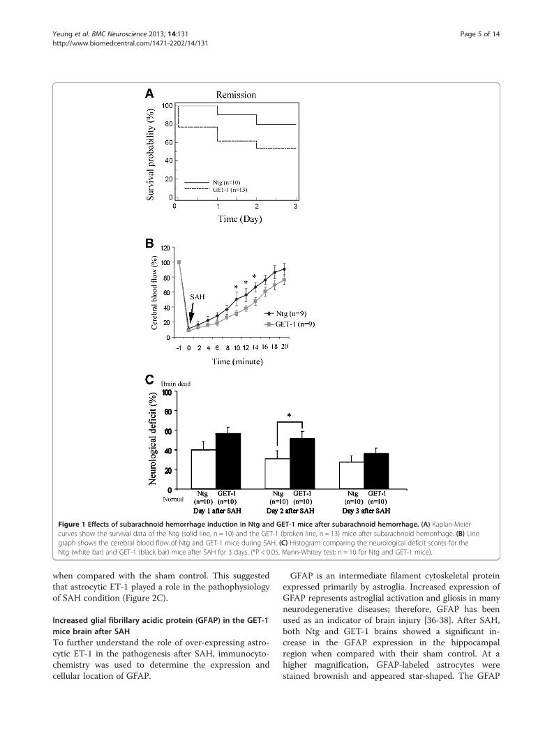
Figure 1 Effects of subarachnoid hemorrhage induction in Ntg and GET-1 mice after subarachnoid hemorrhage. (A) Kaplan-Meiercurves show the survival data of the Ntg (solid line, n = 10) and the GET-1 (broken line, n = 13) mice after subarachnoid hemorrhage. (B) Linegraph shows the cerebral blood flow of Ntg and GET-1 mice during SAH. (C) Histogram comparing the neurological deficit scores for theNtg (white bar) and GET-1 (black bar) mice after SAH for 3 days. (*P < 0.05, Mann-Whitey test; n = 10 for Ntg and GET-1 mice).
Yeung et al. BMC Neuroscience 2013, 14:131 Page 5 of 14http://www.biomedcentral.com/1471-2202/14/131
when compared with the sham control. This suggestedthat astrocytic ET-1 played a role in the pathophysiologyof SAH condition (Figure 2C).
Increased glial fibrillary acidic protein (GFAP) in the GET-1mice brain after SAHTo further understand the role of over-expressing astro-cytic ET-1 in the pathogenesis after SAH, immunocyto-chemistry was used to determine the expression andcellular location of GFAP.
GFAP is an intermediate filament cytoskeletal proteinexpressed primarily by astroglia. Increased expression ofGFAP represents astroglial activation and gliosis in manyneurodegenerative diseases; therefore, GFAP has beenused as an indicator of brain injury [36-38]. After SAH,both Ntg and GET-1 brains showed a significant in-crease in the GFAP expression in the hippocampalregion when compared with their sham control. At ahigher magnification, GFAP-labeled astrocytes werestained brownish and appeared star-shaped. The GFAP
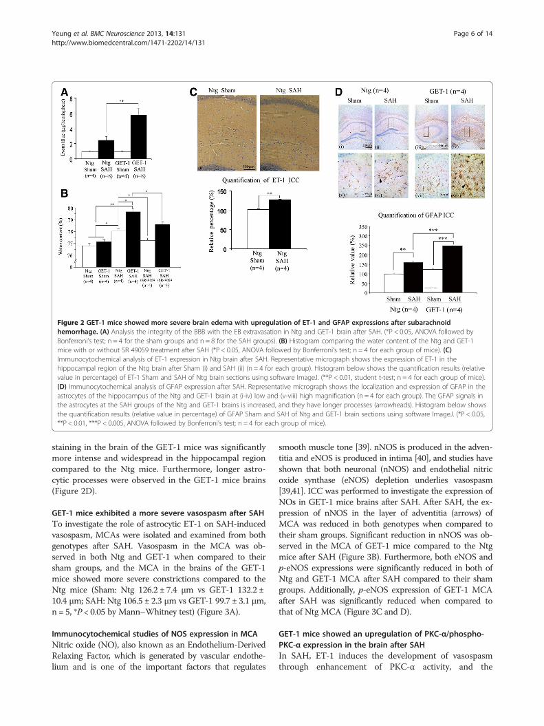
Figure 2 GET-1 mice showed more severe brain edema with upregulation of ET-1 and GFAP expressions after subarachnoidhemorrhage. (A) Analysis the integrity of the BBB with the EB extravasation in Ntg and GET-1 brain after SAH. (*P < 0.05, ANOVA followed byBonferroni’s test; n = 4 for the sham groups and n = 8 for the SAH groups). (B) Histogram comparing the water content of the Ntg and GET-1mice with or without SR 49059 treatment after SAH (*P < 0.05, ANOVA followed by Bonferroni’s test; n = 4 for each group of mice). (C)Immunocytochemical analysis of ET-1 expression in Ntg brain after SAH. Representative micrograph shows the expression of ET-1 in thehippocampal region of the Ntg brain after Sham (i) and SAH (ii) (n = 4 for each group). Histogram below shows the quantification results (relativevalue in percentage) of ET-1 Sham and SAH of Ntg brain sections using software ImageJ. (**P < 0.01, student t-test; n = 4 for each group of mice).(D) Immunocytochemical analysis of GFAP expression after SAH. Representative micrograph shows the localization and expression of GFAP in theastrocytes of the hippocampus of the Ntg and GET-1 brain at (i-iv) low and (v-viii) high magnification (n = 4 for each group). The GFAP signals inthe astrocytes at the SAH groups of the Ntg and GET-1 brains is increased, and they have longer processes (arrowheads). Histogram below showsthe quantification results (relative value in percentage) of GFAP Sham and SAH of Ntg and GET-1 brain sections using software ImageJ. (*P < 0.05,**P < 0.01, ***P < 0.005, ANOVA followed by Bonferroni’s test; n = 4 for each group of mice).
Yeung et al. BMC Neuroscience 2013, 14:131 Page 6 of 14http://www.biomedcentral.com/1471-2202/14/131
staining in the brain of the GET-1 mice was significantlymore intense and widespread in the hippocampal regioncompared to the Ntg mice. Furthermore, longer astro-cytic processes were observed in the GET-1 mice brains(Figure 2D).
GET-1 mice exhibited a more severe vasospasm after SAHTo investigate the role of astrocytic ET-1 on SAH-inducedvasospasm, MCAs were isolated and examined from bothgenotypes after SAH. Vasospasm in the MCA was ob-served in both Ntg and GET-1 when compared to theirsham groups, and the MCA in the brains of the GET-1mice showed more severe constrictions compared to theNtg mice (Sham: Ntg 126.2 ± 7.4 μm vs GET-1 132.2 ±10.4 μm; SAH: Ntg 106.5 ± 2.3 μm vs GET-1 99.7 ± 3.1 μm,n = 5, *P < 0.05 by Mann–Whitney test) (Figure 3A).
Immunocytochemical studies of NOS expression in MCANitric oxide (NO), also known as an Endothelium-DerivedRelaxing Factor, which is generated by vascular endothe-lium and is one of the important factors that regulates
smooth muscle tone [39]. nNOS is produced in the adven-titia and eNOS is produced in intima [40], and studies haveshown that both neuronal (nNOS) and endothelial nitricoxide synthase (eNOS) depletion underlies vasospasm[39,41]. ICC was performed to investigate the expression ofNOs in GET-1 mice brains after SAH. After SAH, the ex-pression of nNOS in the layer of adventitia (arrows) ofMCA was reduced in both genotypes when compared totheir sham groups. Significant reduction in nNOS was ob-served in the MCA of GET-1 mice compared to the Ntgmice after SAH (Figure 3B). Furthermore, both eNOS andp-eNOS expressions were significantly reduced in both ofNtg and GET-1 MCA after SAH compared to their shamgroups. Additionally, p-eNOS expression of GET-1 MCAafter SAH was significantly reduced when compared tothat of Ntg MCA (Figure 3C and D).
GET-1 mice showed an upregulation of PKC-α/phospho-PKC-α expression in the brain after SAHIn SAH, ET-1 induces the development of vasospasmthrough enhancement of PKC-α activity, and the
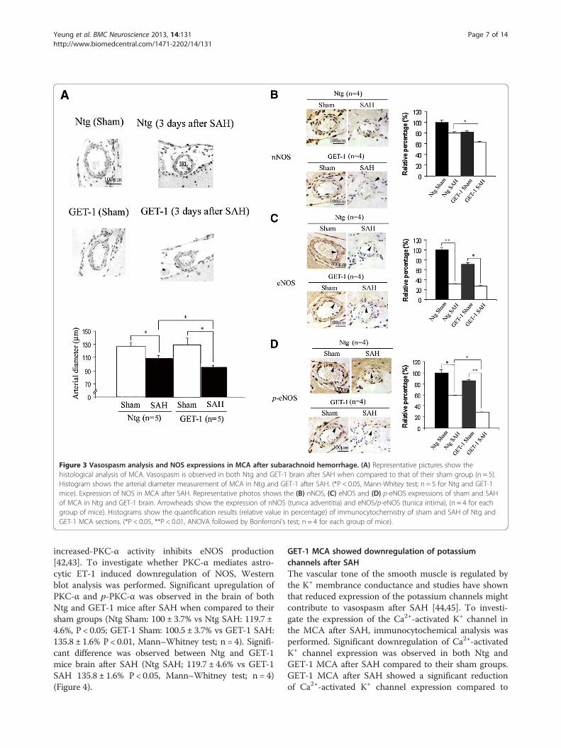
Figure 3 Vasospasm analysis and NOS expressions in MCA after subarachnoid hemorrhage. (A) Representative pictures show thehistological analysis of MCA. Vasospasm is observed in both Ntg and GET-1 brain after SAH when compared to that of their sham group (n = 5).Histogram shows the arterial diameter measurement of MCA in Ntg and GET-1 after SAH. (*P < 0.05, Mann-Whitey test; n = 5 for Ntg and GET-1mice). Expression of NOS in MCA after SAH. Representative photos shows the (B) nNOS, (C) eNOS and (D) p-eNOS expressions of sham and SAHof MCA in Ntg and GET-1 brain. Arrowheads show the expression of nNOS (tunica adventitia) and eNOS/p-eNOS (tunica intima), (n = 4 for eachgroup of mice). Histograms show the quantification results (relative value in percentage) of immunocytochemistry of sham and SAH of Ntg andGET-1 MCA sections. (*P < 0.05, **P < 0.01, ANOVA followed by Bonferroni’s test; n = 4 for each group of mice).
Yeung et al. BMC Neuroscience 2013, 14:131 Page 7 of 14http://www.biomedcentral.com/1471-2202/14/131
increased-PKC-α activity inhibits eNOS production[42,43]. To investigate whether PKC-α mediates astro-cytic ET-1 induced downregulation of NOS, Westernblot analysis was performed. Significant upregulation ofPKC-α and p-PKC-α was observed in the brain of bothNtg and GET-1 mice after SAH when compared to theirsham groups (Ntg Sham: 100 ± 3.7% vs Ntg SAH: 119.7 ±4.6%, P < 0.05; GET-1 Sham: 100.5 ± 3.7% vs GET-1 SAH:135.8 ± 1.6% P < 0.01, Mann–Whitney test; n = 4). Signifi-cant difference was observed between Ntg and GET-1mice brain after SAH (Ntg SAH; 119.7 ± 4.6% vs GET-1SAH 135.8 ± 1.6% P < 0.05, Mann–Whitney test; n = 4)(Figure 4).
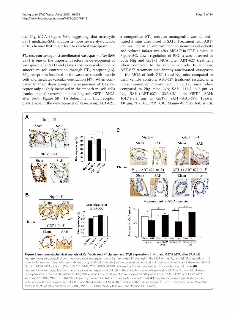
GET-1 MCA showed downregulation of potassiumchannels after SAHThe vascular tone of the smooth muscle is regulated bythe K+ membrance conductance and studies have shownthat reduced expression of the potassium channels mightcontribute to vasospasm after SAH [44,45]. To investi-gate the expression of the Ca2+-activated K+ channel inthe MCA after SAH, immunocytochemical analysis wasperformed. Significant downregulation of Ca2+-activatedK+ channel expression was observed in both Ntg andGET-1 MCA after SAH compared to their sham groups.GET-1 MCA after SAH showed a significant reductionof Ca2+-activated K+ channel expression compared to
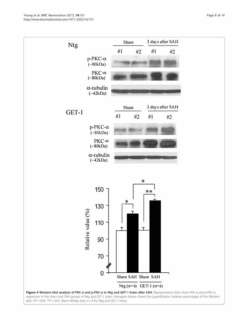
Figure 4 Western blot analysis of PKC-α and p-PKC-α in Ntg and GET-1 brain after SAH. Representative blots show PKC-α and p-PKC-αexpression in the sham and SAH groups of Ntg and GET-1 brain. Histogram below shows the quantification (relative percentage) of the Westernblot. (*P < 0.05, **P < 0.01, Mann-Whitey test; n = 4 for Ntg and GET-1 mice).
Yeung et al. BMC Neuroscience 2013, 14:131 Page 8 of 14http://www.biomedcentral.com/1471-2202/14/131
Yeung et al. BMC Neuroscience 2013, 14:131 Page 9 of 14http://www.biomedcentral.com/1471-2202/14/131
the Ntg MCA (Figure 5A), suggesting that astrocyticET-1 mediated-SAH induces a more severe dysfunctionof K+ channel that might lead to cerebral vasospasm.
ETA receptor antagonist ameliorated vasospasm after SAHET-1 is one of the important factors in development ofvasospasm after SAH and plays a role in vascular tone ofsmooth muscle contraction through ETA receptor [46].ETA receptor is localized in the vascular smooth musclecells and mediates vascular contraction [47]. When com-pared to their sham groups, the expression of ETA re-ceptor only slightly increased in the smooth muscle cells(tunica media) (arrows) in both Ntg and GET-1 MCAafter SAH (Figure 5B). To determine if ETA receptorplays a role in the development of vasospasm, ABT-627,
Figure 5 Immunocytochemical analysis of Ca2+-activated K+ channel aRepresentative micrograph shows the localization and expression of Ca2+-afrom each group of mice). Histogram shows the quantification results (relaNtg and GET-1 MCA sections. (*P < 0.05, **P < 0.01, ***P < 0.005, ANOVA folRepresentative micrograph shows the localization and expression of ETAR iHistogram shows the quantification results (relative value in percentage) ofsections. (*P < 0.05, **P < 0.01, ANOVA followed by Bonferroni’s test; n = 3 fimmunocytochemical expressions of PKC-α and the diameters of MCA aftemeasurements of MCA diameter. (*P < 0.05, **P < 0.01, Mann-Whitey test; n
a competitive ETA receptor anatagonist, was adminis-trated 5 mins after onset of SAH. Treatment with ABT-627 resulted in an improvement in neurological deficitsand reduced infarct size after MCAO in GET-1 mice. InFigure 5C, down-regulation of PKC-α was observed inboth Ntg and GET-1 MCA after ABT-627 treatmentwhen compared to the vehicle controls. In addition,ABT-627 treatment significantly ameliorated vasospasmin the MCA of both GET-1 and Ntg mice compared totheir vehicle controls. ABT-627 treatment resulted in amore promising improvement in GET-1 mice whencompared to Ntg mice (Ntg SAH: 114.5 ± 4.9 μm vsNtg SAH+ABT-627: 133.5 ± 5.1 μm; GET-1 SAH:104.7 ± 3.1 μm vs GET-1 SAH+ABT-627: 138.6 ±1.0 μm, *P < 0.05, **P < 0.01 Mann–Whitney test; n = 3).
nd ETAR expressions in Ntg and GET-1 MCA after SAH. (A)ctivated K+ channel in the MCA of the Ntg and GET-1 after SAH. (n = 3tive value in percentage) of immunocytochemistry of sham and SAH oflowed by Bonferroni’s test; n = 3 for each group of mice). (B)n the smooth muscle cells (arrows) of MCA in Ntg and GET-1 mice.immunocytochemistry of sham and SAH of Ntg and GET-1 MCAor each group of mice). (C) Representative micrograph shows ther treating with ETAR antagonist ABT-627. Histogram below shows the= 3 for Ntg and GET-1 mice).
Yeung et al. BMC Neuroscience 2013, 14:131 Page 10 of 14http://www.biomedcentral.com/1471-2202/14/131
Administration of ETB receptor anatagonist, BQ788, how-ever, did not show any effect in either genotypes (data notshown).
DiscussionElevation of ET-1 levels in the cerebrospinal fluid afterSAH have been reported in SAH patients [18]. ET-1 isknown to be a potent endothelium-derived vasocon-stricting agent [48]. In the CNS, ET-1 can be producedby astrocytes, neurons and pituitary cells under normalphysiological conditions [12,14,49]. ET-1 is also releasedfrom endothelial and smooth muscle cells when stimu-lated by thrombin and oxyhemoglobin [8,50]. However,to date the source and the release mechanisms of ET-1are still largely unknown. In order to address whetherastrocytic ET-1 plays a role in the development of cere-bral edema and vasospasm after SAH, transgenic miceover-expressing ET-1 in astrocytes (GET-1) was used toexaggerate the effects of ET-1 during SAH and delayedischemia after SAH. The GET-1 mice (from 1 week to20 weeks old) show higher endothelin-1 mRNA andpeptide levels in the brain when compared to Ntg brains.In spite of the increased level of astrocytic ET-1, GET-1mice appear normal under physiological condition. Thecerebrovasculature and the mean artery blood pressureis similar in the GET-1 compared to the Ntg mice, sug-gesting that the GET-1 mice show no significant differ-ence in the cerebral artery under normal physiologicalcondition [20]. Although GET-1 mice have been shownto have higher endothelin-1 mRNA and peptide levels inthe brain when compared to Ntg brains [20], their cere-brovasculatures showed no significant difference to thatof Ntg brains. We speculate that the insignificant in-creased level of ET-1 in GET-1 mice may not cause anyexacerbating effects on cerebral vasospasm and edemaformation; however, these mice will be more susceptibleto these pathophysiological processes under stress condi-tions, such as SAH when astrocytic ET-1 level will be in-creased substantially.Vasopressin levels increase in the plasma and cerebro-
spinal fluid after SAH, suggesting that the antidiuretichormone plays a critical role in edema development[51]. SR 49059 is considered to be the most potent andselective non-peptide V1a receptor antagonist in bothanimals and human [52,53]. Recently, SR 49059 wasshown to reduce intracerebral hemorrhagic brain injury-induced cerebral edema in mice [54]. In the presentstudy, arterial puncture technique was used to inducesubarachnoid hemorrhage, and GET-1 mice developedmore severe brain damage and edema after this proced-ure. SR 49059 was administrated intraperitoneally to theNTg and GET-1 mice to evaluate the effect of vasopres-sin V1a receptor antagonist in the edema formation afterSAH. The results showed that SR 49059 significantly
reduced brain water content in both Ntg and GET-1mice 24 hours after SAH, which is in agreement with aprevious study [55] despite using a different type ofhemorrhagic stroke model and paradigm. Collectively,these data suggest that SR 49059 is a promising drug intreating hemorrhagic brain edema and provide a strongrationale to investigate the drug in clinical settings.Although many studies have reported that ET-1 levels
are increased in plasma and CSF during ischemic andhemorrhagic stroke [56,57], it is still unclear whether theproduction and release of ET-1 is the primary responseof the brain cells (astrocytes, endothelial cells, neurons)to the stroke, or the secondary response in which activa-tion of the sympathoadrenal system increases the plasmacatecholamine and vasopressin levels [58], which stimu-lates the release of ET-1 from the peripheral organs. Inthe present study, over-expression of astrocytic ET-1 inGET-1 mice resulted in a more severe brain damage andvasospasm, suggesting that astrocytes may be one of themajor sources of ET-1 production and release underpathological conditions. Previous in vitro data suggestedthat the SAH-induced hypoxia-ischemia in astrocytes ac-counts for the ET-1 release into the subarachnoid space[56]. The present report provides the first documenta-tion for the significance of astrocytic ET-1 in haemor-rhagic stroke in an animal model. Our data demonstratethat overexpression of astrocytic ET-1 excerbates severalpathophysiological processes after SAH, and this couldbe a contributing factor to these processes together withthe physiological levels of astrocytic ET-1, however, wecould not directly conclude that this is the case. Furtherstudies in animals, such as with targeted deletion ofastrocytic ET-1, will be required before drawing theconclusion.In agreement with other studies, we demonstrate that
astrocytic ET-1 also induces vasospasm with a concur-rent elevation of PKC-α protein expression and activa-tion [43,59,60]. ET-1 regulates the vascular tone of thecerebral blood vessels through its receptor subtypes,ETA and ETB. ETB receptors are known to mediate vaso-dilation upon localization to the endothelial cells ofblood vessels. A recent study shows that the expressionof ETB receptors is regulated by initial cerebral bloodflow through the MEK-ERK1/2 signaling pathway [61].ETA receptors are mainly found in smooth muscle cellsand are involved in vasoconstriction; therefore, they arecrucial in cerebral vasospasm [62]. In the present study,immunocytochemical analysis of ETA receptor expres-sion in MCA showed an insignificant change in bothNtg and GET-1 after SAH, which is in agreement withthe previous finding that the expression of smooth-muscle ETA receptors and their mRNA level is un-changed or slightly increased in the cerebral arteriesafter SAH [63,64]. It is demonstrated that an increased
Yeung et al. BMC Neuroscience 2013, 14:131 Page 11 of 14http://www.biomedcentral.com/1471-2202/14/131
coupling of the smooth muscle ETA receptor with thesecond cascade probably contributes to the developmentof cerebral vasospasm [64]. ETA receptor antagonistshave been used in numerous studies in alleviatingSAH-induced cerebral vasospasm [65-67]. However,other studies have also reported that ETA receptor an-tagonists have the potential adverse effects such ashypotension and pneumonia. Moreover, there are no sig-nificant differences in mortality or improving outcomesin the phase 3 clinical trials investigating ETA receptorantagonists as a therapeutic strategy for vasospasm[68-71]. However, ETA receptor antagonists, such as cla-zosentan, have been used in alleviating SAH-inducedcerebral vasospasm [72]. In a clinical study, only highdoses of clazosentan resulted in a significantly reducedvasospasm-related morbidity or all-cause mortalitywithin 6 weeks post SAH, but not at longer time points[69], suggesting that ETA receptor antagonist could beused for treating vasospasm. However, the interferenceby other drugs taken by the patients during the clinicalstudy may reduce the efficiency of the clazosentan at alater time point. In the current study, ETA receptor an-tagonist ABT-627 effectively attenuated SAH-inducedvasospasm in both Ntg and GET-1 mice, and suggested
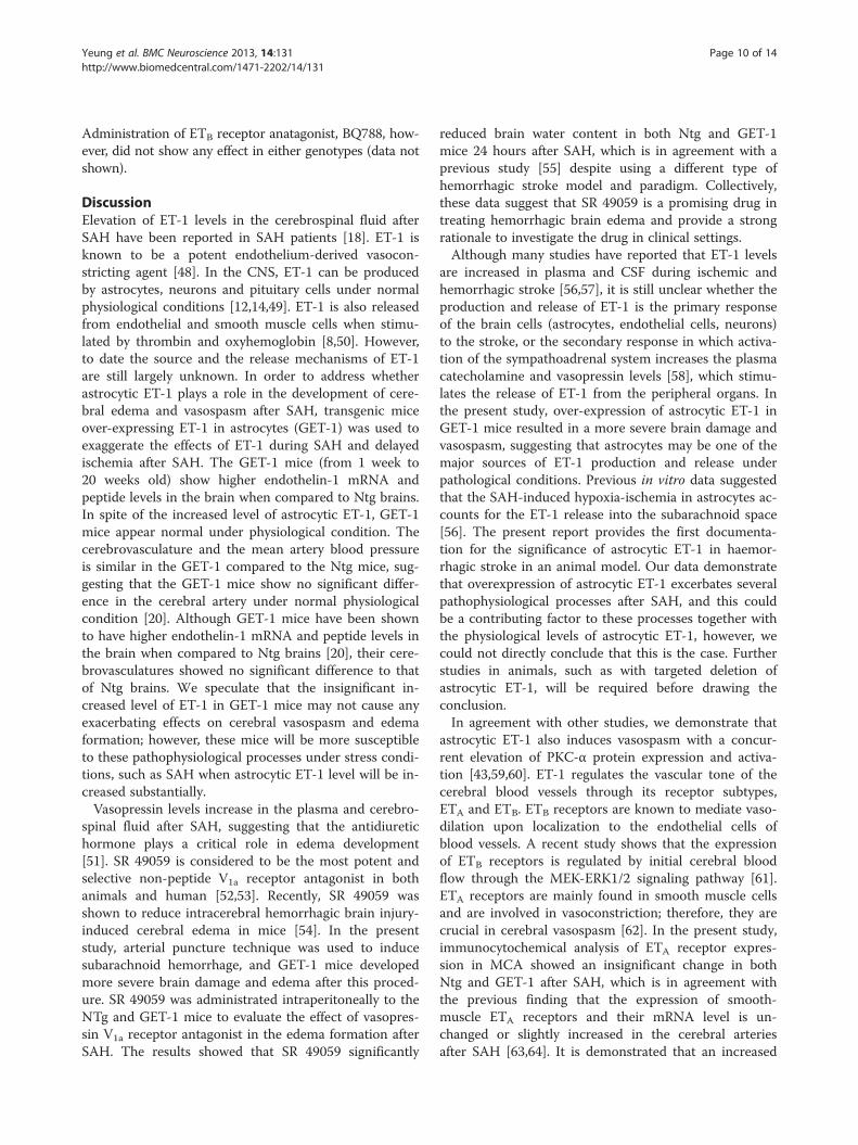
Figure 6 Proposed mechanism of astrocytic ET-1 mediated cerebral vmore severe neurological deficits and vasospasm after SAH. Increased astroreceptor and mediated by PKC-α, which leads to dynfunction in the K+ chathe SAH-induced vasospasm. The impairment of NO system also exaggeratedema and BBB breakdown that further contributes to cerebral vasoconstrreduced the SAH-induced edema, suggesting that astrocytic ET-1 induces e
that pathways elicited by astrocytic ET-1 through ETA
receptor, but not ETB receptor, are involved in SAH-induced vasospasm. ABT-627 is reported to greatly im-prove neurological deficits and reduce infarct size inmice after ischemic stroke, suggesting that the drugcould normalize the vasoconstriction effect of ET-1 [28],as shown in the present study. Although a few ETA
receptor antagonists have been shown to have adverseeffects in the SAH patients, we cannot rule out the pos-sibility that there are pharmacological differences amongETA receptor antagonists as well as testing conductedunder non-optimized conditions. Here we report an-other possible ETA receptor antagonist candidate, ABT-627, which may be an alternative therapeutic drug fortreating SAH-induced vasospasm.ET-1 stimulates the ETB receptor and increases NO
production [73]. NO is suggested to reverse the effectsof the ET-1-induced vasoconstriction, by negative feed-back and reducing the level of ET-1 [73,74]. Diminishedproduction of NO and increased release of ET-1, there-fore, are suggested to be the vital consequences of vaso-spasm in SAH [75]. It is reported that endothelial nitricoxide synthase (eNOS) expression as well as NO pro-duction are impaired by the elevated ET-1 level through
asospasm after SAH. GET-1 mice with over-expressed in ET-1 showedcytic ET-1 during SAH induces cerebral vasospasm through the ETAnnels. Administration of ETA receptor antagonist ABT-627 ameliorateses the vasospasm effect. Astrocytic ET-1 leads to more severe cerebraliction. Vasopressin V1a receptor antagonist, SR 49059, significantlydema in SAH through vasopressin V1a receptor.
Yeung et al. BMC Neuroscience 2013, 14:131 Page 12 of 14http://www.biomedcentral.com/1471-2202/14/131
a PKC-dependent pathway [42]. Since nitric oxide in-duces vascular relaxation by inhibiting PKC activity, thereduced NO production after SAH, therefore, enhancesthe PKC-dependent vasoconstriction [76]. Also, elevatedlevel of ET-1 activates ETA receptor and decreases thevascular sensitivity to NO by a PKC-independent path-way [77]. Down-regulation of PKC-α was observed inboth Ntg and GET-1 MCA after ETA receptor antagonistABT-627 treatment (Figure 5C), suggesting that astro-cytic ET-1 induced vasospasm is also mediated throughETA receptor and PKC. These data collectively provide astrong rationale to investigate ABT-627 as a therapeuticdrug to treat SAH-induced vasospasm.K+ channels are important in regulating the membrane
potential of arterial smooth muscle. Large conductanceCa2+-activated K+ channel is one of the channels that isfound in the arterial smooth muscle and contributes tothe resting membrane potential [45]. Recently, thefunction and expression of ion channels, particularly theCa2+-activated K+ channels, have been investigated inSAH-induced vasospasm. In the present study, the ex-pression of Ca2+-activated K+ channels in MCA waslargely reduced after SAH in both Ntg and GET-1 mice,which is in agreement with others findings that elevatedPKC activity after SAH causes dysfunction in K+ channelactivity and expression [78,79].Here we propose the possible mechanism of the astro-
cytic ET-1 mediated cerebral vasospasm after SAH(Figure 6). The astrocytic ET-1 induces cerebral vaso-spasm through the ETA receptor, which leads to anupregulation of PKC-α.
ConclusionsTo the best of our knowledge, this is the first study toelucidate the role of astrocytic ET-1 in vasogenic edemaformation and vasospasm development in SAH-inducedbrain injured mice. Both the vasopressin V1a receptorantagonist, SR 49059, and ETA receptor inhibitor, ABT-627 significantly ameliorated the astrocytic ET-1 inducedcomplications after SAH. These potential therapeuticdrugs could be used in future for treating SAH patients.
Competing interestsThe authors declare that they have no competing interests.
Authors’ contributionsPKKY performed the majority of the experimental work, designedexperiments, analysed data and wrote the manuscript. JS, SSMC and SKCconceived the idea, designed experiments and edited the manuscript.All authors read and approved the final manuscript.
AcknowledgementThis study was partly supported by the RGC grant to Prof. S.K. Chung andthe Area of Excellence from University Grants Council of Hong Kong on“Molecular Neuroscience: Basic Research and Drug Discovery” (AoE/B-15/01).
Author details1Department of Anatomy, Li Ka Shing Faculty of Medicine, The University ofHong Kong, Hong Kong SAR, China. 2School of Chinese Medicine, TheUniversity of Hong Kong, Hong Kong, SAR, China. 3Research Center of Heart,Brain, Hormone and Healthy Aging, The University of Hong Kong, HongKong, SAR, China. 4Division of Science and Technology, United InternationalCollege, Zhuhai, Guandong, China. 5Department of Anatomy, The Universityof Hong Kong, 1/F, Laboratory Block, Faculty of Medicine Building, 21Sassoon Road, Hong Kong, SAR, China.
Received: 8 April 2013 Accepted: 15 October 2013Published: 25 October 2013
References1. Claassen J, Carhuapoma JR, Kreiter KT, Du EY, Connolly ES, Mayer SA: Global
cerebral edema after subarachnoid hemorrhage: frequency, predictors,and impact on outcome. Stroke 2002, 33(5):1225–1232.
2. Macdonald RL: Pathophysiology and molecular genetics of vasospasm.Acta Neurochir Suppl 2001, 77:7–11.
3. Molnar AH, Varga C, Berko A, Rojik I, Parducz A, Laszlo F, Laszlo FA:Inhibitory effect of vasopressin receptor antagonist OPC-31260 onexperimental brain oedema induced by global cerebral ischaemia.Acta Neurochir (Wien) 2008, 150(3):265–271.
4. Liu X, Nakayama S, Amiry-Moghaddam M, Ottersen OP, Bhardwaj A:Arginine-vasopressin V1 but not V2 receptor antagonism modulatesinfarct volume, brain water content, and aquaporin-4 expressionfollowing experimental stroke. Neurocrit Care 2010, 12(1):124–131.
5. Kleindienst A, Fazzina G, Dunbar JG, Glisson R, Marmarou A: Protectiveeffect of the V1a receptor antagonist SR 49059 on brain edemaformation following middle cerebral artery occlusion in the rat.Acta Neurochir Suppl 2006, 96:303–306.
6. Sano K, Asano T, Tanishima T, Sasaki T: Lipid peroxidation as a cause ofcerebral vasospasm. Neurol Res 1980, 2(3–4):253–272.
7. Kanamaru K, Waga S, Kojima T, Fujimoto K, Niwa S: Endothelium-dependent relaxation of canine basilar arteries. Part 2: Inhibition byhemoglobin and cerebrospinal fluid from patients with aneurysmalsubarachnoid hemorrhage. Stroke 1987, 18(5):938–943.
8. Cocks TM, Malta E, King SJ, Woods RL, Angus JA: Oxyhaemoglobinincreases the production of endothelin-1 by endothelial cells in culture.Eur J Pharmacol 1991, 196(2):177–182.
9. Macdonald RL, Weir BK: A review of hemoglobin and the pathogenesis ofcerebral vasospasm. Stroke 1991, 22(8):971–982.
10. Asano T, Ikegaki I, Suzuki Y, Satoh S, Shibuya M: Endothelin and theproduction of cerebral vasospasm in dogs. Biochem Biophys ResCommun 1989, 159(3):1345–1351.
11. Mima T, Yanagisawa M, Shigeno T, Saito A, Goto K, Takakura K, Masaki T:Endothelin acts in feline and canine cerebral arteries from theadventitial side. Stroke 1989, 20(11):1553–1556.
12. Giaid A, Gibson SJ, Ibrahim BN, Legon S, Bloom SR, Yanagisawa M, Masaki T,Varndell IM, Polak JM: Endothelin 1, an endothelium-derived peptide, isexpressed in neurons of the human spinal cord and dorsal root ganglia.Proc Natl Acad Sci USA 1989, 86(19):7634–7638.
13. Lee ME, de la Monte SM, Ng SC, Bloch KD, Quertermous T: Expression ofthe potent vasoconstrictor endothelin in the human central nervoussystem. J Clin Invest 1990, 86(1):141–147.
14. MacCumber MW, Ross CA, Snyder SH: Endothelin in brain: receptors,mitogenesis, and biosynthesis in glial cells. Proc Natl Acad Sci USA 1990,87(6):2359–2363.
15. Fassbender K, Hodapp B, Rossol S, Bertsch T, Schmeck J, Schutt S, FritzingerM, Horn P, Vajkoczy P, Wendel-Wellner M, et al: Endothelin-1 insubarachnoid hemorrhage: An acute-phase reactant produced bycerebrospinal fluid leukocytes. Stroke 2000, 31(12):2971–2975.
16. Kasuya H, Weir BK, White DM, Stefansson K: Mechanism ofoxyhemoglobin-induced release of endothelin-1 from culturedvascular endothelial cells and smooth-muscle cells. J Neurosurg 1993,79(6):892–898.
17. Ehrenreich H, Lange M, Near KA, Anneser F, Schoeller LA, Schmid R, WinklerPA, Kehrl JH, Schmiedek P, Goebel FD: Long term monitoring ofimmunoreactive endothelin-1 and endothelin-3 in ventricularcerebrospinal fluid, plasma, and 24-h urine of patients withsubarachnoid hemorrhage. Res Exp Med (Berl) 1992, 192(4):257–268.
Yeung et al. BMC Neuroscience 2013, 14:131 Page 13 of 14http://www.biomedcentral.com/1471-2202/14/131
18. Fujimori A, Yanagisawa M, Saito A, Goto K, Masaki T, Mima T, Takakura K,Shigeno T: Endothelin in plasma and cerebrospinal fluid of patients withsubarachnoid haemorrhage. Lancet 1990, 336(8715):633.
19. Suzuki R, Masaoka H, Hirata Y, Marumo F, Isotani E, Hirakawa K: The role ofendothelin-1 in the origin of cerebral vasospasm in patients withaneurysmal subarachnoid hemorrhage. J Neurosurg 1992, 77(1):96–100.
20. Lo AC, Chen AY, Hung VK, Yaw LP, Fung MK, Ho MC, Tsang MC, Chung SS,Chung SK: Endothelin-1 overexpression leads to further wateraccumulation and brain edema after middle cerebral artery occlusion viaaquaporin 4 expression in astrocytic end-feet. J Cereb Blood Flow Metab2005, 25(8):998–1011.
21. Yeung PK, Lo AC, Leung JW, Chung SS, Chung SK: Targetedoverexpression of endothelin-1 in astrocytes leads to more severecytotoxic brain edema and higher mortality. J Cereb Blood Flow Metab2009, 29(12):1891–1902.
22. Bederson JB, Germano IM, Guarino L: Cortical blood flow and cerebralperfusion pressure in a new noncraniotomy model of subarachnoidhemorrhage in the rat. Stroke 1995, 26(6):1086–1091. discussion 1091–1082.
23. Huang Z, Huang PL, Panahian N, Dalkara T, Fishman MC, Moskowitz MA:Effects of cerebral ischemia in mice deficient in neuronal nitric oxidesynthase. Science 1994, 265(5180):1883–1885.
24. Carrillo P, Takasu A, Safar P, Tisherman S, Stezoski SW, Stolz G, Dixon CE, RadovskyA: Prolonged severe hemorrhagic shock and resuscitation in rats does notcause subtle brain damage. J Trauma 1998, 45(2):239–248. discussion 248–239.
25. Kamii H, Kato I, Kinouchi H, Chan PH, Epstein CJ, Akabane A, Okamoto H,Yoshimoto T: Amelioration of vasospasm after subarachnoid hemorrhagein transgenic mice overexpressing CuZn-superoxide dismutase.Stroke 1999, 30(4):867–871. discussion 872.
26. Mesis RG, Wang H, Lombard FW, Yates R, Vitek MP, Borel CO, Warner DS,Laskowitz DT: Dissociation between vasospasm and functionalimprovement in a murine model of subarachnoid hemorrhage.Neurosurg Focus 2006, 21(3):E4.
27. McGirt MJ, Lynch JR, Parra A, Sheng H, Pearlstein RD, Laskowitz DT,Pelligrino DA, Warner DS: Simvastatin increases endothelial nitric oxidesynthase and ameliorates cerebral vasospasm resulting fromsubarachnoid hemorrhage. Stroke 2002, 33(12):2950–2956.
28. Ostrowski RP, Colohan AR, Zhang JH: Mechanisms of hyperbaric oxygen-induced neuroprotection in a rat model of subarachnoid hemorrhage.J Cereb Blood Flow Metab 2005, 25(5):554–571.
29. Altay O, Suzuki H, Hasegawa Y, Caner B, Krafft PR, Fujii M, Tang J, Zhang JH:Isoflurane attenuates blood–brain barrier disruption in ipsilateral hemisphereafter subarachnoid hemorrhage in mice. Stroke 2012, 43(9):2513–2516.
30. Yatsushige H, Ostrowski RP, Tsubokawa T, Colohan A, Zhang JH: Role ofc-Jun N-terminal kinase in early brain injury after subarachnoidhemorrhage. J Neurosci Res 2007, 85(7):1436–1448.
31. Sabri M, Jeon H, Ai J, Tariq A, Shang X, Chen G, Macdonald RL: Anteriorcirculation mouse model of subarachnoid hemorrhage. Brain Res 2009,1295:179–185.
32. Mak KM, Lo AC, Lam AK, Yeung PK, Ko BC, Chung SS, Chung SK: Nuclearfactor of activated T cells 5 deficiency increases the severity of neuronalcell death in ischemic injury. Neurosignals 2012, 20(4):237–251.
33. Lin EY, Li JF, Gnatovskiy L, Deng Y, Zhu L, Grzesik DA, Qian H, Xue XN,Pollard JW: Macrophages regulate the angiogenic switch in a mousemodel of breast cancer. Cancer Res 2006, 66(23):11238–11246.
34. Shuaib A, Xu Wang C, Yang T, Noor R: Effects of nonpeptide V(1)vasopressin receptor antagonist SR-49059 on infarction volume andrecovery of function in a focal embolic stroke model. Stroke 2002,33(12):3033–3037.
35. Laszlo FA, Varga C, Nakamura S: Vasopressin receptor antagonist OPC-31260 prevents cerebral oedema after subarachnoid haemorrhage.Eur J Pharmacol 1999, 364(2–3):115–122.
36. Liao CW, Fan CK, Kao TC, Ji DD, Su KE, Lin YH, Cho WL: Brain injury-associated biomarkers of TGF-beta1, S100B, GFAP, NF-L, tTG, AbetaPP,and tau were concomitantly enhanced and the UPS was impairedduring acute brain injury caused by Toxocara canis in mice.BMC Infect Dis 2008, 8:84.
37. Pekny M, Pekna M: Astrocyte intermediate filaments in CNS pathologiesand regeneration. J Pathol 2004, 204(4):428–437.
38. Pekny M, Wilhelmsson U, Bogestal YR, Pekna M: The role of astrocytesand complement system in neural plasticity. Int Rev Neurobiol 2007,82:95–111.
39. Ignarro LJ: Nitric oxide as a unique signaling molecule in the vascularsystem: a historical overview. J Physiol Pharmacol 2002, 53(4 Pt 1):503–514.
40. Pluta RM: Dysfunction of nitric oxide synthases as a cause andtherapeutic target in delayed cerebral vasospasm after SAH.Acta Neurochir Suppl 2008, 104:139–147.
41. Pluta RM, Thompson BG, Dawson TM, Snyder SH, Boock RJ, Oldfield EH:Loss of nitric oxide synthase immunoreactivity in cerebral vasospasm.J Neurosurg 1996, 84(4):648–654.
42. Ramzy D, Rao V, Tumiati LC, Xu N, Sheshgiri R, Miriuka S, Delgado DH, RossHJ: Elevated endothelin-1 levels impair nitric oxide homeostasis througha PKC-dependent pathway. Circulation 2006, 114(1 Suppl):I319–I326.
43. Nishizawa S, Chen D, Yokoyama T, Yokota N, Otha S: Endothelin-1 initiatesthe development of vasospasm after subarachnoid haemorrhagethrough protein kinase C activation, but does not contribute toprolonged vasospasm. Acta Neurochir 2000, 142(12):1409–1415.
44. Aihara Y, Jahromi BS, Yassari R, Nikitina E, Agbaje-Williams M, Macdonald RL:Molecular profile of vascular ion channels after experimentalsubarachnoid hemorrhage. J Cereb Blood Flow Metab 2004, 24(1):75–83.
45. Standen NB, Quayle JM: K + channel modulation in arterial smoothmuscle. Acta Physiol Scand 1998, 164(4):549–557.
46. Zimmermann M, Seifert V: Endothelin and subarachnoid hemorrhage: anoverview. Neurosurgery 1998, 43(4):863–875. discussion 875–866.
47. Zimmermann M, Seifert V: Endothelin receptor antagonists and cerebralvasospasm. Clin Auton Res 2004, 14(3):143–145.
48. Yanagisawa M, Kurihara H, Kimura S, Tomobe Y, Kobayashi M, Mitsui Y,Yazaki Y, Goto K, Masaki T: A novel potent vasoconstrictor peptideproduced by vascular endothelial cells. Nature 1988, 332(6163):411–415.
49. Yoshizawa T, Shinmi O, Giaid A, Yanagisawa M, Gibson SJ, Kimura S,Uchiyama Y, Polak JM, Masaki T, Kanazawa I: Endothelin: a novel peptide inthe posterior pituitary system. Science 1990, 247(4941):462–464.
50. Ehrenreich H, Costa T, Clouse KA, Pluta RM, Ogino Y, Coligan JE, Burd PR:Thrombin is a regulator of astrocytic endothelin-1. Brain Res 1993,600(2):201–207.
51. Laszlo FA, Varga C, Doczi T: Cerebral oedema after subarachnoidhaemorrhage. Pathogenetic significance of vasopressin. Acta Neurochir(Wien) 1995, 133(3–4):122–133.
52. Serradeil-Le Gal C, Wagnon J, Garcia C, Lacour C, Guiraudou P, Christophe B,Villanova G, Nisato D, Maffrand JP, Le Fur G, et al: Biochemical andpharmacological properties of SR 49059, a new, potent, nonpeptide antagonistof rat and human vasopressin V1a receptors. J Clin Invest 1993, 92(1):224–231.
53. Thibonnier M, Kilani A, Rahman M, DiBlasi TP, Warner K, Smith MC, Leenhardt AF,Brouard R: Effects of the nonpeptide V(1) vasopressin receptor antagonist SR49059 in hypertensive patients. Hypertension 1999, 34(6):1293–1300.
54. Manaenko A, Fathali N, Khatibi NH, Lekic T, Hasegawa Y, Martin R, Tang J,Zhang JH: Arginine-vasopressin V1a receptor inhibition improvesneurologic outcomes following an intracerebral hemorrhagic braininjury. Neurochem Int 2011, 58(4):542–548.
55. Manaenko A, Fathali N, Khatibi NH, Lekic T, Shum KJ, Martin R, Zhang JH, Tang J:Post-treatment with SR 49059 improves outcomes following an intracerebralhemorrhagic stroke in mice. Acta Neurochir Suppl 2011, 111:191–196.
56. Pluta RM, Boock RJ, Afshar JK, Clouse K, Bacic M, Ehrenreich H, Oldfield EH:Source and cause of endothelin-1 release into cerebrospinal fluid aftersubarachnoid hemorrhage. J Neurosurg 1997, 87(2):287–293.
57. Bian LG, Zhang TX, Zhao WG, Shen JK, Yang GY: Increased endothelin-1 inthe rabbit model of middle cerebral artery occlusion. Neurosci Lett 1994,174(1):47–50.
58. Matsumura K, Abe I, Tsuchihashi T, Tominaga M, Kobayashi K, Fujishima M:Central effect of endothelin on neurohormonal responses in consciousrabbits. Hypertension 1991, 17(6 Pt 2):1192–1196.
59. Nishizawa S, Obara K, Koide M, Nakayama K, Ohta S, Yokoyama T:Attenuation of canine cerebral vasospasm after subarachnoidhemorrhage by protein kinase C inhibitors despite augmentedphosphorylation of myosin light chain. J Vasc Res 2003, 40(2):169–178.
60. Yang G, Li T, Xu J, Liu L: PKC plays an important mediated effect inarginine vasopressin induced restoration of vascular responsivenessand calcium sensitization following hemorrhagic shock in rats.Eur J Pharmacol 2010, 628(1–3):148–154.
61. Povlsen GK, Johansson SE, Larsen CC, Samraj AK, Edvinsson L: Earlyevents triggering delayed vasoconstrictor receptor upregulation andcerebral ischemia after subarachnoid hemorrhage. BMC Neurosci2013, 14(1):34.
Yeung et al. BMC Neuroscience 2013, 14:131 Page 14 of 14http://www.biomedcentral.com/1471-2202/14/131
62. Chow M, Dumont AS, Kassell NF: Endothelin receptor antagonists andcerebral vasospasm: an update. Neurosurgery 2002, 51(6):1333–1341.discussion 1342.
63. Hansen-Schwartz J, Ansar S, Edvinsson L: Cerebral vasoconstriction aftersubarachnoid hemorrhage–role of changes in vascular receptorphenotype. Front Biosci 2008, 13:2160–2164.
64. Vatter H, Konczalla J, Weidauer S, Preibisch C, Zimmermann M, Raabe A,Seifert V: Effect of delayed cerebral vasospasm on cerebrovascularendothelin A receptor expression and function. J Neurosurg 2007,107(1):121–127.
65. Clozel M, Watanabe H: BQ-123, a peptidic endothelin ETA receptorantagonist, prevents the early cerebral vasospasm followingsubarachnoid hemorrhage after intracisternal but not intravenousinjection. Life Sci 1993, 52(9):825–834.
66. Kita T, Kubo K, Hiramatsu K, Sakaki T, Yonetani Y, Sato S, Fujimoto M,Nakashima T: Profiles of an intravenously available endothelin A-receptorantagonist, S-0139, for preventing cerebral vasospasm in a caninetwo-hemorrhage model. Life Sci 1998, 63(4):305–315.
67. Zuccarello M, Lewis AI, Rapoport RM: Endothelin ETA and ETB receptors insubarachnoid hemorrhage-induced cerebral vasospasm. Eur J Pharmacol1994, 259(1):R1–R2.
68. Kramer A, Fletcher J: Do endothelin-receptor antagonists prevent delayedneurological deficits and poor outcomes after aneurysmal subarachnoidhemorrhage?: a meta-analysis. Stroke 2009, 40(10):3403–3406.
69. Macdonald RL, Higashida RT, Keller E, Mayer SA, Molyneux A, Raabe A,Vajkoczy P, Wanke I, Bach D, Frey A, et al: Clazosentan, an endothelinreceptor antagonist, in patients with aneurysmal subarachnoidhaemorrhage undergoing surgical clipping: a randomised, double-blind,placebo-controlled phase 3 trial (CONSCIOUS-2). Lancet Neurol 2011,10(7):618–625.
70. Guo J, Shi Z, Yang K, Tian JH, Jiang L: Endothelin receptor antagonists forsubarachnoid hemorrhage. Cochrane Database Syst Rev 2012, 9:CD008354.
71. Zemke D, Farooq MU, Mohammed Yahia A, Majid A: Delayed ischemiaafter subarachnoid hemorrhage: result of vasospasm alone or a broadervasculopathy? Vasc Med 2007, 12(3):243–249.
72. Vatter H, Konczalla J, Weidauer S, Preibisch C, Raabe A, Zimmermann M,Seifert V: Characterization of the endothelin-B receptor expression andvasomotor function during experimental cerebral vasospasm.Neurosurgery 2007, 60(6):1100–1108. discussion 1108–1109.
73. Pluta RM: Delayed cerebral vasospasm and nitric oxide: review, newhypothesis, and proposed treatment. Pharmacol Ther 2005, 105(1):23–56.
74. Thomas JE, Nemirovsky A, Zelman V, Giannotta SL: Rapid reversal ofendothelin-1-induced cerebral vasoconstriction by intrathecaladministration of nitric oxide donors. Neurosurgery 1997, 40(6):1245–1249.
75. Rubanyi GM, Polokoff MA: Endothelins: molecular biology, biochemistry,pharmacology, physiology, and pathophysiology. Pharmacol Rev 1994,46(3):325–415.
76. Nishizawa S, Yokota N, Yokoyama T, Uemura K: Obligatory roles of proteinkinase C and nitric oxide in the regulation of cerebral vascular tone: animplication of a pathogenesis of vasospasm after subarachnoidhaemorrhage. Acta Neurochir (Wien) 1998, 140(10):1063–1068.
77. Gilbert P, Tremblay J, Thorin E: Endothelium-derived endothelin-1reduces cerebral artery sensitivity to nitric oxide by a protein kinaseC-independent pathway. Stroke 2001, 32(10):2351–2355.
78. Jahromi BS, Aihara Y, Ai J, Zhang ZD, Weyer G, Nikitina E, Yassari R,Houamed KM, Macdonald RL: Temporal profile of potassium channeldysfunction in cerebrovascular smooth muscle after experimentalsubarachnoid haemorrhage. Neurosci Lett 2008, 440(1):81–86.
79. Ishiguro M, Morielli AD, Zvarova K, Tranmer BI, Penar PL, Wellman GC:Oxyhemoglobin-induced suppression of voltage-dependent K + channelsin cerebral arteries by enhanced tyrosine kinase activity. Circ Res 2006,99(11):1252–1260.
doi:10.1186/1471-2202-14-131Cite this article as: Yeung et al.: Targeted over-expression of endothelin-1 in astrocytes leads to more severe brain damage and vasospasm aftersubarachnoid hemorrhage. BMC Neuroscience 2013 14:131.
Submit your next manuscript to BioMed Centraland take full advantage of:
• Convenient online submission
• Thorough peer review
• No space constraints or color figure charges
• Immediate publication on acceptance
• Inclusion in PubMed, CAS, Scopus and Google Scholar
• Research which is freely available for redistribution
Submit your manuscript at www.biomedcentral.com/submit