Embed Size (px)
Citation preview
426
Braz J Med Biol Res 42(5) 2009
M.D. Cabral et al.
www.bjournal.com.br
Brazilian Journal of Medical and Biological Research (2009) 42: 426-432ISSN 0100-879X
Normal flow-mediated vasodilatationof the brachial artery and carotid arteryintima-media thickness in subclinicalhypothyroidismM.D. Cabral1, P.F.S. Teixeira1, N.A.O. Silva1, F.F.C. Morais1, D.V. Soares2, E. Salles3,J.M. Henriques2, S.P. Leite4, C.A.B. Montenegro5 and M. Vaisman1
1Serviço de Endocrinologia, Hospital Universitário Clementino Fraga Filho, Departamento de ClínicaMédica, Faculdade de Medicina, Universidade Federal do Rio de Janeiro, Rio de Janeiro, RJ, Brasil2Serviço de Endocrinologia, Hospital Universitário Pedro Ernesto, Universidade Estadual do Rio deJaneiro, Rio de Janeiro, RJ, Brasil3Divisão de Angiologia, Hospital Universitário Clementino Fraga Filho, Universidade Federal do Rio deJaneiro, Rio de Janeiro, RJ, Brasil4Centro de Estudos e Pesquisas da Mulher, Rio de Janeiro, RJ, Brasil5Ultra-Sonografia Botafogo, Rio de Janeiro, RJ, Brasil
Correspondence to: M.D. Cabral, Serviço de Endocrinologia, Hospital Universitário Clementino FragaFilho, UFRJ, R. Guimarães Rosa, 143/204, 22793-090 Rio de Janeiro, RJ, BrasilFax: +55-21-3282-5071. E-mail: [email protected]
Subclinical hypothyroidism (SHT) is a disease for which exact therapeutic approaches have not yet been established. Previousstudies have suggested an association between SHT and coronary heart disease. Whether this association is related to SHT-induced changes in serum lipid levels or to endothelial dysfunction is unclear. The aim of this study was to determine endothelialfunction measured by the flow-mediated vasodilatation of the brachial artery and the carotid artery intima-media thickness (IMT)in a group of women with SHT compared with euthyroid subjects. Triglycerides, total cholesterol, HDL-C, LDL-C, apoprotein A(apo A), apo B, and lipoprotein(a) were also determined. Twenty-one patients with SHT (mean age: 42.4 ± 10.8 years and meanthyroid-stimulating hormone (TSH) levels: 8.2 ± 2.7 μIU/mL) and 21 euthyroid controls matched for body mass index, age andatherosclerotic risk factors (mean age: 44.2 ± 8.5 years and mean TSH levels: 1.4 ± 0.6 μIU/mL) participated in the study. Lipidparameters (except HDL-C and apo A, which were lower) and IMT values were higher in the common carotid and carotidbifurcation of SHT patients with positive serum thyroid peroxidase antibodies (TPO-Ab) (0.62 ± 0.2 and 0.62 ± 0.16 mm for thecommon carotid and carotid bifurcation, respectively) when compared with the negative TPO-Ab group (0.55 ± 0.24 and 0.58 ±0.13 mm, for common carotid and carotid bifurcation, respectively). The difference was not statistically significant. We concludethat minimal thyroid dysfunction had no adverse effects on endothelial function in the population studied. Further investigationis warranted to assess whether subclinical hypothyroidism, with and without TPO-Ab-positive serology, has any effect onendothelial function.
Key words: Subclinical hypothyroidism; Lipid profile; Endothelial function; Carotid artery intima-media thickness
Research supported by Abbott Laboratories.
Received July 8, 2008. Accepted February 19, 2009
427
Braz J Med Biol Res 42(5) 2009
Endothelial function in subclinical hypothyroidism
www.bjournal.com.br
Introduction
Subclinical hypothyroidism (SHT) is a disorder charac-terized by elevated serum thyroid-stimulating hormone(TSH) levels despite normal free-thyroid hormone values(1). The prevalence of SHT ranges from 5 to 10%, and thecondition affects 6 to 10% of women (approximately 15%of women over 60 years of age) and 2.4 to 3% of the overallmale population (2,3). There is much controversy regard-ing the morbidity and clinical significance of SHT (4-7). Anunanswered question is whether individuals with SHThave only biochemical abnormalities that need to be closelymonitored or if further laboratory investigation and thyroidhormone replacement are warranted.
Although not a consistent finding, some studies havefound that subjects with SHT have higher total (TC) andlow-density lipoprotein cholesterol (LDL-C) levels thaneuthyroid subjects (8-10). The reports of lipid and lipopro-tein changes in response to thyroid hormone replacementin the setting of mild thyroid dysfunction have been some-what conflicting (10-12). Although a consensus is stilllacking, the benefit of levothyroxine replacement in pa-tients with SHT seems to be linked to the reduction ofcardiovascular risk (by lowering both TC and LDL-C levels)with restoration of euthyroid status (12-14). Previous stud-ies have suggested an association between SHT andcoronary heart disease (15,16). Whether this associationis related to SHT-induced changes in serum lipid levels isunclear (11,14). In a population-based survey, SHTemerged as a significant risk factor for aortic atherosclero-sis and myocardial infarction in elderly women, independ-ent of serum cholesterol levels (16). Therefore, furthermechanisms should be investigated to clarify the role ofSHT in cardiovascular disease. Data from several studiesof coronary heart disease in subjects with SHT are conflict-ing and the current available evidence for a causal rela-tionship between SHT and mortality is still uncertain (17,18).
The endothelium plays a major role in the maintenanceof vascular function and integrity through the production ofvasodilator and vasoconstrictor substances (19,20). En-dothelial dysfunction, resulting in reduced availability ofnitric oxide, has been identified as an early marker ofatherosclerosis and can be used to predict coronary arterydisease before the development of atherosclerotic changes(21). Recent studies have shown that hypothyroidism andSHT may have adverse effects on endothelial functionindependently of other well-known atherosclerotic risk fac-tors (22-30).
The aim of the present study was to compare endothe-lial function measured by carotid artery intima-media thick-
ness (IMT) and the flow-mediated vasodilatation (FMD) ofthe brachial artery between women with SHT and normalcontrols.
Patients and Methods
All subjects (cases and controls) were women recruitedfrom the Outpatient Clinic of the Clementino Fraga FilhoUniversity Hospital (HUCFF), Federal University of Rio deJaneiro (UFRJ).
Twenty-one women with SHT and 21 euthyroid womenwere included. The inclusion criterion was at least twodocumented laboratory determinations of SHT, at least 6weeks apart, defined by both elevated TSH (>4.0 μIU/mL)and normal free-thyroxine (FT4) levels. The patients hadno previous history of thyroid disease. The maximum TSHvalue accepted for the SHT group was 12.0 μIU/mL, whilecontrols had TSH and serum anti-thyroid peroxidase anti-bodies (TPO-Ab) within the normal range and no history ofthyroid disease. Patients were excluded from the study ifthey had a history of alcohol use, were suffering fromconcomitant non-thyroid illnesses (e.g., diabetes mellitus,arterial hypertension, liver, or renal diseases) or wereusing drugs that could interfere with thyroid, lipoprotein orendothelial function. Cases and controls were matched forbody mass index (BMI), age and atherosclerotic risk fac-tors. The study was approved by the UFRJ InstitutionalEthics Committee and all subjects gave written informedconsent to participate.
A general physical examination was performed includ-ing assessment of height (without shoes), weight and waistcircumference (the minimum value between the iliac crestand the lateral costal margin). BMI was calculated asweight (kg) divided by height squared (m2). Systolic anddiastolic blood pressures were measured from the rightbrachial artery of the subjects in a supine position after 10min of rest using a pneumatic sphygmomanometer.
Venous blood samples were drawn between 8:00 and9:00 am after an overnight fast of 12 h. Serum was centri-fuged and stored at -80°C until assayed. Serum TSH, FT4and TPO-Ab, TC, high-density lipoprotein cholesterol (HDL-C), triglyceride (TG), apoprotein B (apo B), apo A, andlipoprotein(a) were determined in both groups.
Serum TSH, FT4 and TPO-Ab were measured byimmunochemiluminescence (Immulite 2000®; DPC, Diag-nostic Products Corporation, USA). Reference ranges forTSH and FT4 were 0.4-4.0 μIU/mL and 0.8-1.9 ng/dL,respectively. TPO-Ab levels of >35 IU/mL were consideredpositive. The intra-assay coefficients of variation were 3.8-12.5, 4.4-7.5, and 4.3-5.6% for TSH, FT4, and TPO-Ab,respectively. Inter-assay coefficients of variation were 4.6-
428
Braz J Med Biol Res 42(5) 2009
M.D. Cabral et al.
www.bjournal.com.br
12.5, 4.8-9.0, and 7.8-10.5%. TC, TG (Vitros ChemistryProducts assay, USA) and HDL-C (Boehringer MannheimSystems, Germany) were determined enzymatically. LDL-C was calculated by the Friedewald equation: LDL = (TC -HDL) - (TG/5). The reference ranges for TC, HDL-C, TG,and LDL-C were based on the III ATP of the NationalCholesterol Education Program (NCEP) (31).
Lipoprotein(a) concentration was determined by animmunoradiometric assay (Diagnosis System International,Novatech, USA) and apo A and apo B were measured byrate nephelometric immunoassay (Beckman Coulter sys-tem, USA) and their reference ranges were lower than 30,90-200, and 30-100 mg/dL, respectively.
The subjects were investigated by high-resolution color-Doppler ultrasound imaging (Toshiba Nemium, Japan; 14-mH linear probe) of the brachial artery in the dominant arm.The study was performed in a temperature-controlled room(25°C) with subjects resting in the supine position. Studyparticipants fasted for 8 h prior to the exam. Blood pressureand heart rate were recorded on the opposite arm every 3min using an automatic sphygmomanometer. The sub-jects’ dominant arm was comfortably immobilized in theextended position to allow consistent access to the bra-chial artery. Doppler ultrasound measurements were per-formed before and 60 s after reactive hyperemia. To avoidinterobserver variability, all measurements were performedby the same examiner, who was blind to the subjects’clinical status. Brachial artery vasodilation in response toreactive hyperemia was determined by a previously vali-dated technique (32,33). The intraclass correlation coeffi-cient of this technique has been reported previously by ourlaboratory and ranges from R = 0.7001 to R = 0.8420 (P <
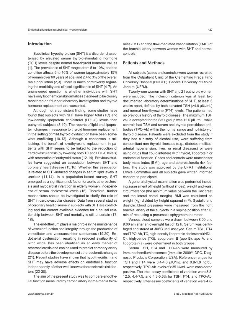
0.05) (34,35). The scans were recorded on S-VHS video-tape. The internal diameter of the brachial artery wasassessed at the end of diastole, and arterial flow wasmeasured using the pulse Doppler sample volume at anangle of 60° or less in the center of the artery. For eachsubject, optimal brachial artery images were obtainedapproximately 5 cm above the antecubital fossa. Armpressure was caused by inflating a pneumatic arm cuff upto 30 mmHg higher than the subject’s systolic arterialpressure for 5 min. The cuff was then deflated, the arterialflow was immediately recorded, and the diameter wasmeasured 60 to 90 s after deflation. Figure 1 illustrates atypical example of an image obtained. For both diameters,one measurement was made and the value was recorded.FMD was calculated according to the formula: FMD = (pos-occlusion diameter - baseline diameter) x 100 / baselinediameter.
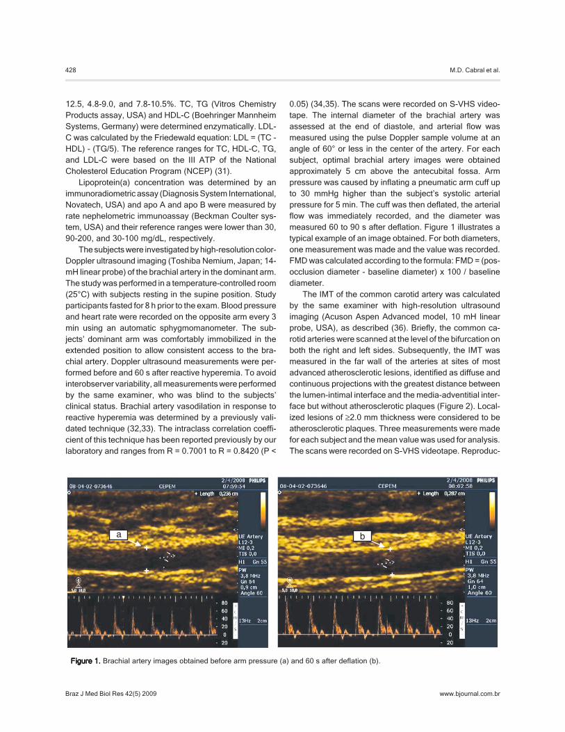
The IMT of the common carotid artery was calculatedby the same examiner with high-resolution ultrasoundimaging (Acuson Aspen Advanced model, 10 mH linearprobe, USA), as described (36). Briefly, the common ca-rotid arteries were scanned at the level of the bifurcation onboth the right and left sides. Subsequently, the IMT wasmeasured in the far wall of the arteries at sites of mostadvanced atherosclerotic lesions, identified as diffuse andcontinuous projections with the greatest distance betweenthe lumen-intimal interface and the media-adventitial inter-face but without atherosclerotic plaques (Figure 2). Local-ized lesions of ≥2.0 mm thickness were considered to beatherosclerotic plaques. Three measurements were madefor each subject and the mean value was used for analysis.The scans were recorded on S-VHS videotape. Reproduc-
Figure 1.Figure 1.Figure 1.Figure 1.Figure 1. Brachial artery images obtained before arm pressure (a) and 60 s after deflation (b).
429
Braz J Med Biol Res 42(5) 2009
Endothelial function in subclinical hypothyroidism
www.bjournal.com.br
ibility of the IMT measurement was acceptable, as demon-strated by coefficients of variation of 7.7 ± 4.3% (36).
Statistical analysisStatistical analysis was performed using SPSS for
Windows program, version 10.0. The mean values of thenumerical variables were compared between groups bythe Mann-Whitney test; if not normally distributed by theKolmogorov-Smirnov Liliefors test. Parametric variableswere compared by the t-test.
Data are reported as median values (interquartile range)of continuous non-parametric variables, as means ± SDvalues of parametric variables or frequencies of qualitativevariables. Non-parametric variables are reported as me-dian and range. A P value of <0.05 was considered to besignificant.
Results
The mean age of the 21 SHT subjects was 42.4 ± 10.8years and the mean age of the 21 subjects in the controlgroup was 44.2 ± 8.5 years. The cause of SHT wasspontaneous and 14 (70%) SHT subjects had abnormalTPO-Ab levels. No significant difference was observedwith respect to age, BMI, waist circumference, activitylevel, or menopausal status. There were no statisticallysignificant differences in lipid parameters between casesand controls, except for LDL-C (148.7 ± 46.3 vs 112.9 ±33.3 mg/dL; P = 0.04) and apo B (128.2 ± 37.7 vs 104.4 ±28.3 mg/dL; P = 0.02), which were significantly higher inthe SHT group. The baseline clinical and laboratory char-acteristics of all participants are summarized in Table 1.
The median and range TSH values were 8.3 (4.3) μIU/mL for the SHT group and 1.2 (0.7) μIU/mL for the euthy-roid group. TSH was 4 to 8 μIU/mL in eight patients (38%),and 8 to 12 μIU/mL in 13 (62%).
No significant differences were observed with respect toFMD or IMT between the SHT and control groups (Table 2).When the SHT group was subdivided into low (≥4 but >8 μIU/mL) or high (≥8 but <12 μIU/mL) TSH levels, no differences
were observed in metabolic, or vascular pa-rameters. Lipid parameters, FMD, and IMTvalues were higher in the common carotid ar-tery and carotid bifurcation of SHT patientswith positive serum TPO-Ab (0.62 ± 0.2 and0.62 ± 0.16 mm, for common carotid artery andcarotid bifurcation, respectively) compared tothe negative TPO-Ab group (0.55 ± 0.24 and0.58 ± 0.13 mm, for common carotid and ca-rotid bifurcation, respectively) although the dif-ference was not statistically significant.
Figure 2.Figure 2.Figure 2.Figure 2.Figure 2. The greatest distance between the lumen-intima inter-face and the media-adventitia interface to calculate intima-mediathickness of the carotid artery (a).
Table 1. Table 1. Table 1. Table 1. Table 1. Clinical and laboratory characteristics of the womenwith subclinical hypothyroidism.
EU (N = 21) SHT (N = 21)
Age (years) 44.2 ± 8.5 42.4 ± 10.8Sedentarism (%) 88.2% 84.2%Menopause (%) 61.9% 71.4%Body mass index (kg/m2) 24.6 ± 3.7 26.8 ± 4.8Waist circumference (cm) 84.2 ± 13.6 85.1 ± 10.2TSH (μIU/mL) 1.2 (0.7) 8.3 (4.3)*FT4 (ng/dL) 1.2 (0.3) 1.0 (0.1)*TG (mg/dL) 110 (61) 98 (47.5)HDL-C (mg/dL) 54.4 ± 12.8 55.6 ± 10.8TC (mg/dL) 201.1 ± 35.4 227 ± 49.4LDL-C (mg/dL) 112.9 ± 33.3 148.7 ± 46.3*apo A (mg/dL) 145.2 ± 26.2 156.9 ± 29.1apo B (mg/dL) 104.4 ± 28.3 128.2 ± 37.7*Lp(a) (mg/dL) 15.3 (28.6) 18.6 (71.05)
Data are reported as means ± SD or median (interquartile range).EU = euthyroid group; SHT = subclinical hypothyroid group; TSH =thyroid-stimulating hormone; FT4 = normal free thyroxine; TG =tryglicerides; HDL-C = high-density lipoprotein cholesterol; TC =total cholesterol; LDL-C = low-density lipoprotein cholesterol; apoA = apoprotein A; apo B = apoprotein B; Lp(a) = lipoprotein(a). *P< 0.05 compared to EU group (t-test and Mann-Whitney test).
Table 2.Table 2.Table 2.Table 2.Table 2. Brachial and carotid artery parameters.
EU (N = 21) SHT (N = 21)
Flow-mediated dilatation (%) 16.1 (8.8%) 20.6 (11.2%)Intima-media thickness of CCA (mm) 0.6 (0.1) 0.6 (0.1)Intima-media thickness of bifurcation (mm) 0.6 (0.2) 0.6 (0.1)
Data are reported as median (interquartile range). EU = euthyroid group; SHT =subclinical hypothyroid group; CCA = common carotid artery. There were nostatistically significant differences between the EU and SHT groups (Mann-Whitney test).
430
Braz J Med Biol Res 42(5) 2009
M.D. Cabral et al.
www.bjournal.com.br
Discussion
We did not find a significant association between thy-roid function and vascular parameters in subjects whowere similar with respect to age, BMI, smoking, meno-pausal status, and endothelial function modifiers, but whodiffered in thyroid function. The exclusion of participantswith concomitant endocrinologic, renal, or hepatic dis-eases allowed us to control our analysis for confoundingvariables and yielded a homogenous population. In addi-tion, by excluding co-morbid conditions, the mean age wasreduced, allowing a smaller sample size for the study (21subjects in each group). This may be the reason we did notdetect a difference between SHT and control subjects invasodilation parameters.
We examined endothelial function using a simple non-invasive, harmless method that enables accurate andreproducible assessment of the vascular response to flowincrease. FMD is currently the main method used to as-sess endothelial function and is convenient for clinicalpractice (37).
Lekakis et al. (24) were the first to describe the nega-tive association between borderline and mild hypothyroid-ism and FMD. In their study, cholesterol did not differsignificantly among groups, but tended to be higher in thehypothyroidism and SHT groups and the authors con-cluded that higher cholesterol levels may be associatedwith endothelial dysfunction. LDL-C and apo B levels weresignificant higher in our SHT group, and we did not findaltered FMD compared to the euthyroid group.
Cikim et al. (26) compared 25 subclinical hypothyroidpatients (mean age: 32.28 ± 9.67 years) and 23 euthyroidsubjects (mean age: 35.87 ± 9.67 years) strictly matchedfor atherosclerotic risk factors and demonstrated that thesubclinical hypothyroid group had significantly lower FMDvalues (15.92 ± 7.92% for the euthyroid group and 10.68 ±3.71% for the SHT group; P < 0.05). No significant differ-ences were observed between groups with respect toother vascular parameters, including carotid artery IMT.Since the lipid profile was comparable between groups,the authors suggested that endothelial function could de-teriorate prior to the emergence of hypothyroidism-inducedmetabolic changes. Cholesterol levels were lower in their
study than in the present one (181.04 ± 36.71 mg/dL for theeuthyroid group and 179.56 ± 30.21 mg/dL for the SHTgroup). We found similar TSH levels (8.85 ± 6.86 mIU/L),but thyroid antibody levels were elevated in their SHTpatients (mean: 496.71 ± 677.09 IU/mL), reflecting theautoimmune nature of hypothyroidism. Cikim et al. (26)evaluated the association of thyroid autoantibodies andthe FMD/IMT ratio and found no significant correlation. Wealso could not find any significant association betweenanti-TPO concentrations and FMD or IMT.
The B-mode ultrasound measurement of IMT in thecommon carotid artery permits easy evaluation of athero-sclerosis. Studies evaluating IMT values in hypothyroidand SHT patients showed conflicting results, which variedfrom low IMT values in subjects with elevated TSH levels(27) to high (22,23), or unchanged levels (38) compared toeuthyroid subjects. The results are controversial but thereare important differences between studies. Monzani et al.(22) excluded individuals over 55 years of age, and themean IMT of their SHT patients was significantly higher inthe subgroup older than 35 years. Levothyroxine replace-ment therapy reduced both LDL-C and mean IMT, sug-gesting that lipid infiltration of the arterial wall may repre-sent the main mechanism underlying the increase in IMT inSHT. We previously reported no significant differences inmean carotid IMT in a group of subclinical hypothyroidpatients compared to a euthyroid group, suggesting thatmild SHT is not associated with an increase in cardiovas-cular risk when assessed by carotid IMT (38).
The causative agent of endothelial damage in thyroiddysfunction conditions is unclear, but seems to be associ-ated with decreased endothelial nitric oxide synthesis dueto low hormone levels or inflammation induced by thyroidautoantibodies (39).
Based on our data, minimal thyroid dysfunction had noadverse effects on endothelial function in the patients withSHT. Current evidence is insufficient to support the asso-ciation between minimal thyroid disease and endothelialdysfunction. Larger studies are necessary to identify ifthere is any beneficial effect of levothyroxine treatment onendothelial function in patients with subclinical hypothy-roidism.
References
1. Ayala A, Wartofsky L. Minimally symptomatic (subclinical)hypothyroidism. Endocrinologist 1997; 7: 44-50.
2. Canaris GJ, Manowitz NR, Mayor G, Ridgway EC. TheColorado thyroid disease prevalence study. Arch Intern Med
2000; 160: 526-534.3. Cooper DS. Clinical practice. Subclinical hypothyroidism. N
Engl J Med 2001; 345: 260-265.4. Surks MI, Ortiz E, Daniels GH, Sawin CT, Col NF, Cobin
431
Braz J Med Biol Res 42(5) 2009
Endothelial function in subclinical hypothyroidism
www.bjournal.com.br
RH, et al. Subclinical thyroid disease: scientific review andguidelines for diagnosis and management. JAMA 2004;291: 228-238.
5. McDermott MT, Ridgway EC. Subclinical hypothyroidism ismild thyroid failure and should be treated. J Clin EndocrinolMetab 2001; 86: 4585-4590.
6. Chu JW, Crapo LM. The treatment of subclinical hypothy-roidism is seldom necessary. J Clin Endocrinol Metab 2001;86: 4591-4599.
7. Gharib H, Tuttle RM, Baskin HJ, Fish LH, Singer PA,McDermott MT. Subclinical thyroid dysfunction: a joint state-ment on management from the American Association ofClinical Endocrinologists, the American Thyroid Associa-tion, and the Endocrine Society. J Clin Endocrinol Metab2005; 90: 581-585.
8. Cabral MD, Costa AJL, Santos M, Vaisman M. Lipid profilealterations in subclinical hypothyroidism. Endocrinologist2004; 14: 121-125.
9. Teixeira PFS, Reis FA, Reuters V, Almeida C, Vaisman M.Hipotireoidismo subclínico e risco cardiovascular. RevSOCERJ 2004; 17: 50-56.
10. Arem R, Escalante DA, Arem N, Morrisett JD, Patsch W.Effect of L-thyroxine therapy on lipoprotein fractions in overtand subclinical hypothyroidism, with special reference tolipoprotein(a). Metabolism 1995; 44: 1559-1563.
11. Caraccio N, Ferrannini E, Monzani F. Lipoprotein profile insubclinical hypothyroidism: response to levothyroxine re-placement, a randomized placebo-controlled study. J ClinEndocrinol Metab 2002; 87: 1533-1538.
12. Danese MD, Ladenson PW, Meinert CL, Powe NR. Clinicalreview 115: effect of thyroxine therapy on serum lipopro-teins in patients with mild thyroid failure: a quantitative re-view of the literature. J Clin Endocrinol Metab 2000; 85:2993-3001.
13. Tanis BC, Westendorp GJ, Smelt HM. Effect of thyroidsubstitution on hypercholesterolaemia in patients with sub-clinical hypothyroidism: a reanalysis of intervention studies.Clin Endocrinol 1996; 44: 643-649.
14. Teixeira PF, Reuters VS, Ferreira MM, Almeida CP, ReisFA, Melo BA, et al. Treatment of subclinical hypothyroidismreduces atherogenic lipid levels in a placebo-controlleddouble-blind clinical trial. Horm Metab Res 2008; 40: 50-55.
15. Bastenie PA, Vanhaelst L, Bonnyns M, Neve P, Staquet M.Preclinical hypothyroidism: a risk factor for coronary heart-disease. Lancet 1971; 1: 203-204.
16. Hak AE, Pols HA, Visser TJ, Drexhage HA, Hofman A,Witteman JC. Subclinical hypothyroidism is an independentrisk factor for atherosclerosis and myocardial infarction inelderly women: the Rotterdam Study. Ann Intern Med 2000;132: 270-278.
17. Monzani F, Dardano A, Caraccio N. Does treating subclini-cal hypothyroidism improve markers of cardiovascular risk?Treat Endocrinol 2006; 5: 65-81.
18. Volzke H, Schwahn C, Wallaschofski H, Dorr M. Review:The association of thyroid dysfunction with all-cause andcirculatory mortality: is there a causal relationship? J ClinEndocrinol Metab 2007; 92: 2421-2429.
19. Vane JR, Anggard EE, Botting RM. Regulatory functions ofthe vascular endothelium. N Engl J Med 1990; 323: 27-36.
20. Healy B. Endothelial cell dysfunction: an emerging endo-crinopathy linked to coronary disease. J Am Coll Cardiol
1990; 16: 357-358.21. Bonetti PO, Lerman LO, Lerman A. Endothelial dysfunction:
a marker of atherosclerotic risk. Arterioscler Thromb VascBiol 2003; 23: 168-175.
22. Monzani F, Caraccio N, Kozakowa M, Dardano A, Vittone F,Virdis A, et al. Effect of levothyroxine replacement on lipidprofile and intima-media thickness in subclinical hypothy-roidism: a double-blind, placebo-controlled study. J ClinEndocrinol Metab 2004; 89: 2099-2106.
23. Nagasaki T, Inaba M, Henmi Y, Kumeda Y, Ueda M, TaharaH, et al. Decrease in carotid intima-media thickness in hypo-thyroid patients after normalization of thyroid function. ClinEndocrinol 2003; 59: 607-612.
24. Lekakis J, Papamichael C, Alevizaki M, Piperingos G,Marafelia P, Mantzos J, et al. Flow-mediated, endothelium-dependent vasodilation is impaired in subjects with hypo-thyroidism, borderline hypothyroidism, and high-normal se-rum thyrotropin (TSH) values. Thyroid 1997; 7: 411-414.
25. Papaioannou GI, Lagasse M, Mather JF, Thompson PD.Treating hypothyroidism improves endothelial function. Me-tabolism 2004; 53: 278-279.
26. Cikim AS, Oflaz H, Ozbey N, Cikim K, Umman S, Meric M,et al. Evaluation of endothelial function in subclinical hypo-thyroidism and subclinical hyperthyroidism. Thyroid 2004;14: 605-609.
27. Volzke H, Robinson DM, Schminke U, Ludemann J, RettigR, Felix SB, et al. Thyroid function and carotid wall thick-ness. J Clin Endocrinol Metab 2004; 89: 2145-2149.
28. Erbil Y, Ozbey N, Giris M, Salmaslioglu A, Ozarmagan S,Tezelman S. Effects of thyroxine replacement on lipid pro-file and endothelial function after thyroidectomy. Br J Surg2007; 94: 1485-1490.
29. Taddei S, Caraccio N, Virdis A, Dardano A, Versari D,Ghiadoni L, et al. Impaired endothelium-dependent vasodi-latation in subclinical hypothyroidism: beneficial effect oflevothyroxine therapy. J Clin Endocrinol Metab 2003; 88:3731-3737.
30. Razvi S, Ingoe L, Keeka G, Oates C, McMillan C, WeaverJU. The beneficial effect of L-thyroxine on cardiovascularrisk factors, endothelial function, and quality of life in sub-clinical hypothyroidism: randomized, crossover trial. J ClinEndocrinol Metab 2007; 92: 1715-1723.
31. National Cholesterol Education Program (USA). Expertpanel on detection, evaluation, and treatment of high bloodcholesterol in adults. Third report of the National CholesterolEducation Program (NCEP; adult treatment panel III): finalreport. Bethesda: National Institutes of Health; 2002.
32. Celermajer DS, Sorensen KE, Gooch VM, Spiegelhalter DJ,Miller OI, Sullivan ID, et al. Non-invasive detection of endo-thelial dysfunction in children and adults at risk of athero-sclerosis. Lancet 1992; 340: 1111-1115.
33. Corretti MC, Anderson TJ, Benjamin EJ, Celermajer D,Charbonneau F, Creager MA, et al. Guidelines for the ultra-sound assessment of endothelial-dependent flow-mediatedvasodilation of the brachial artery: a report of the Interna-tional Brachial Artery Reactivity Task Force. J Am CollCardiol 2002; 39: 257-265.
34. Meirelles CM, Leite SP, Montenegro CA, Gomes PS. Confi-abilidade da medida da dilatação fluxo-mediada da artériabraquial pela ultra-sonografia. Arq Bras Cardiol 2007; 89:160-183.
432
Braz J Med Biol Res 42(5) 2009
M.D. Cabral et al.
www.bjournal.com.br
35. Garrido KU, Leite S, Montenegro C, Koch H, Soares A.Dilatação fluxo-mediada da arteria braquial: estudo dafunção endotelial em mulheres na menopausa. Rev BrasEcocardiog 2008; 21: 22-26.
36. Soares DV, Spina LD, de Lima Oliveira Brasil RR, da SilvaEM, Lobo PM, Salles E, et al. Carotid artery intima-mediathickness and lipid profile in adults with growth hormonedeficiency after long-term growth hormone replacement.Metabolism 2005; 54: 321-329.
37. Faulx MD, Wright AT, Hoit BD. Detection of endothelialdysfunction with brachial artery ultrasound scanning. Am
Heart J 2003; 145: 943-951.38. Almeida CA, Teixeira PF, Soares DV, Cabral MD, Costa
SM, Salles EF, et al. Espessura íntima-média carotídeacomo marcador de risco cardiovascular em pacientes comhipotireoidismo subclínico. Arq Bras Endocrinol Metabol2007; 51: 472-477.
39. Taddei S, Caraccio N, Virdis A, Dardano A, Versari D,Ghiadoni L, et al. Low-grade systemic inflammation causesendothelial dysfunction in patients with Hashimoto’s thy-roiditis. J Clin Endocrinol Metab 2006; 91: 5076-5082.