Embed Size (px)
Citation preview
May-Thurner Syndrome: Diagnosis and Treatment
Jason R. Moore, MD, FACS, RPVISentara Rockingham Memorial Hospital
Presenter nameTitleDate
Disclosures
• None
Presenter nameTitleDate
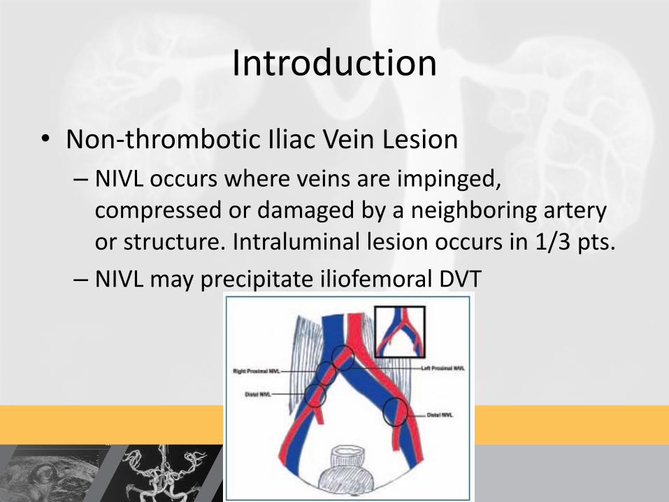
Introduction
• Non-thrombotic Iliac Vein Lesion– NIVL occurs where veins are impinged,
compressed or damaged by a neighboring artery or structure. Intraluminal lesion occurs in 1/3 pts.
– NIVL may precipitate iliofemoral DVT
Presenter nameTitleDate
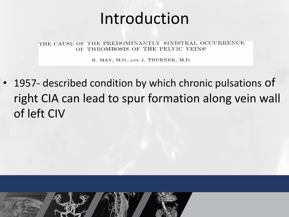
Introduction
• 1957- described condition by which chronic pulsations of right CIA can lead to spur formation along vein wall of left CIV
Presenter nameTitleDate
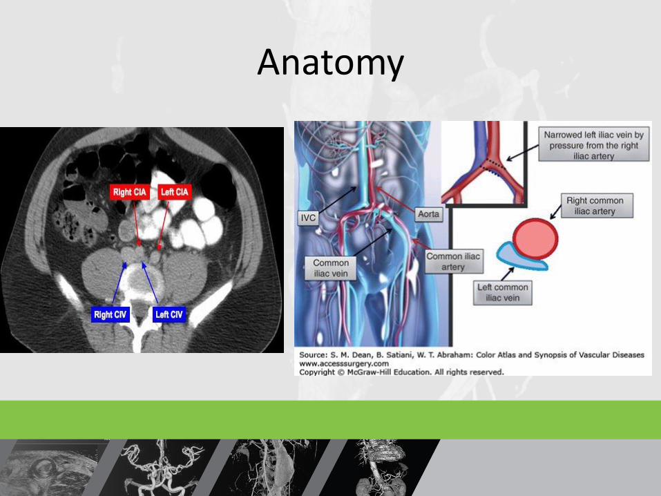
Anatomy
Presenter nameTitleDate
Prevalence• True prevalence of May–Thurnersyndrome unknown• 20% people may have asymptomatic
compression: “Permissive anomaly”• Old data suggests women between
30-50 years are primarily affected• Newer data indicates prevalence is
more significant than thought1. Al-Nouri O, Milner R. May-Thurner Syndrome. Vas Disease Mgt. 2011;3:53-56.
Presenter nameTitleDate
Prevalence• Patients with severe chronic venous disease
(37% >50% stenosis)1
• Reported to be 600,000 DVT hospitalizations
per year in US
• 50-65% of DVTs occur in left leg
• Iliac vein compression thought to occur ~18 - 69%
DVT 1,2
1. Al-Nouri O, Milner R. May-Thurner Syndrome. Vas Disease Mgt. 2011;3:53-56.
2. Rosen E, Groben L, et al. Rare Case of Bilateral Common Iliac Vein Compression by Arterial Stents and Calcification. Vas Disease Mgt.
2012:9(11):E172-E174.
Presenter nameTitleDate
Stages• Stage 1: Iliac vein compression withoutstructural vein changes= Asymptomatic• Stage 2: Venous spur formation which arefibrous shelves eventually developing inthe vein, restricting blood flow andincreasing risk for edema and DVT.Asymptomatic.• Stage 3: Symptomatic obstruction: DVT,edema and the formation of varicoseveins.
Presenter nameTitleDate
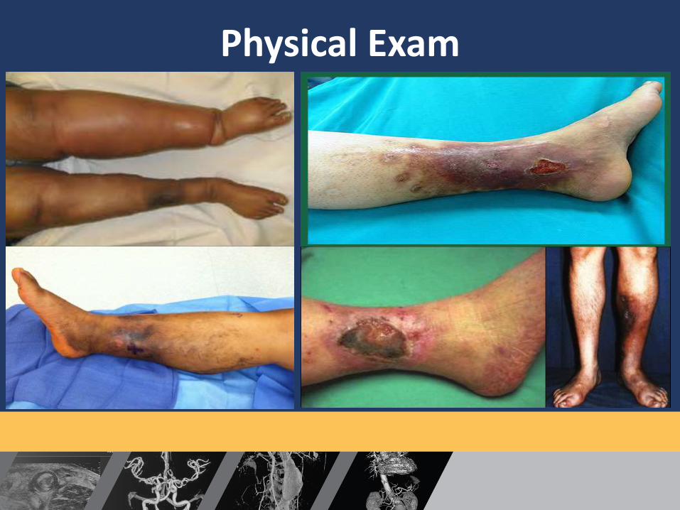
Symptoms• Dull aching, heaviness, or cramping in legs• Pain that gets worse when standing• Pain that gets better when legs are raised• Redness of the legs and ankles• Skin color changes around the ankles• Varicose veins on the surface (superficial)• Thickening & hardening of the skin on thelegs & ankles• Ulcers on the legs and ankles• DVT
Presenter nameTitleDate
Physical Exam
Presenter nameTitleDate
Clinical, Etiology, Anatomic,Pathophysiology
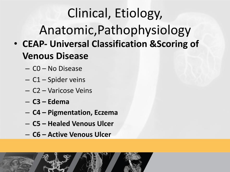
• CEAP- Universal Classification &Scoring of Venous Disease– C0 – No Disease– C1 – Spider veins– C2 – Varicose Veins– C3 – Edema– C4 – Pigmentation, Eczema– C5 – Healed Venous Ulcer– C6 – Active Venous Ulcer
Presenter nameTitleDate
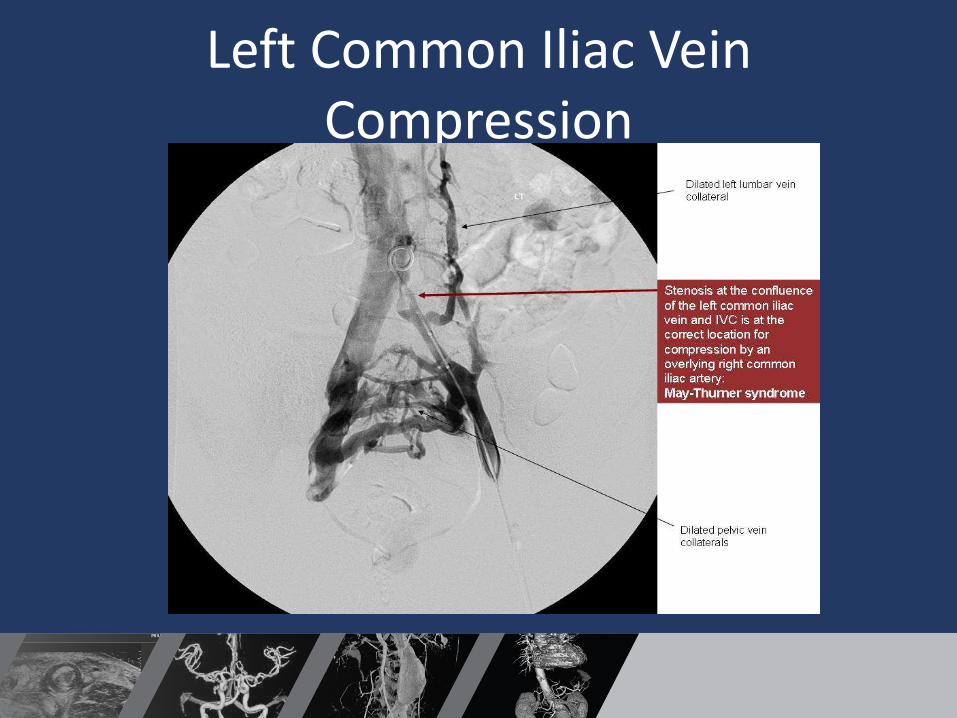
Left Common Iliac Vein Compression
Presenter nameTitleDate
Imaging
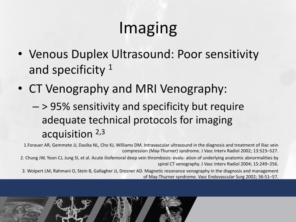
• Venous Duplex Ultrasound: Poor sensitivity
and specificity 1
• CT Venography and MRI Venography:
– > 95% sensitivity and specificity but require
adequate technical protocols for imaging
acquisition 2,3
1.Forauer AR, Gemmete JJ, Dasika NL, Cho KJ, Williams DM. Intravascular ultrasound in the diagnosis and treatment of iliac vein
compression (May-Thurner) syndrome. J Vasc Interv Radiol 2002; 13:523–527.
2. Chung JW, Yoon CJ, Jung SI, et al. Acute iliofemoral deep vein thrombosis: evalu- ation of underlying anatomic abnormalities by
spiral CT venography. J Vasc Interv Radiol 2004; 15:249–256.
3. Wolpert LM, Rahmani O, Stein B, Gallagher JJ, Drezner AD. Magnetic resonance venography in the diagnosis and management
of May-Thurner syndrome. Vasc Endovascular Surg 2002; 36:51–57.
Presenter nameTitleDate
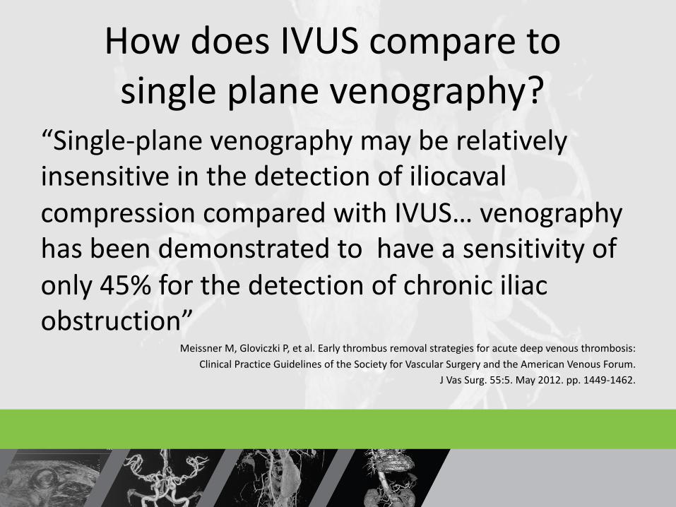
How does IVUS compare tosingle plane venography?
“Single-plane venography may be relatively insensitive in the detection of iliocavalcompression compared with IVUS… venography has been demonstrated to have a sensitivity of only 45% for the detection of chronic iliac obstruction”
Meissner M, Gloviczki P, et al. Early thrombus removal strategies for acute deep venous thrombosis:Clinical Practice Guidelines of the Society for Vascular Surgery and the American Venous Forum.
J Vas Surg. 55:5. May 2012. pp. 1449-1462.
Presenter nameTitleDate
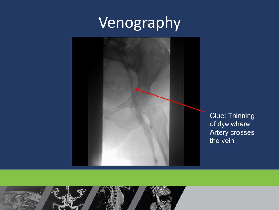
Venography
Clue: Thinningof dye whereArtery crossesthe vein
Presenter nameTitleDate
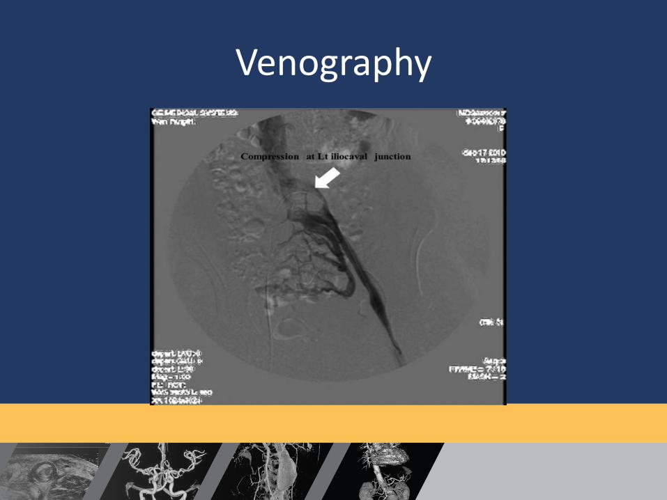
Venography
Presenter nameTitleDate
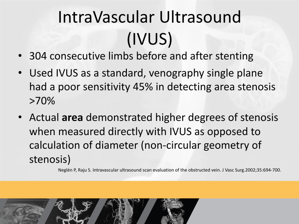
IntraVascular Ultrasound(IVUS)
• 304 consecutive limbs before and after stenting
• Used IVUS as a standard, venography single plane had a poor sensitivity 45% in detecting area stenosis >70%
• Actual area demonstrated higher degrees of stenosis when measured directly with IVUS as opposed to calculation of diameter (non-circular geometry of stenosis)
Neglén P, Raju S. Intravascular ultrasound scan evaluation of the obstructed vein. J Vasc Surg.2002;35:694-700.
Presenter nameTitleDate
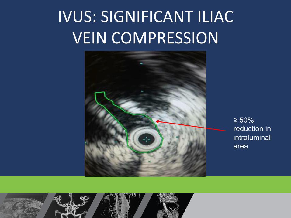
IVUS: SIGNIFICANT ILIACVEIN COMPRESSION
≥ 50%reduction inintraluminalarea
Presenter nameTitleDate
IVUS
• Since IVUS has a diagnostic sensitivity of >90% and is free of radiation, it has become the diagnostic standard in iliac vein compression
1. Raju S. Iliac vein outflow obstruction. Phlebolymphology. Vol 15. No. 1. 2008
Presenter nameTitleDate
Conventional Managment
• Compression stockings to decrease swelling• Wound Care Centers for open wounds sores or
infections• Laser or RF ablation of incompetent veins• Surgery (varicose vein stripping)• Diuretics for edema resolution• Lymphedema Pump
Presenter nameTitleDate
Treatment
• Given “spur” and scar formation that occurs from MTS, it is clear that venous angioplasty is not in itself an effective treatment. 1
• 1/3 patients treated with thrombolysis for iliofemoralDVT required stenting and that the stented patients had significantly higher patency than those who were not stented. 2
1.Park JY, Ahn JH, Jeon YS, et al. Iliac vein stenting as a durable option for residual stenosis after catheter-directed thrombolysis and angioplasty of iliofemoral deep vein thrombosis secondary to May-Thurner syndrome.Phlebology2014;29:461-70.
2.Nazarian GK, Bjarnason H, Dietz CA, Jr, et al. Iliofemoral venous stenoses: effectiveness of treatment with metallic endovascular stents. Radiology 1996;200:193-9.
Presenter nameTitleDate
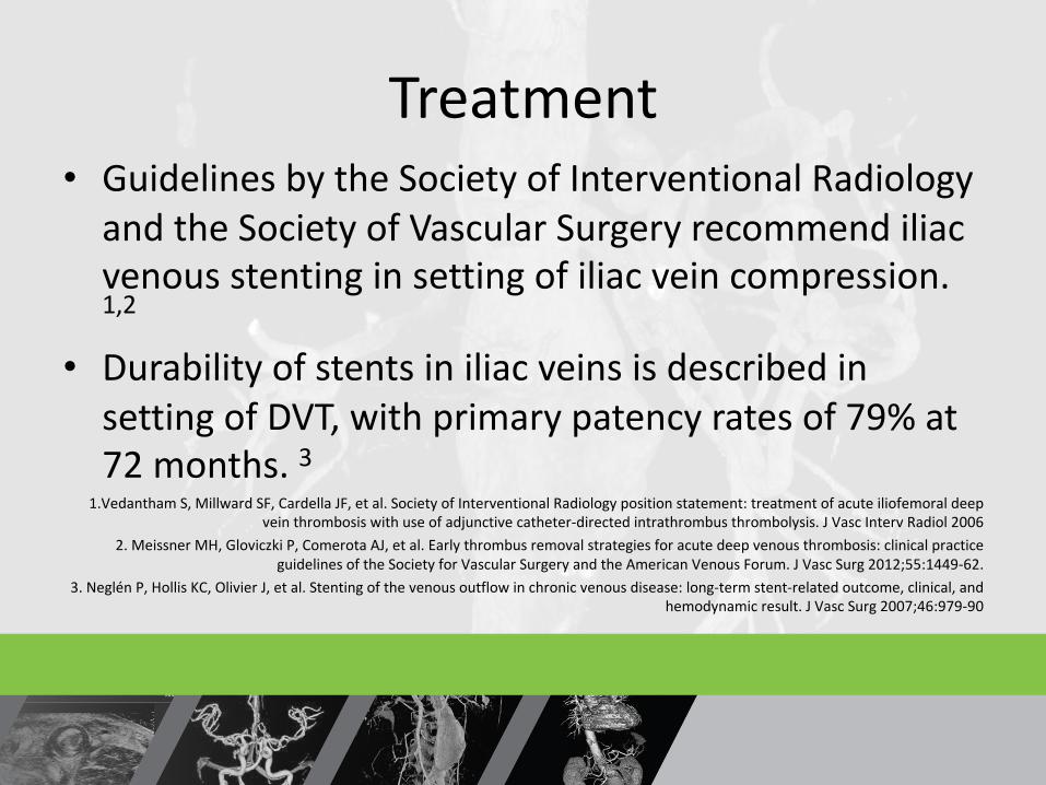
Treatment• Guidelines by the Society of Interventional Radiology
and the Society of Vascular Surgery recommend iliac venous stenting in setting of iliac vein compression. 1,2
• Durability of stents in iliac veins is described in setting of DVT, with primary patency rates of 79% at 72 months. 3
1.Vedantham S, Millward SF, Cardella JF, et al. Society of Interventional Radiology position statement: treatment of acute iliofemoral deep vein thrombosis with use of adjunctive catheter-directed intrathrombus thrombolysis. J Vasc Interv Radiol 2006
2. Meissner MH, Gloviczki P, Comerota AJ, et al. Early thrombus removal strategies for acute deep venous thrombosis: clinical practice guidelines of the Society for Vascular Surgery and the American Venous Forum. J Vasc Surg 2012;55:1449-62.
3. Neglén P, Hollis KC, Olivier J, et al. Stenting of the venous outflow in chronic venous disease: long-term stent-related outcome, clinical, and hemodynamic result. J Vasc Surg 2007;46:979-90
Presenter nameTitleDate
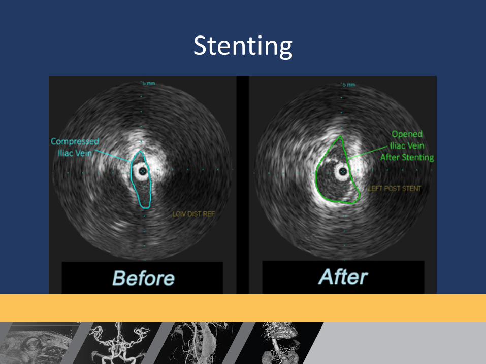
Stenting
Presenter nameTitleDate
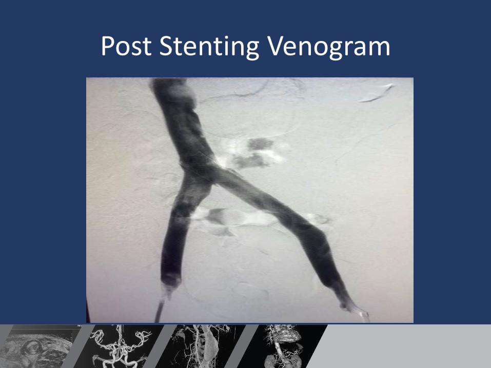
Post Stenting Venogram
Presenter nameTitleDate
Conclusion
May-Thurner syndrome: a not so uncommon cause of a common condition
This anatomic finding has been shown to be present in over 20% of the population; however, it is rarely considered in the differential diagnosis of leg edema, DVT, and chronic venous disease particularly in patients with other risk factors. Systemic anticoagulation, compression therapy, and venous ablation are ineffective or insufficient treatment, and a more aggressive approach is necessary to prevent complications

Presenter nameTitleDate
Thank You


![Bilateral Iliac Vein Stenting without Contrast in a ... · severe CVI (active or healed venous ulcer) is estimated to be around 1-2% [1]. Venous ulceration is more common in patients](https://img.pdfslide.us/doc/110x75/5e6cb7e3a7d5ea244a33c5e5/bilateral-iliac-vein-stenting-without-contrast-in-a-severe-cvi-active-or-healed.jpg)