Embed Size (px)
Citation preview
i
MATHEMATICAL MODEL FOR CANCER CELL INVASION OF TISSUE
NORUL HIDAYAH BINTI ABDULLAH
UniversitiTeknologi Malaysia
PSZ 19:16 (Pind. 1/07)
UNIVERSITI TEKNOLOGI MALAYSIA
DECLARATION OF THESIS / UNDERGRADUATE PROJECT PAPER
AND COPYRIGHT
Author’s full name : NORUL HIDAYAH BINTI ABDULLAH
Date of birth : 10/03/1987
Title : MATHEMATICAL MODEL FOR CANCER
: CELL INVASION OF TISSUE
Academic Session : 2013/2014
I declare that this thesis is classified as :
CONFIDENTIAL (Contains confidential information under the
Official Secret Act 1972)*
RESTRICTED (Contains restricted information as specified by
the organization where research was done)*
OPEN ACCESS I agree that my thesis to be published as online
open access (full text)
I acknowledged that Universiti Teknologi Malaysia reserves the right as follows:
1. The thesis is the property of Universiti Teknologi Malaysia.
2. The Library of Universiti Teknologi Malaysia has the right to make copies for
the purpose of research only.
3. The Library has the right to make copies of the thesis for academic exchange.
Certified by,
Hidayah
SIGNATURE SIGNATURE OF SUPERVISOR
870310-46-5014 Prof. Dr. Norsarahaida S Amin
(NEW IC NO. /PASSPORT NO.) NAME OF SUPERVISOR
Date : 19th
JUNE 2014 Date : 19th
JUNE 2014
NOTES
* If the thesis is CONFIDENTIAL or RESTRICTED, please attach with the letter from
the organization with period and reasons for confidentiality or restriction.
“I hereby that I have read this dissertation and in my opinion
this dissertation is sufficient in terms of scope and quality for the award of
the degree of Master of Science (Engineering Mathematics)”
Signature :
Name : PROF. DR. NORSARAHAIDA S AMIN
Date : 19 JUNE 2014
i
MATHEMATICAL MODEL FOR CANCER CELL INVASION OF TISSUE
NORUL HIDAYAH BINTI ABDULLAH
A dissertation submitted in partial fulfilment of the
requirements for the award of the degree of
Master of Science (Engineering Mathematics)
Faculty of Science
UniversitiTeknologi Malaysia
June 2014
ii
I declared that this dissertation entitled “Mathematical Model for Cancer Invasion of
Tissue” is the result of my own research except as cited in the references. The
dissertation has not been accepted for any degree and is not concurrently submitted
in candidature of any other degree.
Signature : Hidayah
Name : NORUL HIDAYAH BT ABDULLAH
Date : 19 JUNE 2014

iii
To my beloved family
iv
ACKNOWLEDGEMENT
In preparing this dissertation, I was in contact with many people, researchers
and academicians. They have contributed towards my understanding and thoughts. In
particular, I wish to express my sincere appreciation to my dissertation supervisor,
Prof. Dr. Norsarahaida S Amin, for her guidance, advices and motivation. Without
her continued support and interest, this dissertation would not have been the same as
presented here.
My sincere appreciation also extend to my postgraduate colleagues and others
who have provided assistance at various occasions. Their views and tips are useful
indeed. My fellow colleagues and students at Kolej Matrikulasi Johor should also be
recognised for their support. Unfortunately, it is not possible to list all of them in this
limited space.
Last but not least, I am grateful to all my family members for their support
and understanding throughout my journey as a postgraduate student.
v
ABSTRACT
Cancer cell invasion of tissue is a complex biological process which during
cell migration through extracellular matrix, facilitated by the secretion of degradative
enzymes, is a central process. Cells can be deform their cytoplasm to produce
pseudopodia, anchor these pseudopodia to neighbouring spatial locations in the tissue
and detach earlier bonds, to enable them to move and therefore migrate in a specified
direction. Genetic mutations, chemoattractant gradient or a lack of nutrients in their
current location can stimulate cell motility and cause them to migrate, thereby
invading new territory. In this paper, we propose a hybrid discrete-continuum model
to study the early growth of solid tumour and their ability to degrade and migrate into
the surrounding extracellular matrix. Considering the importance of chemoattractant
gradients in the invasion process, the model consists of a system of partial
differential equations describing the interactions of enzyme and the surrounding
matrix. Having formulated, then the system has been simplified to simpler system
which give an analytical solution in Fourier series. Results from simulation show that
the degradation of the extracellular matrix mirrors the tumour’s growth. High
enzyme concentrations, which cause maximum degradation of extracellular matrix
leads to a rise in concentration of attractant and growth factors causes cells to
migrate outward and to proliferate. Finally concluding remarks are made and
recommendation for future work are indicated in the last section.
vi
ABSTRAK
Serangan sel kanser pada tisu adalah satu proses biologi yang kompleks
dimana semasa migrasi sel menerusi matrik luar sel, serangan ini dibantu oleh
rembesan enzim degradasi. Sel ini dapat mengubah bentuk sitoplasma mereka
menjadi pseudopodia dengan mencengkam pseudopodia ini pada ruangan yang
berhampiran pada tisu tersebut dan memutuskan ikatan yang sebelumnya. Ini
membolehkan sel-sel bergerak dan seterusnya merebak kearah yang tertentu. Mutasi
genetik, kecerunan tarikan kimia ataupun kekurangan nutrisi pada lokasi tersebut
boleh meransang pergerakan sel dan menyebabkan sel-sel merebak, seterusnya
menyerang kawasan baru. Dalam kertas kajian ini, kami mencadangkan model
hybrid discrete-continuum untuk mengkaji pertumbuhan awal sel tumor dan
kebolehan berdegradasi serta merebak ke sekeliling matrik di luar sel. Dengan
mempertimbangkan kepentingan kecerunan tarikan kimia dalam proses serangan
berlaku, model ini terdiri daripada sistem persamaan pembezaan separa yang
menggambarkan interaksi antara enzim dengan matrik luar sel. Sistem yang telah
diformulakan kemudian diringkaskan menjadi system yang lebih ringkas untuk
memberikan penyelesaian dalam bentuk siri Fourier secara analitikal. Hasil simulasi
menunjukkan kepekatan enzim yang tinggi, dimana menyebabkan degradasi matrik
luar sel yang maksimum membawa kepada peningkatan kecerunan tarikan kimia dan
juga faktor pertumbuhan yang menyebakan sel merebak keluar dan terus
berkembang. Akhirnya, kesimpulan dibuat dan cadangan untuk kajian masa hadapan
disertakan pada seksyen terakhir.
vii
TABLE OF CONTENTS
CHAPTER TITLE
PAGE
DECLARATION ii
DEDICATION iii
ACKNOWLEDGEMENTS iv
ABSTRACT v
ABSTRAK Vi
TABLE OF CONTENTS Vii
LIST OF TABLES X
LIST OF FIGURES Xi
LIST OF SYMBOLS Xiv
1 INTRODUCTION
1.1 Research Background 1
1.2 Problem Statements 6
1.3 Objective of research 8
1.4 Scope of the study 8
1.5 Significance of the study 9
1.6 Project overview
1.7 Terms Definition
10
11
viii
2 LITERATURE REVIEW
2.0 Introduction
2.1 The Gompertz Growth Model
2.2 The Continuum Mathematical Model
13
14
17
3
PROBLEM FORMULATION
3.0 Introduction 20
3.1 Modeling The Chemicals and the ECM
21
4 SOLUTION PROCEDURES
4.0 Introduction 25
4.1 Interaction of One Cell in One Spatial Domain
4.2 Finding Solution of Degraded ECM by Solving
Nonhomogeneous PDE
26
28
5 RESULTS AND DISCUSSION
5.0 Introduction 45
5.1 Numerical Simulations
5.2 Simulation Results
5.3 Discussion
46
48
52
ix
6 CONCLUSION AND RECOMMENDATION
6.1 Conclusion 62
6.2 Recommendation
REFERENCES
63
65
x
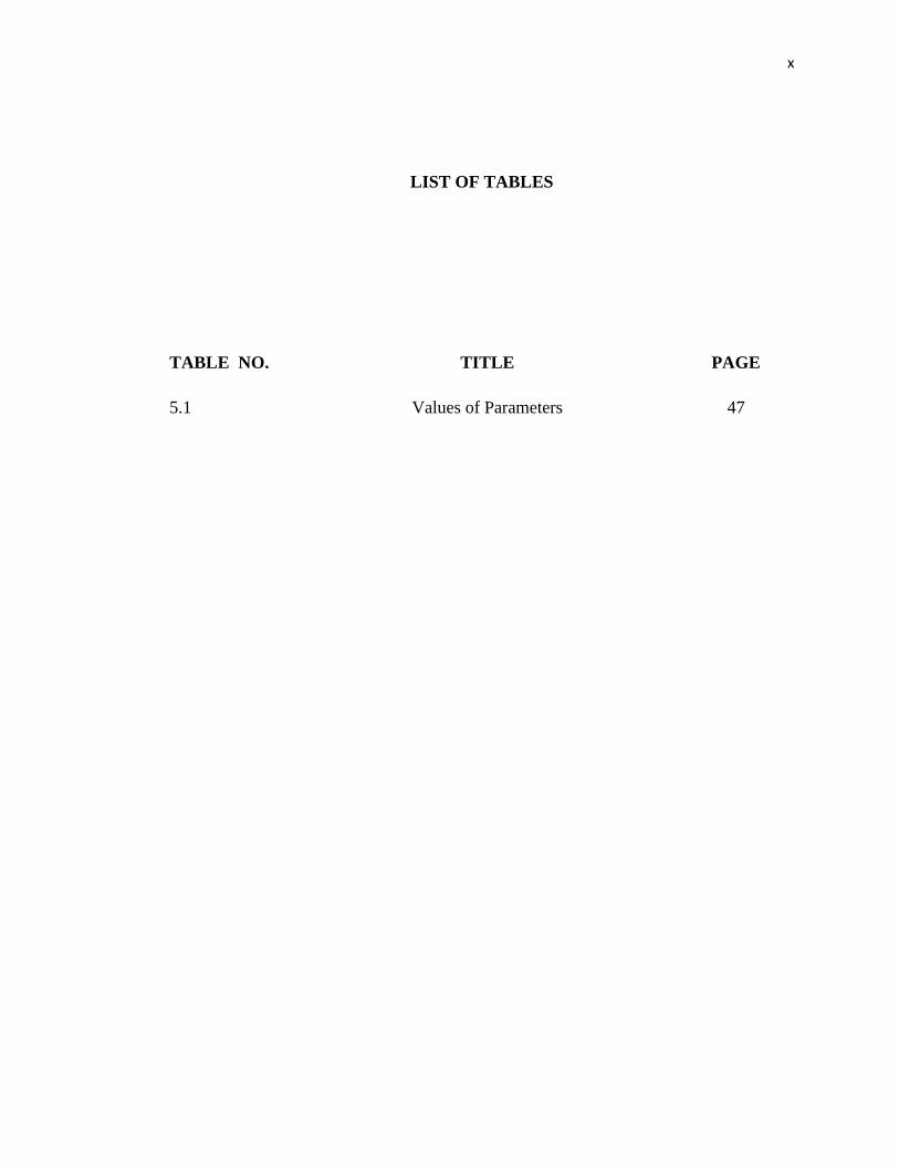
LIST OF TABLES
TABLE NO. TITLE PAGE
5.1 Values of Parameters 47
xi
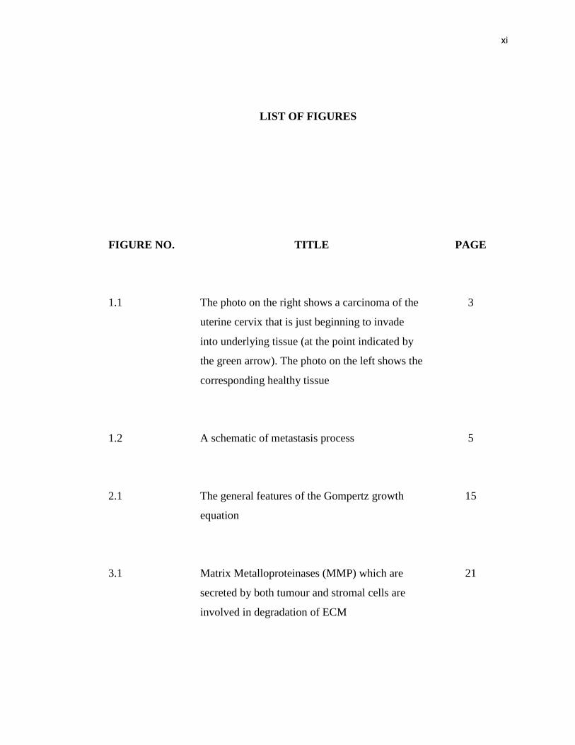
LIST OF FIGURES
FIGURE NO.
TITLE PAGE
1.1 The photo on the right shows a carcinoma of the
uterine cervix that is just beginning to invade
into underlying tissue (at the point indicated by
the green arrow). The photo on the left shows the
corresponding healthy tissue
3
1.2 A schematic of metastasis process
5
2.1 The general features of the Gompertz growth
equation
15
3.1 Matrix Metalloproteinases (MMP) which are
secreted by both tumour and stromal cells are
involved in degradation of ECM
21
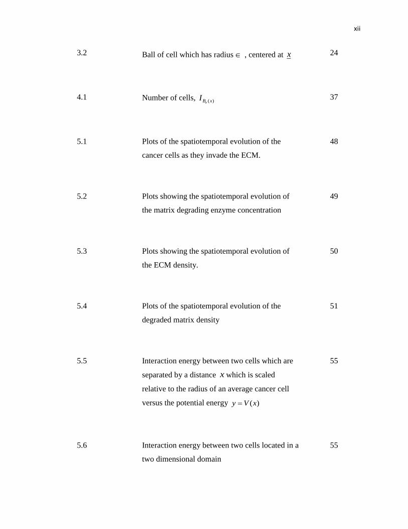
xii
3.2 Ball of cell which has radius , centered at x
24
4.1 Number of cells, )(xBe
I
37
5.1 Plots of the spatiotemporal evolution of the
cancer cells as they invade the ECM.
48
5.2 Plots showing the spatiotemporal evolution of
the matrix degrading enzyme concentration
49
5.3 Plots showing the spatiotemporal evolution of
the ECM density.
50
5.4 Plots of the spatiotemporal evolution of the
degraded matrix density
51
5.5 Interaction energy between two cells which are
separated by a distance x which is scaled
relative to the radius of an average cancer cell
versus the potential energy )(xVy
55
5.6 Interaction energy between two cells located in a
two dimensional domain
55
xiii
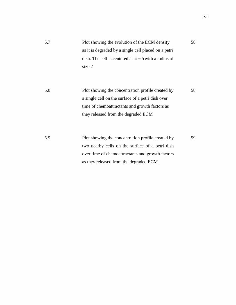
5.7 Plot showing the evolution of the ECM density
as it is degraded by a single cell placed on a petri
dish. The cell is centered at 5x with a radius of
size 2
58
5.8 Plot showing the concentration profile created by
a single cell on the surface of a petri dish over
time of chemoattractants and growth factors as
they released from the degraded ECM
58
5.9 Plot showing the concentration profile created by
two nearby cells on the surface of a petri dish
over time of chemoattractants and growth factors
as they released from the degraded ECM.
59
xiv
LIST OF SYMBOLS
MMP - Matrix Metalloproteinases
ECM - Extracellular Matrix
PDE - Partial Differential Equation
BC - Boundary Condition
IC - Initial Condition
ODE - Ordinary Differential Equation
CHAPTER 1
INTRODUCTION
1.1 Research Background
A healthy human body consists of normal growing cells which carry out the life
processes in a normal and orderly manner. The body is made up of trillions of living
cells. Normal body cells grow, divide, and die in an orderly fashion. During the early
years of a person’s life, normal cells divide faster to allow the person to grow. After the
person becomes an adult, most cells divide only to replace worn-out or dying cells or to
repair injuries.
A normal living cell can, for various unfortunate reasons, turn abnormal or
cancerous. Cancer is a disorder of cells and although it usually appears as a tumour
made up of a mass of cells, the visible tumours is the end of result of whole series of
changes which may have taken many years to develop (Knowles, M.A. and Selby, P.J.
2005). It multiplies in the body rapidly and excessively, forming a group of cells of
uncontrollable growth resulting in a swelling. Tumours are usually recognized by the
fact that the cells have shown abnormal proliferation, so that a reasonably acceptable
definition is that tumour cells differ from normal cells in their lack of response to
normal control mechanisms. Tumours can be classified into three main groups
(Knowles, M.A. and Selby, P.J. 2005):
(1) Benign tumours may arise in any tissue, grow locally, and cause damage by
local pressure or obstruction. However, the common feature is that they do
not spread to distant sites.
(2) In situ tumours usually develop in epithelium and are usually but not
invariably, small. The cells have morphological appearance of cancer cells
but remain in the epithelial layer. They do not invade the basement
membrane and supporting mesenchyme.
(3) Cancer are fully developed (malignant) tumours with a specific capacity to
invade and destroy underlying mesenchyme.
The formation of a tumor begins with the failure in the replication of a cell’s
DNA which leads to the uncontrolled division of the cell. People can inherit abnormal
DNA, but most DNA damage is caused by mistakes that happen while the normal cell is
reproducing or by something in the environment. Sometimes the cause of the DNA
damage may be something obvious like cigarette smoking or sun exposure. But it’s rare
to know exactly what caused any one person’s cancer.
This initial failure in DNA replication occurs at a molecular level in the cell
nucleus. The result of such instability are new daughter cells which interact with the
environment in a two-scale physical-chemical framework. At the cellular level,
dynamics have in general a much longer space scale and a slower time scale than events
at the molecular level. For example, a reaction such as the enzymatic degradation of a
substrate can occur in milliseconds whereas the replication of a cell can take about one
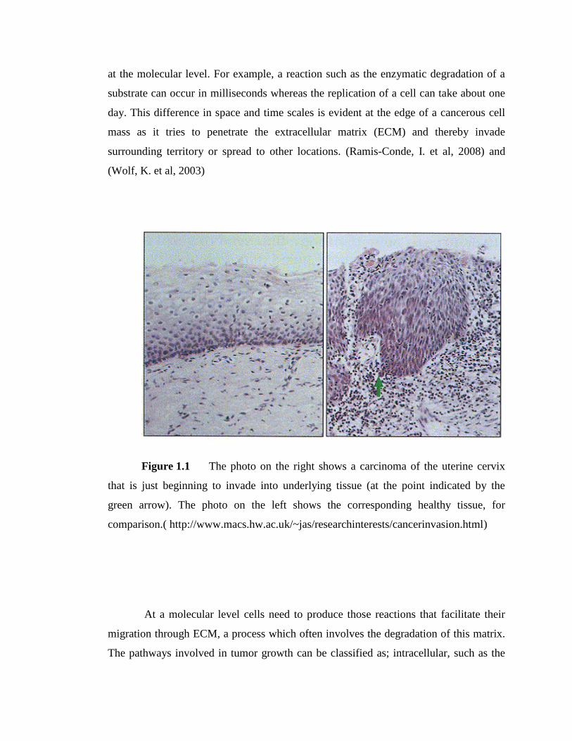
day. This difference in space and time scales is evident at the edge of a cancerous cell
mass as it tries to penetrate the extracellular matrix (ECM) and thereby invade
surrounding territory or spread to other locations. (Ramis-Conde, I. et al, 2008) and
(Wolf, K. et al, 2003)
Figure 1.1 The photo on the right shows a carcinoma of the uterine cervix
that is just beginning to invade into underlying tissue (at the point indicated by the
green arrow). The photo on the left shows the corresponding healthy tissue, for
comparison.( http://www.macs.hw.ac.uk/~jas/researchinterests/cancerinvasion.html)
At a molecular level cells need to produce those reactions that facilitate their
migration through ECM, a process which often involves the degradation of this matrix.
The pathways involved in tumor growth can be classified as; intracellular, such as the
formation of actin filaments to produce pseudopia; extracellular, for example, the
reorganization of the collagen filaments of the ECM; and certain others that connect
intracellular dynamics with the extracellular stroma, for instance, the uptake of growth
factors release from the remodeled ECM, which promote cell mitosis. (Trusolina, L. and
Comoglio, P.M., 2002)
From a cellular perspective, physical interactions with the ECM are crucial in
determining the nature of the tumour’s invasion-front. Invasion of surrounding tissue
normally occurs after the tumour had reached a certain size and the peripheral rim of
cells has started to disaggregate. At this point the cells on the tumour surface initate
different invasion mechanisms including the fingering process, Indian lines, cluster
detachment, etc.
Invasion is the main feature that allows a tumour to be characterized as malignant.
The progression of a benign tumour and delimited growth to a tumour that is invasive
and potentially metastatic is the major cause of poor clinical outcome in cancer patients,
in terms of theraphy and prognosis. Understanding tumour invasion could potentially
lead to a design of therapeutical strategies. Biomedically, invasion involves the
following tumour cell processes:
Tumour cell migration, which is a resut of down-regulation of cadherins, that is
loss of cell-cell adhesion
Tumour cell-extracellular matrix (ECM) interactions, such as cell-ECM
adhesion, and ECM degradation. These processes allow for the penetration of
the migrating tumour cells into host tissue barriers
Tumour cell proliferation
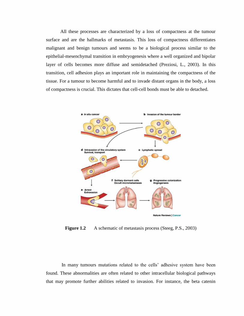
All these processes are characterized by a loss of compactness at the tumour
surface and are the hallmarks of metastasis. This loss of compactness differentiates
malignant and benign tumours and seems to be a biological process similar to the
epithelial-mesenchymal transition in embryogenesis where a well organized and bipolar
layer of cells becomes more diffuse and semidetached (Preziosi, L., 2003). In this
transition, cell adhesion plays an important role in maintaining the compactness of the
tissue. For a tumour to become harmful and to invade distant organs in the body, a loss
of compactness is crucial. This dictates that cell-cell bonds must be able to detached.
Figure 1.2 A schematic of metastasis process (Steeg, P.S., 2003)
In many tumours mutations related to the cells’ adhesive system have been
found. These abnormalities are often related to other intracellular biological pathways
that may promote further abilities related to invasion. For instance, the beta catenin
pathways is thought to be related to tumour invasion: up-regulation of beta-catenin in
the cytoplasm is linked to a poor prognosis for cancer patients (Wong, A. & Gumbiner,
M., 2003). This increased invasive ability can be associated with cell-cycle progression
or increased cellular motility but, in addition, the beta catenin pathway is closely related
to the intracellular domain of the E-cadherin adhesive system. Even if increased cell
motility and proliferation contribute greatly to the invasive ability of the tumour, in
order for metastasis to occur the detachment of intercellular bonds is necessary.
Cancer cells employ different methods of invasion both individually and in
combination to allow tumours to grow. Before a tumour becomes invasive, the
roughness of its surface is caused by variations in how groups of peripheral cells
degrade the ECM they are in contact with. This degradation is achieved by the tumour
cells secreting matrix-degrading enzymes, mainly of the type Matrix Metalloproteinases
(MMPs) and Urokinase Plasminogen Actovators (uPAs). Cancer –induced degradation
leads to the reorganization of the protein network that forms the ECM and, in many
cases, to the production of chemicals that promote cell migration and proliferation.
(Bogenrieder, T. & Herlyn, M., 2003)
1.2 Problem statement
Modelling aspect of cancer growth has been approached using a wide range of
mathematical models. Most models fall into two broad categories based on how the
tumour tissue is represented: discrete cell-based models and continuum models.
Although the continuum and discrete approaches have each provided important insight
into cancer-related processes occurring at particular lenght and time scales, the
complexity of cancer and the interactions between the cell-and tissue-level scales may
be elucidated further by means of multiscale (hybrid) approach that uses both
continuum and discrete representations of tumor cells and components of the tumor
microenvironment (Cristini, V. and Lowengrub, J., 2010).
Continuum models using PDEs are usually too large scale and are inappropriate
when the problem focuses on a relatively small number of individual cancer cells. In
discrete model, it is relatively easy to describe the detailed behaviour of individual cells,
which includes random and biased migration, cell-cell and cell-ECM interactions. On
the other hand, global population interactions among the group of cancer cells that
interact with cannot be captured by discrete approach.
Recently, third category of models has emerged; hybrid model that allows the
modellers to maximise the advantages and minimize drawbacks of both continuum and
discrete models, thus providing more comprehensive understanding of cell migration
leading to more accurate and efficient prediction of tumour morphology and potential of
invassion. The advantages are clear when dealing with organisms that involve processes
at different scales; microscale and macroscale since cancer cells employ different
methods of invasion both individually and in combination to allow tumours to grow.
1.3 Objectives
The purpose of this dissertation of hybrid model for cancer invasion are:
To formulate the governing equation of enzyme’s interaction with the adjacent
stroma proposed by Ramis-Conde et al (2008).
To obtain the solution of interactions of one cell in one spatial dimension
interaction analytically.
1.4 Scope
Scope of this study is based on international journal of Mathematical and
Computer Modelling entitled “Mathematical Modelling of Cancer Cell Invasion of
Tissue” by Ramis-Conde et al (2008) which a hybrid discrete-continuum mathematical
model for cancer invasion have been presented in this paper. The continuum part of the
hybrid model describe the interaction of the chemicals with the ECM whereas the
discrete part models the individual cells. The equations that governs the enzymes’s
interactions with the adjacent stroma are given in form of nonhomogeneous system of
partial differential equation which then been simplified to one spatial dimension, give
an analytical solution in Fourier series. Cells are considered to be discrete particles that
interact with each other physically via a potential function and with surrounding
environment reacting to the contact with the ECM.
In order to highlight the importance of chemoattractant gradient in cell invasion,
the problem formulation and procedure solution are only considering the continuum
part, and then will be integrated with discrete part during numerical simulation. In this
paper, we do not reproduce the simulation and therefore the results obtained by Ramis-
Conde et al (2008) were made as an example of simulation results to be discussing in
Chapter five.
1.5 Significance
The dynamics of tumour growth constitutes a complex process involving cell-
cell and cell-ECM interactions, proliferations of individual cells, secretion of MDE,
transport of nutrient, and so on, taking place at different scales both in spaces and in
time. Although experimentally, it is difficult to isolate individual effects from a
synchronously integrated tumour growth process while keeping other effects intact,
mathematical modeling enables us to examine individual effects, one by one or in any
combination, with relative ease.
Through the years there has been increasingly understanding and identifying the
causes of cancer and in developing strategies for prevention, diagnose and treatment.
Although cancer can develop in virtually any of the body’s tissues, each cancer has its
unique features based on the invaded tissues in the body originated and the particular
person himself. Thus, better understanding of tumour growth and invasion would be
realized with the aid of mathematical models, which could ultimately lead to treatments
being developed which can localize cancer and prevent metastasis and improve
therapeutic efficiency by predicting the outcome of a specific treatment on certain types
of tumours. Hence, not surprisingly there have been many attempts to mathematically
and computationally describe the dynamics of tumour growth.
In order to better understand the dynamics of cancer growth, mathematical
modelling, analysis and simulations have been employed to study the cancer behavior
which can contribute in medical field especially in treating cancer patient. The
significance of numerous developed mathematical modelling is for modelling and
simulation to aid in the development of individualized therapy protocols that minimize
patient suffering while maximizing treatment effectiveness. (Cristini, V. and
Lowengrub, J., 2010)
1.6 Overview of Dissertation
This study contains six chapters started with introductory chapter. First chapter
described briefly about the research background, problem statements, objectives,
scopes, significance of this study and definition of several related biological terms.
Literature review of this study will be considered in the next chapter. This
chapter explained briefly about previous study of tumour growth, illustrating the
development of mathematical approaches from past few decade to the present time. The
development of this field of research through the interactions of these different
approaches is illuminated, demonstrating the origins of our current understanding of the
invasion.
Then, the Chapter 3 will discuss the problem formulation of hybrid model which
consists of both continuum and discrete part. The continuum part of the model
described the interactions of the chemicals with the ECM whereas the individual-based
part models the individuals cells.
Subsequently in Chapter 4, the solution procedure is detailed out. This chapter
show the mathematical analysis done in order to find the solution of the one-spatial
dimension simplified system of partial differential equation analytically.
Following the mathematical modeling and procedures detailed out in the
previous two chapters, analysis is made up and results are obtained presented in Chapter
5. Last but not least in Chapter 6, conclusion is drawn from the elaboration in the earlier
chapters. Recommendations also will be included in this chapter to develop more future
work and potential research areas which can contribute to current cancer treatments.
1.7 Terms Definition
Angiogenesis: Blood vessel formation. Tumor angiogenesis is the growth of
new blood vessels that tumors need to grow. This process is caused by the
release of chemicals by the tumor and by host cells near the tumor.
Chemoattractant gradient: The movement of a microorganism or cell in
response to a chemical stimulus.
Chemotaxis: Motions towards high concentration of diffusive chemical
substance
Extracellular matrix: Components that are extracellular and composed of
secreted fibrous proteins (e.g. collagen) and gel-like polysaccharides binding
cells and tissue together.
Haptotaxis: Derected motion of cells along adhesion gradients of fixed
substrates in the ECM, such as integrins.
Metastasis: The spread of a cancer from one organ or part to another non-
adjacent organ or part. Cancer spreads by metastasis, the ability of cancer cells
to penetrate into lymphatic and blood vessels, circulate through the bloodstream,
and then invade and grow in normal tissues elsewhere.
Proliferation: To grow or multiply by rapidly producing new tissue, parts, cells,
or offspring
Mitosis: The process where a single cell divides resulting in generally two
identical cells, each containing the same number of chromosomes and genetic
content as that of the original cell.
65
REFERENCES
Anderson, A.R.A., and Pitcairn, A.W. (2003). Application of the Hybrid Discrete-
continuum Technique. In Alt, et al (Ed). Polymer and Cell Dynamics-Multiscale
Modeling and Numerical Simulation. (261-279). Germany: Birkhauser
Anderson, A.R.A. (2003). A Hybrid Discrete-continuum Technique for Individual-
based Migration Models. In Alt, et al (Ed). Polymer and Cell Dynamics-
Multiscale Modeling and Numerical Simulation. (251-259). Germany:
Birkhauser
Anderson, A.R.A. (2005). A Hybrid Mathematical Model of Solid Tumour Invasion:
The Importance of Cell Adhesion. Mathematical Medicine and Biology. 22,
163-186. Oxford University Press.
Anderson, A.R.A. (2005). A Hybrid Mathematical Model of Solid Tumour Invasion:
The Importance of Cell Adhesion. Mathematical Medicine and Biology. 22,
163-186. Oxford University Press
Bogenrieder, T. & Herlyn, M. (2003). Axis of evil: Molecular Mechanisms of Cancer
Metastasis. Oncogene 22 (2003)
Byrne, H.M. & Chaplain, M.A.J. (1996). Modelling the Role of Cell-Cell Adhesion in
the Growth and Development of Carcinomas. Mathl. Compt. Modelling. 24(12),
1-17. Elsevier Science Ltd
66
Chaplain, M.A.J., Lachowicz, M., Szymanska, Z. and Wrzosek, D. (2011).
Mathematical Modelling of Cancer Invasion: The Importance of Cell-Cell
Adhesion and Cell-Matrix Adhesion. Mathematical Models and Methods in
Applied Sciences. 21(4), 719. World Scientific
Cristini, V., and Lowengrub, J. (2010). Multiscale Modeling of Cancer: An Integrated
Experimental and Mathematical Modeling Approach. United Kingdom:
Cambridge University Press
Gerisch, A. and Chaplain, M.A.J. (2008). Mathematical Modelling of Cancer Cell
Invasion of Tissue: Local and non-local models and the effect of adhesion.
Journal of Theoritical Biology. 250, 684-704. Elsevier Ltd.
Knowles, M.A. & Selby, P.J. (2005). Introduction to the Cellular and Molecular
Biology of Cancer. New York:Oxford University Press
Kozusko, F, and Bajzer, Z. (2003). Combining Gompertzian Growth and Cell
Population Dynamics. Math. Biosci., 185 (2003), No. 2, 153–67.
Ramis-Conde, I., Chaplain, M.A.J. and Anderson, A.R.A. (2008). Mathematical
Modelling of Cancer Cell Invasion of Tissue. Mathematical and Computer
Modelling. 47(5-6), 533-545. Elsevier Ltd.
Rudek, M.A., Pharm, D. and Venitz, J. (2002). Matrix Metalloproteinase Inhibitors: Do
They Have A Place in Anticancer. Pharmacotheraphy. 22(6). Medscope
Multispecialty.
Preziosi, L. (Ed.) (2003). Cancer Modelling and Simulation. London: Chapman &
Hall/RC
Steeg, P.S. (2003). Metastasis Supressors Alter The Signal Transduction Of Cancer
Cells. Nature Review Cancer (3), 55-63. Nature Publishing Group
67
Wheldon, T.E. (1988). Mathematical Models In Cancer Research. England: Adam
Hilger
Wodarz, D. and Kamarova, N.L. (2005). Computational Biology of Cancer: Lecture
Notes and Mathematical Modeling. Singapore: World Scientific Publishing Co.
Pte. Ltd
Young, S.A., Graf, R. & Stupack, D.G. Neuroblastoma Integrins. In: Shimada, H. ed.
Neuroblastoma. USA: Intech. 210-230; 2013