Embed Size (px)
Citation preview

MATERIALS AND METHODS
Standard bacterial strain of Enterobacter sakazakii was procured from MTCC - 2958
(Microbial Type Culture Collection) Chandigarh, India and the isolate of Enterobacter
sakazakii was procured from the cell biotechnology lab, DEI, Agra.
1. Creating Aseptic Conditions for the Bacteria
Aseptic condition was created for the cultures. Glassware were washed with lab
detergent solution, and packed with the help of cotton, paper, aluminum foil and
thread. Subsequently they were autoclaved at a pressure of 15 atm and a
temperature of 1210C for 15-20 minutes. All the transfers were done inside the
laminar hood chamber which was properly sterilized with spirit.
2. Reviving the Standard Culture of Enterobacter sakazakii (MTCC- 2958)
Standard strain of Enterobacter sakazakii was first enriched in EE (Enterobacter
enrichment) broth and incubated for 24 hrs.
Glasswares were properly sterilized by autoclaving under 15 atm pressure at 1210C
for 15-20 minutes. EE broth prepared in a sterilized conical flask was poured into
the sterilized tubes and was steamed for 20-25 minutes and sterility was checked
before using it for enriching the culture, by incubating at 370C for 24 hrs.
EE Broth
CHEMICALS QUANTITY
EE broth dehydrated 43.5 gm
29

Distilled water 1000ml
3. Reviving the isolated and confirmed Enterobacter sakazakii, procured
from lab
Enterobacter sakazakii, isolated from goat milk samples and confirmed by PCR
(Sharma and Prakash, 2013), was procured from the lab and was enriched in EE
(Enterobacter enrichment) broth and incubated for 24 hrs.
4. Biochemical characterization
The revived cultures of Enterobacter sakazakii (MTCC- 2958) and the isolate were
biochemically characterized at the beginning of the experiment.
4.1 Methyl Red Test
I. PRINCIPLE: Bacteria are able to ferment glucose to produce sufficient amount
of acidity which gives red color with the help of methyl red indicator.
II.
II. REAGENTS USED:
Glucose Phosphate Broth
CHEMICALS QUANTITY
MR-VP medium 17 gm
Distilled water 1000 ml
This was autoclaved at 15 atm, 1210C for 15- 20 minutes. The sterility of broth was checked by 24 hr incubation at 370C.
Methyl Red Indicator
CHEMICALS QUANTITY
30

Methyl Red 0.1 gm
95% Alcohol 300 ml
Distilled Water 200ml
III. PROCEDURE:
In 1ml of sterile glucose phosphate broth, loopful of colonies were inoculated.
This was incubated overnight at 370C
A few drops of methyl red indicator were added and change in color was
observed.
IV. INTERPRETATION:
Red color - Positive test
Yellow or orange color- Negative test
4.2 Voges-Proskauer Test
I. PRINCIPLE: Some bacteria are able to produce acetone when cultured in
glucose phosphate broth. Acetone produced by the fermentation of glucose is
converted to diacetyl under alkaline condition and on exposure to air it forms a
red-pink compound with α-naphthol.
II. REAGENTS USED:
Glucose Phosphate Broth
CHEMICALS QUANTITY
MR-VP 17 gm
Distilled Water 1000 ml
31

This was autoclaved at 15 atm, 1210C for 15- 20 minutesThe sterility of broth was
checked by incubating it at 370C for 24hrs.
Naphthol Solution
CHEMICALS QUANTITY
α-Naphthol 5 gm
Absolute Alcohol 100 ml
Potassium Hydroxide (40%)
CHEMICALS QUANTITY
KOH 40gm
Distilled Water 100ml
III. PROCEDURE:
In 1ml of sterile broth loopful of colonies was inoculated and was incubated
overnight at 370C.
0.6 ml α-naphthol solution and 0.2 ml potassium hydroxide (40%) was added.
After the addition of reagents the tube was vortexed without the cotton
plugs.
The test tubes were left at room temperature for an hour.
Development of red-pink color was awaited.
IV. INTERPRETATION
Red-pink color – Positive test
No pink color- Negative test
32

4.3 Indole Test
I. PRINCIPLE: Certain bacteria have the ability to split amino acid tryptophan into
indole and pyruvic acid. Indole can be easily detected with kovac’s reagent,
which reacts with indole and produces a dark pink colored compound.
II. REAGENTS USED :
Tryptone Broth
CHEMICALS QUANTITY
Tryptone 10gm
Distilled Water 1000 ml
This was autoclaved at 15 atm, 1210C for 15- 20 minutes.
The sterility of broth was checked by 24hr incubation at 370C.
Kovac’s Reagent
*HCl- till pale yellow color develops
III. PROCEDURE:
In 1 ml of sterile tryptone broth, loopful of test organisms was inoculated and
was incubated overnight at 370C.
A few drops of kovac’s reagent were added.
Development of pink color on the surface layer was awaited for 10 minutes.
CHEMICALS QUANTITY
Para-dimethyl amino bezaldehyde 5 gm
Isoamyl alcohol 75 ml
33
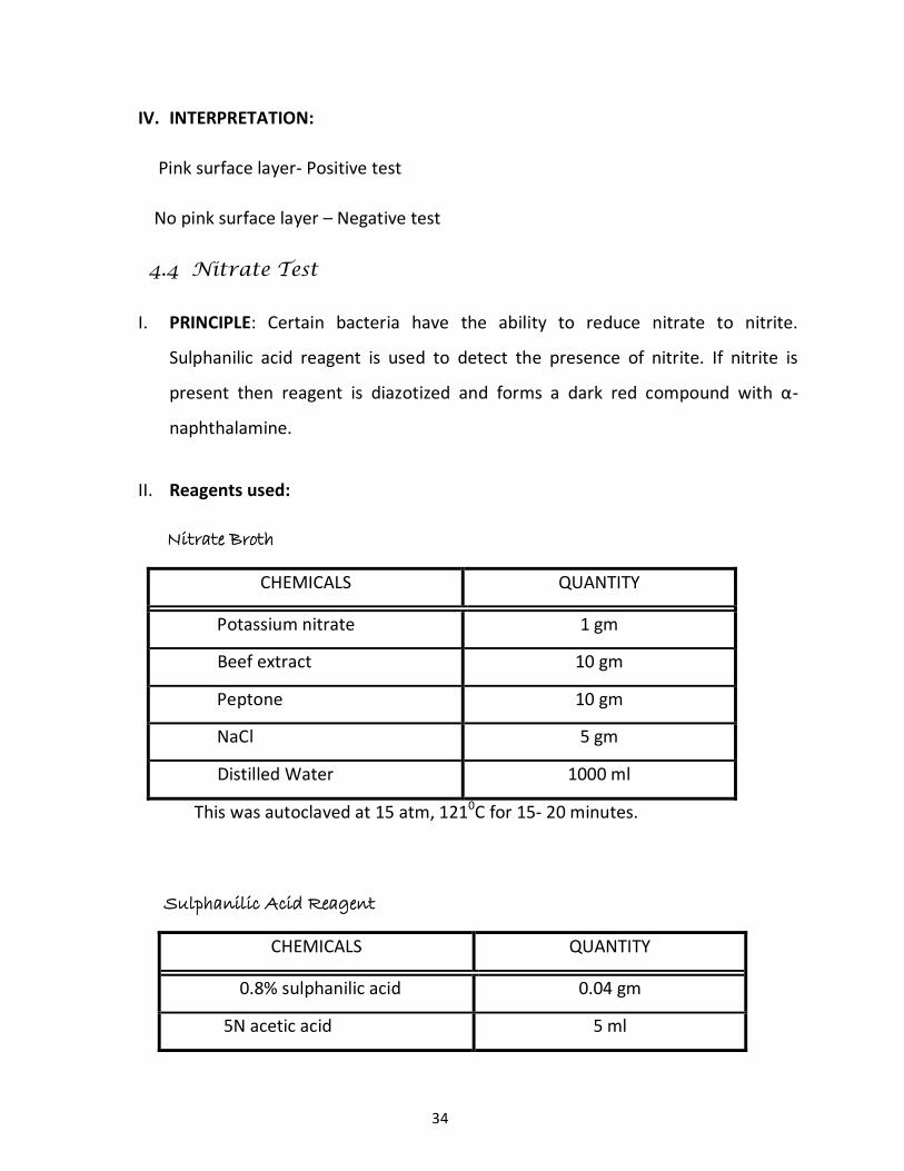
IV. INTERPRETATION:
Pink surface layer- Positive test
No pink surface layer – Negative test
4.4 Nitrate Test
I. PRINCIPLE: Certain bacteria have the ability to reduce nitrate to nitrite.
Sulphanilic acid reagent is used to detect the presence of nitrite. If nitrite is
present then reagent is diazotized and forms a dark red compound with α-
naphthalamine.
II. Reagents used:
Nitrate Broth
CHEMICALS QUANTITY
Potassium nitrate 1 gm
Beef extract 10 gm
Peptone 10 gm
NaCl 5 gm
Distilled Water 1000 ml
This was autoclaved at 15 atm, 1210C for 15- 20 minutes.
Sulphanilic Acid Reagent
CHEMICALS QUANTITY
0.8% sulphanilic acid 0.04 gm
5N acetic acid 5 ml
34

α-naphthylamine reagent
CHEMICALS QUANTITY
0.5% α-naphthylamine 0.025 gm
5N acetic acid 5 ml
III. PROCEDURE:
In 1 ml of sterile nitrate broth loopful of test organism was inoculated and was
incubated for 24 hrs at 370C.
Both the reagents (sulphanilic acid and α-naphthylamine reagent) were mixed in a
ratio of 1:1.
1-2 drops of the reagent were added and color change was observed.
IV. INTERPRETATION:
Red color- Positive test
No red color- Negative test
4.5 Catalase Test
I. PRINCIPLE: Catalase is an enzyme, which converts hydrogen peroxide to water
and oxygen. Catalase producing organisms when brought into contact with
hydrogen peroxide produce oxygen bubbles.
II. Reagents used:
Hydrogen peroxide
35

III. PROCEDURE:
Using a sterile loop the test organism cells were immersed in 1-2 ml of hydrogen
peroxide.
Immediately bubble formation was observed.
IV. INTERPRETATION:
Active bubbling- Positive test
No release of bubbles- Negative test
4.6 Oxidase Test
I. PRINCIPLE: Organism producing enzyme oxidase, oxidises phenylenediamine to
produce a blue-purple color.
II. Reagents used:
Oxidase Reagent
CHEMICALS QUANTITY
Tetra methyl p-phenylenediamine
dihydro-chloride
0.1 gm
Distilled water 10 ml
III. PROCEDURE:
On a piece of whatman no. 2 filter paper, placed in a petridish, several drops
of oxidase reagent were added.
With the help of tooth pick or glass rod a colony of test organism was picked
smeared was made over a small area of filter paper.
Purple-blue color within 10-15 seconds was awaited.
36

IV. INTERPRETATION :
Blue-purple color- Positive test
No blue-purple color- Negative test
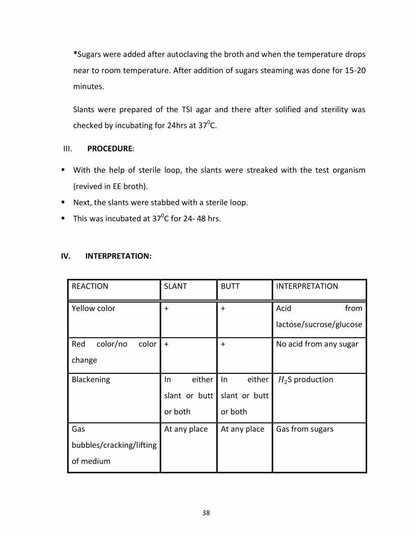
4.7 Triple Sugar Iron (TSI) Agar Test
I. PRINCIPLE: This test is used to differentiate Enterobacteria by producing multiple
tests on a single medium. Acid from sugars (glucose, sucrose and lactose) and S
production was observed.
II. REAGENTS USED:
TSI Agar
CHEMICALS QUANTITY
Peptone 20.0 gm
Yeast extract 3.0 gm
Beef extract 3.0 gm
Lactose 10.0 gm
Sucrose 10.0 gm
Dextrose 1.0 gm
NaCl 5.0 gm
Ferric citrate 0.3 gm
Sodium thiosulphate 0.3 gm
Phenol red 0.024 gm
Agar 22 gm
Distilled water 1000 ml
This was autoclaved at 15 atm pressure and 121 0C for 15-20 minutes.
37
*Sugars were added after autoclaving the broth and when the temperature drops
near to room temperature. After addition of sugars steaming was done for 15-20
minutes.
Slants were prepared of the TSI agar and there after solified and sterility was
checked by incubating for 24hrs at 370C.
III. PROCEDURE:
With the help of sterile loop, the slants were streaked with the test organism
(revived in EE broth).
Next, the slants were stabbed with a sterile loop.
This was incubated at 370C for 24- 48 hrs.
IV. INTERPRETATION:
REACTION SLANT BUTT INTERPRETATION
Yellow color + + Acid from
lactose/sucrose/glucose
Red color/no color
change
+ + No acid from any sugar
Blackening In either
slant or butt
or both
In either
slant or butt
or both
S production
Gas
bubbles/cracking/lifting
of medium
At any place At any place Gas from sugars
38

4.8 Citrate Utilization Test
I. PRINCIPLE: Test organism is cultured in a medium containing sodium citrate,
ammonium salt and bromothymol blue indicator which changes from green to
blue when the growth of organism causes alkalinity.
II. REAGENTS USED:
Simmon’s Citrate Agar
CHEMICALS QUANTITY
Simmon’s Citrate Agar dehydrated 22.28 gm
Agar 22 gm
Distilled Water 1000 ml
C for 15- 20 minutes and was later
poured into the test tubes
C.
III. PROCEDURE:
With the help of a sterile loop the slope was streaked with the test organism.
This was incubated at 370C for 24 hrs and color change was observed in the
medium.
IV. INTERPRETATION:
Bright blue color- Positive test
No color change- Negative test
39

4.9 Acid from Sugar
I. PRINCIPLE: The test organism is cultured in the medium which contains
specific sugars that is fermented by the test organism and lactic acid id
produced. Lactic acid production is detected by the change of color due to the
phenol red indicator used.
II. REAGENTS USED:
Sugar Broth
CHEMICALS QUANTITY
Peptone 10 gm
Beef extract 3 gm
NaCl 5 gm
Distilled Water 1000ml
Phenol Red 12ml
The broth was autoclaved at 15 atm, 1210C for 15- 20 minutes
1% of broth, appropriate sugar was added to the broth (when temperature drops near to room temperature)
1 ml of broth was poured in test tubes and steaming was done for 25 minutes.
Sterility of broth was checked by overnight incubation at 370C.
III. PROCEDURE:
Loopful of test organism was inoculated in the broth and incubated overnight
at 370C.
Color change was observed.
40

IV. INTERPRETATION:
Color change from red to yellow- Positive test
No color change or orange color- Negative test
5. Growing and Fixation of Biofilms for SEM
5.1. Growing of the biofilms of standard culture of Enterobacter sakazakii
(MTCC- 2958) and the isolate.
The revived cultures of standard Enterobacter sakazakii (MTCC- 2958) and
the isolate were streaked on TSA (Trptone Soya Agar) plates.
TSA (Trptone Soya Agar)
CHEMICALS QUANTITY
Caesin Peptone/Tryptone 15 gm
Soya Peptone 15 gm
Sodium Chloride 5 gm
Agar 22 gm
Distilled Water 1000 ml
This was autoclaved under 15 atm pressure at 1210C for 15-20 minutes.
Sterility of the plates was checked by incubating at 370C for 24hours
A colony, from the colonies so appeared on TSA was then picked up and
inoculated in 10ml TSB (tryptic soya broth) with 1% glucose for the growth
hike of biofilms (Bose et al., 2009) along with the three different substrates
such as glass, steel coupons and aluminium to compare the adhering of
41

biofilms with the various type of substrata which are used in our day to day
life.
The substrates were cleaned properly with soap solution followed by acid
wash and were then autoclaved under 15 atm pressure at 1210C for 15-20
minutes.
Sterilized substrates were then introduced in different tubes containing
TSB+1%glucose and the colonies of standard Enterobacter sakazakii (MTCC-
2958) and the isolate. Such as :-
S.No. Substrates Revived Culture TSB
+1%glucose
Test tube 1 Aluminium Standard
(MTCC- 2958)
Test tube 2 Aluminium Isolate of
c.sakazakii
Test tube 3 Steel Standard
(MTCC- 2958)
Test tube 4 Steel Isolate of
c.sakazakii
Test tube 5 Glass Standard
(MTCC- 2958)
Test tube 6 Glass Isolate of
c.sakazakii
They were incubated for 24 hours at 370C.
42

TSB (Tryptic Soya Broth)
CHEMICALS QUANTITY
TSB 30ml
Distilled water 1000ml
The broth was autoclaved at 15 atm, 1210C for 15- 20 minutes.
1% of broth, appropriate sugar was added to the broth (when temperature
dropped close to room temperature).
10 ml of broth was poured in tubes and steaming was done for 15-20minutes.
Sterility of broth was checked by overnight incubation at 370C.
5.2 Fixation of Biofilms on Substrates
REAGENTS FOR FIXING SOLUTION
PBS – phosphate buffer saline (pH=7.3)
CHEMICALS QUANTITY
0.2M Disodium hydrogen phosphate 1.4195 gm
0.2M Sodium dihydrogen phosphate 1.56 gm
0.8% (w/v) NaCl 0.4 gm
Distilled Water 100 ml
0.05M Sodium cacodylate buffer (pH=7.2)
CHEMICALS QUANTITY
Glutharaldehyde 2.5%
Formaldehyde 2.5%
Sodium cacodylate 0.05M in distilled water
43

Calculation for 100ml 0.05M Sodium cacodylate buffer (pH=7.2)
Mass = concentration × volume × molecular weight
Mass = 0.05M × 100/1000 × 159.98
Mass = 0.7999gm sodium cacodylate in 100ml distilled water
Gluthaldehyde = 2.5% of 100ml
= 2.5ml
Formaldehyde = 2.5% of 100ml
= 2.5ml
0.001M Calcium chloride in distilled water
Calculation for 10ml 0.001M Calcium chloride
Mass = concentration × volume × molecular weight
Mass= 0.001 × 10/1000 × 110.984
Mass = 0.00110984gm
Osmium tetraoxide*
1% in distilled water (required in very small quantity)
*handle with care
Acetone gradients
25% acetone (For 20ml)
44

CHEMICALS QUANTITY
Acetone 5ml
Distilled water 15ml
50% acetone (For 20ml)
CHEMICALS QUANTITY
Acetone 10ml
Distilled water 10ml
75% acetone (For 20ml)
CHEMICALS QUANTITY
Acetone 15ml
Distilled water 05ml
90%acetone (For 20ml)
CHEMICALS QUANTITY
Acetone 18ml
Distilled water 02ml
100% acetone (For 20ml)
CHEMICALS QUANTITY
Acetone 20ml
45

After 24 hours of incubation the biofilms stick to the various substrates. The
broth was decanted and the substrata with biofilms was washed with PBS buffer
(pH=7.3)
After washing, the substrates were dried and were fixed in fixing solution by
modified Karnovsky’s method. Various steps followed up for the procedure were:
1. Substrates were incubated for 24hours in 0.05M sodium cacodylate
buffer of pH 7.2 and 0.001M calcium chloride.
2. After incubation, the substrates were washed with 0.05M sodium
cacodylate of pH 7.2 buffer three times for about 10 minutes.
3. Fixation was done in osmium tetraoxide (1% in distilled water) for 1
hour at ambient temperature.
4. Washed thrice with distilled water.
5. The biofilms on substrates were dehydrated in acetone gradients of
25%, 50%, 75%, 90% and 100% for 2-3 minutes in each gradient. The
respective gradient was added to the tube in which the substratum
was contained, thrice consecutively.
6. The substrates were transferred to critical point apparatus for
complete drying.
7. Every substratum was sputter coated with gold.
8. The samples were subjected to scanning electron microscopy (SEM)
for comparison of the biofilms.
46

6. Preparation of planktonic cells for SEM
The enriched cultures of standard Enterobacter sakazakii (MTCC- 2958) and
the isolate were centrifuged at 6000rpm for 10 minutes.
The supernatant was discarded and pellet obtained was washed with PBS
buffer. This was again centrifuged for 5minutes at 6000 rpm. Supernatant was
discarded and pellet was obtained.
The pellet was used to make the smear on slide and was allowed to dry for
fixation process.
Fixation was carried out in the same manner as that for biofilms.
These arrested cells were then subjected for SEM.
7. Control of Biofilms
The biofilms on the substrates were washed with PBS buffer and were incubated
in EE broth overnight so that the biofilms adhering to the substrates get into the
broth.
7.1. Antibiotic susceptibility
Antibiotic susceptibility was checked by agar well diffusion method on MHA
(Muller-Hinton agar) plates. MHA was prepared and was inoculated with 150µl of
the culture of biofilms in EE broth. Wells of 6mm diameter were made in which
40µl of antibiotics of different concentrations was inoculated (Stock and
Wiedemann, 2002). This was incubated at 370C for 24hours. Cronobacter
sakazakii is found to be susceptible to gentamycin and tetracycline. Thereby
these antibiotics were selected for this experiment. Inhibition zones were
measured and recorded.
47

MHA (Muller-Hinton Agar)
CHEMICALS QUANTITY
MHA 38gm
Agar 6gm
Distilled water 1000ml
Calculations for the drug Tetracycline amount for different concentrations
Powdered drug was diluted with distilled water as per the concentration stated by Stock and Weidemann, 2002. MIC of drug is 40mg/L = 40µg/ml
... 1ml = 40 µg of drug
And 1 µl= .04 µg of drug
As per serial dilution procedure the drug was diluted in the ratio of 9:1 (water: drug)
0.4 µl dilution 0.04µl dilution 0.004 µl dilution 0.0004µl dilution =1.6×10⁻²µg drug =1.6×10⁻³µg drug =1.6×10⁻⁴µg drug =1.6×10⁻⁵µg drug
100µl in
900µl H2O
40 µg in
1ml
100µl in
900µl H2O
100µl in
900µl H2O
100µl in
900µl H2O
48

Calculations for the drug Gentamycin amount for different concentrations
7.2. MIC of natural products against the biofilms
Minimum inhibitory concentration was checked for plant extracts (obtained
from the lab) on the biofilms of standard and the isolate by agar well diffusion
method. Alcoholic crude extracts of Triphala, Har (Terminalia chebula),
Amla(Phyllanthus emblica) and Laung (Syngium aromaticum) were checked
against the cultures. 150 µl of the culture was inoculated in MHA which was
poured in plates and 6mm diameter wells were made in which 40 µl of extracts
were added and incubated for 24hours. Inhibition zones were measured and
recorded.
Powdered drug was diluted with distilled water as per the concentration stated by Stock and Weidemann, 2002. MIC of drug is 2.5mg/L =2.5µg/ml
... 1ml = 2.5µg of drug
And 1 µl= 2.5×10⁻³
µg of drug
As per serial dilution procedure the drug was diluted in the ratio of 9:1 (water: drug)
2.5×10⁻²
µl dilution 2.5×10⁻³
µl dilution 2.5×10⁻⁴
µl dilution 2.5×10⁻⁵
µl dilution =1×10⁻³ µg drug =1×10⁻⁴ µg drug =1×10⁻⁵ µg drug =1×10⁻⁶ µg drug
2.5 µg in
1ml
100µl in
900µl H2O
100µl in
900µl H2O
100µl in
900µl H2O
100µl in
900µl H2O
49

7.3. Control of biofilms by LAB
The biofilms of the standard and the isolate cultured on different substrata were
subjected to LAB (lactic acid bacteria) to check the MIC for the biofilms. This was
done by agar well diffusion method. Antimicrobial effect of Lactobacillus
fermentum (MTCC- 903), Lactobacillus lactis (MTCC- 1423) and
Pediococcus acidilactici (MTCC-7742) was checked by inoculating 150 µl culture
in MHA and then 40 µl of LAB in the wells. This was incubated for 24 hours and
the inhibition zones were measured and recorded.
8. Control of Planktonic cells of the isolate and the standard.
The enriched planktonic cells were subjected to antimicrobial effect of plant
extracts, antibiotics and LAB by agar well diffusion method. This was done by
pouring 150 µl of the culture of these planktonic cells in MHA and the wells were
made of 6mm diameter after solidification of the plates. In the wells 40 µl of
extracts, antibiotics of various dilutions and LAB of the three strains were added.
Zone of inhibition was measured and recorded.
50