Embed Size (px)
Citation preview
INACTIVATION OF CRONOBACTER (ENTEROBACTER) SAKAZAKII USING DIFFERENT
ANTIMICROBIAL AGENTS AND THE EFFECT OF SANITIZERS ON BIOFILM
FORMATION PROPERTIES
By
LUIS FERNANDO CASTRO
A thesis submitted in partial fulfillment of
the requirements for the degree of
MASTER OF SCIENCE IN FOOD SCIENCE
WASHINGTON STATE UNIVERSITY
School of Food Science
DECEMBER 2009
ii
To the Faculty of Washington State University:
The members of the Committee appointed to examine the thesis of LUIS FERNANDO
CASTRO find it satisfactory and recommend that it be accepted.
Barbara Rasco, Ph.D.,Chair
Barry G. Swanson, Ph.D.
Gülhan Ünlü, Ph.D.

iii
AKNOWLEDGEMENTS
I would like to express my gratitude to all those people who made it possible for this
research project to be completed. I would first of all like to thank my advisor Dr. Barbara Rasco,
for accepting me as her graduate student, for all of her advice regarding my project, and for
always showing me the humor in bad situations. I will always cherish the conversations we had
in her office. I would also like to thank my committee members Dr. Barry Swanson and Dr.
Gülhan Ünlü, for reading my entire thesis and for all of their advice, and a special thanks to Dr.
Dong-Hyun Kang, for his presentation about Enterobacter sakazakii in my food microbiology
class, which is what got me started on this research project, also for all of his good advice from
the beginning of this project.
I believe special thanks are in order to Dr. Murad Al-Holy, it was my privilege to work
with him on my project throughout the entire summer of 2008. This thesis would not have been
completed without his crucial help. And of course a huge thank you to the wonderful
administrative personnel in our department, Jodi Anderson, Carolee Armfield, Marsha Appel,
and Rich Hoeft, they really have made this entire journey go a lot smoother.
Special thanks to Maria, my office partner and most importantly my “friend” during the
last two years. I really want to thank her for her patience, for trying to teach me how to cook and
feeding me when I was out of food, I will always remember the shopping trips to Spokane, the
countless hours we have spent watching movies and talking about life, and I am very grateful for
all of her support when times got very difficult for me in Pullman. Thank you Maria for being
my friend and for teaching me the value of true friendship, I honestly don’t know what I would
do without you.

iv
Thanks to my parents for all of their support, I honestly would not be here if it wasn’t for
them, thank you for always being there for me and for supporting my decisions, you guys are the
best. Mom and Dad thank you for teaching me discipline and responsibility, for always making
sure I have everything I need, for keeping me going when times get tough, and most of all thank
you for all of your love and support.
Finally I would like to thank my grandfather Fernando, abuelito thank you for everything
you have done for me, for your incredible sense of humor and for teaching me that there is no
such thing as a free lunch, if you really want something in life you have to work for it, I miss
you.
v
INACTIVATION OF CRONOBACTER (ENTEROBACTER) SAKAZAKII USING DIFFERENT
ANTIMICROBIAL AGENTS AND THE EFFECT OF SANITIZERS ON BIOFILM
FORMATION PROPERTIES
Abstract
By Luis Fernando Castro, M.S.
Washington State University
December 2009
Chair: Barbara Rasco
Enterobacter sakazakii is an opportunistic pathogen that represents a health risk to
neonates. It is a known contaminant of infant formula milk (IFM) and has been associated with
cases of necrotizing enterocolitis and infant meningitis. Many studies have been performed on
the growth conditions and inactivation of the microorganism. Control of this microbe is
important since it affects highly susceptible populations. The purpose of this study was to
investigate the inactivation of E. sakazakii (five strains) inoculated into IFM using different
antimicrobials, and to evaluate the effectiveness of different disinfectants for the removal of
biofilms on plastic surfaces. Results showed that out of all the antimicrobials used, the
combination of lactic acid (0.2% v/v) and copper (50 ppm) proved to be the most effective,
where the individual use of copper at 50 ppm and lactic acid at 0.2% v/v did not have a
significant effect on the growth of E. sakazakii (P > 0.05), but the combination of both agents
produced a significant inhibition of growth in both rehydrated infant formula milk (RIFM) and
powdered infant formula milk (PIFM) after 2 hours (P < 0.05). The effectiveness of three

vi
different household dishwashing liquids, and a sodium hypochlorite solution in the removal of
biofilms formed on plastic surfaces was also studied, and it was found that quaternary
ammonium compounds are the most effective, and that treatment time plays an important role in
the elimination of the biofilms, since higher reductions in population were observed as the
treatment times increased.
vii
TABLE OF CONTENTS
AKNOWLEDGEMENTS.............................................................................................................. iii
ABSTRACT .................................................................................................................................... v
LIST OF TABLES .......................................................................................................................... x
LIST OF FIGURES ....................................................................................................................... xi
CHAPTER ONE ............................................................................................................................. 1
LITERATURE REVIEW ............................................................................................................ 1
1.1 Enterobacter sakazakii Characteristics .................................................................................. 1
1.1.1 Taxonomy and Biochemical Characteristics of E. sakazakii ......................................... 1
1.1.2 Occurrence of E. sakazakii in Foods and the Environment ............................................ 3
1.1.3 Resistance to Osmotic Pressure and Desiccation ........................................................... 4
1.1.4 Heat Resistance............................................................................................................... 6
1.1.5 Antimicrobial Resistance .............................................................................................. 10
1.1.6 Biofilm Formation ........................................................................................................ 12
1.2 Detection of E. sakazakii ................................................................................................... 13
1.2.1 FDA Recommended Method ........................................................................................ 13
1.2.2 Chromogenic and Fluorogenic Media .......................................................................... 15
1.2.3 DNA Based Methods .................................................................................................... 17
1.3 E. sakazakii Caused Illness ................................................................................................. 18
1.3.1 History and Pathogenicity Associated with E. sakazakii Infection .............................. 18
1.3.2. Presence of E. sakazakii in Infant Formula Milk ........................................................ 22
1.3.3. Risk Management of E. sakazakii in Infant Formula Milk ........................................ 25
1.4. Inactivation of E. sakazakii ................................................................................................ 27
CHAPTER TWO .......................................................................................................................... 32

viii
MATERIALS AND METHODS .............................................................................................. 32
2.1. Bacterial Strains ................................................................................................................. 32
2.2. Inactivation Studies ............................................................................................................ 32
2.2.1. Inoculation of Re-hydrated Infant Formula Milk (RIFM) ........................................... 32
2.2.3. Inactivation of E. sakazakii using Denatured Lysozyme ............................................ 33
2.2.4. Inactivation of E. sakazakii using Lactic Acid ............................................................ 34
2.2.5 Inactivation of E. sakazakii using lactic acid and nisin ................................................ 34
2.2.6 Inactivation using Copper and Lactic Acid .................................................................. 34
2.2.7 Inoculation of Powdered Infant Formula Milk (PIFM) ................................................ 35
2.2.8 Inactivation of E. sakazakii in PIFM using Copper and Lactic Acid .......................... 36
2.2.9 Enumeration of E. sakazakii ......................................................................................... 36
2.2.9.1 Confirmation of Enumeration Results Using Enrichment Broth............................... 37
2.3 Effectiveness of Different Sanitizers on E. sakazakii Biofilm Formation .......................... 37
2.3.1. Bacterial Strains ........................................................................................................... 37
2.3.2. Biofilm Formation ....................................................................................................... 38
CHAPTER THREE ...................................................................................................................... 40
RESULTS AND DISSCUSSION ............................................................................................. 40
3.1 Inactivation of E. sakazakii using Lysozyme and Denatured Lysozyme ............................ 40
3.2. Inactivation of E. sakazakii using Lactic Acid ................................................................... 42
3.3 Inactivation of E. sakazakii using Nisin and Lactic Acid ................................................... 44
3.4 Inactivation of E. sakazakii using copper and Lactic Acid ................................................. 46
3.5 Biofilm formation and disinfectant efficacy ....................................................................... 51
CONCLUSIONS........................................................................................................................... 53
FUTURE RESEARCH ................................................................................................................. 54

ix
REFERENCES ............................................................................................................................. 55
x
LIST OF TABLES
Table 1. Summary of Enterobacteriaceae cultured from different infant formulations ............... 60
Table 2. Surveys conducted on powdered infant formula for E. sakazakii contamination .......... 61
Table 3. Disinfectants evaluated for efficacy against E. sakazakii biofilms ................................ 62
Table 4. Antimicrobial activity of copper on the growth of E. sakazakii in RIFM as measured by
the log CFU/mL. Values with the same superscript within a column are not significantly
different from the control (P ≤ 0.05). ............................................................................................ 63
Table 5. Mean OD570 of stained biofilms, three replicates. Bold values are significantly lower
from control ( P ≤ 0.05). Values with the same superscript across a row are not significantly
different (P ≥ 0.05). ....................................................................................................................... 64
xi
LIST OF FIGURES
Figure 1. FDA standard protocol for isolation of Enterobacter sakazakii from powdered .......... 65
Figure 2. Effect of different concentrations of lysozyme (A) and denatured lysozyme (B) on the
growth of Enterobacter sakazakii (Log CFU/mL) in reconstituted infant formula milk stored at
room temperature. ......................................................................................................................... 66
Figure 3. Survival curve of E. sakazakii in reconstituted infant formula milk under three different
concentrations of lactic acid……………………………………………………………………...67
Figure 4. Effect of different concentrations of nisin against E. sakazakii in reconstituted infant
formula stored at room temperature. ............................................................................................. 68
Figure 5. Effect of copper at different concentrations on the growth of E. sakazakii in RIFM
stored at room temperature. .......................................................................................................... 69
Figure 6. The combined effect of lactic acid and copper on the growth of Enterobacter sakazakii
in reconstituted infant formula stored at room temperature. ......................................................... 70
Figure 7. The effect of copper and lactic acid applied individually and in combination on the
growth of Enterobacter sakazakii in powdered infant formula milk stored at room temperature.
....................................................................................................................................................... 71

1
CHAPTER ONE
LITERATURE REVIEW
1.1 Enterobacter sakazakii Characteristics
1.1.1 Taxonomy and Biochemical Characteristics of E. sakazakii
Enterobacter sakazakii is an emerging opportunistic human pathogen associated with
bacterial infections in infants. The microorganism belongs to the family Enterobacteriaceae, and
is a Gram-negative, motile, facultatively anaerobic, non-sporeforming organism (Drudy and
others, 2006). It was known as “yellow pigmented E. cloacae” until 1980, when Farmer and
others (1980) designated it as a new species based on the results of DNA-DNA hybridization,
yellow pigment production, biochemical reactions, and antibiotic susceptibility studies. The
organism is named in honor of Riichi Sakazakii, a Japanese bacteriologist (Iversen and Forsythe,
2003).
In the genus Enterobacter there are 14 species or biogroups, and E. sakazakii is a biovar
in the genus (Gurtler and others, 2005). E. sakazakii exhibits many biochemical characteristics
similar to those of E. cloacae, but is D-sorbitol negative, produces an extracellular
deoxyribonuclease, and most strains produce yellow pigmented colonies (Farmer and others,
1980). However, some strains of E. sakazakii recently isolated ferment D-sorbitol, and do not
produce yellow pigment, leading to misidentification as E. cloacae (Farber, 2008).
In 1977, researchers suggested that the yellow pigmented E. cloacae was a new species,
basing this conclusion on DNA-DNA hybridization studies which showed less than 50%
homology with non-pigmented strains. Farmer and others (1980) distinguished 57 strains of
2
yellow-pigmented E. sakazakii based on DNA hybridization, antibiotic susceptibility and
biochemical reactions, they also observed other characteristics that distinguished the bacterium
from E. cloacae. Some characteristics were greater pigment production at temperatures less than
36 °C, optimum pigment production at 25 °C, survival of cells in stock cultures stored at 17 – 30
°C without transfer for up to 8 years, the use of citrate as a sole carbon source, a 31 – 49% DNA-
DNA homology with E. cloacae, and a 57% guanine + cytosine ratio (Farmer and others, 1980;
Gurtler and Beuchat, 2007).
Enzymatic profiles of E. sakazakii were also used to differentiate the microorganism. A
study by Muytjens and others (1984) used the APITM
ZYM identification system to evaluate 226
strains of Enterobacter, including 129 strains of E. sakazakii. Two major differences between E.
sakazakii and the other Enterobacter species were observed, the first is α-glucosidase activity,
present in all strains of E. sakazakii but absent from the other Enterobacter strains (Muytjens and
others, 1984). Famer and others (1985) reported that 53 of 57 strains of E. sakazakii were
positive for α-glucosidase. The second difference observed by Muytjens and others (1984) was
Tween 80 esterase activity unique to E. sakazakii.
The use of gas chromatography to differentiate E. sakazakii from closely related
Enterobacter and Citrobacter species was recently proposed. A study by Whitaker and others
(2007) used capillary gas chromatography with flame ionization detection to compare the
cellular fatty acid profiles for 134 Enterobacter sakazakii strains, to the cellular fatty acid
profiles of closely related Enterobacter and Citrobacter species. The study demonstrated a clear
differentiation in fatty acid composition amongst the species, making this a useful tool to
differentiate E. sakazakii strains from other closely related species (Whittaker and others, 2007).
3
1.1.2 Occurrence of E. sakazakii in Foods and the Environment
The natural habitat of E. sakazakii is not currently well understood, but the
microorganism is widely distributed in the environment and foods, with water, soil and
vegetables being the most probable sources of the organism. Iversen and Forsythe (2004) stated
that because of the organism’s physiological features, its most likely habitat is on plant material.
These features aid environmental survival, and they include the ability to produce a yellow
pigment that protects the cells from UV rays in sunlight, the capsular and fimbriae formation to
aid in adherence to surfaces that include other cells, and its remarkable resistance to desiccation
over long periods of time (Iversen and Forsythe, 2004; Mullane and others, 2006).
E. sakazakii has been isolated from water, sediment, and soil; but in a study performed by
Muytjens and Kolle (1990), the researchers were unable to isolate it from select environmental
sources including: raw cow milk, cattle, rodents, grain, bird feces, domestic animals, surface
water, soil, mud, and rotting wood (Muytjens and Kollee, 1990). Kandhai and others (2004)
evaluated the occurrence of the organism in selected households and food factory environments,
isolating E. sakazakii from almost all of the environments analyzed; these environments included
milk powder manufacturing facilities and common household vacuum cleaners (Kandhai and
others, 2004).
The microorganism was also isolated from floor drains, air, a vacuum canister, broom
bristles, a room heater and electrical control box, a floor dryer, floor and condensate in a dry
product processing environment in the United States. Other environmental sources for E.
sakazakii reported by Iversen and Forsythe (2003), they include air in a hospital, clinical
materials, rats, soil, sediment and wetlands, crude oil and cutting fluids (Gurtler and others,
2005).

4
Dairy products such as milk powders, cheese products, and baby foods are known
reservoirs of E. sakazakii, as well as minced beef, sausage meat, and vegetables. There are also
documented reports on the isolation of E. sakazakii from rats in Kenya and from Mexican fruit
flies in California (Farber, 2008). Several reports implicate rehydrated powdered infant formula
milk as a source of the organism in neonatal infections. The first outbreak linked to powdered
infant formula milk from previously un-opened cans was reported by the CDC in 2001, and in
another outbreak the powdered formula tested negative for E. sakazakii, but the blender used to
prepare the formula was contaminated. It is very likely that the contamination was a result of a
previous batch of contaminated powdered infant formula, and even though the blender was
washed daily, the cleaning procedure may not have been sufficient to eliminate the
contamination (Gurtler and others, 2005).
Out of all the possible sources of E. sakazakii, its presence in powdered infant formula
milk is of particular concern, due to its use for the feeding of newborn babies. The details
regarding the presence of E. sakazakii in infant formula milk will be discussed later.
1.1.3 Resistance to Osmotic Pressure and Desiccation
Enterobacter sakazakii is frequently isolated from powdered infant formula milk, which
suggests that it has an unusual ability to survive in dry environments. Barron and Forsythe
(2007) analyzed the dry stress and survival time of E. sakazakii and other Enterobacteriaceae in
powdered infant formula; they assessed the generation of sub-lethally injured
Enterobacteriaceae during desiccation, and its persistence in powdered infant formula over a
storage period of 2.5 years at room temperature. The study included ten strains of E. sakazakii,
and the recovery of the organism under dry stress decreased an average of 0.58 log cycles during
5
the first month, with a decrease of 3.34 log observed during the first 6 months. The average
recovery decreased 1.88 log cycles over the next 24 months, resulting in a total decline in viable
counts of 4.52 log cycles.
Five out of the ten E. sakazakii strains were still recoverable after two years, which
shows that some of the microorganism strains can remain viable during long-term storage in
powdered infant formula milk (Barron and Forsythe, 2007). The formation of an extracellular
polysaccharide may provide E. sakazakii with the protection to survive physical and
environmental stress; however, during the first 18 mo of this study there was no correlation
between capsulation and recovery. After 2 years, four of the five strains recovered from the
powdered infant formula milk were capsulated, and after 2.5 years only the capsulated strains
were recoverable. Capsulation may play an important role in recovery following long term
dessication. E. sakazakii can survive in dehydrated environments for long periods of time
(Barron and Forsythe, 2007; Lehner and others, 2005).
Edelson-Mammel (2005) studied the survival of a single strain of E. sakazakii inoculated
at approximately 105 CFU/g in powdered infant formula for a period of two years at room
temperature, during which the contaminated formula was periodically rehydrated and analyzed
for viable E. sakazakii. During the initial five mo of storage, there was a decrease in viable count
of approximately 2.4 log cycles, and during the next 19 mo the concentration of viable E.
sakazakii declined an additional 1.0 log cycle.
The calculated D-value for the first five months was 73.8 days and 684.9 days for the
period between 5 and 19 mo. The concentration of E. sakazakii in the infant formula when
rehydrated at the end of the study was approximately 300 CFU/mL (Edelson-Mammel and
others, 2005). The results were consistent with those observed previously by Breeuwer and
6
others (2003), they found that E. sakazakii was more resistant to osmotic stress and desiccation
than other members of the Enterobacteriaceae family (Breeuwer and others, 2003). Even when a
majority of E. sakazakii cells are inactivated by storage in dehydrated powdered infant formula, a
portion of the cells are highly resistant to dry storage conditions and can survive for up to 2 years
(Edelson-Mammel and others, 2005).
Breeuwer and others (2003) studied the genetic basis for the survival of E. sakazakii
exposed to dry conditions and concluded that the desiccation of the microorganism leads to an
induction of seven genes associated with its heat shock regulation, four genes from the cyclic
AMP receptor protein regulon, six genes involved in the stringent response, and a number of
genes involved in trehalose synthesis and cell wall functions such as lipid A and
lipopolysaccharide biosynthesis. Researchers concluded that the response of E. sakazakii to dry
stress involves a genome-wide expression of functionally different groups of genes (Gurtler and
others, 2005).
The high tolerance of E. sakazakii to desiccation provides a competitive advantage in dry
environments, such as conditions found in milk powder factories, and increases the risk of post-
pasteurization contamination of the finished products. The high desiccation resistance of the
microorganism will also allow it to survive in powdered infant formula at water activities as low
as 0.2.
1.1.4 Heat Resistance
The thermal tolerance of E. sakazakii was first investigated by Nazarowec-White and
Farber (1997), they used 10 strains of E. sakazakii to inoculate re-hydrated infant formula at
selected temperatures to determine the D and z values. They reported that the D-values at five
7
selected temperatures were: D52°C = 54.8 min, D54°C = 23.7 min, D56°C = 10.3 min, D58°C = 4.20
min, and D60°C = 2.50 min, with a pooled z-value of 5.82 °C. The comparison of the D-values
obtained for E. sakazakii with those of other Enterobacteriaceae in dairy products, confirms that
E. sakazakii is one of the most thermo-tolerant microorganisms (NazarowecWhite and Farber,
1997c).
Many thermal processes or pasteurization practices require a 4.0 -7.0 D log reduction of
microorganisms as a process control. To obtain a 6.0 – 7.0 log reduction of E. sakazakii, a heat
treatment of 60 °C for 15 – 17.5 minutes is required. The microorganism is more thermo-tolerant
than other Enterobacteriaceae in dairy products (NazarowecWhite and Farber, 1997c).
Iversen and others (2004) determined that the D-values for a specific E. sakazakii strain
were D54°C = 16.4 min, D56°C = 5.1 min, D58°C = 2.6 min, D60°C = 1.1 min and D62°C = 0.3 min. A
capsulated strain showed smaller D values, but the z-values for both strains were 5.7 and 5.8 °C.
The authors theorized that a high temperature short-time pasteurization of 15 s at 71.2 °C will
result in a 21-D kill when dried infant formula is processed, indicating that E. sakazakii will not
survive this commercial pasteurization process, therefore the persistence of the microorganism in
powdered infant formula milk is attributed to post-process contamination (Iversen and others,
2004b).
A study by Edelson-Mammel and Buchanan (2004) reported a 20 fold difference in
thermal resistance between the most and least heat-resistant of 12 strains of E. sakazakii in
rehydrated infant formula at 58 °C. Half of the strains exhibited a D-value smaller than 50s, and
the other half were greater than 300 s, so the authors divided the two thermally resistant groups
into two phenotypes. The z-value for the most thermally resistant strain was 5.6 °C. Further
examination after inoculation of infant powder formula with the strain and rehydration with de-
8
ionized water at temperatures of 50, 60, 70, 80, 90 and 100 °C, resulted in no inactivation at 50
°C, but a 4.0-D and greater inactivation was observed at 70 °C. Rehydration of infant formula
milk with water at a temperature of 70 °C is a good alternative to reduce the risk of E. sakazakii
survival in the rehydrated product by at least four orders of magnitude. E. sakazakii is seldom
isolated at levels above 1 CFU/100g of powdered infant formula milk. A 4.0-D thermal treatment
will virtually eliminate E. sakazakii (Edelson-Mammel and Buchanan, 2004).
The rates of growth of the organism at ambient and cool temperatures are necessary to
predict the possible doses of E. sakazakii that might be ingested by an infant fed a rehydrated,
stored infant formula. To better understand the thermal behavior of the microorganism, Kim and
Park (2007) determined the thermal characteristics of selected strains of E. sakazakii at 52, 56
and 60 °C in a saline solution, rehydrated powdered infant formula, and dried baby food.
The study concluded that D-values were larger for rehydrated infant formula in
comparison with the saline solution, but smaller than in dried baby food. The overall calculated
z-value was 6.0 – 8.0 for saline, 8.0 – 10.0 for powdered infant formula, and 9.0 – 11.0 for dried
baby food, and thermal inactivation did not occur for 20 minutes when infant formula milk was
rehydrated with water at 50 °C, while a reduction of 1.0 – 2.0 log CFU/g and 4.0 – 6.0 log
CFU/g was observed when the rehydration water was at 60 °C and 70 °C, respectively. The
conclusions of the study confirm the report of Edelson-Mammel and Buchanan (2004), that to
reduce the risk of E. sakazakii contamination from powdered infant formula, the water used for
rehydration must be at a temperature higher than 50 °C (Kim and Park, 2007).
Breeuwer and others (2003) used five strains of E. sakazakii in the stationary phase to
perform thermal inactivation studies, they used a disodium hydrogen phosphate/potassium
dehydrogenate phosphate buffer with a neutral pH for the heating medium, and reported D58°c
9
values that ranged from 0.39 to 0.60 min, and z values between 3.1 and 3.6 °C for two strains.
The z-values are slightly smaller than z-values obtained in the studies performed by Edelson-
Mammel and others (2004) and Nazarowec-White and Farber (1997), but the difference in the
heating media used may be the reason for the observed difference. The high amount of fat,
protein, and carbohydrate in infant formula milk may serve to protect E. sakazakii against
thermal inactivation, and result in larger D-values (Breeuwer and others, 2003).
Microwave radiation is also being studied as a method to inactivate E. sakazakii in re-
hydrated infant formula milk. Kindle and others (1996) heated infant formula milk inoculated
with a population of 5 log10 CFU/mL until the first signs of boiling, and cooled it down and
analyzed it for surviving cells. Five samples of heated infant formula milk were analyzed, and
four of the five samples tested negative for E. sakazakii following the microwave treatment,
while the other formula contained 20 CFU/mL.
Based on the results of this study, it is highly recommended that rehydrated infant
formula milk be rewarmed using a microwave, instead of more traditional methods. Kinle and
others (1996) reported that the microwaving of infant formula milk in baby bottles for 85 – 100 s
at 82 - 93°C will achieve a 4 log10 CFU/mL reduction in the E. sakazakii population (Kindle and
others, 1996).
From the data gathered on thermal resistance of E. sakazakii, the World Health
Organization concluded that inactivation of the pathogen occurs very rapidly at temperatures
above 70 °C. The WHO proposed that infant formula milk be rehydrated at temperatures greater
than 70 °C to reduce the risk posed by E. sakazakii infection. The data also demonstrates that E.
sakazakii will not survive the pasteurization step, so factory environments and post-

10
pasteurization processes should be very carefully monitored to avoid the contamination of
powdered infant formula milk (Farber, 2008; Iversen and Forsythe, 2003).
1.1.5 Antimicrobial Resistance
Antimicrobial resistance of E. sakazakii is another interesting characteristic of the
microorganism. Farmer and others (1980) reported that strains of E. sakazakii used in their study
were susceptible to gentamicin, kanamycin, chloramphenicol, and ampicillin, while 87% were
susceptible to nalidixic acid, streptomycin, tetracycline, and carbenicillin; 71 and 67% were
susceptible to sulfadiazine and colistin, respectively, and only 13% were susceptible to
cephalothin. The strains of E. sakazakii in this study were resistant to penicillin, while only 1
strain showed multiple antibiotic resistance (Farmer and others, 1980; Iversen and Forsythe,
2003). The natural antibiotic susceptibilities of 35 clinical isolates of E. sakazakii to 69 selected
antimicrobial agents; identified isolates susceptible to most β-lactam and cephalosporin
antibiotics, but highly resistant to benzylpenicillin and oxacilin. The results confirmed the
observations of Farmer and others (1980) (Iversen and Forsythe, 2003).
Muytjens and Vanderrosvandere (1986) determined the minimum inhibitory
concentration (MIC) of 29 antimicrobial agents against 195 strains of E. sakazakii, and reported
that 90% of them exhibited a lower value than those of E. cloacae, but resistance to cephalothin
and sulfamethoxazole was observed (Muytjens and Vanderrosvanderepe, 1986). A study
performed by Nazarowec-White and Farber (1999) tested the antibiotic resistance of 17 strains of
E. sakazakii, and reported that five food isolates and eight clinical strains were resistant to
sulphisoxazole and cephalohin, but susceptible to ampicillin, cefotaxime, chloramphenico,
11
gentamicin, kanamycin, plymyxin-B, trimethoprim-sulphanmethoxzole, tetracycline and
streptomycin (Gurtler and others, 2005).
Bacterial meningitis must be effectively treated to prevent death of the patient, so
antibiotic resistance of E. sakazakii is of extreme concern when treating neonatal infections. The
infection resulting from E. sakazakii are usually treated with ampicillin in combination with
gentamicin or chloramphenicol, with the ampicillin-gentamicin combination being the preferred
treatment, but unfortunately E. sakazakii developed resistance to both antibiotics by means of
transposable elements, as well as resitance to β-lactams by the production of β-lactamase.
The Enterobacter inactivates a broad spectrum of penicillins and cephalosporins due to β-
lactamase production, and this trend is increasing among strains of E. sakazakii. These
observations led scientists to suggest a switch to carbapenems or the newer third-generation
cephalosporins in combination with an aminoglycoside or trimethoprim-sulfamethoxsazole ot
eliminate E. sakazakii. This treatment improved the outcome of E. sakazakii meningitis, even
though the resistance of Enterobacter spp. to these antibiotics is increasing (Gurtler and others,
2005; Iversen and Forsythe, 2003).
Willis and Robinson (1988) reported two cases of E. sakazakii associated neonatal
meningitis initially unresponsive to ampicillin-gentamicin therapy, but resulted in complete
abatement after treatment with moxalactam. Based on the observations on antimicrobial activity,
general assumptions regarding antibiotic therapy against E. sakazakii cannot be made; treatment
should be decided upon clinical observation and in vitro susceptibility testing (Gurtler and
others, 2005).
12
1.1.6 Biofilm Formation
Pure cultures of microorganisms often grown on broth media were studied for many
years by microbiologists, but it is now accepted that bacteria grow preferentially as bio-films.
Biofilms are complex communities that grow on a surface and are surrounded by a
polysaccharide material known as glycocalyx. The films contain about 90 – 97% water and vary
from a few micrometers to several millimeters in thickness. Biofilms usually consist of mixed
species, and the bacterial cells may be accompanied by eukaryotes, a variety of extracellular
polysacharides, enzymes, proteins, bacteriocins, and low mass solutes accompanied with nucleic
acids present as a result of lysis (Brooks and Flint, 2008).
Very little was known about biofilms before 1980, but today there is an enormous
amount of evidence suggesting food processing equipment is often contaminated by biofilms,
often resulting in contamination of food. Some of the hazards related to biofilms include film
accumulation on food contact surfaces and microbial colonization of milk storage tanks, fouling
of heat exchangers and adhesion of spores on packaging material surfaces. Biofilms can also
form on food itself, providing potential for cross-contamination and post-process contamination
of foods as well (Brooks and Flint, 2008).
The formation of biofilms on biotic and a-biotic surfaces is suggested as a survival
strategy for food associated microorganisms such as Listeria monocytogenes and Escherichia
coli. Biofilms provide the cells with protection to environmental stress factors such as UV light,
osmotic stress, heat, starvation, acids, detergents, antibiotics, phagocytes, antibodies, and
bacteriophages (Lehner and others, 2005). A study on thermo-tolerance and biofilm formation
confirmed that when E. sakazakii was grown in infant formula milk, the microorganism was
capable of producing biofilms on latex, silicon, and stainless steel; materials commonly used for
13
processing of infant formula milk, for infant feeding, and in formula preparation areas (Iversen
and others, 2004b).
Lehner and others (2005) conducted experiments to identify biofilm formation and the
production of cellulose as one of the components of the extracellular matrix, adherence of E.
sakazakii to hydrophilic and hydrophobic surfaces, the production of extracellular
polysaccharides, and the ability of E. sakazakii to produce cell-to-cell signaling molecules. Of
the 56 strains of E. sakazakii used, 44 formed biofilms in nutrient rich media. Lehner and others
(2005) also observed that 42.8% of the strains were attached to glass surfaces, while 58.9% of
the strains formed biofilms on PVC microtiter wells under the same conditions. Extracellular
polysaccharide production was confirmed for 41.8% of the isolates, with greater production by
food and environmental isolates than by human isolates (Lehner and others, 2005).
The ability of E. sakazakii to form biofilms on stainless steel and enteral feeding tubes at
selected temperatures and conditions was studied by Kim and others (Kim and others, 2008).
Five strains grown in selected media including infant formula milk were examined at 12 and 25
°C, and larger populations attached at 25 °C on both surfaces. The research also concluded that
nutrient availability plays a very important role in the biofilm formation process, since some
biofilms were not formed when tryptic soy broth and lettuce juice broth were used to grow the
strains (Kim and others, 2008).
1.2 Detection of E. sakazakii
1.2.1 FDA Recommended Method
E. sakazakii has a very low infectious dose estimated at around 1000 CFU/mL in re-
constituted infant formula milk, which is why a good detection method is of extreme importance
14
to address the food safety issue. It is possible that due to the lack of an appropriate detection
method, there is underestimation of the presence of E. sakazakii in the environment and foods,
and the number of reported cases related directly to the organism (Al-Holy and others, 2008).
The initial method for the detection of E. sakazakii in powdered infant formula was
developed by Muytjens and others (1988), and is currently the U.S. Food and Drug
Administration (FDA) recommended method. The method consists of three key steps, starting
with a pre-enrichment step, where the infant formula is rehydrated in sterile distilled water
overnight at 36 °C. The second step is the enrichment of the rehydrated infant formula in
Enterobacteriaceae enrichment (EE) broth and overnight incubation at 36 °C, after which the
rehydrated infant formula is surface plated and streaked onto violet red bile glucose agar
(VRBGA), the plates are incubated overnight at 36 °C and then sub-culturing of presumptive
positive colonies is performed onto tryptic soy agar (TSA). These plates are incubated for 48 to
72 hours at 25 °C, and only yellow-pigmented colonies are selected. Colony identification is
confirmed using the API 20E biochemical identification system, requiring an additional 18 to 24
hours to complete. A summary of the method can be observed in Figure 1 (Mullane and others,
2006; Farber, 2008).
The current method used by the FDA is very time consuming, requiring a minimum of 5
days to complete. In addition to time consumption, the method will not detect non-pigmented E.
sakazakii strains and uses only one biochemical test kit, whereas other end tests are more
reliable. Another problem is that approximately 2% of E. sakazakii srains are sensitive to EE
broth and will be missed by the procedure. The method is a presumptive, most-probable number
test, selective for Enterobacteriaceae and not specific for E. sakazakii. The final step requires the
biochemical identification of presumptive positive E. sakazakii colonies. Because of the
15
problems associated with this method, several other methods are being developed to detect E.
sakazakii in food and environmental samples (Farber, 2008; Muytjens and others, 1988; Al-Holy
and others, 2008).
1.2.2 Chromogenic and Fluorogenic Media
A variety of media were recently developed for the specific detection of E. sakazakii
instead of just the Enterobacteriaceae. The media include Druggan-Forsythe-Iversen (DFI)
medium, OK medium, Enterobacter sakazakii isolation agar (ESIA), and Enterobacter sakazakii
plating medium (ESPM). These media take advantage of one key biochemical characteristic of
the microorganism which is the production of α-glucosidase. Muytjens and others (1984) were
the first to report the α-glucosidase enzyme activity of E. sakazakii, which is not observed in
other Enterobacter species (Farber, 2008).
The OK medium was developed by Oh and Kang (2004), it is a selective differential
medium that contains a fluorogenic substrate 4-methyl-umbelliferyl-α-D-glucopyranoside, which
detects α-glucosidase activity. The fluorogen is an indicator of the production of α-glucosidase
by E. sakazakii. The media also contains bile salts no. 3 for the selection of enteric bacteria, and
ferric citrate along with sodium thiosulfate to differentiate H2S producing Enterobacteriaceae
(Oh and Kang, 2004).
Druggan and others (2004) developed the DFI media, a chromogenic agar that contains
the substrate 5-bromo-4-chloro-3-indolyl-α-D-glucopyranoside as an indicator of α-glucosidase
activity. The substrate is cleaved by α-glucosidase and blue-green E. sakazakii colonies are
created. The medium was developed as a differential, selective medium for the recovery of E.
sakazakii in powdered infant formula after pre-enrichment and enrichment in preference to
16
VRBGA. Sodium desoxycholate, sodium thiosulfate and ferric ammonium citrate are also
present in the medium as selective agents, the latter act as indicators of hydrogen sulfide
production to differentiate E. sakazakii from Proteus and Salmonella. Proteus and Salmonella
can also grow on the agar and appear as black-colored colonies, while E. sakazakii is H2S
negative (Iversen and others, 2004a).
The ESPM medium contains chromogens that produce blue-dark or blue-gray colonies of
E. sakazakii. ESPM medium contains sorbitol, D-arabitol and adonitol and two chromogens: X-
α-D-glucopyranoside and X-α-D-cellobioside. The inclusion of bile salts, vancomycin, and
cefsulodin is what achieves the selectivity, and the presumptive isolates are confirmed by the
lack of acid production on a second agar that detects acid production from sucrose or melibiose
after 6 h at 35 °C (Restaino and others, 2006).
A culture method for the detection of E. sakazakii from environmental samples was
developed by Guillaume-Gentil and others (2005). The method is based on the selective
enrichment for the isolation of E. sakazakii from environmental samples. For this purpose a
modified enrichment broth containing 0.5 M NaCl and 10 mg of vancomycin/liter in lauryl
sulfate broth (mLSB) was tested. The enrichment is carried out at 45 °C for 22 to 24 h, and is
followed by streaking onto TSA and incubation at 37 °C for an additional 24 h. The plates are
exposed to artificial white light to induce pigment production and yellow colonies are selected
for further identification using API 20E and ribotyping. Vancomycin is added to the mLSB in the
enrichment stage to suppress the growth of Gram-positive bacteria (Farber, 2008).
The method of Guillaume-Gentil and others (2005) is modified by the vertical ISO
method, where chromogenic agar Enterobacter sakazakii isolation agar (ESIA) is used after
enrichment. The ESIA is incubated at 44 °C for 24 h, and the presumptive E. sakazakii colonies
17
are confirmed by pigment production on TSA at 25 °C, not 37 °C, with enhanced light exposure.
ESIA contains 5-bromo-4-chloro-3-indolyl-α-D-glucopyranoside and crystal violet, and colonies
appear blue-green after incubation.
There is a great variety of media for the detection of E. sakazakii, but there is still
considerable variation in the recovery of this organism by the different protocols. Farmer and
others (1980) reported that not all strains of E. sakazakii are yellow pigmented, and sometimes
yellow pigment production depends upon temperature. The FDA method incubates TSA plates at
25 °C, the protocol by Guillaurne-Gentil and others (2005) incubated the TSA plates at 37 °C,
and some other selection methods use incubation temperatures of 44 to 45 °C. Nazarowec-White
and Farber (1997) reported that only 3 of 11 E. sakazakii strains grew at 44 to 45 °C, with the
maximum growth temperature for 4 of 11 strains being 41 °C. The final biochemical
identification of E. sakazakii is also problematic, and there are reports of discrepancies between
different kits (NazarowecWhite and Farber, 1997c; Farber, 2008).
1.2.3 DNA Based Methods
Studies based on the 16S rRNA gene and hsp60 indicate that culture methods and
biotyping lack necessary discriminatory power to confidently identify distinct phylogenetic
lineages among E. sakazakii isolates. The sequence analysis of both partial 16S rRNA genes and
hsp60 showed four distinct clusters. More recent full-length 16S rRNA sequence analyses and
DNA-DNA hybridization determinations confirmed four lineages of E. sakazakii. This diversity
must to be considered when implementing DNA-based detection methods (Farber, 2008).
Recently the FDA developed a 5’ – nuclease real-time PCR assay for the rapid detection
of E. sakazakii in infant formula. The method uses a TaqMan approach to specifically amplify
18
part of the macromolecular synthesis operon, the rpsU gene 3’ end, and the primase (dnaG) gene
5’ end. The assay was specific for differentiating E. sakazakii, E. cloacae, and almost 50 other
genera of Enterobacteriaceae, allowing detection of as few as 100 CFU of E. sakazakii per
milliliter of infant formula milk without enrichment (Gurtler and others, 2005).
1.3 E. sakazakii Caused Illness
1.3.1 History and Pathogenicity Associated with E. sakazakii Infection
Infections caused by E. sakazakii lead to important life-threatening meningitis,
septicemia, and necrotizing enterocolitis in infants, with premature and low birth-weight infants
at higher risk than older infants. Even though the incidence of infection is low, the prognosis is
poor with mortality rates of 33 – 80%. About 94% of those infants that survive develop
irreversible neurological sequelae which results in quadriplegia, developmental impedance, and
impaired sight and hearing. These symptoms are attributed to secondary cerebral infarcts (Drudy
and others, 2006).
The epidemiology of E. sakazakii is not well understood, since E. sakazakii infection is
very rare and not commonly reported in most countries. The first reported cases of E. sakazakii
infection occurred in 1958 in England, where two infants died as a consequence of meningitis
produced by the bacterium. However, at the time the microorganism was described as an unusual
pigmented strain of E. cloacae. Other cases of neonatal and E. sakazakii infections were
reported in Canada, Belgium, Denmark, Germany, Greece, Israel, The Netherlands, Spain and
the United States. At least nine states reported neonatal infections caused by the microorganism
in the United States, and at least 76 cases occurred worldwide between 1958 and 2003 (Iversen
and Forsythe, 2003).

19
The incidence of invasive infant infection is very likely increasing according to some
reports, but the apparent geographic and temporal trends in E. sakazakii infection is probably
caused by differences in surveillance and reporting in different countries. The incidence of E.
sakazakii invasive infection is greater among infants than older groups, and E. sakazakii caused
meningitis is only reported in infants. A survey conducted by the Foodborne Diseases Active
Surveillance Network (FoodNet) identified four cases of E. sakazakii meningitis or bacteremia
among the catchment population during 2002. A survey of 19 U.S. neonatology centers revealed
one episode of E. sakazakii bacteremia among 10,660 infants with low birth weight (Farber,
2008).
Although E. sakazakii is most associated with infections in neonates, it can also lead to
many other types of infections in many age groups. Patients with extended hospital visits,
particularly in intensive care units, are at a higher risk of contracting infections from
Enterobacter species, along with those patients previously treated with antibiotics, the immuno-
compromised, the elderly, patients with medical implants and those with acute, chronic, or
serious illnesses. (Drudy and others, 2006; Farber, 2008).
Very few reports exist of E. sakazakii infections in adults, and it is not usually life
threatening. In fact most of the adults with reported E. sakazakii infections had already
experienced serious underlying disease, such as malignancies. In the United Kingdom a total of
173 cases of E. sakazakii infection were observed between 2001 and 2003. Out of the eleven
detailed adult cases reported in the literature, four were associated with E. sakazakii bacteremia,
and all occurred in persons over 70 years of age; two of the cases occurred nosocomially
following complicated abdominal surgeries, a third case had no clear source, and the fourth
patient had a history of urinary retention and developed urosepsis. The remaining seven cases did
20
not present bacteremia, two had pneumonia, with E. sakazakii isolated from sputum, two had E.
sakazakii isolated from soft tissue infections, and one had diabetes-associated osteomyelitis of
the foot with a bone biopsy that yielded E. sakazakii, S. epidermidis, and enterococci (Farber,
2008).
E. sakazakii was also isolated in association with pneumonia, conjunctivitis, vaginitis and
appendicitis, but the central nervous system infection is the best described (Farber, 2008). The
pathogenesis in neonates usually involves bacteremia and/or sepsis, cerebrospinal fluid infection
and meningitis, brain abscess and infarction, ventricle compartmentalization due to necrosis of
brain tissue, or liquefaction of white cerebral matter, cranial cystic changes, fluid collection and
dilated ventricles and hemorrhagic and non-hemorrhagic intercerebral infarcations, leading to a
softening of the brain, and it has also been associated with necrotizing enterocolitis (Gurtler and
others, 2005; NazarowecWhite and Farber, 1997a).
Meningitis is an acute inflammation of the membranes of the brain that surround the
brain and the spinal cord, and if left untreated can lead to death. It is the most frequent reported
condition associated with E. sakazakii infections, and it leads to brain abcesses approximately
90% of the time. The infection increases the inner cranial pressure, which then requires the
aspiration of fluid and drainage of cerebral infarction, and even the occasional insertion of a
ventriculoperitoneal shunt to prevent cerebral damage (Muytjens and others, 1983).
Low birth weight is a contributing risk factor for contracting the illness, and neonatal
meningitis caused by the Enterobacter species is very high, with a mortality rate of up to 92%
when E. cloacae is involved. Meningitis caused by E. sakazakii arises between the fourth and
fifth day after birth, and it can lead to death within a few hours to several days after the first
clinical signs appear. Seizure activity has been reported in approximately one third of the cases
21
of neonatal E. sakazakii meningitis, and some of the physical responses include grunting, bulging
fontanelles, convulsions, twitching, and an increase in cranial circumference. The infection can
cause hemorrhagic and non-hemorrhagic intercerebral infarctions, which leads to cystic
encephalomalacia; and 20% of newborns will develop very serious neurological complications
after infection (Muytjens and others, 1983; Gurtler and others, 2005).
Pathogenesis of neonatal meningitis caused by E. sakazakii is not well understood. It is
believed that there is a translocation by the bacterium through the chordus plexus, and
subsequent cellular invasion by means of pathogenic secretory factors like proteases or
endotoxins, which increase blood-brain barrier permeability, and with this gaining access to the
nutrient–rich cerebral matter (Iversen and Forsythe, 2003). Researchers have also reported a
similarity between the tropism of E. sakazakii and C. diversus for the invasion and infection in
the central nervous system (Gurtler and others, 2005).
Some neonatal cases of E. sakazakii meningitis may have some level of relation with
necrotizing enterocolitis, which is associated with several bacterial pathogens and is the most
common gastrointestinal disease in newborns. The disease affects about 2-5% of premature
neonates with a mortality rate of 10-55%, and can be characterized by ischaemia, bacterial
colonization of the intestinal tract, and increased levels of protein in the gastrointestinal lumen,
which has been correlated to the consumption of infant formula milk (Iversen and Forsythe,
2003). Some researchers have suggested that there is a correlation between necrotizing
enterocolitis and oral formula feeding; babies fed infant formula instead of milk formula are 10
times more likely to contract necrotizing enterocolitis according to a study by Lucas and Cole in
1990, while another study confirmed that prior to the administration of antibiotics, Enterobacter
species were the most prevalent bacteria (Gurtler and others, 2005).
22
The virulence factors of E. sakazakii are not known yet, and the minimum infectious dose
had not been addressed until a study was performed on the suckling mouse by Pagotto and others
(2003). The production of enterotoxins was studied for nine clinical isolates and eight food
isolates of E. sakazakii, it also included negative and positive controls. One strain of E.
sakazakii was positive for enterotoxin production, and the minimum lethal oral dose was 108
CFU/mouse, while one clinical isolate at a 107
CFU/mouse dose was lethal by the peroral route,
and one food isolate was lethal by intraperitoneal injection at doses as low as 105
CFU/mouse.
Extrapolation of the results lead to the conclusion that a minimum lethal dose in neonates would
require an unusually high number of viable cells, as might occur over time in temperature-abused
infant formula (Pagotto and others, 2003).
There are many possible sources for the transmission of E. sakazakii, but the only vehicle
which has been epidemiologically and microbiologically associated with infection is powdered
infant formula, and the equipment used for its preparation (Iversen and others, 2004b; Muytjens
and others, 1983).
1.3.2. Presence of E. sakazakii in Infant Formula Milk
E. sakazakii’s ability to survive in powdered infant formula and its potential to multiply
in reconstituted formulas are risk factors for the infection of those neonates who are fed with
these products. Dried cow milk and milk products are potential sources of pathogenic bacteria
for humans, and many pathogenic bacteria are known to have been occasionally isolated from
infant formula, such as Citrobacter, Klebsiella, Enterobacter, Yersinia, Staphylococcus and
Streptococcus. The FDA has published bulletins highlighting the dangers of bacterial
23
contamination of formula products which contain powdered milk as the major ingredient (Gurtler
and others, 2005).
Powdered infant formula contains a water activity of approximately 0.2, and it is
manufactured to mimic the nutritional characteristics of human milk, it is usually manufactured
in three different ways: wet-mix, dry-mix and a combination of the processes. The wet mix
approach is usually the most popular one; it involves the combination of all the essential
ingredients with liquid skimmed milk and fat components, the mixture is then heated at 81 °C for
20 s, and the rest of the components are added to the mixture and heated to 107 – 110 °C for 60 s
followed by concentration using a falling film evaporator. The mixture is heated to 80 °C before
spray drying (NazarowecWhite and Farber, 1997a; NazarowecWhite and Farber, 1997c). The dry
process consists of the dry blending of pasteurized evaporated skim milk with the balance of
essential ingredients, such as essential fatty acids, vitamins, whey, stabilizers and emulsifiers,
and the pasteurization for 60 s at 110 °C followed by spray drying. This method has a higher
probability of post process contamination, ingredient contamination during mixing, and problems
due to ingredient separation (Gurtler and others, 2005).
It is difficult to determine which of the two processes is most likely to result in E.
sakazakii contamination, but it is most likely that contamination from the processing
environment is what introduces the microorganism into the product; this could be the result of
contaminated materials or surfaces where the powder is manufactured. Since heat treatments like
pasteurization are known to eliminate E. sakazakii, it has been generally assumed that
contamination comes from the processing environment at post-pasteurization steps. The ability
of the organism to resist dry and osmotic stress is probably the reason for its survival in the
drying stage (Mullane and others, 2006).
24
Control measures must be implemented in order to reduce the exposure of the
manufacturing process to the surrounding environment, in order to avoid contamination from
pathogenic organisms including E. sakazakii. Examples of these measures are: monitoring raw
materials, rejection of contaminated materials, separation of the different processes in the plant
(dry and wet processing), cleaning regimes, hygienic zoning around the dry areas of the facility,
and maintenance of a low-moisture environment post pasteurization. Continuous monitoring is
essential to evaluate the effectiveness of each of the control measures implemented, and since the
organism is found in the processing environment, even the strictest hygienic practices will not
guarantee complete elimination, which translates into low level sporadic contamination of the
finished product. It is important to emphasize that the risk of formula contamination is a function
of the factor environment, and not only the manufacturing process (Mullane and others, 2006;
Gurtler and others, 2005).
The distribution and production of infant formula is regulated by the 1980 Infant Formula
Act, it sets minimum standards for 29 nutrients and mandates adherence to good manufacturing
practices and clear labeling, but there are more stringent good manufacturing and sanitation
requirements for formula compared to other food, but no requirement for sterility. E. sakazakii
has been isolated from infant formula in various studies and surveys, for example a study
conducted by Muytjens and others in 1988 detected the presence of the microorganism in 14.9 %
of 141 samples of infant formula from 35 different countries. The formulas were cultured for
Enterobacter species, and E. sakazakii was the third most commonly isolated bacterium (Table
1). The bacterial numbers obtained from this particular study were under 1 CFU/g of dry powder
, which was in accordance at that time with the microbiological guidelines set by The
International Commission on Microbiological Specification for Foods (ICMSF), but the
25
microbiological criteria has been recently revised by the European Union, and they are presently
under revision by the Codex Alimentarius Commission (Muytjens and others, 1988).
Four strains of E. sakazakii were recovered from powdered milk and two from powdered
infant formula in a survey conducted in the former Czechoslovakia, and other reports have
confirmed infant powdered formula as the source of bacteria in several cases of neonatal
necrotizing enterocolitis. The CDC has reported that a definitive link exists between the presence
of E. sakazakii in powdered infant formula in an unopened can and an outbreak of infection
caused by the microorganism (Gurtler and others, 2005; Baker, 2002). In 1997 a study examined
infant formula powder manufactured by different companies, and isolated E. sakazakii in 3.3 %
of the cans they used, at an average level of 0.36 CFU/100g. (NazarowecWhite and Farber,
1997b).
Due to the increased awareness of the possible contamination of powdered infant formula
with E. sakazakii, many surveys have been conducted (Table 2), and the presence of the
microorganisms ranged from 2 to 14% in the analyzed products. The concentration has never
been reported to be higher than 1 CFU/g, but it is important to emphasize that if poor hygienic
practices are used in the preparation and handling of infant formula milk, the number of cells can
rise quickly due to the short doubling time the organism has at room temperature, and this is why
this food safety issue must be addressed (Al-Holy and others, 2008; Farber, 2008).
1.3.3. Risk Management of E. sakazakii in Infant Formula Milk
There are a few steps that could prove very beneficial for the prevention of E. sakazakii
contamination of infant powder formula. It is necessary to control raw materials upon receipt,
and although the source of E. sakazakii contamination is currently uncertain, it is a known fact
26
that it is an environmental organism, which can be present in many raw ingredients as well as in
milk. This control measure can eliminate the possibility of the microorganism finding its way
into the final product, and with the development of better detection methods it could improve the
safety of infant formula milk.
It has been stated that E. sakazakii has a very high thermal tolerance compared to other
members of Enterobacteriaceae, and this helps in its prevalence in powders such as infant
formula powder, but thermal treatment is an important strategy for its prevention. Kindle and
others (1996) have reported for example that the E. sakazakii viable count can be reduced from
105 to 20 CFU/mL after exposure to microwaves for 85s at 82°C in infant formula milk, while E.
coli and Ps. aeruginosa did not survive this treatment (Kindle and others, 1996).
As stated earlier it is very possible that E. sakazakii contamination occurs in the post-
processing steps, therefore prevention must be strongly enforced by keeping clean areas and
sanitary practices throughout the entire process. The application of microbiological criteria is
also an important part of risk management in this particular case. The FAO’s code of hygienic
practice for foods for infants and children (Codex Alimentarius CAC/RCP 21-1979) requires a
minimum of 4-5 samples with ≤ 3 coliforms/g and a maximum of 1/5 control samples with ≥ 3
but ≤ 20 coliforms/g. The criteria has not been exceeded by the numbers of E. sakazakii found in
infant formula milk, including those associated with outbreaks; so it is probable that infant
formula milk associated with E. sakazakii outbreaks have met the current statutory criteria.
Therefore these criteria must be reviewed and possibly more stringent statutory ones need to be
enforced.
It could be argued that criteria of ≤ 1 enteric pathogen cells/25 g should be adopted for
the manufacture of infant formula milk, this is a level of microbial contamination that can be
27
achieved and is already applied for the presence of Salmonella. In order to implement these strict
microbiological criteria it is also necessary to validate the recommended and newly developed
detection methods (Iversen and Forsythe, 2003).
Since powdered infant formulas are not commercially sterile products, the FDA has
recommended certain practices for their preparation in neonatal intensive care units. These
recommendations include the preparation of the feed by trained personnel, immediate
refrigeration, discarding of any food that has not been used in 24 h, continuous enteral feeding
should not exceed 4h, and that alternatives to the powdered form should be chosen when
possible. Even though these strategies could help in the prevention of infection by the
microorganism, there is clearly a need for a review of the accepted criteria for E. sakazakii in
infant food products, and for a rapid detection test; one that is reliable and feasible for its use in
the food manufacturing industry (Iversen and Forsythe, 2003).
1.4. Inactivation of E. sakazakii
Due to the risk posed by E. sakazakii to a newborn child, health authorities and
researchers are exploring ways to eliminate the bacterium. Although heat treatment has been a
general method of inactivation or reduction of E. sakazakii, studies on its thermal resistance in
infant formula indicate that it is one of the most thermo-tolerant bacteria in the
Enterobacteriaceae (Iversen and others, 2004b). Among the technologies being considered for
this purpose, research is being conducted on the inactivation of the organism using gamma
radiation, pulsed field and high-pressure processing.
Gonzalez and others (2005) studied the effects of high-pressure processing on strains of
E. sakazakii, they inoculated four strains (A, B, C, D) of the organism intro tryptic soy broth
28
(TSB) and reconstituted formula, and exposed them to high pressure processing using pressures
of 200, 400, and 600 MPa for 1 minute in each medium. Pressures of 600 Pa at 25 °C for 1
minute achieved a 6 log reduction in the population of E. sakazakii strains A, C and D in TSB,
and in the population of strains A and C in reconstituted infant formula. A 5 log reduction was
obtained for strain D in infant formula, while strain B was more pressure tolerant than the other
strains in both TSB and infant formula. A 3 log reduction was obtained for this particular strain.
When subjected to pressures of 400 MPa, some of the strains achieved a 3 log reduction
in the broth (strains A and D), while a 0.5 to 2 log reduction was observed in infant formula for
all strains, where A and D were the most sensitive, and strains B and C the most resistant to
pressure treatment in infant formula. The effect of pressurization at 200 MPa was negligible for
all strains.
Other research has also examined the effects of pressure treatments on other
Enterobacteriaceae using longer exposure times. The data collected from all of the different
research show that the log reduction achieved for the organism is very similar to the ones
obtained for other Enterobacteriaceae. Variations in pressure resistance were observed between
strains, but the high-pressure processing did effectively inactivate some strains in broth and
infant formula, more so at 600 MPa. More research needs to be performed to compare different
exposure temperatures and times in combination with different pressures, in order to establish the
cause for the pressure resistance variation, and design an optimal treatment for the inactivation of
E. sakazakii from infant formula milk (Gonzalez and others, 2006).
Pulsed electric field (PEF) is a technology which has potential for use in the food
industry, it is a relatively “friendly” technology in relation to thermo-sensitive components of
foods, and its effectiveness as a treatment to inactivate E. sakazakii was investigated by Perez
29
and others (2007). Infant formula milk and a reference medium were used in the study; they were
inoculated with E. sakazakii in order to obtain 109 CFU/mL and treated. The electric field
intensity and treatment time were varied from 10 to 40 kV cm-1
and from 60 to 389 µs
respectively, and it was found that the population of the organism was reduced by a maximum of
2.70 log units when inoculated into the reference medium and treated by PEF at 40 kV cm-1
for
360 µs, the same conditions provided a 1.22 log reduction in the population of E. sakazakii when
infant formula milk was treated (Perez and others, 2007).
Thermal inactivation, pasteurization or ultra-high temperature (UHT) processing are
widely used treatments for the inactivation of pathogenic microorganisms and enzymes in milk,
but heat can modify components in the product, causing decreases in Ca2+
, denaturation of serum
proteins and degradation of vitamins. The PEF treatment uses temperatures that do not exceed 40
°C. Results from the PEF study show that it is possible to achieve a significant reduction in the
population of E. sakazakii by the application of different PEF treatment conditions. Normally it
is recommended that infant formulas be re-constituted with water at temperatures of 50 and 70
°C, or heated after re-constitution, but this practice may have negative effects on flavor and
possible nutrient loss, reasons why the viability of PEF as an alternate technology for processing
powdered infant formula before feeding in hospitals should be considered. It could be possible to
develop PEF equipment for hospital use because of its relatively small dimensions and easy
processing mechanism, reducing the microbial load of E. sakazakii present in this type of food
(Perez and others, 2007).
Gamma radiation has also been investigated for the possible inactivation of E. sakazakii,
Lee and others (2007) carried out research to investigate the effect of irradiation on the
inactivation of the microorganism in dehydrated infant formula. A particular strain was
30
inoculated into a commercial dehydrated infant formula purchased at a local supermarket. To
determine the D10 value the strain was cultured in TSB and irradiated with 0.0 (control) and 0.3 –
2.0 kGy, the infant formula was also irradiated with 0.0 (control) and 1.0 – 4.0 kGy in a
powdered state.
Effective reduction of the population of the bacterium was achieved with irradiation in
both the TSB and de-hydrated infant formula. A 2.5 log reduction was obtained at 1.0 kGy for
the TSB, but only a 1.0 log reduction was seen in the dehydrated infant formula using 1.0 kGy.
The D10 value for the strain in TSB was found to be 0.27 ± 0.5 kGy, and 0.76 ± 0.5 kGy in the
dehydrated infant formula. The difference between the results obtained are possibly due to the
fact that factors such as temperature during irradiation, stage of growth, presence of oxygen and
availability of water affect the sensitivity of microorganisms to irradiation.
Re-growth experiments were also performed by the researchers, where the infant formula
was irradiated again in a powdered state after freeze drying, and the initial population mean in
the control samples was around 8.5 lg CFU/g, while irradiation with 5 kGy reduced the
population to below the detection level, which means that gamma-irradiation is an effective
technique to be considered for the elimination of E. sakazakii from infant formula milk (Lee and
others, 2007).
Another study on the effect of gamma radiation on the inactivation of E. sakazakii found
that the D10 values of five strains of the microorganisms ranged from 1.06 to 1.71 kGy in de-
hydrated infant formula milk. The D10 values were higher in the infant milk formula when
compared to those obtained in brain heart infusion broth, which means that the microbe is more
resistant to ionizing radiation in the infant formula, possibly due to a protective effect from the
presence of protein, fat, and carbohydrates.
31
Oral infection doses of 103
cells and contamination levels of less than 102 CFU/100g of
dehydrated infant formula have been approximated for E. sakazakii, which suggests that the use
of ionizing radiation to reduce the level of the organism by 3 log is a viable alternative for the
control of the microorganism (Osaili and others, 2007).
32
CHAPTER TWO
MATERIALS AND METHODS
2.1. Bacterial Strains
Four strains of E. sakazakii from the American Type Culture Collection (ATCC) and one
from the FSM collection were used for this study. The cultures were ATCC 29004, ATTC
51329, ATTC 12868, ATTC 29544, and FSM 287. The strains were obtained from the culture
collection in the School of Food Science, at Washington State University, Pullman, USA. The
five strains were transferred individually from stock aqueous glycerol (15%) solutions stored at -
20 °C to slants of tryptic soy agar (TSA) (Difco, Becton Dikinson, Spark, MD) and kept
refrigerated at 4 °C.
2.2. Inactivation Studies
2.2.1. Inoculation of Re-hydrated Infant Formula Milk (RIFM)
Prior to the experiments, the five strains were individually transferred to Brain Heart
Infusion (BHI) broth (Difco, Becton Dickinson, Spark, MD, USA), and grown for 24 hours at 37
⁰C to reach the stationary phase of growth. After incubation, the five strains were transferred to
a sterilized centrifuge tube, creating a bacterial cocktail. The tubes were then centrifuged for 15
min at 4000 rpm (Fisher Scientific, Fisher AccuSpinTM
Model 400 Benchtop Centrifuge,
Pittsburgh, PA, USA) to harvest bacterial cells. The pellet was re-suspended in 9 mL sterile 0.1%
peptone water solution and centrifuged at the same conditions described earlier. The supernatant
was discarded, and the pellet re-suspended in 9 mL of a sterile 0.1% peptone water solution, the
initial level of E. sakazakii was determined by spread plating on TSA and incubation at 37 °C for
33
24 h, and was approximately 1 x 10⁹
CFU/mL. Two ten-fold dilutions using 0.1% peptone water
were carried out to obtain a final concentration of 1 x 10⁷
CFU/mL to be used for inoculation of
the rehydrated infant formula milk.
Milk-based powdered infant formula (Enfamil Lipil Milk Based Infant Formula with
Iron-Powder, MeadJohnson Nutrition, Glenview, IL) was bought from a local grocery store, and
was rehydrated with sterile distilled water according to the manufacturer’s instructions, one
scoop (approximately 7.7 grams) was mixed with 60 mL of sterile distilled water under aseptic
conditions and mixed with a sterile spatula (this material will be referred to throughout the thesis
as RIFM). Nine mL of the RIFM were placed in a sterile test tube, and inoculated with 1 mL of
the inoculums solution to give approximately 1 x 10⁶ CFU/mL.
2.2.2 Inactivation of E. sakazakii using Lysozyme
A 1000 mg/L solution of lysozyme was prepared by placing 0.1 g of lysozyme (MP
Biomediclas, LLC, Solon, OH, USA) into 100 mL of a 10 mM potassium phosphate buffer pH 8.
Different volumes of the lysozyme stock solution were added to the inoculated infant formula
milk to obtain concentrations of 100, 300 and 400 mg/L of lysozyme. RIFM inoculated with E.
sakazakii but without treatment was used as a control, every treatment was carried out in
triplicate.
2.2.3. Inactivation of E. sakazakii using Denatured Lysozyme
The potassium phosphate buffer solution (pH = 8) was heated to 80 °C and 100 mg of
lysozyme were added to 100 mL of buffer to obtain a 1 mg lysozyme/1 mL 10 mM buffer
solution, the mixture was heated for 20 minutes to achieve thermal denaturation of the lysozyme
(Ibrahim and others, 1996). Different volumes of the denatured lysozyme solution were added to
34
the inoculated RIFM to obtain concentrations of 100, 300 and 400 mg/L of denatured lysozyme.
RIFM inoculated with E. sakazakii but without treatment was used as a control, every treatment
was carried out in triplicate.
2.2.4. Inactivation of E. sakazakii using Lactic Acid
Lactic acid was added to inoculated RIFM in concentrations of 0.1, 0.2 and 0.3% v/v.
Additions were done from a 10% v/v solution of lactic acid (DL-Lactic Acid 85%, Acros
Organics, NY, USA). RIFM inoculated with E. sakazakii but without treatment was used as a
control, every treatment was carried out in triplicate.
2.2.5 Inactivation of E. sakazakii using lactic acid and nisin
A stock solution of 10 000 IU/mL of nisin (Sigma-AldrichTM
, Inc., St. Louis,MO,USA)
was prepared by dissolving 500 mg of nisin in 50 mL of 0.02M HCl. Different volumes of the
stock solution were added to inoculated RIFM to obtain concentrations of 1000, 2000, 3000 ,
4000 and 5000 IU nisin/mL RIFM. RIFM inoculated with E. sakazakii but without treatment was
used as a control, every treatment was carried out in triplicate.
2.2.6 Inactivation using Copper and Lactic Acid
Copper sulfate penta-hydrate (JT Baker Chemical Co., Phillipsburg, N.J., USA) and
lactic acid (DL-Lactic Acid 85%, Acros Organics, NY, USA) were added separately and in
different amounts to RIMF inoculated with E. sazkazakii. Concentrations of 0.5, 1, 10, 25, 40,
50, 100 and 200 ppm of elemental copper were used, and concentrations of 0.1, 0.2 and 0.3% v/v
of lactic acid were used. Additions were done from an autoclaved 10 000 mg/L solution of
copper, prepared from copper sulfate penta-hydrate, and a 10% v/v prepared solution of lactic
35
acid. RIFM inoculated with E. sakazakii but without treatment, was used as a control, every
treatment was carried out in triplicate.
A combined treatment was performed which consisted in the addition of both copper (10, 25, 40
and 50 ppm) and lactic acid (0.2% v/v) to 10 mL of inoculated re-hydrated infant milk formula.
RIMF inoculated with E. sakazakii but without treatment, was used as a control, all treatments
were carried out in triplicate.
2.2.7 Inoculation of Powdered Infant Formula Milk (PIFM)
Milk-based infant formula was bought from a local grocery store. E. sakazakii cells were
desiccation stressed by inoculating 24-h old cultures of the cocktail of the five strains of E.
sakazakii (ATCC 29004, 51329, 12868, 29544, and FSM 287) onto the surface of the PIFM.
PIFM was placed into 1000 ml sterile beaker, and the inoculum (100 μl) was sprinkled onto the
dry formula in a drop-wise manner. After inoculation, the PIFM with the inoculum was mixed
vigorously with a sterile spatula for 3 min, the initial level of E. sakazakii was determined by the
overlay method (spread plating on TSA (2 h incubation at 37 ⁰C) followed by applying a layer of
VRBA and incubation at 37 ⁰C for additional 22 h) (Al-Holy and others, 2008). The initial level
of E. sakazakii was ca 1 × 10⁶ CFU/g. Five grams of infant formula powder inoculated with E.
sakazaki (ca 1 x 10⁶
CFU/g) were mixed with 45 grams of un-inoculated infant powder formula
to obtain a concentration of ca 1 x 10⁵
CFU/g, then 1 gram of this mixture is mixed with 99 g of
un-inoculated infant powder formula to obtain a population of 1000 CFU/g. A sterile spatula was
used for the process, as well as sterile beakers, and the entire procedure was performed under
aseptic conditions.
36
2.2.8 Inactivation of E. sakazakii in PIFM using Copper and Lactic Acid
The same treatments used for the RIFM were used for the PIFM. An aliquot of 2,400 µL
of the 10% v/v solution of lactic acid were added to 15.5 g of PIFM, the powder was re-hydrated
with 120 mL of sterile distilled water (according to manufacturer’s instructions) and mixed with
a sterile spatula for a final lactic acid concentration of 0.2%v/v. For the copper treatment, 0.02
grams of copper sulfate penta-hydrate (CuSO4 ·5H2O) were added to 15.5 grams of PIFM
powder, re-hydration was carried out the same way as for lactic acid treatment to obtain a final
copper concentration of 50 ppm. A final combined treatment was performed, adding both lactic
acid and cooper sulfate penta-hydrate in the same quantities as specified earlier to 15.5 grams of
PIFM powder, and re-hydration was performed in the same way as described for both lactic acid
and copper treatments. For control, PIMF inoculated with E. sakazakii but without treatment was
used, all treatments were carried out in triplicate.
2.2.9 Enumeration of E. sakazakii
Ten-fold serial dilutions of the inoculated infant milk formulas with and without
treatment were done in 0.1% peptone water, at intervals of 0h, 2h, 4h and 6h. The enumeration of
E. sakazakii was performed using a minor modification of the overlay method developed by Al-
Holy and others (2008). A volume of 0.1 mL of the samples was spread plated on TSA, instead
of TSA supplemented with 0.1% (w/v) of sodium pyruvate. The plates were incubated for 2
hours at 37 ⁰C, then a thin layer of violet red bile agar (VRBA) (Difco, Becton Dickinson, Spark,
MD) was overlaid onto TSA, the plates were incubated for an additional 22 hours at 37 ⁰C. (Al-
Holy and others, 2008).
37
2.2.9.1 Confirmation of Enumeration Results Using Enrichment Broth
Copper and lactic acid were added to 15.5 g of milk powder containing 103
CFU/mL of
E. sakazakii to obtain concentrations of 50 ppm of copper and 0.2% v/v of lactic acid upon
rehydration. The mixture was re-hydrated with 120 mL of sterile de-ionized water (according to
manufacturer’s instructions) and mixed well, 1 mL of this formula was added to 9 mL of freshly
prepared E.E. broth (Difco, Becton Dikinson, Spark, MD, USA) and incubated for 24 h at 37 °C.
After 24 hours two ten-fold dilutions of the E.E. broth were made into 0.1% peptone
water to obtain 10-2
and 10-4
dilutions, 0.1 mL of these dilutions were streaked on OK medium
and incubated for 24 hours. The overlay method was also used to test the results of the
enrichment.
Copper sulfate penta-hydrate and lactic acid were added in combination to RIMF
inoculated with E. sazkazakii to obtain concentrations of 50 ppm and 0.2% v/v respectively, and
the same confirmation procedure used for the PIFM was performed.
For control, PIMF and RIFM inoculated with E. sakazakii but without treatment was
used, all treatments were carried out in triplicate.
2.3 Effectiveness of Different Sanitizers on E. sakazakii Biofilm Formation
2.3.1. Bacterial Strains
Five strains of E. sakazakii were used for this study. The cultures were ATCC 29004,
ATTC 51329, ATTC 12868, ATTC 29544, and FSM 287. The strains were obtained from the
culture collection in the School of Food Science, at Washington State University. The five strains
were transferred individually from stock aqueous glycerol (15%) solutions stored at -20 °C to
38
slants of tryptic soy agar (TSA) (Difco, Becton Dikinson, Spark, MD, USA) and kept
refrigerated at 4 °C.
Prior to the experiment, the five strains were individually transferred to sterilized test
tubes containing 10 mL of Brain Heart Infusion (BHI) broth (Difco, Becton Dickinson, Spark,
MD, USA), and incubated at 37 ⁰C for 24 hours. After incubation, the five strains were
transferred to a sterilized centrifuge tube, creating a bacterial cocktail. The tubes were then
centrifuged at room temperature for 15 min at 4000 rpm (Fisher Scientific, Fisher AccuSpinTM
Model 400 Benchtop Centrifuge, Pittsburgh, PA, USA) to harvest bacterial cells. The pellet was
re-suspended in 9 mL sterile 0.1% peptone water solution and centrifuged at the same conditions
as described earlier. The supernatant was discarded, and the pellet re-suspended in 9 mL of a
sterile 0.1% peptone water solution, to give and approximate concentration of 1 x 10⁹
CFU/mL.
The grown cultures were used for inoculation into the infant formula milk, 20 µL of the cultures
was poured in the wells of a 96-well flat-bottomed polystyrene microplate (Falcon, Becton
Dickinson, NJ, USA) for subsequent determination of effectiveness of different disinfectants on
biofilm formation on plastic surfaces.
2.3.2. Biofilm Formation
Biofilm production in the plastic microtiter plates was based on the method developed by
Stepanovic and others (Stepanovic and others, 2000), the wells of a sterile 96-well flat bottomed
polystyrene microplate were filled with 230 µL of re-hydrated infant formula milk (rehydrated
according to manufacturer’s instructions). Of the grown cultures 20 µL were added into each
well, except for those wells used as negative controls. The plates were incubated for 24 h at 37
°C. The content of the plates was poured off and the wells washed three times with 300 µL of
39
sterile distilled water. The efficacy of four different common household disinfectants on biofilm
formation was tested, they were: Clorox, Sodium Hypochlorite solution, Concentrated Ultra and
SunSations (chemical composition of these materials is provided in table 3), 200 µL of each
disinfectant were added in triplicate into different wells that contained the grown overnight
culture, except in those wells used as controls (negative and positive). The disinfectants were
added for 1, 5 and 10 min at room temperature. After treatment the content of the wells was
poured off and washed three times with 300 µL of sterile distilled water.
The remaining attached bacteria were fixed with 250 µL of methanol per well for 15 min,
and the liquid discarded and then the cells were air-dried. The microplate wells were then stained
with 250 µL of crystal violet for 5 min. Excess stain was rinsed off by placing the microplate
under running tap water. After this, the microplates were air-dried, the dye bound to the adherent
cells was resolubilized with 250 µL of 33% (v/v) glacial acetic acid per well. The OD of each
well was measured at 570 nm using an automated 96-well microplate reader, PowerWave X
(Bio-Tek Instruments, Winooski, VT, USA).
For the efficacy tests, the cutoff OD (ODc) was defined as three standard deviations
above the mean OD of the negative control (infant formula milk with no bacterium added). The
adherence capabilities with and without each treatment were classified as follows: OD ≤ ODc =
no bio-film was produced, ODc ≤ OD ≤ (2 x ODc) = weak bio-film formation and (2 x ODc) ≤
OD = strong bio-film formation. All tests were carried out in triplicate and the results averaged.
40
CHAPTER THREE
RESULTS AND DISSCUSSION
3.1 Inactivation of E. sakazakii using Lysozyme and Denatured Lysozyme
Consumers today are more aware than ever about the safety of their foods, and demand
safe products with as little artificial chemicals preservatives as possible to control foodborne
pathogens. Lysozyme is a good option to fulfill these requirements, since it has antimicrobial
properties and is a natural protein.
Lysozymes are a family of enzymes that can damage bacterial cell walls by the catalysis
of hydrolysis of the 1,4-β-linkage between N-acetylmuramic acid and N-acetyl-D-glucosamine
residues in a peptidoglycan, and between N-acetyl-D-glucosamine residues in chitodextrins. The
protein can be found in secretions such as tears, saliva and mucus; and is also present in egg
whites (Ibrahim, 1996).
Both lysozyme and denatured lysozyme were investigated in this study as possible
antimicrobials, and the results can be seen in figure 2. The graph shows that concentrations of
300 and 400 ppm of lysozyme had a higher effect on the growth of E. sakazakii than 100 ppm
concentration; the latter produced an initial 0.1 log reduction in the E. sakazakii count, but a
strong re-growth was observed after 4 and 6 hours. A higher reduction is observed after 2 hours
for both the 300 and 400 ppm (0.4 and 0.46 log respectively) concentrations of E. sakazakii, but
just the same as in the case of the 100 ppm concentration, re-growth was observed, and after 6
hours the counts almost reach their initial levels.
Denatured lysozyme at 100 ppm had no effect on the growth of the bacteria in the infant
formula milk. A 100 ppm concentration produced no reduction on the initial count, while the 300
41
and 400 ppm concentrations produced initial count reductions of 0.33 and 0.52 log respectively,
but as was observed for the lysozyme treatment, re-growth was observed after 4 hours, and the
counts almost reached their initial levels after 6 hours.
The results clearly indicate that neither lysozyme nor denatured lysozyme, are good
antimicrobial agents against E. sakazakii in infant formula milk. A study by Ibrahim and others
(1994) had suggested that the antimicrobial action of lysozyme could include Gram-negative
bacteria by altering its surface hydrophobicity through genetic and chemical modifications
(Ibrahim and others, 1994). To test their hypothesis, the researchers studied the antimicrobial
action against Gram-negative and Gram-positive bacteria of irreversibly heat-denatured
lysozyme at different pH.
Amongst their many findings, the study revealed that denaturation of lysozyme at
increasing temperatures for 20 min at pH 6.0 resulted in progressive loss of enzyme activity,
while greatly promoting its antimicrobial actions towards Gram-negative bacteria. Lysozyme
when heated at 80 °C and pH 7.0 exhibited strong bactericidal activity against Gram-negative
and Gram-positive bacteria, which suggested that its antimicrobial activity is independent of
catalytic function. When heated at 80 °C and pH 6.0, lysozyme exhibited the strongest
antimicrobial activity towards both Gram-positive and Gram-negative bacteria, retaining 50% of
its native enzymatic activity, which exhibited a 14 fold increase in surface hydrophobcity, with
two exposed thiol groups.
The results from this study strongly indicated that partial unfolding of lysozyme with the
proper acquisition of the hydrophobic pocket to the surface could alter its antimicrobial activity
to include Gram-negative bacteria without a detrimental effect on the inherent bactericidal effect
against Gram-positive ones (Ibrahim and others, 1996).
42
Based on the results obtained from the lysozyme studies, it was determined that it could
prove to be a good candidate as a natural antimicrobial for the control of E. sakazakii in infant
formula milk, however it was not the case. One major difference between this research and the
one performed by Ibrahim and others (1996), is that they tested the lysozyme and denatured
lysozyme antimicrobial activity against Gram-negative bacteria in a peptone broth, and this
research used a food matrix (reconstituted infant formula), which has a much different chemical
composition, for example a high concentration of simple sugars, fat, and a higher protein
concentration.
The study on the peptone broth concluded that the enhanced antimicrobial activity of
denatured lysozyme was due to an increased hydrophobicity of the lysozyme, which allowed for
a stronger and more disruptive interaction with the bacterial cell wall, and an eventual
perturbation of its function. It is very possible that some of the components found in the formula
milk (i.e. proteins, fat, carbohydrates etc.) could have provided some protection or interfered
with the ability of denatured lysozyme to disturb the cell membrane, resulting in the negligible
antimicrobial activity observed.
3.2. Inactivation of E. sakazakii using Lactic Acid
Lactic acid is a natural antimicrobial widely used for food treatment, it lacks acute and
chronic toxicity and hence has a generally recognized as safe (GRAS) status at high levels of
use. It is able to inhibit the growth of many types of food spoilage bacteria, including Gram-
negative species of the families Enterobacteriaceae and Pseudomonadaceae. The antimicrobial
activity is largely assigned to its ability in the un-dissociated form to penetrate the cytoplasmic
43
membrane, which results in a reduced intracellular pH and disruption of the transmembrane
proton motive force (Alakomi and others, 2000).
Lactic acid seemed like a good candidate for the control of E. sakazakii, and no research
was found on infant formula milk concerning the use of lactic acid for the control of E.
sakazakii. Figure 3 shows the survival curves for the pathogen when lactic acid was used in three
different concentrations.
The results show that lactic acid at a concentration of 0.1% v/v apparently does not have
an antibacterial effect on the growth of the microorganism, and the number of E. sakazakii was
not significantly different (P ≥ 0.05) from the control after 4 and 6 hours of storage at room
temperature. Lactic acid at 0.2% v/v imparted a bacteriostatic effect on the growth of E.
sakazakii, only a subtle increase was noticed after 6 hours of storage at room temperature, which
was significantly (P ≤ 0.05) lower compared to the control. In contrast, lactic acid at 0.3% v/v
elicited the most pronounced bactericidal effect against E. sakazakii compared to the other
concentrations (0.1 and 0.2% v/v). A noticeable gradual reduction in the numbers of the
organism was noticed during the whole storage period. In this treatment, the numbers of E.
sakazakii were significantly (P ≤ 0.05) lower compared to the other lactic acid concentrations
and the control for every single time interval studied.
Besides its antimicrobial activity, lactic acid is a strong outer membrane disintegrating
agent. According to a study by Alakomi and others (2000), it permeabilizes the outer membrane
of gram negative bacteria, a property that could help other antimicrobials penetrate bacterial
cells, and produce a toxic effect. The permeabilizing properties of lactic acid make it both an
antimicrobial, and a possible potentiator for other antimicrobial substances (Alakomi and others,
2000).
44
3.3 Inactivation of E. sakazakii using Nisin and Lactic Acid
Nisin was tested in combination with lactic acid for the control of E. sakazakii in
reconstituted formula milk, the combined effect of the two antimicrobials was observed for any
hint of a synergistic effect. The level of lactic acid used was 0.2 % L.A., since it had showed a
bacteriostatic effect in the previous experiments, and nisin was used at three different
concentrations (1000, 3000 and 5000 IU/mL) to try and find the most useful combination. The
results of this trial can be seen in figure 4.
From figure 4 it is quite obvious that the combination of antimicrobials exhibited no
synergistic effect. The use of lactic acid alone shows the bacteriostatic effect previously observed
when the antimicrobial was tested at different levels for the control of the pathogen, but the
combination with nisin did not improve this effect, in fact a higher growth rate can be observed.
The lactic acid treatment produced less than a 1 log increase in the count of E. sakazakii after a 4
hour period of storage at room temperature, while the control and combinations of nisin with
lactic acid showed a count increase greater than 1 log.
As the concentration of nisin is increased in combination with lactic acid, a lower growth
rate is also observed, meaning that a 5000 IU concentration of nisin reduces growth more than
the 1000 IU concentration of nisin, however; the differences are not significant (P ≥ 0.05)
between themselves or with the control for the entire storage period.
Nisin is a bacteriocin produced by Lactococcus lactis spp. lactis which is primarily active
against Gram-positive bacteria, it is a compound which could be used for the preservation of
foods, especially since it is regarded as “natural” by consumers. Its action against Gram-negative
bacteria is limited, since the bacterial cell walls are protected by the outer membrane. But it has
45
been hypothesized that the potency of nisin could be increased if combined with other
antimicrobials (Gill and Holley, 2003), which provided the justification for this study.
Branen and Davidson (2004) conducted research to evaluate the enhancement of nisin in
the presence of EDTA against strains of L. monocytogenes, E. coli, S. enteritidis and P.
fluorescens. EDTA is known to release LPS from the outer membrane of Gram-negative bacteria
by chelating Ca and Mg salts necessary for LPS to bind to the cell wall, this would eliminate the
outer membrane shield protecting the Gram-negative bacteria’s cytoplasmic membrane, allowing
nisin to form pores and cause depletion of the proton motive force, collapse of the pH gradient,
reduction of ATP, as well as other disruptions. This would explain any synergy found by the use
of EDTA and nisin.
The results of the study showed that low levels of EDTA acted synergistically with nisin
in tryptic soy broth (TSB) against two enterohemorrhagic E. coli strains and L. monocytogenes,
but were ineffective in 2% fat UHT milk stored at mild thermal abuse temperatures. It is possible
that the EDTA activity was neutralized by calcium and other cations present in milk (Branen and
Davidson, 2004).
Another study tested the use of nisin with EDTA against meat spoilage pathogenic
bacteria, and just like in the research conducted by Branen and Davidson (2004) it was found
that no enhanced antimicrobial effect against Gram-negative bacteria was present (Gill and
Holley, 2003).
No enhanced effect was observed for the combination of nisin and lactic acid evaluated in
this study with reconstituted infant formula. Many authors have reported however that an
increased antimicrobial activity is observed against Gram-negative bacteria when nisin is
combined with membrane disrupting agents, but the majority o these reports are based upon
46
experiments conducted on organism suspended in buffers or pure broth media, rather than in a
rich nutrient media. The reconstituted infant formula provides a nutrient rich environment for E.
sakazakii, and the experiment effectively evaluated the interactions between the antimicrobial
agents of interest without nutrient restriction, and the failure of a synergistic effect could indicate
the interaction between antimicrobials and some of the components in the food system.
3.4 Inactivation of E. sakazakii using copper and Lactic Acid
The antibacterial activity of copper against E. sakazakii in RIFM is shown in figure 5. Six
different concentrations (0.5, 1, 10, 50, 100 and 200 ppm) of copper were used, a dose dependent
relationship can be observed. As the concentration of copper increased, the extent of inhibition
increased accordingly. Copper concentrations of 0.5, 1 and 10 exhibited a slight inhibition on the
growth of E. sakazakii during the storage period, but the counts were not significantly different
from the control ( P ≥ 0.05).
At concentrations of 50, 100 and 200 ppm a significant decrease (P ≤ 0.05) in the number
of E. sakazakii cells was observed after 2 hours, but a re-growth occurred in the samples with 50
and 100 ppm of copper, and the E. sakazakii cells reached about 5.15 and 3.50 logs respectively
after 6 hours of storage. The number of cells is not significantly different (P ≥ 0.05) between the
50 ppm treatment and the control after 4 hours, only the 100 and 200 ppm dose remain
significantly different from the control throughout the entire storage period. Copper at 200 ppm
exerted the most remarkable antimicrobial activity against E. sakazakii, where a 3.94 log
reduction was achieved after the storage period (Table 4).
Copper ions, alone or in combination have been used for centuries to disinfect liquids,
solids and human tissue. It is a metallic element which is essential to human health, and along
47
with amino and fatty acids and vitamins, it is required for normal metabolic processes. The adult
body contains between 1.4 and 2.1 mg of copper per kg of body weight, estimates indicate that a
human eats and drinks about 1 mg of copper every day.
Copper is considered safe to humans, which is demonstrated by the widespread and
prolonged use of copper intrauterine devices, but in contrast to the low sensitivity of human
tissue, microorganisms are extremely susceptible to copper. The use of copper has been recently
demonstrated as a bacterial inhibitor in various stages of food processing, for example, it has
been shown that metallic copper surfaces inhibited the growth of Salmonella enterica and
Campylobacter jejuni, two of the most prevalent bacterial pathogens that cause foodborne
diseases. In a similar way, the addition of copper to drinking glasses has been shown to reduce
biofilm formation of Streptococcus sanguis, reducing the risk of oral infections (Borkow and
Gabbay, 2005).
Metals at high concentrations are toxic to microorganism, and toxicity occurs through
many mechanisms, such as displacement of essential metals from their native binding sites of
enzymes through ligand interactions. The toxic effect of copper on bacteria is possibly attributed
to displacement of essential ions, hence inactivating enzymes and obstructing functional groups
of proteins, which may lead to the production of hydroperoxide free radicals and affecting
membrane integrity (Nies, 1999). For the copper ions to have any physiological or toxic effect,
they first have to enter the cell, and it is possible that at concentrations as low as 0.5, 1, 10 and
even 50 ppm of copper, not enough ions are reaching the cell membrane to produce a toxic
effect.
Based on the results obtained, there is obviously a potential for copper to be used as a
preservative agent to inhibit the growth of pathogens in food products, but very little information
48
is currently available on the effect of copper on the growth inhibition of food-borne pathogens in
food products. A study using pigs suggested that the combination of lactic acid and copper may
be an effective combination against undesired microbes (Ibrahim and others, 2008).
Beal and others (2004) conducted a study in which the addition of lactic acid (150 mM)
and copper sulfate (50 ppm) to liquid pig feed caused a 10-fold dcrease in the Dvalue of
Salmonella typhimurium DT104:30 (Beal and others, 2004). The same combination was used in
a study by Ibrahim and others (2008), but this time they tested copper and lactic acid, alone and
in combination, to test the survival and growth of Salmonella spp. and E. coli O157:H7 in
laboratory medium and carrot juice. The study found that the growth of bacterial strains was
significantly inhibited when both lactic acid and copper were tested in BHI broth and carrot juice
within the time frame of the study (Ibrahim and others, 2008).
The results obtained for the combination of lactic acid and copper from the previous
studies are of particular importance, since this antimicrobial combinations were used against
gram-negative bacteria similar to E. sakazakii, which suggests that they could be a good
candidate for an inhibition study of the microorganism. Of special importance is the use of carrot
juice in the study by Ibrahim and others (2008), since they were using a food matrix containing
many components at a near neutral pH, and not a pure laboratory medium.
The studies performed using lactic acid and copper alone on the growth of E. sakazakii
could be considered preliminary studies to determine the lowest concentration of copper and
lactic acid alone that caused slight growth inhibition on the bacteria. Lactic acid was already
shown to have a bacteriostatic effect on the growth of E. sakazakii, and at levels of 50 ppm of
copper a significant reduction (P ≤ 0.05) was observed after 2 hours of storage. Therefore,
49
combinations of 0.2% lactic acid and four concentrations of copper (10, 25, 40 and 50 ppm) were
used in combination to test for the presence of a synergistic effect between both antimicrobials.
The effects of lactic acid in combination with copper on the growth of E. sakazakii in a
RIFM sample can be seen in figure 6. In the control samples, the number of E. sakazakii
increased from 5.19 to 7.71 log CFU/mL after a 6 hour incubation period at room temperature.
The addition of 0.2% lactic acid and 10 ppm of copper did not produce a significant growth
inhibition (P ≥ 0.05). The treatment achieved an initial decrease of less than 1 log after 2 hours,
and the bacterial population further decreased to 4 log CFU/mL after 6 hours of storage at room
temperature. The addition of 0.2% lactic acid and 25 ppm of copper produced a significant
inhibition of growth (P ≤ 0.05), the E. sakazakii counts decreased to 3.71 log CFU/mL after 6
hours of storage; a reduction of 4 logs when compared to the counts for the control after 6 hours
of storage. The combinations of 0.2% lactic acid with 40 and 50 ppm copper respectively
produced the most pronounced antimicrobial activity against E. sakazakii, a 2.08 log reduction
was achieved after 2 hours when 40 ppm of copper were used, and a 4.42 log reduction was
observed after 2 hours when 50 ppm of copper were added. Both treatments produced complete
inhibition of the pathogen after 4 hours, and no growth was observed at all after the 6 hour
storage period.
As shown in figure 6, after the application of a combined treatment of copper and lactic
acid, a synergistic effect was obtained, where a complete elimination of the organism was
obtained after the end of the storage period when 40 and 50 ppm of copper were used in
combination with 0.2% lactic acid. The validity of the combined treatment against E. sakazakii
was further explored in PIFM, samples of PIFM were inoculated with E. sakazakii and kept
50
under dry conditions at room temperature to simulate the dry stress conditions that the
microorganism may encounter under normal storage conditions.
The PIFM was treated with individual doses of lactic acid (0.2 % v/v) and copper (50
ppm), as well as a combined treatment of both antimicrobials. Figure 7 shows the results of the
individual treatment, where the number of E. sakazakii increased from 1.68 to 3.25 log CFU/mL
after 6 hours at room temperature. The cells increased by about 1 log CFU/mL for both the lactic
acid and copper treatments after 6 hours of storage.
As figure 8 shows, the combined treatment achieved complete elimination of the
organism after 2 hours, and until the end of the storage period. The numbers in the untreated
samples (control) increased by about 1 log after 6 hours of storage at room temperature.
Results for both RIFM and PIFM show a synergistic effect when the two antimicrobials
are combined; as stated earlier when 50 ppm of copper are added it is possible that not enough
ions are entering the cell in order to produce a toxic effect. The study by Alakomi and others
(2000) suggested that lactic acid in addition to its antimicrobial properties, also functions as a
pemeabilizer of the gram negative bacterial outer membrane, and thus could enable other
compounds to penetrate it, and increase the cells susceptibility (Alakomi and others, 2000).
The results seem to indicate that the permeabilizer properties of lactic acid could have
facilitated the entry of copper ions into the E. sakazakii cells and produce the toxic effect, as well
as producing a toxic effect itself. The combination of inhibitory activity and disruption of the
outer membrane of E. sakazakii by lactic acid may have lead to potentiation of the antimicrobial
activity of copper and consequently resulted in the inhibition of E. sakazakii growth.
51
3.5 Biofilm formation and disinfectant efficacy
The results of biofilm formation using the crystal violet biofilm staining method
developed by Stepanovic and others (2000) showed that the E. sakazakii cocktail used produced
a weak biofilm on the plastic surface. The mean OD570 for the positive control (infant formula
milk with no bacterium or treatment added) was higher than the cutoff OD (ODc) but less than 2
x ODc, which classifies the cells as weak biofilm producers.
Three common household dishwashing disinfectants and a solution of sodium
hypochlorite were used to test their efficacy in the removal of the biofilms produced on plastic
surfaces. It is important to asses this risk, since the production of biofilms protects the cells from
being affected by disinfectants, making it harder to wash such bacteria off form the surface they
have attached to, increasing the chance of cross-contamination. This is of particular importance
for the sanitation of bottles used to feed newborns, since a biofilm presence could lead to many
infections and ultimately death.
The results for the disinfectant tests are shown in table 5. The sodium hypochlorite
solution was the least effective in removing the biofilm produced by the bacterial cocktail, none
of the treatment times were significantly different from the control (P ≥ 0.05), although a
reduction in OD570 was observed as the treatment time increased. Treatment with Clorox
presented significant difference from the control (P ≤ 0.05) after 10 minutes of treatment; the
OD570 was reduced from 0.176 to 0.131 which is below the ODc, meaning that no bio-film was
present. The treatments after 1 minute and 5 minutes with Clorox were not significantly
different from the control (P ≥ 0.05).
The treatment with Concentrated Ultra dishwashing liquid was very similar to the one
with Clorox, a significant reduction (P ≤ 0.05) was observed after 10 minutes of treatment where
52
the OD570 reached 0.122, a value lower than the ODc. The result shows that biofilm is still
present after 1 and 5 minutes of treatment with this disinfectant. The Sun Sations Dishwashing
liquid was the most effective against the bacterial biofilms, the OD570 reached levels below the
ODc after 5 and 10 minutes of treatment, meaning that it worked the fastest, these values were
significantly different (P ≤ 0.05) from the control.
The disinfectants evaluated in the study showed different levels of efficacy against
biofilms of E. sakazakii on plastic surfaces, depending on their composition. The results show
that the longer the treatment time the more effective the sanitizing step will be, which is very
important for the proper cleaning of plastic bottles used to prepare infant formula, otherwise the
biofilms could lead to cross-contamination and infection of newborns.
53
CONCLUSIONS
Combined lactic acid and copper showed a significant inhibitory activity against the
growth of E. sakazakii in RIF. A combination of sub-lethal concentrations of lactic acid (0.2%
v/v) and copper (5 ppm) resulted in a synergistic inhibitory activity against E. sakazakii in RIF
stored at room temperature; where complete elimination was achieved after 4 hours on RIFM
and 2 hours in PIFM. The use of the synergistic interactive combination of lactic acid and copper
could be beneficial to control E. sakazakii in the infant formula industry, and maybe other
combinations of different antimicrobials could result as useful as the ones used in this study.
After the evaluation of different common household disinfectants against E. sakazakii
biofilms on plastic surfaces, it can be concluded that quaternary ammonium compounds are the
most effective. In addition, treatment time plays an important role in the elimination of the
biofilms, since higher reductions in population were observed as the treatment times increased.
The results provide information useful in reducing E. sakazakii embedded on surfaces used for
infant formula preparation.
54
FUTURE RESEARCH
E. sakazakii has caused much concern in the food industry, as well as in regulatory and
academic communities. As matter of fact, there has been more research done in the last 2 to 3
years than in the past 25 years, for example; there have been at least 85 publications about the
organism in the last 2 years alone (Farber, 2008).
In response to concerns and the potential threat posed by E. sakazakii to vulnerable
groups (i.e. neonates) by contaminated infant formula milk, the Codex Committee on Food
Hygiene (CCFH) organized a working panel to review current knowledge in this area. At the 36th
session of CCFH in February 2004, a decision was taken to revise the International Code of
Hygienic Practice for Foods for Infants and Children, to include an extensive risk assessment
model for E. sakazakii. For the completion of this important objective, knowledge on certain key
areas regarding this pathogen must be addressed (Mullane and others, 2006).
The key areas prioritized for future research are: the discovery of the sources of E.
sakazakii in the manufacturing setting, incorporating all environmental and ingredient routes of
transmission, effectiveness of different physical and chemical treatments in the management of
E. sakazakii, and information on possible virulence factors of E. sakazakii.
Future research must provide improved detection methods, and standardized subtyping
approaches must be implemented to facilitate the identification of reservoirs, and contribute to
the elimination of this pathogen from the final food product (Mullane and others, 2006).

55
REFERENCES
Al-Holy MA, Lin M, Al-Qadiri HM & Rasco BA. 2008. A comparative study between overlay
method and selective-differential media for recovery of stressed Enterobacter sakazakii cells
from infant formula. Food Microbiol. 25(1):22-28.
Alakomi HL, Skytta E, Saarela M, Mattila-Sandholm T, Latva-Kala K & Helander IM. 2000.
Lactic acid permeabilizes gram-negative bacteria by disrupting the outer membrane. Appl.
Environ. Microbiol. 66(5):2001-2005.
Baker RD. 2002. Infant formula safety. Pediatrics 110(4):833-835.
Barron JC & Forsythe SJ. 2007. Dry stress and survival time of Enterobacter sakazakii and other
Enterobacteriaceae in dehydrated powdered infant formula. J. Food Prot. 70(9):2111-2117.
Beal JD, Niven SJ, Campbell A & Brooks PH. 2004. The effect of copper on the death rate of
Salmonella typhimurium DT104 : 30 in food substrates acidified with organic acids. Lett. Appl.
Microbiol. 38(1):8-12.
Borkow G & Gabbay J. 2005. Copper as a biocidal tool. Current Medicinal Chemistry
12(18):2163-2175.
Branen JK & Davidson PM. 2004. Enhancement of nisin, lysozyme, and monolaurin
antimicrobial activities by ethylenediaminetetraacetic acid and lactoferrin. Int. J. Food Microbiol.
90(1):63-74.
Breeuwer P, Lardeau A, Peterz M & Joosten HM. 2003. Desiccation and heat tolerance of
Enterobacter sakazakii. Journal of Applied Microbiology 95(5):967-973.
Brooks JD & Flint SH. 2008. Biofilms in the food industry: problems and potential solutions. Int.
J. Food Sci. Technol. 43(12):2163-2176.
Drudy D, Mullane NR, Quinn T, Wall PG & Fanning S. 2006. Enterobacter sakazakii: An
emerging pathogen in powdered infant formula. Clinical Infectious Diseases 42(7):996-1002.
Edelson-Mammel SG & Buchanan RL. 2004. Thermal inactivation of Enterobacter sakazakii in
rehydrated infant formula. J. Food Prot. 67(1):60-63.
56
Edelson-Mammel SG, Porteous MK & Buchanan RL. 2005. Survival of Enterobacter sakazakii
in a dehydrated powdered infant formula. J. Food Prot. 68(9):1900-1902.
Farber JM, Forsythe, S.J. 2008. Emerging Issues in Food Safety: Enterobacter sakazakii,first ed.
Washington D.C.: ASM Press.
Farmer JJ, Asbury MA, Hickman FW & Brenner DJ. 1980. Enterobacter sakazakii: A new
species of Enterobacteriaceae isolated from clinical specimens. International Journal of
Systematic Bacteriology 30(3):569-584.
Gill AO & Holley RA. 2003. Interactive inhibition of meat spoilage and pathogenic bacteria by
lysozyme, nisin and EDTA in the presence of nitrite and sodium chloride at 24 degrees C. Int. J.
Food Microbiol. 80(3):251-259.
Gonzalez S, Flick GJ, Arritt FM, Holliman D & Meadows B. 2006. Effect of high-pressure
processing on strains of Enterobacter sakazakii. J. Food Prot. 69(4):935-937.
Gurtler JB & Beuchat LR. 2007. Survival of Enterobacter sakazakii in powdered infant formula
as affected by composition, water activity, and temperature. J. Food Prot. 70(7):1579-1586.
Gurtler JB, Kornacki JL & Beuchat LR. 2005. Enterobacter sakazakii: A coliform of increased
concern to infant health. Int. J. Food Microbiol. 104(1):1-34.
Ibrahim HR, Higashiguchi S, Koketsu M, Juneja LR, Kim M, Yamamoto T, Sugimoto Y & Aoki
T. 1996. Partially unfolded lysozyme at neutral pH agglutinates and kills Gram-negative and
Gram-positive bacteria through membrane damage mechanism. Journal of Agricultural and Food
Chemistry 44(12):3799-3806.
Ibrahim HR, Yamada M, Matsushita K, Kobayashi R & Kato A. 1994. Enhanced bactericidal
action of lyzozyme to Escherichia coli by inserting a hydrophobic pentapeptide into its C-
terminus. J. Biol. Chem. 269(7):5059-5063.
Ibrahim SA, Yang H & Seo CW. 2008. Antimicrobial activity of lactic acid and copper on
growth of Salmonella and Escherichia coli O157 : H7 in laboratory medium and carrot juice.
Food Chemistry 109(1):137-143.
Iversen C, Druggan P & Forsythe S. 2004a. A selective differential medium for Enterobacter
sakazakii, a preliminary study. Int. J. Food Microbiol. 96(2):133-139.

57
Iversen C & Forsythe S. 2003. Risk profile of Enterobacter sakazakii, an emergent pathogen
associated with infant milk formula. Trends Food Sci. Technol. 14(11):443-454.
Iversen C & Forsythe S. 2004. Isolation of Enterobacter sakazakii and other Enterobacteriaceae
from powdered infant formula milk and related products. Food Microbiol. 21(6):771-777.
Iversen C, Lane M & Forsythe SJ. 2004b. The growth profile, thermotolerance and biofilm
formation of Enterobacter sakazakii grown in infant formula milk. Lett. Appl. Microbiol.
38(5):378-382.
Kandhai MC, Reij MW, Gorris LGM, Guillaume-Gentil O & van Schothorst M. 2004.
Occurrence of Enterobacter sakazakii in food production environments and households. Lancet
363(9402):39-40.
Kim H, Bang J, Beuchat LR & Ryu JH. 2008. Fate of Enterobacter sakazakii attached to or in
biofilms on stainless steel upon exposure to various temperatures or relative humidities. J. Food
Prot. 71(5):940-945.
Kim SH & Park JH. 2007. Thermal resistance and inactivation of Enterobacter sakazakii isolates
during rehydration of powdered infant formula. J. Microbiol. Biotechnol. 17(2):364-368.
Kindle G, Busse A, Kampa D, MeyerKonig U & Daschner FD. 1996. Killing activity of
microwaves in milk. Journal of Hospital Infection 33(4):273-278.
Lee JW, Oh SH, Byun EB, Kim JH, Kim JH, Woon JH & Byun MW. 2007. Inactivation of
Enterobacter sakazakii of dehydrated infant formula by gamma-irradiation. Pergamon-Elsevier
Science Ltd. p. 1858-1861.
Lehner A, Riedel K, Eberl L, Breeuwer P, Diep B & Stephan R. 2005. Biofilm formation,
extracellular polysaccharide production, and cell-to-cell signaling in various Enterobacter
sakazakii strains: Aspects promoting environmental persistence. J. Food Prot. 68(11):2287-2294.
Mullane NR, Drudy D, Whyte P, O'Mahony M, Scannell AGM, Wall PG & Fanning S. 2006.
Enterobacter sakazakii: biological properties and significance in dried infant milk formula (IMF)
powder. p. 102-111.
Muytjens HL & Kollee LAA. 1990. Enterobacter sakazakii meningitis in neonates: causative
role of formula? Pediatr. Infect. Dis. J. 9(5):372-373.
58
Muytjens HL, Roelofswillemse H & Jaspar GHJ. 1988. Quality of powdered substitutes for
breast milk with regard to members of the family Enterobacteriaceae. J. Clin. Microbiol.
26(4):743-746.
Muytjens HL & Vanderrosvanderepe J. 1986. Comparative invitro susceptibilities of 8
Enterobacter species, with special reference to Enterobacter sakazakii. Antimicrob. Agents
Chemother. 29(2):367-370.
Muytjens HL, Vanderrosvanderepe J & Vandruten HAM. 1984. Enzymatic profiles of
Enterobacter sakazakii and related species with special reference to the alpha-glucosidase
reaction and reproducibility of the test system. J. Clin. Microbiol. 20(4):684-686.
Muytjens HL, Zanen HC, Sonderkamp HJ, Kollee LA, Wachsmuth IK & Farmer JJ. 1983.
Analysis of 8 cases of neonatal meningitis and sepsis due to Enterobacter sakazakii. J. Clin.
Microbiol. 18(1):115-120.
NazarowecWhite M & Farber JM. 1997a. Enterobacter sakazakii: A review. Int. J. Food
Microbiol. 34(2):103-113.
NazarowecWhite M & Farber JM. 1997b. Incidence, survival, and growth of Enterobacter
sakazakii in infant formula. J. Food Prot. 60(3):226-230.
NazarowecWhite M & Farber JM. 1997c. Thermal resistance of Enterobacter sakazakii in
reconstituted dried-infant formula. Lett. Appl. Microbiol. 24(1):9-13.
Nies DH. 1999. Microbial heavy-metal resistance. Applied Microbiology and Biotechnology
51(6):730-750.
Oh SW & Kang DH. 2004. Fluorogenic selective and diffrerential medium for isolation of
Enterobacter sakazakii. Appl. Environ. Microbiol. 70(9):5692-5694.
Osaili TM, Shaker RR, Abu Al-Hasan AS, Ayyash MM & Martin EM. 2007. Inactivation of
Enterobacter sakazakii in infant milk formula by gamma irradiation: Determination of D-10-
value. Journal of Food Science 72(3):M85-M88.
Pagotto FJ, Nazarowec-White M, Bidawid S & Farber JM. 2003. Enterobacter sakazakii:
infectivity and enterotoxin production in vitro and in vivo. J. Food Prot. 66(3):370-375.
59
Perez MCP, Aliaga DR, Bernat CF, Enguidanos MR & Lopez A. 2007. Inactivation of
Enterobacter sakazakii by pulsed electric field in buffered peptone water and infant formula
milk. Int. Dairy J. 17(12):1441-1449.
Restaino L, Frampton EW, Lionberg WC & Becker RJ. 2006. A chromogenic plating medium
for the isolation and identification of Enterobacter sakazakii from foods, food ingredients, and
environmental sources. J. Food Prot. 69(2):315-322.
Stepanovic S, Vukovic D, Dakic I, Savic B & Svabic-Vlahovic M. 2000. A modified microtiter-
plate test for quantification of staphylococcal biofilm formation. J. Microbiol. Methods
40(2):175-179.
Whittaker P, Keys CE, Brown EW & Fry FS. 2007. Differentiation of Enterobacter sakazakii
from closely related Enterobacter and Citrobacter species using fatty acid profiles. Journal of
Agricultural and Food Chemistry 55(11):4617-4623.
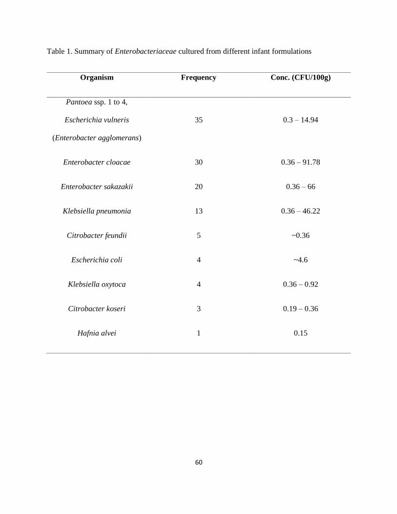
60
Table 1. Summary of Enterobacteriaceae cultured from different infant formulations
Organism Frequency Conc. (CFU/100g)
Pantoea ssp. 1 to 4,
Escherichia vulneris
(Enterobacter agglomerans)
35 0.3 – 14.94
Enterobacter cloacae 30 0.36 – 91.78
Enterobacter sakazakii 20 0.36 – 66
Klebsiella pneumonia 13 0.36 – 46.22
Citrobacter feundii 5 ~0.36
Escherichia coli 4 ~4.6
Klebsiella oxytoca 4 0.36 – 0.92
Citrobacter koseri 3 0.19 – 0.36
Hafnia alvei 1 0.15
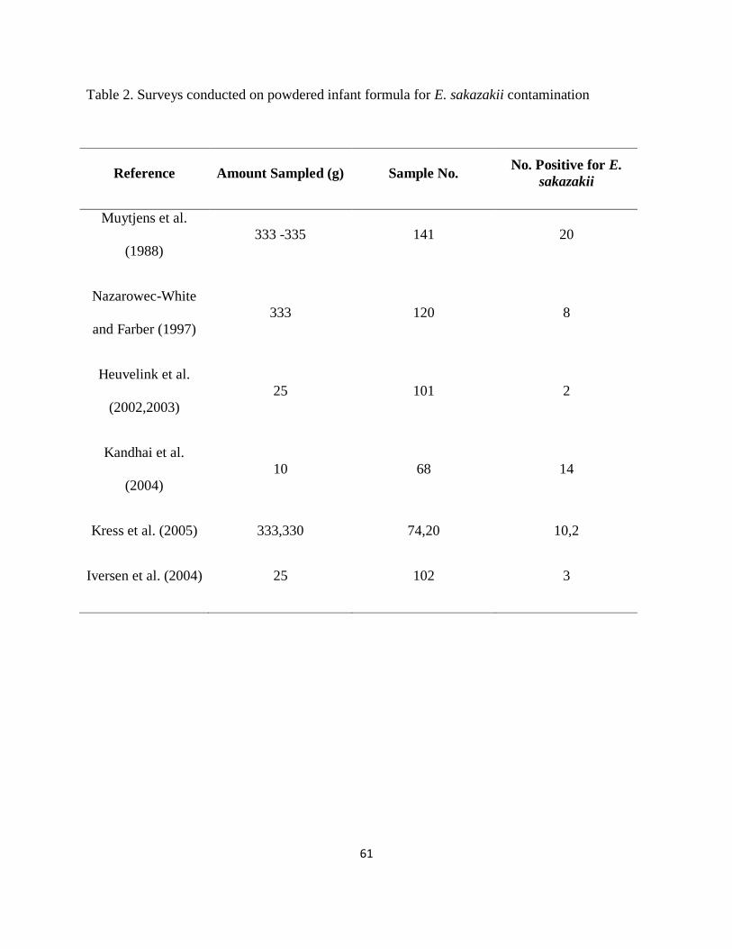
61
Table 2. Surveys conducted on powdered infant formula for E. sakazakii contamination
Reference Amount Sampled (g) Sample No. No. Positive for E.
sakazakii
Muytjens et al.
(1988)
333 -335 141 20
Nazarowec-White
and Farber (1997)
333 120 8
Heuvelink et al.
(2002,2003)
25 101 2
Kandhai et al.
(2004)
10 68 14
Kress et al. (2005) 333,330 74,20 10,2
Iversen et al. (2004) 25 102 3
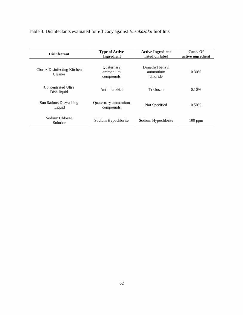
62
Table 3. Disinfectants evaluated for efficacy against E. sakazakii biofilms
Disinfectant Type of Active
Ingredient
Active Ingredient
listed on label
Conc. Of
active ingredient
Clorox Disinfecting Kitchen
Cleaner
Quaternary
ammonium
compounds
Dimethyl benzyl
ammonium
chloride
0.30%
Concentrated Ultra
Dish liquid Antimicrobial Triclosan 0.10%
Sun Sations Diswashing
Liquid
Quaternary ammonium
compounds Not Specified 0.50%
Sodium Chlorite
Solution Sodium Hypochlorite Sodium Hypochlorite 100 ppm
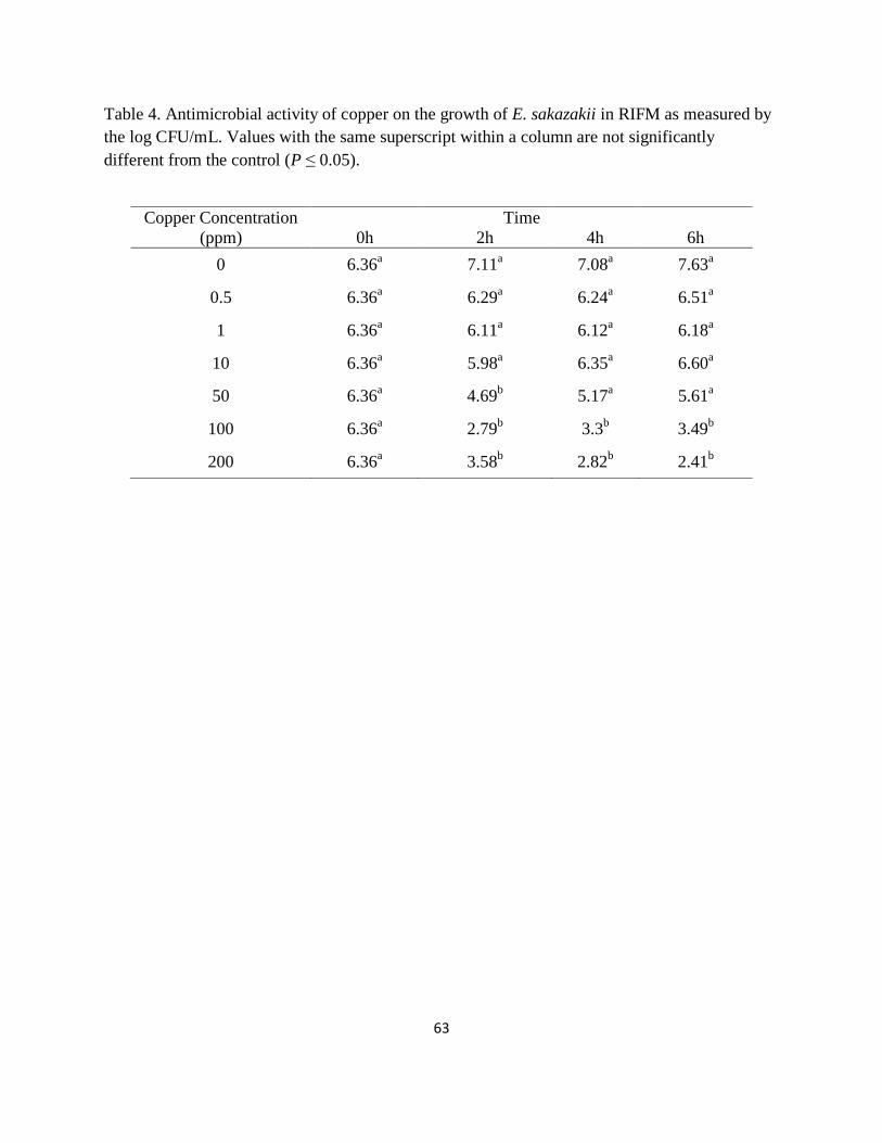
63
Table 4. Antimicrobial activity of copper on the growth of E. sakazakii in RIFM as measured by
the log CFU/mL. Values with the same superscript within a column are not significantly
different from the control (P ≤ 0.05).
Copper Concentration
(ppm)
0h
Time
2h
4h
6h
0 6.36a
7.11a
7.08a
7.63a
0.5 6.36a
6.29a
6.24a
6.51a
1 6.36a
6.11a
6.12a
6.18a
10 6.36a
5.98a
6.35a
6.60a
50 6.36a
4.69b
5.17a
5.61a
100 6.36a
2.79b
3.3b
3.49b
200 6.36a
3.58b
2.82b
2.41b
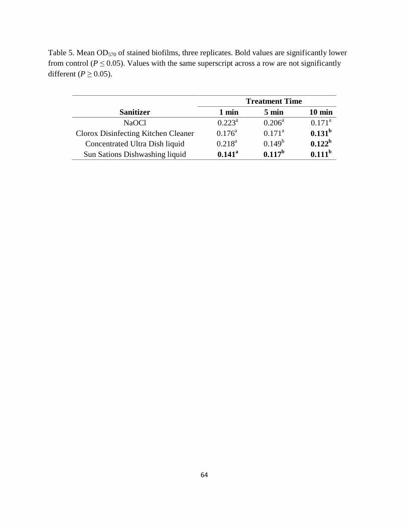
64
Table 5. Mean OD570 of stained biofilms, three replicates. Bold values are significantly lower
from control (P ≤ 0.05). Values with the same superscript across a row are not significantly
different (P ≥ 0.05).
Treatment Time
Sanitizer 1 min 5 min 10 min
NaOCl 0.223a
0.206a
0.171a
Clorox Disinfecting Kitchen Cleaner 0.176a
0.171a
0.131b
Concentrated Ultra Dish liquid 0.218a
0.149b
0.122b
Sun Sations Dishwashing liquid 0.141a
0.117b
0.111b
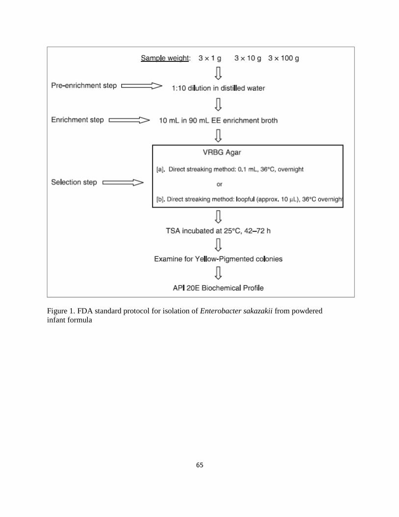
65
Figure 1. FDA standard protocol for isolation of Enterobacter sakazakii from powdered
infant formula
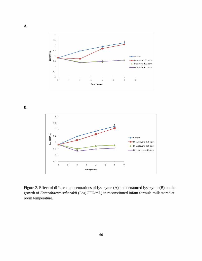
66
A.
B.
Figure 2. Effect of different concentrations of lysozyme (A) and denatured lysozyme (B) on the
growth of Enterobacter sakazakii (Log CFU/mL) in reconstituted infant formula milk stored at
room temperature.
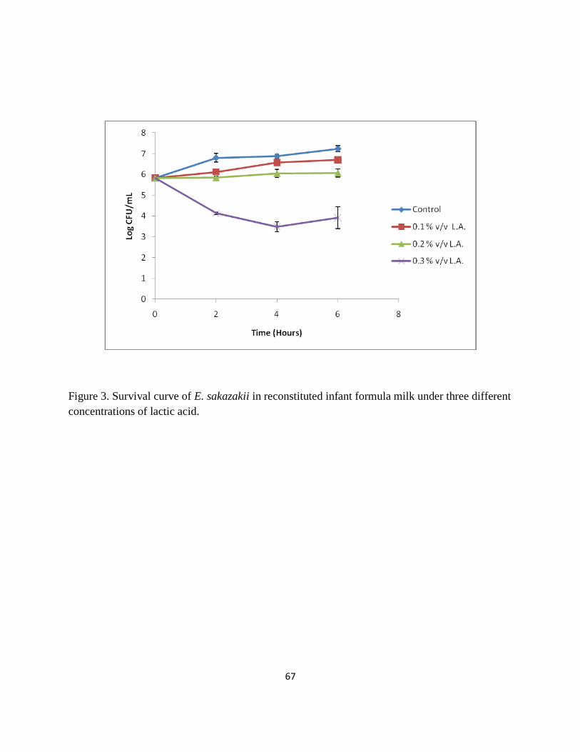
67
Figure 3. Survival curve of E. sakazakii in reconstituted infant formula milk under three different
concentrations of lactic acid.
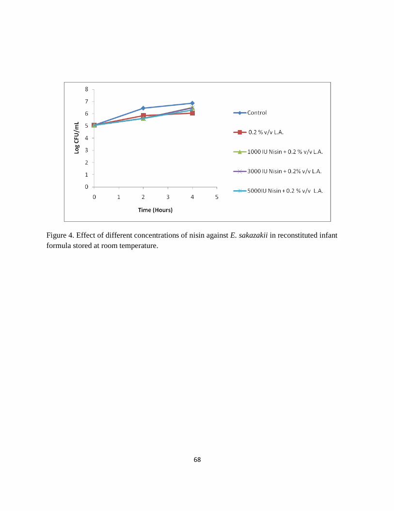
68
Figure 4. Effect of different concentrations of nisin against E. sakazakii in reconstituted infant
formula stored at room temperature.
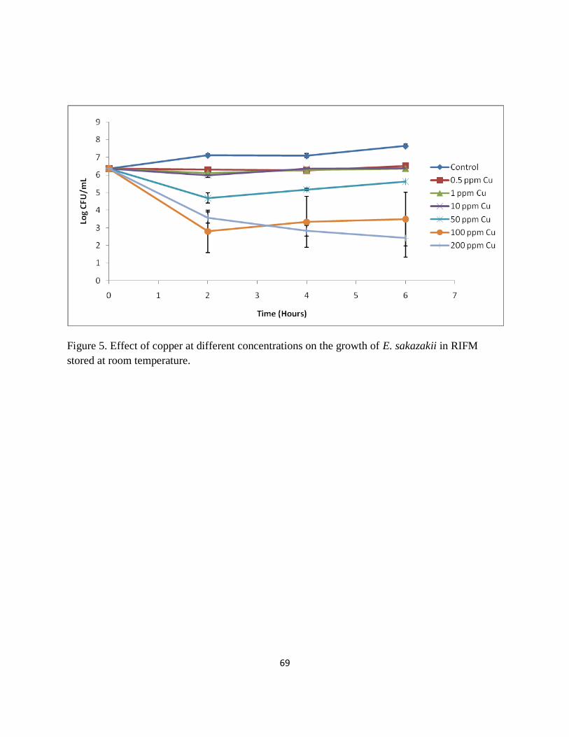
69
Figure 5. Effect of copper at different concentrations on the growth of E. sakazakii in RIFM
stored at room temperature.
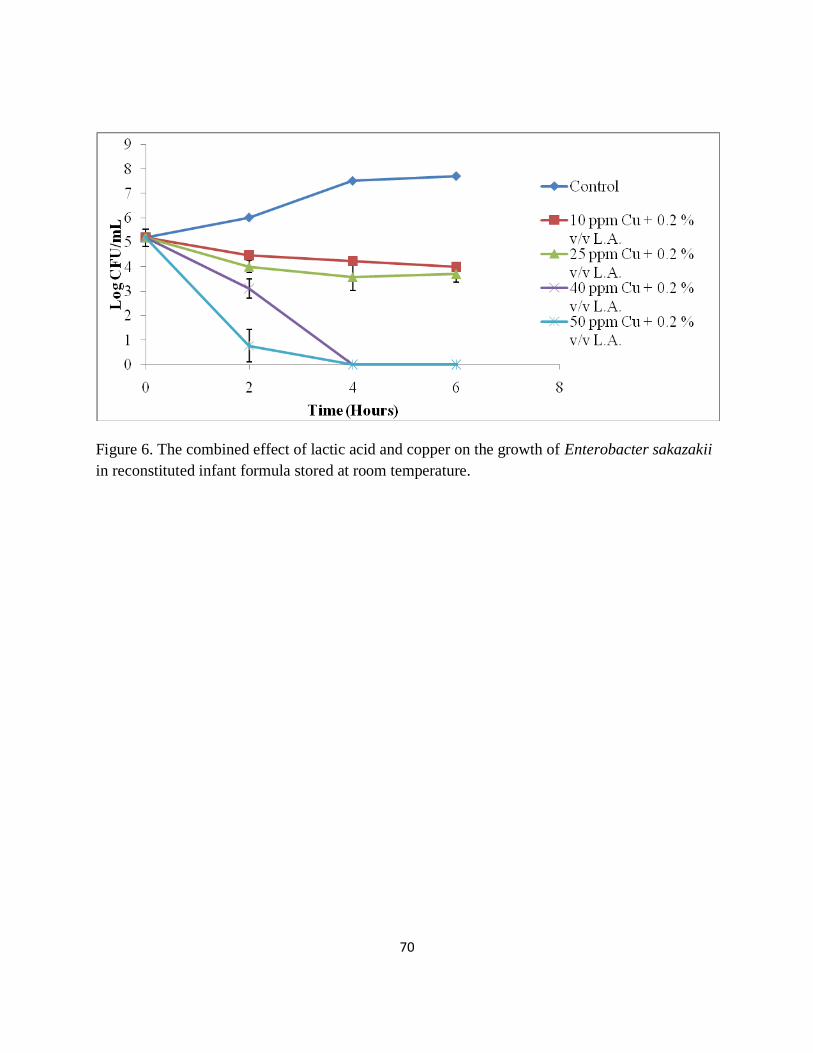
70
Figure 6. The combined effect of lactic acid and copper on the growth of Enterobacter sakazakii
in reconstituted infant formula stored at room temperature.
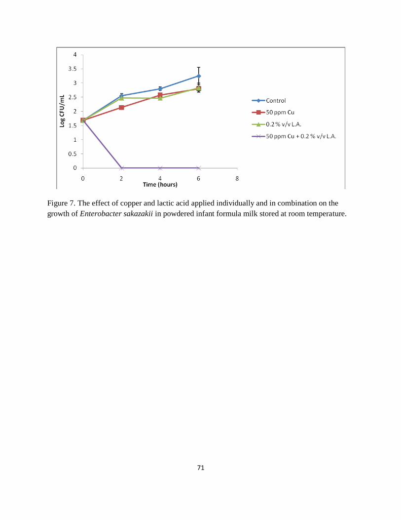
71
Figure 7. The effect of copper and lactic acid applied individually and in combination on the
growth of Enterobacter sakazakii in powdered infant formula milk stored at room temperature.

72