Embed Size (px)
Citation preview
MATE2 Mediates Vacuolar Sequestration of FlavonoidGlycosides and Glycoside Malonates inMedicago truncatula C W OA
Jian Zhao, David Huhman, Gail Shadle, Xian-Zhi He, Lloyd W. Sumner, Yuhong Tang, and Richard A. Dixon1
Plant Biology Division, Samuel Roberts Noble Foundation, Ardmore, Oklahoma 73401
The majority of flavonoids, such as anthocyanins, proanthocyanidins, and isoflavones, are stored in the central vacuole, but
the molecular basis of flavonoid transport is still poorly understood. Here, we report the functional characterization of a
multidrug and toxin extrusion transporter (MATE2), from Medicago truncatula. MATE 2 is expressed primarily in leaves and
flowers. Despite its high similarity to the epicatechin 39-O-glucoside transporter MATE1, MATE2 cannot efficiently transport
proanthocyanidin precursors. In contrast, MATE2 shows higher transport capacity for anthocyanins and lower efficiency for
other flavonoid glycosides. Three malonyltransferases that are coexpressed with MATE2 were identified. The malonylated
flavonoid glucosides generated by these malonyltransferases are more efficiently taken up into MATE2-containing
membrane vesicles than are the parent glycosides. Malonylation increases both the affinity and transport efficiency of
flavonoid glucosides for uptake by MATE2. Genetic loss of MATE2 function leads to the disappearance of leaf anthocyanin
pigmentation and pale flower color as a result of drastic decreases in the levels of various flavonoids. However, some
flavonoid glycoside malonates accumulate to higher levels in MATE2 knockouts than in wild-type controls. Deletion of
MATE2 increases seed proanthocyanidin biosynthesis, presumably via redirection of metabolic flux from anthocyanin
storage.
INTRODUCTION
Anthocyanins and proanthocyanidins (PAs; also called con-
densed tannins) are abundant flavonoids found in seed coats,
leaves, fruits, flowers, and bark of many plant species (Ariga
et al., 1981; Gabetta et al., 2000; Gu et al., 2004; Dixon et al.,
2005). PAs and anthocyanins play protective roles against mi-
crobial pathogens, insect attack, and UV irradiation, and antho-
cyanins are commonly utilized to attract insect pollinators. For
humans, both classes of compounds have beneficial effects on
cardiac health, immunity, and longevity (Santos-Buelga and
Scalbert, 2000; Skibola and Smith, 2000), and the presence of
modest levels of PAs in the leaves and stems of protein-rich
forage crops is an important agronomic trait, as they protect
ruminant animals frompasture bloat, enhance ruminant nutrition,
and reduce protein degradation in silage (Lees, 1992).
Attempts to engineer PA production in the forage legume
alfalfa (Medicago sativa) have so far led to the accumulation of
anthocyanins rather than PAs (Peel et al., 2009). Flavan 3-ol-
derived PA oligomers and anthocyanins are derived from the
same precursors, anthocyanidins, and competition between
these parallel pathways for metabolic flux might be an important
regulatory mechanism determining PA biosynthesis (Lepiniec
et al., 2006; Figure 1). Anthocyanidins (such as cyanidin, pelar-
gonidin, and delphinidin) from the flavonoid pathway can be
either immediately modified by glycosylation, acylation, and/or
methylation to generate diverse anthocyanins or further reduced
to generate flavan 3-ol precursors of PAs, such as epicatechin
formed by the action of anthocyanidin reductase (ANR; Figure 1;
Xie et al., 2003). Seeds of knockout mutants in ANR accumulate
many more anthocyanins than wild-type seeds (Marinova et al.,
2007b), and an inverse relationship between the expression of
ANR and anthocyanidin 3-O-glucosyltransferase has been dem-
onstrated for selective direction of cyanidin into either PA or
anthocyanin biosynthesis (Lee et al., 2005).
The family I glycosyltransferases UTG78G1 and UTG72L1
catalyze the glucosylation of anthocyanidins and epicatechin,
respectively (Modolo et al., 2007; Pang et al., 2008; Peel et al.,
2009). Glycosylation of epicatechin and cyanidin is essential for
their transport into the vacuole by the multidrug and toxin
extrusion transporters MATE1 and TT12 in the seed coats of
Medicago truncatula and Arabidopsis thaliana, respectively
(Marinova et al., 2007b; Zhao and Dixon, 2009; Figure 1). Many
flavonoid glycosides accumulate in the vacuole with acyl sub-
stituents on the sugar residues, but the exact function of the
acylation remains unclear. Whereas acylation with malonyl res-
idues has been suggested to be essential for the retention of
some glycosides within the vacuole (Matern et al., 1983), other
studies indicate the involvement of acylation in flavonoid trans-
port per se. For example, proton gradient–dependent vacuolar
uptake by unknown transporters of anthocyanidin-3-O-sinapoyl
1 Address correspondence to [email protected] author responsible for distribution of materials integral to thefindings presented in this article in accordance with the policy describedin the Instructions for Authors (www.plantcell.org) is: Richard A. Dixon([email protected]).CSome figures in this article are displayed in color online but in blackand white in the print edition.WOnline version contains Web-only data.OAOpen Access articles can be viewed online without a subscription.www.plantcell.org/cgi/doi/10.1105/tpc.110.080804
The Plant Cell, Vol. 23: 1536–1555, April 2011, www.plantcell.org ã 2011 American Society of Plant Biologists
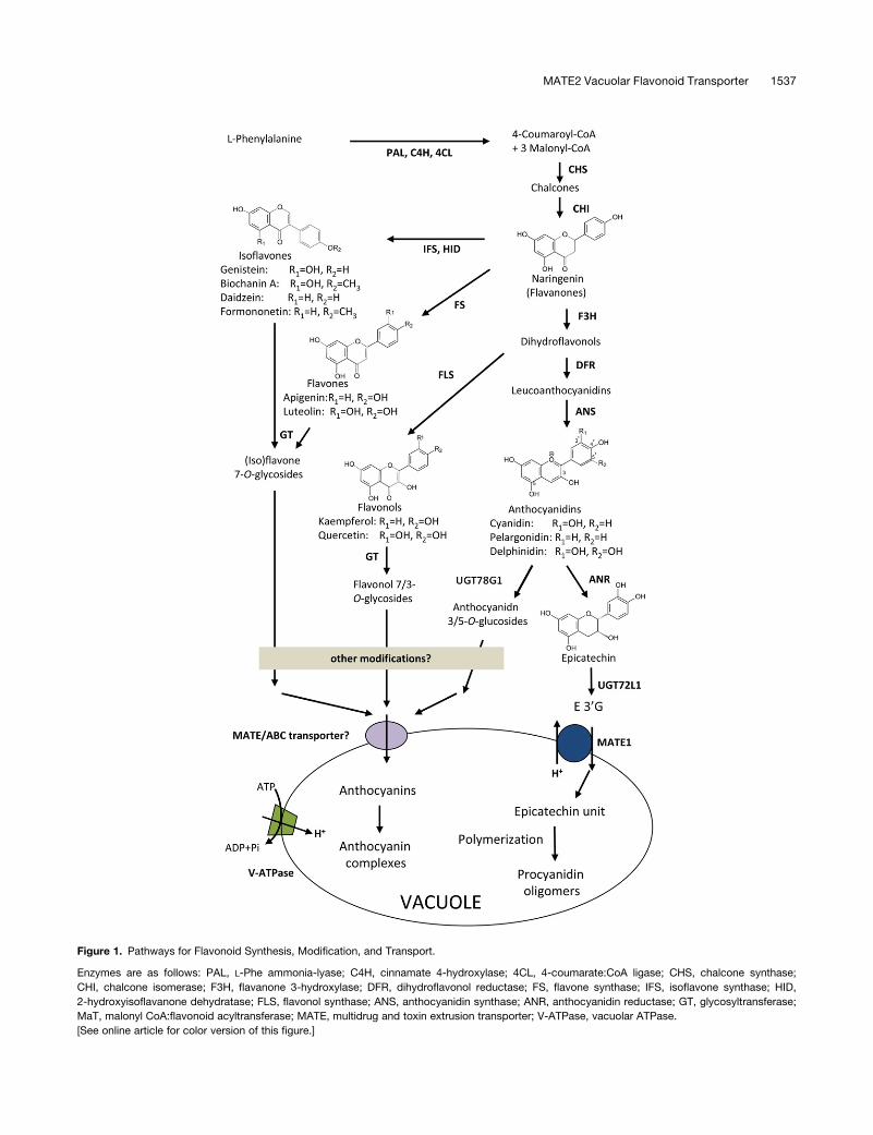
Figure 1. Pathways for Flavonoid Synthesis, Modification, and Transport.
Enzymes are as follows: PAL, L-Phe ammonia-lyase; C4H, cinnamate 4-hydroxylase; 4CL, 4-coumarate:CoA ligase; CHS, chalcone synthase;
CHI, chalcone isomerase; F3H, flavanone 3-hydroxylase; DFR, dihydroflavonol reductase; FS, flavone synthase; IFS, isoflavone synthase; HID,
2-hydroxyisoflavanone dehydratase; FLS, flavonol synthase; ANS, anthocyanidin synthase; ANR, anthocyanidin reductase; GT, glycosyltransferase;
MaT, malonyl CoA:flavonoid acyltransferase; MATE, multidrug and toxin extrusion transporter; V-ATPase, vacuolar ATPase.
[See online article for color version of this figure.]
MATE2 Vacuolar Flavonoid Transporter 1537
xyloseglucosylgalactoside or apigenin 7-O-(699-O-malonylglu-
coside) strongly depended on acylation of the attached sugars
(Matern et al., 1986; Hopp and Seitz, 1987). Biochemical studies
of other vacuolar transport systems for flavonoids have not led to
a consistent view of the substrate specificity requirements (Klein
et al., 1996, 2000; Bartholomew et al., 2002; Frangne et al.,
2002).
The ATP binding cassette (ABC) transporter MRP3 frommaize
(Zea mays; Goodman et al., 2004), the MATE transporters AM1
and AM3 from grapevine (Vitis vinifera; Gomez et al., 2009), and
FFT fromArabidopsis (Thompson et al., 2010) were suggested to
be involved in anthocyanin/flavonol transport. However, due to
the lack of complete data sets on transport properties, substrate
preferences, in vivo substrates, or genetically determined in
planta functions for each of these transporters, our overall pic-
ture of flavonoid transport is still incomplete. The study of inter-
cellular or subcellular transport and storage of plant specialized
metabolites, including flavonoids, has emerged as an important
and rapidly growing area and is expected to have major impacts
on our understanding of plant specialized metabolism and met-
abolic engineering (Zhao and Dixon, 2010).
During our studies on the tonoplast epicatechin 39-O-
glucoside (E39G) transporter MATE1 (Zhao and Dixon, 2009),
we identified another transporter, MATE2, based on homology to
Arabidopsis TT12. Here, we report that MATE2 is a flavonoid
transporter with a wide substrate spectrum. This vacuolar trans-
porter can transport flavonoid glycosides, particularly anthocy-
anin and flavone glucosides. However, MATE2 prefers to
transport malonylated flavonoid glucosides. The flavonoid ma-
lonylation reaction is performed by three malonyl CoA:flavonoid
malonyltransferases, encoded by genes that are coexpressed
with MATE2 in M. truncatula tissues. Characterization of mate2
knockout mutants confirms that MATE2 is a flavonoid trans-
porter involved in vacuolar sequestration of anthocyanins and
other flavonoids in flowers and leaves.
RESULTS
Identification ofMedicagoMATE2
During characterization of MATE1 as the tonoplast transporter
for E39G (Zhao and Dixon, 2009), we found six related putative
transporter proteins by BLAST analysis of the M. truncatula
genome sequence with the Arabidopsis TT12 protein sequence.
Of these, only one, designated MATE2, showed uptake activity
for flavonoids when expressed in yeast (see below). MATE2
shares 54 and 50% amino acid sequence similarity with MATE1
and TT12, respectively (see Supplemental Figure 1 online).
BLAST and sequence alignment of MATE2 with several charac-
terized MATE transporters from other species, however, indi-
cated that Medicago MATE2 is more similar to the tomato
(Solanum lycopersicum) MATE transporter MTP77 (66% identity
and 80% similarity), the Arabidopsis putative MATE transporter
At4g25640 (65% identity and 79% similarity), and the two
grapevine MATE transporters AM3 (62% identity and 79%
similarity) and AM1 (61% identity and 79% similarity) than to
Medicago MATE1 and TT12 (see Supplemental Figure 1 online).
All of these reported MATE transporters are confirmed or puta-
tive flavonoid transporters (Mathews et al., 2003; Marinova et al.,
2007b; Gomez et al., 2009; Zhao and Dixon, 2009; Thompson
et al., 2010). To further predict and distinguish MATE2 function,
phylogenetic analysis was performed with protein sequences of
different types of known and putative MATE transporters (Sup-
plemental Data Set 1). These proteins grouped into three clades
(Figure 2A). The first clade includes three subclades: one con-
tains MATE2 as well as other known anthocyanin/flavonoid
MATE transporters; whereas Medicago MATE1, Arabidopsis
TT12, and their putative grapevine and canola (Brassica napus)
orthologs are grouped into another subclade; and two vacuolar
tobacco (Nicotiana tabacum) nicotine transporters, Nt MATE1/2
(Shoji et al., 2009), form the third subclade. The nicotine trans-
porter JAT1 from tobacco alongwith ALF5 and the toxin exporter
DTX1 from Arabidopsis (Diener et al., 2001; Li et al., 2002; Morita
et al., 2009) are grouped together in an independent clade. Three
plasma membrane–localized organic acid exporters, Sb MATE1
from sorghum (Sorghum bicolor), Hv AACT1 from barley (Hor-
deum vulgare), and FRD3 from Arabidopsis (Durrett et al., 2007;
Furukawa et al., 2007; Magalhaes et al., 2007), are grouped with
their Medicago counterpart AC122162_1.4 into a clade much
further from the secondary metabolite MATE transporters. The
Arabidopsis MATE transporter EDS5 is in a separate clade
(Nawrath et al., 2002). The phylogenetic analysis suggested
that MATE2 may function as a flavonoid transporter, possibly
associated with the transport of anthocyanins or flavonols.
MATE2 Transports a Wide Range of Flavonoid Glucosides
MATE2 was expressed in yeast for comparison of substrate
transport preference to that of MATE1, which transports both
E39G and cyanidin 3-O-glucoside (Cy3G), but with a strong
kinetic preference for E39G (Zhao and Dixon, 2009). Isolated
microsomes from yeast cells expressing MATE2 took up Cy3G,
whereas uptake by microsomes from vector control cells was
significantly less (Figure 2B; see Supplemental Figure 2 online).
Similar to MATE1, MATE2-expressing membrane vesicles did
not show significant transport activity toward cyanidin, epicate-
chin, or other flavonoid aglycones (see Supplemental Figure 2
online).
Inhibitors of the vacuolar H+-ATPase or uncouplers of the
proton motive force (Luvisetto and Azzone, 1989; Drose and
Altendorf, 1997; Rodrigues et al., 1999) inhibited Cy3G transport,
indicating that MATE2-mediated Cy3G transport is H+-gradient
dependent, as is transport of E39G by MATE1 (Zhao and Dixon,
2009; Figure 2C). Furthermore, MATE2 was not inhibited by the
ATPase inhibitor vanadate, which preferentially inhibits ABC
transporters (Pezza et al., 2002).
Of 10 flavonol, flavone, and isoflavone glucosides tested as
potential substrates for MATE2, apigenin 7-O-glucoside (A7G)
exhibited the highest uptake, but this was less than that of Cy3G
(Figures 2D and 2E). In particular, MATE2 appears not to trans-
port E39G, which is the preferred substrate for MATE1 (Figure
2D). In competition assays, A7G, naringenin 7-O-glucoside,
luteolin 7-O-glucoside, and pelargonidin 3-O-glucoside (P3G;
all at 250mM)weremost effective in inhibiting the uptake of Cy3G
(at 100 mM), whereas E39G and isoflavone glycosides did not
1538 The Plant Cell
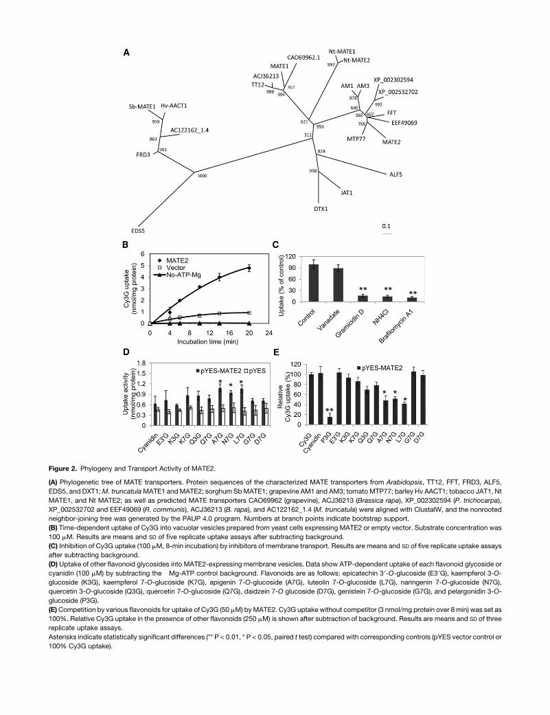
Figure 2. Phylogeny and Transport Activity of MATE2.
(A) Phylogenetic tree of MATE transporters. Protein sequences of the characterized MATE transporters from Arabidopsis, TT12, FFT, FRD3, ALF5,
EDS5, and DXT1;M. truncatulaMATE1 andMATE2; sorghum SbMATE1; grapevine AM1 and AM3; tomatoMTP77; barley Hv AACT1; tobacco JAT1, Nt
MATE1, and Nt MATE2; as well as predicted MATE transporters CAO69962 (grapevine), ACJ36213 (Brassica rapa), XP_002302594 (P. trichocarpa),
XP_002532702 and EEF49069 (R. communis), ACJ36213 (B. rapa), and AC122162_1.4 (M. truncatula) were aligned with ClustalW, and the nonrooted
neighbor-joining tree was generated by the PAUP 4.0 program. Numbers at branch points indicate bootstrap support.
(B) Time-dependent uptake of Cy3G into vacuolar vesicles prepared from yeast cells expressing MATE2 or empty vector. Substrate concentration was
100 mM. Results are means and SD of five replicate uptake assays after subtracting background.
(C) Inhibition of Cy3G uptake (100 mM, 8-min incubation) by inhibitors of membrane transport. Results are means and SD of five replicate uptake assays
after subtracting background.
(D) Uptake of other flavonoid glycosides into MATE2-expressing membrane vesicles. Data show ATP-dependent uptake of each flavonoid glycoside or
cyanidin (100 mM) by subtracting the �Mg-ATP control background. Flavonoids are as follows: epicatechin 39-O-glucoside (E39G), kaempferol 3-O-
glucoside (K3G), kaempferol 7-O-glucoside (K7G), apigenin 7-O-glucoside (A7G), luteolin 7-O-glucoside (L7G), naringenin 7-O-glucoside (N7G),
quercetin 3-O-glucoside (Q3G), quercetin 7-O-glucoside (Q7G), daidzein 7-O glucoside (D7G), genistein 7-O-glucoside (G7G), and pelargonidin 3-O-
glucoside (P3G).
(E)Competition by various flavonoids for uptake of Cy3G (50 mM) byMATE2. Cy3G uptake without competitor (3 nmol/mg protein over 8 min) was set as
100%. Relative Cy3G uptake in the presence of other flavonoids (250 mM) is shown after subtraction of background. Results are means and SD of three
replicate uptake assays.
Asterisks indicate statistically significant differences (** P < 0.01, * P < 0.05, paired t test) compared with corresponding controls (pYES vector control or
100% Cy3G uptake).
cause inhibition (Figure 2E). Overall, the data suggest that
MATE2 can transport a wide range of flavonoids, including
anthocyanins and flavone glycosides, albeit with different effi-
ciencies.
Tissue-Level Expression of MATE2 and Flavonoid
Modification Genes
Mining microarray data from the Medicago Gene Expression
Atlas (MtGEA) database (http://mtgea.noble.org/v2/; Benedito
et al., 2008) showed that MATE2 is most strongly expressed in
flowers, followed by roots and vegetative buds, leaves, and
seeds (see Supplemental Figure 3 online). Since MATE2 trans-
ports glycosylated flavonoids rather than flavonoid aglycones,
flavonoid glycosyltransferases may be spatially coexpressed
with MATE2. UGT78G1, previously identified as a Medicago
anthocyanidin glycosyltransferase (Modolo et al., 2007; Peel
et al., 2009), shows a similar tissue expression pattern to that of
MATE2, being highly expressed in pod, flower, vegetative bud,
root, and leaf tissues (see Supplemental Figure 3 online). To
further examine genes coexpressed withMATE2 andUGT78G1,
hierarchical clustering was conducted based on Pearson corre-
lation of expression data from the MtGEA, and a coregulated
group of genes involved in flavonoid biosynthesis, modification,
and transport was generated (Figure 3A; see Supplemental
Figure 4 online). Two major clusters were identified based on
these data: genes involved in the early steps and genes involved
in the later steps of flavonoid biosynthesis. The effectiveness of
clustering is shown by coclustering of ANS, ANR, and UGT72L1
involved in the PA biosynthesis pathway (Xie et al., 2003; Pang
et al., 2008). The coordinated expression patterns ofMATE2 and
UGT78G1 are consistent with their functional corelationship.
Interestingly, two putative malonyltransferases (probe sets
Mtr.19777.1.S1_at and Mtr.19945.1.S1_at, named MaT4 and
MaT5, respectively) also clustered with these flavonoid biosyn-
thesis, modification, and transport genes (Figure 3A). Malonyla-
tion is a major modification on anthocyanins and (iso)flavonoid
glycosides in M. truncatula (Farag et al., 2008); and the expres-
sion patterns therefore suggested that thesemalonyltransferases
might be involved in the modification of flavonoid glucosides.
Quantitative RT-PCR analysis of flavonoid biosynthesis–
related genes indicated that most genes showed diverse tis-
sue expression patterns, presumably because of the complex
and widespread accumulation of (iso)flavonoid compounds in
M. truncatula (see Supplemental Figure 3 online). However,
UGT78G1, MaT4, MaT5, and MATE2 are all expressed in flower,
root, and leaf to varying levels (Figure 3B). Examination of the
developmental stage–dependent expression of MATE2 indi-
cated strong expression in flowers compared with vegetative
buds and leaves (Figure 3C). To further examine the cell type
expression of MATE2, in situ hybridization was conducted with
both floral and leaf tissues. Consistent with the RT-PCR data,
MATE2 transcripts showed higher signal intensity in younger
flower and leaf tissues than in older tissues (Figure 3D). MATE2
signal was mainly in the mesophyll cells of young leaves but was
evenly distributed throughout the cells of the vegetative bud. In
cross sections of young flowers, although the petals and pollen
were labeled, the strongest signals were in the outer petals and
sepals of the flower. In cross sections of the petals of fully
opened flowers, MATE2 transcript signal was strong in both
epidermal cells and vascular bundles of veins (Figure 3D).
Malonyltransferases Modify Flavonoid Glucosides
Based on the above expression patterns, we selected and
cloned the putative malonyltransferases to test their functions.
Because the transport efficiency of MATE2 toward anthocyanins
and flavonol glucosides is lower than that of other MATE trans-
porters, such asMATE1 toward E39G (Zhao andDixon, 2009), we
also tested whether flavonoid glucosidemalonates generated by
the action of these enzymes would be better substrates for
MATE2 transport than are the nonsubstituted glycosides. For this
purpose, we also cloned a third putative malonyltransferase,
MaT6, which is weakly expressed in leaf and root (Figure 3) but
was not represented on the Medicago Affymetrix gene chip.
Protein sequence alignment showed that MaT4, MaT5, and
MaT6 not only have the HXXXD and DFGWG domains that are
conserved in all BAHD (for BEAT, AHCTs, HCBT, and DAT; St.
Pierre and De Luca, 2000) family proteins but also have the con-
served NYXGNC domain found only in malonyl CoA:flavonoid
malonyltransferases (see Supplemental Figure 5 online). Phylo-
genetic analysis showed that MaT4 to -6, MaT1 to -3, and other
anthocyanin and flavonoid malonyltransferases are clustered
into the same clade (Figure 4A), while other putative malonyl-
transferases from M. truncatula are separated from the known
flavonoid malonyltransferases (Supplemental Data Set 2).
Recombinant His-taggedMaT4 to -6 proteins were expressed
in Escherichia coli, purified on a nickel-resin column (Figure 4B),
and examined for in vitro malonyltransferase activity. Using
the aliphatic thioester donors malonyl CoA and acetyl CoA or
the aromatic acyl donor p-coumaroyl-CoA in combination with
flavonoid glucosides as acceptors, each of the three MaTs
showed different substrate preferences and donor specificities
(Figures 4C to 4N). All three enzymes used only malonyl CoA
to acylate their preferred substrates (see Supplemental Table
1 and Supplemental Figure 5 online). MaT4 prefers to acylate 7-O
glucosides, including isoflavone, flavonol, or flavanone 7-O-
glucosides, but shows little activity toward 3-O-glucosides (see
Supplemental Table 1 and Supplemental Figure 6 online). MaT5
and MaT6 have similar activity toward anthocyanins (cyanidin,
delphinidin, and pelargonidin conjugates) and exhibit a regio-
specific preference for 3-O-glucosides, with less activity with 3,5-
di-O-glucosides (see Supplemental Table 1 and Supplemental
Figure 7 online). Electrospray ionization-liquid chromatography-
mass spectrometry (ESI-LC-MS) profiling of the reaction products
revealed that MaT4 converts isoflavone and flavanone 7-O-
glycosides into the corresponding monomalonylated products
(see Supplemental Figure 8 online). Although identification of
the position of malonylation of the sugar is beyond the scope of
this study, it is likely that the products are the flavonoid 7-O-
(699-O-malonyl)-b-D-glucosides, with the malonate moiety at
the same position on the sugar as in the compounds generated
by the M. truncatula MaT1, MaT2, and MaT3 malonyltransfer-
ases (Yu et al., 2008; see Supplemental Figure 9 online). The
reaction product displayed a UV spectrum similar to that of
its parental substrate and amass increment of 86m/z, themass
1540 The Plant Cell
of the malonyl group. However, MaT5- and MaT6-catalyzed
malonylation of anthocyanins was more complex: multiple
malonylation products were obtained with five different antho-
cyanins, with the anthocyanidin glycoside monomalonate and
dimalonate being the dominant products (see Supplemental
Figure 9 online).
MATE2 Prefers to Transport Malonylated Flavonoids
MATE2 exhibits a Km of 88.9 mM and a Vmax of 0.91 nmol/mg
protein/min for the uptake of Cy3G (Table 1; see Supplemental
Figure 10 online). MATE2 also mediates the uptake of other
anthocyanidin glucosides, such as delphinidin 3-O-glucoside
(D3G; Km of 96.5 mM and Vmax of 0.95 nmol/mg protein/min),
delphinidin 3,5 di-O-diglucoside, P3G, and pelargonidin 3,5-di-
O-diglucoside (Table 1; see Supplemental Figure 10 online).
To determinewhetherMATE2 prefers to transportmalonylated
compared with nonmalonylated flavonoids, we performed up-
take assays using pure compounds (A7G and K7G and their
malonates A7GM and K7GM) or mixtures of malonylated and
nonmalonylated substrates generated from the MaT5 assays
(Cy3G and P3G and their malonates Cy3GM and P3GM). The
mixtures were used because of the instability of the anthocya-
nidin malonyl glycosides during purification. In these latter
assays, the relative proportions of the two types of compound
were compared in the original mixture and the fraction extracted
from the MATE2-containing yeast membrane vesicles. MATE2
took up more than five times more A7G malonate than A7G and
about two timesmore kaempferol 7-O-glucoside (K7G)malonate
than nonmodified K7G (Figures 5A and 5B), suggesting that the
transporter preferentially takes up flavonoid glucoside malon-
ates rather than the unsubstituted glycosides. This was con-
firmed by kinetic analysis (Table 1; see Supplemental Figure 11
online). MATE2 also took up more malonylated anthocyanins,
Cy3G and P3G malonates, than nonmodified Cy3G and P3G
from malonylation reaction mixtures in standard uptake assays
with yeast microsomes (Figures 5C and 5D). Although kinetic
parameters for Cy3GM and P3GM uptake could not be
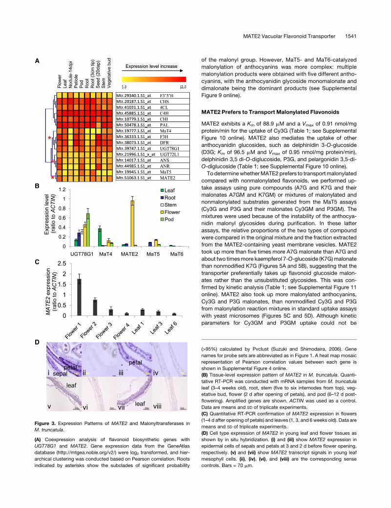
Figure 3. Expression Patterns of MATE2 and Malonyltransferases in
M. truncatula.
(A) Coexpression analysis of flavonoid biosynthetic genes with
UGT78G1 and MATE2. Gene expression data from the GeneAtlas
database (http://mtgea.noble.org/v2/) were log2 transformed, and hier-
archical clustering was conducted based on Pearson correlation. Roots
indicated by asterisks show the subclades of significant probability
(>95%) calculated by Pvclust (Suzuki and Shimodaira, 2006). Gene
names for probe sets are abbreviated as in Figure 1. A heat map mosaic
representation of Pearson correlation values between each gene is
shown in Supplemental Figure 4 online.
(B) Tissue-level expression pattern of MATE2 in M. truncatula. Quanti-
tative RT-PCR was conducted with mRNA samples from M. truncatula
leaf (3–4 weeks old), root, stem (five to six internodes from top), veg-
etative bud, flower (2 d after opening of petals), and pod (6–12 d post-
flowering). Amplified genes are shown. ACTIN was used as a control.
Data are means and SD of triplicate experiments.
(C) Quantitative RT-PCR confirmation of MATE2 expression in flowers
(1–4 d after opening of petals) and leaves (1, 3, and 6 weeks old). Data are
means and SD of triplicate experiments.
(D) Cell type expression of MATE2 in young leaf and flower tissues as
shown by in situ hybridization. (i) and (iii) show MATE2 expression in
epidermal cells of sepals and petals at 3 and 2 d before flower opening,
respectively. (v) and (vii) show MATE2 transcript signals in young leaf
mesophyll cells. (ii), (iv), (vi), and (viii) are the corresponding sense
controls. Bars = 70 mm.
MATE2 Vacuolar Flavonoid Transporter 1541
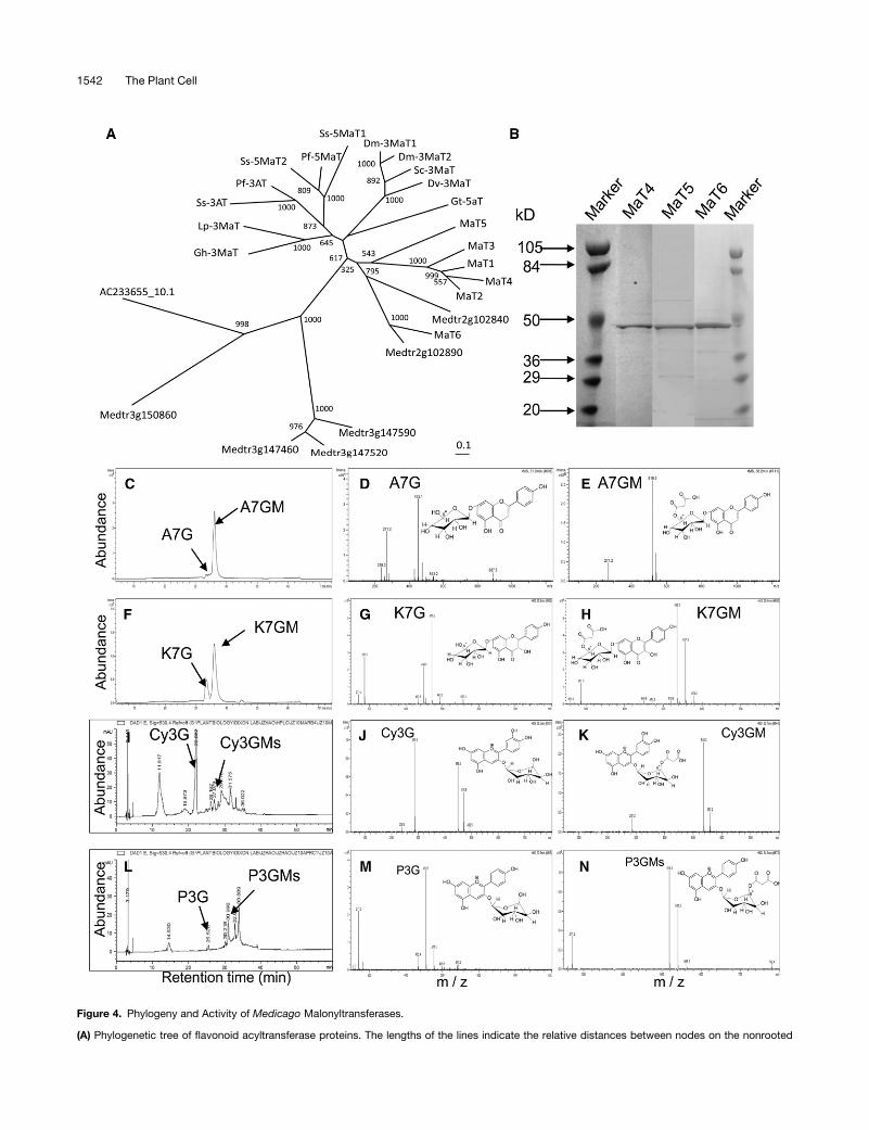
Figure 4. Phylogeny and Activity of Medicago Malonyltransferases.
(A) Phylogenetic tree of flavonoid acyltransferase proteins. The lengths of the lines indicate the relative distances between nodes on the nonrooted
1542 The Plant Cell
determined due to instability and multiple malonylation reaction
products with anthocyanins, the mixed substrate competition
uptake assays with malonylation reaction mixtures suggested
that MATE2 has a kinetic preference for malonylated anthocy-
anins over the nonmodified glycosides (Figures 5C and 5D).
MATE2 Is a Tonoplast Transporter, Whereas Flavonoid
Malonyltransferases Are Cytosolic or Endoplasmic
Reticulum Proteins
Several known flavonoidMATE transporters, including AM1, AM3,
MATE1, and TT12, are localized to the tonoplast membrane, as
shown by imaging of green fluorescent protein (GFP)-tagged
transporters or membrane proteomics studies (Marinova et al.,
2007a; Gomez et al., 2009; Zhao and Dixon, 2009). Topological
prediction suggests that MATE2 has 12 putative transmembrane
domains in the same pattern as other known MATE transporters
(see Supplemental Figure 12 online). To further determine the
subcellular localization of MATE2 protein, a GFP fusion to the C
terminus of MATE2 was constructed and expressed in both yeast
and plant cells. This GFP fusion protein was competent in antho-
cyanin transport when expressed in yeast microsomes (see Sup-
plemental Figure 13A online). When driven by the glyceraldehyde
3-phosphate dehydrogenase promoter in yeast cells, MATE2-
GFP localized to the tonoplast, as indicated by the ring-like
endomembrane structure labeled with the MATE-GFP fluores-
cence signal (see Supplemental Figure 13B online). The MATE2-
GFP–labeled endomembranes cofractionated with vacuolar
membrane–enriched fractions on sucrose-density gradient
centrifugation (see Supplemental Figure 13C online). Similarly,
MATE2-GFP localized to vacuolar membranes when 35S
promoter–driven MATE2-GFP constructs were stably expressed
in Arabidopsis (Figure 6; see Supplemental Figure 14 online).
MATE2-GFP appeared to label both vacuolar membranes and
membrane structures between the vacuole and the nucleus in
Arabidopsis epidermal cells (Figures 6A to 6C). When compared
with chloroplast autofluorescence in Arabidopsis petiole cells
expressing MATE2-GFP, MATE2-GFP–labeled endomembranes
were clearly interior to chloroplasts, suggesting that the MATE2-
GFP–labeled endomembrane is the vacuolar membrane rather
than the plasma membrane (Figures 6A to 6C). This conclusion
was further strengthened by comparison of the MATE2-GFP
fluorescence pattern in Arabidopsis petiole cells in which the
plasma membrane had been labeled with the red fluorescent dye
FM4-64 (Figures 6D to 6F). The FM4-64–labeled plasma mem-
brane was external to the MATE2-GFP–labeled vacuolar mem-
brane. Similar vacuolar membrane localization was also seen
when MATE2-GFP was expressed in onion (Allium cepa) and
tobacco epidermal cells, comparedwith the general distribution of
free GFP control (see Supplemental Figure 14 online).
The three malonyltransferases MaT4, MaT5, and MaT6 were
also fused to GFP for determination of their subcellular
Figure 4. (continued).
neighbor-joining tree. Numbers at branch points indicate bootstrap support. Proteins and their accession numbers are given in Methods. Putative
acyltransferases Medtr3g150860, Medtr3g147460, Medtr3g147520, Medtr3g147590, Medtr2g102890, Medtr2g102840, and AC233655_10.1 were
from M. truncatula IMGAG version MT3.0 (http://www.medicago.org/genome/downloads/Mt3/).
(B) Purified recombinant MaT4, MaT5, and MaT6 malonyltransferase proteins separated by SDS-PAGE. The enzymes were expressed in E. coli as His-
tagged fusions (;48 kD) and purified with nickel resin.
(C) to (H) Analysis of MaT4 activity.
(C) and (F) HPLC chromatographs showing substrates and products of malonyltransferase reactions catalyzed by recombinant MaT4 with A7G or K7G
as substrate. Apigenin 7-O-glucoside malonate (A7GM) and kaempferol 7-O-glucoside malonate (K7GM) are indicated as products.
(D) and (G) Mass spectra for A7G and K7G, respectively.
(E) and (H) Mass spectra for the malonylated products A7GM and K7GM, respectively.
(I) to (N) Analysis of MaT5 activity.
(I) and (L) HPLC chromatographs showing substrates and products of malonyltransferase reactions catalyzed by recombinant MaT5 with Cy3G or P3G
as substrate. Cyanidin 3-O-glucoside malonate (Cy3GM) and pelargonidin 3-O-glucoside malonate (P3GM) are indicated as products.
(J) and (M) Mass spectra for Cy3G and P3G, respectively.
(K) and (N) Mass spectra for the malonylated products Cy3GM and P3GM, respectively.
Note that the exact position of malonylation remains to be determined.
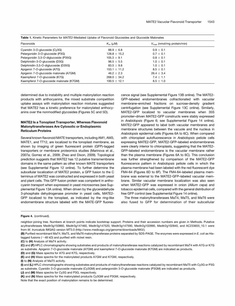
Table 1. Kinetic Parameters for MATE2-Mediated Uptake of Flavonoid Glucosides and Glucoside Malonates
Flavonoids Km (mM) Vmax (nmol/mg protein/min)
Cyanidin 3-O-glucoside (Cy3G) 88.9 6 6.8 0.9 6 0.1
Pelargonidin 3-O-glucoside (P3G) 128.8 6 15.2 0.7 6 0.1
Pelargonidin 3,5-O-diglucoside (P35G) 105.3 6 9.1 0.8 6 0.1
Delphinidin-3-O-glucoside (D3G) 96.5 6 5.5 1.0 6 0.1
Delphinidin-3,5-O-diglucoside (D35G) 83.5 6 9.8 1.0 6 0.1
Apigenin 7-O-glucoside (A7G) 153.1 6 11.2 8.5 6 0.1
Apigenin 7-O-glucoside malonate (A7GM) 46.2 6 2.3 26.4 6 3.4
Kaempferol 7-O-glucoside (K7G) 268.0 6 34.2 7.4 6 1.1
Kaempferol 7-O-glucoside malonate (K7GM) 100.5 6 12.1 8.5 6 1.0
MATE2 Vacuolar Flavonoid Transporter 1543
localization. Compared with optical sections of free GFP control
(;20 sections), which show signals on both nucleus and many
thick cytosolic bundles in tobacco epidermal cells (Figure 6G),
GFP-MaT4 was observed to be cytosolic, exhibiting no signal
inside the nucleus but only around the nuclear membrane and
thick cytosolic bundles (Figure 6H), whereas GFP-MaT5 ap-
peared to exhibit both endoplasmic reticulum (ER) and cytosolic
localization (Figure 6I). GFP-MaT6 was evenly distributed in the
thin ER network as well as around the nuclear membrane (Figure
6J).
To further confirm the subcellular localization of the malonyl-
transferases, total protein extracts from tobacco leaves express-
ing free GFP and the three GFP-MaT fusions were fractionated
into soluble and microsomal membrane fractions, which were
then analyzed on protein gel blots probedwith anti-GFP antibody
and an antibody against BiP, an ER marker. Free GFP and GFP-
MaT4 were recovered in the soluble fraction, whereas GFP-
MaT5 was detected in both soluble and microsomal fractions,
and GFP-MaT6 was mainly in the microsomal fraction (see
Supplemental Figure 15 online).
Loss of MATE2 Expression Changes Leaf Pigmentation,
Flower Color, and (Malonylated) Flavonoid Profiles
To address the physiological function of MATE2 in Medicago,
Tnt1 retrotransposon insertion mutants that abolished MATE2
expression were isolated from the Medicago Tnt1 insertion
population established at the Noble Foundation (Tadege et al.,
2008). By PCR screening of Tnt1 population DNA pools with both
Tnt1 left and right border primers and MATE2 gene-specific
primers (see Supplemental Table 2 online), as well as searching
the flanking sequence tag collection from the population (http://
bioinfo4.noble.org/mutant/database.php) with the MATE2 ge-
nomic sequence, we obtained four lines with independent Tnt1
insertions in the MATE2 gene (Figure 7A). Apart from mate2-1,
with a Tnt1 insertion in the first exon, the insertion sites inMATE2
were all in introns; however, MATE2 transcripts were effectively
abolished in all homozygous lines (Figure 7B).
Under normal growth conditions, homozygous mate2 mutant
lines did not show altered vegetative growth or morphological
phenotypes. However, the characteristic purple pigmentation in
the trifoliate leaves of 1- to 2-week-old M. truncatula wild-type
R108 was strongly reduced or even disappeared inmate2mutant
lines (Figure 7C), and this effect was most apparent when viewing
cross sections of leaves ofmate2mutants comparedwith thewild
type, especially leaves from which the epidermis had been
stripped (Figure 7D; see Supplemental Figure 16 online). Quanti-
fication of anthocyanins from mate2 and wild-type R108 plants
showed that total anthocyanin levels decreased by more than
50% inmutants as comparedwith thewild type (Figure 7E). All four
mate2 mutant lines had paler flowers than wild- type R108 plants
(Figure 7F). However, RT-PCR indicated that anthocyanin biosyn-
thetic genes were still expressed in the mate2 mutants (see
Supplemental Figure 17 online). The decreased yellow pigmenta-
tion inmate2 mutant lines may result from reduced accumulation
of flavonoids in flower petals, sinceMATE2 is strongly expressed
in epidermal cells and vascular bundles in flowers. Flavonoid
metabolites were therefore profiled in the mutants and corre-
sponding null segregant controls. In M. truncatula leaves, apige-
nin, genistein, kaempferol, and quercetin represent more than
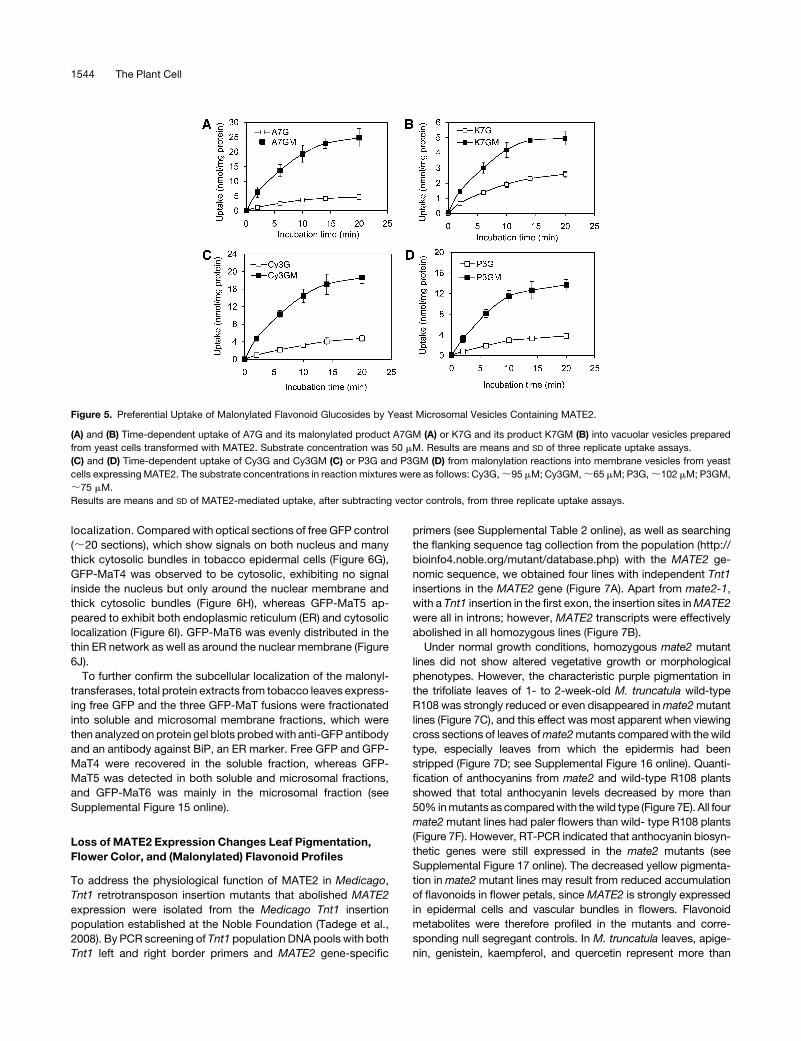
Figure 5. Preferential Uptake of Malonylated Flavonoid Glucosides by Yeast Microsomal Vesicles Containing MATE2.
(A) and (B) Time-dependent uptake of A7G and its malonylated product A7GM (A) or K7G and its product K7GM (B) into vacuolar vesicles prepared
from yeast cells transformed with MATE2. Substrate concentration was 50 mM. Results are means and SD of three replicate uptake assays.
(C) and (D) Time-dependent uptake of Cy3G and Cy3GM (C) or P3G and P3GM (D) from malonylation reactions into membrane vesicles from yeast
cells expressing MATE2. The substrate concentrations in reaction mixtures were as follows: Cy3G,;95 mM; Cy3GM,;65 mM; P3G,;102 mM; P3GM,
;75 mM.
Results are means and SD of MATE2-mediated uptake, after subtracting vector controls, from three replicate uptake assays.
1544 The Plant Cell
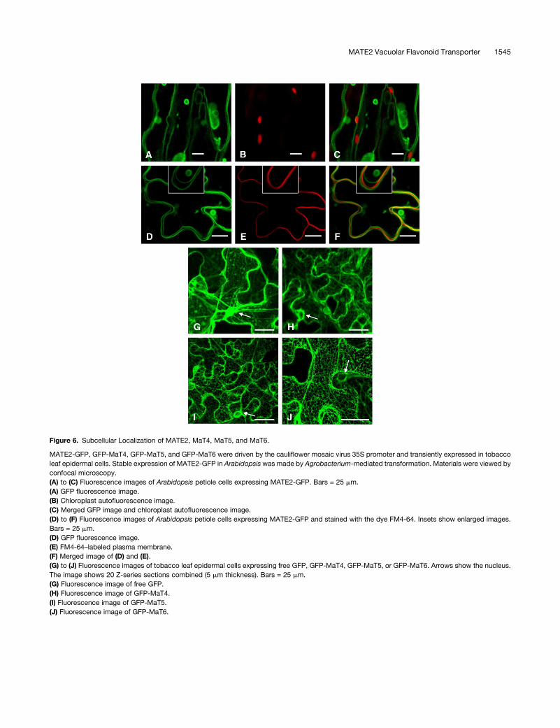
Figure 6. Subcellular Localization of MATE2, MaT4, MaT5, and MaT6.
MATE2-GFP, GFP-MaT4, GFP-MaT5, and GFP-MaT6 were driven by the cauliflower mosaic virus 35S promoter and transiently expressed in tobacco
leaf epidermal cells. Stable expression of MATE2-GFP in Arabidopsiswas made by Agrobacterium-mediated transformation. Materials were viewed by
confocal microscopy.
(A) to (C) Fluorescence images of Arabidopsis petiole cells expressing MATE2-GFP. Bars = 25 mm.
(A) GFP fluorescence image.
(B) Chloroplast autofluorescence image.
(C) Merged GFP image and chloroplast autofluorescence image.
(D) to (F) Fluorescence images of Arabidopsis petiole cells expressing MATE2-GFP and stained with the dye FM4-64. Insets show enlarged images.
Bars = 25 mm.
(D) GFP fluorescence image.
(E) FM4-64–labeled plasma membrane.
(F) Merged image of (D) and (E).
(G) to (J) Fluorescence images of tobacco leaf epidermal cells expressing free GFP, GFP-MaT4, GFP-MaT5, or GFP-MaT6. Arrows show the nucleus.
The image shows 20 Z-series sections combined (5 mm thickness). Bars = 25 mm.
(G) Fluorescence image of free GFP.
(H) Fluorescence image of GFP-MaT4.
(I) Fluorescence image of GFP-MaT5.
(J) Fluorescence image of GFP-MaT6.
MATE2 Vacuolar Flavonoid Transporter 1545
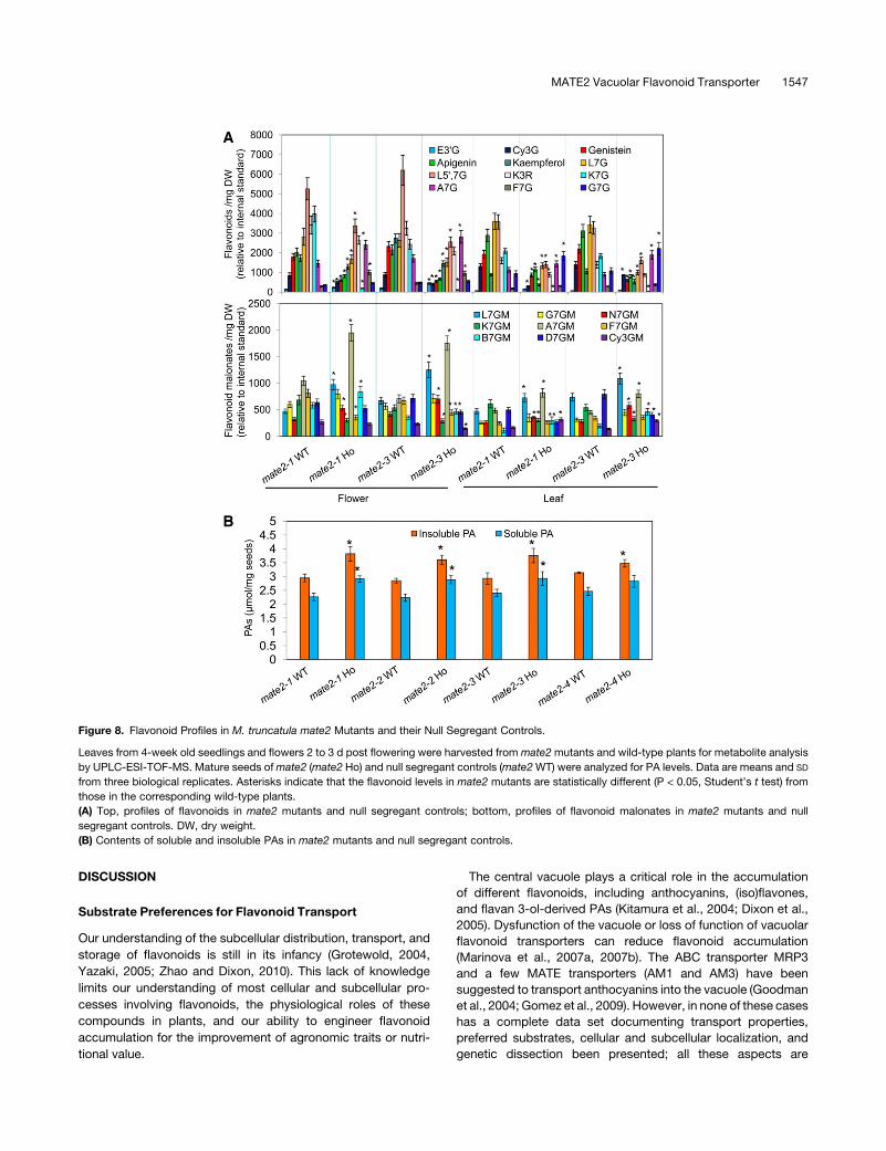
30% of total flavonoids, and luteolin and kaempferol glycosides
are the major flavonoid glycosides. All mate2 mutants showed
generally decreased flavonoid glycoside levels in both leaves and
flowers (Figure 8A; see Supplemental Figure 18 online). Levels of
Cy3G and luteolin 7-O-glucoside in both leaves and flowers
decreased inmate2mutants comparedwith controls; in particular,
K7G and kaempferol 3-O-rhamnoside contents were drastically
reduced. However, A7G, naringenin 7-O-glucoside, biochanin A
7-O-glucoside, and formononetin 7-O-glucoside levels appeared
to be increased inmate2mutants compared with controls. Levels
of minor anthocyanins, such as D3G, P3G, D35G, and pelargoni-
din 3,5-di-O-glucoside (P35G),werealso reduced inmate2 leaves,
but less so in flowers. Levels of flavonoid aglycones were also
reduced in themate2mutants, suggesting either feedback into the
biosynthetic pathway or turnover of incorrectly stored products.
Because malonylated flavonoid glycosides were the preferred
substrates for MATE2 in vitro, and these compounds, although
abundant in roots and cell suspension cultures of M. truncatula
(Farag et al., 2008), have not been described from the aerial
portions of the plant, it was important to demonstrate their relative
levels in wild-type and mate2 plants. Both flower and leaf tissues
contained the flavonoid glucoside malonates that were identified
as MaT4 and MaT5 enzymatic products. However, the levels of
these various flavonoid malonates in these tissues varied greatly
over a wide range: L7G malonate, K7G malonate, G7G malonate,
and A7G malonate were present at relatively high levels, whereas
levels of Cy3Gmalonate and biochanin A 7-O-glucosidemalonate
were lower (Figure 8A). Loss of MATE2 expression altered these
flavonoid malonate profiles as compared with wild-type controls.
Most flavonoid malonates, including L7G, naringenin 7-O-
glucoside, A7G, and Cy3G malonates, accumulated to higher
levels in mate2 mutants than in wild-type plants (differences
statistically significant at P < 0.05; Figure 8A). Whereas K7G
malonate levels were reduced in a similarmanner to K7G levels in
mate2 mutants, levels of G7G malonate showed no change.
Changes in flavonoid malonate levels were also dependent on
tissue type; for example, the levels of Cy3G malonate increased
in mate2 leaf tissues but not in flower tissues, whereas formo-
nonetin 7-O-glucoside malonate levels were reduced in mate2
flower tissues but not in leaf tissues (Figure 8A).
LossofFunctionofMATE2AffectsPABiosynthesis inSeeds
E39G levels were higher inmate2mutant flowers than in wild-type
control flowers, suggesting that blocking the transport of antho-
cyanins into the vacuole may feed back to direct more anthocya-
nidin precursor flux into the PA biosynthesis pathway, leading to
moreE39Gproduction (Figure 8A). To further confirm that PAor PA
precursor biosynthesis can indeed be enhanced by blocking
anthocyanin transport, PA contents of seeds of mate2 mutants
and wild-type controls were determined. Four mate2 mutant
alleles all showed increased contents of both soluble and insoluble
PAs in their seeds comparedwith those in their corresponding null
segregant controls (Figure 8B). The increases in the mate2-1,
mate2-2, and mate2-3 lines were significant (P < 0.01–0.05), but
the increase in mate2-4 was not. Consistent with increased PA
accumulation, decreased anthocyanin levels were also observed
in the mate2 lines (see Supplemental Figure 19 online).
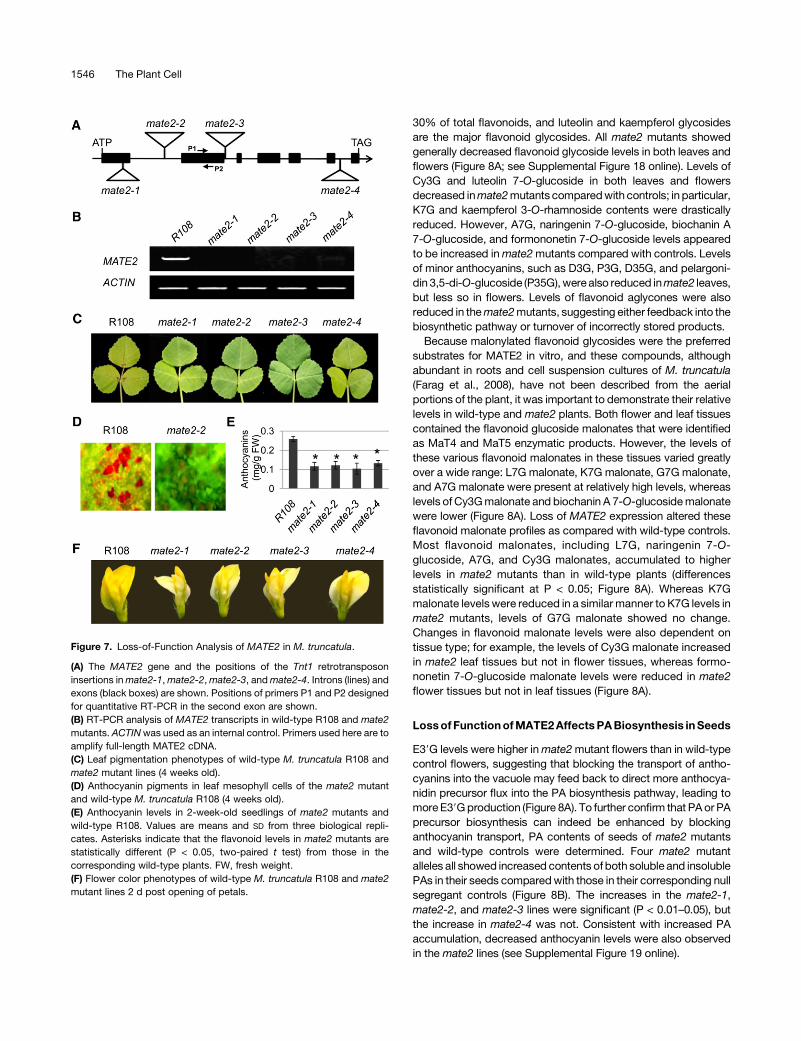
Figure 7. Loss-of-Function Analysis of MATE2 in M. truncatula.
(A) The MATE2 gene and the positions of the Tnt1 retrotransposon
insertions inmate2-1,mate2-2,mate2-3, andmate2-4. Introns (lines) and
exons (black boxes) are shown. Positions of primers P1 and P2 designed
for quantitative RT-PCR in the second exon are shown.
(B) RT-PCR analysis of MATE2 transcripts in wild-type R108 and mate2
mutants. ACTINwas used as an internal control. Primers used here are to
amplify full-length MATE2 cDNA.
(C) Leaf pigmentation phenotypes of wild-type M. truncatula R108 and
mate2 mutant lines (4 weeks old).
(D) Anthocyanin pigments in leaf mesophyll cells of the mate2 mutant
and wild-type M. truncatula R108 (4 weeks old).
(E) Anthocyanin levels in 2-week-old seedlings of mate2 mutants and
wild-type R108. Values are means and SD from three biological repli-
cates. Asterisks indicate that the flavonoid levels in mate2 mutants are
statistically different (P < 0.05, two-paired t test) from those in the
corresponding wild-type plants. FW, fresh weight.
(F) Flower color phenotypes of wild-type M. truncatula R108 and mate2
mutant lines 2 d post opening of petals.
1546 The Plant Cell
DISCUSSION
Substrate Preferences for Flavonoid Transport
Our understanding of the subcellular distribution, transport, and
storage of flavonoids is still in its infancy (Grotewold, 2004,
Yazaki, 2005; Zhao and Dixon, 2010). This lack of knowledge
limits our understanding of most cellular and subcellular pro-
cesses involving flavonoids, the physiological roles of these
compounds in plants, and our ability to engineer flavonoid
accumulation for the improvement of agronomic traits or nutri-
tional value.
The central vacuole plays a critical role in the accumulation
of different flavonoids, including anthocyanins, (iso)flavones,
and flavan 3-ol-derived PAs (Kitamura et al., 2004; Dixon et al.,
2005). Dysfunction of the vacuole or loss of function of vacuolar
flavonoid transporters can reduce flavonoid accumulation
(Marinova et al., 2007a, 2007b). The ABC transporter MRP3
and a few MATE transporters (AM1 and AM3) have been
suggested to transport anthocyanins into the vacuole (Goodman
et al., 2004; Gomez et al., 2009). However, in none of these cases
has a complete data set documenting transport properties,
preferred substrates, cellular and subcellular localization, and
genetic dissection been presented; all these aspects are
Figure 8. Flavonoid Profiles in M. truncatula mate2 Mutants and their Null Segregant Controls.
Leaves from 4-week old seedlings and flowers 2 to 3 d post flowering were harvested from mate2mutants and wild-type plants for metabolite analysis
by UPLC-ESI-TOF-MS. Mature seeds ofmate2 (mate2 Ho) and null segregant controls (mate2WT) were analyzed for PA levels. Data are means and SD
from three biological replicates. Asterisks indicate that the flavonoid levels in mate2 mutants are statistically different (P < 0.05, Student’s t test) from
those in the corresponding wild-type plants.
(A) Top, profiles of flavonoids in mate2 mutants and null segregant controls; bottom, profiles of flavonoid malonates in mate2 mutants and null
segregant controls. DW, dry weight.
(B) Contents of soluble and insoluble PAs in mate2 mutants and null segregant controls.
MATE2 Vacuolar Flavonoid Transporter 1547
essential for a full understanding of flavonoid transporter func-
tion. Furthermore, the relationship between flavonoid modifica-
tion and transport is still unclear, in particular whether the acyl
modifications commonly found with anthocyanins and (iso)fla-
vonoids are essential for transport and/or stabilization of the
parent compounds once localized to the vacuole (Matern et al.,
1983; Suzuki et al., 2002; Luo et al., 2007; Zhao andDixon, 2010).
This study, therefore, was undertaken to provide a complete
description and dissection of flavonoid transport and modifica-
tion in a genetically tractable system.
MATE2 Is a Vacuolar Flavonoid Transporter
MATE2 is phylogenetically related to the known flavonoid trans-
porters MATE1, TT12, AM1, and AM3, all of which appear to be
localized to the vacuolar membrane on the basis of GFP fusion
imaging (Marinova et al., 2007a; Gomez et al., 2009; Zhao and
Dixon, 2009). Using the same approach, MATE2-GFP also
appears to be targeted to the vacuolar membrane in plant cells
(tobacco, Arabidopsis, and onion epidermal cells) as well as in
yeast cells. It is always a challenge to clearly distinguish the
tonoplast from the plasma membrane in mature and highly
vacuolated epidermal cells. However, by comparing MATE2-
GFP signal with red chloroplast autofluorescence and plasma
membrane labeling with FM4-64, we were able to conclude that
MATE2-GFP is indeed a vacuolar membrane protein. Based on
proteomic analysis, another MATE transporter, EET, was shown
to be present on the tonoplast in Arabidopsis (Jaquinod et al.,
2007).
MATE2 localization in yeast cells was confirmed both with
MATE2-GFP imaging under confocal microscopy and by co-
fractionation of MATE2-GFP with vacuolar membranes following
sucrose-density gradient centrifugation. MATE2 expressed in
yeast not only transports several anthocyanin glucosides but
also transports other flavonoid glucosides into vacuolar vesicles
at a low efficiency.
MATE2 Preferentially Transports Malonylated Flavonoids
Many MATE transporters, such as DTX1 from Arabidopsis (Li
et al., 2002), show a relatively broad substrate specificity. This is
also the case for MATE2. In contrast toMATE1, which transports
E39G at a high efficiency and Cy3G at a lower efficiency but
shows almost no transport activity toward other flavonoid glu-
cosides, MATE2 transports various anthocyanidin glucosides
with different hydroxylation patterns, flavonol glucosides, and
flavone glucosides at lower efficiencies. However,MATE2 shows
little transport activity toward E39G and has a very different
tissue-specific expression pattern from that ofMATE1, indicating
that the two transporters clearly possess different physiological
functions.
Malonylated flavonoids are more efficiently transported by
MATE2 than are their nonacylated precursors. Malonylation of
flavonoid glucosides byMaT4,MaT5, orMaT6 increases not only
their affinity for MATE2 but also their transport efficiency, as
indicated by increased Vmax values. Similar to other flavonoid
transporters (Marinova et al., 2007a; Gomez et al., 2009; Zhao
and Dixon, 2009), MATE2 shows almost no transport activity
toward flavonoid aglycones. Therefore, MATE2 specially trans-
ports glycosyl- and malonyl-glycosyl–modified flavonoids, com-
pounds that are widely present in M. truncatula (Farag et al.,
2008; this work).
The nature of the forms in which flavonoids are preferentially
transported has been actively discussed for many years (Zhao
and Dixon, 2010). The anthocyanins that accumulate in the
vacuoles of different plants are largely present in acylated forms
(Matern et al., 1983; Markham et al., 2000; Zhang et al., 2006),
and biochemical analysis suggests that the grapevine MATE
transporters AM1 and AM3 specifically transport p-coumaroyl–
acylated anthocyanidin glucosides (Gomez et al., 2009). Two
major types of acylation, aromatic acylation (such as p-cou-
maroyl group addition) and aliphatic acylation (such as malonyl
group addition), may have different physiological functions
(Strack and Wray, 1994). It has been suggested that aromatic
acylation enhances the color of anthocyanins, whereas aliphatic
acylation (usually malonylation) may stabilize flavonoids and
increase the resistance of flavonoid glucoside malonates to
enzymatic degradation (Suzuki et al., 2002; Luo et al., 2007). For
example, the levels of (iso)flavone glucoside malonates in M.
truncatula suspension cultures show much less change after
elicitation of cells with yeast extract than do the levels of
isoflavone glucosides inside the cells and corresponding agly-
cones in the culture medium (Farag et al., 2008), suggesting that
the flavonoid glucoside malonates are a stable storage form.
Malonylation of flavonoids occurs widely in plants, and malo-
nyltransferases have been characterized from several different
species (Suzuki et al., 2002, 2003, 2004; Luo et al., 2007; Yu
et al., 2008). An NMR study showed that malonylation, but not
p-coumaroylation, of flavonoids induces a conformational
change that might facilitate their transport and storage in the
vacuole (Matern et al., 1983), possibly through improved solu-
bility and reaction with other vacuolar components (Matern et al.,
1983; Markham et al., 2000; Zhang et al., 2006). However, the
precise physiological function of flavonoid malonylation has yet
to be defined (Boller and Wiemken, 1986; Springob et al., 2003;
Luo et al., 2007; Yu et al., 2008). A role for malonylation in
targeting compounds for vacuolar transport as opposed to
extracellular secretion is supported by the recent demonstration
that malonylation of xenobiotic phenolic glucosides leads to their
preferential transport to the vacuole in Arabidopsis (Taguchi
et al., 2010).
This study demonstrates that malonylation of anthocyanins or
other flavonoids facilitates their transport into the vacuole by a
defined transporter and shows that MaT4, localized to the
cytoplasm, MaT5, localized to the ER, and MaT6, localized to
both the cytoplasm and ER, are likely important for flavonoid
modification and therefore transport. The fact that the malonyl
group addition to the sugar moiety of flavonoid glucosides in-
creases their affinity for, and transport efficiency by, the MATE2
transporter effectively links cytosolic flavonoid modifications to
transport and storage in the vacuole. This link could be further
addressed upon the availability of Tnt1 insertion mutants in the
malonyltransferase genes.
Mechanisms in addition to acylation have been also suggested
to facilitate anthocyanin transport. Although genetic evidence
indicates that the maize ABC transporter MRP3 is involved in
1548 The Plant Cell
vacuolar transport of anthocyanins (Goodman et al., 2004), and
Marrs et al. (1995) showed that Cy3G conjugated with glutathi-
one by glutathione S-transferase (GST) was recognized as a
substrate for transport into vacuoles by the GS-X pump inmaize,
it is now believed that MRP3 and other related transporters are
unlikely to transport glutathionylated anthocyanins (Kitamura,
2006). This is because such compounds have yet to be identified
in plants, and the GST activity of TT19, a GST required for PA
accumulation in Arabidopsis, is not required for its function
(Mueller et al., 2000).
Coregulation of MATE2 with Flavonoid Biosynthesis and
Modification Genes
Flavonoid biosynthetic genes are usually coordinately regulated
by developmental and environmental cues such as growth stage
(in culture) and light (Lepiniec et al., 2006). Furthermore, meta-
bolic channeling has been proposed for flavonoid biosynthetic
enzymes (Winkel, 2004; Jørgensen et al., 2005), suggesting the
need for tight coregulation of protein amounts. Tissue-specific
coregulation of core flavonoid biosynthetic genes, glycosyltrans-
ferases, malonyltransferases, and MATE2 are evident from anal-
ysis ofmicroarray databases andour experiments. UGT78G1 is a
glycosyltransferase that shows activity toward anthocyanins and
a range of other flavonoids in vitro and is, along with MATE2 and
MaT4, controlled in Medicago by the anthocyanin-regulatory
MYB transcription factor LAP1 (Modolo et al., 2007; Peel et al.,
2009). Genetic evidence indicates that the major function of
UGT78G1 in vivo is the 3-O-glycosylation of anthocyanidins
(Peel et al., 2009). The anthocyanin malonyltransferase gene
MaT5 is primarily expressed in flowers and roots, while MaT6 is
primarily expressed in leaf, stem, and flowers. Since MaT5 and
MaT6 showed very similar substrate specificities, it is probable
thatM. truncatula utilizes these two genes in different tissues for
similar functions.
Physiological Function of MATE2
Flavonoid pigments, including flavonols, flavones, and anthocy-
anins, can interact with each other for copigmentation and
aggregation within the vacuole or be selectively compartmen-
talized, as in petal cells; these fates can influence color hue,
intensity, and stability (Markham et al., 2000). The abundant
flavonols and flavones, as well as their complexes with proteins
or lipids, often contribute to the yellow petal color (Markham
et al., 2000). Similar to mutants affecting other flavonoid trans-
porters such asMRP3, TT12, andMATE1 (Goodman et al., 2004;
Marinova et al., 2007a; Zhao and Dixon, 2010), mate2 mutant
alleles display reduced leaf pigmentation and pale flower color,
as compared with wild-type M. truncatula plants, which exhibit
purple anthocyanin pigmentation in the trifoliate leaves and
brownish yellow flowers. Metabolite profiling data suggest that
the leaf pigmentation and flower color phenotypes in mate2
mutants may be attributable to the decreased levels of antho-
cyanins and other flavonoid compounds in leaf and floral tissues.
These data indicate that MATE2 plays an important role in
flavonoid accumulation in Medicago, and the differentially al-
tered levels of upstream intermediates, an array of flavonoid
glucosides and aglycones, in the mate2 mutants suggests that
MATE2 activity exerts positive feedback on upstream metabo-
lism. Mutation of the MATE transporter FFT (At4g25640) in
Arabidopsis results in a decrease in levels of flavonol glucosides,
but not anthocyanins; this study did not determine levels of
malonylated flavonoids or in vitro transport properties (Thompson
et al., 2010). It appears that FFT has a somewhat different
transport function from MATE2.
It is interesting that the levels of some flavonoid glycoside
malonates are increased in mate2 mutants, whereas others are
reduced. Seeds of the Arabidopsis tt12 mutant accumulate
epicatechin glucoside, as this is the preferred substrate for the
TT2 MATE transporter (Zhao and Dixon, 2009; Kitamura et al.,
2010), and is probably further metabolized to PAs following
transport to the vacuole. The further metabolic fates of flavonoid
malonyl glycosides are poorly understood, as are their potential
stabilities compared with the unmodified glycoside if they are
allowed to accumulate in the cytoplasm due to blockage of
vacuolar transport.
The presence ofMATE2 transcripts in vascular bundles of both
leaf and petal suggests that MATE2 might also be associated
with long-distance transport of flavonoids, but the mechanisms
underlying such transport of flavonoids between shoots and
roots remain to be determined (Buer et al., 2007).
Competition between Anthocyanin and PA Biosynthesis
Several pieces of evidence suggest competition between an-
thocyanin and PA biosynthesis, but the mechanisms remain
unclear (Lee et al., 2005; Marinova et al., 2007a). MATE1 is
specifically expressed in the seed coat where PAs accumulate in
M. truncatula (Zhao and Dixon, 2009). MATE2 is also expressed
in developing seeds, but at low levels. While the levels of Cy3G
are much lower in mate2 flowers than in the wild type, E39Glevels, mainly from the coharvested young pods, are higher,
suggesting that blocking MATE2-mediated transport of antho-
cyanins into the vacuole directs more anthocyanidin into PA
biosynthesis. ANR expression in mate2 mutants was not higher
than in controls, suggesting that this effect is probably one of
substrate availability. This phenomenon is also supported by the
observation that mate2 mutant seeds contain more soluble and
insoluble PAs than controls. Blocking of anthocyanin transport
may lead to temporary accumulation of Cy3GM and Cy3G and
inhibition of modification enzyme activity, which then makes
more cyanidin available for ANR to synthesize epicatechin, which
is then converted to E39G for vacuolar transport (by MATE1) and
PA biosynthesis.
Alternative Transport Pathways for Anthocyanins
AlthoughMATE2 efficiently transports glycosylated/malonylated
anthocyanidins into the vacuole and functions in anthocyanin
accumulation during leaf pigmentation and flavonoid accumu-
lation in flowers, we cannot exclude the possibility of other
mechanisms for flavonoid transport. Anthocyanin pigment–
containing bodies have been observed moving in the cytoplasm
of some plant cells, such as cultured maize and grapevine cells
(Grotewold et al., 1998; Zhang et al., 2006). Membrane vesicle
MATE2 Vacuolar Flavonoid Transporter 1549
trafficking–mediated transport of anthocyanins into the central
vacuole has also beenproposed (Grotewold, 2004; Poustka et al.,
2007; Pourcel et al., 2010). However, such phenomena have only
been observed in a limited number of plants (Grotewold, 2004).
We have not observed anthocyanin bodies inM. truncatula leaf or
flower petal cells. Furthermore, in Medicago plants producing
massive amounts of anthocyanins as a result of constitutive
expression of the LAP1 transcription factor, several membrane
transporters including MATE2 are significantly up-regulated,
whereas genes encoding known proteins involved in vesicle
trafficking, such as SNAREs and GTPases, are not (Peel et al.,
2009). Clearly, MATE2 represents one route for vacuolar trans-
port of anthocyanins and other flavonoids in Medicago, but
others cannot be ruled out, especially as the loss of function of
MATE2 does not result in a complete loss of flavonoid glycoside
accumulation.
METHODS
Plant Materials andMedicago truncatula Tnt1Mutant Screening
Medicago truncatula plants were grown in the greenhouse as described
previously (Zhao and Dixon, 2009). Reverse genetic screening of M.
truncatula Tnt1 mutants in the R108 background was performed as
described previously (Peel et al., 2009). TwoMATE2 Tnt1 insertionmutant
lines, NF5697 (mate2-1) and NF5176 (mate2-2), were identified through
PCR-based screening of 10 superpools of pooled DNA samples from
5000 Tnt1 insertional mutant lines of M. truncatula (Tadege et al., 2008)
using a combination of MATE2- and Tnt1-specific primers (see Supple-
mental Table 2 online). The Tnt1 insertion site in MATE2 was verified by
sequencing. Two other Tnt1 lines, NF0737 and NF3103, were identified in
silico through analysis of Tnt1 flanking sequences in the mutant popu-
lation (http://bioinfo4.noble.org/mutant/line2.php), and the Tnt1 insertion
sites were confirmed by PCR. Loss/reduction in MATE2 transcripts in
homozygous lines of these four independent Tnt1mutants was confirmed
by RT-PCR.
Cloning and Vector Construction
The MATE2, MaT4, MaT5, and MaT6 open reading frames (ORFs) were
cloned from a cDNA library constructed from M. truncatula wild-type
(R108) flower tissues (0–5 d post flowering) with forward and reverse
primers that enabled PCR fragments to be directionally cloned into
pENTR vector (see Supplemental Table 2 online). Total RNA was
extracted from flowers of M. truncatula using the RNeasy plant mini kit
procedure (Qiagen). First-strand cDNA was synthesized from total RNA
with the SuperScript III First-Strand Synthesis system (Invitrogen) ac-
cording to the manufacturer’s protocol. The MATE2 ORF was amplified
with MATE2-59 forward and MATE2-39 reverse primers, and MaT4, -5,
and -6 ORFs were cloned with corresponding forward and reverse primer
pairs (see Supplemental Table 2 online). The PCR products were cloned
into pENTR/D-TOPO vector (Invitrogen) for sequencing, and the con-
firmed clones were subcloned into Gateway destination vectors, the
yeast expression vector pYESDEST52 (uracil selectable marker), His-
tagged fusion protein expression vector pDEST17 for expression in
Escherichia coli, or the plant binary vector pB2GW7 by recombination
using Gateway LR Clonase (Invitrogen).
TheMATE2-GFP fusion was constructed by cloning theMATE2ORF in
framewith the N terminus of GFP. TheMATE2ORFwas amplifiedwith the
primers MATE2-GFP-59 and MATE2-GFP-39 (see Supplemental Table 2
online). PCR products were subcloned into the yeast GFP fusion cassette
shuttle vector pHGDP-GFP (Zhao et al., 2009) in frame with the N
terminus of GFP. The construct pHGDP-MATE2-GFP was transformed
into yeast cells for checking MATE2-GFP expression. A 2.1-kb MATE2-
GFP coding sequence was then amplified with the primer pair MATE2
SFI-59 and MATE2 SFI-39 (see Supplemental Table 2 online) and sub-
cloned into pENTR/D-TOPO vector, followed by subcloning into the plant
binary vector pB2GW7 (Invitrogen; Karimi et al., 2002) by recombination
with Gateway LR Clonase. The 35S:MATE2-GFP construct was used for
localization studies. All clones were confirmed by sequencing.
Chemicals and Reagents
Flavonoid aglycones, anthocyanidins, Cy3G, D3G, D35G, P3G, P35G,
daidzein 7-O-glucoside, and other flavonoid glucosides were obtained
from Indofine Chemical Company. Malonylated flavonoids were gener-
ated using MaT4 and MaT5 enzyme reactions in this study.
Yeast Culture, Transformation, and Preparation of
Microsomal Fractions
Saccharomyces cerevisiae W303a (Mat ade2-1 ura3-1 his3-11,15 trp1-1
leu2-3,112 can1-100) was used to express MATE2. The Gateway yeast
expression vectors pYESDEST52 (Invitrogen) and pYES harboring
MATE2 were transformed into W303a by the polyethylene glycol/lithium
acetate method (Zhao and Dixon, 2009). Yeast growth, induction of
MATE2 expression by galactose, and microsomal preparation were
conducted as described previously (Zhao and Dixon, 2009).
Transport Activity Assays
Flavonoid uptake by vacuolar membrane vesicles was performed at 258C
as described previously (Zhao and Dixon, 2009). Briefly, the 600- to 1000-
mL assay mixtures contained 25mM Tris-MES (pH 8.0), 0.4 M sorbitol, 50
mMKCl, 5 mMMg-ATP, 0.1% (w/v) BSA, the indicated concentrations of
transport substrate, and microsomal preparations (;300 mg of proteins).
The flavonoids taken up into the membrane vesicles were extracted with
50% (v/v) methanol for 1 h on a shaker and analyzed by HPLC. Inhibitor
assays were conducted with 1mMvanadate, 5mMgramicidin D, 0.1 mM
bafilomycin A1, or 5mMNH4Cl in the reactionmixture for 8min. Transport
inhibitor stock solutions were 1 M vanadate in water, 5 mM bafilomycin
A1 in DMSO, 5 mM gramicidin D in DMSO, and 1 M NH4Cl in water.
Corresponding solvent controls were included.
For competition assays with yeast membrane vesicles expressing
MATE2, various concentrations of flavonoid glucosides were included in
the uptake assaywith 100mMCy3Gas substrate. After incubation at 258C
for 10 min, transported flavonoids were extracted from vesicles as
described (Zhao and Dixon, 2009) and measured by HPLC. Reverse-
phase HPLC analyses were performed on an Agilent HP1100 HPLC
apparatus using a gradient mobile phase, solvent A (1%phosphoric acid)
andB (acetonitrile) at 1mL/min flow rate: 0 to 5min, 5%B; 5 to 10min, 5 to
10%B; 10 to 25min, 10 to 17%B; 25 to 30min, 17 to 23%B; 30 to 65min,
23 to 50%B; 65 to 79min, 50 to 100%B; 79 to 80min, 100 to 5%B. Data
were collected at 206 and 530 nm for epicatechin and cyanidin deriva-
tives, respectively. Identifications were based on chromatographic be-
havior and UV spectra compared with those of authentic standards.
Malonyltransferase Expression, Activity Assay, and Enzyme
Product Identification
The full-length ORFs of MaT4, MaT5, and MaT6 were cloned by PCR
using their gene-specific primers (see Supplemental Table 2 online). PCR
products were cloned into pENTR/D/TOPO TA vector (Invitrogen) for
sequencing. Confirmed constructs were used for in vitro recombination
1550 The Plant Cell
into destination vectors. For heterologous expression in E. coli, these
genes were recombined with pDEST17 vector using Invitrogen Clonase
LR. Confirmed pDEST-MaT4, pDEST-MaT5, and pDEST-MaT6 con-
structs were transformed into BL21 (DE3) cells for expression of proteins.
Proteins were induced by 0.4 mM isopropyl-L-thio-b-D-galactopyrano-
side at 168C overnight. After purification with a MagneHis protein puri-
fication system according to the manufacturer’s instructions (Promega),
malonyl transferase activity was assayed in reaction mixtures (100 mL)
consisting of 20 mM potassium phosphate buffer (pH 7.0), 60 mM
malonyl-CoA, 120 mM flavonoid glycoside, and enzyme (;0.5 mg of
protein). The reaction was terminated by adding 200 mL of methanol after
incubation at 308C for 20 min. Enzymatic products in the reaction mixture
were analyzed by reverse-phase HPLC as described previously (Zhao
and Dixon, 2009). Malonylated flavonoid products from enzyme assays
were identified by ESI-LC-MS. HPLC separation was achieved using a
reverse-phase, C18 (5 mm, 4.6 3 250 mm) column (J.T. Baker) on an
Agilent HP1100 HPLC apparatus using a step gradient mobile phase,
solvent A (0.1% acetic acid) and B (acetonitrile) at 0.8 mL/min flow rate: 0
to 5min, 5%B; 5 to 15min, 5 to 15%B; 15 to 20min, 15 to 17%B; 20 to 25
min, 17 to 23%B; 25 to 60min, 23 to 50%B; 60 to 60.5 min, 50 to 95%B;
60.5 to 70.5 min, 95% B. All mass spectra were acquired using a Bruker
Esquire LC apparatus equipped with an electrospray ionization source in
the positive mode. Positive ion ESI was performed using a source voltage
of 3000 V and capillary offset voltage of 270.7 V. Nebulization was
achieved using nitrogen gas at a pressure of 70 p.s.i. Desolvation was
aided by using a counter current gas of nitrogen at a pressure of 12 p.s.i.
The capillary temperature was set at 3508C. Mass spectra were recorded
over the range of 50 to 2200 m/z. The Bruker ion trap was operated under
an ion current control of 20,000 with a maximum acquire time of 100 ms
and a trap drive setting of 50.
Determination of Transport and Enzyme Kinetics
Transport assays were performed with different concentrations of Cy3G,
D3G, D35G, P3G, P35G, and theirmalonylated derivatives (from 25 to 200
mM) and 5 mM Mg-ATP. After incubation at 258C for 10 min, transported
glycosides were measured by HPLC as described above. Kinetics of the
malonyltransferases were determined with various flavonoid glycoside
acceptors in 20-min incubations at 308C. Lineweaver-Burk plots were
used to calculate Km and Vmax values. Protein concentrations were
determined using a Bio-Rad Protein Assay kit with BSA as the standard.
Localization of GFP Fusions by Confocal Microscopy
Five micrograms of plasmid DNA containing 35S:MATE2-GFP or 35S:GFP
was mixed with 20 mL of an aqueous suspension containing 1.0-mm gold
particles. After washing, the gold was spread onto plastic carrier discs for
biolistic bombardment of tobacco (Nicotiana tabacum) and onion (Allium
cepa) epidermal cells using a Bio-Rad 1000/HE particle delivery system.
After 15 to 18h, epidermal cellswere vieweddirectlywith a TCS-SP2AOBS
confocal laser scanning microscope (Leica Microsystems) to examine the
localization patterns of MATE2-GFP. Alternatively, Arabidopsis thaliana
(ecotype Columbia) was transformed with Agrobacterium tumefaciens
AGl1harboring the35S:MATE2-GFPconstructby the floral dippingmethod
(Clough and Bent, 1998). F1 seeds were selected on half-strength
Murashige and Skoog plates containing appropriate antibiotics. Positive
transgenic seedlings were used for imaging.
To compare the difference between MATE2-GFP–labeled tonoplast
and plasma membrane, Arabidopsis leaf epidermal cells expressing
MATE2-GFPwere stainedwith a 10mMaqueous solution of the dye FM4-
64 for 10 min before being observed with the confocal microscope. FM4-
64–labeled plasma membrane was excited at 543 nm with the argon
laser, and emission was detected from 620 to 680 nm. Chloroplasts were
visualized at 560 to 610 nm under excitation at 543 nm. All images were
acquired at a resolution of 512 3 512 pixels using a 603/1.20 water-
immersion objective and analyzed with Leica LAS AF software or a Bio-
Rad MRC 1024 ES confocal laser scanning microscope with Laser Sharp
2000 software for analysis.
Determination of the subcellular localizations of MaT4 and MaT5 was
performed by tobacco leaf infiltration. pGFP-MaT4 and pGFP-MaT5were
transformed into Agrobacterium strain AGL1, and colonies were selected
and grown at 288C in a shaker in Luria-Bertani broth containing 100 mg/
mL spectinomycin and 50 mg/mL ampicillin. Bacterial cells were pelleted
and resuspended in infiltration medium (10 mM MES, pH 5.6, 10 mM
MgSO4, and 150 mM acetosyringone) at an OD600 of;0.5. The bacteria
were then infiltrated into the abaxial epidermal surface of a tobacco leaf
with a syringe, and the plant was grown at room temperature for 2 to 3 d. A
small piece of the infiltrated leaf tissue was then cut out and mounted in
water for confocal microscopy. Imaging of GFP and GFP fusion proteins
was performed using a Leica TCS SP2 inverted confocal microscope
using a 633 water-immersion objective and Leica Confocal software.
Serial optical sections of 0.5 mm were obtained at a resolution of 512 3
512 pixels using an excitation wavelength of 488 nm, and emissions were
collected between 500 and 560 nm and analyzed by Leica LAS AF
software.
In Situ Hybridization
In situ hybridization of MATE2 transcripts in young leaves and flowers of
M. truncatula R108 was conducted as described previously (Zhou et al.,
2010). Leaves (3 weeks old) or flowers (0–3 d before flower opening) were
sampled and fixed for hybridization with probes generated by PCR using
the primers shown in Supplemental Table 2 online.
Quantitative RT-PCR
Total RNAwas isolated fromM. truncatula tissues using the RNeasy plant
mini kit (Qiagen) for cDNA synthesis. Equal amounts of total RNA were
treated with DNase I (Invitrogen) and were subsequently heat inactivated.
cDNAs were synthesized with the SuperScript III First-Strand Synthesis
system (Invitrogen). Diluted fractions were used for quantitative RT-PCR
with an ABI PRISM 7900 HT sequence detection system (Applied Bio-
systems) as described previously (Zhao and Dixon, 2009). Quantitative
RT-PCR data were analyzed using SDS 2.2.1 software (Applied Biosys-
tems). PCR efficiency was estimated using the LinRegPCR software
(Ramakers et al., 2003), and the transcript levels were determined by
relative quantification (Pfaffl, 2001) using theM. truncatula ACTIN gene as
a reference. For RT- PCR (Promega; Mix go polymerase), Medicago
MATE2 and ACTIN were amplified for 25 cycles (948C for 30 s, 588C for
30 s, and 728C for 40 s) as described previously (Zhao and Dixon, 2009).
Primers are shown in Supplemental Table 2 online.
Metabolite Profiling by UPLC-ESI-Quadrupole Time-of-Flight-MS
Leaf and flower tissue was ground into powder in liquid nitrogen and
lyophilized. Dried tissues (;10.0 mg) were weighed into 1-g glass vials.
Three biological replicates were extracted in 2 mL of 80% methanol
containing 4 mg/mL puerarin (internal standard) for 2 h at room temper-
ature with agitation. After centrifugation, the supernatants were analyzed
with a Waters Acquit UPLC system fitted with a hybrid quadrupole time-
of-flight (QTOF) Premier mass spectrometer (Waters). A reverse-phase,
1.7-mm UPLC BEH C18, 2.1 3 150 mm column (Waters) was used for
separations. The mobile phase consisted of eluent A (0.1% [v/v] acetic
acid/water) and eluent B (acetonitrile), and separations were achieved
using a linear gradient of 95 to 30% A over 30 min, 30 to 5% A over 3 min,
and 5 to 95% A over 3 min. The flow rate was 0.56 mL/min, and the
MATE2 Vacuolar Flavonoid Transporter 1551
column temperature was maintained at 608C. Masses of the eluted
compounds were detected in the negative ESI mode from 50 to 2000
mass-to-charge ratio. The QTOF Premier mass spectrometer was oper-
ated under the following instrument parameters: desolvation temperature
of 3758C; desolvation nitrogen gas flow of 850 liters/h; capillary voltage of
2.9 kV; cone voltage of 48 eV; and collision energy of 10 eV. The MS
system was calibrated using sodium formate, and raffinose was used as
the lockmass.Metabolites were identified based on accuratemasses and
retention times relative to authentic standards. Data Bridge (Mass Lynx
version 4.1) was used to convert the raw data files to NetCDF. Relative
abundances were calculated using MET-IDEA, and the peak areas were
normalized by dividing each peak area by the value of the internal
standard peak area.
The flavonoid glucoside malonates generated and identified by LC-
ESI-MS/MS from MaT4-catalyzed flavonoid 7-O-glucoside and MaT5-
catalyzed anthocyanin (Cy3G and P3G)malonylation reactions were run
on UPLC-ESI-QTOF-MS under the above conditions. Using these
flavonoid glucoside malonates and anthocyanin malonates as stan-
dards in our chemical library, the flavonoid malonates in M. truncatula
tissues were identified by comparison of retention times, UV spectra,
and mass spectra, and quantified relative to the same internal standard
(puerarin).
Analysis of PAs and Anthocyanins
Soluble and insoluble PAs from matureM. truncatula seeds were measured
as described previously (Zhao and Dixon, 2009). Epicatechin and proantho-
cyanidin B1 were used for making standard curves for the determination
of soluble and insoluble PAs, respectively. The levels of anthocyanins in
leavesandmature seedswerequantifiedasdescribedpreviously (Panget al.,
2008) with cyanidin-3-O glucoside as the standard.
Protein Gel Blotting
Total protein extracts from yeast cells expressing MATE2-GFP or to-
bacco leaves expressing GFP-MaT4, GFP-MaT5, GFP-MaT6, or free
GFP were subjected to fractionation by sucrose-density gradient centri-
fugation as described previously (Zhao and Dixon 2009). About 20 mg of
proteins from the resulting fractions was loaded and run on precast 4 to
12% gradient SDS-PAGE gels (Nupage; Invitrogen) and transferred to
nitrocellulose or polyvinylidene difluoride membranes. After electropho-
resis and transfer to polyvinylidene difluoride membranes, proteins were
blotted with anti-GFP (Invitrogen), anti-V-H+-ATPase (Agrisera), anti-PM-
H+-ATPase (Agrisera), or anti-BiP (Agrisera) antibody raised in rabbits and
then visualized by horseradish peroxidase–conjugated secondary anti-
body raised against rabbit IgG. Signals were visualized by enhanced
chemiluminescence (ECL kit; Amersham).
Leaf Cross Sectioning
Discs from the bases of trifoliate leaflets of M. truncatula wild-type R108
andmate2mutants were excised and fixed in a specimen block. Leaf disc
sections of 100 mm thickness were made using a vibratome (series 1500;
Ted Pella). Sections were observed and photographs taken using a Nikon
Microphot-FX system with a Nikon DXM 1200 color camera.
Clustering Analysis of Flavonoid Gene Expression Patterns
Clustering analysis used expression data obtained from the publicly
availableMtGEA version 2 (156 chips from 64 experiments; http://bioinfo.
noble.org/gene-atlas/v2/). Expression datawere transformed to log2, and
hierarchical clustering was conducted using MEV software (http://www.
tm4.org/mev/) based on Pearson correlation. Statistical verification of the
probabilities for the roots was determined using Pvclust (Suzuki and
Shimodaira, 2006).
Phylogenetic and Topological Analyses
Alignment of protein sequences was done with ClustalW (http://www.ebi.
ac.uk/clustalw/). Formatting of aligned sequences was done with the box
shade program (http://www.ch.embnet.org/software/BOX_form.html). Se-
quence alignments were analyzed with Mesquite software, and the non-
rooted neighbor-joining tree was generated by PAUP 4.0 BETA programs.
Topological analysis for transmembrane domainswas performed using the
TMHMM2 program in SMART (http://smart.embl-heidelberg.de).
Accession Numbers
Sequence data from this article can be found in the Arabidopsis Genome
Initiative or GenBank/EMBL databases under the following accession
numbers. Medicago truncatula MATE2, HM856605; MATE1, ACX37118;
and predicted MATE transporter, AC122162_1.4. Arabidopsis thaliana
MATE transportersTT12,NP_191462;FFT,BAE98568;FRD3,NP_001154595;
ALF5, AAK21273; EDS5, AAL27003; and DXT1, NP_178496. Solanum
lycopersicum (tomato) MTP77, AAQ55183. Nicotiana tabacum (tobacco)
JAT1, CAQ51477; Nt MATE1, BAF47751; Nt MATE2, BAF47752. Hor-
deum vulgare (barley) Hv ACCT1, BAF75822. Vitis vinifera MATE trans-
porters AM1 and AM3, XP_002282932 and CAO69962, respectively.
Populus trichocarpa predictedMATE transporter XP_002307572.Ricinus
communis predicted MATE transporters XP_002532702 and EEF49069.
Brassica rapa predicted MATE transporter, ACJ36213. Sorghum bicolor
(sorghum) MATE transporter Sb MATE1, ABS89149. Flavonoid malonyl-
transferases from M. truncatula MaT1 (ABY91220), MaT2 (ABY91222),
MaT3 (ABY91221), MaT4 (HM856606), MaT5 (HM856607), MaT6
(HM856608); Salvia splendens Ss 5MaT1 (AAR26386), Ss 3AT (AAR28757),
and Ss 5MaT2 (AAL50566); Dendranthema 3 morifolium Dm 3MaT1
(AAQ63615) and Dm 3MaT2 (AAQ63616); Senecio cruentus Sc 3MaT
(AAO38058); Dahlia variabilis Dv 3MaT (AAO12206); Perilla frutescens Pf
5MaT (AAL50565) and Pf 3AT (BAA93475);Glandularia3 hybridGh 3MaT
(AAS77402); Lamium purpureum Lp 3MaT (AAS77404); and Gentiana
triflora Gt 5AT (BAA74428).
Supplemental Data
The following materials are available in the online version of this article.
Supplemental Figure 1. Amino Acid Alignments of MATE2 with
MATE Transporters from Other Plant Species.
Supplemental Figure 2. Uptake of Cy3G by MATE2.
Supplemental Figure 3. Microarray and Quantitative RT-PCR Anal-
ysis of Tissue-Level Expression of Flavonoid Biosynthetic, Modifica-
tion, and Transporter Genes in Wild-Type M. truncatula R108.
Supplemental Figure 4. Heat Map Mosaic Representation of
Pearson Correlation Values between Each Gene in Figure 3A.
Supplemental Figure 5. Amino Acid Alignments of Conserved Motifs
in MaT4, MaT5, and MaT6 from M. truncatula with Other Plant
Malonyltransferases.
Supplemental Figure 6. HPLC Analysis of MaT4 Malonyltransferase
Activity toward Various Flavonoid Glucosides.
Supplemental Figure 7. HPLC Analysis of Anthocyanin Malonyl-
transferase Reactions Catalyzed by MaT5 or MaT6.
Supplemental Figure 8. ESI-LC-MS Analysis and Identification of
Flavonoid Glucoside Malonates from MaT4 Malonyltransferase En-
zyme Assays.
1552 The Plant Cell
Supplemental Figure 9. Schematic of MaT4-, MaT5-, and MaT6-
Catalyzed Malonylation of Flavonols, Flavones, Isoflavones, and
Anthocyanins.
Supplemental Figure 10. Concentration-Dependent Uptake of Pelar-
gonidin and Delphinidin Monoglucosides and Diglucosides by MATE2.
Supplemental Figure 11. Concentration-Dependent Uptake of Fla-
vones and Flavonol (Malonyl)Glucosides by MATE2.
Supplemental Figure 12. Topological Analysis of MATE2 and Re-
lated MATE Transporters from Other Plant Species.
Supplemental Figure 13. Localization and Transport Activity of
MATE2-GFP Expressed in Yeast.
Supplemental Figure 14. Localization of MATE2-GFP Expressed in
Tobacco and Onion Cells.
Supplemental Figure 15. Protein Gel Blot Analysis of the Subcellular
Localization of MaT4, MaT5, and MaT6 in Tobacco Plants.
Supplemental Figure 16. M. truncatula Leaf Pigmentation and Leaf
Cross Sections of mate2 Mutant and Wild-Type R108.
Supplemental Figure 17. Transcript Analysis of Flavonoid Pathway
Genes in Wild-Type R108 and mate2 Mutants.
Supplemental Figure 18. Flavonoid Profiles of M. truncatula mate2
Mutants and their Null Segregant Controls.
Supplemental Figure 19. Anthocyanin Levels in Seeds of Mature
mate2 Mutant and Null Segregant Controls.
Supplemental Table 1. Kinetic Parameters of MaT4 and MaT5
Malonyltransferases from M. truncatula toward Various Flavonoid
Glucosides.
Supplemental Table 2. Primers Used in This Study.
Supplemental Data Set 1. Alignment Corresponding to the Phylo-
genetic Analysis in Figure 2A.
Supplemental Data Set 2. Alignment Corresponding to the Phylo-
genetic Analysis in Figure 4A.
ACKNOWLEDGMENTS
We thank Xiaofei Cheng and Jiangqi Wen for screening transposon
insertion lines, Junying Ma for assistance with in situ hybridization, Elison
Blancaflor for help with cellular imaging, Carla Welch for assistance with
plant growth, Scott McNeill and Broderick Stearns for assistance with
photography, Darla Boydston for assistance with figure preparation, and
Michael Udvardi and Rujin Chen for critical reading of the manuscript.
The M. truncatula plants utilized in this research project, which are jointly
owned by the Centre National de la Recherche Scientifique, were
created through research funded, in part, by Grant 703285 from the
National Science Foundation. This work was supported by Forage
Genetics International and the Samuel Roberts Noble Foundation.
ReceivedOctober 26, 2010; revisedMarch 17, 2011; acceptedMarch 22,
2011; published April 5, 2011.
REFERENCES
Ariga, T., Asao, Y., Sugimoto, H., and Yokotuska, T. (1981). Occur-
rence of astringent oligomeric proanthocyanidins in legume seeds.
Agric. Biol. Chem. 45: 2705–2708.
Bartholomew, D.M., Van Dyk, D.E., Lau, S.M., O’Keefe, D.P., Rea,
P.A., and Viitanen, P.V. (2002). Alternate energy-dependent path-
ways for the vacuolar uptake of glucose and glutathione conjugates.
Plant Physiol. 130: 1562–1572.
Benedito, V.A., et al. (2008). A gene expression atlas of the model
legume Medicago truncatula. Plant J. 55: 504–513.
Boller, T., and Wiemken, A. (1986). Dynamics of vacuolar compart-
mentation. Annu. Rev. Plant Physiol. 37: 137–164.
Buer, C.S., Muday, G.K., and Djordjevic, M.A. (2007). Flavonoids are
differentially taken up and transported long distances in Arabidopsis.
Plant Physiol. 145: 478–490.
Clough, S.J., and Bent, A.F. (1998). Floral dip: A simplified method for
Agrobacterium-mediated transformation of Arabidopsis thaliana. Plant
J. 16: 735–743.
Diener, A.C., Gaxiola, R.A., and Fink, G.R. (2001). Arabidopsis ALF5, a
multidrug efflux transporter gene family member, confers resistance
to toxins. Plant Cell 13: 1625–1638.
Dixon, R.A., Xie, D.Y., and Sharma, S.B. (2005). Proanthocyanidins—A
final frontier in flavonoid research? New Phytol. 165: 9–28.
Drose, S., and Altendorf, K. (1997). Bafilomycins and concanamycins
as inhibitors of V-ATPases and P-ATPases. J. Exp. Biol. 200: 1–8.
Durrett, T.P., Gassmann, W., and Rogers, E.E. (2007). The FRD3-
mediated efflux of citrate into the root vasculature is necessary for
efficient iron translocation. Plant Physiol. 144: 197–205.
Farag, M.A., Huhman, D.V., Dixon, R.A., and Sumner, L.W. (2008).
Metabolomics reveals novel pathways and differential mechanistic
and elicitor-specific responses in phenylpropanoid and isoflavonoid
biosynthesis in Medicago truncatula cell cultures. Plant Physiol. 146:
387–402.
Frangne, N., Eggmann, T., Koblischke, C., Weissenbock, G.,
Martinoia, E., and Klein, M. (2002). Flavone glucoside uptake into
barley mesophyll and Arabidopsis cell culture vacuoles. Energiza-
tion occurs by H+-antiport and ATP-binding cassette-type mecha-
nisms. Plant Physiol. 128: 726–733.
Furukawa, J., Yamaji, N., Wang, H., Mitani, N., Murata, Y., Sato, K.,
Katsuhara, M., Takeda, K., and Ma, J.F. (2007). An aluminum-
activated citrate transporter in barley. Plant Cell Physiol. 48: 1081–
1091.
Gabetta, B., Fuzzati, N., Griffini, A., Lolla, E., Pace, R., Ruffilli, T., and
Peterlongo, F. (2000). Characterization of proanthocyanidins from
grape seeds. Fitoterapia 71: 162–175.
Gomez, C., Terrier, N., Torregrosa, L., Vialet, S., Fournier-Level, A.,
Verries, C., Souquet, J.M., Mazauric, J.P., Klein, M., Cheynier, V.,
and Ageorges, A. (2009). Grapevine MATE-type proteins act as
vacuolar H+-dependent acylated anthocyanin transporters. Plant
Physiol. 150: 402–415.
Goodman, C.D., Casati, P., and Walbot, V. (2004). A multidrug
resistance-associated protein involved in anthocyanin transport in
Zea mays. Plant Cell 16: 1812–1826.
Grotewold, E. (2004). The challenges of moving chemicals within and
out of cells: Insights into the transport of plant natural products. Planta
219: 906–909.
Grotewold, E., Chamberlin, M., Snook, M., Siame, B., Butler, L.,
Swenson, J., Maddock, S., St. Clair, G., and Bowen, B. (1998).
Engineering secondary metabolism in maize cells by ectopic expres-
sion of transcription factors. Plant Cell 10: 721–740.
Gu, L., Kelm, M.A., Hammerstone, J.F., Beecher, G., Holden, J.,
Haytowitz, D., Gebhardt, S., and Prior, R.L. (2004). Concentrations
of proanthocyanidins in common foods and estimations of normal
consumption. J. Nutr. 134: 613–617.
Hopp, W., and Seitz, H.U. (1987). The uptake of acylated anthocyanin
into isolated vacuoles from a cell suspension culture of Daucus
carota. Planta 170: 74–85.
MATE2 Vacuolar Flavonoid Transporter 1553
Jaquinod, M., Villiers, F., Kieffer-Jaquinod, S., Hugouvieux, V.,
Bruley, C., Garin, J., and Bourguignon, J. (2007). A proteomics
dissection of Arabidopsis thaliana vacuoles isolated from cell culture.
Mol. Cell. Proteomics 6: 394–412.
Jørgensen, K., Rasmussen, A.V., Morant, M., Nielsen, A.H.,
Bjarnholt, N., Zagrobelny, M., Bak, S., and Møller, B.L. (2005).
Metabolon formation and metabolic channeling in the biosynthesis of
plant natural products. Curr. Opin. Plant Biol. 8: 280–291.
Karimi, M., Inze, D., and Depicker, A. (2002). GATEWAY vectors for
Agrobacterium-mediated plant transformation. Trends Plant Sci. 7:
193–195.
Kitamura, S. (2006). Transport of flavonoids: From cytosolic synthesis
to vacuolar accumulation. In The Science of Flavonoids, E. Grotewald,
ed (New York: Springer), pp. 123–146.
Kitamura, S., Matsuda, F., Tohge, T., Yonekura-Sakakibara, K.,
Yamazaki, M., Saito, K., and Narumi, I. (2010). Metabolic profiling
and cytological analysis of proanthocyanidins in immature seeds of
Arabidopsis thaliana flavonoid accumulation mutants. Plant J. 62:
549–559.
Kitamura, S., Shikazono, N., and Tanaka, A. (2004). TRANSPARENT
TESTA 19 is involved in the accumulation of both anthocyanins and
proanthocyanidins in Arabidopsis. Plant J. 37: 104–114.
Klein, M., Martinoia, E., Hoffmann-Thoma, G., and Weissenbock, G.
(2000). A membrane-potential dependent ABC-like transporter medi-
ates the vacuolar uptake of rye flavone glucuronides: Regulation of
glucuronide uptake by glutathione and its conjugates. Plant J. 21:
289–304.
Klein, M., Weissenbock, G., Dufaud, A., Gaillard, C., Kreuz, K., and
Martinoia, E. (1996). Different energization mechanisms drive the
vacuolar uptake of a flavonoid glucoside and a herbicide glucoside.
J. Biol. Chem. 271: 29666–29671.
Lee, Y., Yoon, H.R., Paik, Y.S., Liu, J.R., Chung, W.I., and Choi, G.
(2005). Reciprocal regulation of Arabidopsis UGT78D2 and BANYULS
is critical for regulation of the metabolic flux of anthocyanidins to
condensed tannins in developing seed coats. J. Plant Biol. 48: 356–370.
Lees, G.L. (1992). Condensed tannins in some forage legumes: Their
role in the prevention of ruminant pasture bloat. Basic Life Sci. 59:
915–934.
Lepiniec, L., Debeaujon, I., Routaboul, J.-M., Baudry, A., Pourcel, L.,
Nesi, N., and Caboche, M. (2006). Genetics and biochemistry of
seed flavonoids. Annu. Rev. Plant Biol. 57: 405–430.
Li, L., He, Z., Pandey, G.K., Tsuchiya, T., and Luan, S. (2002).
Functional cloning and characterization of a plant efflux carrier for
multidrug and heavy metal detoxification. J. Biol. Chem. 277: 5360–
5368.
Luo, J., et al. (2007). Convergent evolution in the BAHD family of acyl
transferases: Identification and characterization of anthocyanin acyl
transferases from Arabidopsis thaliana. Plant J. 50: 678–695.
Luvisetto, S., and Azzone, G.F. (1989). Local protons and uncoupling
of aerobic and artificial delta muH-driven ATP synthesis. Biochemistry
28: 1109–1116.
Magalhaes, J.V., et al. (2007). A gene in the multidrug and toxic
compound extrusion (MATE) family confers aluminum tolerance in
sorghum. Nat. Genet. 39: 1156–1161.
Marinova, K., Kleinschmidt, K., Weissenbock, G., and Klein, M.
(2007a). Flavonoid biosynthesis in barley primary leaves requires the
presence of the vacuole and controls the activity of vacuolar flavonoid
transport. Plant Physiol. 144: 432–444.
Marinova, K., Pourcel, L., Weder, B., Schwarz, M., Barron, D.,
Routaboul, J.-M., Debeaujon, I., and Klein, M. (2007b). The Arabi-
dopsis MATE transporter TT12 acts as a vacuolar flavonoid/H+-
antiporter active in proanthocyanidin-accumulating cells of the seed
coat. Plant Cell 19: 2023–2038.
Markham, K.R., Gould, K.S., Winefield, C.S., Mitchell, K.A., Bloor,
S.J., and Boase, M.R. (2000). Anthocyanic vacuolar inclusions—
Their nature and significance in flower colouration. Phytochemistry
55: 327–336.
Marrs, K.A., Alfenito, M.R., Lloyd, A.M., and Walbot, V. (1995). A
glutathione S-transferase involved in vacuolar transfer encoded by the
maize gene Bronze-2. Nature 375: 397–400.
Matern, U., Heller, W., and Himmelspach, K. (1983). Conformational
changes of apigenin 7-O-(6-O-malonylglucoside), a vacuolar pigment
from parsley, with solvent composition and proton concentration. Eur.
J. Biochem. 133: 439–448.
Matern, U., Reichenbach, C., and Heller, W. (1986). Efficient uptake of
flavonoids into parsley (Petroselinum hortense) vacuoles requires
acylated glycosides. Planta 167: 183–189.
Mathews, H., Clendennen, S.K., Caldwell, C.G., Liu, X.L., Connors,
K., Matheis, N., Schuster, D.K., Menasco, D.J., Wagoner, W.,
Lightner, J., and Wagner, D.R. (2003). Activation tagging in tomato
identifies a transcriptional regulator of anthocyanin biosynthesis,
modification, and transport. Plant Cell 15: 1689–1703.
Modolo, L.V., Blount, J.W., Achnine, L., Naoumkina, M.A., Wang, X.,
and Dixon, R.A. (2007). A functional genomics approach to (iso)
flavonoid glycosylation in the model legume Medicago truncatula.
Plant Mol. Biol. 64: 499–518.
Morita, M., Shitan, N., Sawada, K., Van Montagu, M.C., Inze, D.,
Rischer, H., Goossens, A., Oksman-Caldentey, K.M., Moriyama,
Y., and Yazaki, K. (2009). Vacuolar transport of nicotine is mediated
by a multidrug and toxic compound extrusion (MATE) transporter in
Nicotiana tabacum. Proc. Natl. Acad. Sci. USA 106: 2447–2452.
Mueller, L.A., Goodman, C.D., Silady, R.A., and Walbot, V. (2000).
AN9, a petunia glutathione S-transferase required for anthocyanin
sequestration, is a flavonoid-binding protein. Plant Physiol. 123:
1561–1570.
Nawrath, C., Heck, S., Parinthawong, N., and Metraux, J.P. (2002).
EDS5, an essential component of salicylic acid-dependent signaling
for disease resistance in Arabidopsis, is a member of the MATE
transporter family. Plant Cell 14: 275–286.
Pang, Y., Peel, G.J., Sharma, S.B., Tang, Y., and Dixon, R.A. (2008). A
transcript profiling approach reveals an epicatechin-specific gluco-
syltransferase expressed in the seed coat of Medicago truncatula.
Proc. Natl. Acad. Sci. USA 105: 14210–14215.
Peel, G.J., Pang, Y., Modolo, L.V., and Dixon, R.A. (2009). The LAP1
MYB transcription factor orchestrates anthocyanidin biosynthesis and
glycosylation in Medicago. Plant J. 59: 136–149.
Pezza, R.J., Villarreal, M.A., Montich, G.G., and Argarana, C.E.
(2002). Vanadate inhibits the ATPase activity and DNA binding capa-
bility of bacterial MutS. A structural model for the vanadate-MutS
interaction at the Walker A motif. Nucleic Acids Res. 30: 4700–4708.
Pfaffl, M.W. (2001). A new mathematical model for relative quantifica-
tion in real-time RT-PCR. Nucleic Acids Res. 29: e45.
Pourcel, L., Irani, N.G., Lu, Y., Riedl, K., Schwartz, S., and
Grotewold, E. (2010). The formation of anthocyanic vacuolar
inclusions in Arabidopsis thaliana and implications for the sequestra-
tion of anthocyanin pigments. Mol Plant 3: 78–90.
Poustka, F., Irani, N.G., Feller, A., Lu, Y., Pourcel, L., Frame, K., and
Grotewold, E. (2007). A trafficking pathway for anthocyanins overlaps
with the endoplasmic reticulum-to-vacuole protein-sorting route in
Arabidopsis and contributes to the formation of vacuolar inclusions.
Plant Physiol. 145: 1323–1335.
Ramakers, C., Ruijter, J.M., Deprez, R.H., and Moorman, A.F. (2003).
Assumption-free analysis of quantitative real-time polymerase chain
reaction (PCR) data. Neurosci. Lett. 339: 62–66.
Rodrigues, C.O., Scott, D.A., and Docampo, R. (1999). Presence of
a vacuolar H+-pyrophosphatase in promastigotes of Leishmania
1554 The Plant Cell
donovani and its localization to a different compartment from the
vacuolar H+-ATPase. Biochem. J. 340: 759–766.
Santos-Buelga, C., and Scalbert, A. (2000). Proanthocyanidins and
tannin-like compounds—Nature, occurrence, dietary intake and ef-
fects on nutrition and health. J. Sci. Food Agric. 80: 1094–1117.
Shoji, T., Inai, K., Yazaki, Y., Sato, Y., Takase, H., Shitan, N., Yazaki,
K., Goto, Y., Toyooka, K., Matsuoka, K., and Hashimoto, T. (2009).
Multidrug and toxic compound extrusion-type transporters implicated
in vacuolar sequestration of nicotine in tobacco roots. Plant Physiol.
149: 708–718.
Skibola, C.F., and Smith, M.T. (2000). Potential health impacts of
excessive flavonoid intake. Free Radic. Biol. Med. 29: 375–383.
Springob, K., Nakajima, J., Yamazaki, M., and Saito, K. (2003).
Recent advances in the biosynthesis and accumulation of anthocy-
anins. Nat. Prod. Rep. 20: 288–303.
St. Pierre, B., and De Luca, V. (2000). Evolution of acyltransferase
genes: Origin and diversification of the BAHD superfamily of acyl-
transferases involved in secondary metabolism. In Recent Advances
in Phytochemistry. Vol. 34. Evolution of Metabolic Pathways, J.T.
Romeo, R. Ibrahim, L. Varin, and V. De Luca, eds (Amsterdam:
Elsevier), pp. 285–315.
Strack, D., and Wray, V. (1994). The anthocyanins. In The Flavonoids,
J.B. Harborne, ed (Washington: Chapman & Hall/CRC Press), pp. 1–22.
Suzuki, H., Nakayama, T., and Nishino, T. (2003). Proposed mecha-
nism and functional amino acid residues of malonyl-CoA:anthocyanin
5-O-glucoside-699-O-malonyltransferase from flowers of Salvia splen-
dens, a member of the versatile plant acyltransferase family. Bio-
chemistry 42: 1764–1771.
Suzuki, H., Nakayama, T., Yonekura-Sakakibara, K., Fukui, Y.,
Nakamura, N., Yamaguchi, M.A., Tanaka, Y., Kusumi, T., and
Nishino, T. (2002). cDNA cloning, heterologous expressions, and
functional characterization of malonyl-coenzyme A:anthocyanidin
3-O-glucoside-699-O-malonyltransferase from dahlia flowers. Plant
Physiol. 130: 2142–2151.
Suzuki, H., Sawada, S., Watanabe, K., Nagae, S., Yamaguchi, M.A.,
Nakayama, T., and Nishino, T. (2004). Identification and charac-
terization of a novel anthocyanin malonyltransferase from scarlet
sage (Salvia splendens) flowers: An enzyme that is phylogenetically
separated from other anthocyanin acyltransferases. Plant J. 38:
994–1003.
Suzuki, R., and Shimodaira, H. (2006). Pvclust: An R package for
assessing the uncertainty in hierarchical clustering. Bioinformatics 22:
1540–1542.
Tadege, M., Wen, J., He, J., Tu, H., Kwak, Y., Eschstruth, A., Cayrel,
A., Endre, G., Zhao, P.X., Chabaud, M., Ratet, P., and Mysore, K.S.
(2008). Large-scale insertional mutagenesis using the Tnt1 retrotrans-
poson in the model legume Medicago truncatula. Plant J. 54:
335–347.
Taguchi, G., Ubukata, T., Nozue, H., Kobayashi, Y., Takahi, M.,
Yamamoto, H., and Hayashida, N. (2010). Malonylation is a key
reaction in the metabolism of xenobiotic phenolic glucosides in Arab-
idopsis and tobacco. Plant J. 63: 1031–1041.
Thompson, E.P., Wilkins, C., Demidchik, V., Davies, J.M., and
Glover, B.J. (2010). An Arabidopsis flavonoid transporter is required
for anther dehiscence and pollen development. J. Exp. Bot. 61:
439–451.
Winkel, B.S.J. (2004). Metabolic channeling in plants. Annu. Rev. Plant
Biol. 55: 85–107.
Xie, D.Y., Sharma, S.B., Paiva, N.L., Ferreira, D., and Dixon, R.A.
(2003). Role of anthocyanidin reductase, encoded by BANYULS in
plant flavonoid biosynthesis. Science 299: 396–399.
Yazaki, K. (2005). Transporters of secondary metabolites. Curr. Opin.
Plant Biol. 8: 301–307.
Yu, X.H., Chen, M.H., and Liu, C.J. (2008). Nucleocytoplasmic-localized
acyltransferases catalyze the malonylation of 7-O-glycosidic (iso)
flavones in Medicago truncatula. Plant J. 55: 382–396.
Zhang, H., Wang, L., Deroles, S., Bennett, R., and Davies, K. (2006).
New insight into the structures and formation of anthocyanic vacuolar
inclusions in flower petals. BMC Plant Biol. 6: 29.
Zhao, J., Connorton, J.M., Guo, Y., Li, X., Shigaki, T., Hirschi, K.D.,
and Pittman, J.K. (2009). Functional studies of split Arabidopsis
Ca2+/H+ exchangers. J. Biol. Chem. 284: 34075–34083.
Zhao, J., and Dixon, R.A. (2009). MATE transporters facilitate vacuolar
uptake of epicatechin 39-O-glucoside for proanthocyanidin biosyn-
thesis in Medicago truncatula and Arabidopsis. Plant Cell 21: 2323–
2340.
Zhao, J., and Dixon, R.A. (2010). The ‘ins’ and ‘outs’ of flavonoid
transport. Trends Plant Sci. 15: 72–80.
Zhou, R., Jackson, L., Shadle, G., Nakashima, J., Temple, S., Chen,
F., and Dixon, R.A. (2010). Distinct cinnamoyl CoA reductases
involved in parallel routes to lignin in Medicago truncatula. Proc.
Natl. Acad. Sci. USA 107: 17803–17808.
MATE2 Vacuolar Flavonoid Transporter 1555
DOI 10.1105/tpc.110.080804; originally published online April 5, 2011; 2011;23;1536-1555Plant Cell
DixonJian Zhao, David Huhman, Gail Shadle, Xian-Zhi He, Lloyd W. Sumner, Yuhong Tang and Richard A.
Medicago truncatulaMATE2 Mediates Vacuolar Sequestration of Flavonoid Glycosides and Glycoside Malonates in
This information is current as of May 29, 2020
Supplemental Data /content/suppl/2011/04/06/tpc.110.080804.DC1.html
References /content/23/4/1536.full.html#ref-list-1
This article cites 73 articles, 29 of which can be accessed free at:
Permissions https://www.copyright.com/ccc/openurl.do?sid=pd_hw1532298X&issn=1532298X&WT.mc_id=pd_hw1532298X
eTOCs http://www.plantcell.org/cgi/alerts/ctmain
Sign up for eTOCs at:
CiteTrack Alerts http://www.plantcell.org/cgi/alerts/ctmain
Sign up for CiteTrack Alerts at:
Subscription Information http://www.aspb.org/publications/subscriptions.cfm
is available at:Plant Physiology and The Plant CellSubscription Information for
ADVANCING THE SCIENCE OF PLANT BIOLOGY © American Society of Plant Biologists