Embed Size (px)
Citation preview
Reproductive Toxicology 21 (2006) 216–219
Short communication
Mancozeb exposure in vivo impairs mouse oocyte fertilizability
Gianna Rossia, Roberto Buccioneb, Massimiliano Baldassarreb, Guido Macchiarellic,Maria Grazia Palmerinic, Sandra Cecconia,∗
a Dipartimento di Scienze e Tecnologie Biomediche, Universita degli Studi di L’Aquila, 67100 L’Aquila, Italyb Dipartimento di Biologia Cellulare ed Oncologia, Consorzio Mario Negri Sud, Santa Maria Imbaro, 66030 Chieti, Italy
c Dipartimento di Medicina Sperimentale, Universita degli Studi di L’Aquila, 67100 L’Aquila, Italy
Received 22 March 2005; received in revised form 24 August 2005; accepted 26 August 2005Available online 4 October 2005
Abstract
Mancozeb is known to alter reproductive performance in exposed animals, but its specific mechanism of action is still unclear. We investigatedwhether in female mice of the F1 generation, mancozeb could affect oocyte ability to undergo complete meiotic maturation and fertilization. Femalemice were treated with 50 and 500 mg/kg of mancozeb (or vehicle in the controls) from gestational day 2 to postnatal day 20. Results demonstratedt zeb caused as©
K
1
io((cat(
muitstt
e
sy
aluatexic
hemi-o-ant
ause
cant
Tolow
pletel pre-
ice20,
0d
hat only at the highest dose, mancozeb induced a significant decrease in the number of ovulated eggs. Moreover, at this dose mancoignificant decrease of fertilizability related to a reduction of the formation of male and female pronuclei.2005 Elsevier Inc. All rights reserved.
eywords: Mouse; Meiotic maturation; In vitro fertilization; Mancozeb; PN
. Introduction
Among the 25,000 types of pesticides available for var-ous home and agricultural uses, more than 80% are eitherrganophosphate or carbamate compounds. DithiocarbamatesDTCs) and their derivatives like ethylenebisdithiocarbamatesEBDCs) are generally used to protect fruits, vegetables and fieldrops against a large spectrum of fungal diseases. Despite havingshort environmental resistance, one of their main degrada-
ion products, ethylenethiourea (ETU), shows a long persistence5–10 weeks) in soil[1].
Although the risk of acute intoxication by DTCs and EBDCsainly concerns agricultural and industrial workers[2], the pop-lation at large can be chronically exposed to residues present
n foods[3]. Moreover, as EBDC residues are present also inobacco, smoke can represent a chronic source of absorption bymokers and non-smokers[4]. EBDCs are metabolized to ETUhat in exposed animals exerts various toxic effects, includinghyroid and hepatic neoplasms[1].
To date few studies have investigated the effects of chronicxposure to these chemicals in humans, yet a potential role
as thyroid carcinogens, modulators of TSH homeostasi[5]and of the immune system[6] has been considered likely. Bcontrast, numerous studies have been performed to evDTC and EBDC toxicity in laboratory animals. Although toresponse varies depending on the species utilized[7,8], it isgenerally accepted that long-term exposure to these ccals induces either tumours[8–10], or mutagenic and teratgenic effects[10,11]. Concerning fertility, chronic exposure cinduce degenerative changes in testes[12–14], and impairmenof ovulatory LH surge[15–17].
Mancozeb is one of the most widely used EBDCs, becof its lower toxicity in mammals (LD50 = 8 g/kg in rats)[18].However, in vivo exposed female rodents show a signifidecrease in size and number of healthy follicles[19–22], andin embryo implantation rate[23], that are dose-dependent.our knowledge, the specific effects exerted by exposure todose mancozeb on the ability of mammalian oocytes to commeiotic maturation and to be fertilized, that are fundamentarequisites for a successful embryonic development[24], are stillunknown.
Thus, we have collected oocytes from female mexposed to mancozeb from foetal life to postnatal day
∗ Corresponding author. Tel.: +39 0862 433459; fax: +39 0862 433433.E-mail address: [email protected] (S. Cecconi).
and have focused our attention on their ability to formboth male and female pronuclei after in vitro fertilization(IVF).
890-6238/$ – see front matter © 2005 Elsevier Inc. All rights reserved.oi:10.1016/j.reprotox.2005.08.004
G. Rossi et al. / Reproductive Toxicology 21 (2006) 216–219 217
2. Materials and methods
2.1. Chemicals
All chemicals were obtained from Sigma (St. Louis, MO, USA), unless statedotherwise. Mancozeb (78.3% wettable Powder, technical mixture, PESTANAL[manganese–zinc ethylenebis (dithio-carbamate)], #45553) was obtained fromRiedel-de Haen–Sigma–Aldrich. ECL from Amersham (Little Chalfont,UK).
2.2. Animals and treatments
Swiss CD1 mice (Harlan Italy, Udine, Italy) were housed in individual cageswith 12:12 h light:dark regimen, and at a room temperature of 21± 1◦C. All ani-mal experimentation described in this article was conducted in accordance withaccepted standards of animal care. All experimental protocols were approved bythe local committees on animal care and use and according to accepted veterinarymedical practice.
Adult female mice (n = 15; 8–10-weeks-old) were mated with adult malemice (10–12-weeks-old). After assessment of vaginal plugs (day 0), animalswere dosed orally every 2 days with 50 mg/kg (low exposure) and 500 (highexposure) mg/kg mancozeb in sesame oil, from gestational day 2 to postnatalday 20. This schedule was chosen because in mice about 90% of mancozebis excreted within 24–48 h[1,25]. Exposure of the offspring to the fungi-cide occurred through either placenta[26] or mother milk [27]. The dosesof fungicide administered were selected on the basis of: (1) LD50, that forrodents was reported to be greater than 8000 mg/kg/day[18], and (2) lackof evident metabolic and organic effects in treated animals[19,20]. Con-trol animals (n = 12) were treated with vehicle under the same experimentalconditions.
ofs ty-one-d e byi opin( nicG eacho
2
, werecO hase-c on of5
2
G inM ersee (MIIa of thezt d for1 indivu sw i (PN[
2
ataw m-p .03,G llys
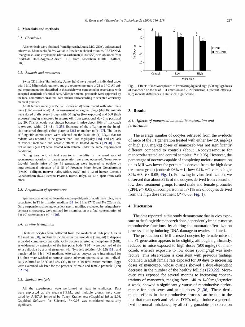
Fig. 1. Effects of in vivo exposure to low (50 mg/kg) and high (500 mg/kg) dosesof mancozeb on the % of PB1 emission and 2PN formation. Different letters (a,b, c) indicate differences in statistical significance.
3. Results
3.1. Effects of mancozeb on meiotic maturation andfertilization
The average number of oocytes retrieved from the oviductsof mice of the F1 generation treated with either low (50 mg/kg)or high (500 mg/kg) doses of mancozeb was not significantlydifferent compared to controls (about 16 oocytes/mouse formancozeb-treated and control samples;P > 0.05). However, thepercentage of oocytes capable of completing meiotic maturationup to MII was lower for germ cells derived from the high dosetreatment group (control: 96%± 1; low: 94%± 2 versus high:84%± 3, P < 0.05;Fig. 1). Following in vitro fertilization, weobserved that about 82% of the oocytes derived from control orlow dose treatment groups formed male and female pronuclei(2PN;P > 0.05), in comparison with 71%± 2 of oocytes derivedfrom the high dose treatment (P < 0.05;Fig. 1).
4. Discussion
The data reported in this study demonstrate that in vivo expo-sure to the fungicide mancozeb dose-dependently impairs mousereproductive functions, by altering the maturation/fertilizationprocess, and by inducing DNA damage to ovaries and uteri.
oft ntly,r an-c inef-f ingso asingd endentdo ncen-t icea rfor-m -m o thef eral-i etion
During treatment, clinical symptoms, mortality or increased ratepontaneous abortion in parent generation were not observed. Twenay-old female mice of the F1 generation were induced to ovulat
ntra-peritoneal injection of 5 IU of Pregnant Mare Serum GonadotrPMSG; Folligon, Intervet Italia, Milan, Italy) and 5 IU of human Corioonadotropin (hCG; Serono Pharma, Rome, Italy), 44–48 h apart fromther.
.3. Preparation of spermatozoa
Spermatozoa, obtained from the cauda epididymis of adult male miceapacitated in T6 fertilization medium[28] for 2 h at 37◦C and 5% CO2 in air.nly suspensions showing excellent sperm motility, evaluated by using p
ontrast microscopy, were utilized for insemination at a final concentrati× 104 spermatozoa ml−1 [29].
.4. In vitro fertilization
Ovulated oocytes were collected from the oviducts at 16 h post hC2 medium[30], and briefly incubated in hyaluronidase (1 mg/ml) to disp
xpanded cumulus-corona cells. Only oocytes arrested at metaphase IIs evidenced by extrusion of the first polar body (PB1), were deprivedona pellucida by a brief treatment with Tyrode’s solution (pH 2.5)[31], andransferred for 1 h in M2 medium. Afterwards, oocytes were inseminateh, then were washed to remove excess adherent spermatozoa, andally cultured at 37◦C and 5% CO2 in air in T6 fertilization medium. Eggere examined 6 h later for the presence of male and female pronucle
32–35].
.5. Statistic analysis
All the experiments were performed at least in triplicates. Dere expressed as the mean± S.E.M., and multiple groups were coared by ANOVA followed by Tukey–Kramer test (GraphPad InStat 2raphPad Software for Science).P < 0.05 was considered statistica
ignificant.
),
id-
)
The production of MII-arrested oocytes by female micehe F1 generation appears to be slightly, although significaeduced in mice exposed to high doses (500 mg/kg) of mozeb, whereas exposure to low doses (50 mg/kg) wasective. This observation is consistent with previous findbtained in adult female rats exposed for 30 days to increoses of mancozeb, whose ovaries showed a dose-depecrease in the number of the healthy follicles[20,22]. More-ver, rats exposed for several months to increasing corations of mancozeb, ranging from 140 to 1400 mg/kg/tw
week, showed a significantly worse of reproductive peance for both sexes and at all doses[21,36]. These detriental effects in the reproductive process can be due t
act that mancozeb and related DTCs might induce a genzed hormonal imbalance, by affecting gonadotropin secr
218 G. Rossi et al. / Reproductive Toxicology 21 (2006) 216–219
[37]. In our study, we found that oocytes from high-dose-treated mice showed a reduced ability to sustain the early stepsof fertilization, i.e. 2PN formation. The formation of pronu-cleate oocytes results from a complex series of ordered pro-cesses, both metabolic and regulatory[29]. Therefore, it canbe proposed that mancozeb exposure may interfere directlywith one or more of the steps leading to successful reproduc-tion.
Both at low and high doses of mancozeb the morpholog-ical features of the ovaries of treated mice were not signifi-cantly different from that of control animals (data not shown).Indeed, also the ovaries of rats treated for several days with500 mg/kg/day of mancozeb did not show significant morpho-logical changes as compared with control[19]. In conclusion,the toxicity of DTCs and EBDCs towards several animal speciesmakes it likely that they are involved in the induction of infer-tility, due to their ability to interfere with normal cell cycleprogression.
Acknowledgements
This study was granted by the Ministero della Salute (Pro-getto di Ricerca Finalizzato 1998) and by MIUR 60% to SCand GM. Thanks are due to Prof. Mauro Maccarrone (Univer-sity of Teramo, Italy) and Dott. Alberto Mantovani (IstitutoS em
R
andPer
enenneva,
ioureSao
oftam
J, erayerEnv-
,ozeb
epati
om-ina-05–
R,enic-01–
[ aacol
[11] Khera KS. Ethylenethiourea: teratogenicity study in rats and rabbits.Teratology 1973;7:243–52.
[12] Mishra VK, Srivastava MK, Raizada RB. Testicular toxicity in rat torepeated oral administration of tetramethylthiuram disulfide (Thiram).Indian J Exp Biol 1998;36:390–4.
[13] Pant N, Shankar R, Srivastava SP. Spermatotoxic effects of carbaryl inrats. Hum Exp Toxicol 1996;15:736–8.
[14] Pant N, Shankar R, Srivastava SP. In utero and lactational exposureof carbofuran to rats: effect on testes and sperm. Hum Exp Toxicol1997;16:267–72.
[15] Goldman JM, Parrish MB, Cooper RL, McElroy WK. Blockade inovulation in the rat by systemic and ovarian intrabursal administra-tion of the fungicide sodium dimethyldithiocarbamate. Reprod Toxicol1997;11:185–90.
[16] Rawlings NC, Cook SJ, Waldbillig D. Effects of the pesticides car-bofuran, chlorpyrifos, dimethoate, lindane, triallate, trifluralin, 2,4-D,and pentachlorophenol on the metabolic endocrine and reproductiveendocrine system in ewes. J Toxicol Environ Health 1998;54:21–36.
[17] Stoker TE, Goldman JM, Cooper RL. Delayed ovulation and pregnancyoutcome: effect of environmental toxicants on the neuroendocrine controlof the ovary (1). Environ Toxicol Pharmacol 2001;9:117–29.
[18] Edwards IR, Ferry DG, Temple WA. Thio-dithiocarbamates. In: KamrinMA, editor. Pesticides profiles of toxicity, environmental impact andfate. Boca Raton, New York: CRC Lewis Publishers; 1997. p. 91–132.
[19] Mahadevaswami MP, Jadaramkunti UC, Hiremath MB, Kaliwal BB.Effect of mancozeb on ovarian compensatory hypertrophy and bio-chemical constituents in hemicastred Albino rat. Reprod Toxicol2000;14:127–34.
[20] Baligar PN, Kaliwal BB. Induction of gonadal toxicity to female:235–
[ toealth
[ ndBasic
[ gi-
[ rs.4. p.
[ CS
[ thencol
[ sidues
[ cytesFertil
[ rtil-
[ ymalheri-
[ cal-sma
[ entprod
[ plas-ing in
uperiore di Sanita, Rome, Italy) for critical reading of thanuscript.
eferences
[1] Houeto P, Bindoula G, Hoffman JR. Ethylenebisdithiocarbamatesethylenethiourea: possible human health hazards. Environ Healthspect 1995;103:568–73.
[2] WHO. Dithiocarbamates pesticides ethylenethiourea, and propylthiourea: a general introduction. Environment Health Criteria 78 GeWorld Health Organization 1988: p. 17–102.
[3] Mestres R, Mestres G. Ethylenebisdithiocarbamates and ethylenethresidue in food. In: Program of the Sixth Congress of ToxicologyPaulo, Brazil, 1989.
[4] Lentza-Rizos C. Ethylenethiourea (ETU) in relation to useethylenebisdithiocarbamate (EBCD) fungicide. Rev Environ ConToxicol 1990;115:1–37.
[5] Steenland K, Cedillo L, Tucker J, Hines C, Sorensen K, Deddensal. Thyroid hormones and cytogenetic outcomes in backpack spusing ethylenebis(dithiocarbamate) (EBDC) fungicides in Mexico.iron Health Perspect 1997;105:1126–30.
[6] Colosio C, Barcellini W, Maroni M, Alcini D, Bersani M, Cavallo Det al. Immunomodulatory effects of occupational exposure to mancArch Environ Health 1996;51:445–51.
[7] Lewerenz HJ, Plass R. Contrasting effects of ethylenethiourea on hmonooxygenases in rats and mice. Arch Toxicol 1984;56:92–5.
[8] Chhabra RS, Eustis S, Haseman JK, Kurtz PJ, Carlton BD. Cparative carcinogenicity of ethylenethiourea with or without pertal exposure in rats and mice. Fundam Appl Toxicol 1992;18:417.
[9] Innes JR, Ulland BM, Valerio MG, Petrucelli L, Fishbein L, Hart Eet al. Bioassay of pesticides and industrial chemicals for tumoregity in mice: a preliminary note. J Natl Cancer Inst 1969;42:1114.
10] Frakes RA. Drinking water guideline for ethylenethiourea,metabolite of ethylene bisdithiocarbamate. Regul Toxicol Pharm1988;8:207–18.
-
-
a
ts
.
c
rats after chronic exposure to mancozeb. Ind Health 2001;3943.
21] Baligar PN, Kaliwal BB. Reproductive toxicity of carbofuranthe female mice: effects on estrous cycle and follicles. Ind H2002;40:345–52.
22] Baligar PN, Kaliwal BB. Morphometric analysis of follicular growth abiochemical constituents in albino rats exposed to mancozeb. JClin Physiol Pharmacol 2004;15:241–62.
23] Bindali BB, Kaliwal BB. Anti-implantation effect of a carbamate funcide mancozeb in albino mice. Ind Health 2002;40:191–7.
24] Yanagimachi R. Mammalian fertilization. In: Knobil E, Neill JD, editoThe physiology of reproduction. New York: Raven Press Ltd.; 199189–317.
25] Kocialski A. Evaluation of some pesticide residues in food. IPINCHEM AGP:1970/M/12/1 WHO/FOOD ADD/71.42, 1970.
26] Shukla Y, Arora A. Transplacental carcinogenic potential ofcarbamate fungicide mancozeb. J Environ Pathol Toxicol O2001;20:127–31.
27] 1993 Fao/Who Joint Meeting On Pesticide Residues. Pesticide rein food. Geneva, 20–29 September 1993; p. 89–101.
28] Quinn P, Barros C, Whittingham DG. Preservation of hamster ooto assay the fertilizing capacity of human spermatozoa. J Reprod1982;66:161–8.
29] Kupker W, Diedrich K, Edwards RG. Principles of mammalian feization. Hum Reprod 1998;3:20–32.
30] Sato M, Ishikawa A. Room temperature storage of mouse epididspermatozoa: exploration of factors affecting sperm survival. Togenology 2004;61:1455–69.
31] Nicolson GL, Yanagimachi R, Yanagimachi H. Ultrastructural loization of lectin binding sites on the zonae pellucidae and plamembranes of mammalian eggs. J Cell Biol 1975;66:263–74.
32] Cecconi S, D’Aurizio R, Colonna R. Role of antral follicle developmand cumulus cells on in vitro fertilization of mouse oocytes. J ReFertil 1996;107:207–14.
33] Redkar AA, Olds-Clarke PJ. An improved mouse sperm-oocytemalemma binding assay: studies on characteristics of sperm bindmedium with or without glucose. J Androl 1999;20:500–8.

G. Rossi et al. / Reproductive Toxicology 21 (2006) 216–219 219
[34] Cohen DJ, Ellerman DA, Cuasnicu PS. Mammalian sperm-egg fusion:evidence that epididymal protein DE plays a role in mouse gametefusion. Biol Reprod 2000;63:462–8.
[35] Hamatani T, Tanabe K, Kamei K, Sakai N, Yamamoto Y, Yoshimura Y.A monoclonal antibody to human SP-10 inhibits in vitro the binding ofhuman sperm to hamster oolemma but not human Zona pellucida. BiolReprod 2000;62:1201–8.
[36] Ivanova-Chemishanska L, Valcheva V, Takeva TZ. Effect of chronicperoral poisoning with perozine (zineb) on the reproduction of whiterats. Acta Med Soc 1973;1:107–8.
[37] Stoker TE, Jeffay SC, Zucker RM, Cooper RL, Perreault SD.Abnormal fertilization is responsible for reduced fecundity follow-ing thiram-induced ovulatory delay in the rat. Biol Reprod 2003;68:2142–9.


![Heat exposure impairs porcine oocyte quality with ... · zona pellucida to contact oocyte with gap junction (GJ) structures distributed on the oolemma [1]. Through TZPs, CCs provide](https://img.pdfslide.us/doc/110x75/5f94c6b4c999425a3d10052a/heat-exposure-impairs-porcine-oocyte-quality-with-zona-pellucida-to-contact.jpg)