Embed Size (px)
Citation preview

Management Of Venous Thromboembolism
Khaled O. Hadeli, MD
Pulmonary and critical care
12/16/99

Introduction
Complex vascular syndrome Multifactorial pathogenesis Wide spectrum 1 in 1,000 people affected 50,000 deaths/year in the USA
Nordstrom et.al, J.IM:232;155-160

Predisposing Factors
Race, age, genetics Thrombophilia Immobility Surgery Trauma Pregnancy and child birth Carcinoma
Clagett,et.al, 1995 Chest 108:312s-334s

30% POST T HROM BOTIC SYNDROM
M ASSIVE PE CLINICALY SEGNIFICANT
30% SYM PT OM ATIC PE
CHRONIC PEPHT N
COR PULM
30% ASYM PTOM ATIC PE 40% NO PE
20% PROXIM AL DVT 80% RESOLVE
CALF VEINS
VTE

Case
68 yo male with acute chest pain R/O (AMI), & R/I PE by spiral CT sent home on Warfarin 1 months later came back with GI bleed
» Htc 45 to 28 Review of the spiral CT, new CT and
leg US NEGATIVE

Cases
62 yo male with acute SOB and chest pain R/O PE by spirat CT
6 weeks later some SOB and no chest pain
Review of CT again show large PE
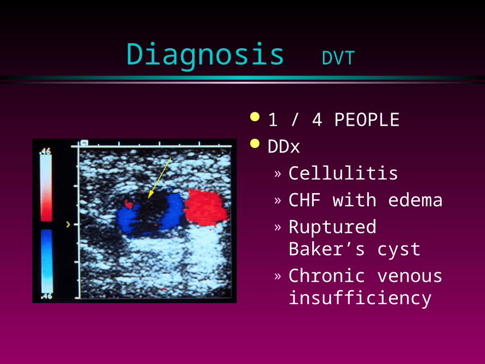
Diagnosis DVT
1 / 4 PEOPLE DDx
» Cellulitis» CHF with edema» Ruptured Baker’s
cyst» Chronic venous
insufficiency
Ginsberg. NEJM 1996

Pulmonary Embolism
Dyspnea, Pleuritic CP, Hemoptysis
Hemodynamic instability & Syncope
mimic indolent Pneumonia, CHF, COPD


Ginsberg. NEJM 1996


5 Reasons why CT is replacing VQ Scan
1. Dr Stern wants to be the next Faculty to own a “Porsche”
2. VQ Scan “Sucks”
3. CT Scan is more available
4. CT Scan is more reliable
5. CT Scan is cheaper
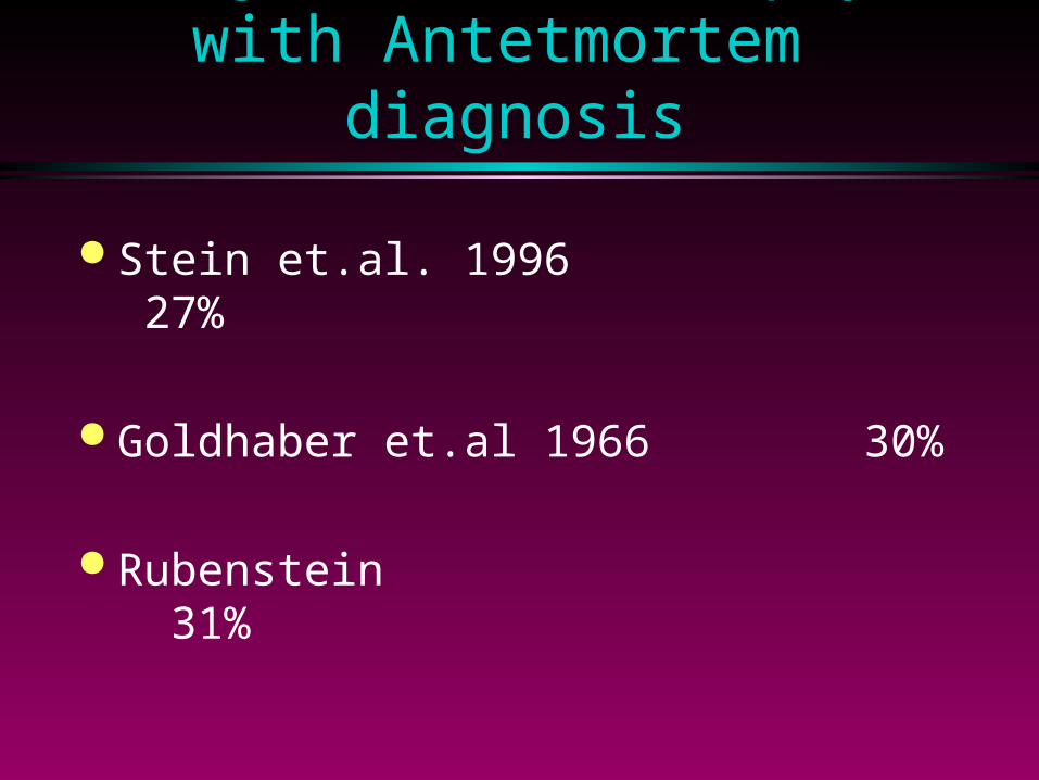
Major PE at Autopsy with Antetmortem diagnosis
Stein et.al. 1996 27%
Goldhaber et.al 1966 30%
Rubenstein 31%
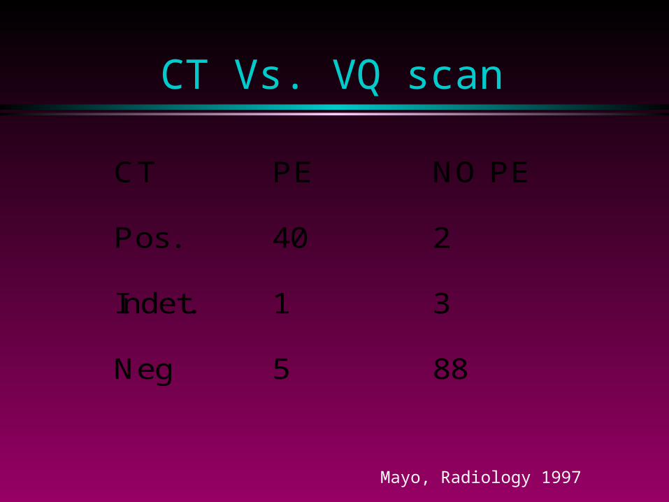
CT Vs. VQ scan
CT PE NO PE
Pos. 40 2
Indet. 1 3
Neg 5 88
Mayo, Radiology 1997

N=164 patients wit Indeterminate VQ and negative leg US
If CT is negative NO Angio, NO Rx 3 month F/U show 2.8% recurrence, and
1 patient died of PE
Ferretti, Radiology 1997

Spiral CT Scan


Case
58 yo male R/I PE by spiral CT 2 months later came back with acute
SOB INR = 2 VQ scan show high probability for PE
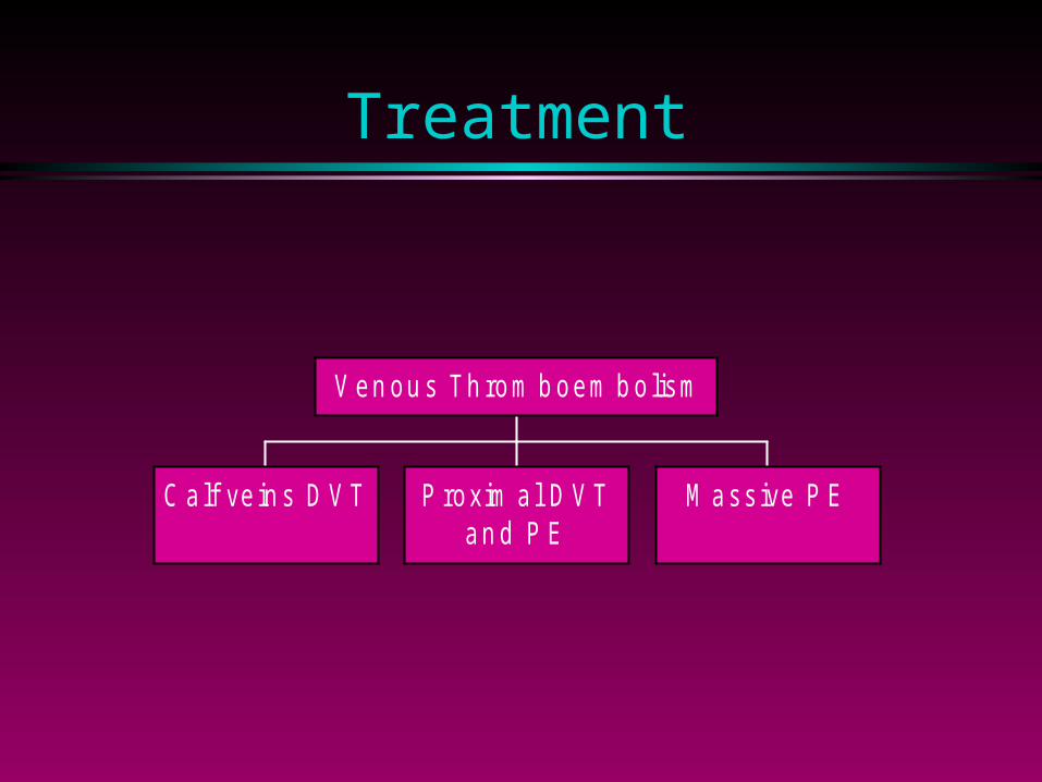
Treatment
C alf ve in s D V T P roxim a l D V Tan d P E
M ass ive P E
V en ou s Th rom b oem b o lism
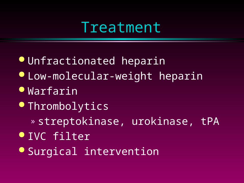
Treatment
Unfractionated heparin Low-molecular-weight heparin Warfarin Thrombolytics
» streptokinase, urokinase, tPA IVC filter Surgical intervention

Unfractionated Heparin
Start heparin before testing High starting dose 0.2 - 0.4 U/ml protamine titration assay Sub therapeutic levels lead to 15X risk
of recurrence


Heparin Resistance
High dose of heparin >40,000U/day High levels of heparin binding proteins High levels of factor Vlll APTT/plasma heparin dissociation
measure plasma heparin levels or change to LMWH
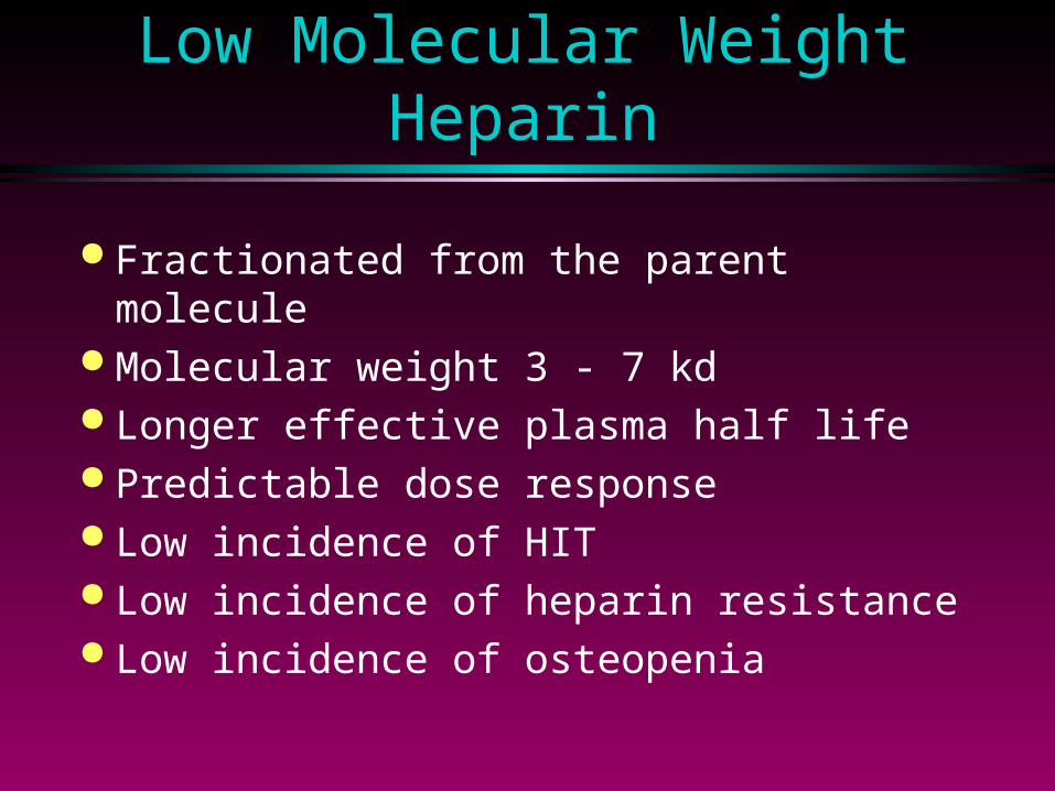
Low Molecular Weight Heparin
Fractionated from the parent molecule Molecular weight 3 - 7 kd Longer effective plasma half life Predictable dose response Low incidence of HIT Low incidence of heparin resistance Low incidence of osteopenia
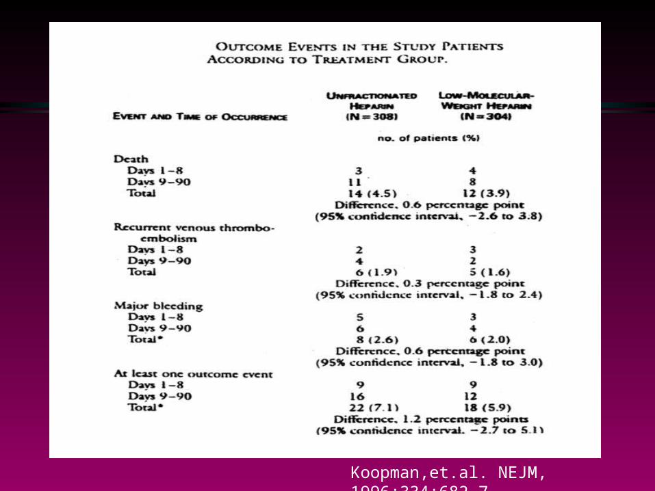
Koopman,et.al. NEJM, 1996;334:682-7

Oral Anticoagulants
Warfarin 1st - 3rd day of therapy 5mg q day INR 2.5 - 3.5 Teratogenic Drug interaction:
» medications, diet, alcohol, and illness

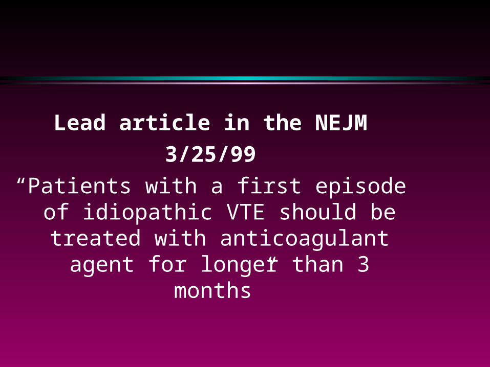
Lead article in the NEJM
3/25/99
“Patients with a first episode of idiopathic VTE should be treated with
anticoagulant agent for longer than 3 months”

Case
38 yo Female on Warfarin for DVT Acute chest pain and SOB +ve Urine pregnancy test

VTE In Pregnancy
Increased risk; late pregnancy and early post partum
Leg studies are not reliable VQ scan is safe Pulmonary angio can be done Warfarin is contraindicated

Thrombophilia
Inherited recurrent VTE» Hyperhomocysteinemia» Protein C deficiency» Protein S deficiency» Antithrombin deficiency» Activated protein C resistance


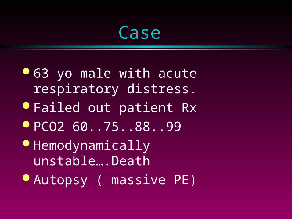
Case
63 yo male with acute respiratory distress.
Failed out patient Rx PCO2 60..75..88..99 Hemodynamically unstable….Death Autopsy ( massive PE)

Massive PE
PE that leads to acute right ventricular failure
Death within 1st 2 hrs Occlusion of 75% of the pulmonary bed Echo findings: Rt Vent dysfunction and
enlargement, TR, increase pulmonary artery size, Rt sided thrombus (18%)
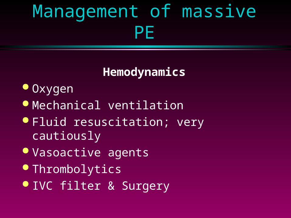
Management of massive PE
Hemodynamics Oxygen Mechanical ventilation Fluid resuscitation; very cautiously Vasoactive agents Thrombolytics IVC filter & Surgery

Thrombolytics

Alpert et.al Arch.1997:157,2550-56

Alpert et.al Arch.1997:157,2550-56

IVC Filter
Surgical» slit like channel, serrated miles, Adam
and De Weese device Percutaneous
» Greenfield» Vena -Tech» Bird’s nest » Simon - Nitinol

No difference in 2 year survival Indications
» contraindication to anticoagulation» failure of anticoagulation» compromised pulmonary vascular bed
Complication» filter migration, erosion, or obstruction
Backer, et.al Arch. 1992: 152:1985-94
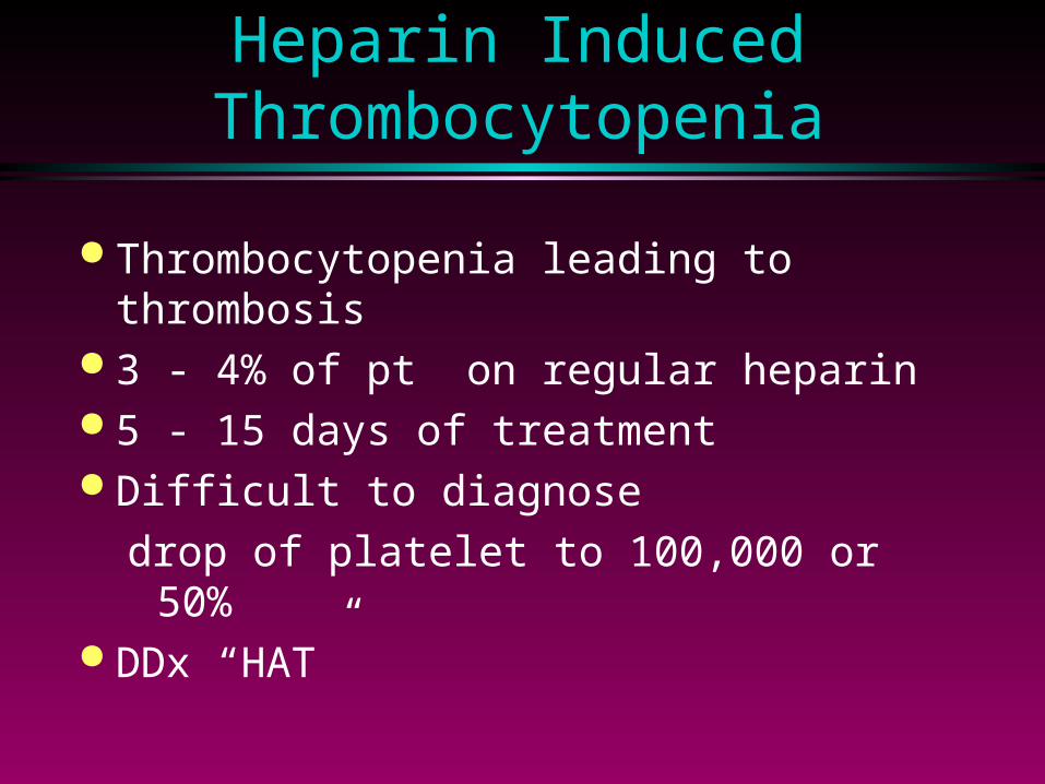
Heparin Induced Thrombocytopenia
Thrombocytopenia leading to thrombosis
3 - 4% of pt on regular heparin 5 - 15 days of treatment Difficult to diagnose
drop of platelet to 100,000 or 50% DDx “HAT”

Heparin Induced Osteoporosis
Associated with prolonged use Partially reversible Dexa scan Calcium and vitamin D questionable
efficacy
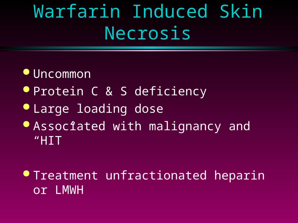
Warfarin Induced Skin Necrosis
Uncommon Protein C & S deficiency Large loading dose Associated with malignancy and “HIT”
Treatment unfractionated heparin or LMWH

VTE in Malignancy
Increased risk Idiopathic recurrent VTE may be due to
occult malignancy Do not respond to Warfarin, need to use
heparin LMWH

Thrombosis and Pulmonary Embolism are today no longer dreaded either by
patients or by physicians, although only few years ago we where still completely
powerless to combat them.
Harry Zilliacus
1946