Embed Size (px)
Citation preview

Management of lymph nodes in the setting of differentiated thyroid cancer
Julie Ann Sosa, MDProfessor of Surgery and Medicine (Oncology)
Chief, Section of Endocrine SurgeryLeader, Endocrine Neoplasia Diseases Group
Duke Cancer Institute and Duke Clinical Research InstituteDurham, NC
Disclosure
Member and ATA representative, Medullary Thyroid Cancer Registry Data Monitoring Committee funded by GlaxoSmithKline, Novo Nordisk, Astra Zeneca, and
Eli Lilly

Maxim
• Routine lymph node dissection that includes the central and lateral neck compartments has shown nodal metastases in up to 80% of patients with differentiated thyroid cancer.
• The incidence of clinically significant lymph nodes is only 25%.

Patterns of lymph node metastases
• 119 patients bilateral neck dissection• Excluded microcarcinomas• Mean tumor size: 3.1cm• 21% had clinically involved lymph nodes
• Cervical lymph node metastases identified in 61%• Pattern of medial to lateral to contralateral
Mirallie E. Localization of Cervical node metastases of papillary thyroid cancer. World J Surg 1999
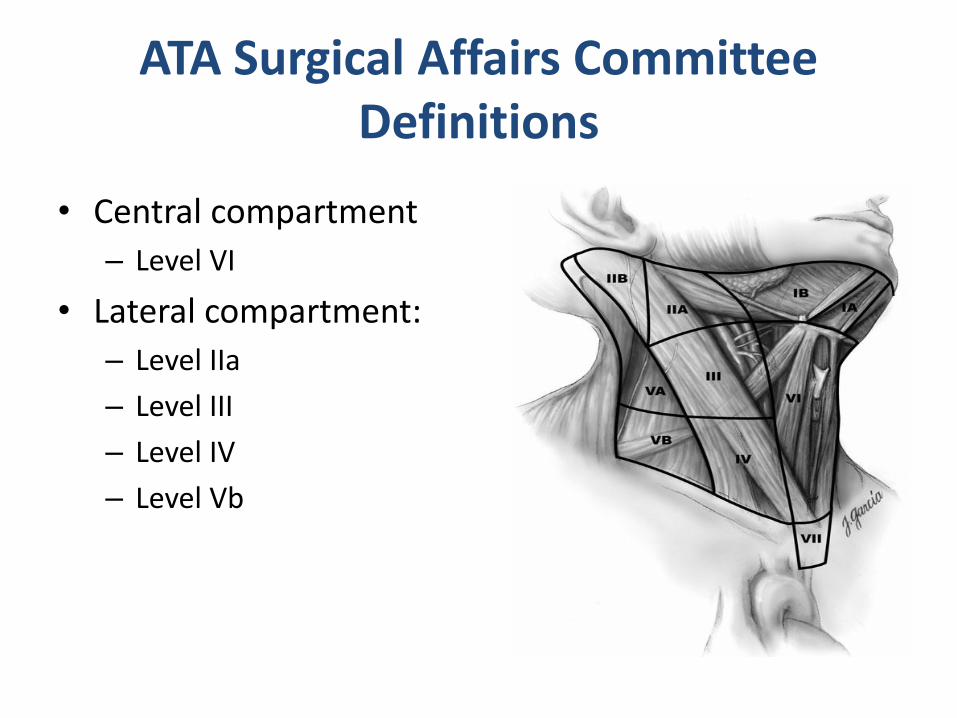
ATA Surgical Affairs Committee Definitions
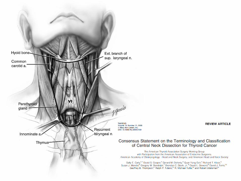
• Central compartment
– Level VI
• Lateral compartment:
– Level IIa
– Level III
– Level IV
– Level Vb

Skip metastases• Lateral compartment metastases without involvement of
central compartment• Most frequently from upper pole tumors• 5-20% of tumors
Fritze D, Surgical management of cervical lymph nodes in differentiated thyroid cancer. Otolaryngol Clin N Am 2010

Risk factors for lymph node mets• Age <45• Tumor Size• Extrathyroidal extension
• Lymphovascular invasion• BRAF mutation• Distant metastases
Ito Y, Clinical significance of lymph node metastases of thyroid papillary carcinoma located in one lobe. World J Surg2006

The first operation is the most important operation.
• Preoperative ultrasound (or other modalities if indicated) of the central and lateral neck compartments (with FNA biopsy when indicated) should be performed prior to initial surgery in the setting of a (+) thyroid biopsy.
• Complete compartmental resection should follow.

Do it right the first time!
Up to 57% of re-operations for thyroid cancer are preventable and a direct result of incomplete initial surgery.
Kouvaraki M. Preventable reoperations for persistent and recurrent papillary thyroid cancer. Surgery 2004.
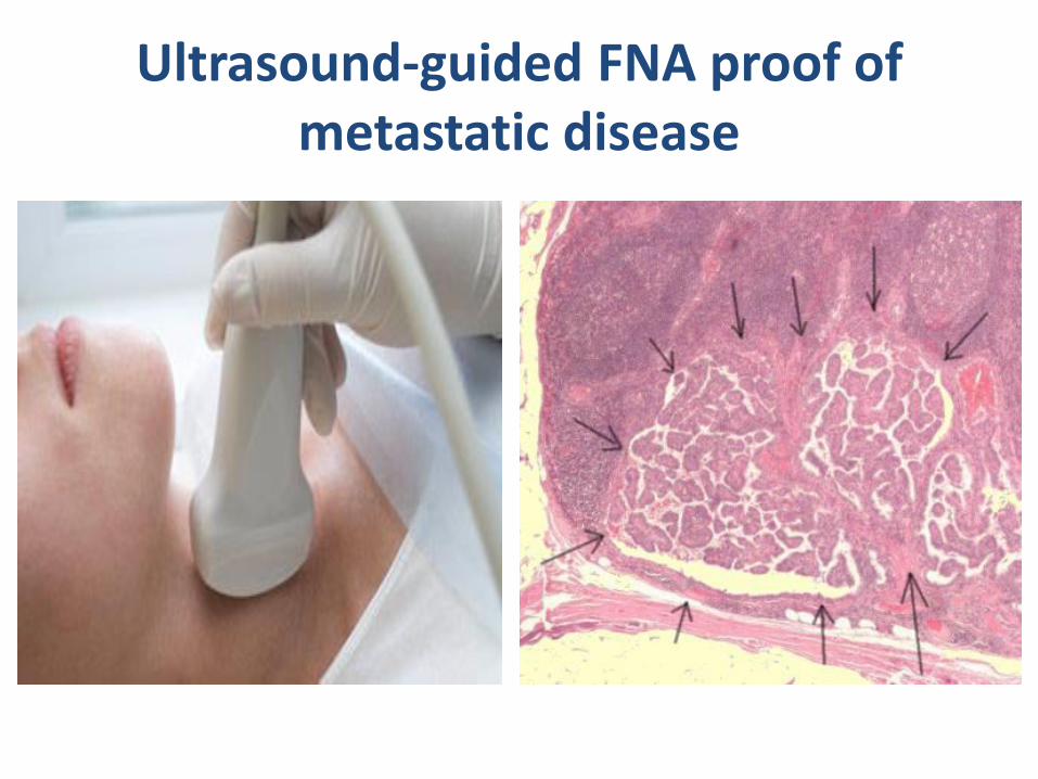
Ultrasound-guided FNA proof of metastatic disease

UltrasoundNormal lymph Node
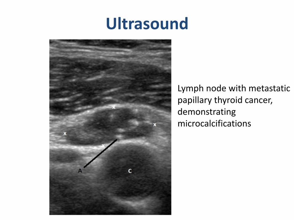
Ultrasound
Lymph node with metastaticpapillary thyroid cancer, demonstrating microcalcifications
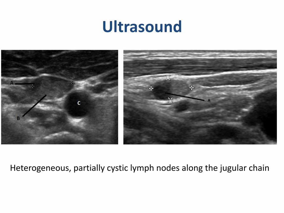
Ultrasound
Heterogeneous, partially cystic lymph nodes along the jugular chain

CT with IV contrast

The role of cross-sectional imaging as an adjunct is increasing.
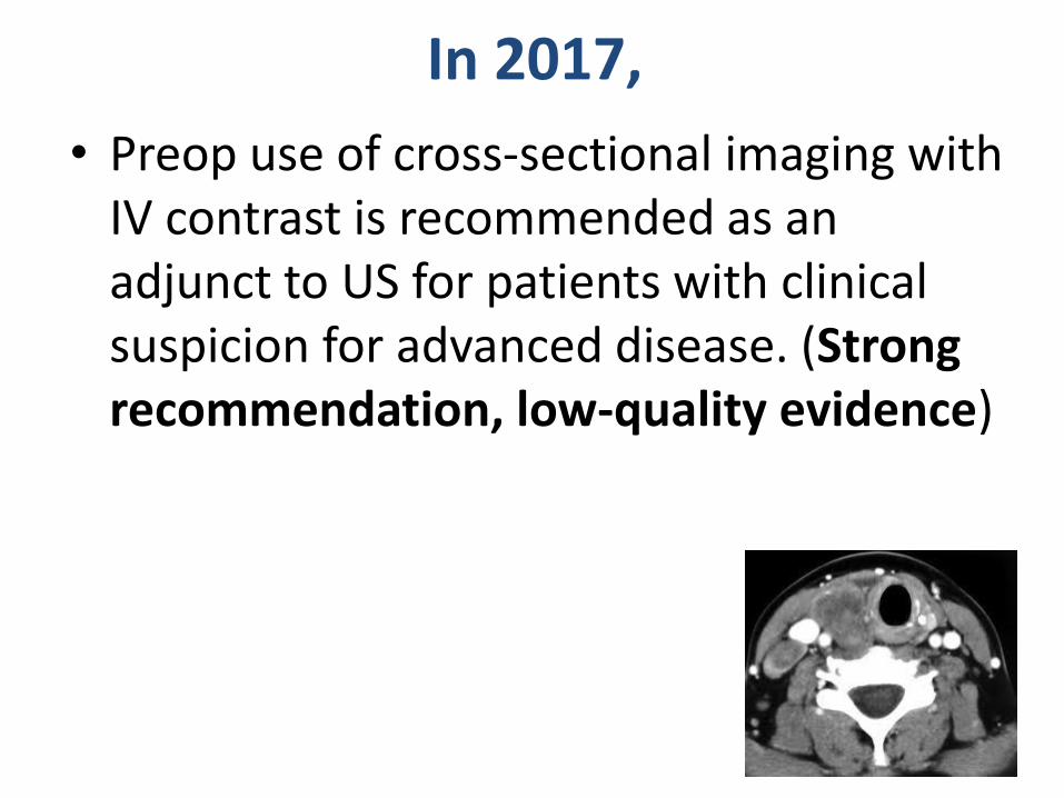
In 2017,
• Preop use of cross-sectional imaging with IV contrast is recommended as an adjunct to US for patients with clinical suspicion for advanced disease. (Strong recommendation, low-quality evidence)
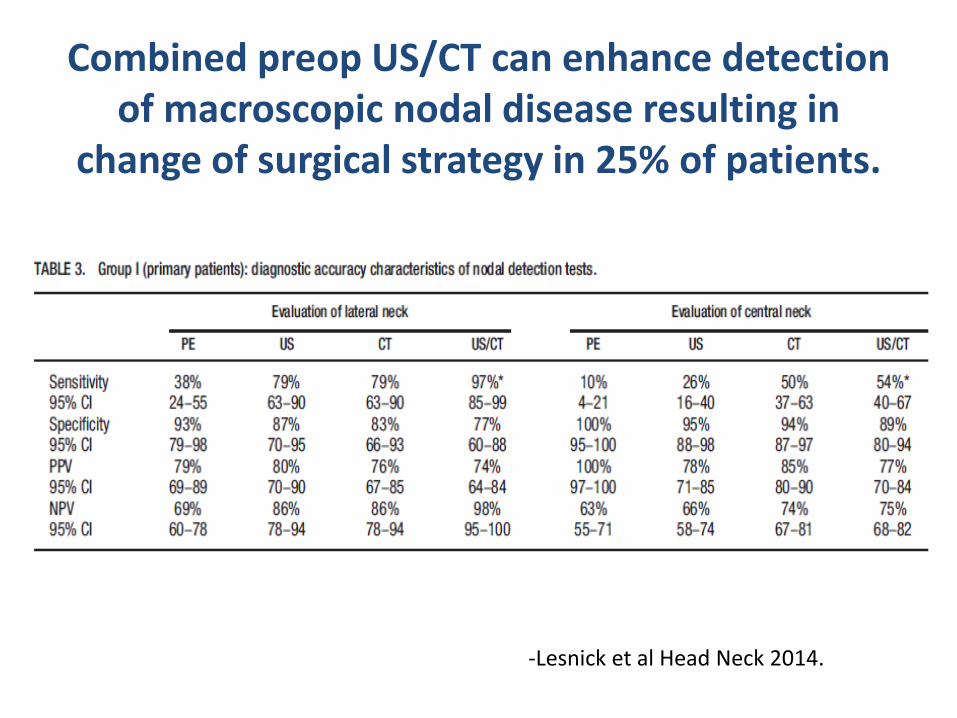
Combined preop US/CT can enhance detection of macroscopic nodal disease resulting in
change of surgical strategy in 25% of patients.
-Lesnick et al Head Neck 2014.

Cross sectional imaging can clarify involvement of:
• Nodal regions difficult to visualize on routine US, including the mediastinum, infraclavicular, retropharyngeal and para-pharyngeal regions.
• Larynx, trachea, esophagus, or blood vessels
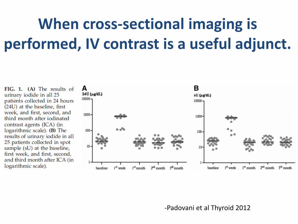
When cross-sectional imaging is performed, IV contrast is a useful adjunct.
-Padovani et al Thyroid 2012
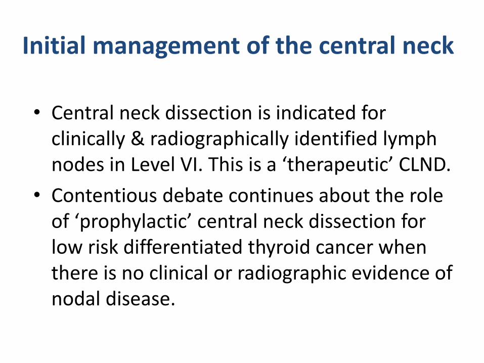
Initial management of the central neck
• Central neck dissection is indicated for clinically & radiographically identified lymph nodes in Level VI. This is a ‘therapeutic’ CLND.
• Contentious debate continues about the role of ‘prophylactic’ central neck dissection for low risk differentiated thyroid cancer when there is no clinical or radiographic evidence of nodal disease.

Central lymph node dissection
• Benefits• Decreases recurrence• Prevents compression/invasion
of local structures• ? improves survival
• Possible complications (transient and permanent?)• RLN injury• SLN injury• Hypoparathyroidism• Bleeding• Lymphatic leak

Is prophylactic central lymph node dissection warranted?
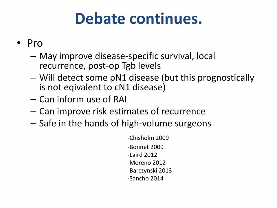
Debate continues.
• Pro– May improve disease-specific survival, local
recurrence, post-op Tgb levels– Will detect some pN1 disease (but this prognostically
is not eqivalent to cN1 disease)– Can inform use of RAI– Can improve risk estimates of recurrence– Safe in the hands of high-volume surgeons
-Chisholm 2009
-Bonnet 2009-Laird 2012-Moreno 2012-Barczynski 2013-Sancho 2014
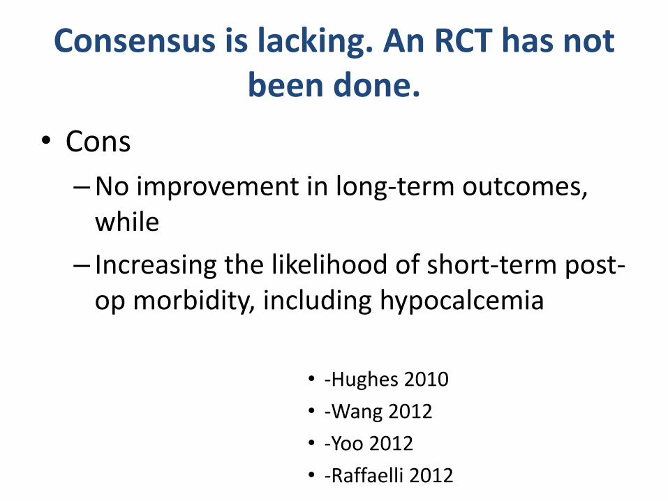
Consensus is lacking. An RCT has not been done.
• Cons
–No improvement in long-term outcomes, while
– Increasing the likelihood of short-term post-op morbidity, including hypocalcemia
• -Hughes 2010
• -Wang 2012
• -Yoo 2012
• -Raffaelli 2012

Locoregional recurrence in 6 comparative studies
Recurrence rate of TT: 7.9% Recurrence rate of TT/pCCND: 4.7% Relative risk of recurrent PTC after TT/pCCND: 0.59 (NS) Number needed to treat to prevent one recurrence: 31
(Wang et al)

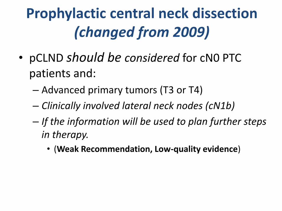
Prophylactic central neck dissection(changed from 2009)
• pCLND should be considered for cN0 PTC patients and:
– Advanced primary tumors (T3 or T4)
– Clinically involved lateral neck nodes (cN1b)
– If the information will be used to plan further steps in therapy.
• (Weak Recommendation, Low-quality evidence)

Prophylactic central neck dissection(unchanged from 2009)
• Thyroidectomy w/o pCLND may be appropriate for patients who are cN0 with:
– Small (T1 or T2) PTCs,
– Non-invasive PTCs, and
– Most follicular cancer.
(Strong Recommendation, Moderate-quality evidence)

Prophylactic lateral dissection is not indicated for DTC.

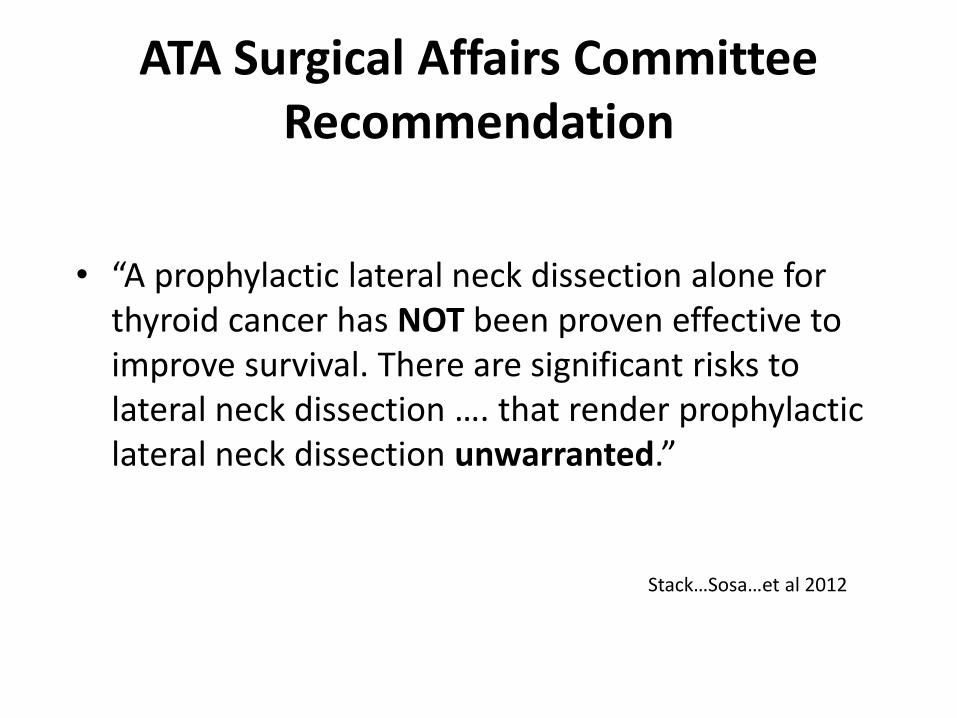
ATA Surgical Affairs Committee Recommendation
• “A prophylactic lateral neck dissection alone for thyroid cancer has NOT been proven effective to improve survival. There are significant risks to lateral neck dissection …. that render prophylactic lateral neck dissection unwarranted.”
Stack…Sosa…et al 2012

ATA RECOMMENDATION 37
• Therapeutic lateral neck compartmental lymph node dissection should be performed for patients with biopsy-proven metastatic lateral cervical lymphadenopathy.
(Strong recommendation, Moderate-quality evidence)
ATA guidelines 2015
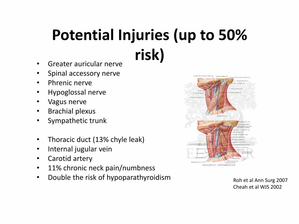
Potential Injuries (up to 50% risk)
• Greater auricular nerve• Spinal accessory nerve• Phrenic nerve• Hypoglossal nerve• Vagus nerve• Brachial plexus• Sympathetic trunk
• Thoracic duct (13% chyle leak)• Internal jugular vein• Carotid artery• 11% chronic neck pain/numbness• Double the risk of hypoparathyroidism Roh et al Ann Surg 2007
Cheah et al WJS 2002

Important tenets
• A prophylactic lateral neck dissection has not been proven effective to improve survival!
• The reported rate of recurrence in the lateral neck is just 5-15%.– Treating the neck when metastases become apparent
does not affect overall outcome.
• Preoperative US is an excellent outcome predictor for DFS and DSS. It can find high-risk disease without a scalpel!
• A sonographically-based surgical approach provides excellent long-term regional control.
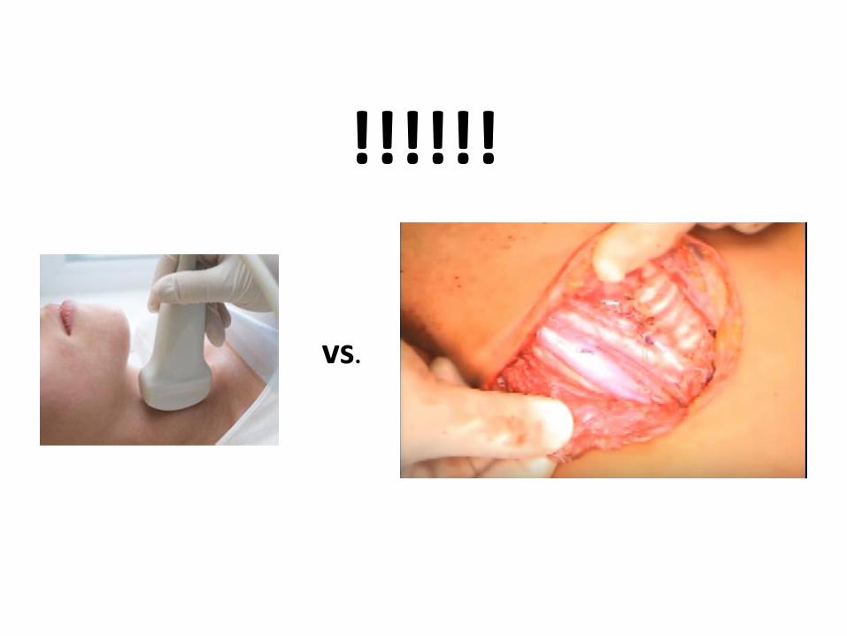
!!!!!!
vvs.
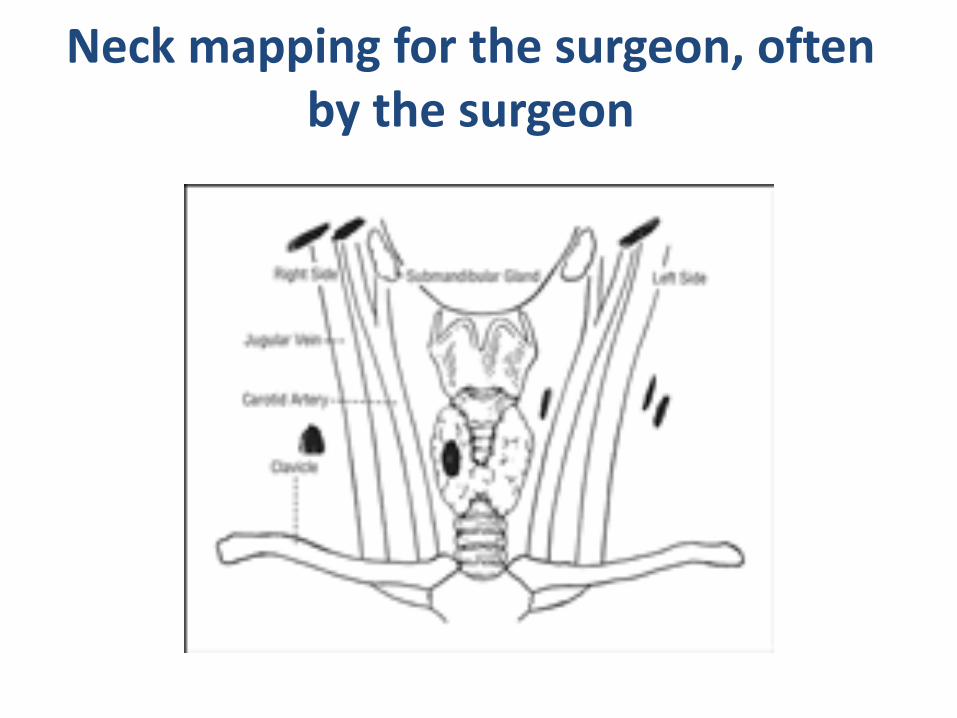
Neck mapping for the surgeon, often by the surgeon

Kaplan-Meier estimate for lateral neck disease-free interval, stratified by sonographic status of the ipsilateral neck
Kaplan-Meier estimate for disease-specific survival, stratified by presence of sonographic abnormalities in the lateral neck compartments
Kaplan-Meier estimate for recurrence-free survival (years torecurrence or death for any reason), stratified by s onographic status of the lateral neck compartments
Moreno et al, Arch OTO 2011
What is the importance of lymph nodes in PTC?
ymph nodes?

Zaydfudim et al. 2008:1. 15,497 PTC patients from SEER
2. Lymph node metastases:
≥45 yrs: associated with survival (HR 1.46, p<0.01)
<45 yrs: NOT associated with survival (HR 1.11, p=0.54) Zaydfudim V, et al. Surgery 2008

Tran Cao et al. 2012: 1. 49,240 pts with DTC in SEER
2. For pts <45 yrs: Lymph node metastases were associated with compromised survival (HR 2.09, p<0.01)
3. Potential limitations:
Extent of surgery
Radioactive iodine therapyTran Cao, et al. Surgery 2012


Hypotheses
1. Presence of lymph node metastases and number of metastatic lymph nodes are associated with compromised survival for patients <45 yrs with Stage I PTC.
2. It is possible to stratify patients’ risk of death based on the number of metastatic lymph nodes.
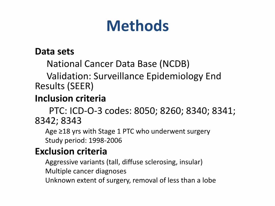
Methods
Data setsNational Cancer Data Base (NCDB)Validation: Surveillance Epidemiology End
Results (SEER) Inclusion criteria
PTC: ICD-O-3 codes: 8050; 8260; 8340; 8341; 8342; 8343
Age ≥18 yrs with Stage 1 PTC who underwent surgeryStudy period: 1998-2006
Exclusion criteriaAggressive variants (tall, diffuse sclerosing, insular)Multiple cancer diagnosesUnknown extent of surgery, removal of less than a lobe
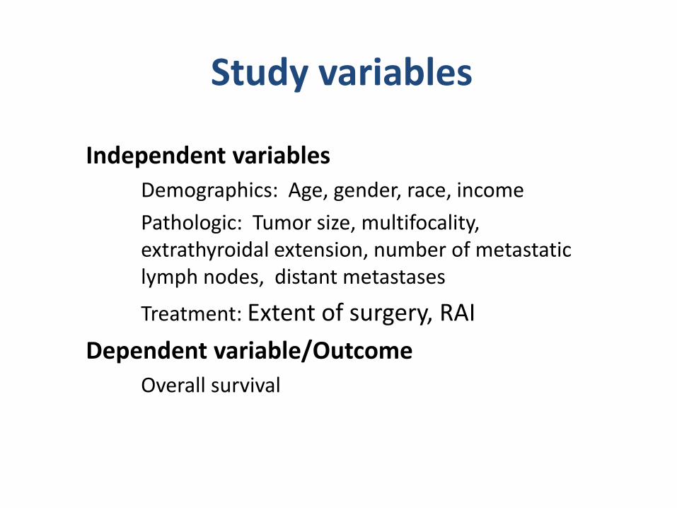
Study variables
Independent variables
Demographics: Age, gender, race, income
Pathologic: Tumor size, multifocality, extrathyroidal extension, number of metastatic lymph nodes, distant metastases
Treatment: Extent of surgery, RAI
Dependent variable/Outcome
Overall survival
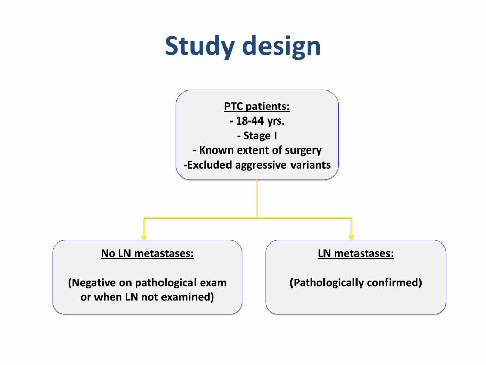
Study design
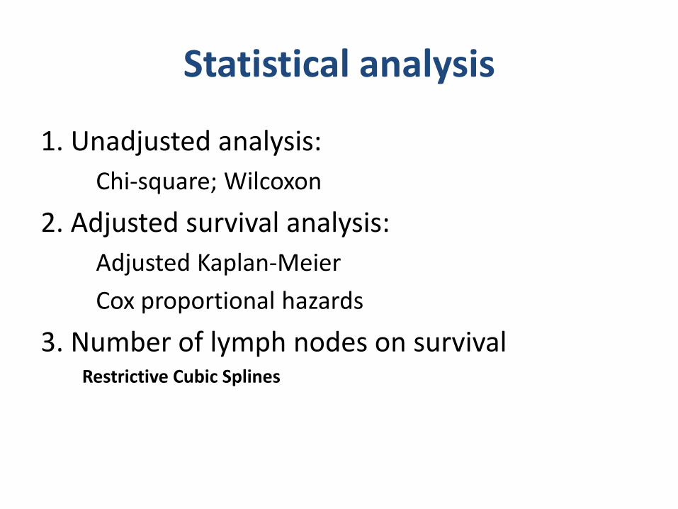
Statistical analysis
1. Unadjusted analysis:
Chi-square; Wilcoxon
2. Adjusted survival analysis:
Adjusted Kaplan-Meier
Cox proportional hazards
3. Number of lymph nodes on survival Restrictive Cubic Splines
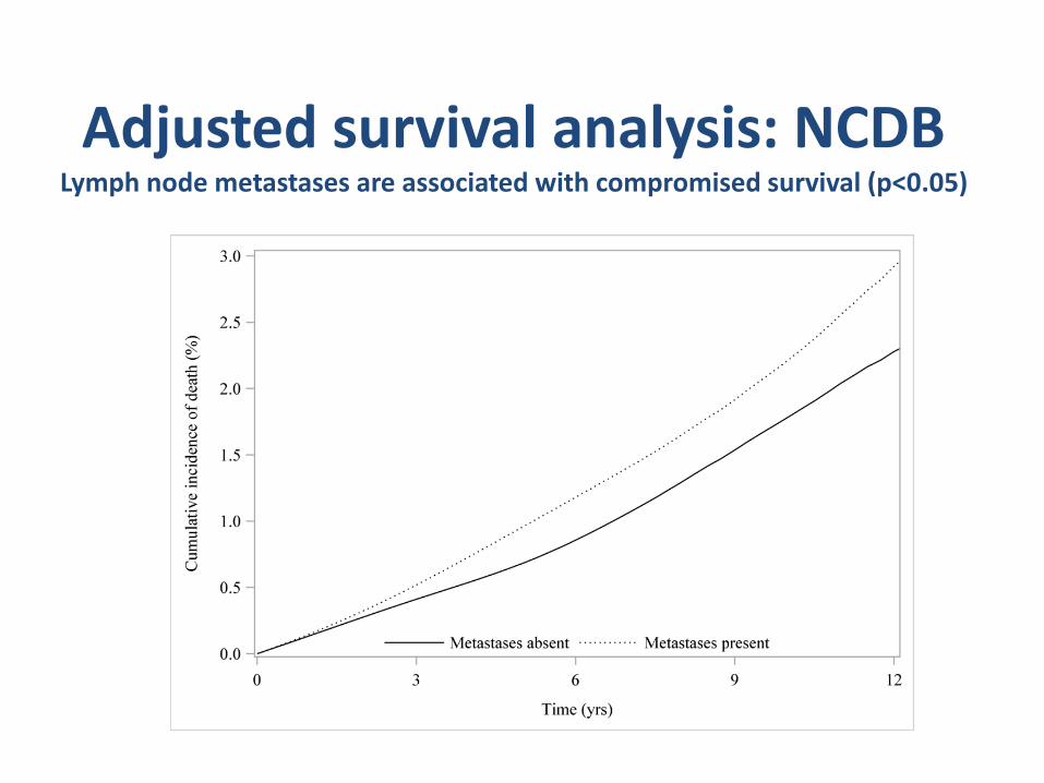
Adjusted survival analysis: NCDBLymph node metastases are associated with compromised survival (p<0.05)

Adjusted survival analysis: SEERLymph node metastases are associated with compromised survival (p<0.05)
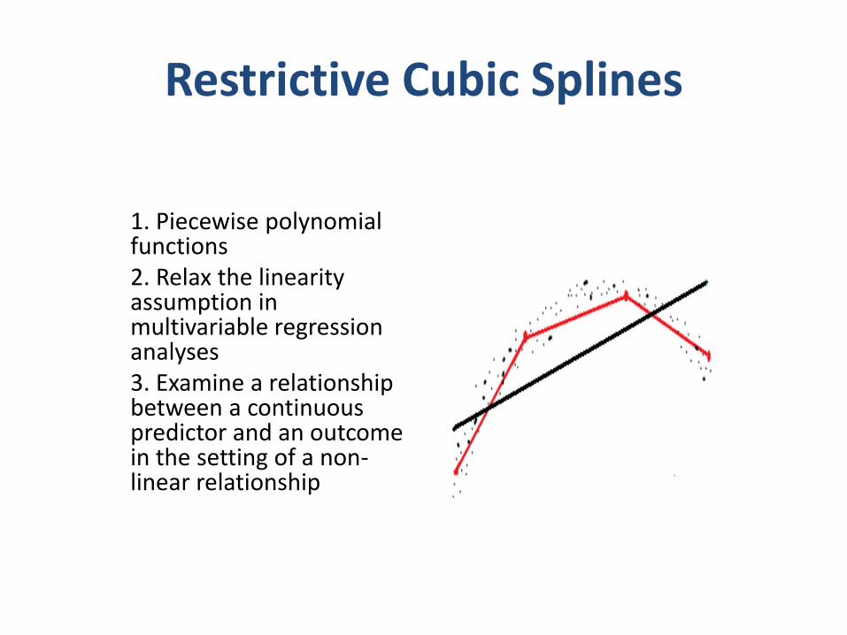
Restrictive Cubic Splines
1. Piecewise polynomial functions 2. Relax the linearity assumption in multivariable regression analyses3. Examine a relationship between a continuous predictor and an outcome in the setting of a non-linear relationship
Stone C, Koo C: Additive Splines in Statistics, American Statistical Association 1986

Metastatic lymph nodes are associated with survival.
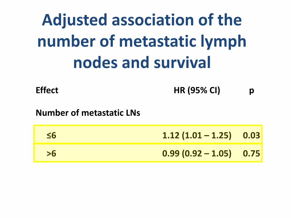
Adjusted association of the number of metastatic lymph
nodes and survival
Effect HR (95% CI) p
Number of metastatic LNs
≤6 1.12 (1.01 – 1.25) 0.03
>6 0.99 (0.92 – 1.05) 0.75

Implications
1. Cervical lymph node metastases are associated with compromised survival among patients <45 yrs.
2. In the current AJCC staging, young patients with nodal metastases may be under-staged.
3. Rigorous preoperative screening for lymph node metastases is warranted for patients <45 yrs.

Cancer-specific survival estimate by patient age

Linear association between patient age and risk of death
Restricted cubic splines Thin plate splines Adaptive smoothers

Conclusions and implications
• Age is associated with an increased risk of disease-specific mortality in a continuous, linear fashion, without an apparent age cut-point demarcating survival difference.
• These results challenge the appropriateness of using a patient age cut-point in current (<45 yrs) and imminent (<55 yrs) AJCC staging systems.
• We might be under-staging young patients with intermediate- and high-risk tumors, and they might be under-treated, as a result.

In 2009

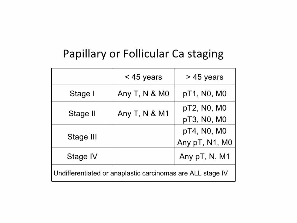
Thyroid cancer staging
• All current staging systems for thyroid cancer incorporate age:– AJCC
– European Organization for Research and Treatment of Cancer (EORTC)
– National Thyroid Cancer Treatment Cooperative Study
– AGES
– AMES
– MACIS
• Is age an appropriate criterion for thyroid cancer staging? If so, is 45 years the right cut-point?


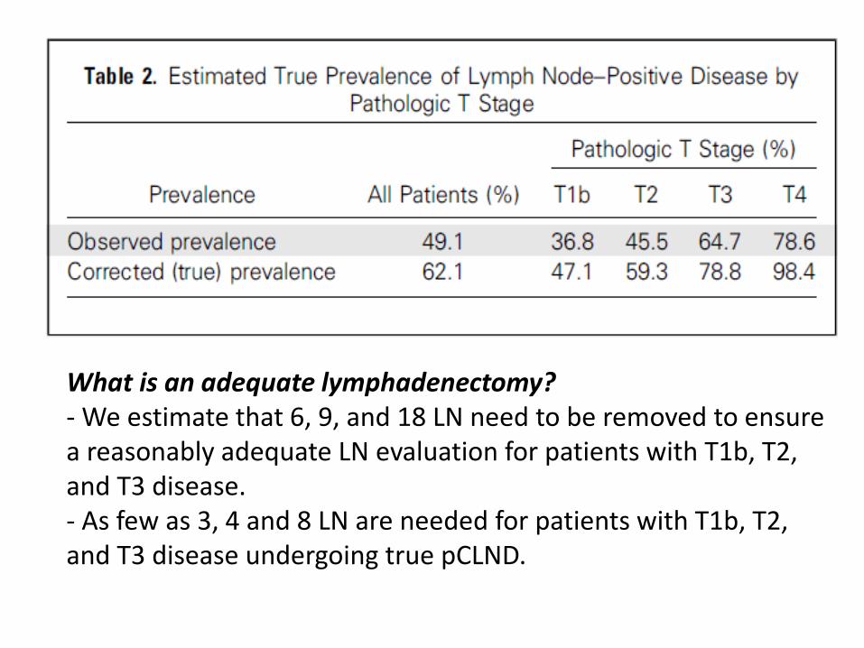
What is an adequate lymphadenectomy?- We estimate that 6, 9, and 18 LN need to be removed to ensure a reasonably adequate LN evaluation for patients with T1b, T2, and T3 disease.- As few as 3, 4 and 8 LN are needed for patients with T1b, T2, and T3 disease undergoing true pCLND.

Acknowledgments Duke Endocrine Neoplasia Research Group
www.dcri.org/our-research/endocrine-neoplasia-research-group


![Annals of Clinical Case Reports Case Report · epithelium are found in axillary lymph nodes [14,15]. Ectopic thyroid tissueis found occasionally in cervical lymph nodes. Among nonepithelial](https://img.pdfslide.us/doc/110x75/5f1cd159d6b56138e82777d7/annals-of-clinical-case-reports-case-epithelium-are-found-in-axillary-lymph-nodes.jpg)