Embed Size (px)
Citation preview

379
MAJOR REVIEW
SURVEY OF OPHTHALMOLOGY
VOLUME 45
•
NUMBER 5
•
MARCH–APRIL 2001
© 2001 by Elsevier Science Inc. 0039-6257/01/$–see front matterAll rights reserved. PII S0039-6257(00)00208-3
Central Disorders of Vision in Humans
Christopher A. Girkin, MD,
1
and Neil R. Miller, MD
2
1
Department of Ophthalmology, University of Alabama—Birmingham, Birmingham, Alabama, and
2
The Neuro-Ophthalmology Unit, The Johns Hopkins Medical Institutions, Baltimore, Maryland, USA
Abstract.
Over the past 20 years, researchers have discovered over 30 separate visual areas in the cor-tex of the macaque monkey that exhibit specific responses to visual and environmental stimuli. Many ofthese areas are homologous to regions of the human visual cortex, and numerous syndromes involvingthese areas are described in the neurologic and ophthalmic literature. The focus of this review is theanatomy and physiology of these higher cortical visual areas, with special emphasis on their relevanceto syndromes in humans. The early visual system processes information primarily by way of two separatesystems: parvocellular and magnocellular. Thus, even at this early stage, visual information is function-ally segregated. We will trace this segregation to downstream areas involved in increasingly complexvisual processing and discuss the results of lesions in these areas in humans. An understanding of theseareas is important, as many of these patients will first seek the attention of the ophthalmologist, oftenwith vague, poorly defined complaints that may be difficult to specifically define. (
Surv Ophthalmol45
:379–405, 2001. © 2001 by Elsevier Science Inc. All rights reserved.)
Key words.
agraphia
•
alexia
•
allesthesia
•
anomia
•
blindsight
•
brain damage
•
brain mapping
•
cerebral cortex
•
color perception
•
dyslexia
•
occipital lobe
•
optic aphasia
•
visual attention
•
visual fields
•
visual hallucinations
•
visual perception
Ophthalmologists tend to view the striate cortexas an afferent structure receiving visual informationmostly from the lateral geniculate nucleus (LGN).Indeed, most of our efforts as ophthalmologists cen-ter on the preservation or restoration of these inputsinto the visual cortex. However, a wide variety of vi-sual disorders may occur from damage to the visualcortex and its occipitofugal connections with asso-ciative visual areas. These syndromes are often called“disorders of higher cortical function,” and they re-mind us that the striate cortex is not the end of theline, but the beginning of a complex system of visualanalysis that ultimately leads to global awareness ofthe visual environment.
I. Functional Segregation of Visual Inputs
A. RETINOGENICULATE PATHWAYS
Functional segregation in the visual system has itsearliest subdivision in the retina. Over 22 types ofganglion cells exist in the primate retina,
212
but onlythree types appear to be involved in visual percep-tion and project to specific locations within theLGN.
55,153,192
The
midget
ganglion cells have smallreceptive fields and project to layers 3–6 of the par-vocellular LGN.
Parasol
cells have much more ex-tensive receptive fields and project to layers 1 and 2of the magnocellular LGN.
261
A third type of re-tinogeniculate ganglion cell, the
bistratified
ganglion

380 Surv Ophthalmol 45 (5) March–April 2001
GIRKIN AND MILLER
cell, projects to the koniocellular layers of theLGN.
56,111,164
The parvocellular system (P-pathway) is a static fir-ing system that conveys information from the retinato the LGN concerning wavelength selectivity andlow-contrast retinal imagery with high spatial re-solution.
157,222,223
High band-pass resolution perime-try (ring perimetry) is an attempt to selectively evalu-ate the P-pathway.
85
In contrast, the magnocellularsystem (M-pathway) conveys high-contrast, low-reso-lution information that is color blind.
157,223
The mag-
nocellular system is a phasic system, thus is wellsuited for the analysis of moving stimuli. Perimetrictechniques, such as frequency-doubled perimetry
113
and motion-detection automated perimetry,
237
areused in an attempt to isolate the M-pathway. The ko-niocellular system (K-pathway) conveys informationconcerning blue-yellow color opponency.
54
Short-wavelength automated perimetry (SWAP) uses ablue stimulus presented against a yellow backgroundand may evaluate the responses processed throughthis pathway (Fig. 1).
Fig. 1. Visual information that is used to create the perceived image of a visual scene (top photograph) is conveyed to thelateral geniculate through three pathways. The lower photographs were manipulated to simulate the characteristics of theperceived image that are conveyed along each of these pathways. The parvocellular pathway (lower left photograph) con-veys fine spatial detail and is thought to be wavelength selective primarily for red and green opponency. The koniocellularpathway conveys information concerning blue-yellow opponency and low spatial detail (lower middle photograph). Themagnocellular pathway (lower right photograph) conveys color-blind information of low spatial detail, sensitive to motion.(Artist: David Fisher.)
CENTRAL DISORDERS OF VISION IN HUMANS
381
The M-, P-, and K-pathway inputs, although differ-ing in response characteristics, have some overlap inthe type of visual stimuli to which they are activated.Thus, perimetric techniques may emphasize onepathway over another, but do not completely isolatethe responses processed through a given pathway. Inaddition, inputs from the parallel systems are pro-cessed in a complex and interrelated manner by thevisual cortex and higher cortical centers.
B. CORTICAL VISUAL AREAS
Over the past 20 years, more than 30 visual corti-cal areas have been isolated in macaque monkeys.These areas comprise almost 50% of the entire corti-cal volume. Although the function of most of theseareas is unclear, studies of the visual cortex in lowerprimates
67,258,266,283
and clinical correlation with cere-bral lesions in patients,
118,207,208,260
along with electro-physiologic studies,
18,80
postmortem histologic exam-inations,
32,38
and functional imaging studies,
48,72,227,
251,282
have identified several cortical areas that mayhave clinical importance in humans.
The visual cortex in the macaque was initially di-vided into six sub-regions named visual areas 1–6(Areas V1–V6). Area V1 is the primary visual cortex,and it corresponds to the striate cortex in bothhumans and lower primates. Areas V2–V6 are ex-tensively interconnected visual areas that lie anteriorto V1 and contain specialized maps of the visualfield.
74,284
These areas are best visualized in themacaque in a horizontal section through the occipi-tal lobe (Fig. 2).
Area V2 is immediately adjacent to area V1 inmost primates, including humans. It corresponds toBrodmann’s area 18 and was previously called the
parastriate cortex.
Hubel and Wiesel initially believed that Brod-mann’s area 19, also called the
peristriate cortex
in hu-mans, was composed entirely of the human homologof V3,
74
but further studies indicated that the peris-triate cortex is composed of two functionally distinctareas, V3 and V3A.
251
Area V4 in the macaque lies in the lateral occipitallobe. Zeki claimed that “human V4” was located inthe caudal lingual and fusiform gyri within Brod-mann’s area 18.
281
This area is involved in color pro-cessing in humans (see below); however, whether ornot it is homologous to area V4 in the macaque iscontroversial.
167
Area V5, also called area MT because of its loca-tion in the middle temporal gyrus of the owl mon-key, is located in humans in the gyrus subangularisof the ventrolateral occipital lobe.
267
Area V6 in the macaque has no clear homolog inhumans; however, an area associated with visuospa-tial processing in the posterior parietal cortex is the
most likely candidate. Fig. 3 illustrates the locationof several of the corresponding visual areas in hu-mans. Only those cortical areas associated with dis-tinct clinical syndromes will be discussed.
C. OCCIPITOFUGAL PATHWAYS
Based on numerous studies of lesions in humans,
59,61
functional imaging of normal subjects,
48
and ex-periments in monkeys,
258
it is clear that the informa-tion processed by the striate cortex and visual asso-ciative areas is projected through two occipitofugalpathways: a ventral occipitotemporal pathway and adorsal occipitoparietal pathway (Fig. 4).
109
The ven-tral pathway, often called the “what” pathway, is in-volved in processing the physical attributes of a vi-sual image that are important to the perception ofcolor, shape, and pattern. These, in turn, are crucialfor object identification and object-based atten-tion.
259
The ventral pathway originates in V1 andprojects through V2 and V4 to specific inferior tem-poral cortical areas, the angular gyrus, and limbicstructures. It provides visual information to areas in-volved in visual identification, language processing,memory, and emotion.
234
Thus, a lesion in this path-way may cause a variety of associative defects, includ-ing visual alexia and anomia, visual agnosia, visualamnesia, and visual hypoemotionality.
The dorsal, or “where” pathway, begins in V1 andprojects through V2 and V3 to V5.
234,259
From V5, thispathway continues to additional areas in the parietaland superior temporal cortex.
232
These projectionsare involved in visuospatial analysis, in the localiza-tion of objects in visual space, and in modulation ofvisual guidance of movements toward these ob-jects.
96,257
Thus, lesions of this pathway may cause avariety of visuospatial disorders, such as Bálint’s syn-drome and hemispatial neglect.
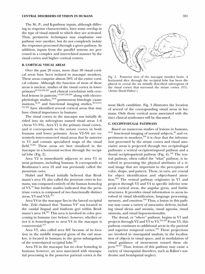
Fig. 2. Posterior view of the macaque monkey brain. Ahorizontal slice through the occipital lobe has been dis-placed to reveal the six initially described sub-regions ofthe visual cortex that surround the striate cortex (V1).(Artist: David Fisher.)
382 Surv Ophthalmol 45 (5) March–April 2001
GIRKIN AND MILLER
Although the ventral and dorsal pathways areclearly involved in the analysis of different aspects ofthe visual environment,
259
they are extensively inter-connected laterally and in feedback and feedfor-ward directions, indicating that the flow of percep-tual processing does not necessarily proceed in astepwise, hierarchic manner.
234
This “what” and“where” dichotomy of visual processing is an over-simplification of how these cortical areas function,but it serves as a useful framework in which to de-velop a clinical model of cortical visual processing. Anumber of specific syndromes in humans involvingthe central processing of visual information can belocalized primarily to one of the six visual cortical ar-eas or one of the two occipitofugal pathways andthus are of clinical value. These are summarized inTable 1.
II. Syndromes Associated with Damage to the Striate Cortex (Area V1)
A. ANTON SYNDROME
Denial of blindness, or Anton syndrome,
7
is an un-common form of anosagnosia that usually follows ex-tensive damage to the striate cortex.
9,177
AlthoughAnton syndrome usually occurs with geniculostriatelesions, it may occur from any etiology, including
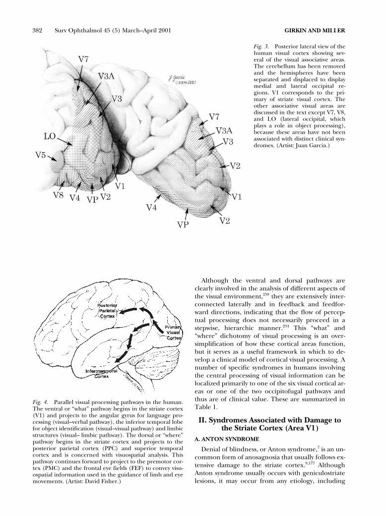
Fig. 3. Posterior lateral view of thehuman visual cortex showing sev-eral of the visual associative areas.The cerebellum has been removedand the hemispheres have beenseparated and displaced to displaymedial and lateral occipital re-gions. V1 corresponds to the pri-mary of striate visual cortex. Theother associative visual areas arediscussed in the text except V7, V8,and LO (lateral occipital, whichplays a role in object processing),because these areas have not beenassociated with distinct clinical syn-dromes. (Artist: Juan Garcia.)
Fig. 4. Parallel visual processing pathways in the human.The ventral or “what” pathway begins in the striate cortex(V1) and projects to the angular gyrus for language pro-cessing (visual–verbal pathway), the inferior temporal lobefor object identification (visual–visual pathway) and limbicstructures (visual– limbic pathway). The dorsal or “where”pathway begins in the striate cortex and projects to theposterior parietal cortex (PPC) and superior temporalcortex and is concerned with visuospatial analysis. Thispathway continues forward to project to the premotor cor-tex (PMC) and the frontal eye fields (FEF) to convey visu-ospatial information used in the guidance of limb and eyemovements. (Artist: David Fisher.)

CENTRAL DISORDERS OF VISION IN HUMANS
383
blindness from prechiasmal disorders, such as opticneuropathies and retinal detachment.
Patients with Anton syndrome will deny they areblind and often confabulate to mask their visual loss.We recently examined a hospitalized patient whowas blind following a bilateral occipital stroke butwho adamantly believed he could see. He could notsee to feed himself and would feel his way aroundthe room, yet he falsely identified objects when pre-sented to him.
There are several theories regarding the etiologyof Anton syndrome, but a definitive etiology remainselusive.
152
Geschwind noted that patients with thiscondition often had altered emotional reactivity,with a “coarse and shallow” affect similar to some pa-tients with frontal lobe lesions. He attributed the de-nial of blindness to damage to higher cognitive cen-ters.
152
Psychiatric denial may explain other cases.
126
Finally, lesions of the geniculostriate pathway thatdisrupt input to the visual cortex may also interferewith output from the visual cortex to areas involvedin the conscious awareness of visual perception. Insuch cases, the striate cortex is unable to communi-cate the nature of the patient’s visual loss to areasconcerned with conscious awareness.
126
B. BLINDSIGHT
Studies of visually guided behavior following re-moval of the striate cortex in monkeys demonstratedefinite preservation of visual sensory function.
178
Asimilar phenomenon is thought to occur in humanswho experience severe damage to one or both occip-ital lobes.
198
Weiskrantz coined the term
blindsight
torefer to this rudimentary level of visual processingthat occurs below the level of visual awareness.
272
Over the past 20 years, the phenomenon of blind-sight has been extensively studied in humans andlower primates. This entity encompasses a wide vari-ety of visual processing mechanisms, all occurringwithout conscious awareness.
240
Some authors expand blindsight to refer to anypreserved visual function that occurs below the levelof visual awareness in patients with cortical blind-ness.
240
These authors include neuroendocrineresponses
53
and visual reflexes,
269
such as preserva-tion of the photic blink reflex,
104
as types of blind-sight. However, most authors refer to blindsightstrictly as preserved higher levels of visual processingfollowing an occipital stroke.
Higher levels of cortical processing have beenevaluated in humans by both implicit processing,which measures induced responses to stimuli pre-sented to the blind field,
162,197,254,271
and direct re-sponses, including forced-choice experiments, sac-cadic localization tasks, and manual pointing toobjects presented in the blind hemifield.
26
Usingthese techniques, researchers have shown that rarepatients exhibit preservation of the ability to detectdirection of motion,
162
wavelength,
241
target displace-ment,
24
stimulus presence,
272
orientation,
180
and ob-ject discrimination.
272
Behavior studies in monkeysand humans have demonstrated that this uncon-scious discrimination exhibits a learning effect, withincreased accuracy with extensive training.
285
How-ever, the potential use of blindsight in visual rehabil-itation is controversial.
285
The methods described above have been criti-cized in the past, leading to the questioning of theexistence of blindsight in humans. Ocular scatterand residual functional islands of striate cortex haveboth been used as alternate explanations of blind-sight.
36,78
However, controlled experiments involvingblind spot stimulation,
180
along with improved fixa-tion controls, have made these alternative hypothe-ses less likely.
270
Additionally, functional imagingstudies have failed to show residual activity in thestriate cortex in several of these patients.
12,79,243
Nev-ertheless, although recent experimental evidencemay indicate that blindsight in humans is more thanjust an experimental artifact, even those who acceptthe idea admit that it can be demonstrated in only a
TABLE 1
Syndromes Localized to One of the Visual Cortical Areas or Fungal Pathways
1. Area V1A. Anton syndromeB. BlindsightC. Riddoch phenomenonD. Transient achromatopsiaE. Visual ataxia
2. Area V2 and V3A. Quadrantic homonymous hemianopia
3. Area V4A. Cerebral achromatopisa
4. Area V5A. Akinetopsia
5. Dorsal occipitofugal pathwayA. Bálint syndromeB. Hemispatial neglectC. Visual allesthesiaD. Enviromental rotation
6. Ventral occipitofugal pathwayA. Visual–verbal disconnection
i. Pure alexiaii. Color anomia
iii. Object anomiaB. Visual–visual disconnection
i. Prosopagnosiaii. Object agnosia
C. Visual–limbic disconnectioni. Visual amnesia
ii. Visual hypoemotionality

384 Surv Ophthalmol 45 (5) March–April 2001
GIRKIN AND MILLER
limited number of patients and is dependent upontraining and time after cortical injury, possibly be-cause of plasticity in the visual cortex seen in pa-tients with long-standing central field defects.
199
Anatomic studies in lower primates,
178,188,189
as wellas functional imaging
14,181
and electrophysiologicstudies
79,231
in humans, have provided some insightinto the functional anatomy of blindsight. Thesestudies suggest that a subcortical pathway involvingthe superior colliculus and pulvinar processes someaspects of the unconscious perception demonstratedin these rare cases (Fig. 5).
18,19,270
However, the re-tino-tecto-pulvinar-cortical pathway lacks informa-tion concerning color opponency, thus cannot ex-plain the presence of color discrimination in somesubjects with blindsight.
50,241,242
Alternatively, blind-sight may be secondary to surviving connectionsfrom the LGN to associative visual areas that bypassthe striate cortex.
51
Although much controversy still surrounds the ex-istence of and pathways involved in blindsight in hu-mans, the availability of improved functional imag-ing methods and techniques of reversible corticaldeactivation may help resolve these conflicts in thefuture. Indeed, an understanding of this conditionmay eventually provide crucial clues toward an un-derstanding of the neural correlates of visual aware-ness and may even contribute to the design of tech-
niques for visual rehabilitation in patients withcortical blindness.
C. RIDDOCH PHENOMENON
In 1917, George Riddoch, a captain in the RoyalArmy Medical Corps, described 10 patients withwounds to the occipital area who were able to per-ceive movements within their blind hemifield.
202
Sev-eral studies of similar patients by other investigatorssubsequently confirmed what became known as the
Riddoch phenomenon
: preservation of motion percep-tion in an otherwise complete scotoma.
172,280
The etiology of the Riddoch phenomenon is notclear. It has been suggested that patients who exhibitthis phenomenon have preserved islands of functionwithin the striate cortex,
202
or that extrastriate areas,V5 in particular, may be involved through activationof subcortical pathways that bypass V1.
36,280
Alterna-tively, statokinetic dissociation may be related to lat-eral summation of moving images. It has been dem-onstrated in normal subjects that a kinetic targetmay be seen more readily in some areas of the visualfield than nonmoving objects of the same intensityand size.
101
Variable degrees of dissociation of per-ception between moving and nonmoving stimulihave been demonstrated in normal subjects
88
and inpatients with compression of the anterior visualpathways.
217,276
Fig. 5. Two proposed pathways involved in blindsight. A: Visual information is transmitted to associative visual areas inthe dorsal occipitofugal pathway via a subcortical pathway that includes the superior colliculus (SC) and pulvinar (Pulv)thus bypassing the pathway to the primary visual cortex through the dorsal lateral geniculate nucleus (LGN). B: Pathwaysfrom the LGN convey visual information to extrastriate cortical areas that may bypass the striate cortex. (Artist: DavidFisher.)

CENTRAL DISORDERS OF VISION IN HUMANS 385
Clinically, the Riddoch phenomenon can be a use-ful prognostic indicator. For example, patients withthe Riddoch phenomenon following an occipitalstroke or in the setting of an occipital lobe tumorare more likely to recover function in the affectedfield, spontaneously in the first setting or after re-moval of the tumor in the second.255
D. TRANSIENT ACHROMATOPSIA
Although cerebral achromatopsia—i.e., loss ofcolor perception from a lesion in one or both cere-bral hemispheres—usually occurs from a lesion in-volving area V4 (see below), transient achromatop-sia has been reported in some patients withvertebrobasilar insufficiency.147 Achromatopsia inthe latter setting may occur from dysfunction ofwavelength-selective regions in areas V1 or V2. Bothof these areas exhibit increased metabolic activitycompared with surrounding areas and thus may bemore vulnerable to ischemia.274
E. VISUAL ATAXIA
Patients with an homonymous hemianopia froman occipital lobe lesion may experience loss of bal-ance associated with a sensation of falling toward theblind hemifield.213 Such patients have an intact vesti-bular system, and this “visual ataxia” is thought to besecondary to unopposed tonic input from the intactcontralateral occipital lobe. Visual ataxia is a distur-bance in balance and should not be confused withoptic ataxia, which is a disturbance in visually-basedlimb guidance (see below).
III. Syndromes Caused by Damage to the Parastriate and Peristriate Visual Cortex
(Areas V2 and V3)Both primate and human studies have demon-
strated that area V2 may play a role in the detectionof illusionary contours.193,194 This perceptual task isof great importance in the detection of obscured ob-jects, such as a camouflaged predator. Patients withearly posterior Alzheimer’s disease demonstrate im-paired detection of illusionary contours, possiblydue to degeneration of area V2.
The effect of lesions of V2 and V3 in humans isunclear. Horton and Hoyt reported two patientswith lesions thought to involve the superior parastri-ate and peristriate cortex.118 Both patients had hom-onymous quadrantic visual field defects that re-spected the horizontal and vertical meridians. Thisreport not withstanding, lesions of V2 induced byibotenic acid fail to produce a visual field defect inmonkeys.171 The apparent discrepancy betweenthese two observations may be related to the fact thatibotenic acid destroys cell bodies while preservingaxons and, therefore, may spare the fibers of the op-
tic radiations passing deep to the parastriate cortex.Of course, a congruous homonymous quadrantano-pia is not specific to parastriate lesions; it not infre-quently results from striate lesions as well.165
IV. Syndromes Caused by Damage to the Human Color Center (Area V4)
A. PERCEPTION OF COLOR AND AREA V4
Unlike a camera, the visual system has the abilityto compensate for the changing spectral compo-nents of a light source. Therefore, in most viewingsituations, a red object will appear red regardless ofthe wavelength of light that illuminates it, eventhough the dominant spectral component reflectedfrom the object may vary with lighting conditions.This effect is called color constancy. The visual systemcreates the concept of color by comparing areas ofthe visual field.142 Thus, a red object appears red notbecause it reflects long-wavelength light, but becauseit reflects relatively more long-wavelength light thando other objects within the visual field. A neuralstructure that performs this comparison of wave-lengths across large areas of the visual field requirescells that can combine wavelength-selective informa-tion obtained from disparate areas of the visualfield.70 The cells in area V4 in the monkey fulfillthese requirements. They respond to both wave-length and perceived color, whereas neurons in ar-eas V1 and V2 respond only to wavelength.279 Thesephysiologic experiments, along with lesion studies inmacaques, have led some investigators to concludethat area V4 is the site of color constancy in lowerprimates.278
Although the localization of a “color center” inhumans has been clearly identified,160 whether ornot this area is homologous to area V4 in monkeysand whether or not cerebral achromatopsia in hu-mans is a defect of color constancy alone are cur-rently unresolved issues.39,114,129 Many authors use theterm human color center interchangeably with the termhuman V4.167 However, the complaints of patientswith cerebral achromatopsia from damage to this re-gion are not entirely explained by loss of color con-stancy.207 In addition, extirpation of area V4 in themonkey does not lead to achromatopsia, for the ani-mals retain the ability to discriminate and order huesdespite clearly impaired form recognition.92,220,221
B. CEREBRAL ACHROMATOPSIA
Cerebral achromatopsia is an acquired defect incolor perception caused by damage to the ventrome-dial visual cortex.278 Affected patients describe aworld that looks faded, gray, and washed out, orcompletely devoid of color, like a black-and-whitephotograph. Patients with the most pure form of ce-

386 Surv Ophthalmol 45 (5) March–April 2001 GIRKIN AND MILLER
rebral achromatopsia cannot arrange graded isolu-minant, but retain normal sorting for graded grayobjects.49 However, many affected patients havesome residual hue discrimination, thus view theworld as if looking though a colored filter. Such pa-tients are probably more correctly classified as hav-ing cerebral dyschromatopsia rather than achro-matopsia.278 Additionally, many patients reported tohave achromatopsia have not received an adequateneuropsychologic assessment and could actually beexhibiting color anomia or aphasia, thus furtherconfusing this issue.278
Unlike patients with congenital achromatopsia,patients with acquired cerebral achromatopsia showpreserved trichromacy and intact cortical responsesto chromatic visual evoked potentials (VEPs).49 Thechromatic pathways from the retina to the striatecortex are intact. Because of preserved function ofwavelength-selective cells in the striate cortex, achro-matopsic patients may still retain the ability to distin-guish the border between two adjacent isoluminantcolored patches49 and may perform well on testingwith pseudoisochromatic plates.168
Verrey, in 1888, provided the first pathologic cor-relation in cerebral achromatopsia, localizing the le-sion to the posterior fusiform and lingual gyri (Fig.6). Subsequent investigators have described thepathologic findings in patients with this condition.278
Bilateral infarction in the posterior cerebral arterydistribution is the most common etiology and is of-ten caused by vertebrobasilar ischemia.147 Additionalcauses include metastatic tumors,97 posterior corticaldementia,82 and herpes simplex encephalitis.113
Transient achromatopsia may occur with migraine,149
focal seizures,2 and vertebrobasilar insufficiency.147
Cerebral achromatopsia is often associated with asuperior homonymous visual field defect from dam-age to the inferior striate cortex.57 In such cases, theresidual inferior field on that side is achromatopsic.Furthermore, because of the ventral location of thelesions in these patients, central achromatopsia isoften accompanied by deficits caused by damage tothe ventral occipitofugal pathway, including pro-sopagnosia, topographagnosia, visual object agnosia,pure alexia, and defects of visual memory (see be-low). Global amnesia may be present if the lesiondamages large areas in the temporal lobe.57 Defectsin pattern processing are also well described, possi-bly reflecting a role for color in form analysis.92
Three-dimensional magnetic resonance imaging(MRI) in patients with cerebral achromatopsia indi-cates that the critical lesion involves the middle thirdof the lingual gyrus or the white matter posterior tothe tip of the lateral ventricle.62 Cerebral achro-matopsia may be complete or affect only one hom-onymous hemifield.190 Bilateral lesions are required
for a complete achromatopsia, whereas unilateral le-sions produce hemiachromatopsia.
Zeki and coworkers used functional MRI (fMRI)to define the representation of the visual field in thehuman color center.167 This study localized the colorcenter to the lateral aspect of the collateral sulcus onthe fusiform gyrus. Additionally, these investigatorsdescribed a retinotopic organization of the fusiformgyrus, with the superior field being representedwithin the medial fusiform gyrus and the inferiorfield located more laterally.
If achromatopsia were due solely to defectivecolor constancy, then patients with the conditionshould not see the world as gray or desaturated, butinstead they should experience dramatic fluctua-tions in color as environmental lighting conditionschange. Since this is not the case, the defect in ach-romatopsia may involve more than just color con-stancy. Indeed, Rizzo et al have hypothesized thatcolor constancy, like lightness constancy, is gener-ated by earlier visual associative areas.211 Alterna-tively, lesions of the fusiform gyrus may disrupt whitematter deep to the collateral sulcus and disconnectthe striate and extrastriate areas from a more rostralcolor center, possibly an area homologous to a wave-
Fig. 6. View of the ventral surface of the brain with thecerebellum removed. The posterior fusiform and lingualgyri, which contain the human color center, are high-lighted. (Artist: David Fisher.)

CENTRAL DISORDERS OF VISION IN HUMANS 387
length-selective inferior temporal area in monkeysthat, when extirpated, produces a deficit similar tocerebral achromatopsia in humans.49 Recently, acolor-selective area has been demonstrated in hu-mans in a similar region and has been labeled V8.105
There is some controversy as to whether this is a newvisual area or merely the anterior portion of V4.281
Although there is still much controversy regardingthe function of the human color center and its rela-tionship to area V4 in lower primates, it is clear thatlesions that produce cerebral achromatopsia in hu-mans are invariably located in the ventromedial oc-cipital lobe (Figs. 3 and 6).
V. Syndromes Caused by Damage toArea V5
A. NEUROPHYSIOLOGY OF MOTION PERCEPTION
Functional imaging;252,282 experiments using mye-lin, cytochrome oxidase, and monoclonal-antibodystaining;253 and cortical stimulation experiments sug-gest that the most likely location of area V5 in hu-mans is the ventrolateral occipital gyrus, a key areainvolved in the perception of visual motion (Fig. 3).In addition, transcranial magnetic deactivation ofthis area demonstrates deficits in motion percep-tion.17 However, the analysis of motion involves acomplex system of several interrelated cortical areasthat are involved in processing various componentsof motion perception and that may adapt to the lossof area V5.77,252 This would explain the preservationof some aspects of visual motion perception in pa-tients with akinetopsia (see below) and the rarity ofthis syndrome in humans.
B. AKINETOPSIA
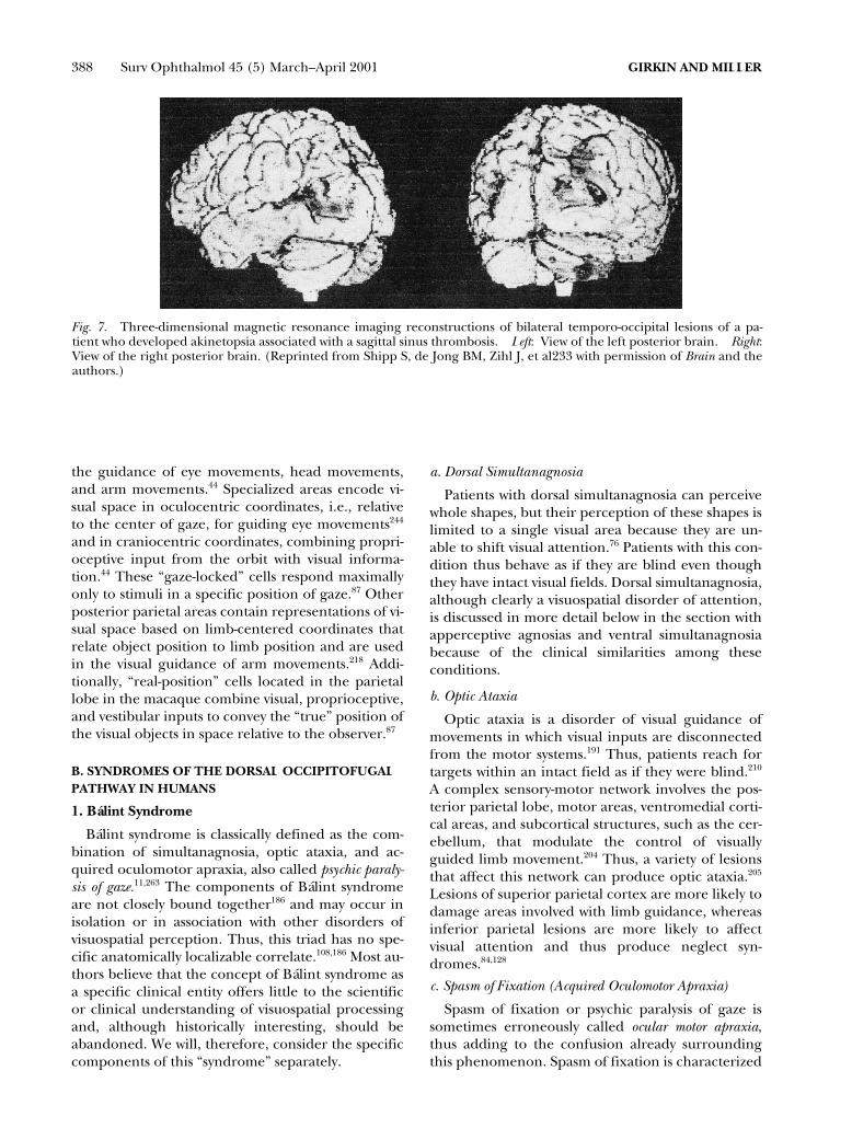
Akinetopsia is the loss of perception of visual mo-tion with preservation of the perception of othermodalities of vision, such as form, texture, andcolor. Although isolated deficits in motion vision inhumans were reported in the early 20th century,these cases were poorly documented.277 It was notuntil the report of Zihl et al in 1983 that an exampleof akinetopsia was clearly described.286 The patient,LM, described by these investigators, is one of onlytwo patents who have been extensively studied. Shedeveloped bilateral cerebral infarctions involvingthe lateral occipital, middle temporal, and angulargyri, secondary to sagittal sinus thrombosis (Fig. 7).She described moving objects as jumping from placeto place. For example, when pouring tea, she ob-served that the liquid appeared frozen like a glacier,and she failed to perceive the tea rising in the cup.She did, however, have definite evidence of residualmotion processing at modest levels of backgroundnoise on psychophysical tests, such as random-dot
cinematograms. Indeed, she was able to distinguishmoving from nonmoving targets and to distinguishshape and three-dimensional structure from mo-tion.208 These abilities may reflect preserved func-tion of associated areas of motion processing andemphasize that motion perception is not completelyisolated to a single cortical region.233
Subtle deficits in motion processing in the con-tralateral hemifield in patients with unilateral occipi-toparietal lesions involving area V5 have also beendescribed.260 This “hemiakinetopsia” is often ob-scured by coexistent incomplete homonymous fielddefects.
In a study using fMRI, Eden et al found almost noactivation of area V5 in dyslexic patients.71 The re-sults of this study support previous psychophysio-logic and anatomic data suggesting that patientswith dyslexia have anomalous magnocellular re-sponses.158 The superior temporal area that is acti-vated while normal patients view moving stimulioverlaps areas involved in language processing, lead-ing some investigators to conclude that some disor-ders of phonologic awareness may result from a glo-bal deficit in processing temporal properties ofvision.158
VI. The Dorsal Occipitofugal Pathway and Visuospatial Processing in Humans
A. NEUROANATOMY AND NEUROPHYSIOLOGY
The dorsal or “where” pathway receives informa-tion primarily from area MT and, to a lesser extent,area V4.259 This information is conveyed along thedorsal longitudinal fascicles to the posterior parietalcortex, frontal motor areas, and frontal eye fields(FEF). This pathway is concerned with spatial local-ization, visuomotor search and guidance, and visu-ospatial synthesis.109 Lesions of the dorsal pathwayproduce visuomotor and attention deficits, in con-trast to the visuoassociative deficits produced by ven-tral lesions.
The posterior parietal cortex is neither a purelysensory nor a purely motor area; rather, it combinescharacteristics of both. Thus, it serves as a junctionbetween multimodal sensory input and motor out-put, linking the afferent and efferent arms of the vi-sual pathways and providing the connection that en-compasses the entire field of neuro-ophthalmology,from the eyes to the extraocular muscles.4
One model proposed for the modulation of spa-tial attention is that the posterior parietal cortexcontains several maps of the visual environment,which are used in visually guided movement,5 modu-late attention,47,179 and plays a role in visuospatialperception. These maps code the location of visualtargets in a variety of coordinate systems tailored to
388 Surv Ophthalmol 45 (5) March–April 2001 GIRKIN AND MILLER
the guidance of eye movements, head movements,and arm movements.44 Specialized areas encode vi-sual space in oculocentric coordinates, i.e., relativeto the center of gaze, for guiding eye movements244
and in craniocentric coordinates, combining propri-oceptive input from the orbit with visual informa-tion.44 These “gaze-locked” cells respond maximallyonly to stimuli in a specific position of gaze.87 Otherposterior parietal areas contain representations of vi-sual space based on limb-centered coordinates thatrelate object position to limb position and are usedin the visual guidance of arm movements.218 Addi-tionally, “real-position” cells located in the parietallobe in the macaque combine visual, proprioceptive,and vestibular inputs to convey the “true” position ofthe visual objects in space relative to the observer.87
B. SYNDROMES OF THE DORSAL OCCIPITOFUGAL PATHWAY IN HUMANS
1. Bálint Syndrome
Bálint syndrome is classically defined as the com-bination of simultanagnosia, optic ataxia, and ac-quired oculomotor apraxia, also called psychic paraly-sis of gaze.11,263 The components of Bálint syndromeare not closely bound together186 and may occur inisolation or in association with other disorders ofvisuospatial perception. Thus, this triad has no spe-cific anatomically localizable correlate.108,186 Most au-thors believe that the concept of Bálint syndrome asa specific clinical entity offers little to the scientificor clinical understanding of visuospatial processingand, although historically interesting, should beabandoned. We will, therefore, consider the specificcomponents of this “syndrome” separately.
a. Dorsal Simultanagnosia
Patients with dorsal simultanagnosia can perceivewhole shapes, but their perception of these shapes islimited to a single visual area because they are un-able to shift visual attention.76 Patients with this con-dition thus behave as if they are blind even thoughthey have intact visual fields. Dorsal simultanagnosia,although clearly a visuospatial disorder of attention,is discussed in more detail below in the section withapperceptive agnosias and ventral simultanagnosiabecause of the clinical similarities among theseconditions.
b. Optic Ataxia
Optic ataxia is a disorder of visual guidance ofmovements in which visual inputs are disconnectedfrom the motor systems.191 Thus, patients reach fortargets within an intact field as if they were blind.210
A complex sensory-motor network involves the pos-terior parietal lobe, motor areas, ventromedial corti-cal areas, and subcortical structures, such as the cer-ebellum, that modulate the control of visuallyguided limb movement.204 Thus, a variety of lesionsthat affect this network can produce optic ataxia.205
Lesions of superior parietal cortex are more likely todamage areas involved with limb guidance, whereasinferior parietal lesions are more likely to affectvisual attention and thus produce neglect syn-dromes.84,128
c. Spasm of Fixation (Acquired Oculomotor Apraxia)
Spasm of fixation or psychic paralysis of gaze issometimes erroneously called ocular motor apraxia,thus adding to the confusion already surroundingthis phenomenon. Spasm of fixation is characterized
Fig. 7. Three-dimensional magnetic resonance imaging reconstructions of bilateral temporo-occipital lesions of a pa-tient who developed akinetopsia associated with a sagittal sinus thrombosis. Left: View of the left posterior brain. Right:View of the right posterior brain. (Reprinted from Shipp S, de Jong BM, Zihl J, et al233 with permission of Brain and theauthors.)

CENTRAL DISORDERS OF VISION IN HUMANS 389
by loss of voluntary eye movements with persistenceof fixation on a target. However, in contrast to trueocular motor apraxia, saccades easily are made toperipheral targets in the absence of a fixation target.Thus, a patient asked to fixate an object centrallyand then move the eyes to a peripheral target can-not do so, whereas a patient who is not fixing on anyobject in particular easily can move the eyes to fixatea peripheral target when asked to do so.
The location of the lesion that causes spasm of fix-ation is obscure. The FEF is required for the releaseof fixation for voluntary saccades, and lesions of thisregion may prolong saccadic latency. Posterior pari-etal, middle temporal, and superior temporal areasmediate cortical maintenance of fixation by inhibi-tion of attention shifts. Thus, damage to the FEFthat spares these regions may prevent the release offixation by disinhibiting the inhibitory effect of thesubstantia nigra pars reticulata on the superior colli-culus, suppressing the generation of saccades.125
2. Hemispatial (Hemifield) Neglect
Complex visual scenes constantly bombard the vi-sual system. Because cognitive and motor activitiesare generally concerned with one object at a time,these elements of the visual scene must compete forthe limited resources of focal attention.13,37 Modula-tion of attention occurs at many levels in the visualsystem, even at the level of area V1.136,262
Numerous lines of evidence suggest that a com-plex network of cortical and subcortical areas prima-rily in the dorsal occipitofugal pathway is involved inthe modulation of spatial attention. These includethe superior colliculus, the posterior parietal cortex,the striatum, the pulvinar, and areas in the prefrontalcortex.43 In particular, the posterior parietal cortex,the FEF, and cingulate gyrus play key roles in spatial-based attention mechanisms. The posterior parietalcortex builds the sensory representation of extra-personal space, the FEF plans and initiates explor-atory movements, and the cingulate gyrus providesthe motivational potential.43
A positron emission tomography (PET) study innormal humans demonstrated activation of the cin-gulate gyri (greater on the right), the posterior pari-etal cortex, and the medial and lateral premotor cor-tical areas during covert shifts of attention.184 Theresults of this study are consistent with the conceptthat these cortical areas form the core of a neuralnetwork for spatial attention.
Damage to the components of the dorsal occipi-tofugal pathway or their interconnections may causehemispatial neglect to the contralateral side.173 Oneattractive hypothesis is that the right hemisphere co-ordinates attention throughout extrapersonal space,whereas the left hemisphere coordinates attention
only in the contralateral right hemispace; thus, onlyleft hemispatial neglect is seen as a persistent phe-nomenon.268 Hemispatial neglect involves multiplesensory modalities,133 but visual extinction often isthe most prominent feature. Affected patients seestimuli presented separately in either their right ortheir left hemifield, but ignore stimuli in the lefthemifield when both hemifields are stimulated si-multaneously. Thus, any patient who appears tohave a homonymous hemianopia when bilateral si-multaneous stimulation confrontation testing is per-formed should undergo testing of each homony-mous hemifield separately to determine if theapparent field defect is real or the consequence ofhemifield extinction. In fact, some patients withhemifield extinction also have an homonymous vi-sual field defect, most commonly a lower left qua-drantanopia. Such patients will demonstrate a hom-onymous quadrantic field defect when eachhemifield is tested separately but complete neglectof the left homonymous hemifield when tested withbilateral simultaneous stimulation. In patients withfull visual fields, double simultaneous stimulation ortesting line bisection are excellent bedside examina-tion techniques to detect hemifield neglect.
3. Visual Allesthesia
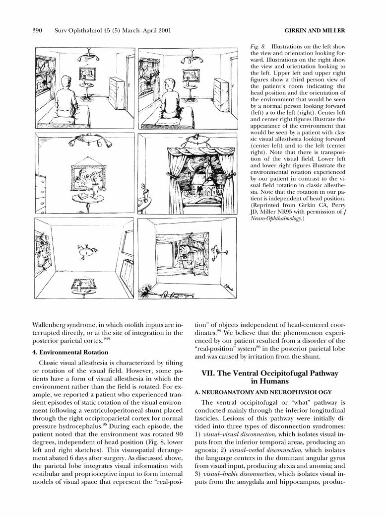
Classic visual allesthesia is a disorder of visuospa-tial perception in which the retinotopic visual field isrotated, flipped, or even inverted (Fig. 8, center leftand right sketches). This syndrome localizes to twoseemingly diverse areas of the brain: the lateral me-dulla and the occipitoparietal area, usually on theright side. Although visual allesthesia is a commoncomponent of the lateral medullary syndrome ofWallenberg214 and is usually due to infarction, a vari-ety of disorders that affect the cerebral cortex canproduce visual allesthesia, including infarction,112
neoplasm, trauma, infection,8 and multiple sclero-sis.226 Transient visual allesthesia can occur duringseizures183 and migraine attacks.103
Several theories have been proposed to explain vi-sual allesthesia; however, an all-encompassing expla-nation remains elusive. Jacobs suggested that alles-thesia may involve transcallosal transmission of thecontralateral hemifield to the damaged parietal cor-tex, with retention of the image as a palinoptic phe-nomenon.121 Although this theory might explain thepatient he described who had transposition of the vi-sual field from left to right, it fails to explain the vari-eties of rotational or inverted allesthesia describedby other patients.
Alternatively, visual allesthesia may be the result ofa disorder of integration of visuospatial input. Thecausative lesion may disturb the integration of visualand otolithic inputs at the level of the medulla, as in
390 Surv Ophthalmol 45 (5) March–April 2001 GIRKIN AND MILLER
Wallenberg syndrome, in which otolith inputs are in-terrupted directly, or at the site of integration in theposterior parietal cortex.249
4. Environmental Rotation
Classic visual allesthesia is characterized by tiltingor rotation of the visual field. However, some pa-tients have a form of visual allesthesia in which theenvironment rather than the field is rotated. For ex-ample, we reported a patient who experienced tran-sient episodes of static rotation of the visual environ-ment following a ventriculoperitoneal shunt placedthrough the right occipitoparietal cortex for normalpressure hydrocephalus.95 During each episode, thepatient noted that the environment was rotated 90degrees, independent of head position (Fig. 8, lowerleft and right sketches). This visuospatial derange-ment abated 6 days after surgery. As discussed above,the parietal lobe integrates visual information withvestibular and proprioceptive input to form internalmodels of visual space that represent the “real-posi-
tion” of objects independent of head-centered coor-dinates.29 We believe that the phenomenon experi-enced by our patient resulted from a disorder of the“real-position” system86 in the posterior parietal lobeand was caused by irritation from the shunt.
VII. The Ventral Occipitofugal Pathwayin Humans
A. NEUROANATOMY AND NEUROPHYSIOLOGY
The ventral occipitofugal or “what” pathway isconducted mainly through the inferior longitudinalfascicles. Lesions of this pathway were initially di-vided into three types of disconnection syndromes:1) visual–visual disconnection, which isolates visual in-puts from the inferior temporal areas, producing anagnosia; 2) visual–verbal disconnection, which isolatesthe language centers in the dominant angular gyrusfrom visual input, producing alexia and anomia; and3) visual–limbic disconnection, which isolates visual in-puts from the amygdala and hippocampus, produc-
Fig. 8. Illustrations on the left showthe view and orientation looking for-ward. Illustrations on the right showthe view and orientation looking tothe left. Upper left and upper rightfigures show a third person view ofthe patient’s room indicating thehead position and the orientation ofthe environment that would be seenby a normal person looking forward(left) a to the left (right). Center leftand center right figures illustrate theappearance of the environment thatwould be seen by a patient with clas-sic visual allesthesia looking forward(center left) and to the left (centerright). Note that there is transposi-tion of the visual field. Lower leftand lower right figures illustrate theenvironmental rotation experiencedby our patient in contrast to the vi-sual field rotation in classic allesthe-sia. Note that the rotation in our pa-tient is independent of head position.(Reprinted from Girkin CA, PerryJD, Miller NR95 with permission of JNeuro-Ophthalmology.)

CENTRAL DISORDERS OF VISION IN HUMANS 391
ing deficits in visual memory and emotion.68 Al-though this disconnection theory was helpful indeveloping a general categorization scheme forthese defects, it is inadequate in that it assumes thatvisual perception, semantic understanding, andmemory are all processed in a staged, modular fash-ion. This separation is not distinct, and patients sel-dom display completely isolated manifestations ofthese syndromes. For example, visual–verbal defectsmay occur in combination with visual-visual defects,and disorders previously categorized as visual–verbaldisconnections, such as pure alexia, are now consid-ered by some authors as subtypes of visual agnosia(see below).
B. LESIONS OF THE VENTRAL OCCIPITOFUGAL PATHWAY IN HUMANS
1. Visual–Visual Disconnection
Visual information processed in area V4 projectsanteriorly to the temporal cortex, where it is inte-grated with stored memory templates.16 Lesions ofthis pathway may cause true visual agnosia; i.e., uni-modal deficits in object knowledge.75 In contrast toobject anomia, patients with pure visual agnosia can-not provide the name or the associative features ofan object, thus indicating a defect in recognition, notjust in naming. For example, a patient with objectanomia cannot name a shovel but may be able to de-scribe it as a tool for digging. A patient with visualagnosia not only is unable to name it, but also is un-able to describe its function. However, a clear dis-tinction between anomic defects and agnosia is diffi-cult in that many patients will exhibit a constellationof defects that include perceptual difficulties, agno-sia, and anomias.272 This again illustrates that theremay not be distinct separation between those cere-bral processes involved in visual perception, objectidentification, and naming.
Neuropsychologists generally divide the agnosiasinto two groups: apperceptive and associative.76 This di-vision was based upon theoretical models developedby Lissauer in 1890,156 which, in turn, were basedupon models of staged visual processing that as-sumed that perception of the visual environment isdistinctly separated from the aquisition of a state ofknowledge concerning that perception. Thus, ap-perceptive agnosia is an inability to accurately de-velop a visual perception of an object, whereas asso-ciative agnosia is an inability to associate theperceived visual object with areas involved in pro-cessing and storing memories to eventually achieveglobal semantic knowledge of the object within thevisual environment. Although these theoreticalmodels are not entirely valid, in the clinical settingpatients tend to fall into two relatively homogenous
groups with characteristics that roughly correspondto these early theories.76
a. Apperceptive Agnosias
The term apperceptive agnosia has been applied topatients with impaired object recognition due toperceptual difficulties in which elementary visualfunction remains intact.247 Perception involves theintegration of visual information to form an internalimage of an object. Thus, such patients may havegood visual acuity, color vision, and brightness dis-crimination, but still may be unable to perceive anobject because of an inability to integrate incomingvisual information. These patients often have visualfield defects, but their perceptual deficit cannot beexplained by their field loss. They perceive their vi-sual environment in a piecemeal fashion, being un-able to integrate multiple characteristics of a visualscene into a global perception.
Although patients with apperceptive agnosia mayexhibit behavior that is superficially similar to thebehavior of patients with associative agnosia, the un-derlying deficit in apperceptive agnosia, when inter-preted in the narrowest sense, applies only to pa-tients who exhibit a disorder in which only localcontour is perceived. These patients have difficultymatching, copying, and recognizing even simpleshapes, and although they can trace these shapes,they often exhibit “derailment” when trying to fol-low the lines that make up an image or symbol.33
Apperceptive agnosia usually develops in associa-tion with diffuse lesions to the posterior brain. It hasmost frequently been described following carbonmonoxide34 or mercury145 poisoning, both of whichcan cause diffuse cerebral injury. Carbon monoxidepoisoning causes multifocal interlaminar disruption,whereas mercury poisoning causes white matterdamage. In both settings, the damage is primarily tothe connections among neurons rather than to theneurons themselves.
Three other conditions are also categorized bysome authors as types of apperceptive agnosia be-cause of the similarity in the “piecemeal” nature ofperception; however, on closer inspection, the na-ture of the deficit in these disorders is quite distinctfrom true apperceptive agnosia.76 These three condi-tions are dorsal simultanagnosia, ventral simultanag-nosia, and perceptual categorization deficit.
i. Dorsal simultanagnosia. Dorsal simultanagnosia re-sults from lesions of the “where” pathway and wasinitially considered part of Bálint syndrome (seeabove). Unlike patients with true apperceptive agno-sias, patients with dorsal simultanagnosia can per-ceive whole shapes, but their perception of theseshapes is restricted to a single visual area because of

392 Surv Ophthalmol 45 (5) March–April 2001 GIRKIN AND MILLER
their inability to shift visual attention. These patientsthus behave as if they are blind even though theyhave intact visual fields.76
Synthesis of the visual environment requires con-stant scanning of visual space and integration of in-formation from multiple foveations to continuouslyupdate the internal representation of externalspace. Patients with dorsal simultanagnosia fail to in-tegrate this information into a global image of visualspace and thus are unable to sustain awareness ofmultiple areas in the visual environment at any giventime despite intact visual inputs.76 They describeonly fragments of a scene that fall within central fixa-tion and sometimes note the disappearance of pe-ripheral objects. Peripheral visual field defects maysimulate simultanagnosia and must be excluded. Si-multanagnosia is not a true agnosia, defined as a uni-modal disorder of recognition, but instead is a disor-der of sustained visual attention to multiple areas invisual space. This is in contrast to hemifield neglect,in which attention is relatively diminished in onehemifield more than the other.132 The cookie-theftpicture (Fig. 9) may be used in the examination ofpatients with simultanagnosia. This scene requireshigher-order synthesis of multiple objects scatteredthroughout four quadrants of the picture to achievea global understanding of the image.
Dorsal simultanagnosia can result from a variety oflesions, most of which cause bilateral damage to thedorsal occipitofugal visual information stream, in-cluding strokes (especially watershed infarcts),209 tu-mors, encephalitis from human immunodeficiencyvirus infection,224 degenerative diseases (e.g., Alzhei-mer’s disease),116 and following rupture of a basilaraneurysm.176
ii. Ventral simultanagnosia. Patients who sustain dam-age to the left inferior temporal region of the brainmay exhibit a syndrome that has been called ventralsimultanagnosia.135 These patients can identify singleobjects; however, they have difficulty with multiplevisual objects and complex visual scenes. Unlikedorsal simultanagnosia, which is a disorder in shift-ing of attention, patients with ventral simultanag-nosia can “see” and shift attention to multiple ob-jects. They cannot, however, integrate singlecomponents into a whole object. Also in contrast topatients with dorsal simultanagnosia, patients withventral simultanagnosia can navigate a room andmanipulate single objects normally. However, theyare unable to recognize a complex object like a car,even though they can identify the components ofthe car, such as the tires or the fender. Thus, ven-tral simultanagnosia is often called integrative simul-tanagnosia.146
iii. Perceptual categorization deficit. Apperceptive ag-nosia has also been applied to a group of patients inwhom perception of an object is difficult only whenthe object is viewed from an unconventional per-spective or in uneven lighting conditions.65
Patients with this condition have little trouble withidentification of objects when they are presented intheir usual fashion. For example, a patient may cor-rectly identify and be able to match faces when theyare presented upright, but when the faces are tiltedor photographed from different angles, the patientfails to identify them.265 This deficit is often elicitedonly in experimental conditions and is usually seenin patients with damage to the right posterior infe-rior parietal lobule.265
Fig. 9. The “Cookie Theft Picture”from the Boston Diagnostic AphasiaExamination. This picture contains abalance of information among thefour visual field quadrants. A patientis asked to describe the events in thepicture, a task that requires assimila-tion of the entire visual scene. A per-son with simultagnosia would only beable to describe disconnected frag-ments of the scene such as the cookiejar or the faucet and would not beable to describe the events related tothe scene.

CENTRAL DISORDERS OF VISION IN HUMANS 393
b. Associative Agnosia
Patients with true associative agnosia have intactperception and can draw and match objects, but areunable visually to identify objects or categories of ob-jects.76 Tactile and auditory recognition is intact inthese patients.216 This condition occurs most oftenfrom lesions that damage the ventral posterior cor-tex bilaterally and disturb occipitotemporal interac-tions responsible for correlation of visual perceptionto memory centers involved in object recognition.60
Although patients with associative agnosia do notdisplay the gross perceptual deficits seen in patientswith apperceptive agnosia, mild perceptual distur-bances can be found in many of these patients withcareful neuropsychiatric testing, indicating that theseparation between perception and semantic under-standing is indistinct.76
c. Subtypes of Agnosia
Agnosias may be generalized or restricted to spe-cific classes of objects. Prosopagnosia and pure al-exia (see below under visual–verbal disconnection)are specific subtypes of agnosia restricted to individ-ual categories of visual objects.
i. Prosopagnosia. Patients with prosopagnosia haveimpaired ability to recognize familiar faces or tolearn new faces, often relying on nonfacial clues,such as posture or voice, to distinguish friends, col-leagues, and family from strangers.60 They are usu-ally but not universally aware of this deficit. The re-tained ability to identify people by nonfacial cuesdifferentiates this disorder from person-specific am-nesia, which has been reported in patients with le-sions of the temporal poles and presumably rendersthe personal identity nodes inaccessible.107 Patientswith prosopagnosia usually can match faces and dis-tinguish among unfamiliar faces, and some can ac-curately judge age, sex, and emotional expressionfrom facial information, thus indicating that percep-tion of some facial information is intact.64 However,performance of these tasks is often abnormal, indi-cating some degree of perceptual disturbance.64
As with all lesions in the ventral stream, prosopag-nosia is often associated with superior homonymousunilateral or bilateral visual field defects from exten-sion of damage to the inferior striate cortex. Often,a left homonymous hemianopia is present,55 andachromatopsia (with bilateral lesions) or hemiachro-matopsia (with unilateral lesions) also is frequently,but not universally present.206 Topographagnosia (arare navigational disorder), agnosia extending toother classes of objects,143 or generalized object ag-nosia may also be demonstrated in these patients.63
In addition, other ventral stream defects may occurin the visual–verbal and visual–limbic pathways. If
the lesion extends superiorly, dorsal stream syn-dromes, such as dorsal simultanagnosia31 and lefthemifield neglect, may occur.122
The majority of cases of prosopagnosia result frombilateral damage to the inferior portions of the oc-cipitotemporal cortex, notably the lingual and fusi-form gyri,60 areas that show increased activity by PETand fMRI studies in response to objects in general,but particularly to faces.127,228 Rare cases of prosopag-nosia have been reported with unilateral lesions130,250
and as a developmental defect.63 The most frequentlesions causing prosopagnosia are infarctions in theterritory of the posterior cerebral artery, trauma,and viral encephalitis.60 An autosomal-dominantcase of developmental prosopagnosia was describedin a patient with the Asperger syndrome of autism.141
ii. Generalized Object Agnosia. Generalized object ag-nosia refers to agnosias that extend to include a widevariety of classes of objects. The causative lesions andassociated findings in generalized object agnosia aresimilar to those of prosopagnosia.130 That agnosiasmay be specific to a variety of classes of objects hasled many researchers to assume that object recogni-tion is achieved in a modular fashion, with specificareas in the brain that are responsible for recogni-tion of various classes of objects.60 However, it ismore likely that these class-specific agnosia resultfrom differences in the way in which different typesof stimuli are processed in the brain.216 It has beenproposed that there are two basic types of associativeagnosias, one that involves the recognition of multi-ple shapes within an image, requiring decompensa-tion of an image into separate elements, and onethat involves the processing of these individual ele-ments themselves.216 The latter type of processing isrequired to identify objects (visual–visual integra-tion) and usually occurs in association with bilaterallesions that involve the inferior temporal-occipitalcortex, whereas the former is required for word rec-ognition (visual–verbal integration) and is caused by le-sions of the left temporal occipital cortex (see below).
2. Visual–Verbal Disconnection
A visual–verbal disconnection produces difficul-ties in naming objects despite intact object recogni-tion. These deficits must be distinguished from ag-nosia, in which identification of objects is defective(see above).148 Three main syndromes of visual–ver-bal disconnection have been described in humans:pure alexia (alexia without agraphia), color anomia,and object anomia (optic aphasia).
a. Pure Alexia (Alexia Without Agraphia)
Patients with pure alexia can write and conversenormally; however, they have profound difficulties
394 Surv Ophthalmol 45 (5) March–April 2001 GIRKIN AND MILLER
reading, even words they have just written. Becauseidentification of lexical stimuli is intact by other sen-sory modalities, patients with pure alexia may beable to identify words by tracing. The degree of defi-cit is variable. Most patients exhibit slow, letter-by-letter reading, whereas others are completely un-able to identify words, letters, or symbols.23 Manycases of pure alexia are overlooked or wrongly attrib-uted to the hemianopic defects frequently seen inthese patients.146
Déjerine published the first postmortem findingsin pure alexia.66 His patient had damage to the leftoccipital lobe and the posterior aspect of the corpuscallosum. Based on this study, as well as on subse-quent anatomic and neuroimaging studies, pure al-exia was thought to result from disconnection of vi-sual inputs from the dominant angular gyrus.195
Some authors consider pure alexia to be a subtypeof visual agnosia specific to lexical symbols,174,264
whereas others consider it a form of ventral simul-tanagnosia, an apperceptive disorder in which multi-ple objects cannot be interpreted simultaneously(see above). This explains the letter-by-letter readingstrategy exhibited by many of these patients.155
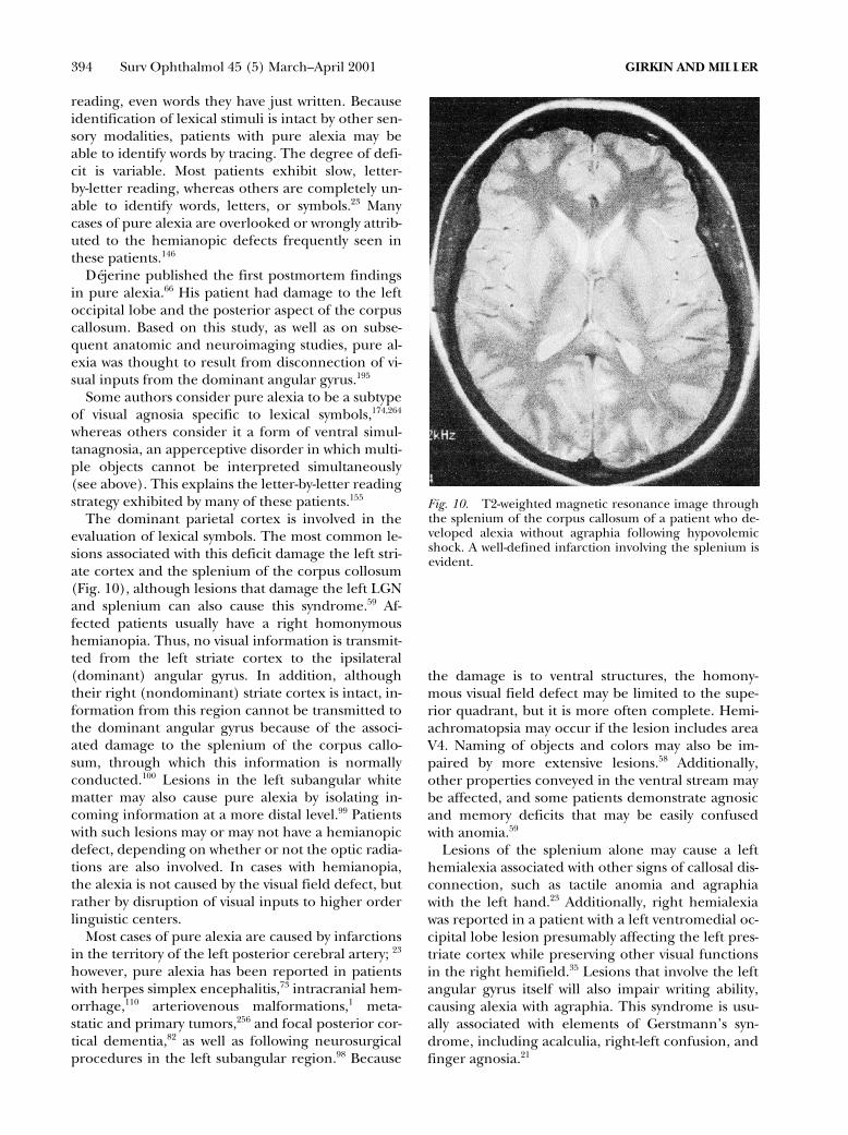
The dominant parietal cortex is involved in theevaluation of lexical symbols. The most common le-sions associated with this deficit damage the left stri-ate cortex and the splenium of the corpus collosum(Fig. 10), although lesions that damage the left LGNand splenium can also cause this syndrome.59 Af-fected patients usually have a right homonymoushemianopia. Thus, no visual information is transmit-ted from the left striate cortex to the ipsilateral(dominant) angular gyrus. In addition, althoughtheir right (nondominant) striate cortex is intact, in-formation from this region cannot be transmitted tothe dominant angular gyrus because of the associ-ated damage to the splenium of the corpus callo-sum, through which this information is normallyconducted.100 Lesions in the left subangular whitematter may also cause pure alexia by isolating in-coming information at a more distal level.99 Patientswith such lesions may or may not have a hemianopicdefect, depending on whether or not the optic radia-tions are also involved. In cases with hemianopia,the alexia is not caused by the visual field defect, butrather by disruption of visual inputs to higher orderlinguistic centers.
Most cases of pure alexia are caused by infarctionsin the territory of the left posterior cerebral artery; 23
however, pure alexia has been reported in patientswith herpes simplex encephalitis,73 intracranial hem-orrhage,110 arteriovenous malformations,1 meta-static and primary tumors,256 and focal posterior cor-tical dementia,82 as well as following neurosurgicalprocedures in the left subangular region.98 Because
the damage is to ventral structures, the homony-mous visual field defect may be limited to the supe-rior quadrant, but it is more often complete. Hemi-achromatopsia may occur if the lesion includes areaV4. Naming of objects and colors may also be im-paired by more extensive lesions.58 Additionally,other properties conveyed in the ventral stream maybe affected, and some patients demonstrate agnosicand memory deficits that may be easily confusedwith anomia.59
Lesions of the splenium alone may cause a lefthemialexia associated with other signs of callosal dis-connection, such as tactile anomia and agraphiawith the left hand.23 Additionally, right hemialexiawas reported in a patient with a left ventromedial oc-cipital lobe lesion presumably affecting the left pres-triate cortex while preserving other visual functionsin the right hemifield.35 Lesions that involve the leftangular gyrus itself will also impair writing ability,causing alexia with agraphia. This syndrome is usu-ally associated with elements of Gerstmann’s syn-drome, including acalculia, right-left confusion, andfinger agnosia.21
Fig. 10. T2-weighted magnetic resonance image throughthe splenium of the corpus callosum of a patient who de-veloped alexia without agraphia following hypovolemicshock. A well-defined infarction involving the splenium isevident.

CENTRAL DISORDERS OF VISION IN HUMANS 395
b. Color Anomia
Although cases exist in which color is dispropor-tionally affected, most cases of color anomia are partof a more general visual–verbal defect with coexist-ent pure alexia.91,218 Patients with color anomia canmatch colors: they do not have achromatopsia or anagnostic deficit. Their semantic recall of color is in-tact, and they are, thus, able to recall accurately thecolor of known objects (i.e., the color of a banana oran apple).
c. Object Anomia (Optic Aphasia)
Object anomia is characterized by a generalizeddefect in visual naming. Affected patients are unableto recall the names of objects presented visually, al-though their recall based on tactile and auditory in-put is preserved. Object matching and recognitionare also intact. Such patients may be able to describethe characteristics of an object and its purpose, butthey cannot provide the name of the object basedsolely on visual information. Often deficits in objectidentification are also present, making the separa-tion between agnosia and aphasia difficult. The ana-tomic bases for color anomia, pure alexia, and ob-ject anomia are not entirely clear, but probablyrepresent variations in the disruption of visual infor-mation reaching the angular gyrus. Object anomiamay results from more extensive isolation of visualinformation from the angular gyrus than that whichproduces color anomia or pure alexia alone.90 How-ever, color anomia, optic aphasia, and pure alexiacan occur together or in isolation. This double disso-ciation implies different anatomic substrates forthese processes.
3. Visual-Limbic Disconnection
The sensory-limbic system plays a critical role inprocessing the emotional impact of sensory stimuliand in reinforcing certain aspects of multimodalsensory memory traces that are emotionally relevantthrough reciprocal circuits involving the temporallobe.90 Thus, lesions of the limbic system may causemultimodal amnestic disorders that impair recall ofthe recent past and an inability to establish newmemories.69 Lesions that disconnect visual input tothis system may cause a modality-specific deficit. Twosuch disorders associated with lesions that disrupt vi-sual axons projecting to the ventromedial temporallobe are visual amnesia and visual hypoemotionality.These syndromes are rarely reported because objectagnosia or prosopagnosia often mask their presence.
a. Visual Amnesia
Visual amnesia is a modality-specific disorder inwhich patients are unable to learn new visual ob-jects, patterns, and faces, or to remember visual sur-
roundings. In contrast to visual agnosias, consoli-dated visual knowledge is intact, whereas visuallearning and recent recall are defective.215 Isolatedvisual amnesia is rare. More commonly, it occurs inassociation with visual agnosia.
b. Visual Hypoemotionality
Isolated visual hypoemotionality is a rare syn-drome in which emotional responses to visual stim-uli are blunted or absent. Emotional reaction toother sensory modalities, such as listening to music,remains intact. Visual hypoemotionality is most fre-quently associated with prosopagnosia.102 The dam-age in patients with this disorder is to the medial oc-cipitotemporal area, sparing the associative areasinvolved in object recognition. One patient with thiscondition was said to be so visually unarousable thathe cancelled his subscription to Playboy magazine.15
VIII. Visual Hallucinations and IllusionsVisual hallucinations are internally generated per-
ceptions. That hallucinations are consciously per-ceived implies that these internal perceptions utilizemechanisms similar to those that generate consciousvisual awareness, presumably through reciprocal ac-tivation of visual processing areas. Thus, we may gaininsight into the neurobiology of visual awarenessthrough the study of these internally generatedstimuli.
Hallucinations may be caused by a variety of con-ditions. They are a common manifestation of a vari-ety of psychiatric disorders, including schizophrenia,narcolepsy,161 psychotic depression, and mania.238
They also occur in patients with neurologic disor-ders, such as Alzheimer’s disease151 and epilepsy,89
metabolic derangements, such as during alcoholwithdrawal,176 and in febrile states. Strokes may alsocause hallucinations through release mechanismsand through seizures.6,131
Strokes involving the mesencephalon may causepeduncular hallucinations.229 These are usually mul-timodal complex hallucinations to which patientsmay lack insight. Generally, peduncular hallucinosisis associated with other signs of midbrain disease,such as ocular motor nerve palsies, hemiparesis, gaitataxia, or hemiparkinsonism.137 Interestingly, thesepatients almost invariably have inversion of thesleep-wake cycle, probably caused by damage to thepedunculopontine nucleus.166 Thalamic infarcts maycause peduncular hallucinations of past events.185
Drugs, both prescribed and illicit, can inducehallucinations.238 Some of the more common drugsassociated with visual hallucinations are indometha-cin,28 digoxin,41 bupropion,3 vincristine,93 cyclo-sporine,239 lithium,219 lidocaine,219 and dopamine.150
Visual hallucinations can also occur in patients given

396 Surv Ophthalmol 45 (5) March–April 2001 GIRKIN AND MILLER
topical homatropine,201 scopolamine,106 atropine,140
or other topical agents.140,287 Withdrawal of certainmedications, such as baclofen,203 may cause halluci-nations. The most frequent types of visual hallucina-tions are release hallucinations, visual migraines,and visual seizures.
A. RELEASE HALLUCINATIONS (CHARLESBONNET SYNDROME)
Visual hallucinations frequently occur in patientswho lose vision in both eyes, regardless of the loca-tion of the causative lesion or lesions. This associa-tion was first reported by Charles Bonnet, a Swissnaturalist, who described complex formed halluci-nations experienced by his 89-year-old grandfather,who, although cognitively intact, was blind from cat-aracts.27 The gentleman reported “amusing visions”of silent “figures of men, women, birds . . . etc.”Cogan postulated that hallucinations occurring inblind or near-blind individuals were “released” by re-moval of normal visual afferent input to associationcortex.42 He therefore called them “release phenom-ena,” and other authors refer to patients experienc-ing them as having the Charles Bonnet syndrome.
Release hallucinations may be simple or complex,with simple hallucinations occurring more fre-quently than complex ones. Simple hallucinationsusually consist of brief flashes of light, phosphenes,or various shapes and textures. Complex hallucina-tions are specific objects, such as people, animals,plants, or imaginary creatures. They may appear asblack-and-white figures, or they may be seen incolor. Some of these complex visions reflect past vi-sual memories. We recently examined a patient withcount fingers vision from retinitis pigmentosa whobegan seeing a visually detailed hallucination of hersister as she appeared 30 years earlier, when the pa-tient still retained relatively good vision. Another pa-tient had recurrent hallucinations of a previous busi-ness partner. Some patients experience progressionfrom simple to complex hallucinations.235
Release hallucinations often begin shortly afterthe loss of vision, but they may not develop untilyears later and may even precede visual loss.139 Theycan occur during sensory deprivation,187 for in-stance, in patients who have undergone bilateral oc-ular patching because of corneal damage from weld-ing injuries or in patients who are bilaterally patchedin preparation for ocular surgery (“black patch psy-chosis”). Patient surveys demonstrate that releasehallucinations are quite common, occurring in 11–13% of blind patients.117 Frequently, these patientsdo not mention them to their physicians, friends, orfamily, because they know they are experiencing hal-lucinations and are afraid that they will be consid-
ered “crazy.” This is in contradistinction to patientswith schizophrenia, who believe that their visual hal-lucinations are real.
Release hallucinations are most common in pa-tients with vision of 20/60 or worse in their bettereye. Age may play a role, as 80% of patients who ex-perience these types of hallucinations are over age60. Social isolation may also predispose to the phe-nomenon.117 Studies associating cognitive impair-ment and the development of dementia in patientswith release hallucinations have yielded conflictingresults.46,279 Also confusing are the development ofsuch hallucinations in patients with poor vision inone eye but normal or near-normal vision in theother. An association with periventricular white mat-ter lesions on MRI has been asserted but not con-firmed.230
Several theories have been proposed to explain re-lease hallucinations.225 Visual seizures have been sus-pected in patients with intracranial pathology, butthese patients tend to have repetitive stereotypedvisual phenomena associated with other signs of aseizure disorder (see below). Theories of sensorydeprivation are supported by sensory deprivation ex-periments and may be analogous to the musical hal-lucinations of deafness and the phantom limb phe-nomenon following amputation.30 Expansion of thereceptive fields of adjacent cortical neurons hasbeen found after retinal ablation in animals. Thedenervated cortical neurons may regain activitywithin several months.94 Thus, spontaneous dis-charges from denervated cells along with alterationsin cortical receptive fields may explain some cases.However, modification and activation of denervatedstriate cortex neurons cannot explain adequately thecomplex hallucinations experienced by some ofthese patients. Direct stimulation of the temporallobe produces complex visual hallucinations. In ad-dition, a recent fMRI study demonstrated activationof cerebral activity in ventral extrastriate visual cor-tex during hallucinations of color, faces, textures,and objects.81 Thus, activation of the associative vi-sual areas, disinhibited by the loss of visual input,may be responsible for at least some complexhallucinations.225
Most patients are not disturbed by release halluci-nations, and some patients may even enjoy them;however, identification of this syndrome is impor-tant to avoid unnecessary neuroimaging and psychi-atric evaluations.248 In rare patients, the hallucina-tions are troublesome, but no consistently effectivetreatment exists to treat them. Anticonvulsants,22 ha-loperidol,235 and tiapride10 have all been used withmixed success. Removing patients from socially iso-lated environments may abolish these hallucina-tions.45

CENTRAL DISORDERS OF VISION IN HUMANS 397
B. VISUAL MIGRAINES AND VISUAL SEIZURES
The typical fortification spectra of visual mi-graines (migraine with visual aura or visual aurawithout headache) usually consist of a moving arc ofeither colored or black-and-white zigzag lines thatexpand toward the periphery with concurrent in-crease in the size of lines (Fig. 11). These scintilla-tions reflect the cytoarchitectural arrangement ofthe orientation columns as a wave of excitatory activ-ity spreads across the striate cortex. The scotomathat trails behind fortification spectra represents anarea of transient inactivation of cortical neurons.These phenomena usually last about 20 minutesand may or may not be followed by headache. A vari-ety of atypical hallucinations also may occur dur-ing migraine attacks, including formed hallucina-tions, micropsia, macropsia, palinopsia, and visualallesthesia.120
Visual seizures involving the occipital or, occasion-ally, the temporal lobe may cause unformed halluci-nations similar to those experienced during a classicmigraine attack.273 Patients with visual seizures usu-ally lack the typical history of migraines, have anatypical frequency and duration of hallucinations,and often exhibit other seizure phenomena, such aseye deviation or rapid blinking. Temporal and evenoccipital lobe seizures may cause formed visual hal-lucinations, thus mimicking the Charles Bonnet syn-drome.138 Although a careful history usually will dif-ferentiate visual seizures from other visualhallucinations, the diagnosis in some cases can bemade only by seizure monitoring.246
C. VISUAL PERSEVERATION
Visual perseveration, or palinopsia (from Greekpalin, meaning “again”), refers to the pathologic
Fig. 11. Artist’s rendition of fortifi-cation spectra seen during a mi-graine. (Reprinted from Hupp SL,Kline LB, Corbett JJ120 with permis-sion of the authors. Artist: WilliamDean Mosher.)
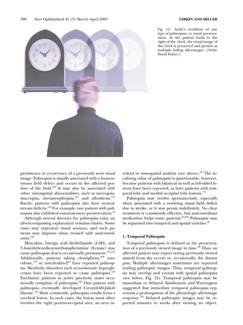
398 Surv Ophthalmol 45 (5) March–April 2001 GIRKIN AND MILLER
persistence or recurrence of a previously seen visualimage. Palinopsia is usually associated with a homon-ymous field defect and occurs in the affected por-tion of the field.169 It may also be associated withother visuospatial abnormalities, such as micropsia,macropsia, metamorphopsia,275 and allesthesia.275
Rarely, patients with palinopsia also have ventral-stream deficits.144 For example, one patient with pali-nopsia also exhibited somatosensory perseveration.52
Although several theories for palinopsia exist, anall-encompassing explanation remains elusive. Somecases may represent visual seizures, and such pa-tients may improve when treated with anticonvul-sants.182
Mescaline, lysergic acid diethylamide (LSD), and3,4-methylenedioxymethamphetamine (Ecstasy) maycause palinopsia that is occasionally permanent.134,154
Additionally, patients taking clomiphene,200 traz-odone,119 or interleukin-283 have reported palinop-sia. Metabolic disorders such as nonketotic hypergly-cemia have been reported to cause palinopsia.124
Psychiatric patients in active psychotic states occa-sionally complain of palinopsia.163 One patient withpalinopsia eventually developed Creutzfeldt-Jakobdisease.163 Most commonly, palinopsia results from acerebral lesion. In such cases, the lesion most ofteninvolves the right parieto-occipital area, an area in-
volved in visuospatial analysis (see above).40 The lo-calizing value of palinopsia is questionable, however,because patients with bilateral as well as left-sided le-sions have been reported, as have patients with tem-poral lobe and medial occipital lobe lesions.175
Palinopsia may resolve spontaneously, especiallywhen associated with a resolving visual field deficitdue to stroke, or it may persist indefinitely. No cleartreatment is consistently effective, but anticonvulsantmedication helps some patients.25,236 Palinopsia maybe separated into temporal and spatial varieties.46
1. Temporal Palinopsia
Temporal palinopsia is defined as the persevera-tion of a previously viewed image in time.20 Thus, anaffected patient may report seeing previously viewedstimuli from the recent or, occasionally, the distantpast. Multiple afterimages sometimes are reportedtrailing palinoptic images. Thus, temporal palinop-sia may overlap and coexist with spatial palinopsia(see below, Fig. 12). Temporal palinopsia may beimmediate or delayed. Kinsbourne and Warringtonsuggested that immediate temporal palinopsia rep-resents a prolongation of the physiologic afterimageresponse.134 Delayed palinoptic images may be re-ported minutes to weeks after viewing an object.
Fig. 12. Artist’s rendition of onetype of palinopsia, or visual persever-ation. As the patient looks to theright of the clock, the visual image ofthe clock is preserved and persists asmultiple fading afterimages. (Artist:David Fisher.)

CENTRAL DISORDERS OF VISION IN HUMANS 399
Some patients describe a combination of bothtypes.169
2. Spatial Palinopsia
Spatial palinopsia is defined as persistence of a vi-sual image in space. This form of palinopsia can besubdivided into illusory visual spread and cerebralpolyopia. In illusory visual spread, patients report ex-tension (bleeding) of a pattern beyond the bound-aries of the object being viewed.20 In these cases, apattern from one object, such as striped shirt,spreads to appear to cover adjacent objects. In cere-bral polyopsia, copies of an object are reproducedwithin the visual field, often in geometric rows orcolumns.
In contrast to ocular polyopia caused by opticalaberrations, the images in cerebral polyopia are seenwith equal clarity, do not improve with pinhole test-ing, and lack the ghost-image quality seen with opti-cal disorders.115 Patients may report seeing only twoimages, in which case strabismus must be a consider-ation. Other patients, however, report seeing dozensor even hundreds of images (entomopia).159
A homonymous field defect is often present in pa-tients with cerebral polyopia, as may achromatopsiaor ventral-stream deficits.20 Most often, cerebralpolyopia develops in patients with dominant or non-dominant parietal or parieto-occipital lesions. Oc-cipital lobe injures, encephalitis, seizures, multiplesclerosis, migraines,170 and tumors245 have all beenreported to cause this disorder. The origin of cere-bral polyopia remains unclear.
IX. Concluding RemarksAs investigators uncover the complexities of the
higher visual system, a greater understanding ofthese clinical syndromes will become clear. Familiar-ity with these syndromes will enable the ophthalmol-ogist to identify patients with such disorders and per-form appropriate testing. Additionally, the study ofhigher cortical disorders may provide insight intothe mechanisms of visual awareness and the globalsense of the perception of our environment. Under-standing this crucial function of the mind will bean integral step in forming a global theory ofconsciousness.
Methods of Literature SearchThe literature search was conducted through
Medline, including all years covered in the database.Secondary sources primarily for historic articles wereobtained from our own files. Several references andthe overall organizational scheme were obtainedfrom Rizzo and Barton’s extensive review of this sub-ject, “Central Disorders of Visual Function” in the1998 edition of Walsh and Hoyt. Keywords used in
the Medline search were: alexia, agraphia, anomia,Anton syndrome, allesthesia, blindsight, brain damage,brain mapping, color perception, color vision defects, cere-bral cortex, dyslexia, epilepsy, form perception, limbic sys-tem, motion perception, neglect, parietal lobe, palinopsia,psychophysics, retinal ganglion cells, space perception, sleepdeprivations, spatial neglect, space perception, occipitallobe, optic aphasia, visual cortex, visual hallucinations,visual fields, visual pathways, visual hypoemotionality, vi-sual attention, visual perception, vision disorders, V1, V2,V3, V3A, V4, and V5. Additionally, specific authornames that were used in the search were: Anderson,Barbur, Dacey, Damasio, Hubel, Livingstone, Rizzo, Un-gerleider, Weiskrantz, Zeki, and Zihl.
References1. Ajax ET, Schenkenberg T, Kosteljanetz M: Alexia without
agraphia and the inferior splenium. Neurology 27:685–8,1977
2. Aldrich MS, Vanderzant CW, Alessi AG, et al: Ictal corticalblindness with permanent visual loss. Epilepsia 30:116–20,1989
3. Ames D, Wirshing WC, Szuba MP: Organic mental disor-ders associated with bupropion in three patients. J Clin Psy-chiatry 53:53–5, 1992
4. Andersen RA: Multimodal integration for the representa-tion of space in the posterior parietal cortex. Philos TransR Soc Lond B Biol Sci 352:1421–8, 1997
5. Andersen RA, Zipser D: The role of the posterior parietalcortex in coordinate transformations for visual-motor inte-gration. Can J Physiol Pharmacol 66:488–501, 1988
6. Anderson SW, Rizzo M: Hallucinations following occipitallobe damage: the pathological activation of visual represen-tations. J Clin Exp Neuropsychol 16:651–63, 1994
7. Anton G: Über Herderkrankungren der Gehirns, welchevom Patienten selbst nicht wahrgenommmen werden.Wiener Klinsche Wochenshrift 33:227–9, 1989
8. Ardila A, Botero M, Gomez J: Palinopsia and visual allesthe-sia. Int J Neurosci 32:775–82, 1987
9. Argenta PA, Morgan MA: Cortical blindness and Antonsyndrome in a patient with obstetric hemorrhage. ObstetGynecol 91:810–2, 1998
10. Badino R, Trucco M, Caja A, et al: Release hallucinationsand tiapride. Ital J Neurol Sci 15:183–7, 1994
11. Balint R: Seelenlähmung des Schauens, optische Ataxie,räumliche Strörung der Aufmerksamkeit. Monatsschr Psy-chiat Neurol 25:51–81, 1909
12. Barbur JL, Watson JD, Frackowiak RS, Zeki S: Conscious vi-sual perception without V1. Brain 116:1293–302, 1993
13. Barrett AM, Beversdorf DQ, Crucian GP, Heilman KM: Ne-glect after right hemisphere stroke: a smaller floodlight fordistributed attention [see comments]. Neurology 51:972–8,1998
14. Barton JJ: Higher cortical visual function. Curr Opin Oph-thalmol 9:40–5, 1998
15. Bauer RM: Visual hypoemotionality as a symptom of visual-limbic disconnection in man. Arch Neurol 39:702–8, 1982
16. Beason-Held LL, Purpura KP, Van Meter JW, et al: PET re-veals occipitotemporal pathway activation during elemen-tary form perception in humans. Vis Neurosci 15:503–10,1998
17. Beckers G, Homberg V: Cerebral visual motion blindness:transitory akinetopsia induced by transcranial magneticstimulation of human area V5. Proc R Soc Lond B Biol Sci249:173–8, 1992
18. Beckers G, Zeki S: The consequences of inactivating areasV1 and V5 on visual motion perception. Brain 118:49–60,1995

400 Surv Ophthalmol 45 (5) March–April 2001 GIRKIN AND MILLER
19. Bender DB: Electrophysiological and behavioral experi-ments on the primate pulvinar. Prog Brain Res 75:55–65,1988
20. Bender MB, Feldman M, Sobin AJ: Palinopsia. Brain 91:321–38, 1968
21. Benton AL: Gerstmanns syndrome. Arch Neurol 49:445–7,1992
22. Bhatia MS, Khastgir U, Malik SC: Charles Bonnet syn-drome. Br J Psychiatry 161:409–10, 1992
23. Binder JR, Mohr JP: The topography of callosal readingpathways. A case-control analysis. Brain 115:1807–26, 1992
24. Blythe IM, Bromley JM, Kennard C, Ruddock KH: Visualdiscrimination of target displacement remains after dam-age to the striate cortex in humans. Nature 320:619–21,1986
25. Blythe IM, Bromley JM, Ruddock KH, et al: A study of sys-tematic visual perseveration involving central mechanisms.Brain 109:661–75, 1986
26. Blythe IM, Kennard C, Ruddock KH: Residual vision in pa-tients with retrogeniculate lesions of the visual pathways.Brain 110:887–905, 1987
27. Bonnet C. Essai Analytique Sur Les Facultes de L’Ame.Copenhagen, Philber, 1769, ed 2
28. Braddock LE, Heard RN: Visual hallucinations due to in-domethacin: a case report. Int Clin Psychopharmacol 1:263–6, 1986
29. Brandt T, Dieterich M: The vestibular cortex. Its locations,functions, and disorders. Ann NY Acad Sci 871:293–312,1999
30. Brasic JR: Hallucinations. Percept Mot Skills 86:851–77,1998
31. Bruyer R, Laterre C, Seron X, et al: A case of prosopagno-sia with some preserved covert remembrance of familiarfaces. Brain Cogn 2:257–84, 1983
32. Burkhalter A, Bernardo KL: Organization of corticocorti-cal connections in human visual cortex. Proc Natl Acad SciUSA 86:1071–5, 1989
33. Campion J: Apperceptive agnosia : the specification anddescription of constructs, in Humphery GW, Riddoch MJ(eds): Visual Object Processing: A Cognitive Neuropsycho-logical Approach. London, Lawrence Erlbaum Associates,1987
34. Campion J, Latto R: Apperceptive agnosia due to carbonmonoxide poisoning. An interpretation based on criticalband masking from disseminated lesions. Behav Brain Res15:227–40, 1985
35. Castro-Caldas A, Salgado V: Right hemifield alexia withouthemianopia. Arch Neurol 41:84–7, 1984
36. Celesia GG, Bushnell D, Toleikis SC, Brigell MG: Corticalblindness and residual vision: is the second visual system inhumans capable of more than rudimentary visual percep-tion? Neurology 41:862–9, 1991
37. Chelazzi L: Neural mechanisms for stimulus selection incortical areas of the macaque subserving object vision. Be-hav Brain Res 71:125–34, 1995
38. Clarke S, Miklossy J: Occipital cortex in man: organizationof callosal connections, related myelo- and cytoarchitec-ture, and putative boundaries of functional visual areas. JComp Neurol 298:188–214, 1990
39. Clarke S, Walsh V, Schoppig A, et al: Colour constancy im-pairments in patients with lesions of the prestriate cortex.Exp Brain Res 123:154–8, 1998
40. Cleland PG, Saunders M, Rosser R: An unusual case of vi-sual perseveration. J Neurol Neurosurg Psychiatry 44:262–3, 1981
41. Closson RG: Visual hallucinations as the earliest symptomof digoxin intoxication. Arch Neurol 40:386, 1983
42. Cogan DG: Visual hallucinations as release phenomena. Al-brecht Von Graefes Arch Klin Exp Ophthalmol 188:139–50, 1973
43. Colby CL: The neuroanatomy and neurophysiology of at-tention. J Child Neurol 6 Suppl:S90–118, 1991
44. Colby CL, Duhamel JR: Spatial representations for action inparietal cortex. Brain Res Cogn Brain Res 5:105–15, 1996
45. Cole MG: Charles Bonnet hallucinations: a case series. CanJ Psychiatry 37:267–70, 1992
46. Cole MG: Charles Bonnet hallucinations: a case series. CanJ Psychiatry 37:267–70, 1992
47. Corbetta M, Miezin FM, Shulman GL, Petersen SE: A PETstudy of visuospatial attention. J Neurosci 13:1202–26, 1993
48. Courtney SM, Ungerleider LG: What fMRI has taught usabout human vision. Curr Opin Neurobiol 7:554–61, 1997
49. Cowey A, Heywood CA: There’s more to colour than meetsthe eye. Behav Brain Res 71:89–100, 1995
50. Cowey A, Stoerig P: Spectral sensitivity in hemianopicmacaque monkeys. Eur J Neurosci 11:2114–20, 1999
51. Cowey A, Stoerig P: The neurobiology of blindsight.Trends Neurosci 14:140–5, 1991
52. Cummings JL, Syndulko K, Goldberg Z, Treiman DM: Pali-nopsia reconsidered. Neurology 32:444–7, 1982
53. Czeisler CA, Shanahan TL, Klerman EB, et al: Suppressionof melatonin secretion in some blind patients by exposure tobright light [see comments]. N Engl J Med 332:6–11, 1995
54. Dacey DM: Circuitry for color coding in the primate retina.Proc Natl Acad Sci USA 93:582–8, 1996
55. Dacey DM: Physiology, morphology and spatial densities ofidentified ganglion cell types in primate retina. Ciba FoundSymp 184:12–28; discussion 28–34, 63–70, 1994
56. Dacey DM, Lee BB: The blue-on opponent pathway in pri-mate retina originates from a distinct bistratified ganglioncell type. Nature 367:731–5, 1994
57. Damasio A, Yamada T, Damasio H, et al: Central achro-matopsia: behavioral, anatomic, and physiologic aspects.Neurology 30:1064–71, 1980
58. Damasio AR, Damasio H: Hemianopia, hemiachromatop-sia and the mechanisms of alexia. Cortex 22:161–9, 1986
59. Damasio AR, Damasio H: The anatomic basis of pure al-exia. Neurology 33:1573–83, 1983
60. Damasio AR, Damasio H, Van Hoesen GW: Prosopagnosia:anatomic basis and behavioral mechanisms. Neurology 32:331–41, 1982
61. Damasio AR, McKee J, Damasio H: Determinants of perfor-mance in color anomia. Brain Lang 7:74–85, 1979
62. Damasio H, Frank R: Three-dimensional in vivo mappingof brain lesions in humans. Arch Neurol 49:137–43, 1992
63. De Haan EH, Campbell R: A fifteen year follow-up of a caseof developmental prosopagnosia. Cortex 27:489–509, 1991
64. De Haan EH, Young A, Newcombe F: Faces interfere withname classification in a prosopagnosic patient. Cortex 23:309–16, 1987
65. De Renzi E, Scotti G, Spinnler H: Perceptual and associa-tive disorders of visual recognition. Relationship to the sideof the cerebral lesion. Neurology 19:634–42, 1969
66. Dejerine J: Contribution a l’etude anatomoclinique et clin-ique des differentes varietes de cecite verbale. Memoires dela Soc de Biologie 4:61–90, 1892
67. Desimone R, Schein SJ: Visual properties of neurons inarea V4 of the macaque: sensitivity to stimulus form. J Neu-rophysiol 57:835–68, 1987
68. Desimone R, Schein SJ, Moran J, Ungerleider LG: Con-tour, color and shape analysis beyond the striate cortex. Vi-sion Res 25:441–52, 1985
69. Drew WG, Weet CR, De Rossett SE, Batt JR: Effects of hip-pocampal brain damage on auditory and visual recentmemory: comparison with marijuana-intoxicated subjects.Biol Psychiatry 15:841–58, 1980
70. Dufort PA, Lumsden CJ: Color categorization and colorconstancy in a neural network model of V4. Biol Cybern 65:293–303, 1991
71. Eden GF, VanMeter JW, Rumsey JM, et al: Abnormal pro-cessing of visual motion in dyslexia revealed by functionalbrain imaging [see comments]. Nature 382:66–9, 1996
72. Engel SA, Rumelhart DE, Wandell BA, et al: fMRI of hu-man visual cortex [letter] [published erratum appears inNature 1994 Jul 14;370(6485):106]. Nature 369:525, 1994
73. Erdem S, Kansu T: Alexia without either agraphia or hemi-anopia in temporal lobe lesion due to herpes simplex en-cephalitis. J Neuroophthalmol 15:102–4, 1995

CENTRAL DISORDERS OF VISION IN HUMANS 401
74. Essen DC, Zeki SM: The topographic organization ofrhesus monkey prestriate cortex. J Physiol (Lond) 277:193–226, 1978
75. Farah MJ: Agnosia. Curr Opin Neurobiol 2:162–4, 199276. Farah MJ. Visual Agnosia: Disorders of Object Recognition
and What They Tell us about Normal Vision. Cambridge,MIT Press, 1990
77. Felleman DJ, Van Essen DC: Distributed hierarchical pro-cessing in the primate cerebral cortex. Cereb Cortex 1:1–47, 1991
78. Fendrich R, Wessinger CM, Gazzaniga MS: Residual visionin a scotoma: implications for blindsight [see comments].Science 258:1489–91, 1992
79. ffytche DH, Guy CN, Zeki S: Motion specific responsesfrom a blind hemifield. Brain 119:1971–82, 1996
80. ffytche DH, Guy CN, Zeki S: The parallel visual motion in-puts into areas V1 and V5 of human cerebral cortex. Brain118:1375–94, 1995
81. ffytche DH, Howard RJ, Brammer MJ, et al: The anatomyof conscious vision: an fMRI study of visual hallucinations.Nat Neurosci 1:738–42, 1998
82. Freedman L, Costa L: Pure alexia and right hemiachro-matopsia in posterior dementia. J Neurol Neurosurg Psy-chiatry 55:500–2, 1992
83. Friedman DI, Hu EH, Sadun AA: Neuro-ophthalmic com-plications of interleukin 2 therapy. Arch Ophthalmol 109:1679–80, 1991
84. Friedrich FJ, Egly R, Rafal RD, Beck D: Spatial attentiondeficits in humans: a comparison of superior parietal andtemporal-parietal junction lesions. Neuropsychology 12:193–207, 1998
85. Frisen L: High-pass resolution perimetry. A clinical review.Doc Ophthalmol 83:1–25, 1993
86. Galletti C, Battaglini PP, Fattori P: Eye position influenceon the parieto-occipital area PO (V6) of the macaque mon-key. Eur J Neurosci 7:2486–501, 1995
87. Galletti C, Battaglini PP, Fattori P: Parietal neurons encod-ing spatial locations in craniotopic coordinates. Exp BrainRes 96:221–9, 1993
88. Gandolfo E: Stato-kinetic dissociation in subjects with nor-mal and abnormal visual fields. Eur J Ophthalmol 6:408–14, 1996
89. Gastaut H, Zifkin BG: Ictal visual hallucinations of numer-als. Neurology 34:950–3, 1984
90. Geschwind N: Disconnexion syndromes in animals andman. II. Brain 88:585–644, 1965
91. Geschwind N, Fusillo M: Color-naming defects in associa-tion with alexia. Arch Neurol 15:137–46, 1966
92. Ghose GM, Tso DY: Form processing modules in primatearea V4. J Neurophysiol 77:2191–6, 1997
93. Ghosh K, Sivakumaran M, Murphy P, et al: Visual halluci-nations following treatment with vincristine. Clin Lab Hae-matol 16:355–7, 1994
94. Gilbert CD, Wiesel TN: Receptive field dynamics in adultprimary visual cortex. Nature 356:150–2, 1992
95. Girkin CA, Perry JD, Miller NR: Visual environmental rota-tion: a novel disorder of visiospatial integration. J Neu-roophthalmol 19:13–6, 1999
96. Goldberg ME, Segraves MA: Visuospatial and motor atten-tion in the monkey. Neuropsychologia 25:107–18, 1987
97. Green GJ, Lessell S: Acquired cerebral dyschromatopsia.Arch Ophthalmol 95:121–8, 1977
98. Greenblatt SH: Left occipital lobectomy and the preangu-lar anatomy of reading. Brain Lang 38:576–95, 1990
99. Greenblatt SH: Subangular alexia without agraphia orhemianopsia. Brain Lang 3:229–45, 1976
100. Greenblatt SH: Alexia without agraphia or hemianopsia. An-atomical analysis of an autopsied case. Brain 96:307–16, 1973
101. Greve EL: Single and multiple stimulus static perimetry inglaucoma; the two phases of perimetry. Thesis. Doc Oph-thalmol 36:1–355, 1973
102. Habib M: Visual hypoemotionality and prosopagnosia asso-ciated with right temporal lobe isolation. Neuropsycholo-gia 24:577–82, 1986
103. Hachinski VC, Porchawka J, Steele JC: Visual symptoms inthe migraine syndrome. Neurology 23:570–9, 1973
104. Hackley SA, Johnson LN: Distinct early and late subcompo-nents of the photic blink reflex: response characteristics inpatients with retrogeniculate lesions. Psychophysiology 33:239–51, 19
105. Hadjikhani N, Liu AK, Dale AM, et al: Retinotopy andcolor sensitivity in human visual cortical area V8 [see com-ments]. Nat Neurosci 1:235–41, 1998
106. Hamborg-Petersen B, Nielsen MM, Thordal C: Toxic effectof scopolamine eye drops in children. Acta Ophthalmol(Copenh) 62:485–8, 1984
107. Hanley JR, Pearson NA, Young AW: Impaired memory fornew visual forms. Brain 113:1131–48, 1990
108. Hausser CO, Robert F, Giard N: Balints syndrome. Can JNeurol Sci 7:157–61, 1980
109. Haxby JV, Grady CL, Horwitz B, et al: Dissociation of objectand spatial visual processing pathways in human extrastri-ate cortex. Proc Natl Acad Sci USA 88:1621–5, 1991
110. Henderson VW: Anatomy of posterior pathways in reading:a reassessment. Brain Lang 29:119–33, 1986
111. Hendry SH, Yoshioka T: A neurochemically distinct thirdchannel in the macaque dorsal lateral geniculate nucleus.Science 264:575–7, 1994
112. Heo K, Kim SJ, Kim JH, et al: False lateralization of seizureperceived by a patient with infarction of the right parietallobe who showed the neglect syndrome. Epilepsia 38:122–3, 1997
113. Heywood CA, Cowey A, Newcombe F: On the role of parvo-cellular (P) and magnocellular (M) pathways in cerebralachromatopsia. Brain 117:245–54, 1994
114. Heywood CA, Gadotti A, Cowey A: Cortical area V4 and itsrole in the perception of color. J Neurosci 12:4056–65,1992
115. Hirst LW, Miller NR, Johnson RT: Monocular polyopia.Arch Neurol 40:756–7, 1983
116. Hof PR, Bouras C, Constantinidis J, Morrison JH: Selectivedisconnection of specific visual association pathways incases of Alzheimers disease presenting with Balints syn-drome. J Neuropathol Exp Neurol 49:168–84, 1990
117. Holroyd S, Rabins PV, Finkelstein D, et al: Visual hallucina-tions in patients with macular degeneration [see com-ments]. Am J Psychiatry 149:1701–6, 1992
118. Horton JC, Hoyt WF: Quadrantic visual field defects. A hall-mark of lesions in extrastriate (V2/V3) cortex. Brain 114:1703–18, 1991
119. Hughes MS, Lessell S: Trazodone-induced palinopsia. ArchOphthalmol 108:399–400, 1990
120. Hupp SL, Kline LB, Corbett JJ: Visual disturbances of mi-graine. Surv Ophthalmol 33:221–36, 1989
121. Jacobs L: Visual allesthesia. Neurology 30:1059–63, 1980122. Jacome DE: Subcortical prosopagnosia and anosognosia.
Am J Med Sci 292:386–8, 1986123. Johnson CA, Samuels SJ: Screening for glaucomatous visual
field loss with frequency-doubling perimetry. Invest Oph-thalmol Vis Sci 38:413–25, 1997
124. Johnson SF, Loge RV: Palinopsia due to nonketotic hyper-glycemia. West J Med 148:331–2, 1988
125. Johnston JL, Sharpe JA, Morrow MJ: Spasm of fixation: aquantitative study. J Neurol Sci 107:166–71, 1992
126. Joseph R: Confabulation and delusional denial: frontallobe and lateralized influences. J Clin Psychol 42:507–20,1986
127. Kanwisher N, McDermott J, Chun MM: The fusiform facearea: a module in human extrastriate cortex specialized forface perception. J Neurosci 17:4302–11, 1997
128. Karnath HO: Spatial orientation and the representation ofspace with parietal lobe lesions. Philos Trans R Soc Lond BBiol Sci 352:1411–9, 1997
129. Kennard C, Lawden M, Morland AB, Ruddock KH: Colouridentification and colour constancy are impaired in a pa-tient with incomplete achromatopsia associated with pres-triate cortical lesions. Proc R Soc Lond B Biol Sci 260:169–75, 1995
402 Surv Ophthalmol 45 (5) March–April 2001 GIRKIN AND MILLER
130. Kertesz A: Visual agnosia: the dual deficit of perceptionand recognition. Cortex 15:403–19, 1979
131. Kim SM, Park CH, Intenzo CM, Zhang J: Brain SPECT in apatient with post-stroke hallucination. Clin Nucl Med 18:413–6, 1993
132. Kinsbourne M: Hemi-neglect and hemisphere rivalry. AdvNeurol 18:41–9, 1977
133. Kinsbourne M: A model for the mechanism of unilateralneglect of space. Trans Am Neurol Assoc 95:143–6, 1970
134. Kinsbourne M, Warrington E: A study of visual persevera-tion. Brain:321–338, 1963
135. Kinsbourne M, Warrington E: A disorder of simultaneousfrom perception. Brain 85:461–8, 1962
136. Knierim JJ, van Essen DC: Neuronal responses to static tex-ture patterns in area V1 of the alert macaque monkey. JNeurophysiol 67:961–80, 1992
137. Kolmel HW: Peduncular hallucinations. J Neurol 238:457–9, 1991
138. Kolmel HW: Complex visual hallucinations in the hemian-opic field. J Neurol Neurosurg Psychiatry 48:29–38, 1985
139. Kolmel HW: Coloured patterns in hemianopic fields. Brain107:155–67, 1984
140. Kortabarria RP, Duran JA, Chacon JR, et al: Toxic psychosisfollowing cycloplegic eyedrops. DICP 24:708–9, 1990
141. Kracke I: Developmental prosopagnosia in Asperger syn-drome: presentation and discussion of an individual case[see comments]. Dev Med Child Neurol 36:873–86, 1994
142. Land EH, McCann JJ: Lightness and retinex theory. J OptSoc Am 61:1–11, 1971
143. Landis T, Cummings JL, Benson DF, Palmer EP: Loss of to-pographic familiarity. An environmental agnosia. ArchNeurol 43:132–6, 1986
144. Landis T, Cummings JL, Christen L, et al: Are unilateralright posterior cerebral lesions sufficient to causeprosopagnosia? Clinical and radiological findings in six ad-ditional patients. Cortex 22:243–52, 1986
145. Landis T, Graves R, Benson DF, Hebben N: Visual recogni-tion through kinaesthetic mediation. Psychol Med 12:515–31, 1982
146. Landis T, Grusser OJ. Visual agnosias and other distur-bances of visual perception, in Cronly-Dillong JR (ed): Vi-sion and Visual Dysfunction, Vol. 12. Macmillan Press
147. Lapresle J, Metreau R, Annabi A: Transient achromatopsiain vertebrobasilar insufficiency. J Neurol 215:155–8, 1977
148. Larrabee GJ, Levin HS, Huff FJ, et al: Visual agnosia con-trasted with visual-verbal disconnection. Neuropsychologia23:1–12, 1985
149. Lawden MC, Cleland PG: Achromatopsia in the aura of mi-graine. J Neurol Neurosurg Psychiatry 56:708–9, 1993
150. Lera G, Vaamonde J, Rodriguez M, Obeso JA: Cabergolinein Parkinsons disease: long-term follow-up [see com-ments]. Neurology 43:2587–90, 1993
151. Lerner AJ, Koss E, Patterson MB, et al: Concomitants of visualhallucinations in Alzheimers disease. Neurology 44:523–7, 1994
152. Lessel S: Higher disorders of visual function: negative phe-nomena, in Glaser J, Smith J (eds): Neuro-ophthalmology,Vol 8. St. Louis, Mosby, 1975, pp 3–4
153. Leventhal AG, Rodieck RW, Dreher B: Retinal ganglioncell classes in the Old World monkey: morphology and cen-tral projections. Science 213:1139–42, 1981
154. Levi L, Miller NR: Visual illusions associated with previousdrug abuse. J Clin Neuroophthalmol 10:103–10, 1990
155. Levine DN, Calvanio R: A study of the visual defect in ver-bal alexia-simultanagnosia. Brain 101:65–81, 1978
156. Lissauer H: Das Krankheitschild der Apraxie. Monatss-chrift fur Psychiatrie und Neurologie 21:222–70, 1890
157. Livingstone MS, Hubel DH: Psychophysical evidence forseparate channels for the perception of form, color, move-ment, and depth. J Neurosci 7:3416–68, 1987
158. Livingstone MS, Rosen GD, Drislane FW, Galaburda AM:Physiological and anatomical evidence for a magnocellulardefect in developmental dyslexia [published erratum ap-pears in Proc Natl Acad Sci USA 1993 Mar 15;90(6):2556].Proc Natl Acad Sci USA 88:7943–7, 1991
159. Lopez JR, Adornato BT, Hoyt WF: Entomopia: a remark-able case of cerebral polyopia. Neurology 43:2145–6, 1993
160. Lueck CJ, Zeki S, Friston KJ, et al: The colour centre in thecerebral cortex of man. Nature 340:386–9, 1989
161. Manford M, Andermann F: Complex visual hallucinations.Clinical and neurobiological insights. Brain 121: 1819-40,1998
162. Marcel AJ: Blindsight and shape perception: deficit of vi-sual consciousness or of visual function? Brain 121:1565–88, 1998
163. Marneros A, Korner J: Chronic palinopsia in schizophre-nia. Psychopathology 26:236–9, 1993
164. Martin PR, White AJ, Goodchild AK, et al: Evidence thatblue-on cells are part of the third geniculocortical pathwayin primates. Proc Natl Acad Sci USA 27:5900–5, 1997
165. McFadzean RM, Hadley DM: Homonymous quadrantano-pia respecting the horizontal meridian. A feature of striateand extrastriate cortical disease. Neurology 49:1741–6,1997
166. McKee AC, Levine DN, Kowall NW, Richardson EP Jr: Pe-duncular hallucinosis associated with isolated infarction ofthe substantia nigra pars reticulata. Ann Neurol 27:500–4,1990
167. McKeefry DJ, Zeki S: The position and topography of thehuman colour centre as revealed by functional magneticresonance imaging. Brain 120:2229–42, 1997
168. Meadows JC: Disturbed perception of colours associatedwith localized cerebral lesions. Brain 97:615–32, 1974
169. Meadows JC, Munro SS: Palinopsia. J Neurol NeurosurgPsychiatry 40:5–8, 1977
170. Melen O, Olson SF, Hodes BL: Visual disturbances in mi-graine. Postgrad Med 64:139–43, 1978
171. Merigan WH, Nealey TA, Maunsell JH: Visual effects of le-sions of cortical area V2 in macaques. J Neurosci 13:3180–91, 1993
172. Mestre DR, Brouchon M, Ceccaldi M, Poncet M: Percep-tion of optical flow in cortical blindness: a case report. Neu-ropsychologia 30:783–95, 1992
173. Mesulam MM: Large-scale neurocognitive networks anddistributed processing for attention, language, and mem-ory. Ann Neurol 28:597–613, 1990
174. Mesulam MM: Patterns in behavioral neuroanatomy, inMesulam MM (ed): Principles of Behavioral Neurology.Philadelphia, FA Davis Company, 1985, pp 1–70
175. Michel EM, Troost BT: Palinopsia: cerebral localizationwith computed tomography. Neurology 30:887–9, 1980
176. Miller NR: Bilateral visual loss and simultagnosia after lum-boperitoneal shunt for pseudotumor cerebri. J Neurooph-thalmol 17:36–8, 1997
177. Misra M, Rath S, Mohanty AB: Anton syndrome and corti-cal blindness due to bilateral occipital infarction. Indian JOphthalmol 37:196, 1989
178. Mohler CW, Wurtz RH: Role of striate cortex and superiorcolliculus in visual guidance of saccadic eye movements inmonkeys. J Neurophysiol 40:74–94, 1977
179. Morecraft RJ, Geula C, Mesulam MM: Architecture of con-nectivity within a cingulo-fronto-parietal neurocognitivenetwork for directed attention. Arch Neurol 50:279–84,1993
180. Morland AB, Ogilvie JA, Ruddock KH, Wright JR: Orienta-tion discrimination is impaired in the absence of the striatecortical contribution to human vision. Proc R Soc Lond BBiol Sci 263:633–40, 1996
181. Morris JS, Ohman A, Dolan RJ: A subcortical pathway tothe right amygdala mediating unseen fear. Proc Natl AcadSci USA 96:1680–5, 1999
182. Muller T, Buttner T, Kuhn W, et al: Palinopsia as sensoryepileptic phenomenon. Acta Neurol Scand 91:433–6, 1995
183. Nakajima M, Yasue M, Kaito N, et al: [A case of visual alles-thesia]. No To Shinkei 43:1081–5, 1991
184. Nobre AC, Sebestyen GN, Gitelman DR, et al: Functionallocalization of the system for visuospatial attention usingpositron emission tomography. Brain 120:515–33, 1997
185. Noda S, Mizoguchi M, Yamamoto A: Thalamic experiential
CENTRAL DISORDERS OF VISION IN HUMANS 403
hallucinosis. J Neurol Neurosurg Psychiatry 56:1224–6,1993
186. Ogren MP, Mateer CA, Wyler AR: Alterations in visually re-lated eye movements following left pulvinar damage inman. Neuropsychologia 22:187–96, 1984
187. Olbrich HM, Engelmeier MP, Pauleikhoff D, Waubke T: Vi-sual hallucinations in ophthalmology. Graefes Arch ClinExp Ophthalmol 225:217–20, 1987
188. Pasik T, Pasik P: Extrageniculostriate vision in the monkey.IV. Critical structures for light vs. no-light discrimination.Brain Res 56:165–82, 1973
189. Pasik T, Pasik P: The visual world of monkeys deprived ofstriate cortex: effective stimulus parameters and the impor-tance of the accessory optic system. Vis Res (Suppl):419–35,1971
190. Paulson HL, Galetta SL, Grossman M, Alavi A: Hemiachro-matopsia of unilateral occipitotemporal infarcts. Am JOphthalmol 118:518–23, 1994
191. Perenin MT, Vighetto A: Optic ataxia: a specific disruptionin visuomotor mechanisms. I. Different aspects of the defi-cit in reaching for objects. Brain 111:643–74, 1988
192. Perry VH, Oehler R, Cowey A: Retinal ganglion cells thatproject to the dorsal lateral geniculate nucleus in themacaque monkey. Neuroscience 12:1101–23, 1984
193. Peterhans E, von der Heydt R: Functional organization ofarea V2 in the alert macaque. Eur J Neurosci 5:509–24,1993
194. Peterhans E, von der Heydt R: Mechanisms of contour per-ception in monkey visual cortex. II. Contours bridginggaps. J Neurosci 9:1749–63, 1989
195. Petersen SE, Fox PT, Posner MI, et al: Positron emission to-mographic studies of the cortical anatomy of single- wordprocessing. Nature 331:585–9, 1988
196. Platz WE, Oberlaender FA, Seidel ML: The phenomenol-ogy of perceptual hallucinations in alcohol-induced delir-ium tremens. Psychopathology 28:247–55, 1995
197. Poppel E: Long-range colour-generating interactionsacross the retina. Nature 320:523–5, 1986
198. Poppel E, Held R, Frost D: Leter: Residual visual functionafter brain wounds involving the central visual pathways inman. Nature 243:295–6, 1973
199. Poppel E, Stoerig P, Logothetis N, et al: Plasticity and rigid-ity in the representation of the human visual field. ExpBrain Res 68:445–8, 1987
200. Purvin VA: Visual disturbance secondary to clomiphene cit-rate. Arch Ophthalmol 113:482–4, 1995
201. Reilly KM, Chan L, Mehta NJ, Salluzzo RF: Systemic toxicityfrom ocular homatropine. Acad Emerg Med 3:868–71,1996
202. Riddoch G: Dissociation of visual perceptions due to occip-tial injuries, with especial refernce to appreciation of move-ment. Brain:15–7, 1917
203. Rivas DA, Chancellor MB, Hill K, Freedman MK: Neurolog-ical manifestations of baclofen withdrawal. J Urol 150:1903–5, 1993
204. Rizzo M, Butler A, Darling W: Sensorimotor transforma-tion inpatients with lateral cerebellar damage. Soc Neuro-sci Abstr:415, 1995
205. Rizzo M, Darling W, Damasio H: Disorders of reaching withlesions of the posterior cerebral hemisphere. Soc NeurosciAbstr:265, 1995
206. Rizzo M, Hurtig R, Damasio AR: The role of scanpaths infacial recognition and learning. Ann Neurol 22:41–5, 1987
207. Rizzo M, Nawrot M, Blake R, Damasio A: A human visualdisorder resembling area V4 dysfunction in the monkey[see comments]. Neurology 42:1175–80, 1992
208. Rizzo M, Nawrot M, Zihl J: Motion and shape perception incerebral akinetopsia. Brain 118:1105–27, 1995
209. Rizzo M, Robin DA: Simultanagnosia: a defect of sustainedattention yields insights on visual information processing.Neurology 40:447–55, 1990
210. Rizzo M, Rotella D, Darling W: Troubled reaching afterright occipito-temporal damage. Neuropsychologia 30:711–22, 1992
211. Rizzo M, Smith V, Pokorny J, Damasio AR: Color percep-tion profiles in central achromatopsia. Neurology 43:995–1001, 1993
212. Rodieck RW, Watanabe M: Survey of the morphology ofmacaque retinal ganglion cells that project to the pretec-tum, superior colliculus, and parvicellular laminae of thelateral geniculate nucleus. J Comp Neurol 338:289–303,1993
213. Rondot P, Odier F, Valade D: Postural disturbances due tohomonymous hemianopic visual ataxia. Brain 115 Pt 1:179-88, 1992
214. Ropper AH: Illusion of tilting of the visual environment.Report of five cases. J Clin Neuroophthalmol 3:147–51,1983
215. Ross ED: Sensory-specific and fractional disorders of recentmemory in man. I. Isolated loss of visual recent memory.Arch Neurol 37:193–200, 1980
216. Rubens AB, Benson DF: Associative visual agnosia. ArchNeurol 24:305–16, 1971
217. Safran AB, Glaser JS: Statokinetic dissociation in lesions ofthe anterior visual pathways. A reappraisal of the Riddochphenomenon. Arch Ophthalmol 98:291–5, 1980
218. Sakata H, Taira M, Murata A, Mine S: Neural mechanismsof visual guidance of hand action in the parietal cortex ofthe monkey. Cereb Cortex 5:429–38, 1995
219. Sandyk R, Gillman MA: Lithium-induced visual hallucina-tions: evidence for possible opioid mediation [letter]. AnnNeurol 17:619–20, 1985
220. Schiller PH: Effect of lesions in visual cortical area V4 onthe recognition of transformed objects. Nature 376:342–4,1995
221. Schiller PH: The effects of V4 and middle temporal (MT)area lesions on visual performance in the rhesus monkey.Vis Neurosci 10:717–46, 1993
222. Schiller PH, Logothetis NK: The color-opponent andbroad-band channels of the primate visual system. TrendsNeurosci 13:392–8, 1990
223. Schiller PH, Logothetis NK, Charles ER: Role of the color-opponent and broad-band channels in vision. Vis Neurosci5:321–46, 1990
224. Schnider A, Landis T, Regard M: Balints syndrome in sub-acute HIV encephalitis [see comments]. J Neurol Neuro-surg Psychiatry 54:822–5, 1991
225. Schultz G, Melzack R: The Charles Bonnet syndrome:phantom visual images. Perception 20:809–25, 1991
226. Seigel AM: Inverted vision in MS [letter]. Neurology 38:1335, 1988
227. Sereno MI, Dale AM, Reppas JB, et al: Borders of multiplevisual areas in humans revealed by functional magnetic res-onance imaging [see comments]. Science 268:889–93,1995
228. Sergent J, Zuck E, Levesque M, MacDonald B: Positronemission tomography study of letter and object processing:empirical findings and methodological considerations.Cereb Cortex 2:68–80, 1992
229. Serra Catafau J, Rubio F, Peres Serra J: Peduncular halluci-nosis associated with posterior thalamic infarction. J Neu-rol 239:89–90, 1992
230. Shedlack KJ, McDonald WM, Laskowitz DT, Krishnan KR:Geniculocalcarine hyperintensities on brain magnetic reso-nance imaging associated with visual hallucinations in theelderly. Psychiatry Res 54:283–93, 1994
231. Shefrin SL, Goodin DS, Aminoff MJ: Visual evoked potentialsin the investigation of blindsight. Neurology 38:104–9, 1988
232. Shipp S, Blanton M, Zeki S: A visuo-somatomotor pathwaythrough superior parietal cortex in the macaque monkey:cortical connections of areas V6 and V6A. Eur J Neurosci10:3171–93, 1998
233. Shipp S, de Jong BM, Zihl J, et al: The brain activity relatedto residual motion vision in a patient with bilateral lesionsof V5. Brain 117:1023–38, 1994
234. Shipp S, Zeki S: Segregation of pathways leading from areaV2 to areas V4 and V5 of macaque monkey visual cortex.Nature 315:322–5, 1985

404 Surv Ophthalmol 45 (5) March–April 2001 GIRKIN AND MILLER
235. Siatkowski RM, Zimmer B, Rosenberg PR: The CharlesBonnet syndrome. Visual perceptive dysfunction in sensorydeprivation. J Clin Neuroophthalmol 10:215–8, 1990
236. Silva JA, Tekell JL, Penny G, Bowden CL: Resolution ofpalinopsia with carbamazepine [letter]. J Clin Psychiatry58:30, 1997
237. Silverman SE, Trick GL, Hart WM Jr: Motion perception isabnormal in primary open-angle glaucoma and ocular hy-pertension. Invest Ophthalmol Vis Sci 31:722–9, 1990
238. Sokolski KN, Cummings JL, Abrams BI, et al: Effects of sub-stance abuse on hallucination rates and treatment re-sponses in chronic psychiatric patients. J Clin Psychiatry 55:380–7, 1994
239. Steg RE, Garcia EG: Complex visual hallucinations and cy-closporine neurotoxicity. Neurology 41:1156, 1991
240. Stoerig P, Cowey A: Blindsight in man and monkey. Brain120:535–59, 1997
241. Stoerig P, Cowey A: Wavelength discrimination in blind-sight. Brain 115:425–44, 1992
242. Stoerig P, Cowey A: Increment-threshold spectral sensitivityin blindsight. Evidence for colour opponency. Brain 114:1487-512, 1991
243. Stoerig P, Kleinschmidt A, Frahm J: No visual responses indenervated V1: high-resolution functional magnetic reso-nance imaging of a blindsight patient. Neuroreport 9:21–5,1998
244. Stricanne B, Andersen RA, Mazzoni P: Eye-centered, head-centered, and intermediate coding of remembered soundlocations in area LIP. J Neurophysiol 76:2071–6, 1996
245. Sugiyama K, Mochida H, Karasawa H, et al: Palinopsiacaused by tentorial meningioma. Case report. Neurol MedChir (Tokyo) 29:324–7, 1989
246. Sveinbjornsdottir S, Duncan JS: Parietal and occipital lobeepilepsy: a review [published erratum appears in Epilepsia1994 Mar–Apr;35(2):467]. Epilepsia 34:493–521, 1993
247. Teuber HL: Alteration of perception and memory in man,in Weiskrantz L(ed): Analysis of Behavioral Change. NewYork, Harper and Row, 1968
248. Teunisse RJ, Cruysberg JR, Hoefnagels WH, et al: Visualhallucinations in psychologically normal people: CharlesBonnets syndrome [see comments]. Lancet 347:794–7,1996
249. Tiliket C, Ventre-Dominey J, Vighetto A, Grochowicki M:Room tilt illusion. A central otolith dysfunction. Arch Neu-rol 53:1259–64, 1996
250. Tohgi H, Watanabe K, Takahashi H, et al: Prosopagnosiawithout topographagnosia and object agnosia associatedwith a lesion confined to the right occipitotemporal re-gion. J Neurol 241:470–4, 1994
251. Tootell RB, Mendola JD, Hadjikhani NK, et al: Functionalanalysis of V3A and related areas in human visual cortex. JNeurosci 17:7060–78, 1997
252. Tootell RB, Reppas JB, Kwong KK, et al: Functional analysisof human MT and related visual cortical areas using mag-netic resonance imaging. J Neurosci 15:3215–30, 1995
253. Tootell RB, Taylor JB: Anatomical evidence for MT and ad-ditional cortical visual areas in humans. Cereb Cortex 5:39–55, 1995
254. Torjussen T: Residual function in cortically blind hemi-fields. Scand J Psychol 17:320–3, 1976
255. Troost BT, Newton TH: Occipital lobe arteriovenous mal-formations. Clinical and radiologic features in 26 cases withcomments on differentiation from migraine. Arch Oph-thalmol 93:250–6, 1975
256. Uitti RJ, Donat JR, Romanchuk K: Pure alexia withouthemianopia [letter]. Arch Neurol 41:1130, 1984
257. Ungerleider LG, Brody BA: Extrapersonal spatial orienta-tion: the role of posterior parietal, anterior frontal, and in-ferotemporal cortex. Exp Neurol 56:265–80, 1977
258. Ungerleider LG, Desimone R: Cortical connections of visualarea MT in the macaque. J Comp Neurol 248:190–222, 1986
259. Ungerleider LG, Mishkin M: Two cortical visual systems, inI. DJ, G. MA and M. RJW (ed): Analysis of Visual Behaviour.Cambridge, MIT Press, 1982, pp 549–86
260. Vaina LM: Selective impairment of visual motion interpre-
tation following lesions of the right occipito-parietal area inhumans. Biol Cybern 61:347–59, 1989
261. von Noorden GK, Middleditch PR: Histology of the mon-key lateral geniculate nucleus after unilateral lid closureand experimental strabismus: further observations. InvestOphthalmol 14:674–83, 1975
262. Vuilleumier P, Landis T: Illusory contours and spatial ne-glect. Neuroreport 9:2481–4, 1998
263. Wang MY, Chen L: [The Balint syndrome]. Chung HuaShen Ching Ching Shen Ko Tsa Chih 22:84–5, 126
264. Warrington EK, Shallice T: Word-form dyslexia. Brain 103:99–112, 1980
265. Warrington EK, Taylor AM: The contribution of the rightparietal lobe to object recognition. Cortex 9:152–64, 1973
266. Watanabe M: [Visual information processing from the retinato the prefrontal cortex]. Shinrigaku Kenkyu 56:365–78, 1986
267. Watson JD, Myers R, Frackowiak RS, et al: Area V5 of thehuman brain: evidence from a combined study usingpositron emission tomography and magnetic resonanceimaging. Cereb Cortex 3:79–94, 1993
268. Weintraub S, Mesulam MM: Right cerebral dominance inspatial attention. Further evidence based on ipsilateral ne-glect. Arch Neurol 44:621–5, 1987
269. Weiskrantz L: Pupillary responses with and without aware-ness in blindsight. Conscious Cogn 7:324–326, 1998
270. Weiskrantz L: Blindsight revisited. Curr Opin Neurobiol 6:215–20, 1996
271. Weiskrantz L: The Ferrier lecture, 1989. Outlooks forblindsight: explicit methodologies for implicit processes.Proc R Soc Lond B Biol Sci 239:247–78, 1990
272. Weiskrantz L, Warrington EK, Sanders MD, Marshall J: Vi-sual capacity in the hemianopic field following a restrictedoccipital ablation. Brain 97:709–28, 1974
273. Williamson PD, Thadani VM, Darcey TM, et al: Occipitallobe epilepsy: clinical characteristics, seizure spread pat-terns, and results of surgery. Ann Neurol 31:3–13, 1992
274. Yoshioka T, Dow BM: Color, orientation and cytochromeoxidase reactivity in areas V1, V2 and V4 of macaque mon-key visual cortex. Behav Brain Res 76:71–88, 1996
275. Young WB, Heros DO, Ehrenberg BL, Hedges TR 3d:Metamorphopsia and palinopsia. Association with periodiclateralized epileptiform discharges in a patient with malig-nant astrocytoma. Arch Neurol 46:820–2, 1989
276. Zappia RJ, Enoch JM, Stamper R, et al: The Riddoch phe-nomenon revealed in non-occipital lobe lesions. Br J Oph-thalmol 55:416–20, 1971
277. Zeki S: Cerebral akinetopsia (visual motion blindness). Areview. Brain 114:811–24, 1991
278. Zeki S: A century of cerebral achromatopsia. Brain 113:1721–77, 1990
279. Zeki S: Colour coding in the cerebral cortex: the reactionof cells in monkey visual cortex to wavelengths and colours.Neuroscience 9:741–65, 1983
280. Zeki S, ffytche DH: The Riddoch syndrome: insights into theneurobiology of conscious vision. Brain 121:25–45, 1998
281. Zeki S, McKeefry DJ, Bartels A, Frackowiak RS: Has a newcolor area been discovered? [letter; comment]. Nat Neuro-sci 1:335–6, 1998
282. Zeki S, Watson JD, Lueck CJ, et al: A direct demonstrationof functional specialization in human visual cortex. J Neu-rosci 11:641–9, 1991
283. Zeki SM: Representation of central visual fields in prestri-ate cortex of monkey. Brain Res 14:271–91, 1969
284. Zeki SM: The secondary visual areas of the monkey. BrainRes 13:197–226, 1969
285. Zihl J: Blindsight: improvement of visually guided eyemovements by systematic practice in patients with cerebralblindness. Neuropsychologia 18:71–7, 1980
286. Zihl J, von Cramon D, Mai N: Selective disturbance ofmovement vision after bilateral brain damage. Brain 106:313–40, 1983
287. Zoldan J, Friedberg G, Livneh M, Melamed E: Psychosis inadvanced Parkinsons disease: treatment with ondansetron,a 5-HT3 receptor antagonist [see comments]. Neurology45:1305–8, 1995

CENTRAL DISORDERS OF VISION IN HUMANS 405
OutlineI. Functional segregation of visual inputs
A. Retinogeniculate pathwaysB. Cortical visual areasC. Occipitofugal pathways
II. Syndromes associated with damage to thestriate cortex (Area V1)A. Anton syndromeB. BlindsightC. Riddoch phenomenonD. Transient achromatopsiaE. Visual ataxia
III. Syndromes caused by damage to the parastriate and peristriate visualcortex (V2 and V3)
IV. Syndromes caused by damage to the humancolor center (V4)A. Perception of color and area V4B. Cerebral achromatopsia
V. Syndromes caused by damage to area V5A. Neurophysiology of motion perceptionB. Akinetopsia
VI. The dorsal occipitofugal pathway andvisuospatial processing in humansA. Neuroanatomy and neurophysiologyB. Syndromes of the dorsal occipitofugal
pathway in humans1. Bálint Syndrome
a. Dorsal simultagnosiab. Optic ataxiac. Spasm of fixation (acquired
oculomotor apraxia)2. Hemispatial (hemifield) neglect3. Visual allesthesia4. Environmental rotation
VII. The ventral occipitofugal pathway in humansA. Neuroanatomy and neurophysiology
B. Lesions of the ventral occipitofugalpathway in humans1. Visual–visual disconnection
a. Apperceptive agnosiai. Dorsal simultagnosia
ii. Ventral simultagnosiaiii. Perceptal categorzation deficit
b. Associative agnosiac. Subtypes of agnosia
i. Prosopagnosiaii. Generalized object agnosia
2. Visual–verbal disconnectiona. Pure alexiab. Color anomiac. Object anomia (optic aphasia)
3. Visual–limbic disconnectiona. Visual amnesiab. Visual hypoemotionality
VIII. Visual hallucinations and illusionsA. Release hallucinations (Charles Bonnet
syndrome)B. Visual migraines and visual seizuresC. Visual perseveration
1. Temporal palinopsia2. Spatial palinopsia
IX. Concluding remarks
We would like to thank Juan Garcia (Johns Hopkins University,Figs. 3 and 8) and David Fisher (University of Alabama, Figs. 1, 2,4–6, 12) for their preparation of the figures in this review.
This work was supported in part by an unrestricted grant fromResearch to Prevent Blindness, Inc., New York, New York; TheHeed Ophthalmic Foundation; and The Alabama Eye Institute(Dr. Girkin).
Reprint address: Christopher A. Girkin, MD, UAB Dept. ofOphthalmology, 700 South 18th Street, Suite 601, Birmingham,AL 35233.