Embed Size (px)
Citation preview
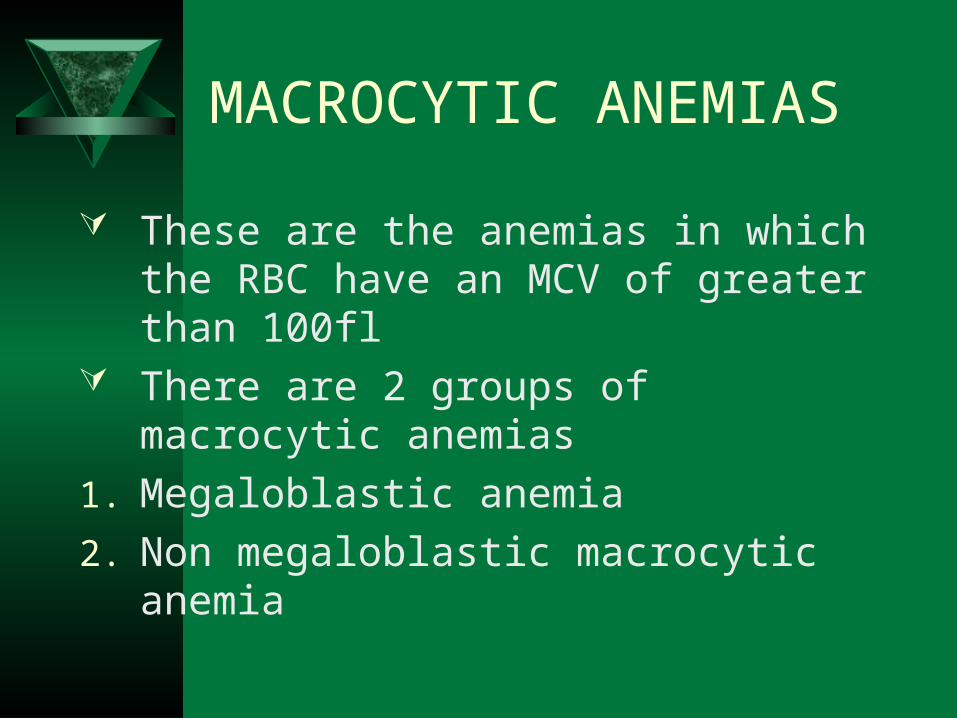
MACROCYTIC ANEMIAS
MACROCYTIC ANEMIAS
These are the anemias in which the RBC have an MCV of greater than 100fl
There are 2 groups of macrocytic anemias
1. Megaloblastic anemia
2. Non megaloblastic macrocytic anemia
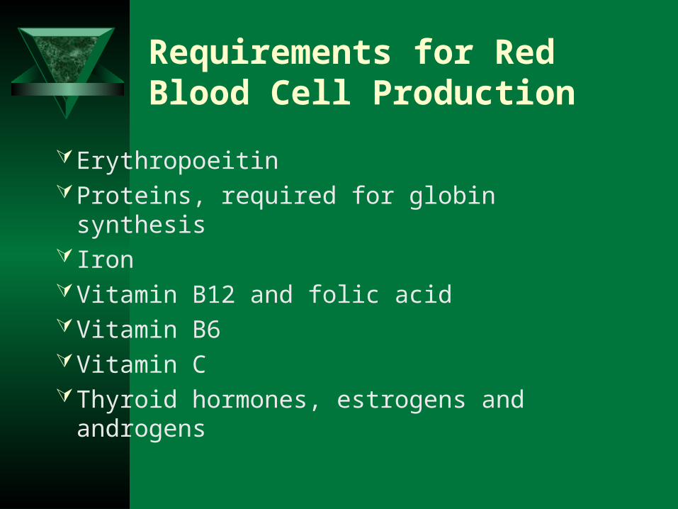
Requirements for Red Blood Cell Production
ErythropoeitinProteins, required for globin synthesis Iron Vitamin B12 and folic acidVitamin B6 Vitamin C Thyroid hormones, estrogens and
androgens
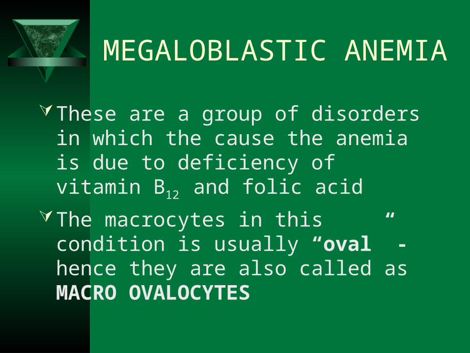
MEGALOBLASTIC ANEMIA
These are a group of disorders in which the cause the anemia is due to deficiency of vitamin B12 and folic acid
The macrocytes in this condition is usually “oval” - hence they are also called as MACRO OVALOCYTES
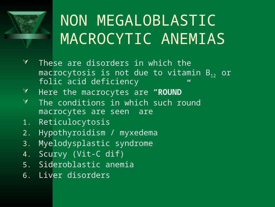
NON MEGALOBLASTIC MACROCYTIC ANEMIAS
These are disorders in which the macrocytosis is not due to vitamin B12 or folic acid deficiency
Here the macrocytes are “ROUND” The conditions in which such round macrocytes are seen
are1. Reticulocytosis2. Hypothyroidism / myxedema3. Myelodysplastic syndrome4. Scurvy (Vit-C dif) 5. Sideroblastic anemia6. Liver disorders
MEGALOBLASTIC ANEMIA
Vitamin B12 and folic acid are important nutrients required in the process of nuclear maturation
They are required during erythropoiesis (during DNA synthesis)
These anemias may be caused because of a nutritional deficiency or impaired absorption mainly.
MEGALOBLASTIC ANEMIA
Impaired DNA synthesis leading to defective cell maturation and cell division
Nuclear maturation delays from the cytoplasmic maturation – NUCLEAR CYTOPLASMIC ASYNCHRONY
Abnormally large erythroid precursors and red cells
Affect all marrow elements. Neurologic symptoms (dorsal
columns)Ineffective erythropoiesis:
High indirect bilirubinVery high LDH
MEGALOPLASTIC ANAEMIA.
Folic Acid:– It a vitamin which yellow in colour, water soluble,
necessary for the production of the RBC, WBC and platelets.
– It is not synthesized in the body.– It is found in large number of green fresh vegetables,
fruits.Daily requirement: The human body needs about 100-150 µg daily.
Absorption: It is absorbed in the Duodenum and Jejunum.Transportation: Weakly bound to albumin.
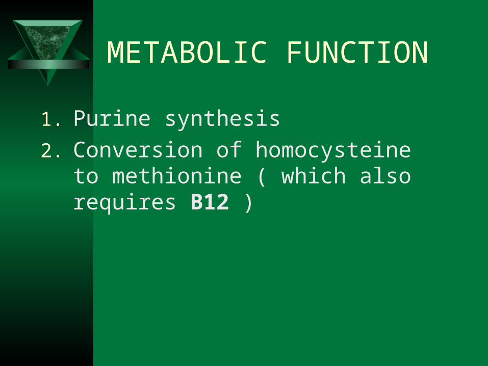
METABOLIC FUNCTION
1. Purine synthesis
2. Conversion of homocysteine to methionine ( which also requires B12 )
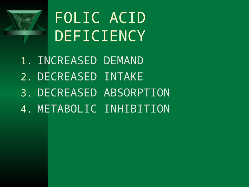
FOLIC ACID DEFICIENCY
1. INCREASED DEMAND
2. DECREASED INTAKE
3. DECREASED ABSORPTION
4. METABOLIC INHIBITION
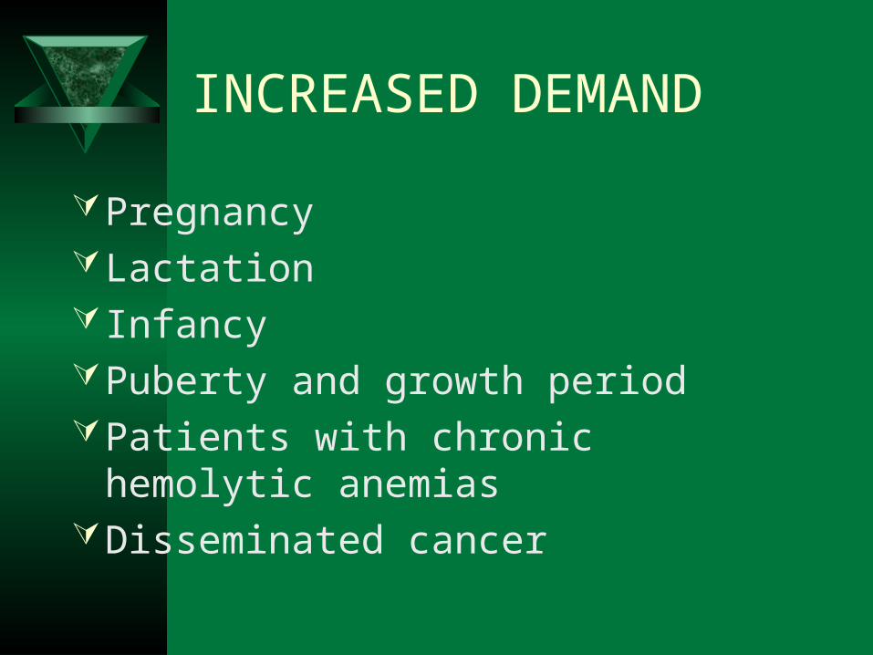
INCREASED DEMAND
PregnancyLactation Infancy Puberty and growth periodPatients with chronic hemolytic anemiasDisseminated cancer

DECREASED INTAKE
ElderlyLower socio economic statusChronic alcoholics
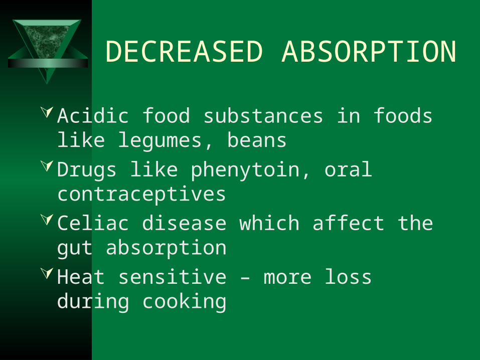
DECREASED ABSORPTION
Acidic food substances in foods like legumes, beans
Drugs like phenytoin, oral contraceptivesCeliac disease which affect the gut
absorptionHeat sensitive – more loss during cooking

METABOLIC INHIBITION

Vitamin B12:
This vitamin is synthesized in nature by micro-organism in the intestine of man and animals, but we can not obtain it from the bacteria in our bodies, because it is synthesizing in the large colon after the site of absorption and it is wasted in the faeces in about 5µg/day. So we obtain it from animal food such as liver, kidney, meat and dairy products as milk and cheese.

VITAMIN B12
Abundant in animal foodsMicroorganisms are the ultimate origin of
cobalamin It is stored in liver for many years It is efficiently reabsorbed from bile It is resistant to cooking and boiling
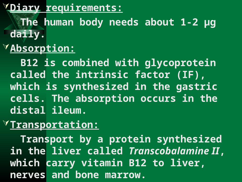
Diary requirements:
The human body needs about 1-2 µg daily.Absorption:
B12 is combined with glycoprotein called the intrinsic factor (IF), which is synthesized in the gastric cells. The absorption occurs in the distal ileum.
Transportation:
Transport by a protein synthesized in the liver called Transcobalamine II, which carry vitamin B12 to liver, nerves and bone marrow.
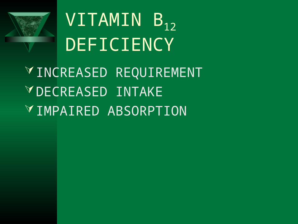
VITAMIN B12 DEFICIENCY
INCREASED REQUIREMENTDECREASED INTAKE IMPAIRED ABSORPTION
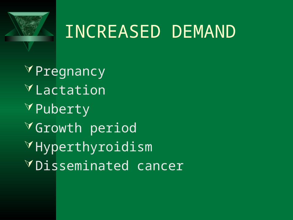
INCREASED DEMAND
PregnancyLactationPubertyGrowth periodHyperthyroidismDisseminated cancer

DECREASED INTAKE
Inadequate intakeVegetarian diet
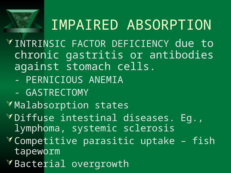
IMPAIRED ABSORPTION INTRINSIC FACTOR DEFICIENCY due to
chronic gastritis or antibodies against stomach cells.
- PERNICIOUS ANEMIA- GASTRECTOMY
Malabsorption statesDiffuse intestinal diseases. Eg., lymphoma,
systemic sclerosisCompetitive parasitic uptake – fish tapewormBacterial overgrowth
CLINICAL FEATURES
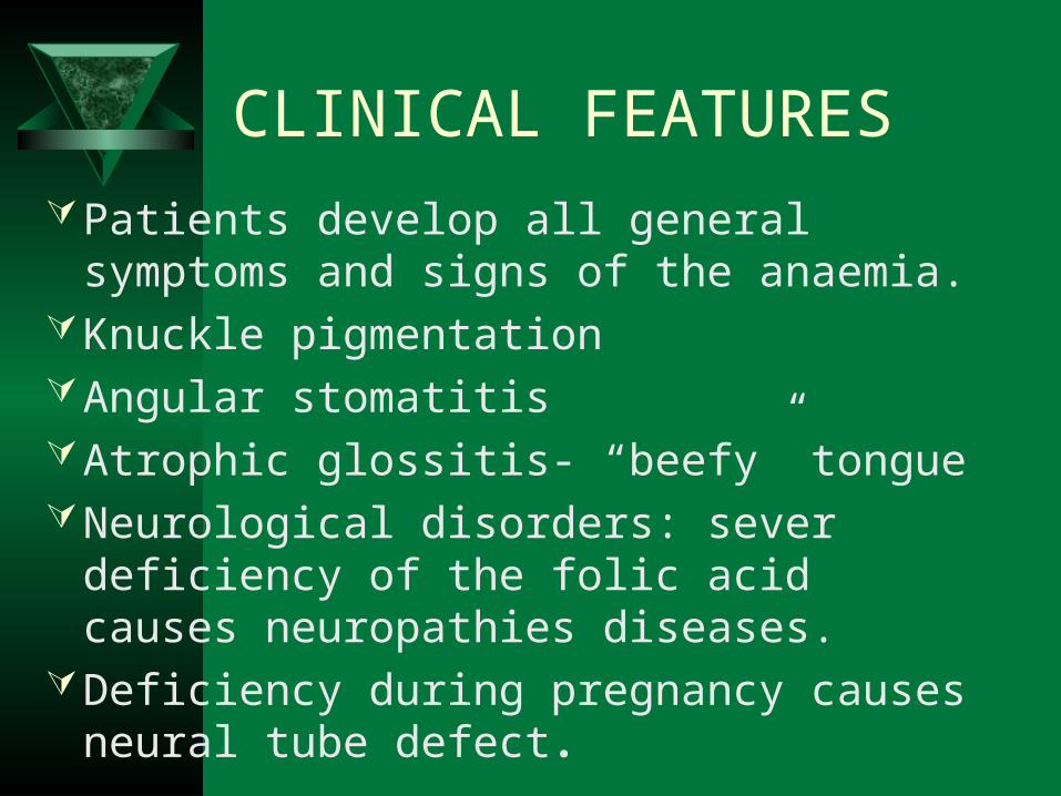
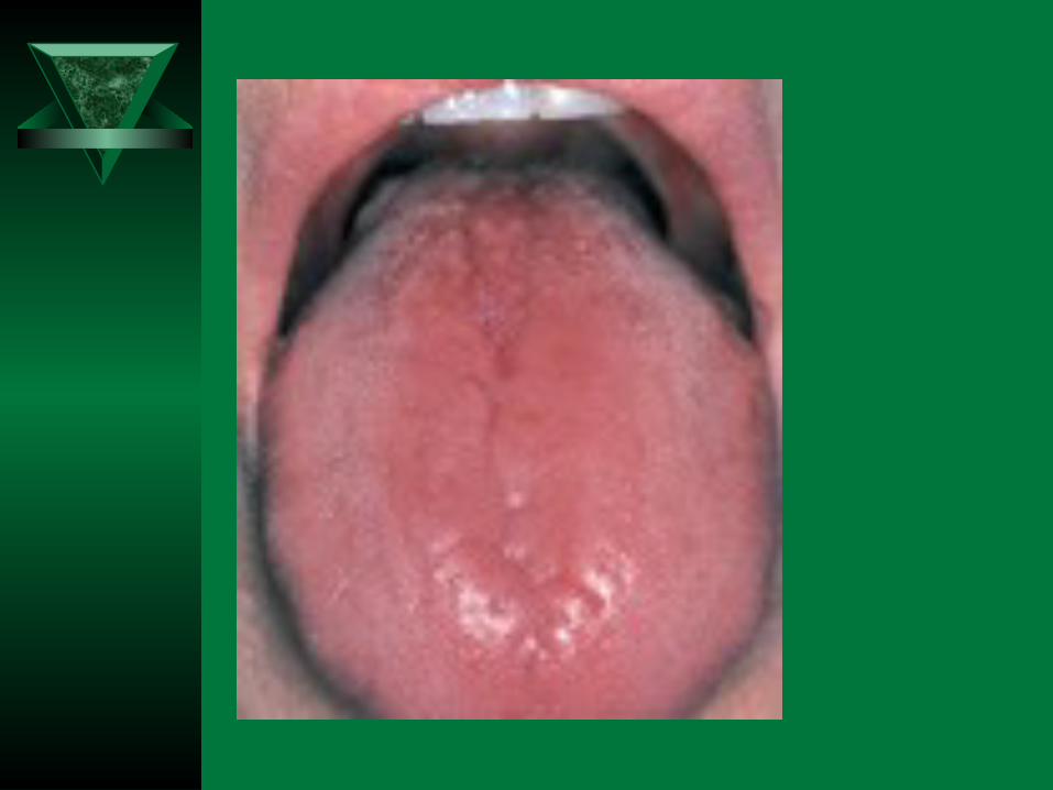
Patients develop all general symptoms and signs of the anaemia.
Knuckle pigmentation Angular stomatitisAtrophic glossitis- “beefy” tongueNeurological disorders: sever deficiency of
the folic acid causes neuropathies diseases.Deficiency during pregnancy causes neural
tube defect.
PERIPHERAL BLOOD FINDINGS
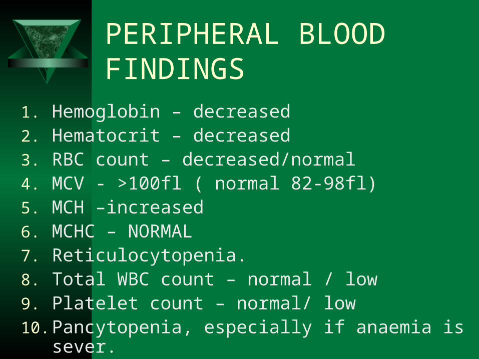
1. Hemoglobin – decreased2. Hematocrit – decreased3. RBC count – decreased/normal4. MCV - >100fl ( normal 82-98fl)5. MCH –increased6. MCHC – NORMAL7. Reticulocytopenia.8. Total WBC count – normal / low9. Platelet count – normal/ low10. Pancytopenia, especially if anaemia is sever.
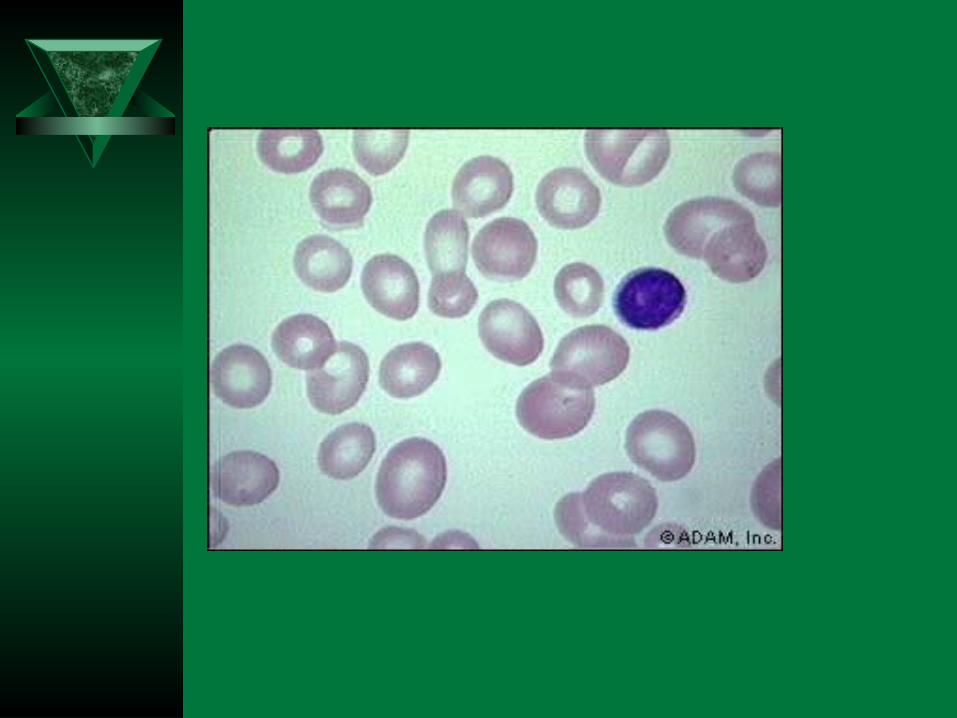
PERIPHERAL SMEAR
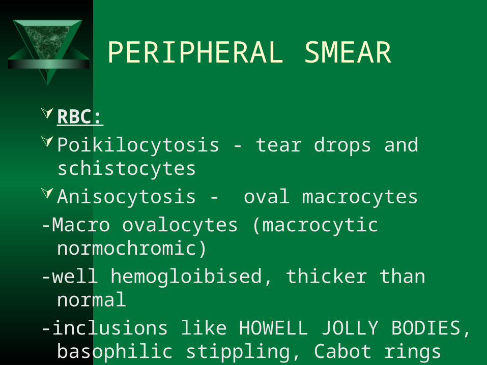
RBC:Poikilocytosis - tear drops and schistocytesAnisocytosis - oval macrocytes
-Macro ovalocytes (macrocytic normochromic)
-well hemogloibised, thicker than normal
-inclusions like HOWELL JOLLY BODIES, basophilic stippling, Cabot rings
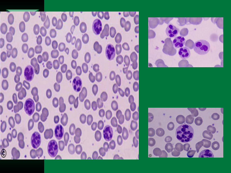
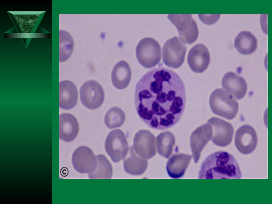
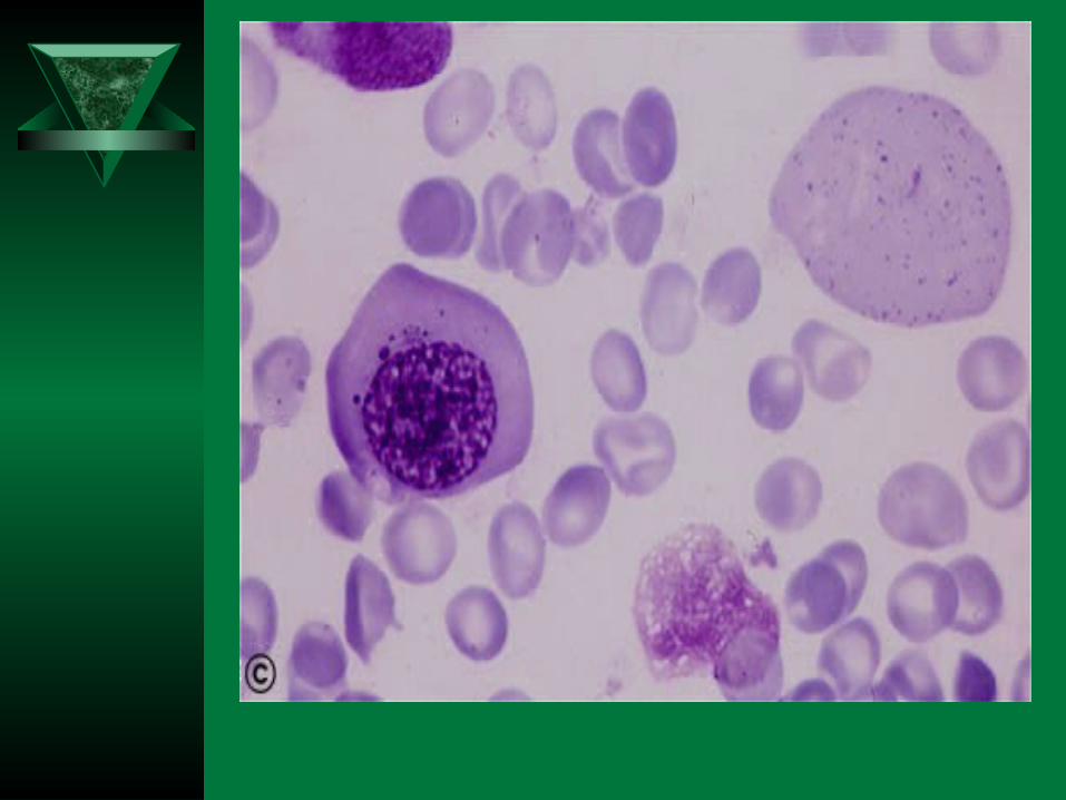
PERIPHERAL SMEAR
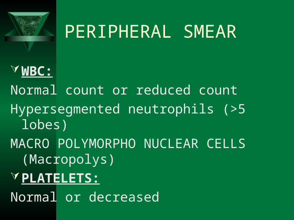
WBC:
Normal count or reduced count
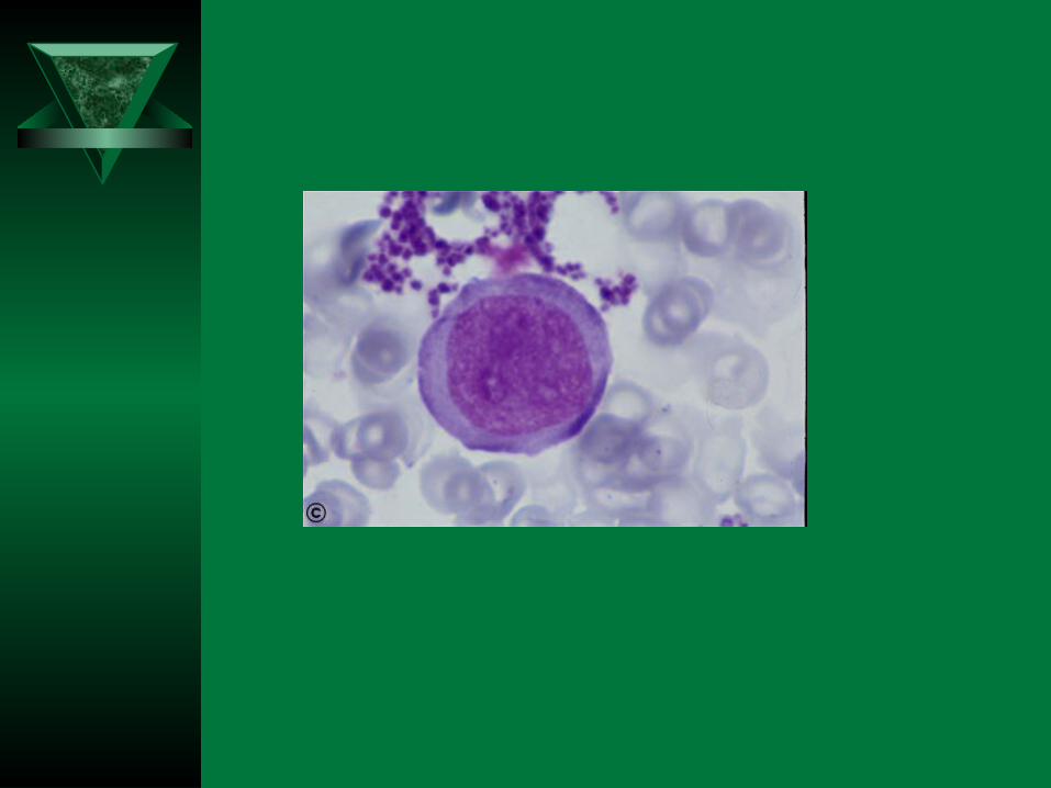
Hypersegmented neutrophils (>5 lobes)
MACRO POLYMORPHO NUCLEAR CELLS (Macropolys)
PLATELETS:
Normal or decreased
BONE MARROW
Markedly hypercellularMyeloid : erythroid ratio decreased or
reversed. (Normally, there are three myeloid precursors for each erythroid precursor resulting in a 3:1 ratio, known as the M:E (myeloid to erythroid) ratio)
Erythropoiesis : MEGALOBLASTIC
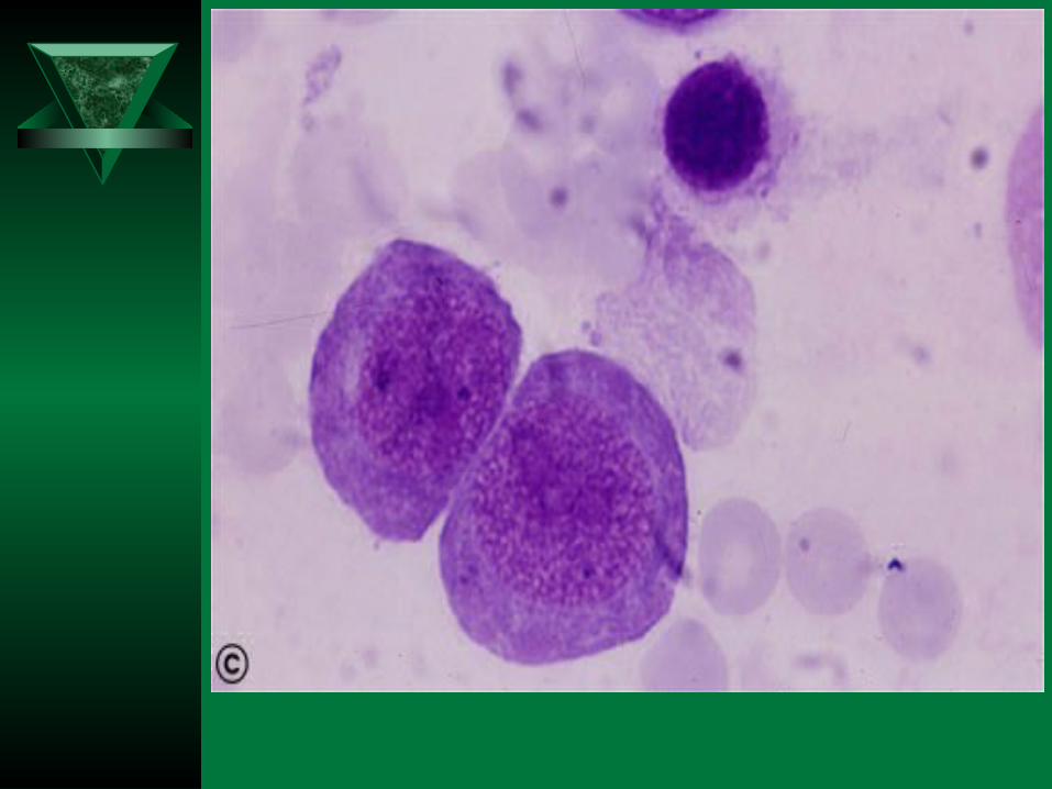
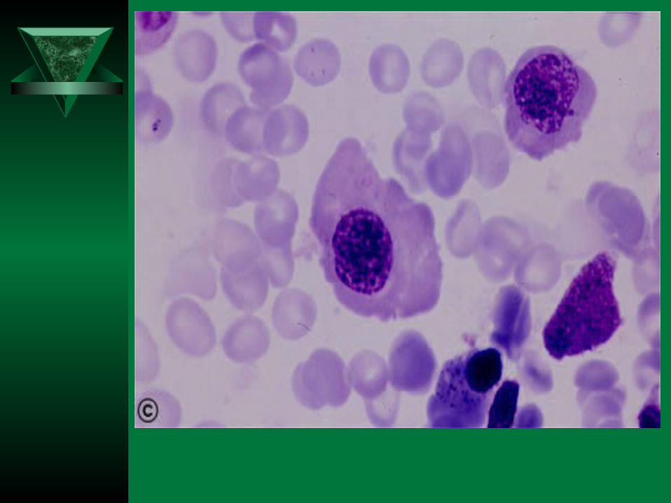
MEGALOBLAST
1. Abnormally large precursor
2. Deeply basophilic royal blue cytoplasm
3. Fine chromatin with prominent nucleoli
4. Nuclear cytoplasmic asynchrony
5. Abnormal mitoses
6. Maturation arrest
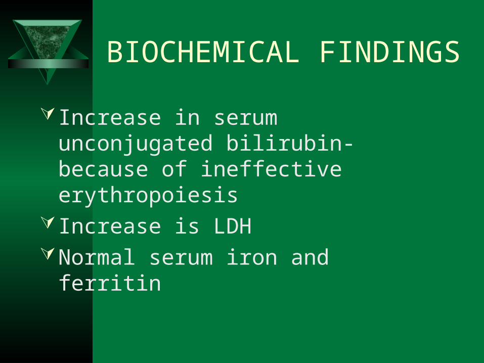
BIOCHEMICAL FINDINGS
Increase in serum unconjugated bilirubin- because of ineffective erythropoiesis
Increase is LDHNormal serum iron and ferritin
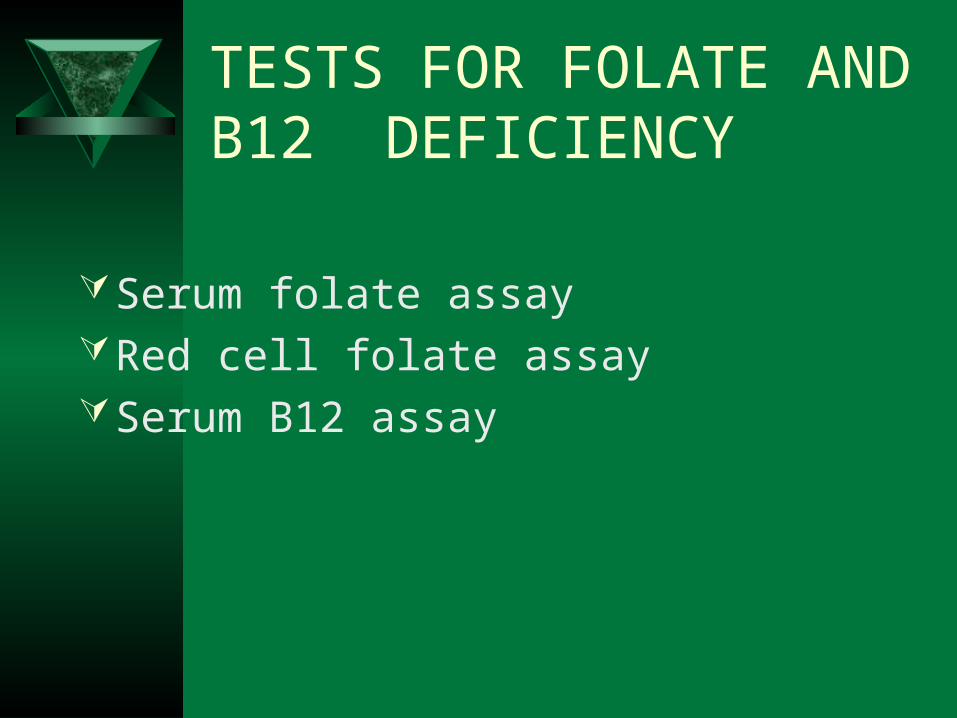
TESTS FOR FOLATE AND B12 DEFICIENCY
Serum folate assay Red cell folate assaySerum B12 assay
PERNICIOUS ANEMIA
Scandinavian countries more prevalentDisease of elderly – 5th to 8th decadesGenetic predispositionTendency to form antibodies against
multiple self antigens
PATHOGENESIS
Immunologically mediated, autoimmune destruction of gastric mucosa
CHRONIC ATROPHIC GASTRITIS – marked loss of parietal cells
Three types of antibodies:a) Type I antibody- 75% - blocks vitamin B12 and IF
bindingb) Type II antibody – prevents binding of IF-B12 complex
with ileal receptorsc) Type III antibody – 85-90% patients – against specific
structures in the parietal cell Associated with other autoimmune diseases like
autoimmune thyroiditis
DIAGNOSTIC FEATURES
1. Moderate to severe megaloblastic anemia2. Leucopenia with hypersegmented neutrophils3. Mild to moderate thrombocytopenia4. Mild jaundice due to ineffective erythropoiesis
and peripheral hemolysis5. Neurologic changes 6. Low levels of serum B12
7. Elevated levels of homocysteine 8. Striking reticulocytosis after parenteral
administration of vitamin B12
9. Serum antibodies to intrinsic factor