Embed Size (px)
Citation preview
Remedy Publications LLC.
Annals of Atherosclerosis Research
2018 | Volume 1 | Issue 3 | Article 10111
Long Term Effects of Mesoglycan on Brachial Arterial Stiffness and MMP-9/TIMP-1 System in Patients with
Metabolic Syndrome
OPEN ACCESS
*Correspondence:Ugo Oliviero, Department of
Translational Medical Sciences, Federico II University, Italy,
E-mail: [email protected] Date: 01 Jan 2018Accepted Date: 16 Apr 2018Published Date: 27 Apr 2018
Citation: Valvano A, Bosso G, Ricci S, Di Carlo
A, Oliviero U. Long Term Effects of Mesoglycan on Brachial Arterial
Stiffness and MMP-9/TIMP-1 System in Patients with Metabolic Syndrome. Ann
Atheroscler Res. 2018; 1(3): 1011.
Copyright © 2018 Ugo Oliviero. This is an open access article distributed under
the Creative Commons Attribution License, which permits unrestricted
use, distribution, and reproduction in any medium, provided the original work
is properly cited.
Research ArticlePublished: 27 Apr, 2018
AbstractObjectives: The aim of this study was to evaluate the chronic effects of mesoglycan on the vascular remodeling in patients with metabolic syndrome (Mets).
Background: MetS is defined by a clustering of vascular risk factors that require both pharmacologic and non-pharmacologic interventions, including body weight reductions and physical activity. The correction of vascular remodeling associated with MetS has lately received increasing interest.
Methods: Thirty consecutive ambulatory patients affected by MetS were 2:1 randomized in a double-blind fashion to receive mesoglycan or placebo, respectively. At the beginning and after 90 days of oral treatment we appraised the effects of mesoglycan (50 mg per os bid) or placebo on vascular remodeling, as assessed by the measurement of arterial wall elastic properties. Moreover, the matrix metalloproteinase’s (MMPs) type 9 and tissue inhibitor of metalloproteinase (TIMP) type 1 were analyzed by enzyme-linked immune sorbent assay (ELISA) and gelatin substrate zymography at the beginning of the study and after 90 days of treatment.
Results: After 90 days of treatment, a marked improvement of arterial distensibility and compliance was detected in Mesoglycan group, with associated significant reduction of arterial stiffness, and a significant reduction of serum levels of MMP-9 and TIMP-1 and significant reduction of enzyme activity of MMPs.
Conclusions: This small, preliminary study shows that mesoglycan exerts relevant effects on vascular remodeling after three-month treatment, in patients affected by metabolic syndrome.
Keywords: Mesoglycan; Arterial stiffness; Coefficient of distensibility; Matrix metallo proteinases; Metabolic syndrome
IntroductionMetabolic syndrome (MetS) is defined by a clustering of risk factors, including hypertension,
dyslipidaemia, excess body weight, and altered glucose homeostasis, which leads to increased risk for cardiovascular disease (CVD) [1]. Insulin Resistance (IR) could be the common pathogenic pathway of these risk factors [2,3], whereas appropriate life style changes represent the gold standard treatment for MetS. However, body weight reduction and physical activity programs [4-6] together to the control of blood pressure values and altered glucose and/or lipid profiles could not be sufficient to achieve an adequate management of MetS patients, determining the necessity of pharmacological treatments [1]. In this regard endothelial dysfunction, as well as pro-inflammatory and pro-thrombotic states [7-10], could represent an interesting pharmacological target. Mesoglycan is a glycosaminogly can compound extracted from porcine intestinal mucosa and composed of heparan sulphate (48%), derma tan sulphate (36%), and electro phoretically slow-moving heparin (8%), and chondroitin sulphate (8%). Heparan and dermatan sulphate are thrombin inhibitors that act through complementary pathways [11,12], heparan sulphate also inhibits factor Xa [11]. Mesoglycan exerts several effects on the antithrombotic and profibrinolitic pathways and, according to some clinical studies, could also be useful in patients with cerebral vascular disease [13,14]. Moreover, mesoglycan treatment reduces thrombophlebitis recurrences in patients with previous deep vein thrombosis [15], and increases the free-pain walking distance in non diabetic patients affected with peripheral artery disease [16]. The complete effects of mesoglycan on the
Antonio Valvano1, Giorgio Bosso1, Serena Ricci1, Angelina Di Carlo2 and Ugo Oliviero1*1Department of Translational Medical Sciences, Federico II University, Italy
2Department of Medico-Surgical Sciences and Biotechnologies, University of Rome "La Sapienza", Italy
Ugo Oliviero, et al., Annals of Atherosclerosis Research
Remedy Publications LLC. 2018 | Volume 1 | Issue 3 | Article 10112
mechanisms of the vascular remodeling have not yet completely recognized, even if it has well been documented that endothelial cells bind and internalize exogenous sulphated polysaccharides, such as heparin and heparan sulphate [17,18], and are able to generate endogenous heparan sulphate [19]. In a previous study, we observed that acute and chronic treatment with mesoglycan improved the vascular reactivity in patients affected with Metabolic Syndrome [20]. Moreover, we also observed a worsened arterial elasticity in MetS patients in respect to healthy control subjects at the beginning, as observed in other patients at high cardiovascular risk, particularly in hypertensive’s [20-24]. It has been also reported a possible landmark role of the endopeptidates MMPs in the development of higher vascular stiffness in hypertensive and diabetic patients [25-27], which has been associated with an increase of cardiovascular morbidity and mortality in these patients [28-31]. According to our previous report [20], the aim of this study was to evaluate the long-term effects of mesoglycan on arterial stiffness through the impact of the substance
on MMP/TIMP system in a group of MetS patients.
Population, Materials and MethodsThirty consecutive ambulatory patients affected by MetS and
recruited from the Department of Translational Medical Sciences of the University Federico II of Naples were enrolled in a clinical trial between May 2013 and June 2014. MetS was defined according to the American Heart Association criteria [1] based on the presence of any 3 of the following 5 abnormalities: abdominal obesity, hyper triglyceridemia, high Blood Pressure (BP), low High-Density Lipoprotein Cholesterol (HDL-C), and elevated fasting glucose. The exclusion criteria were the presence of a critical illness (i.e., heart failure, severe valve heart disease, and neoplasms, advanced renal or liver disease), history of vascular disease or adverse side effects of mesoglycan and heparinoids, bleeding, pregnancy, surgery in the previous three months, and insulin treatment. At the beginning of the trial patients were double-blindly randomized into two arms, according to a 2:1 scheme: one group (20 patients) received oral tablets of mesoglycan 50 mg twice a day (mesoglycan group) and the other (10 patients) received placebo (placebo group). We opted for this randomization scheme because it offered the advantage of increasing the statistical power for paired comparisons of the treated group (baseline vs. after treatment). Mesoglycan, 50 mg capsules, and matching placebos (capsules containing excipients only, respectively) were provided by Mediolanum Farmaceutici, Milan, Italy. At baseline and at the end of the 90-day period, all patients underwent an ultrasound evaluation to assess the elastic arterial wall properties, using standard parameters, such as Distensibility Coefficient (DC), brachial artery Compliance Coefficient (CC), brachial artery stiffness (β), Gosling index (pulsatility index, PI), and Pourcelot index (Resistive Index, RI). Moreover, all patients underwent a laboratory evaluation at the basal visit and after 90 days, including the measurements of metalloproteasis MMP-9 and specific tissue inhibitor TIMP-1 by ELISA and as gelatin substrate zymography. Thirty well matched healthy subjects were also recruited and served as a control group for comparison with the MetS group at baseline. These subjects were studied only at baseline and did not receive any treatment. All participants gave their informed written consent. The protocol was approved by the Ethics Committee of the University Federico II and registered at ClinicalTrial.gov with # NCT02254850.
MMP-9 and TIMP-1Peripheral venous blood samples were collected from all
individuals. Native serum was prepared using plastic tubes without coagulation accelerators, to prevent the release of gelatinases during platelet activation. Tubes were centrifuged at 1600 g for 10 min, 30 min after blood collection. For each sample, determination of protein concentration was performed using the method of Bradford [32]. Sera were aliquoted and stored at -20ºC until used. Each aliquot was used only once in order to prevent enzyme activation due to freeze- thawing processes.
ELISA assay of MMP-9 and TIMP-1 MMP-9 and TIMP-1 levels were detected by quantitative
sandwich ELISA using commercial kits obtained from R&D Systems (Minneapolis, MN, USA). These assays are based on a two-site sandwich format using two antibodies directed against various epitopes of the molecule. All analyses were performed according to the manufacturer’s instructions. Gelatin substrate zymography. Gelatinolytic activity was performed as previously described and,
Figure 1: MMP-9 and TIMP-1 values in Mesoglycan Groups at baseline (Time 0) and after 90 days (Time 90) of treatment. Data are expressed as mean ± Standard Deviation and compared with Wilcoxon signed Rank test.
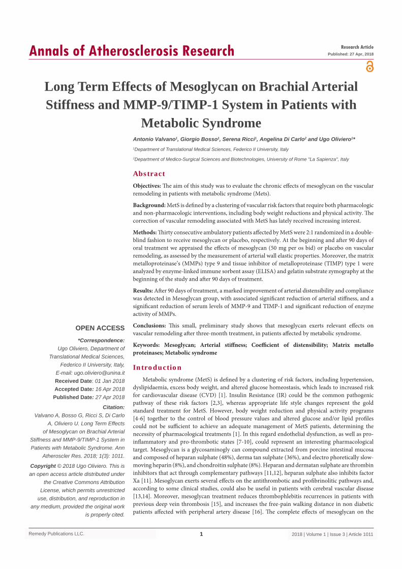
Figure 2: MMP-9 92 kDa (A) and MMP-9 kDa (B) values in Mesoglycan Groups at baseline (Time 0) and after 90 days (Time 90) of treatment.Data are expressed as mean ± Standard Deviation and compared with Wilcoxon signed Rank test
Ugo Oliviero, et al., Annals of Atherosclerosis Research
Remedy Publications LLC. 2018 | Volume 1 | Issue 3 | Article 10113
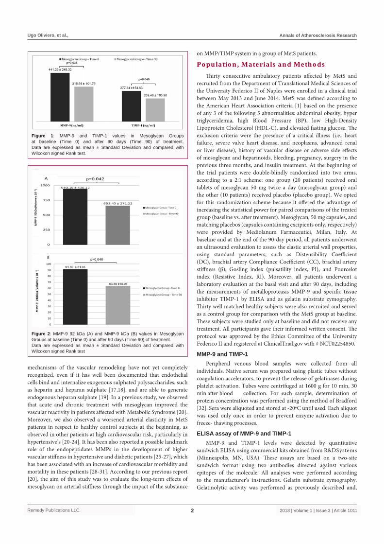
following zymography, the degree of gelatin digestion was quantified. We used image analysis software (Image Quant TL, Amersham Bioscience, and Chicago, IL, USA): the image of the gel was inverted revealing dark bands on a white background. The molecular weight, volume and background of each band were determined. The relative amounts of the different forms of both serum and urine gelatinizes were expressed as the integrated density x 10-3 (volume) of all the pixels above the background of each band.
Brachial artery elastic propertiesAll subjects performed the evaluation of brachial artery elastic
properties at baseline and at the end of the 90 days treatment. They were evaluated in a quiet, temperature-controlled room (22ºC) after 12 h of fasting, including caffeine, and abstaining from cigarette smoking and drugs from the day before the examination. Tests were performed at the same time of day for each patient (approximately 12:00). An ultrasound system equipped with a multi-frequency transducer with a minimum frequency of 7.5 MHz was used (Aplio XG Imaging System, Toshiba, Japan). The parameters measured were: diastolic arterial diameter, Dd (mm); variation in arterial diameter over time, ΔD (mm); thickness of the arterial wall, IMTb (mm); variation in arterial pressure, ΔP (kPa). Arterial distensibility, compliance, and stiffness, were then derived using the following equations:
DC= (2ΔD*Dd+ ΔD2)/ ΔP*Dd2
CC= π (2Dd* ΔD + ΔD2)/4 ΔP
β= ln(SBP/DBP)/(ΔD/Dd)
Where ln is the natural logarithm and SBP and DBP are the brachial systolic and diastolic pressures, respectively [34-36]. We also evaluated Pourcelot Index and Gosling Index, which represent partial
quantitative parameters of blood flow as assessed by the ratio between the difference of the brachial artery maximum systolic velocity (Vs) and diastolic velocity (Vd) to the maximum systolic velocity (Resistive
Figure 3: Gelatin zymography examples in patient treated with Mesoglycan (A) or Placebo (B) at baseline and after 90 days of treatment.
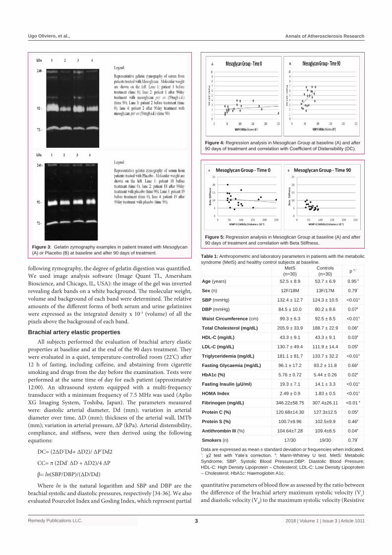
Figure 4: Regression analysis in Mesoglican Group at baseline (A) and after 90 days of treatment and correlation with Coefficient of Distensibility (DC).
Figure 5: Regression analysis in Mesoglican Group at baseline (A) and after 90 days of treatment and correlation with Beta Stiffness.
MetS (n=30)
Controls (n=30) p Ϯ,*
Age (years) 52.5 ± 8.9 53.7 ± 6.9 0.95 Ϯ
Sex (n) 12F/18M 13F/17M 0.79*
SBP (mmHg) 132.4 ± 12.7 124.3 ± 10.5 <0.01Ϯ
DBP (mmHg) 84.5 ± 10.0 80.2 ± 8.6 0.07Ϯ
Waist Circumference (cm) 99.3 ± 6.3 92.5 ± 8.5 <0.01Ϯ
Total Cholesterol (mg/dL) 205.9 ± 33.9 188.7 ± 22.9 0.06Ϯ
HDL-C (mg/dL) 43.3 ± 9.1 43.3 ± 9.1 0.03Ϯ
LDL-C (mg/dL) 130.7 ± 49.4 111.9 ± 14.4 0.05Ϯ
Triglyceridemia (mg/dL) 181.1 ± 81.7 133.7 ± 32.2 <0.01Ϯ
Fasting Glycaemia (mg/dL) 96.1 ± 17.2 93.2 ± 11.8 0.66Ϯ
HbA1c (%) 5.76 ± 0.72 5.44 ± 0.26 0.02Ϯ
Fasting Insulin (μU/ml) 19.3 ± 7.1 14.1 ± 3.3 <0.01Ϯ
HOMA Index 2.49 ± 0.9 1.83 ± 0.5 <0.01Ϯ
Fibrinogen (mg/dL) 346.22±58.75 307.4±26.11 <0.01 Ϯ
Protein C (%) 120.68±14.30 127.3±12.5 0.05Ϯ
Protein S (%) 100.7±9.96 102.5±9.9 0.46Ϯ
Antithrombin III (%) 104.64±7.28 109.4±8.5 0.04Ϯ
Smokers (n) 17/30 19/30 0.79*
Table 1: Anthropometric and laboratory parameters in patients with the metabolic syndrome (MetS) and healthy control subjects at baseline.
Data are expressed as mean ± standard deviation or frequencies when indicated.*: χ2 test with Yate’s correction. Ϯ: Mann-Whitney U test. MetS: Metabolic Syndrome; SBP: Systolic Blood Pressure;DBP: Diastolic Blood Pressure; HDL-C: High Density Lipoprotein – Cholesterol; LDL-C: Low Density Lipoprotein – Cholesterol; HbA1c: Haemoglobin A1c.
Ugo Oliviero, et al., Annals of Atherosclerosis Research
Remedy Publications LLC. 2018 | Volume 1 | Issue 3 | Article 10114
Index, RI) or to the time-averaged velocity (Pulsatility Index, PI) [37]. The coefficient of variation for the arterial DC in our laboratory was 5%. To guarantee blinding, three operators were involved: one measured the distensibility parameters in all patients and was blinded to the treatment assigned, another operator performed the offline
readings of the vascular exams and was blinded both to the treatment assigned and time of examination, one additional operator assigned to the patients a code number corresponding to mesoglycan or placebo, in undistinguishable packaging.
Statistical analysisData are presented as the means ± standard deviation. Statistical
significance between groups was tested by the Mann-Witney U test for independent samples, Wilcox on test for paired samples, and chi-square test with Yates correction for non-continuous variables. Spearman’s rank correlation test and stepwise regression analysis were performed. A p value less than 0.05 was considered statistically significant. All calculations were performed with the SPSS software, version 13 (SPSS Inc., Chicago IL).
ResultsTable 1 shows the clinical and laboratory characteristics of
the MetS patients compared with a control group of matched
MetS (n=30) Controls (n=30) pϮ
DC (10-3/kPa) 2.38 ± 1.14 3.03 ± 1.12 0.03
CC (mm2/kPa) 7.49 ± 3.57 9.51 ± 3.52 0.04
β 10.11 ± 3.95 6.51 ± 3.16 <0.01
PI 7.21 ± 2.42 5.42 ± 2.18 <0.01
RI 0.92 ± 0.02 0.88 ± 0.06 <0.01
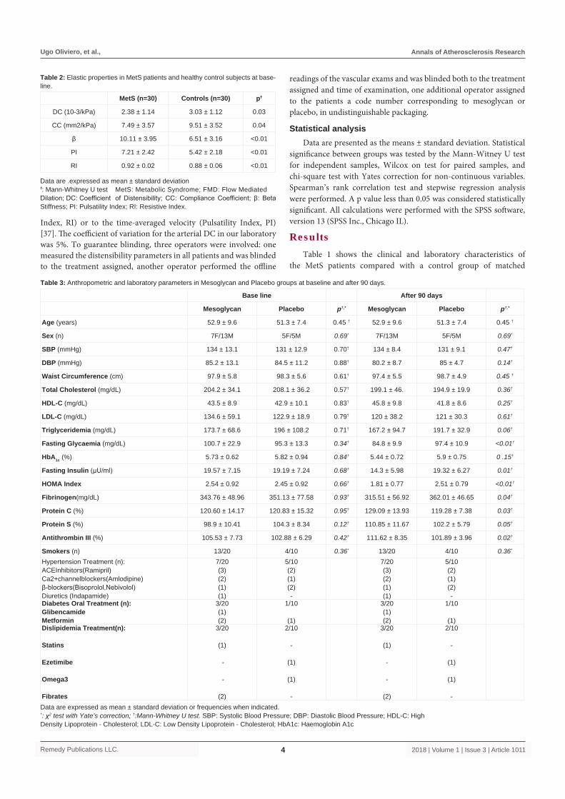
Table 2: Elastic properties in MetS patients and healthy control subjects at base-line.
Data are .expressed as mean ± standard deviationϮ: Mann-Whitney U test MetS: Metabolic Syndrome; FMD: Flow Mediated Dilation; DC: Coefficient of Distensibility; CC: Compliance Coefficient; β: Beta Stiffness; PI: Pulsatility Index; RI: Resistive Index.
Base line After 90 days
Mesoglycan Placebo p†,* Mesoglycan Placebo p†,*
Age (years) 52.9 ± 9.6 51.3 ± 7.4 0.45 † 52.9 ± 9.6 51.3 ± 7.4 0.45 †
Sex (n) 7F/13M 5F/5M 0.69* 7F/13M 5F/5M 0.69*
SBP (mmHg) 134 ± 13.1 131 ± 12.9 0.70† 134 ± 8.4 131 ± 9.1 0.47†
DBP (mmHg) 85.2 ± 13.1 84.5 ± 11.2 0.88† 80.2 ± 8.7 85 ± 4.7 0.14†
Waist Circumference (cm) 97.9 ± 5.8 98.3 ± 5.6 0.61† 97.4 ± 5.5 98.7 ± 4.9 0.45 †
Total Cholesterol (mg/dL) 204.2 ± 34.1 208.1 ± 36.2 0.57† 199.1 ± 46. 194.9 ± 19.9 0.36†
HDL-C (mg/dL) 43.5 ± 8.9 42.9 ± 10.1 0.83† 45.8 ± 9.8 41.8 ± 8.6 0.25†
LDL-C (mg/dL) 134.6 ± 59.1 122.9 ± 18.9 0.79† 120 ± 38.2 121 ± 30.3 0.61†
Triglyceridemia (mg/dL) 173.7 ± 68.6 196 ± 108.2 0.71† 167.2 ± 94.7 191.7 ± 32.9 0.06†
Fasting Glycaemia (mg/dL) 100.7 ± 22.9 95.3 ± 13.3 0.34† 84.8 ± 9.9 97.4 ± 10.9 <0.01†
HbA1c (%) 5.73 ± 0.62 5.82 ± 0.94 0.84† 5.44 ± 0.72 5.9 ± 0.75 0 .15†
Fasting Insulin (μU/ml) 19.57 ± 7.15 19.19 ± 7.24 0.68† 14.3 ± 5.98 19.32 ± 6.27 0.01†
HOMA Index 2.54 ± 0.92 2.45 ± 0.92 0.66† 1.81 ± 0.77 2.51 ± 0.79 <0.01†
Fibrinogen(mg/dL) 343.76 ± 48.96 351.13 ± 77.58 0.93† 315.51 ± 56.92 362.01 ± 46.65 0.04†
Protein C (%) 120.60 ± 14.17 120.83 ± 15.32 0.95† 129.09 ± 13.93 119.28 ± 7.38 0.03†
Protein S (%) 98.9 ± 10.41 104.3 ± 8.34 0.12† 110.85 ± 11.67 102.2 ± 5.79 0.05†
Antithrombin III (%) 105.53 ± 7.73 102.88 ± 6.29 0.42† 111.62 ± 8.35 101.89 ± 3.96 0.02†
Smokers (n) 13/20 4/10 0.36* 13/20 4/10 0.36*
Hypertension Treatment (n): ACEInhibitors(Ramipril)Ca2+channelblockers(Amlodipine) β-blockers(Bisoprolol,Nebivolol) Diuretics (Indapamide)
7/20 (3) (2) (1) (1)
5/10 (2) (1) (2) -
7/20 (3) (2) (1) (1)
5/10 (2) (1) (2) -
Diabetes Oral Treatment (n): Glibencamide Metformin
3/20 (1) (2)
1/10
(1)
3/20 (1) (2)
1/10
(1)
Dislipidemia Treatment(n): Statins Ezetimibe Omega3 Fibrates
3/20
(1) - -
(2)
2/10 -
(1)
(1) -
3/20
(1) - -
(2)
2/10 -
(1)
(1) -
Table 3: Anthropometric and laboratory parameters in Mesoglycan and Placebo groups at baseline and after 90 days.
Data are expressed as mean ± standard deviation or frequencies when indicated.*: χ2 test with Yate’s correction; †:Mann-Whitney U test. SBP: Systolic Blood Pressure; DBP: Diastolic Blood Pressure; HDL-C: HighDensity Lipoprotein - Cholesterol; LDL-C: Low Density Lipoprotein - Cholesterol; HbA1c: Haemoglobin A1c
Ugo Oliviero, et al., Annals of Atherosclerosis Research
Remedy Publications LLC. 2018 | Volume 1 | Issue 3 | Article 10115
healthy subjects. The parameters adopted as inclusion criteria for MetS were, as expected, significantly different from the controls. Fasting glucose was only slightly higher in MetS patients, while HbA1c resulted significantly different pointing to abnormal gluco regulation, as reported in our previous study [20]. As summarized in (Table 2), the MetS patients showed marked alterations in the basal parameters of elastic properties in respect to healthy control subjects, in particular the indices of arterial performance showed concordant abnormalities of distensibility, compliance, and stiffness. Table 3 shows the clinical and laboratory characteristics of the
patients after they were randomized into the two groups, receiving either mesoglycan or placebo. There were no differences in any of the parameters considered. As shown in the Table, some of the patients were under pharmacologic treatment for hypertension, diabetes, or dyslipidaemia. No changes in the pharmacological treatments were made throughout the study period. After 90 days of treatment with mesoglycan, the serum levels of MM9 and TIMP, as well as MMP-9 activity at 92 kDa and 240 kDa, significantly decreased (Figure 1.2 A,B). Gelatin zymography examples in patient treated with Mesoglycan or Placebo at baseline and after 90 days of treatment
MESOGLYCAN PLACEBO
Baseline After 90 Days p# Baseline After 90 Days p#
SBP(mmHg) 134 ± 13.1 134 ± 8.4 0.88 131 ± 12.9 131 ± 9.1 0.95
DBP(mmHg) 85.2 ± 13.1 80.2 ± 8.7 0.05 84.5 ± 11.2 85 ± 4.7 0.94
WC(cm) 97.9 ± 5.8 97.4 ± 5.5 0.39 98.3 ± 5.6 98.7 ± 4.9 0.68
Total Cholesterol(mg/dL) 204.2 ± 34.1 199.1 ± 46.2 0.76 208.1 ± 36.2 194.9 ± 19.9 0.24
HDL-C(mg/dL) 43.5 ± 8.9 45.8 ± 9.8 0.29 42.9 ± 10.1 41.8 ± 8.6 0.63
LDL-C(mg/dL) 134.6 ± 59.1 120 ± 38.2 0.7 122.9 ± 18.9 121 ± 30.3 0.86
Triglyceridemia(mg/dL) 173.7 ± 68.6 167.2 ± 94.7 0.75 196 ± 108.2 191.7 ± 32.9 0.84
Fasting Glycaemia(mg/dL) 100.7 ± 22.9 84.8 ± 9.9 0.03 95.3 ± 13.3 97.4 ± 10.9 0.88
HbA1c(%) 5.73 ± 0.62 5.44 ± 0.72 0.08 5.82 ± 0.94 5.9 ± 0.75 0.51
Fasting Insulin(μU/ml) 19.57 ± 7.15 14.3 ± 5.98 0.04 19.19 ± 7.24 19.32 ± 6.27 0.17
HOMA Index 2.54 ± 0.92 1.81 ± 0.77 0.05 2.45 ± 0.92 2.51 ± 0.79 0.21
Fibrinogen(mg/dL) 343.76±48.96 315.51±56.92 0.04 351.13±77.58 362.01±46.65 0.72
Protein C(%) 120.60±14.17 129.09±13.94 0.02 120.83±15.32 119.28±7.38 0.72
Protein S(%) 98.9±10.41 110.85±11.67 <0.01 104.3±8.34 102.2±5.79 0.33
Antithrombin III(%) 105.53±7.73 111.62±8.35 0.04 102.88±6.3 101.89±3.96 0.58
MMP-2 (72 kDa)Volume x 10-3 428.37± 176.08 475.28±210.15 0.28 421.99±283.34 496.90±246.20 0.56
MMP-9 (92 kDa)Volume x 10-3 940.15 ± 436.12 653.41± 271.22 0.042 1093.23 ± 578.47 949.97 ± 528.14 0.57
MMP-9 (240 kDa)Volume x 10-3 91.50 ± 57.39 62.87 ± 17.91 0.046 98.11 ± 52.88 82.07 ± 35.72 0.51
MMP-9 (ng/ml) 441.23 ± 248.32 315.98 ± 101.79 0.035 537.90 ± 183.52 437.20 ± 120.22 0.49
TIMP-1 (ng/ml) 277.34 ± 154.63 209.48 ± 105.98 0.049 195.80 ± 113.19 189.72 ± 110.76 0.73
DC(10-3/kPa) 2.25 ± 0.93 4.54 ± 1.64 <0.01 2.65 ± 1.49 2.60 ± 1.25 0.88
CC(mm2/kPa) 7.07 ± 2.92 14.30 ± 5.17 <0.01 8.33 ± 4.69 8.19 ± 3.94 0.88
β 10.45 ± 4.01 5.83 ± 1.79 <0.01 9.45 ± 3.96 8.7 ± 5.61 0.58
RI 0.96 ± 0.02 0.88 ± 0.08 <0.01 0.95 ± 0.02 0.94 ± 0.03 0.51
PI 7.20 ± 2.7 5.22 ± 1.86 0.02 7.19 ± 1.6 6.85 ± 1.84 0.87
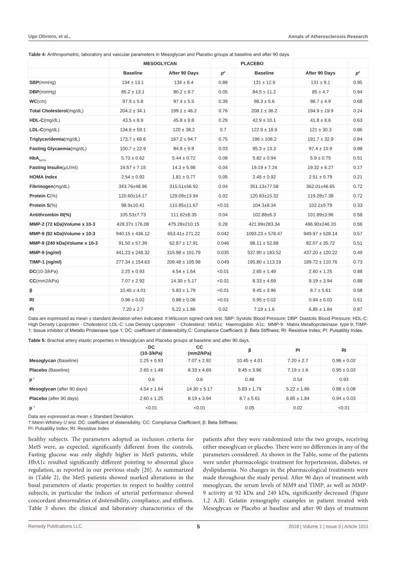
Table 4: Anthropometric, laboratory and vascular parameters in Mesoglycan and Placebo groups at baseline and after 90 days.
Data are expressed as mean ± standard deviation when indicated. #:Wilcoxon signed-rank test. SBP: Systolic Blood Pressure; DBP: Diastolic Blood Pressure; HDL-C: High Density Lipoprotein - Cholesterol; LDL-C: Low Density Lipoprotein - Cholesterol; HbA1c: Haemoglobin A1c; MMP-9: Matrix Metalloproteinase type 9; TIMP-1: tissue inhibitor of Metallo Proteinase type 1; DC: coefficient of distensibility;C: Compliance Coefficient; β: Beta Stiffness; RI: Resistive Index; PI: Pulsatility Index.
DC (10-3/kPa)
CC (mm2/kPa) β PI RI
Mesoglycan (Baseline) 2.25 ± 0.93 7.07 ± 2.92 10.45 ± 4.01 7.20 ± 2.7 0.96 ± 0.02
Placebo (Baseline) 2.65 ± 1.49 8.33 ± 4.69 9.45 ± 3.96 7.19 ± 1.6 0.95 ± 0.02
p † 0.6 0.6 0.48 0.54 0.93
Mesoglycan (after 90 days) 4.54 ± 1.64 14.30 ± 5.17 5.83 ± 1.79 5.22 ± 1.86 0.88 ± 0.08
Placebo (after 90 days) 2.60 ± 1.25 8.19 ± 3.94 8.7 ± 5.61 6.85 ± 1.84 0.94 ± 0.03
p † <0.01 <0.01 0.05 0.02 <0.01
Table 5: Brachial artery elastic properties in Mesoglycan and Placebo groups at baseline and after 90 days.
Data are expressed as mean ± Standard Deviation.†:Mann-Whitney U test. DC: coefficient of distensibility; CC: Compliance Coefficient; β: Beta Stiffness;PI: Pulsatility Index; RI: Resistive Index
Ugo Oliviero, et al., Annals of Atherosclerosis Research
Remedy Publications LLC. 2018 | Volume 1 | Issue 3 | Article 10116
are showed in (Figure 3 A,B). Fasting glucose and HbA1c were also significantly reduced by mesoglycan, indicating an improvement of glucoregulation in treated patients. Protein C, Protein S, and ATIII activities increased after mesoglycan treatment, whereas fibrinogen, but also MMP-9 and TIMP-1, both quantitatively and qualitatively decreased (Table 4). None of the arterial elastic properties measured were different between the mesoglycan and placebo groups at baseline. Moreover, after 90 days of treatment, in the mesoglycan group there was a marked improvement in arterial elasticity, as documented by increased distensibility and compliance, reduced stiffness, and improvements in both pulsatile and resistive indices (Table 5). The data were also analyzed by stepwise multiple regression to determine which factor was predictive of vascular improvement. In this analysis, β-stiffness and Coefficient of Distensibility (DC) were the dependent variables whereas glycaemia, HbA1c, SBP, DBP, total cholesterol, LDL-cholesterol, HDL-cholesterol, triglycerides, MM9 and TIMP1 were used as independent variables. The results indicate a significant predictive value for the MMP-9 240kDa values (standardized β= -0.07, p=0.005 for Beta stiffness and standardized β= 0.05, p=0.03 for DC, respectively) as summarized in (Figure 4A,B) and (Figure 5A,B). All patients completed the chronic treatment with mesoglycan without reporting major side effects. Four patients in the mesoglycan group and two patients in the placebo group experienced mild dyspepsia.
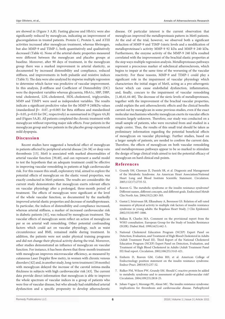
DiscussionRecent studies have suggested a beneficial effect of mesoglycan
in patients affected by peripheral arterial disease [16-38] or deep vein thrombosis [15]. MetS is associated with marked abnormalities of arterial vascular function [39,40], and can represent a useful model to test the hypothesis that an adequate treatment could be effective in improving vascular remodeling in patients at high cardiovascular risk. For this reason this small, exploratory trial, aimed to explore the potential effects of mesoglycan on the elastic vessel properties, was exactly conducted in MetS patients. The results are considerable: the current study demonstrates that mesoglycan exerts relevant effects on vascular physiology after a prolonged, three-month period of treatment. The effects of mesoglycan were significant at the level of the whole vascular function, as documented by the markedly improved arterial elastic properties and decrease of metalloproteases. In particular, the indices of distensibility and compliance increased, whereas arterial stiffness, a marker of increased cardiovascular risk in diabetic patients [41], was reduced by mesoglycan treatment. The vascular effects of mesoglycan seem reflect an action of mesoglycan per se on arterial wall remodeling. Other potential confounding factors which could act on vascular physiology, such as waist circumference and BMI, remained stable during treatment. In addition, the patients were not under physical training programs and did not change their physical activity during the trial. Moreover, other studies demonstrated an influence of mesoglycan on vascular function. For instance, it has been shown that three-month treatment with mesoglycan improves microvascular efficiency, as measured by cutaneous Laser Doppler flow metry, in women with chronic venous disorders [42] and, in another study, long-term treatment (18 months) with mesoglycan delayed the increase of the carotid intima-media thickness in subjects with high cardiovascular risk [43]. The current data provide direct information that mesoglycan is able to improve the whole spectrum of vascular indices in a group of patients who were free of vascular disease, but who already had established arterial dysfunction and a specific propensity to develop atherosclerotic
disease. Of particular interest is the current observation that mesoglycan improved the metalloproteasis pattern in MetS patients. At the end of the trial, however, we observed both a significant reduction of MMP-9 and TIMP-1sieric levels and a modification of metalloproteinase’s activity MMP-9 92 kDa and MMP-9 240 kDa. Furthermore, the enzyme activity of the MMP-9 240 kDa resulted correlated with the improvement of the brachial elastic properties at the step ways multiple regression analysis. Metalloproteases pathways represent a precocious marker of subclinical atherosclerosis, which begins to impair at the same time of the worsening of the vascular reactivity. For these reasons, MMP-9 and TIMP-1 could play a significant role in the impairment of vascular physiology which characterizes the initial stages of MetS, acting as a pro-atherogenic factor which can cause endothelial dysfunction, inflammation, and, finally, concurs to the impairment of vascular remodeling [1,40,41,44-48]. The decrease of the sieric levels of metalloproteases, together with the improvement of the brachial vascular properties, could explain the anti atherosclerotic effects and the clinical benefits carried out by mesoglycan in some previous studies, even if the exact molecular mechanisms whereby mesoglycan exerts its vascular effects remains largely unknown. Therefore, our study was conducted on a small sample of patients, who were recruited from and studied in a single centre. Thus, the results of this small trial should be taken as preliminary information regarding the potential beneficial effects of mesoglycan on vascular physiology. Further studies, based on a larger sample of patients, are needed to confirm the current data. Therefore, the effects of mesoglycan on both vascular remodeling and metalloproteases pathways appear to be so marked to stimulate the design of large clinical trials aimed to test the potential efficacy of mesoglycan on hard clinical end-points.
References1. Grundy SM, Cleeman JI, Daniels SR, et al. Diagnosis and Management
of the Metabolic Syndrome. An American Heart Association/National Heart Lung and Blood Institute Scientific Statement. Circulation. 2005;112(17):2735-52.
2. Reaven G. The metabolic syndrome or the insulin resistance syndrome? Different names, different concepts, and different goals. Endocrinol Metab Clin North Am. 2004;33(2):283-303.
3. Gustat J, Srinivasan SR, Elkasabany A, Berenson GS. Relation of self-rated measures of physical activity to multiple risk factors of insulin resistance syndrome in young adults: the Bogalusa Heart Study. J Clin Epidemiol. 2002;55(10):997-1006.
4. Balkau B, Charles MA. Comment on the provisional report from the WHO consultation. European Group for the Study of Insulin Resistance (EGIR). Diabet Med. 1999;16(5):442-3.
5. National Cholesterol Education Program (NCEP) Expert Panel on Detection, Evaluation, and Treatment of High Blood Cholesterol in Adults (Adult Treatment Panel III). Third Report of the National Cholesterol Education Program (NCEP) Expert Panel on Detection, Evaluation, and Treatment of High Blood Cholesterol in Adults (Adult Treatment Panel III) final report. Circulation. 2002;106(25):3143-421.
6. Einhorn D, Reaven GM, Cobin RH, et al. American College of Endocrinology position statement on the insulin resistance syndrome. Endocr Pract. 2003;9(3):237-52.
7. Ridker PM, Wilson PW, Grundy SM. Should C-reactive protein be added to metabolic syndrome and to assessment of global cardiovascular risk? Circulation. 2004;109(23):2818-25.
8. Juhan-Vague I, Morange PE, Alessi MC. The insulin resistance syndrome: implications for thrombosis and cardiovascular disease. Pathophysiol
Ugo Oliviero, et al., Annals of Atherosclerosis Research
Remedy Publications LLC. 2018 | Volume 1 | Issue 3 | Article 10117
Haemost Thromb. 2002;32(5-6):269-73.
9. Deanfield JE, Halcox JP, Rabelink TJ. Endothelial function and dysfunction: testing and clinical relevance. Circulation. 2007;115(10):1285-95.
10. Prieto D, Contreras C, Sánchez A. Endothelial dysfunction, obesity and insulin resistance. Curr Vasc Pharmacol. 2014;12(3):412-26.
11. Ofosu FA, Modi GJ, Smith LM, Cerksus AL, Hirsh J, Blajchman MA. Heparan sulfate and dermatan sulfate inhibit the generation of thrombin activity in plasma by complementary pathways. Blood. 1984;64(3):742-7.
12. Tollefsen DM. Insight into the mechanism of action of heparin cofactor II. Thromb Haemost. 1995;74(5):209-14.
13. Orefice G, Brancaccio V, Coppola G. Comparative effects of mesoglycan and ticlopidine treatment on some coagulative parameters in patients with previous ischemic stroke: results of a randomized controlled trial. Current Therapeutic Research. 2002;63(5):337-43.
14. Forconi S, Battistini N, Guerrini M, Passero SG for the SIAM Group. A randomized, ASA-controlled trial of mesoglycan in secondary prevention after cerebral ischemic events. Cerebrovasc Dis. 1995;5(5):334-41.
15. Andreozzi GM. Effectiveness of mesoglycan in patients with previous deep venous thrombosis and chronic venous insufficiency. Minerva Cardioangiol. 2007;55(6):741-53.
16. Nenci GG, Gresele P, Ferrari G, Santoro L, Gianese F. Mesoglycan Intermittent Claudication Group. Treatment of intermittent claudication with mesoglycan—a placebo-controlled, double-blind study. Thromb Haemost 2001;86(5):1181-7.
17. Barzu T, Molho P, Tobelem G, Petitou M, Caen J. Binding and endocytosis of heparin by human endothelial cells in culture. Biochim Biophys Acta. 1985;845(2):196-203.
18. Dawes J, Pepper DS. Human vascular endothelial cells catabolise exogenous glycosaminoglycans by a novel route. Thromb Haemostas 1992;67(4):468-72.
19. Rosenberg RD, Edelberg JM, Zhang L. The heparin/antithrombin system: a natural anticoagulant mechanism. In: Colman RW et al (Eds.). Hemostasis and thrombosis. Basic principles and clinical practice. Lippincott Williams & Wilkins, Philadelphia. 2001.
20. Valvano A, Bosso G, Apuzzi V, Riccone F, Saccà L, Oliviero U. Mesoglycan improves vascular reactivity and insulin sensitivity in patients with metabolic syndrome. Atherosclerosis. 2015;243(2):407-13.
21. Lopes-Vicente WR, Rodrigues S, Cepeda FX. Arterial stiffness and its association with clustering of metabolic syndrome risk factors. Diabetol Metab Syndr. 2017;9:87.
22. Chan DT1, Watts GF, Irish AB. Insulin resistance and the metabolic syndrome are associated with arterial stiffness in patients with chronic kidney disease. Am J Hypertens. 2013;26(9):155-61.
23. Kangas P, Tikkakoski AJ, Tahvanainen AM. Metabolic syndrome may be associated with increased arterial stiffness even in the absence of hypertension: a study in 84 cases and 82 controls. Metabolism. 2013;62(8):1114-22.
24. London GM, Marchais SJ, Guerin AP, Pannier B. Arterial stiffness: pathophysiology and clinical impact. Clin Exp Hypertens. 2004;26(7-8):689-99.
25. Tan J, Hua Q, Xing X. Impact of the Metalloproteinase-9/Tissue Inhibitor of Metalloproteinase-1 System on Large Arterial Stiffness in Patients with Essential Hypertension Jing. Hypertens Res. 2007;30:959-63
26. Peeters SA, Engelen L, Buijs J. Circulating matrix metalloproteinases are associated with arterial stiffness in patients with type 1 diabetes: pooled analysis of three cohort studies. Cardiovascular Diabetology. 2017;16:139.
27. Yasmin, Sharon Wallace, Carmel M McEniery. Matrix Metalloproteinase-9
(MMP-9), MMP-2, and Serum Elastase Activity Are Associated With Systolic Hypertension and Arterial Stiffness. Arteriosclerosis, Thrombosis, and Vascular Biology. 2005;25(2):372-78.
28. Daniel FJK, Magnus Back. The role of Matrix Metalloproteinases in Atherothrombosis. Curr Atheroscler Rep. 2011;13(2):162-9.
29. Flamant M, Placier S, Dubroca C. Role of matrix metalloproteinases in early hypertensive vascular remodeling. Hypertension. 2007;50(1):212-8.
30. Tayebjee MH, Nadar SK, MacFayden RJ, Lip GY. Tissue inhibitor of metalloproteinase-1 and metalloproteinase-9 levels in patients with hypertension: relationship to tissue Doppler indices of diastolic relaxation. Am J Hypertens. 2004;17(9):770-4.
31. Hopps E, Caimi G. Matrix metalloproteinases in metabolic syndrome. Eur J Intern Med. 2012;23(2):99-104.
32. Bradford MM. A rapid and sensitive method for the quantification of microgram quantities of protein utilizing the principle of protein-dye binding. Analytical Biochemistry. 1976;72:248-54.
33. Ricci S, D'Esposito V, Oriente F, Formisano P, Di Carlo A. Substrate-zymography: a still worthwhile method for gelatinases analysis in biological samples. Clin Chem Lab Med. 2016;54(8):1281-90.
34. van Bortel LC, Duprez D, Starmans-Kool MJ. Clinical Applications of Arterial Stiffness, Task Force III: Recommendations for User Procedures. Am J Hypertens. 2002;15(5):445-52.
35. Messas E, Pernot M, Couade M. Arterial wall elasticity: State of the art and future prospects. Diagnostic and Interventional Imaging. 2013;94(5):561-9.
36. Jourdan C, Wühl E, Litwin M. Normative values for intima–media thickness and distensibility of large arteries in healthy adolescents. J Hypertens. 2005,23(9):1707-15.
37. Nakatou T, Nakata K, Nakamura A, Itoshima T. Carotid haemodynamic parameters as risk factors for cerebral infarction in Type 2 diabetic patients. Diabet Med. 2004;21(3):223-9.
38. Gossetti B, Irace L, Felli M. Effect of mesoglycan on walking distance in patients affected by chronic peripheral arterial occlusive disease. The International Journal of Medicine. 2008;1:23-5.
39. Chantler PD, Frisbee JC. Arterial Function in Cardio-Metabolic Diseases: From the microcirculation to the Large Conduits. Prog Cardiovasc Dis. 2015.
40. Mulè G, Calcaterra I, Nardi E, Cerasola G, Cottone S. Metabolic Syndrome in Hypertensive patients: An unholy alliance. World J Cardiol. 2014;6(9):890-907.
41. Van Sloten TT, Schram MT, van den Hurk K. Local stiffness of the carotid and femoral artery is associated with incident cardiovascular events and all-cause mortality: the Hoorn study. J Am Coll Cardiol. 2014;63(17):1739-47
42. Maresca L, Foggia C, Leonardo G. Restoring microvascular efficiency with mesoglycan in women affected by moderate chronic venous disease. Minerva Cardioangiol. 2015;63(2):105-11.
43. Laurora G, Cesarone MR, Belcaro G. Control of the progress of arteriosclerosis in high risk subjects treated with mesoglycan. Measuring the intima media. Minerva Cardioangiol. 1998;46(3):41-7.
44. Eckel RH, Grundy SM, Zimmet PZ. The Metabolic Syndrome. Lancet. 2005;365(9468):1415-28.
45. Cersosimo E, DeFronzo RA. Insulin resistance and endothelial dysfunction: the road map to cardiovascular diseases. Diabetes Metab Res Rev. 2006;22(6):423-36.
46. Huang PL. eNOS, metabolic syndrome and cardiovascular disease. Trends Endocrinol Metab. 2009;20(6):295-302.

Ugo Oliviero, et al., Annals of Atherosclerosis Research
Remedy Publications LLC. 2018 | Volume 1 | Issue 3 | Article 10118
47. Muniyappa R, Sowers JR. Role of insulin resistance in endothelial dysfunction. Rev Endocr Metab Disord. 2013;14(1):5-12.
48. Shulmann GI. Ectopic Fat in Insulin Resistance, Dyslipidemia, and Cardiometabolic Disease. N Engl J Med. 2014;371(12):1131-41.