Embed Size (px)
Citation preview
A Cadaveric study on variations in brachial artery – An Anatomical
perspective
Chada Jayasree1, C Kishan Reddy2
1Tutor, Department of Anatomy, Rajiv Gandhi Institute of Medical Sciences, Adilabad, 2Professor and Head,
Department of Anatomy, Prathima Institute of Medical Sciences, Nagunur, Karimnagar , Telangana, India.
Address for correspondence: Chada Jayasree, Tutor, Department of Anatomy, Rajiv Gandhi Institute of Medical Sciences,
Adilabad,Telangana,India.
Email: [email protected]
ABSTRACT
Introduction : Brachial artery being the direct
continuation of axillary artery at the lower border of Teres
major is the vital feeder vessel of the upper limb. Deviations in
the normal course & branching pattern of the brachial artery
were observed during routine undergraduate curricular
cadaveric dissection.
Aims & Objectives : The present study was done to
evaluate the incidence of brachial artery anatomical variations
& enumerate its impact on clinical implications.
Materials & Methods: In the present study, a total of
50 adult upper limbs of 25 cadavers were studied. The upper
limbs were dissected & observed using standard procedures
during routine undergraduate curricular cadaveric dissection
in Prathima Institute of Medical Sciences – Karimnagar.
Results: Deviations in anatomical course with high
branching pattern were observed in two specimens. An unusual
branch of brachial artery was observed in one more specimen.
Conclusion: High division of the brachial artery has
profund clinical importance varying across the spectrum, from
a simple procedure of recording blood pressure using
auscultatory method to advanced radiovascular procedures
and nevertheless vascular surgeries. These variations are to
be considered before proceeding on to any vascular surgeries
and interpretation of arteriograms involving these vessels. An
unusual superficial ulnar branch of brachial artery is vulnerable
to injury and accidental arterial injections. Therefore, sound
knowledge of such unusual variations or deviations in brachial
artery is important for both Radiologists and Surgeons to
reduce diagnostic & therapeutic errors.
Keywords: Arterial variations, anatomical variations,
bifurcation, axillary artery, brachial artery, ulnar artery, radial
artery
INTRODUCTION
The primary vital feeder vessel of the upper limb is the
axillary artery, which continues down in the brachium as the
brachial artery at the level of distal border of teres major
muscle. Initially the brachial artery lies medial to the humerus
and gradually crosses over to anterior aspect of humerus as
its journey continues to the cubital fossa. In the cubital fossa,
brachial artery is crossed superficially by the median nerve via
an lateral to medial course. Approximately at the level of neck
of radius, the brachial artery terminates by dividing into right
& the left branches named corresponding to the antebrachial
bones as radial & ulnar arteries traditionally. It is noted that
the brachial artery gives off following branches before
terminating into radial & ulnar arteries (ie) profunda brachii,
nutrient, superior & inferior ulnar collateral along with
muscular branches.
If an artery is superficial, it is at risk during intravenous
intervention, injections, infusions and draining of blood.
Brachial artery with variations in the branches may present a
superficial pulse and hazard to venaepuncture (Hazlett 1949)
and lead to intra-arterial injections or ligature instead of vein
in cubital fossa - “Pabst1 and Lippest2”.
Literatures along with various cadaveric & clinical
studies suggest that various variations have been observed in
course & branching pattern of the brachial artery. Conventional
knowledge of brachial artery & its branches have played a
major role in vascular surgeries involving these vessels. In
current study, we have tried to elaborate the current existing
knowledge regarding, the course & branching pattern of
brachial artery
MATERIALS AND METHODS
The present study was done in the Department of
Anatomy of Prathima Institute of Medical Sciences over a
period of two years, as part of routine under graduate
dissection teaching schedule. Historically relevant dissection
technique as per Cunningham’s manual of practical Anatomy
was used to dissect the specimens.
Original Article
29
Totally 50 upper limbs of adult embalmed cadvers
were dissected, to study the course & branching pattern of
brachial artery The observations were noted and statistically
computed to obtain relevant data.
RESULTS
Among the 50 upper limbs studied, 3(6%) upper limbs
in total showed variations in brachial artery. 2(4%) upper limbs
showed high bifurcation of brachial artery in bilateral upper
limbs of a cadaver. 1(2%) upper limb showed unusual branching
pattern of brachial artery [Table 1]
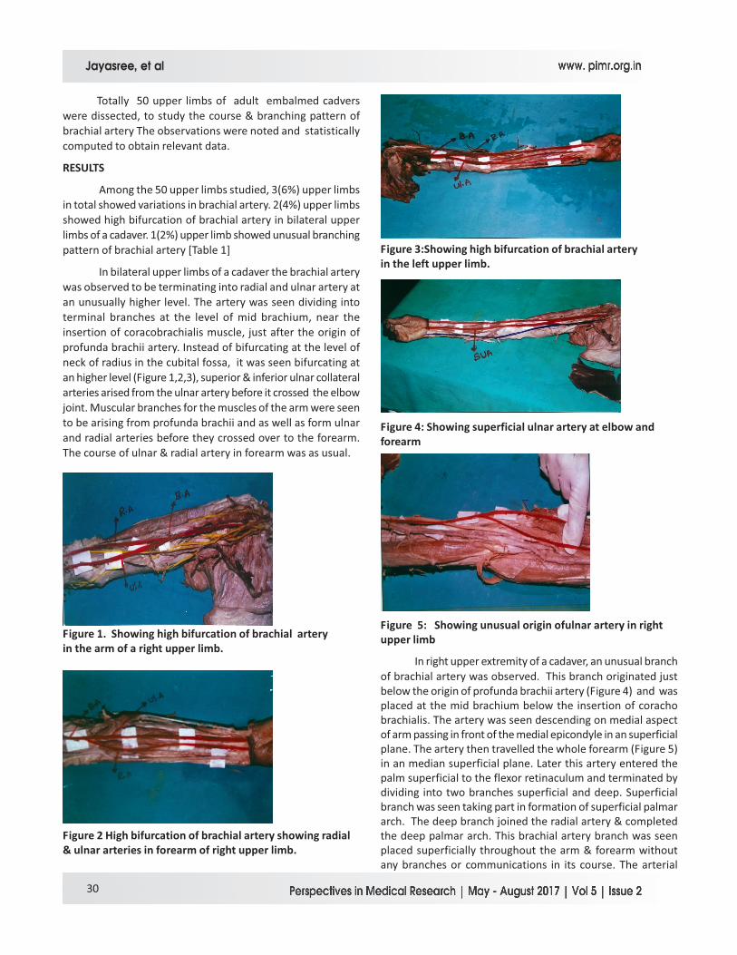
In bilateral upper limbs of a cadaver the brachial artery
was observed to be terminating into radial and ulnar artery at
an unusually higher level. The artery was seen dividing into
terminal branches at the level of mid brachium, near the
insertion of coracobrachialis muscle, just after the origin of
profunda brachii artery. Instead of bifurcating at the level of
neck of radius in the cubital fossa, it was seen bifurcating at
an higher level (Figure 1,2,3), superior & inferior ulnar collateral
arteries arised from the ulnar artery before it crossed the elbow
joint. Muscular branches for the muscles of the arm were seen
to be arising from profunda brachii and as well as form ulnar
and radial arteries before they crossed over to the forearm.
The course of ulnar & radial artery in forearm was as usual.
Figure 1. Showing high bifurcation of brachial artery
in the arm of a right upper limb.
Figure 2 High bifurcation of brachial artery showing radial
& ulnar arteries in forearm of right upper limb.
Figure 3:Showing high bifurcation of brachial artery
in the left upper limb.
Figure 4: Showing superficial ulnar artery at elbow and
forearm
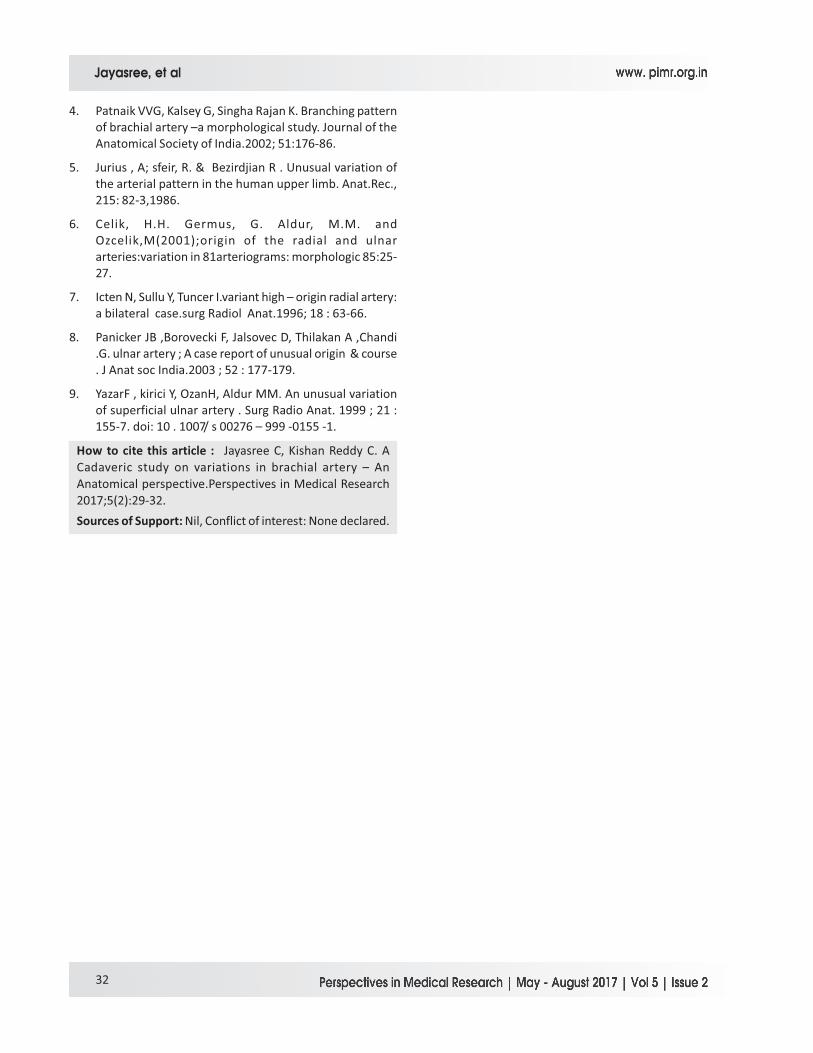
Figure 5: Showing unusual origin ofulnar artery in right
upper limb
In right upper extremity of a cadaver, an unusual branch
of brachial artery was observed. This branch originated just
below the origin of profunda brachii artery (Figure 4) and was
placed at the mid brachium below the insertion of coracho
brachialis. The artery was seen descending on medial aspect
of arm passing in front of the medial epicondyle in an superficial
plane. The artery then travelled the whole forearm (Figure 5)
in an median superficial plane. Later this artery entered the
palm superficial to the flexor retinaculum and terminated by
dividing into two branches superficial and deep. Superficial
branch was seen taking part in formation of superficial palmar
arch. The deep branch joined the radial artery & completed
the deep palmar arch. This brachial artery branch was seen
placed superficially throughout the arm & forearm without
any branches or communications in its course. The arterial
Jayasree, et al
30
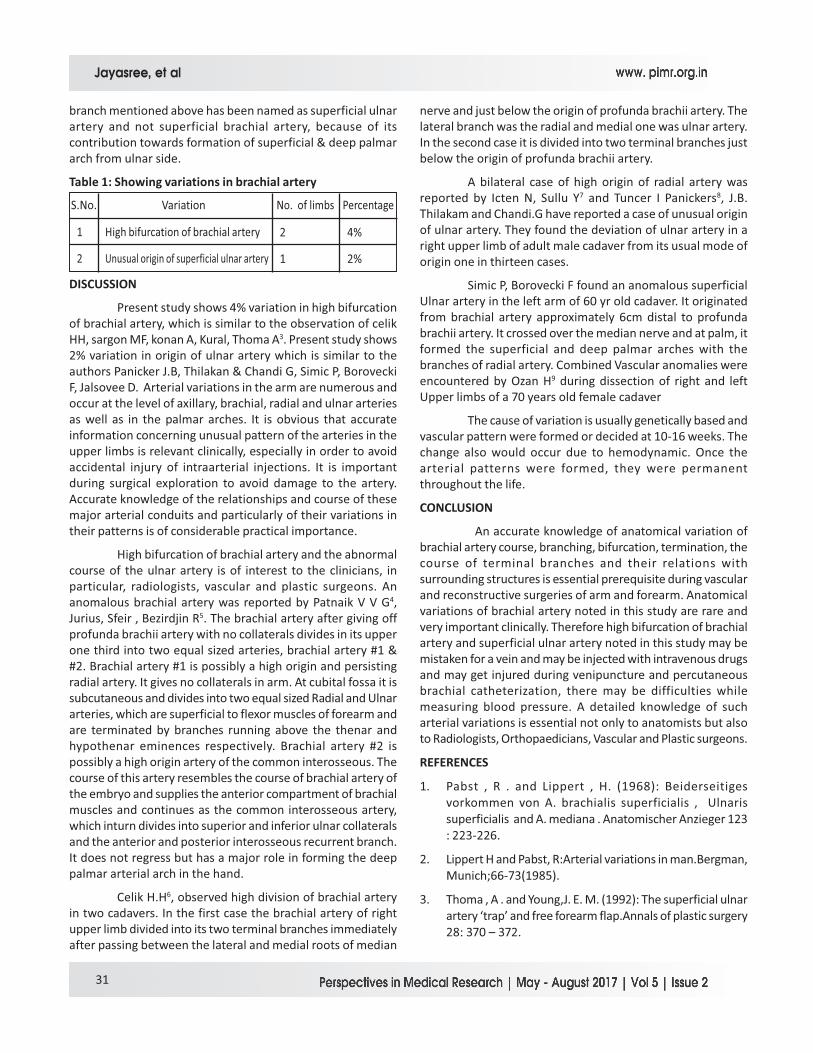
branch mentioned above has been named as superficial ulnar
artery and not superficial brachial artery, because of its
contribution towards formation of superficial & deep palmar
arch from ulnar side.
Table 1: Showing variations in brachial artery
1
2
4%
2%
2
1
No. of limbsS.No. Percentage
High bifurcation of brachial artery
Unusual origin of superficial ulnar artery
Variation
DISCUSSION
Present study shows 4% variation in high bifurcation
of brachial artery, which is similar to the observation of celik
HH, sargon MF, konan A, Kural, Thoma A3. Present study shows
2% variation in origin of ulnar artery which is similar to the
authors Panicker J.B, Thilakan & Chandi G, Simic P, Borovecki
F, Jalsovee D. Arterial variations in the arm are numerous and
occur at the level of axillary, brachial, radial and ulnar arteries
as well as in the palmar arches. It is obvious that accurate
information concerning unusual pattern of the arteries in the
upper limbs is relevant clinically, especially in order to avoid
accidental injury of intraarterial injections. It is important
during surgical exploration to avoid damage to the artery.
Accurate knowledge of the relationships and course of these
major arterial conduits and particularly of their variations in
their patterns is of considerable practical importance.
High bifurcation of brachial artery and the abnormal
course of the ulnar artery is of interest to the clinicians, in
particular, radiologists, vascular and plastic surgeons. An
anomalous brachial artery was reported by Patnaik V V G4,
Jurius, Sfeir , Bezirdjin R5. The brachial artery after giving off
profunda brachii artery with no collaterals divides in its upper
one third into two equal sized arteries, brachial artery #1 &
#2. Brachial artery #1 is possibly a high origin and persisting
radial artery. It gives no collaterals in arm. At cubital fossa it is
subcutaneous and divides into two equal sized Radial and Ulnar
arteries, which are superficial to flexor muscles of forearm and
are terminated by branches running above the thenar and
hypothenar eminences respectively. Brachial artery #2 is
possibly a high origin artery of the common interosseous. The
course of this artery resembles the course of brachial artery of
the embryo and supplies the anterior compartment of brachial
muscles and continues as the common interosseous artery,
which inturn divides into superior and inferior ulnar collaterals
and the anterior and posterior interosseous recurrent branch.
It does not regress but has a major role in forming the deep
palmar arterial arch in the hand.
Celik H.H6, observed high division of brachial artery
in two cadavers. In the first case the brachial artery of right
upper limb divided into its two terminal branches immediately
after passing between the lateral and medial roots of median
nerve and just below the origin of profunda brachii artery. The
lateral branch was the radial and medial one was ulnar artery.
In the second case it is divided into two terminal branches just
below the origin of profunda brachii artery.
A bilateral case of high origin of radial artery was
reported by Icten N, Sullu Y7 and Tuncer I Panickers8, J.B.
Thilakam and Chandi.G have reported a case of unusual origin
of ulnar artery. They found the deviation of ulnar artery in a
right upper limb of adult male cadaver from its usual mode of
origin one in thirteen cases.
Simic P, Borovecki F found an anomalous superficial
Ulnar artery in the left arm of 60 yr old cadaver. It originated
from brachial artery approximately 6cm distal to profunda
brachii artery. It crossed over the median nerve and at palm, it
formed the superficial and deep palmar arches with the
branches of radial artery. Combined Vascular anomalies were
encountered by Ozan H9 during dissection of right and left
Upper limbs of a 70 years old female cadaver
The cause of variation is usually genetically based and
vascular pattern were formed or decided at 10-16 weeks. The
change also would occur due to hemodynamic. Once the
arterial patterns were formed, they were permanent
throughout the life.
CONCLUSION
An accurate knowledge of anatomical variation of
brachial artery course, branching, bifurcation, termination, the
course of terminal branches and their relations with
surrounding structures is essential prerequisite during vascular
and reconstructive surgeries of arm and forearm. Anatomical
variations of brachial artery noted in this study are rare and
very important clinically. Therefore high bifurcation of brachial
artery and superficial ulnar artery noted in this study may be
mistaken for a vein and may be injected with intravenous drugs
and may get injured during venipuncture and percutaneous
brachial catheterization, there may be difficulties while
measuring blood pressure. A detailed knowledge of such
arterial variations is essential not only to anatomists but also
to Radiologists, Orthopaedicians, Vascular and Plastic surgeons.
REFERENCES
1. Pabst , R . and Lippert , H. (1968): Beiderseitiges
vorkommen von A. brachialis superficialis , Ulnaris
superficialis and A. mediana . Anatomischer Anzieger 123
: 223-226.
2. Lippert H and Pabst, R:Arterial variations in man.Bergman,
Munich;66-73(1985).
3. Thoma , A . and Young,J. E. M. (1992): The superficial ulnar
artery ‘trap’ and free forearm flap.Annals of plastic surgery
28: 370 – 372.
Jayasree, et al
31
4. Patnaik VVG, Kalsey G, Singha Rajan K. Branching pattern
of brachial artery –a morphological study. Journal of the
Anatomical Society of India.2002; 51:176-86.
5. Jurius , A; sfeir, R. & Bezirdjian R . Unusual variation of
the arterial pattern in the human upper limb. Anat.Rec.,
215: 82-3,1986.
6. Celik, H.H. Germus, G. Aldur, M.M. and
Ozcelik,M(2001);origin of the radial and ulnar
arteries:variation in 81arteriograms: morphologic 85:25-
27.
7. Icten N, Sullu Y, Tuncer I.variant high – origin radial artery:
a bilateral case.surg Radiol Anat.1996; 18 : 63-66.
8. Panicker JB ,Borovecki F, Jalsovec D, Thilakan A ,Chandi
.G. ulnar artery ; A case report of unusual origin & course
. J Anat soc India.2003 ; 52 : 177-179.
9. YazarF , kirici Y, OzanH, Aldur MM. An unusual variation
of superficial ulnar artery . Surg Radio Anat. 1999 ; 21 :
155-7. doi: 10 . 1007/ s 00276 – 999 -0155 -1.
How to cite this article : Jayasree C, Kishan Reddy C. A
Cadaveric study on variations in brachial artery – An
Anatomical perspective.Perspectives in Medical Research
2017;5(2):29-32.
Sources of Support: Nil, Conflict of interest: None declared.
Jayasree, et al
32