Embed Size (px)
Citation preview

Localisation and function of Slam in the early
Drosophila embryo
Dissertation
for the award of the degree
“Doctor rerum naturalium (Dr.rer.nat.)”
in the GGNB program “Genes and Development”
at the Georg-August-Universität Göttingen
Faculty of Biology
submitted by
Sreemukta Acharya
born in Akividu, India
Göttingen, August 2014

MEMBERS OF THE THESIS COMMITTEE
Prof. Jörg Großhans (Supervisor, reviewer)
Department of Developmental Biochemistry, University of Göttingen
Prof. Reinhard Schuh (Reviewer)
Department of Molecular Developmental Biology, Max Planck Institute for Biophysical Chemistry
Prof. Henning Urlaub
Bioanalytical Mass Spectometry Group, Max Planck Institute for Biophysical Chemistry
Date of oral examination: 20.10.2014

AFFIDAVIT
I hereby declare that I prepared the PhD thesis “Localisation and function of Slam in
the early Drosophila embryo” on my own with no other sources and aids than quoted.
__________________
Sreemukta Acharya,
Göttingen, 29.08.2014

LIST OF PUBLICATIONS
Acharya, S.*, Laupsien, P.*, Wenzl, C., Yan, S., and Großhans, J. (2014). Function and dynamics of slam in furrow formation in early Drosophila embryo. Dev. Biol. 386, 371–384.
*These authors contributed equally.
V
TABLE OF CONTENTS
ACKNOWLEDGEMENTS ....................................................................................... VIII
SUMMARY ................................................................................................................ IX
LIST OF FIGURES ..................................................................................................... X
LIST OF TABLES .................................................................................................... XII
LIST OF MOVIES .................................................................................................... XIII
ABBREVIATIONS .................................................................................................... XV
1. INTRODUCTION .................................................................................................... 1
1.1 Drosophila early development ....................................................................... 1
1.2 Cleavage furrow site specification ................................................................. 4
1.3 Initiation and maintenance of membrane invagination ................................... 6
1.3.1 slow as molasses (slam) ............................................................................. 7
1.3.2 nullo ............................................................................................................ 8
1.3.3 RhoGEF2 .................................................................................................... 9
1.3.4 diaphanous (dia) ....................................................................................... 10
1.3.5 abelson and enabled ................................................................................. 10
1.3.6 Cytoskeletal components .......................................................................... 11
1.4 Membrane transport ........................................................................................ 12
1.5 Basal closure ................................................................................................... 15
1.6 Aim of the work ................................................................................................ 15
2. MATERIALS AND METHODS ............................................................................. 17
2.1 Materials .......................................................................................................... 17
2.1.1 Reagents ................................................................................................... 17
2.1.2 Buffers and solutions ................................................................................. 17
2.1.3 Media for bacterial culture ......................................................................... 22
2.1.4 Media for flies ............................................................................................ 23
2.1.5 Enzymes and Kits ..................................................................................... 24
2.1.6 Chromatography ....................................................................................... 24
2.1.7 Oligonucleotides used in the study ............................................................ 24
2.1.8 Plasmid constructs used in the study ........................................................ 25
2.1.9 Primary antibodies .................................................................................... 26
2.1.10 Secondary antibodies/dyes ..................................................................... 27
2.1.11 Bacterial cell lines ................................................................................... 27
2.1.12 Fly stocks ................................................................................................ 27
2.1.13 Microscopy .............................................................................................. 29

Table of contents VI
2.1.14 Other materials ........................................................................................ 29
2.1.15 Other equipment ..................................................................................... 30
2.1.16 Softwares ................................................................................................ 30
2.2 Methods .......................................................................................................... 31
2.2.1 DNA methods ............................................................................................ 31
2.2.1.1 Molecular cloning ................................................................................ 31
2.2.1.2 Polymerase chain reaction (PCR) ....................................................... 31
2.2.1.3 Site-directed mutagenesis .................................................................. 31
2.2.1.4 In-fusion cloning .................................................................................. 32
2.2.1.5 Purification of DNA/RNA by phenol-chloroform extraction .................. 32
2.2.1.6 DNA sequencing ................................................................................. 32
2.2.1.7 In-vitro transcription ............................................................................ 33
2.2.1.8 Reverse transcription .......................................................................... 34
2.2.1.9 Quantitative real-time PCR (qRT-PCR) .............................................. 34
2.2.2 Biochemical methods ................................................................................ 35
2.2.2.1 Protein purification .............................................................................. 35
2.2.2.2 Affinity purification of antibodies ......................................................... 36
2.2.2.3 Western blot ........................................................................................ 37
2.2.2.4 EMSA .................................................................................................. 37
2.2.2.5 Immunoprecipitation............................................................................ 38
2.2.2.6 Pulldown of GFPslam using paramagnetic Streptavidin beads ........... 39
2.2.3 Fly embryo methods .................................................................................. 39
2.2.3.1 Fractionation of embryos .................................................................... 39
2.2.3.2 RNA isolation following fractionation of embryos ................................ 40
2.2.3.3 Embryo fixation ................................................................................... 41
2.2.3.4 Immunostaining of embryos ................................................................ 41
2.2.3.5 Fluorescence in-situ hybridisation (FISH) ........................................... 41
2.2.3.6 Generation of germline clones ............................................................ 43
2.2.3.7 Fly genetics ......................................................................................... 43
2.2.3.8 Induction of shibire phenotype ............................................................ 43
2.2.3.9 Microinjection of protein/drug into embryos ........................................ 44
2.2.4. Microscopy ............................................................................................... 44
2.2.4.1. Live-imaging of fly embryos ............................................................... 44
2.2.4.2 Fluorescence recovery after photobleaching (FRAP) ......................... 44
2.2.4.3 Laser ablation of centrosomes ............................................................ 44

Table of contents VII
3. RESULTS ............................................................................................................. 46
3.1 Factors contributing to the accumulation of Slam protein at the furrow canal . 46
3.1.1 Centrosomes define the site of invagination ............................................. 46
3.1.2 Accumulation of Slam is dependent on vesicular transport ....................... 50
3.2 Dynamics of Slam protein, mobility and life-time ............................................. 59
3.2.1 A fraction of Slam is membrane-associated .............................................. 59
3.2.2 Slam protein is stable during cellularisation .............................................. 60
3.2.3 Mobility of Slam is independent of new translation.................................... 63
3.2.4 Mobility of Slam is not directly affected by the recycling endosome .......... 64
3.3 Role of Slam during cellularisation .................................................................. 66
3.1.1 Additional factors apart from Slam are needed for furrow specification .... 66
3.3.1 nullo and slam together control the specification of the cleavage furrow .. 67
3.3.2 spire – an interactor of slam in yeast two-hybrid screen ........................... 70
3.3.3 Other interactors of Slam .......................................................................... 73
3.4 Slam protein properties and the RNP complex................................................ 74
3.4.1 Slam has a predicted structured N-terminal half ....................................... 74
3.4.2 GFP tag at the N-terminus of Slam partially interferes with its function ..... 75
3.4.3 Slam protein and slam mRNA are present in a complex ........................... 76
4. DISCUSSION ....................................................................................................... 81
4.1 Centrosomes specify the site of cleavage furrow and restrict Slam to the
furrow canal ....................................................................................................... 81
4.2 Slam restriction at the furrow canal is dependent upon the recycling
endosome .......................................................................................................... 82
4.3 Role of Slam during cellularisation ............................................................... 85
4.4 Mobility of Slam at the furrow canal ............................................................. 86
4.5 Slam protein and mRNA form a complex ..................................................... 89
5. REFERENCES ..................................................................................................... 91

VIII
ACKNOWLEDGEMENTS
I would firstly like to thank my supervisor Prof. Jörg Großhans for allowing me
to carry out my PhD work in his laboratory and for always being available for
discussions and to answer questions, no matter how simple (or stupid). I really
appreciate his patience, optimism and politeness towards his students. His hard-
working nature and passion for science are truly inspiring.
I am very thankful to my thesis committee members Prof. Reinhard Schuh and
Prof. Henning Urlaub for their helpful suggestions and constructive criticism during the
thesis committee meetings. Their positivity was always very encouraging.
I express my gratitude to my current and past colleagues for creating an
excellent working atmosphere, providing with great discussions and German lessons
over coffee and fly-sorting. I would especially like to thank everyone involved in the
‘Slam project’ - Dr. Shuling Yan, Dr. Philip Laupsien, Dr. Christian Wenzl and
Stephanie Gröning for sharing results, exciting discussions and helping me with the
methods. Without their cooperation, it would have been impossible to carry out this
collaborative work. I am grateful to Dr. Maike Claußen for helping me with the EMSA
experiments and for suggestions regarding the project. I thank Olaf Bernhard from Dr.
Bernhard Schmidt’s lab for carrying out Mass spectrometric analysis for us.
I am thankful to all members of the Department of developmental biochemistry
for their support and cooperation (and cakes). I could not have asked for a better
working environment.
I am indebted to my parents who always believed in me and supported me
through thick and thin. The seeds of scientific enquiry and critical thinking sowed by
my father and my mother’s practical advice and math lessons were crucial to reach
this stage. I am thankful to my brother Sreekanth Acharya for being there for me
whenever I felt downtrodden for any reason.
I would like to thank Roman Petrovsky for fascinating discussions, insightful
comments and constant support in the lab as well as outside of it.
Last but not the least, I would like to thank Mansi Karkhanis for ceaseless
support, constant encouragement and general suggestions.

IX
SUMMARY
Embryogenesis of many insects starts with a syncytial stage characterised by
13 rapid nuclear divisions. During the following interphase, the plasma membrane of
the embryo invaginates between each nucleus to give rise to the first epithelial layer of
the embryo. This type of cytokinesis during interphase has been named cellularisation.
Two important processes involved in cellularisation are a) specification of the site of
cleavage furrow and b) initiation and maintenance of membrane invagination. We
investigated the role of a gene slam in these processes. Although it has been shown
earlier that slam is necessary for cellularisation and the formation of the basal domain
of the invaginating membrane (furrow canal), it has remained unclear how Slam
reaches the furrow canal and interacts with the membrane. The aim of the study was
to uncover the mechanism of Slam accumulation at the furrow canal, elucidate its
mobility dynamics at the membrane and elaborate its role in cellularisation.
By means of live imaging and centrosome ablation, I could show that
centrosomes are the initial source of signals for the accumulation of Slam. Using
shibire temperature-sensitive mutants, I found that the initial accumulation of Slam at
the new furrow was vesicle-dependent but its maintenance at the old furrow was not.
Analysis of nuf mutants revealed that although Slam is not directly transported on
Rab11-positive vesicles, its proper targeting to the basal domain is indirectly
dependent upon the recycling endosome. I identified a role of slam together with
another gene nullo in establishing the furrow. I found that slam and nullo act
redundantly to each other. Furthermore, using fluorescent recovery after
photobleaching experiments, I could show that membrane-associated Slam undergoes
a switch-like change from high mobility at the onset of cellularisation to low mobility at
mid-cellularisation. Slam mobility in mid-cellularisation is independent of new
translation and vesicular trafficking. Finally, I showed that Slam is a ribonucleoprotein
complex (RNP) and that slam mRNA was more enriched at the membrane.
I propose that the recycling endosome that is organised by the centrosome
restricts a potential Slam receptor to the prospective basal domain of the membrane
to which the Slam RNP is recruited from the cytoplasm. Once at the furrow, slam acts
together with nullo to establish the furrow and initiate cellularisation without further
recruitment of Slam RNP and Slam protein synthesis.

X
LIST OF FIGURES
Fig 1.1 Schematic representation of Drosophila early development
1
Fig 1.2 Schematic representation of pseudocleavage furrow formation and cellularisation
3
Fig 1.3 Schematic representation of Rappaport and cleavage furrows
5
Fig 3.1 Lonesome centrosomes are capable of specifying ectopic furrows
47
Fig 3.2 SAS6GFP overexpression results in extra centrosomes in the embryo due to additional centrosomal divisions
48
Fig 3.3 GFPslam accumulates around the additional duplicate/singular lonesome centrosomes to form ectopic furrows in SAS6GFP embryos
49
Fig 3.4 Functional ablation of a centrosome induces loss of its furrow-specifying ability
50
Fig 3.5 Slam accumulation at the furrow canal is dependent on vesicular transport
51
Fig 3.6 Loss of Slam from the furrow canal is a specific effect
51
Fig 3.7 Rab11 is associated with the perinuclear recycling endosome and the recycling endosome vesicles
52
Fig 3.8 Slam is not transported on Rab11 vesicles
53
Fig 3.9 Graph depicting the variability of nuf embryos
54
Fig 3.10 nuf embryos show variable phenotype ranging from mild to severe
55
Fig 3.11
Slam is not restricted to the basal domain in nuf embryos. 56
Fig 3.12 Slam mislocalisation corresponds to the severity of nuf phenotype
57
Fig 3.13 Interference with Rab11 function in the embryo causes delay in Slam restriction to the furrow canal
58
Fig 3.14 Slam is a membrane-associated protein
60
Fig 3.15 Effect of blocking protein translation in different stages of embryo
61
Fig 3.16 Slam protein is stable during cellularisation
62
Fig 3.17 Mobility of Slam during cellularisation is independent of new translation
63

List of figures XI
Fig 3.18 Slam is stably associated to the membrane during interphase 13 and 14 and switches to a high-mobility state at the onset of cellularisation
65
Fig 3.19 Slam mobility is slightly increased in a subset of nuf embryos
65
Fig 3.20 The mobility of Slam during cellularisation is not dependent on vesicular trafficking
66
Fig 3.21 Schematic representation of alleles of slam
67
Fig 3.22 Furrow specification requires factors additional to slam
68
Fig 3.23 nullo and slam together control the specification of the furrow
69
Fig 3.24 Schematic representation of interaction of Slam and Spire in a Yeast two-hybrid screen
70
Fig 3.25 spir alleles show cell-cycle defects and occasional cellularisation defects
71
Fig 3.26 Spire antibody against the KIND domain detects several isoforms of Spire
72
Fig 3.27 Immunostaining against Spire shows apical staining during cellularisation
73
Fig 3.28 Pulldown of Slam and GFPslam
74
Fig 3.29 Schematic representation of disordered regions in Slam protein
75
Fig 3.30 GFP tag partially interferes with Slam function
75
Fig 3.31 GFPslam is less efficiently bound to the membrane
76
Fig 3.32 slam mRNA and protein are present in a complex
77
Fig 3.33 slam mRNA is enriched at the membrane
78
Fig 3.34 slam mRNA and protein also colocalise ectopically 79
Fig 3.35 Binding assay with SlamC651 protein and slam mRNA localising elements
80
Fig 4.1 Model for the role of centrosomes in targeting of recycling endosome vesicles to the site of cleavage furrow
84
Fig 4.2 Mobility of Slam and the role of recycling endosome in its restriction
88

XII
LIST OF TABLES
Table 2.1 Oligonucleotides used in the study
25
Table 2.2 Plasmids used in the study
25
Table 2.3 Antibodies used in the study
26
Table 2.4 Fly stocks used in the study 27
Table 3.1 Penetrance of cell-cycle and cellularisation defects in different spire alleles
72
Table 3.2 qRT-PCR threshold cycle (Ct) numbers for three independent fractionation experiments
79

XIII
LIST OF MOVIES
Movie 1 Extra centrosome replication (Fig 3.2)
Movie 2 Duplicate lonesome centrosomes (Fig 3.3 A)
Movie 3 Singular lonesome centrosome (Fig 3.3 B)
Movie 4 Centrosome ablation (Fig 3.4)
Movie 5 GFPslam control 32°C (Fig 3.5)
Movie 6 GFPslam in shibire 32°C (Fig 3.5)
Movie 7 GFPslamRab11YFP (Fig 3.8)
Movie 8 GFPslam WT (Fig 3.11)
Movie 9 GFPslam nuf embryo 1 (Fig 3.11)
Movie 10 GFPslam nuf embryo 2 (Fig 3.11)
Movie 11 GST injected in GFPslam embryo (Fig 3.13 A)
Movie 12 Rab11S25N injected in GFPslam embryo (Fig 3.13A)
Movie 13 Buffer injected into hisRFP embryo (Fig 3.15A)
Movie 14 Cycloheximide into hisRFP embryo (Fig 3.15A)
Movie 15 Buffer injected into GFPslam embryo at anaphase of cycle 13 (Fig 3.15B)
Movie 16 Cycloheximide injected into GFPslam embryo at anaphase of cycle 13 (Fig 3.15B)
Movie 17 Buffer injected into GFPslam embryo at onset of cycle 14 (Fig 3.15C)
Movie 18 Cycloheximide injected into GFPslam embryo at onset of cycle 14 (Fig 3.15C)
Movie 19 Buffer injected into GFPslam embryo at mid-cellularisation (Fig 3.15D)
Movie 20 Cycloheximide injected into GFPslam embryo at mid-cellularisation (Fig 3.15D)
Movie 21 GFPslam FRAP in buffer injected embryos (Fig 3.17)

List of movies XIV
Movie 22 GFPslam FRAP in cycloheximide injected embryos (Fig 3.17)
Movie 23 GFPslam FRAP in WT background (Fig 3.19 A)
Movie 24 GFPslam FRAP in nuf embryo1 (Fig 3.19C)
Movie 25 GFPslam FRAP in nuf embryo2 (Fig 3.19E)
Movie 26 GFPslam FRAP in shibire (Fig 3.20)

XV
ABBREVIATIONS
aa amino acid(s)
ß-gal beta-galactosidase
bp base pairs
cDNA complementary DNA
DAPI 4’, 6’ – Diamidino-2-phenylindole
ddH2O double distilled water
°C degree Celsius
DNA deoxyribonucleic acid
DTT 1,4-dithiothreitol
deletion
C C-terminal deletion
E.coli Escherichia coli
EDTA ethylenediaminetetraacetic acid
EMSA electrophoretic mobility shift assay
FRT flippase recognition target
FRAP fluorescence recovery after photobleaching
GEF guanidyl nucleotide exchange factor
GFP green fluorescent protein
GST Glutathion-S-transferase
g gram(s)
glc germline clone
h hour(s)
IPTG Isopropyl-ß-D-thiogalactopyranoside
kb kilobases
kDa kiloDalton
l litre(s)
m milli-
micro-
min minute(s)
mRNA messenger RNA

Abbreviations XVI
Nr number
PCR polymerase chain reaction
PMSF Phenylmethylsulfonylfluorid
RNA ribonucleic acid
RNAi RNA interference
RNP ribonucleoprotein
rpm revolutions per minute
RT room temperature
SDS sodiumdodecylsulphate
SDS-PAGE SDS-polyacylamide gel electrophoresis
Tris tris(hydroxymethyl)aminomethne hydrochloride
WT wild-type
w white gene
Y2H yeast two-hybrid
1
1. INTRODUCTION
1.1 Drosophila early development
The early embryonic divisions of a Drosophila embryo, similar to many insects,
take place in a syncytium. Subsequent to fertilisation of the oocyte by a sperm, the
nucleus undergoes thirteen rounds of mitotic divisions (Zalokar and Erk, 1976; Foe and
Alberts, 1983). However, the nuclear divisions are not accompanied by cytokinesis of
the embryo, resulting in the daughter nuclei lying in a common cytoplasm. The first
eight mitotic cycles take place in the embryonic core and are referred to as the
preblastoderm cycles. These cycles are very rapid, lasting 8 min on an average and
consist only of the synthetic phase (S-phase) and the mitotic phase (M-phase) of cell
cycle (Zalokar and Erk, 1976). From cycle 9 onwards, most of the nuclei undergo
microtubule-dependent cortical migration, which involves their migration from the
centre of the embryo to the cortex. This stage of the embryo where the embryo contains
a cortical monolayer of nuclei in a syncytium is called syncytial blastoderm. Some
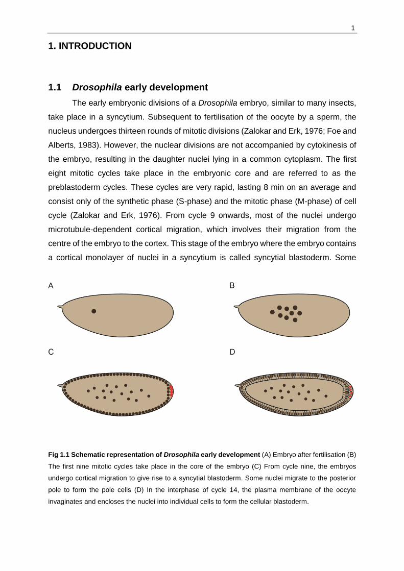
Fig 1.1 Schematic representation of Drosophila early development (A) Embryo after fertilisation (B)
The first nine mitotic cycles take place in the core of the embryo (C) From cycle nine, the embryos
undergo cortical migration to give rise to a syncytial blastoderm. Some nuclei migrate to the posterior
pole to form the pole cells (D) In the interphase of cycle 14, the plasma membrane of the oocyte
invaginates and encloses the nuclei into individual cells to form the cellular blastoderm.

Introduction 2
nuclei migrate to the posterior pole of the embryo to form the pole cells, which are germ
cell precursors. The nuclei at the cortex remain anchored beneath the plasma
membrane where they undergo further mitotic divisions. These divisions include a
second gap phase (G2 phase), whose length progressively increases following each
cycle. After the end of the thirteenth cell cycle, there is an extended interphase of cycle
14, which lasts about an hour (Foe and Alberts, 1983). During this interphase, the
plasma membrane of the embryo starts invaginating between each nucleus and
progressively encloses each nucleus into an individual cell, in a process called
cellularisation (Fig 1.1).
The cortex of the embryo is rich in actin that undergoes a cycle of redistribution
under the influence of centrosomal asters during the syncytial cycles 10 to 13 (Kao and
Megraw, 2009). During interphases, it accumulates as actin caps above each nucleus.
As the nuclei progress through prophase and enter metaphase, the plasma membrane
of the embryo invaginates between adjacent nuclei, reaching a depth of up to
approximately 8m. These structures are called pseudocleavage furrows or
metaphase furrows. Metaphase furrows prevent fusion of spindle from adjacent nuclei
(Sullivan et al., 1993). As the cell cycle approaches telophase, the metaphase furrows
start regressing and the actin rearranges itself again as actin caps. This cycling of actin
distribution occurs until the embryo reaches the stage of cellularisation during which
actin persists at the tip of the invaginating membrane (Fig 1.2).
At the onset of cellularisation during the cycle 14 interphase, microtubules
emanate from the apically-located centrosomes. These microtubules extend
downwards to encompass the nuclei into an inverted basket-like structure and are
necessary for furrow invagination.
The process of cellularisation has been divided into four distinct phases on the
basis of morphology and speed of invagination of the membrane (Knoblich, 2000;
Lecuit and Wieschaus, 2000). At Phase one, the cortical region above each nucleus is
rich in microvilli while the region at the middle of adjacent nuclei, which is called furrow
canal, is devoid of microvilli (Lecuit and Wieschaus, 2000). Furrow canals form the
leading edge of the invaginating furrows (Fullilove and Jacobson, 1971). Phase one,
which lasts about 10 minutes, is also accompanied by nuclear elongation. Phase two
is characterised by the completion of nuclear elongation but very slow membrane

Introduction 3
Fig 1.2 Schematic representation of pseudocleavage furrow formation and cellularisation. (A)
Cycle 13 interphase: actin is located apical to the nuclei as caps (B) Cycle 13 mitosis: Metaphase furrows
are formed and actin redistributes itself to the tip of the furrows (C) Cycle 14 interphase: actin forms
caps again (D) Onset of cellularisation: Furrow canal and basal adherens junctions are formed, actin
redistributes itself to the furrow canal (E, F) apical adherens junctions are formed and cellularisation
proceeds first slowly (slow phase) and then the rate speeds up (fast phase).
invagination. In phase three, the invaginating membrane reaches to the plane of the
basal region of the nuclei. When the membrane reaches about 35 m in depth, it starts
contracting laterally in a process called basal closure, which encloses each nucleus
from beneath to give rise to blastoderm cells. Cellularisation is a precisely regulated
process requiring establishment and maintenance of membrane polarity, specification
of the cleavage site, regulated membrane growth and finally basal closure to enclose

Introduction 4
the nuclei into cells. These processes are discussed in further detail in the following
sections.
Cellularisation involves formation of a polarised epithelium. As the plasma
membrane invaginates during this process, it becomes compartmentalised into apical,
lateral and basal domain. It has been shown that the plasma membrane of the embryo
displays a polarised organisation already during the syncytial blastoderm stage
(Mavrakis et al., 2009). Two distinct plasma membrane domains have been identified
during the syncytial cycles a) above each nucleus and b) at the lateral region of the
nuclei. These regions contain distinct membrane markers and are unable to diffuse
outside of their own domain, indicating towards the existence of a diffusion barrier. This
diffusion barrier was shown to be actin-dependent (Mavrakis et al., 2009).
Establishment and maintenance of membrane polarity during cellularisation is
also dependent upon several other factors such as proper vesicle targeting through
the recycling endosome and the proper localisation of initial furrow canal markers such
as Actin and Slam. The role of these factors has been discussed later under individual
sections.
1.2 Cleavage furrow site specification
Cellularisation of the Drosophila embryo is a modified cytokinesis and hence,
many of the mechanisms involved in the positioning of the cytokinetic furrow during
cellularisation are similar to conventional cytokinesis. The cytokinetic furrow in
eukaryotic animal cells is positioned at the cell cortex midway down the longitudinal
axis of the mitotic spindle (Balasubramanian et al., 1992; Bi et al., 1998; Fujiwara and
Pollard, 1976; Mabuchi and Okuno, 1977, 1977). The positioning of the furrow is
carried out by the central spindle or astral microtubules, depending on the cell size and
type. Rappaport’s classic experiments with marine invertebrate embryos suggested
that astral microtubules are the source of the furrow initiation signal. He created a
binucleate embryo artificially and found that when these nuclei divided, apart from the
two furrows that formed between the respective metaphase plates, a third ectopic
furrow formed between neighbouring centrosomes in the absence of a nucleus or a
central spindle (Rappaport, 1961). He proposed that the signal for initiation of the
ectopic furrow must be generated by overlapping astral microtubules (Fig 1.3). It was
further demonstrated that a minimal distance between the two centrosomes as well as

Introduction 5
between the centrosomes and cortex were essential for furrow induction (Rappaport,
1986).
Fig. 1.3 Schematic representation of Rappaport and cleavage furrows. Artificially induced
Rappaport furrows are formed between overlapping asters of adjacent centrosomes while cleavage
furrows are formed between the overlapping spindles between a centrosome pair (Adapted from
Sullivan, 2009).
However, studies in cultured cells have suggested that the region of overlapping
antiparallel microtubules called the central spindle is responsible for furrow positioning
(Wheatley and Wang, 1996; Williams et al., 1995). A number of proteins required for
cytokinesis are recruited to the central spindle. Rappaport had also showed that
flattening the sand dollar embryos so as to allow the central spindle to interact with the
cortex could induce a furrow (Rappaport, 1985). Recent studies on Caenorhabditis
elegans (C.elegans) embryos have suggested both astral microtubules as well as the
central spindle contribute to furrow induction (Bringmann and Hyman, 2005; Motegi et
al., 2006). In these embryos, it seems that the initial furrow induction signal comes from
the asters, while the completion of cytokinesis depends on the sustenance of the signal
from the central spindle (Baruni et al., 2008; Bringmann and Hyman, 2005).
In addition to forming the astral microtubules, other centrosome-associated
activities are also essential for furrow specification. The Drosophila Centrosomin
(CNN) is a core centrosomal protein required for normal pericentriolar material
organisation and astral microtubule assembly (Li and Kaufman, 1996; Megraw et al.,
1999). A hypomorphic allele of cnn called cnnB4 has been used to dissect these two

Introduction 6
functions since this allele shows severe disruptions in furrow formation, despite having
an apparently normal microtubule organisation (Kao and Megraw, 2009). Kao and
Megraw identified another protein Centrocortin (CEN), which interacts with CNN. CEN
partially colocalises with CNN at the centrosomes and is also found on cleavage
furrows. Strong cen alleles show similar phenotype as cnnB4 allele where cleavage
furrows are weak or broken. Like cnnB4, cen mutants show no discernable defects in
microtubule organisation. This study demonstrates the role of centrosomal protein
CNN in relaying a furrow initiation signal to via CEN, thus suggesting that centrosomes
possess additional information for furrow specification which is independent of astral
microtubule assembly.
Various studies have been done suggesting centrosomes are capable of
determining the site of furrow invagination independent of the nuclei. Anucleate
C.elegans embryos containing only a pair of centrosomes are able to form a cytokinetic
furrow and attempt to divide (Baruni et al., 2008). In Drosophila syncytial embryos, it
has been shown that if centrosome and nuclear divisions are uncoupled by inhibiting
S-phase, and nuclear migration to the cortex prevented, the centrosomes that migrate
to the posterior pole are able to initiate pole cell formation in the absence of nuclei (Raff
and Glover, 1989). In another study, blocking nuclei from entering mitosis by
simultaneous RNAi of all three mitotic cyclins in the early Drosophila embryos showed
that centrosomes are able to organise Myosin ‘cages’ around them (McCleland and
O’Farrell, 2008). The timing of cellularisation was unaffected despite the nuclear arrest
(McCleland and O’Farrell, 2008) which suggests that the temporal information for the
initiation of cellularisation is not regulated by the number of nuclear divisions.
In summary, positioning of the furrow is controlled by astral microtubules or
central spindle or both depending upon the size and type of the cell. Centrosomal
activity, in addition to its microtubular organizing capacity, is required during
cellularisation in Drosophila embryos. Also, centrosomes have the ability to organise
the furrow position in the absence of nuclei in several systems.
1.3 Initiation and maintenance of membrane invagination
Several maternal as well as zygotic genes are necessary for the initiation of
membrane invagination. Some of them are discussed below.

Introduction 7
1.3.1 slow as molasses (slam)
One of the zygotic genes that plays an important role in the initiation of
membrane invagination is slow as molasses (slam). slam is highly expressed at the
onset of cellularisation. Zygotic deficiency of slam shows a very strong and penetrant
cellularisation phenotype (Lecuit et al., 2002). It showed defects primarily in the slow
phase of cellularisation causing delayed furrow invagination, which is why it was
named ‘slow as molasses’ originally (Lecuit et al., 2002). However, removal of both
maternal and zygotic contributions has demonstrated that slam is in fact essential for
furrow invagination. Embryos devoid of maternal and zygotic slam display no furrow
invagination (Stein et al., 2002; Acharya et al., 2014; Dr. Philip Laupsien, PhD
dissertation).
Already at the onset of cellularisation, the respective morphologies of the apical
and the basal regions of the plasma membrane are distinguishable. The apical part,
which lies above the nuclei, consists of many villous projections. Adjacent to this region
is a region of depressed plasma membrane that lacks villous projections. As the slow
phase progresses, formation of the basal junction brings together the adjacent lateral
regions of the plasma membrane, thus delimiting the basal domain from the lateral
domain. In slam zygotic deficiency embryos, although the prospective site of furrow
invagination shows a slight depression, no basal junction is formed and the villous
projections extend into the prospective furrow canal (Lecuit et al., 2002). Despite the
abnormal furrow canal organisation, a delayed membrane invagination can be seen in
the presence of maternal Slam contribution (Lecuit et al., 2002). Membrane
invagination is completely abolished only when the maternal contribution is also
removed (Acharya et al., 2014; Dr. Philip Laupsien, PhD dissertation). This suggests
that Slam has a dual role – one in polarity establishment and second in membrane
invagination during cellularisation.
The apical domain of the plasma membrane rich in villous projections shows
higher endocytic behaviour compared to the prospective furrow canal region (Lecuit
and Wieschaus, 2000). slam zygotic deficiency embryos show reduced endocytosis at
the apical region (Lecuit et al., 2002). This suggests that Slam might establish the
polarity of the membrane by regulating transcytosis at the onset of cellularisation
(Lecuit et al., 2002). However, Slam localises exclusively at the furrow canal
(Grosshans et al., 2005; Lecuit et al., 2002) so it is unclear how exactly Slam might
regulate endocytosis at the apical region.

Introduction 8
Zygotic expression of hypomorphic alleles of slam (slamwaldo1 and slamwaldo2)
does not show a cellularisation phenotype but instead display germ cell migration
defects. During the process of germ cell migration, neither slam mRNA nor the protein
are detectable in the embryo. Therefore, the mechanism behind the role of slam in
germ cell migration is unknown. It has been speculated that during cellularisation itself,
Slam might be recruiting factors necessary for the subsequent germ cell migration.
1.3.2 nullo
nullo was first identified as a locus on the X-chromosome that was necessary
for cellularisation (Rose and Wieschaus, 1992; Simpson and Wieschaus, 1990;
Wieschaus and Sweeton, 1988). In nullo mutant embryos, the cleavage furrow is
unable to invaginate resulting in the generation of multinucleate cells. Nullo stabilises
the accumulation of components of the basal junction; in nullo mutants, Armadillo and
E-cadherin are not concentrated in the junctional region and are instead spread across
the lateral membrane (Hunter and Wieschaus, 2000). It was suggested that due to the
failure of formation of the basal junction, compartmentalisation of the invaginating
membrane was compromised. However, subsequent studies found that furrow
compartmentalisation does not require the basal adherens junctions and that rather
the reduced F-actin levels at furrow canals in nullo mutants is the cause of polarity
disruption (Sokac and Wieschaus, 2008b).
Nullo protein is rapidly degraded before the completion of cellularisation and
apical adherens junction formation. Prolonging the expression of Nullo into late
cellularisation causes blockage of the apical clustering of Armadillo, -catenin and E-
cadherin. These junctional defects cause a disrupted cell morphology resulting in a
failure to form the ventral furrow. These studies indicate that establishment of the apical
junction and the formation of the ventral furrow is dependent on the rapid degradation
of Nullo in late cellularisation (Hunter and Wieschaus, 2000).
Nullo contains an N-terminal myristoylation site that is important for its targeting
to the plasma membrane during cycle 14. Interestingly, Nullo protein devoid of the
myristoylation site still shows normal localisation to the plasma membrane of the
metaphase furrows during cycle 13 (Hunter et al., 2002). This suggests that at the
onset of cellularisation, there is alteration of some aspect of Nullo targeting or
membrane association.

Introduction 9
Another gene that shows similar multinucleate phenotype as nullo mutants is
serendipity-(sry-) (Merrill et al., 1988; Schweisguth et al., 1990). nullo mutants show
severely impaired localisation of Sry-at the furrow canal and the basolateral
membrane (Postner and Wieschaus, 1994; Schweisguth et al., 1990). Therefore it is
likely that nullo and sry-act in the same pathway where nullo acts upstream to sry-.
1.3.3 RhoGEF2
RhoGEF2 is a guanine triphosphate (GTP) exchange factor which is localised
apically in epithelial cells throughout embryogenesis. During cellularisation, RhoGEF2
is localised at the furrow canal. RhoGEF2 germline clones show defects in
cellularisation with furrow canals often missing between adjacent nuclei, resulting in
multinucleate cells. During later stage of cellularisation, irregularities in nuclear
arrangement were observed. Analysis of the furrow canal morphology through electron
microscopy revealed that the furrow canals are often enlarged and do not retain the
typical hairpin loop structure. RhoGEF2 germline clones however, show normal rate of
membrane invagination (Grosshans et al., 2005). This is in contrast to slam where
removal of either the maternal or the zygotic contribution causes reduction in the rate
of membrane invagination and no membrane invagination occurs in the slam null
situation (Acharya et al., 2014, Dr. Philip Laupsien, PhD dissertation). RhoGEF2
recruitment to the furrow canal is however, dependent upon Slam. Slam is necessary
and sufficient for the localisation of RhoGEF2 (Wenzl et al., 2010).
RhoGEF2 genetically interacts with Rho1 and acts as its specific positive
regulator during cellularisation (Barrett et al., 1997; Grosshans et al., 2005). Rho1 is a
small GTPase that plays an important role in the regulation of the actomyosin
cytoskeleton (Allen et al., 1997). Rho1 also localises to the furrow canal where
Diaphanous and Myosin II are its two key targets. Diaphanous is an actin-nucleator
while non-muscle Myosin II is an actin-binding protein that can cross-link actin and
regulate contractility. Therefore, RhoGEF2 regulates the actomyosin network at the
furrow canal via Rho signalling through downstream effectors such as Diaphanous and
Myosin II.

Introduction 10
1.3.4 diaphanous (dia)
Dia belongs to a family of proteins called Formins, which associate themselves
at the plus end (barbed end) of an actin filament and mediate nucleation and
polymerisation (for a review, see Chesarone et al., 2010). In addition, it has been
proposed that Dia also regulates microtubule stability (Ishizaki et al., 2001; Palazzo et
al., 2001; Wen et al., 2004). Consistent with its function, dia hypomorphic embryos
display severe morphological defects after nuclear cycle 11. Actin is absent from the
hexagonal arrays of metaphase furrows. Two other furrow components, Anilin and
Peanut (a Drosophila septin), also fail to recruit to the metaphase furrows. Additionally,
Myosin II accumulation at the metaphase furrows is also weak (Afshar et al., 2000).
During cellularisation, Dia localises to the furrow canal. dia embryos show
variable defects in actin and microtubule organisation during this stage. The least
severe cases show morphological and positional defects in nuclei or microtubular
structure. A more severe phenotype is characterised by irregularities or absence of
Actin localisation at the furrow canals. Also, the positioning of nuclei and their
associated microtubular baskets is affected (Afshar et al., 2000).
Dia also promotes stability of the basal domain by suppressing endocytosis at
the region. It has been proposed that the linear actin filaments generated by Dia form
a dense cortical layer giving rise to mechanical constraints which prevent endocytic
budding. dia embryos show spreading of the lateral membrane markers such as Discs-
large (Dlg) and Armadillo/ß-Catenin (Arm) into the basal domain suggesting that Dia
plays a role in the establishment of baso-lateral polarity (Yan et al., 2013).
1.3.5 abelson and enabled
abelson (abl) is a gene encoding a highly conserved nonreceptor tyrosine
kinase which regulates cortical actin (for a review, see Van Etten, 1999). ablM mutants
(embryos lacking maternal contribution while half of the embryos also lack zygotic
contribution) display defects or absence of metaphase furrows in cycle 13. During
cellularisation, they show multinucleate cells with abnormal microtubule baskets. In
these embryos, actin is reduced at the metaphase furrows while it shows excess
accumulation at the apical region. Abl is localised in the syncytial blastoderm to the
Actin caps and metaphase furrows. It has been shown that Abl regulates actin
polymerisation primarily by downregulating another protein Enabled (Ena) through an

Introduction 11
unknown mechanism (Grevengoed et al., 2003). Ena belongs to a family of actin
modulators Ena/VASP Homology proteins (Ena/Vasodilator-stimulated
phosphoproteins) which are involved in cell motility (for a review, see (Krause et al.,
2002). Ena/VASP proteins promote the growth of filamentous actin, possibly by binding
to the barbed end and negatively regulating capping protein that restrains
polymerisation (Bear et al., 2002). ablM mutants show excess accumulation of Ena at
the apical region which is the likely cause of excessive Actin accumulation. ena
deficient embryos however, show no defects in the hexagonal actin arrays during
cellularisation (Fox and Peifer, 2007). Also, Ena is not localised at the furrow canal
which suggests that it probably does not have a direct role in actin organisation at the
furrow canal. Other pathways for actin polymerisation at the furrow canal, one of them
being the Rho1-Diaphanous pathway are more directly involved. Arp2/3-mediated
(Actin-related proteins 2/3) branched actin polymerisation has also been implicated to
have some, but not a major role in the organisation of f-actin at the furrow canal
(Stevenson et al., 2002).
1.3.6 Cytoskeletal components
A proper cytoskeletal organisation is essential for cellularisation. All the genes
mentioned above that disrupt cellularisation act via directly or indirectly affecting the
cytoskeleton. Therefore it is expected that mutating/downregulating cytoskeletal
components would disrupt cellularisation. The role of cytoskeleton during syncytial
divisions and cellularisation has been long established (Foe and Alberts, 1983; Foe et
al., 1993). Intact microtubules, microfilaments and f-actin are necessary for
cellularisation. When microtubules are depolymerised by injection of Colcemid in
embryos at the onset of cellularisation, the plasma membrane invagination is
abolished. However, when injected at the beginning of the fast phase, there is no
influence on the invagination. Depolymerisation of microfilaments through injection of
Cytochalasin B at the onset of cellularisation causes blockage of membrane
invagination and even a likely retraction (Foe et al., 1993). Actin is dynamically
associated at the furrow canal as revealed by its quick recovery following
photobleaching. Preventing actin polymerisation by injection of Latrunculin A into
cellularising embryos caused loss of furrows showing that maintenance of actin at the
furrow through continuous polymerisation is necessary to sustain plasma membrane
integrity (Cao et al., 2008).

Introduction 12
It is also known that microtubular motors Kinesin and Dynein activity is
necessary for furrow formation and membrane invagination. It has been reported that
injection of the function-inhibiting anti-Khc (Kinesin1 heavy chain) antibody causes
disruption in cellularisation (Susan L. Tran and Michael A. Welte, unpublished – as
mentioned in Shubeita et al., 2008). The role of a kinesin-6, Pavarotti-KLP (Pavarotti-
Kinesin-like protein; Pav-KLP) has been studied in early Drosophila embryos. In
syncytial embryos, Pav-KLP is seen to be localised on the spindle where it colocalises
with microtubules. It has been proposed to be involved in the organisation of spindle.
Injection of an antibody against Pav-KLP at this stage shows defects in chromosome
segregation, spindle collapse and perturbed or absent telophase midbodies. Pav-KLP
is also seen to localise at the cortex, colocalising with actin caps. Blocking Pav-KLP
function during cellularisation showed reduced furrow invagination. Recruitment of
actin at the furrow canal was also lost, thus suggesting that Pav-KLP is involved in
actin remodelling (Sommi et al., 2010).
Use of two lethal hypomorphic alleles of Dynein-heavy chain Dhc64C6-6 and
Dhc64C6-8 that complement each other has allowed to study the role of Dynein in the
early Drosophila embryo. Embryos from Dhc64C6-6/ Dhc64C6-8 adult females lack the
maternal contribution of dynein heavy chain. Mitotic defects were observed during the
syncitial cycles and majority of the embryos fail to properly cellularise and complete
gastrulation (Robinson et al., 1999).
Another gene that has been implicated in furrow canal establishment by
regulating Dynein-mediated transport is the MAST kinase Drop out (Dop). In dop
mutants, membrane invagination is severely delayed (Hain et al., 2014; Meyer et al.,
2006). Membrane polarity is also disrupted as Slam restriction to the furrow canal is
delayed and Dlg is spread into the basal domain. dop controls phosphorylation of Short
wing [Drosophila Dynein intermediate chain (Dic)] either directly or indirectly (Hain et
al., 2014). This study shows that dynein-based transport is crucial for cellularisation.
1.4 Membrane transport
Addition of membrane to the invaginating furrows is necessary in order for
cellularisation to proceed. The apical domain of the plasma membrane is rich in
microvilli which unfold as the furrow ingresses (Figard et al., 2013; Figard and Sokac,
2014). Another source of membrane is through transcytosis in which endocytic vesicles

Introduction 13
bud off from the apical domain and fuse to the lateral domain (Lee and Harris, 2013;
Pelissier et al., 2003). However, new membrane supply is also provided to the apical
region at phase 1 and to the apico-lateral region at phase 2 of cellularisation. Injection
of colchicine blocks the transport of Neurotactin, a transmembrane protein that
localises apically and laterally. This shows that the transport of secretory membranes
from Golgi-apparatus to the ingressing plasma membrane is mediated by microtubules
(Lecuit and Wieschaus, 2000). Lava lamp (Lva) is a Golgi-associated coiled coil protein
which when inactivated using antibodies, inhibits furrow invagination. Consistent with
this observation, injection of Brefeldin A, an inhibitor of Golgi-derived membrane
vesicle transport caused inhibition of membrane invagination (Sisson et al., 2000). It
has been shown that Lva is necessary for dynein-mediated targeting of the secretory
machinery and that it specifically associates with Golgi spectrin as well as dynein,
dynactin and cytoplasmic linker protein-190 (CLIP-190) (Papoulas et al., 2005).
Several studies have shown that vesicular trafficking is required for the transfer
of membranes and furrow components to the ingressing membrane during
cellularisation. Dynamin is a conserved protein that is necessary for the scission of
clathrin-coated vesicles (Hinshaw and Schmid, 1995). The Drosophila homologue of
dynamin, shibire, is indispensable for cellularisation (Swanson and Poodry, 1981).
Using a temperature-sensitive allele, it was shown that shibire is required for
membrane invagination in the slow phase of cellularisation (Pelissier et al., 2003). This
is due to inhibition of apical endocytosis from the plasma membrane as well as
impairment of vesicle trafficking from the trans-golgi network and recycling endosome.
Injection of a dominant negative variant of Rab5, an early endosomal protein, leads to
reduction of speed of furrow invagination, indicating that vesicle trafficking through the
early endosome is also necessary for cellularisation (Pelissier et al., 2003).
The importance of vesicular trafficking for cellularisation is also demonstrated
by the necessity of a functional exocyst complex for this process. Exocyst is an
octameric protein complex that defines the sites at which vesicles tether and fuse to
the plasma membrane during cytokinesis (Finger et al., 1998). It has been shown that
Sec5, a component of the exocyst, is necessary for the invagination of cleavage
furrows. sec5ts1 (a temperature-sensitive mutant allele) embryos display no cleavage
furrow invagination and fail to deposit Neurotactin, a transmembrane protein, at the
plasma membrane. It has been suggested that exocyst is likely to direct polarised

Introduction 14
addition of new membrane at the apico-lateral region of the invaginating membrane
(Murthy et al., 2010).
The recycling endosome is an endosomal compartment located at the
pericentrosomal region apical to the nuclei in the Drosophila embryo. Rab11 is a key
mediator of the recycling endosome function and is required for the proper targeting of
recycling endosome vesicles (Wilson et al., 2005). Interfering with Rab11 function by
injecting a dominant negative variant of the protein into cellularising embryos causes
inhibition of membrane invagination during slow phase (Pelissier et al., 2003). Rab11
physically interacts with another protein called Nuclear fallout (Nuf), a Rab11 effector
needed to maintain the structural integrity of the recycling endosome (Rothwell et al.,
1999; Riggs et al., 2003; Horgan et al., 2007). Both Rab11 and Nuf are necessary for
each other’s localisation at the pericentrosomal region (Riggs et al., 2003). Nuf is a
structural and functional homologue of a mammalian ADP ribosylation factor (Arf)
effector called Arfo2 (Arfophilin-2) (Hickson et al., 2003). nuf (nuclear fallout) is a
maternal effect mutation where the embryos show defects during cellularisation due to
reduction of efficient RhoGEF2 recruitment at the furrow, thus affecting F-actin levels
at the furrow. Rab11 mutants show a similar phenotype. It has been suggested that
the recycling endosome has an important role in the recruitment of RhoGEF2 and
therefore in the maintenance of furrow integrity (Cao et al., 2008).
Endocytosis is tightly regulated at the furrow by a number of factors. During
early cellularisation, the furrow displays Amphiphysin-positive tubules at the basal
region of the furrow (Sokac and Wieschaus, 2008a). Amphiphysin is a protein that can
sense and bind to curved endocytic membrane via its conserved BAR domain (for a
review, see Daumke et al., 2014). In shibire mutants, the number of tubules is
increased, suggesting that these tubules represent intermediate structures formed
during endocytic scission, and delay or impairment in endocytosis causes them to
become extended (Sokac and Wieschaus, 2008a). As cellularisation progresses, the
Amphiphysin-positive tubules are reduced in length and number (Sokac and
Wieschaus, 2008a). Overexpression of a cytohesin Arf-GEF Steppke, a positive
regulator of endocytosis also causes increase in tubule formation (Lee and Harris,
2013). This indicates that endocytosis at the furrow canal is a highly regulated process
and perturbing this balance leads to morphological as well as functional defects in the
furrow canal.

Introduction 15
1.5 Basal closure
Basal closure during cellularisation involves formation of actin-myosin
contractile rings which gradually contract and decrease in diameter, eventually
enclosing each nucleus basally. It has been suggested that the timing of basal closure
is not dependent on the depth of invagination. Rather, it is regulated in a temporal
manner (Royou et al., 2004). bottleneck (bnk) is one of the genes regulating basal
closure (Merrill et al., 1988; Schejter and Wieschaus, 1993; Wieschaus and Sweeton,
1988). bnk mutant embryos display premature contraction of actin-myosin network.
This causes pinching off of the apical regions of nuclei, which gives the nuclei a
bottleneck-like appearance (Schejter and Wieschaus, 1993). Apart from actin and
myosin II, other furrow components such as anillin and septin are also required for
(Adam et al., 2000; Field et al., 2005). The tyrosine-kinases src64 and tec29 are also
required for contraction of microfilaments (Thomas and Wieschaus, 2004).
RhoGEF2 has also been implicated in regulating basal closure. RhoGEF2
mutants show defective Bnk localisation to the furrow canal in late cellularisation and
ectopic RhoGEF2 expression leads to recruitment of Bnk (Padash Barmchi et al.,
2005). bnk/RhoGEF2 double-mutants show a stronger phenotype compared to
RhoGEF2 mutants in the later stages, suggesting their role in a common pathway
involved in the regulation of basal closure.
Another pathway of regulation of the furrow canal during late cellularisation is
via negative regulation of Rho1 by Steppke. steppke mutants show elevated Rho1 at
the furrows which extend perpendicularly and displace the nuclei. The elevation in
Rho1 activity is not accompanied by elevated RhoGEF2, suggesting that Sreppke acts
independently on Rho1. It is proposed that Steppke keeps the plasma membrane
growth in check by acting on a specific sub-population of endocytic events (Lee and
Harris, 2013).
1.6 Aim of the work
The main focus of this study is the gene slam. The following questions were addressed
in this work:-
a) What upstream signals are needed for Slam accumulation at the site of furrow
invagination?

Introduction 16
Slam is one of the first markers to reach the furrow canal and as mentioned earlier, is
essential for cellularisation. However, it was found that furrow specification takes place
even in the absence of Slam. Therefore, the first aim was to determine the origin of
signals that are necessary for the specification of the site of invagination that ultimately
lead to the accumulation of Slam at the furrow canal.
b) What is the mechanism of Slam localisation and maintenance at the furrow canal?
The second aim was to elaborate on the underlying mechanism behind the localisation
of Slam at the furrow canal. Using different approaches, we aimed to determine
whether Slam is transported directly/indirectly via microtubules/vesicles. We attempted
to learn about its maintenance at the furrow canal by studying the mobility dynamics
of Slam protein at the furrow.
c) What is the function of Slam?
Slam is a non-conserved protein with no known functional domains and therefore its
biochemical function is yet to be discovered. We have tried to gain some insight into
the function of slam by analysing its interactions with other genes such as nullo and
spire. Also, Slam protein has been shown to colocalise with its mRNA (Wenzl et al.,
2010). The relationship of the protein and the mRNA has been explored further.

17
2. MATERIALS AND METHODS
2.1 Materials
2.1.1 Reagents
All standard chemicals were purchased from AppliChem GmbH (Darmstadt), Gibco
BRL (Eggenstein), Invitrogen (Carlsbad, USA), Merck (Darmstadt), Carl Roth GmbH
(Karlsruhe) or Sigma-Aldrich (St. Louis, USA) unless otherwise mentioned. RNAse-
free water for EMSA was purchased from Ambion.
2.1.2 Buffers and solutions
All buffers were prepared according to Sambrook and Russel, 2001 unless otherwise
stated.
a) For Immunostaining and western blot
- PBS 130 mM NaCl
7 mM Na2HPO4
3 mM NaH2PO4
pH 7.4
- PBST 0.1% Tween 20
1X PBS
- Embryo fixation solution 4.5ml 1X PBS
0.5 or 1ml Formaldehyde (37%)
5ml Heptane
- Immunostaining blocking buffer 1X PBS
5% BSA
- Western blot blocking buffer 1X PBST
5% milk powder
- Wet transfer buffer 25mM Tris

Materials and Methods 18
175mM Glycine
20% Methanol
b) For mini prep of plasmid DNA
- Solution I 50mM Tris/HCl, pH 8.0
10mM EDTA
- Solution II 1% SDS
0.2M NaOH
- Solution III 3M Potassium acetate
Adjusted to pH 5.4
with acetic acid
c) For protein purification under native conditions (with GST tag)
- Lysis buffer 50mM Tris/HCl pH 8.0
100mM NaCl
1mM DTT
- Wash buffer 50mM Tris/HCl pH 8.0
500mM NaCl
1mM DTT
- Elution buffer 50mM Tris/HCl pH 8.0
50mM NaCl
10mM Glutathione (freshly added from 10X stock stored at -20°C)
1mM DTT
- Storage buffer 1X PBS
Glyecerol added to 10% after buffer exchange
All buffers were filtered prior to use

Materials and Methods 19
d) For protein purification under native conditions (with His tag)
- Lysis buffer 20mM Na-Phosphate pH 8.0
500mM NaCl
20mM Imidazol
- Wash buffer 20mM Na-Phosphate pH 8.0
500mM NaCl
40mM Imidazol
- Elution buffer 20mM Na-Phosphate pH 8.0
500mM NaCl
250mM Imidazol
- Storage buffer 20mM Na-Phosphate pH 8.0
150mM NaCl
Glycerol added to 10% after buffer
exchange
All buffers were filtered prior to use.
e) For protein purification under denaturing conditions (with His tag)
- Lysis buffer 20mM Na-Phosphate pH 8.0
500mM NaCl
20mM Imidazol
- Buffer A 0.1M Na-Phosphate
10mM Tris pH 8.0 (NaOH)
6M Guanidine hydrochloride
(synthesis grade)
(pH adjusted prior to use)
- Buffer C 0.1M Na-Phosphate
10mM Tris pH 6.3 (HCl)
8M Urea

Materials and Methods 20
(pH adjusted prior to use)
- Buffer E 0.1M Na-Phosphate
10mM Tris pH 4.5 (HCl)
8M Urea
(pH adjusted prior to use)
All buffers were filtered prior to use
f) For protein coupling to CNBr beads
- Wash buffer for CNBr beads 1mM HCl
- Coupling buffer 100mM NaHCO3/NaOH pH 8.3
300mM NaCl
- Blocking buffer 0.1M Tris/HCl pH 8.0
- Wash buffer I 0.1M Na-acetate
0.5M NaCl
pH adjusted to 4.0
- Wash buffer II 0.1M Tris/HCl
0.5M NaCl
pH adjusted to 8.0
g) For affinity purification of antibodies
- Wash buffer 1X PBS
300mM NaCl
- Elution buffer 50mM Glycine
pH adjusted to 2.5
- Neutralisation buffer 1M Tris/HCl pH 11.0
- 20% Sodium Azide (NaN3)

Materials and Methods 21
h) For immunoprecipitation
- RIPA buffer 10mM Tris/HCl pH 7.5
150mM NaCl
0.1% SDS
1% TritonX 100
1% Deoxycholate
5mM EDTA
2mM PMSF (freshly added)
1X Roche protease inhibitor cocktail (freshly added)
i) For fractionation
- Lysis (and wash) buffer 50mM Tris pH 7.5
75mM NaCl
1mM MgCl2
0.05% NP-40
1mM DTT
2mM PMSF (freshly added)
1X Roche Protease inhibitor cocktail (freshly added)
0.01U/l Rnase inhibitor
- High salt buffer 1M NaCl in Lysis buffer
- 6X Lämmli buffer 375mM Tris HCl
10% SDS
50% Glycerol
0.6M DTT
0.06% Bromophenol blue
j) For EMSA
- 5X EMSA binding buffer 5mg/ml Heparin
1% Glycerol
50mM KCl
10mM DTT
5.2mM HEPES pH 7.0
Materials and Methods 22
1mM MgCl2
0.1mM EDTA
40mg/ml tRNA or yeast RNA
- EMSA loading dye 50 mM Tris/Cl pH 7.5
50% Glycerol
0.01% Bromophenol blue
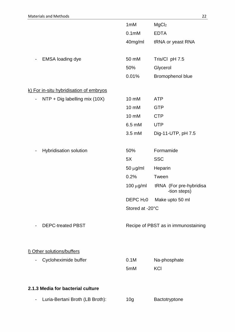
k) For in-situ hybridisation of embryos
- NTP + Dig labelling mix (10X) 10 mM ATP
10 mM GTP
10 mM CTP
6.5 mM UTP
3.5 mM Dig-11-UTP, pH 7.5
- Hybridisation solution 50% Formamide
5X SSC
50 g/ml Heparin
0.2% Tween
100 g/ml tRNA (For pre-hybridisa -tion steps)
DEPC H20 Make upto 50 ml
Stored at -20°C
- DEPC-treated PBST Recipe of PBST as in immunostaining
l) Other solutions/buffers
- Cycloheximide buffer 0.1M Na-phosphate
5mM KCl
2.1.3 Media for bacterial culture
- Luria-Bertani Broth (LB Broth): 10g Bactotryptone
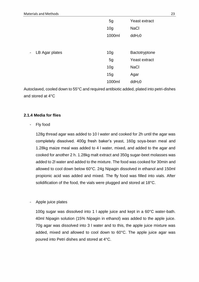
Materials and Methods 23
5g Yeast extract
10g NaCl
1000ml ddH20
- LB Agar plates 10g Bactotryptone
5g Yeast extract
10g NaCl
15g Agar
1000ml ddH20
Autoclaved, cooled down to 55°C and required antibiotic added, plated into petri-dishes
and stored at 4°C
2.1.4 Media for flies
- Fly food
128g thread agar was added to 10 l water and cooked for 2h until the agar was
completely dissolved. 400g fresh baker’s yeast, 160g soya-bean meal and
1.28kg maize meal was added to 4 l water, mixed, and added to the agar and
cooked for another 2 h. 1.28kg malt extract and 350g sugar-beet molasses was
added to 2l water and added to the mixture. The food was cooked for 30min and
allowed to cool down below 60°C. 24g Nipagin dissolved in ethanol and 150ml
propionic acid was added and mixed. The fly food was filled into vials. After
solidification of the food, the vials were plugged and stored at 18°C.
- Apple juice plates
100g sugar was dissolved into 1 l apple juice and kept in a 60°C water-bath.
40ml Nipagin solution (15% Nipagin in ethanol) was added to the apple juice.
70g agar was dissolved into 3 l water and to this, the apple juice mixture was
added, mixed and allowed to cool down to 60°C. The apple juice agar was
poured into Petri dishes and stored at 4°C.

Materials and Methods 24
2.1.5 Enzymes and Kits
All restriction enzymes were obtained either from Fermentas (St. Leon-Rot) or New
England Biolabs (Ipswich, USA) and used according to the manufacturer’s instructions.
Additionally, the following enzymes were used:-
- Taq Polymerase (expressed and purified in the lab)
- Pfu DNA Polymerase (expressed and purified in the lab)
- T7 RNA Polymerase (expressed and purified in the lab)
- Transcriptor Reverse transcriptase (Roche)
- DnaseI (Roche)
Following kits were used according to the manufacturer’s instructions:-
- MiniElute Gel extraction Kit Qiagen, Hilden
- Plasmid Midi Kit Nucleobond AX Macherey-Nagel
- In-fusion HD cloning kit Clontech
- RNeasy Micro kit (for RNA purification) Qiagen, Hilden
- Individual Cy3 tyramide reagent pack PerkinElmer
2.1.6 Chromatography
- GST columns (GSTrap HP) GE Healthcare Life Sciences
- CNBr activated Sepharose 4B GE Healthcare Life Sciences
- Ni-sepharose beads Amersham Pharmacia Biotech
- PD-10 desalting columns GE Healthcare Life Sciences
- Dynabeads protein A(paramagnetic beads) Life technologies
- Dynabeads MyOne Streptavidin T1 beads Life technologies
2.1.7 Oligonucleotides used in the study
All oligonucleotides used in this study were ordered from Eurofins genomics. The
nucleotides in red show the changed nucleotide in site-directed mutagenesis.

Materials and Methods 25
Table 2.1 Oligonucleotides used in the study
Rab11 cloning
Serial
Nr.
Seqeunce (5’ – 3’) Description
SG1 CTCCGGTGTTGGCAAAAATAATTT
GCTCTCACG
Forward primer for site-directed
mutagenesis of the 25th amino acid of
Rab11 from S to N
SG2 CAAATTATTTTTGCCAACACCGGA
GTCACCGATAAG
Reverse primer for site-directed
mutagenesis of the 25th amino acid of
Rab11 from S to N
SG3 TGGATCCCCGGAATTCATGGGTG
CAAGAGAAGACGAG
Forward primer for in-fusion cloning of
Rab11 and Rab11S25N into PGEX-4T1
SG4 GTCGACCCGGGAATTCTCACTGAC
AGCACTGTTTGCG
Reverse primer for in-fusion cloning of
Rab11 and Rab11S25N into PGEX-4T1
Sequencing
CW20a ACCCAATGTGCCTGGATGC Forward sequencing primer for PGEX-
4T1
CW20b CGGGAGCTGCATGTGTCAGA Reverse sequencing primer for PGEX-
4T1
qRTPCR
SY94 GCGAGATCTACCACGCTTTTTCGC
GGTC
Forward primer spanning exon-exon
junction in the 5’UTR of slam
SY95 CGTGGATCCTTTGCTAATAGCTTA
TATACAATG
Reverse primer for detection of slam
mRNA
SA12 GTTAAATCGAACAAAAAGCTTAC Forward primer spanning exon-exon
junction of first intron of actin
SA13 GTGAGGATACCACGCTTGC Reverse primer for detection of actin
mRNA
2.1.8 Plasmid constructs used in the study
Table 2.2 Plasmids used in the study
Name Description Source
pGEX6OH GST-His fusion protein Prof. Jörg Großhans
pBS (SK-)-rab11 (EST) Rab11 cDNA Bloomington GM06568

Materials and Methods 26
pGEX-4T1-rab11 N-terminal GST tag fused with
Rab11 protein
Stephanie Gröning
(Pelissier et al., 2003)
pGEX-4T1-rab11S25N N-terminal GST tag fused with
Rab11 protein with a point mutation
changing the 25th amino acid from
Serine to Asparagine
Stephanie Gröning
(Pelissier et al., 2003)
pProEXHTb-p150-KIND p150-KIND with an N-terminal his-
tag
Prof. E. Kerkhoff
pQEslamC651 Slam N-terminal fragment from
amino acid 1 to 545 fused with a 6X
His tag at the C-terminal
Prof. Jörg Großhans
pCS2GFP GFP sequence under SP6 promoter Dr. Shuling Yan
pCS2slam2-1(slam20) N-terminal GFP sequence fused
with slam sequence coding from
amino acid 200 to 330
Dr. Shuling Yan
pCS2slam2-2(slam21) N-terminal GFP sequence fused
with slam sequence coding from
amino acid 331 to 476
Dr. Shuling Yan
2.1.9 Primary antibodies
Table 2.3 Antibodies used in the study
Antibody Raised
in
Dilution and working concentration
Source Staining Western
ß-Gal Mouse 1:5000 (0.2g/ml) - Roche
Dia Rabbit 1:1000 - ZMBH
Dlg Mouse 1:100 (~0.4g/ml) - Hybridoma bank 4F3
Nullo Mouse 1:10 (~0.3g/ml) - Hybridoma bank 5C3-12
Rab11 Rabbit 1:1000 - Prof. D.F. Ready
Slam Rabbit 1:5000(~1g/ml) 1:5000(~1.1g/ml) Prof. Jörg Großhans
Slam Guinea
pig
1:5000 1:5000 Prof. Jörg Großhans
Slam (affinity
purified)
Rabbit - 1:5000 (~1.2g/ml) Purified by me
Spire (affinity
purified)
Guinuea
pig
1:1000 (~10g/ml) 1:2000 (~5g/ml) Purified by me
Materials and Methods 27
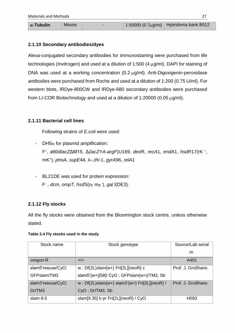
-Tubulin Mouse - 1:50000 (0.7g/ml) Hybridoma bank B512
2.1.10 Secondary antibodies/dyes
Alexa-conjugated secondary antibodies for immunostaining were purchased from life
technologies (Invitrogen) and used at a dilution of 1:500 (4g/ml). DAPI for staining of
DNA was used at a working concentration (0.2g/ml). Anti-Digoxigenin-peroxidase
antibodies were purchased from Roche and used at a dilution of 1:200 (0.75 U/ml). For
western blots, IRDye-800CW and IRDye-680 secondary antibodies were purchased
from LI-COR Biotechnology and used at a dilution of 1:20000 (0.05g/ml).
2.1.11 Bacterial cell lines
Following strains of E.coli were used:
- DH5for plasmid amplification:
F–, ø80dlacZΔM15, Δ(lacZYA-argF)U169, deoR, recA1, endA1, hsdR17(rK –,
mK+), phoA, supE44, λ–,thi-1, gyrA96, relA1
- BL21DE was used for protein expression:
F- , dcm, ompT, hsdS(rB -mB
-), gal l(DE3).
2.1.12 Fly stocks
All the fly stocks were obtained from the Bloomington stock centre, unless otherwise
stated.
Table 2.4 Fly stocks used in the study
Stock name Stock genotype Source/Lab serial
nr.
oregon-R +/+ A401
slam5’rescue/CyO;
GFPslam/TM3
w ; Df(2L)slam{w+} Frt[2L]{neoR} c
slam5’{w+}[58]/ CyO ; GFPslam{w+}/TM3, Sb
Prof. J. Großhans
slam3’rescue/CyO;
Dr/TM3
w ; Df(2L)slam{w+} slam3’{w+} Frt[2L]{neoR} /
CyO ; Dr/TM3, Sb
Prof. J. Großhans
slam-9.5 slam[9.35] b pr Frt[2L]{neoR} / CyO H093

Materials and Methods 28
slam-B4.1 slam[B4.1] b pr Frt[2L]{neoR} / CyO H094
Flp122; ovoD2L hs-Flp[122]; ovoDFrt2L[40A]/If/CyO, hs-hid Maintained in the
lab
mat-GFPslam w ; tub-Gal4-VP16[67]{w+} ; UASp-GFP-
slam[1]{w+}
H016
GFPslam w ; GFPslam[68]{w+} H087
SAS6GFP/TM6c w ; ubi-GFP-SAS6{w+} / TM6c, Sb Prof. J. Raff
Sp/CyO; GFP-
SAS6/TM6c
w ; Sp/CyO; ubi-GFP-SAS6{w+} / TM6c, Sb Zhiyi Lv
shibire w shi[1] / FM6, y B A119
shibire; Sp/CyO w shi[1] / FM7c, y B; Sp/CyO Zhiyi Lv
rab11YFP-CHR rab11YFP-wt[CHR-7]/TM3, Sb H008
GFPslam; rab11YFP w ; tub-Gal4-VP16[67]{w+} UASp-GFP-
slam{w+}/CyO; rab11YFP-wt[CHR-7]/TM3, Sb
Prof. J. Großhans
nuf/TM6B nuf[1] sr e ca / TM6B, Tb Hu H010
Sp/CyO; nuf/TM3 Sp/CyO; nuf[1] sr e ca/ TM3, Sb Prof. J. Großhans
eos-slam w; UASp-meos2-slam{w+} Dr. P. Laupsien
mat67.15 tub-Gal4-VP16[67]{w+}; tub-Gal4-
VP16[15]{w+}
Prof. D. St.
Johnston, UK
his-RFP w; histone2Av-mRFP[1]{w+} B384
nullo/FM7; If/CyO Df(1)nullo[6F12] / FM7; If/CyO (Hunter and
Wieschaus, 2000)
Dfslam/hb-lacz Df(2L)BSC5/CyO, hb-lacZ Prof. J. Großhans
spir1 spir[1]cn[1]bw[1]/CyO, I(2)DTS513[1] H064
Dfspire Df(2L)Exel6046 Bloomington
Following alleles of spire were obtained from Tübingen stock centre Tübingen Stock nr.
spir2L-62-29 w (P[hs-Flp]122); al? dp? b? pr P[FRT,
neoR]40A/CyO, P[hs-hid, w+]
L210
spir2L-75-28 w (P[hs-Flp]122); al? dp b? pr P[FRT,
neoR]40A/CyO, P[hs-hid, w+]
L214
spir2L-133-31 w (P[hs-Flp]122); al? dp? b? pr P[FRT,
neoR]40A/CyO, P[hs-hid, w+]
L228
spir2L-146-30 w (P[hs-Flp]122); al dp b pr P[FRT,
neoR]40A/CyO, P[hs-hid, w+]
L235
spir2L-210-2(ts) w (P[hs-Flp]122); al? dp b pr P[FRT,
neoR]40A/CyO, P[hs-hid, w+]
L253

Materials and Methods 29
spir2L-216-18 w (P[hs-Flp]122); al? dp? b? pr? P[FRT,
neoR]40A/CyO, P[hs-hid, w+]
L258
spir2L-244-35 w (P[hs-Flp]122); al? dp b? pr P[FRT,
neoR]40A/CyO, P[hs-hid, w+]
L265
Generated in this work:
gfpslam; nuf tub-Gal4-VP16[67]{w+} ; UASp-GFP-
slam{w+}/ CyO; nuf[1] sr e ca /TM3
-
shi; gfpslam w shi[1] / FM7c, y B; tub-Gal4-VP16[67]{w+} ;
UASp-GFP- slam{w+}/ CyO
-
nullo/FM7;
Dfslam5’/CyO
Df(1)nullo[6F12] / FM7 ; Df(2L)slam{w+}
Frt[2L]{neoR} c slam5’{w+}/ CyO
-
2.1.13 Microscopy
- Zeiss Stemi 2000 Carl Zeiss
- Leica MZ125 Leica
- Microinjection microscope Carl Zeiss
- LSM 780 Carl Zeiss
- Zeiss Axiovert 200 M Ultra-view spinning Disc confocal microscope Carl Zeiss
- Zeiss Axioplan 2 Fluorescence microscope Carl Zeiss
2.1.14 Other materials
- Ribolock RNAse inhibitor Thermo Scientific
- Cy3-UTP Perkin Elmer
- Cycloheximide Sigma
- Complete Mini (EDTA-free)
Protease Inhibitor Cocktail Roche
- Aquapolymount Polysciences, Inc.
- Coverslips Menzel
- Glass slides Thermo Scientific
- Fly vials Greiner
- Glass pipettes (25ml, 20ml, 10ml, 5ml) Silber Brandt
- Pasteur pipettes Brandt
- Glass homogenizer B. Braun Biotech International

Materials and Methods 30
- Dynamag – spin magnet Life technologies
- Petri dishes Greiner
- Pipet-aid Drummond
- Micropipettes (1000 l, 200l, 20l, 2l) Gilson
- Micropipette tips (1000 l, 200l, 2l) Eppendorf
- Safe-seal RNase-free tips 1000 l, 200l,
20l, 10l) Biozym
- Falcon tubes (50 ml, 15 ml) BD Falcon
- Eppendorf tubes (1.5ml, 2ml, 5ml) Eppendorf
- PCR tubes Brand, Wertheim
- 10S and 3S VoltaLef Halocarbon oil Lehmann & Voss & Co.
2.1.15 Other equipment
- UGA-40 MLC ablation system with
DPSL-355/14 laser Rapp Optoelectronic
- Äkta pure GE Healthcare Life Sciences
- Odyssey CLx Infrared imaging system LI-COR Biosciences
- CFX96 Real time PCR detection system/
C1000 touch thermal cycler Biorad
- Typhoon variable mode imager Amersham
2.1.16 Softwares
- Adobe Photoshop CS6 Adobe
- Adobe Illustrator CS6 Adobe
- FIJI NIH
- Zen 2012 Carl Zeiss
- Lasergene GATC biotech
- Microsoft excel Microsoft
- Microsoft word Microsoft
- Zotero Roy Rosenzweig Center
for History and New Media

Materials and Methods 31
2.2 Methods
2.2.1 DNA methods
2.2.1.1 Molecular cloning
All the molecular cloning methods were carried out according to Sambrook and Russel,
2001, unless otherwise stated.
2.2.1.2 Polymerase chain reaction (PCR)
PCR reactions were carried out using Taq or Pfu DNA polymerase. Standard PCR
reactions were carried out with the following reagents in the mentioned concentrations:
50-200 ng DNA template, 0.5 M forward and reverse primers, 50 M dNTP (each),
1X PCR buffer (depending on the polymerase), 1-2 units (per 50 l of reaction) Taq or
Pfu polymerase.
Following conditions were used for a general reaction:
Step 1 (Initial denaturation): 95°C - 2 min
Step 2 (Denaturation): 95°C - 30 sec
Step 3 (Annealing): 50-60°C - 1 min
(depending on the annealing temperatures of the primers)
Step 4 (Elongation): 72°C - 1 min/Kb to be amplified
Step 5 (Repetition of cycles) Steps 2 to 4 - 30 cycles
Step 6 (Final elongation) 72°C - 7 min
Step 7 (Hold) 4°C - ∞
2.2.1.3 Site-directed mutagenesis
Site-directed mutagenesis was carried out to change the 25th amino acid in Rab11
protein from Serine to Asparagine (AGT to AAT). The following PCR program was
used:

Materials and Methods 32
Step 1 (Initial denaturation): 95°C - 5 min
Step 2 (Denaturation): 95°C - 50 sec
Step 3 (Annealing): 62°C - 50 sec
Step 4 (Elongation): 68°C - 5.5 min
Step 5 (Repetition of cycles) Steps 2 to 4 - 18 cycles
Step 6 (Final elongation) 68°C - 7 min
Step 7 (Hold) 4°C - ∞
2.2.1.4 In-fusion cloning
In-fusion cloning was carried out according to the manufacturer’s instructions.
2.2.1.5 Purification of DNA/RNA by phenol-chloroform extraction
DNA/RNA sample was made up to 200 l by adding ddH20/DEPC-H20. To this, 200 l
of Phenol:Chloroform:Isoamylalcohol was added and mixed well. The sample was
centrifuged at 13,000 rpm for 5 min. The upper phase was transferred to a new
eppendorf. 200 l of Chloroform was added to this and mixed well. The sample was
centrifuged again at 13,000 rpm for 5 min. The upper phase was again transferred to
a fresh eppendorf. To this, 20 l of 3M NaAc and 500 l Ethanol was added and mixed.
The sample was kept at -20°C overnight (or -80°C for 1 hour). Then the sample was
pelleted by centrifuging at 14,000 rpm for 30 min at 4°C. The supernatant was
discarded. 100 l of 70% Ethanol was added to the pellet. Again the sample was
centrigued at 14,000 rpm for 15 min. Supernatant was discarded and pellet dried either
in the Speedvac or under the hood. The dried pellet was resuspended in 20l of
ddH20/DEPC-H20.
2.2.1.6 DNA sequencing
DNA sequencing was carried out with the sequencing facility at the Department of
Developmental Biochemistry, GZNB, University of Göttingen.

Materials and Methods 33
2.2.1.7 In-vitro transcription
In-vitro transcription was carried out to prepare Cy3-labelled GFP, slam2-1 and slam2-
2 fragments for EMSA. The pCS2-GFP, pCS2-slam2-1 and pCS2-slam2-2 plasmids
was restriction digested using NotI and purified using phenol-chloroform extraction
(Refer to 2.2.1.5). The linearised plasmids were used as a template for in-vitro
transcription. The following components were mixed into an eppendorf for the in-vitro
transcription reaction:
Component Volume Stock Concentration
Linearised plasmid 5 l 0.2 g/l
Transcription buffer 2.5 l 10X
rCTP 1 l 10 mM
rGTP 1 l 10 mM
rATP 1 l 10 mM
rUTP 0.64 l 10 mM
Cy3-UTP 0.36 l 100 nmol
DTT 0.75 l 1M
Ribolock RNAse-inhibitor 1 l 40 U/l
SP6 polymerase 1 l 20 U/l
Pyrophosphatase 0.2 l 0.5 U/l
dH20 (Rnase-free) 10.55 l
Total Volume 25 l
The reaction was kept at 37°C, overnight. 1 l of DnaseI (Stock:10 U/l) was added
and kept at 37°C for 30 min. The RNA was purified using Qiagen RNeasy kit, eluted in
30 l RNAse-free water and stored at -20°C until further use.

Materials and Methods 34
2.2.1.8 Reverse transcription
5 l RNA sample (ranging from 0.2 to 1.2 g/l) was mixed with 7 l dH20 (RNase-
free) and 1l oligo(dT) (100 pmol/l) and kept at 65°C for 10 min. After that, the
following components were added to the sample (Stock concentration in brackets):
5X RT buffer - 4 l
RNAse inhibitor (40 U/l) - 0.5l
dNTP mix (10mM) - 2 l
Reverse transcriptase (20 U/l ) - 0.5 l
The sample (total volume 20 l), was kept at 55°C for 30 min and then at 85°C for 10
min. The cDNA was then stored at -20°C.
2.2.1.9 Quantitative real-time PCR (qRT-PCR)
The following components were added for each qPCR reaction:
SYBr green qPCR mix - 12.5 l
dH20 - 8 l
Forward primer - 1.25 l
Reverse primer - 1.25 l
cDNA - 2 l
The following qPCR program was used for detection of slam and actin mRNA:
Step 1 (Initial denaturation): 95°C - 3 min
Step 2 (Denaturation): 95°C - 10 sec
Step 3 (Annealing): 60°C - 15 sec
Step 4 (Elongation): 72°C - 30 sec
Step 5 (Repetition of cycles) Steps 2 to 4 - 40 cycles
Step 6 (Melting curve) 55°C to 95°C - 10 sec + plate read
(Increment of 0.5°C)

Materials and Methods 35
2.2.2 Biochemical methods
2.2.2.1 Protein purification
Expression of all recombinant proteins was carried out in E.coli. BL21DE
a) Purification of GST-tagged proteins under native conditions:
GST, GST-Rab11 and GST-Rab11S25N recombinant protein expression was induced
with 0.2 mM IPTG at 18°C overnight. The cells were harvested by centrifuging at
5000rpm for 30 min. They were resuspended in lysis buffer for GST-tagged proteins
and incubated with 1mg/ml lysozyme and a pinch of DNase for 30 min on ice. The cells
were then lysed using a microfluidiser. The soluble part was obtained by centrifuging
twice at 12,000 rpm for 20 min each to remove the insoluble fraction. The supernatant
(soluble fraction) was passed through a Glutathione Sepharose prepacked column
(GSTrap HP column), washed with approximately 10 ml of wash buffer and eluted with
elution buffer. The eluted protein was then exchanged to PBS using a PD10 column
and the protein was concentrated to 1 mg/ml for injection into GFPslam embryos.
b) Purification of His-tagged proteins under native conditions:
Expression of SlamC651 protein was induced with 0.1 mm IPTG at 18°C overnight.
The cells were harvested and lysed as described earlier, but in lysis buffer for his-
tagged proteins. The supernatant (soluble fraction) was passed through a Nickel-
Sepharose prepacked column (HisTrap HP column), washed with approximately 10 ml
of wash buffer and eluted with elution buffer. The eluted protein was then exchanged
to storage buffer using a PD10 column and the protein was concentrated to 1.5 mg/ml.
Glycerol was added to the protein sample to make the final concentration 10% glycerol
and the protein was snap-frozen in liquid Nitrogen and stored at -80°C.
c) Purification of His-tagged proteins under denaturing conditions:
Expression of p150-KIND was induced with 0.5 mM IPTG at 37°C for three hours. The
cells were harvested and lysed as described earlier, but in lysis buffer for his-tagged
proteins under denaturing conditions. The supernatant (soluble fraction) was rocked
with 5 ml Nickel-Sepharose beads for one hour at room temperature. The suspension
was added to a gravity column and washed with approximately 50 ml of wash buffer

Materials and Methods 36
and eluted with elution buffer. The eluted protein was then exchanged to storage buffer
using a PD10 column and the protein was concentrated to 1.5 mg/ml. Glycerol was
added to the protein sample to make the final concentration 10% glycerol and the
protein was snap-frozen in liquid Nitrogen and stored at -80°C, before sending it out to
the company for antibody production.
2.2.2.2 Affinity purification of antibodies
a) Preparation of the CNBr-activated Sepharose beads:
1 g of dry CNBr-sepharose beads were swelled in 10 ml of 1 mM HCl for 15 min. The
beads were washed on a sintered glass filter with about 200 ml of 1 mM HCl. Washes
were carried out in several aliquots.
b) Coupling of protein to beads:
The gel was washed with 5 ml coupling buffer. A280 of the protein solution was taken.
The protein solution was then added to the gel in a 1:2 gel:buffer ratio. The suspension
was mixed on a rocker for 3 hr at room temperature. The beads were allowed to settle
down and the supernatant removed after taking the A280 reading to confirm binding.
Blocking buffer was added to the gel and mixed for 2 hr at room temperature. The
suspended beads were poured into a column. The column was washed with five cycles
of alternating low and high pH buffers (Buffer I and Buffer II). Then the column was
washed with five column volumes of PBS. The column was stored at 4°C.
c) Loading of serum and elution of antibodies:
The serum was centrifuged twice at 15,000 rpm for 20 min each at 4°C. The cleared
serum was then applied to the column at a flow rate of 0.5 mg/ml. The flow-through
was collected and saved. The column was washed first with ten column volumes of
PBS + 500 mM NaCl and then with PBS + 1 M NaCl. For elution, 100 l of neutralisation
buffer was added to the collection tubes and 900 l fractions were taken via elution
with low pH buffer. The peak fractions were identified using A280 and pooled together.
The pooled sample was then exchanged to PBS using PD-10 columns. The antibody
solution was then concentrated (~5 mg/ml for Rabbit anti-Slam antibodies and ~10
mg/ml for Guinuea pig anti-KIND antibodies). Na-Azide was added to 0.02% to the
antibodies and they were stored at 4°C.

Materials and Methods 37
2.2.2.3 Western blot
a) Embryo collection:
Embryos were staged from 1.5 to 2.5 hours on apple-juice agar plates and
dechorionated in 50% Klorix bleach for 90 seconds. The dechorionated embryos were
collected into a weighed eppendorf tube and spun down gently. The weight of the
embryos was determined (~1mg =100 embryos). 1X Lämmlli buffer was added and the
embryos were homogenised. The sample was then heated to 95°C for 5 min and
centrifuged at 14,000 rpm for 1 min. The embryos were either loaded immediately on
an SDS-gel for a western blot or stored in -20°C for future use.
b) Western blot procedure:
A volume corresponding to 10 embryos was loaded on an SDS-PAGE gel for a general
western blot, unless otherwise stated. The gel was run at 16 mA constant current until
the dye front just ran off. The proteins from the gel were transferred onto a
nitrocellulose membrane either using a semi-dry transfer for 1 hour (Westerns for Slam
and -tubulin) or wet transfer for overnight (Westerns for GFP-slam and Spire). The
membrane was then blocked in 5% milk powder in PBT for 35 min and incubated with
primary antibody either overnight at 4°C or 2 hours at room temperature. The
membrane was rinsed thrice in PBT and given four 10 minute-PBT washes. The
membrane was incubated with secondary antibody for 1 and a half hours at room
temperature, protected from light. Then it was again rinsed thrice in PBT and given
four 10 minute-PBT washes, after which the bands were detected using the Odyssey
CLx Infrared Imaging system.
2.2.2.4 EMSA
Six serial dilutions of the protein were carried out in protein storage buffer. The Cy3-
labelled RNA was heated to 95°C for 1 min and then allowed to cool to 25°C slowly on
a thermomixer. A mastermix for twelve samples was prepared with the following
components:
Component Volume per sample
RNase-free water 12 l

Materials and Methods 38
Yeast RNA (10 g/l) 1 l
5X binding buffer 4 l
Cy3-labelled RNA (~200 ng/l) 1 l
The above mastermix was aliquoted into 12 eppendorfs (18 l each). To each reaction
tube, 3 l of protein dilution was added. Storage buffer without any protein was used
as a negative control. The reaction tubes were mixed and kept at room temperature
for 30 min for binding. 5 l of EMSA loading dye was added to samples and mixed.
The samples were loaded on 1% Agarose gels and run at 40 V constant voltage until
the dye front ran three-fourths of the gel length. The RNA bands were visualised using
Typhoon phosphoimager.
2.2.2.5 Immunoprecipitation
Staged wild-type embryos were collected by dechorionating the embryos (Refer to
2.2.2.3 a). After collecting the embryos in an eppendorf, spinning them down and
weighing, they were snap-frozen in liquid nitrogen and stored at -80°C. 40 l
dynabeads protein A magnetic beads were washed twice with 1 ml PBS each and
incubated with rabbit slam antibody for 1 hour. After one hour, the beads were washed
thrice with PBS and kept on ice. Approximately 100 mg of embryos (~10,000 embryos)
were lysed in 1 ml RIPA buffer using a dounce homogeniser and transferred into a cool
eppendorf using a pre-cooled pasteur pipette. The lysate was centrifuged at 14,000
rpm at 4°C for 15 min. The lipid layer floating on the top was sucked off using an
aspirator pump. Input sample was taken such that the final concentration in 1X Lämmlli
buffer was 1 embryo/l. The sample was boiled at 95°C immediately for 5 min. The
rest of the lysate was added to the antibody-conjugated dynabeads and rotated on a
wheel for 2 hours at 4°C. Subsequently, the beads were settled down using Dynamag
spin-magnetic and an unbound sample (supernatant) was taken in the same way as
input sample. The beads were washed thrice with RIPA buffer and subsequently,
protein was eluted from the beads such that the final concentration in 1X Lämmlli buffer
was 50 embryos/l. Sample was heated immediately at 95°C for 5 min. The input (~10
embryos), unbound (~10 embryos) and bound (~500 embryos) samples were loaded
onto an 8% SDS-PAGE gel to carry out a western blot.

Materials and Methods 39
2.2.2.6 Pulldown of GFPslam using paramagnetic Streptavidin beads
Staged embryos expressing GFPslam (as well as endogenous Slam) were collected
as mentioned in the previous section (2.2.2.5). 40l of Streptavidin MyOne T1
dynabeads (Streptavidin paramagnetic beads) were washed twice with 1X PBS. About
5000 embryos were lysed in 500l of RIPA buffer and input sample taken. The lysate
was then incubated with ~45nM final concentration of biotinylated GFP-binding protein
for one hour on ice (biotinylated-GBP – a gift from Prof. Dirk Görlich). The lysate with
biotinylated-GBP was added to the beads and rotated on a wheel at 4°C for 1h. The
beads were settled down and unbound sample taken. The beads were washed thrice
with RIPA buffer. The bound protein was eluted by adding 1X Lämmlli buffer and
heating at 95°C for 5 min.
2.2.3 Fly embryo methods
2.2.3.1 Fractionation of embryos
a) WT embryos followed by western and qPCR:
Approximately 7500 staged embyros were lysed in 1.5 ml of fractionation buffer (Refer
to 2.1.2 under ‘For fractionation’ for buffer recipes). The lysate was centrifuged twice
at 2500 rpm for 5 min at 4°C to pellet the nuclei. 1.3 ml supernatant (total lysate) was
transferred into a new eppendorf. ‘Total lysate’ protein sample was taken such that the
final concentration in 1X Lämmlli buffer was 1embryo/l. For ‘Total lysate’ RNA sample,
200l (~1000 embryos) was used and immediately processed for RNA isolation (For
protocol, refer to 2.2.3.2). 1 ml of the remaining total lysate (~5000 embryos) was
transferred into a new eppendorf tube and centrifuged at 14,000 rpm for 15 min at 4°C.
The lipid layer was generously removed by an aspirator. 500 l of supernatant
(Cytoplasm) was carefully transferred into a new eppendorf. The rest of the
supernatant was discarded and the pellet (Membrane) was re-suspended in 1 ml of
fresh fractionation buffer until a fine suspension was formed. The ‘Cytoplasm’ and the
‘Membrane’ tubes were centrifuged again at 14,000 rpm for 15 min at 4°C. If there was
a significant pellet in the ‘Cytoplasm’ fraction, it was included in the ‘Membrane’ tube.
The wash step was repeated again. After the second wash, ‘Cytoplasm’ protein and
RNA sample was taken from the supernatant in a similar manner to the ‘Total lysate’
sample. The ‘Membrane’ pellet was re-suspended in 500 l fractionation buffer.

Materials and Methods 40
‘Membrane’ protein sample was taken such that the final concentration in 1X Lämmlli
was 3 embryos/l. For the ‘Membrane’ RNA sample, 100 l (membrane derived from
~1000 embryos) was used. All protein samples were heated immediately at 95°C for 5
min and kept at room temperature until the end of protocol. They were loaded on an
8% SDS gel and a western blot for Slam and -tubulin was carried out. The detection
of Slam was done by IRDye 800 secondary antibody while the detection of -tubulin
was done by IRDye 680 secondary antibody on the same membrane.
Fractionation of genomic GFPslam embryos was carried out exactly in the same
manner, except that the RNA samples were not taken.
b) Fractionation followed by high salt membrane extraction:
Fractionation of wild-type embryos was carried out in a similar manner as described
above. Only protein samples were taken. After taking the ‘Membrane’ protein sample,
NaCl was added to a final concentration of 1M. The sample was kept on ice with
intermittent resuspension for 5 min. It was then centrifuged at 14,000 rpm for 15 min
at 4°C. The supernatant sample was dissolved in 1X Lämmlli such that 20 l sample
contained 250 embryos.
2.2.3.2 RNA isolation following fractionation of embryos
The ‘Membrane’ RNA lysate sample (100 l volume) was made up to 200 l using
fractionation buffer to make the volume equal to the ‘Total lysate’ and ‘Cytoplasm’
fractions. 20l of 10% SDS and 1l (20 g/l) glycogen and 200 l
Phenol:Chloroform:Isoamyl alcohol was added to the lysates, mixed well and
centrifuged at 14,000 rpm for 5 min at 4°C. The upper phase was transferred into fresh
tubes and 200 l chloroform was added and mixed well. The samples were centrifuged
at 14,000 rpm for 5 min at 4°C. The upper phase was transferred to fresh tubes and
40 l of 3M NaAc (DEPC-treated) was added and mixed. Then 500 l of 100% Ethanol
was added, mixed and kept at -20°C overnight. The RNA was pelleted by centrifuging
at 14,000 rpm for 30 min at 4°C, washed with 100 l 80% DEPC-treated Ethanol and
centrifuged again at 14,000 rpm for 15 min at 4°C. The pellet was dried under a hood
for 10-15 min and dissolved in 40 l RNAse-free water. The samples were stored at -
20°C.

Materials and Methods 41
2.2.3.3 Embryo fixation
Embryos were dechorionated as mentioned earlier (2.2.2.3a) and collected into a net
and transferred into a scintillation vial with 5 ml heptane and 4.5 ml PBS. To this, 0.5
ml of 37% formaldehyde was added (final concentration of 4%). The embryos were
fixed for 20 min with constant shaking. The lower phase was removed and the embryos
were washed twice by adding and removing 4.5 ml PBS. 5 ml of methanol was added
to the embryos and the vial was vortexed for 30 sec to devitellinise or ‘pop’ the
embryos. The popped embryos sink to the bottom. They were collected using a Pasteur
pipette and transferred to a fresh eppendorf tube. They were washed twice with
methanol and also stored in methanol at -20°C.
2.2.3.4 Immunostaining of embryos
The embryos stored in methanol were rinsed thrice and washed twice for 5 min in PBT.
Blocking was carried out for 1 hour in 1 ml of 5% BSA in PBT. The primary antibodies
were added in the respective dilutions in PBT and embryos were incubated at 4°C with
constant rotation. Embryos were then rinsed thrice in PBT and given four 15 min
washes. Appropriate secondary antibodies were added in PBT and incubated for 2
hours. Embryos were again rinsed thrice in PBT and given four 15 min washes.
Embryos were stained with DAPI for 5 min, rinsed thrice with PBT, washed once for 5
min with PBT and were mounted using aquapolymount mounting medium.
2.2.3.5 Fluorescence in-situ hybridisation (FISH)
a) Preparation of Dig-labelled slam mRNA probe:
Following components were mixed together and incubated at 37°C for 2 h:
Template DNA (1 g/ml) - 1 l (linearised OTslam plasmid)
10X NTP+Dig labelling mix - 2 l
10X transcription buffer - 2 l
T7 RNA polymerase (~200 U/l) - 1 l
RNAse inhibitor - 0.5 l

Materials and Methods 42
DEPC-treated water - 13.5l
The RNA was treated with 2l DNaseI (10 U/l) and incubated for 15 min at 37°C. The
RNA was then purified using phenol-chloroform extraction (Refer to 2.2.1.5). The pellet
was dissolved in 20l DEPC-treated water and stored at -20°C.
b) in-situ hybridisation of Drosophila embryos:
Day 1: Dechorionated and fixed embryos were rinsed thrice with PBT and washed
twice for 5 min each. The embryos were incubated for 10 min in 1:1 hybridisation
solution:PBT. Then the embryos were incubated in hybridisation solution (hyb-sol) for
10 min. The embryos were prehybridised in hyb-sol for 1 hour at 57°C. 1 l of slam
probe and 1 l of tRNA was mixed in 20 l DEPC-H20 and heated at 95°C for 4 min.
The probe was chilled rapidly in ice-water. 200 l of chilled hybridisation solution was
added to this. After the prehybridisation of embryos, the probe solution was added to
the embryos and incubated overnight at 57°C.
Day 2: The probe solution was removed and the embryos were rinsed thrice with
prewarmed hyb-sol. The embryos were then give three 30 min washes in hyb-sol at
57°C. The embryos were then given four 10 min washes with 4:1 hyb-sol/PBT (at
57°C), 3:2 hyb-sol/PBT (at 57°C), 2:3 hyb-sol/PBT(at 57°C) and 1:4 hyb-sol/PBT (at
room temperature) respectively. Then the embryos were washed twice, 20 min each
with 1% BSA in PBT at room temperature. Digoxigenin antibody coupled with
peroxidase was added to the embryos in a 1:200 dilution in PBT and incubated for 2
hours at room temperature. The embryos were rinsed thrice with PBT and washed four
times, 15 min each. TSA-Cy3 stock solution was diluted 1:200 in reaction buffer and
incubated with embryos for ~1 min, protected from light. The staining reaction was
stopped by washing with PBT. Then primary antibodies for protein staining were added
in appropriate dilutions in PBT and incubated overnight, protected from light.
Day 3: Immunostaining was carried out for proteins according to the protocol
mentioned in 2.2.3.4. Immunostaining was carried out in dark.

Materials and Methods 43
2.2.3.6 Generation of germline clones
Germline clones in the Drosophila germline were created mainly according to Chou
and Perrimon, 1992. Heat shock for the expression of Flippase was carried out at 37°C
for 60 min on two consecutive days after 24 hours post-hatching.
2.2.3.7 Fly genetics
slam germline clones were generated using Df(2L)slam Frt(2L) slam 5’rescue
chromosome.
nullo,slam double mutants were created by crossing females of Df(1)nullo6F/hs-
Flp[122]; Df(2L)slam Frt[2L] slam5’rescue/ovo[2L] with Df(2L)BSC5/CyO, hb-lacZ
males.
To test whether GFPslam68 can rescue viability, Df(2L)slam slam5’rescue/CyO;
GFPslam/TM3 females were crossed with Df(2L)slam slam3’rescue/CyO; +/+ males.
Rescued flies would consist of straight wings.
To test whether GFPslam68 can rescue cellularisation phenotype, Df(2L)slam Frt[2L]
slam5’rescue/ovo[2L]; GFPslam68 females were crossed with Df(2L)BSC5/CyO, hb-
lacZ males. All resultant embryos consisted of a single copy of GFPslam68 as maternal
contribution. Half of the embryos (lacZ positive) had a zygotic rescue from a single
copy of endogenous slam. One-fourth of the embryos (lacZ negative) contained a
single copy of zygotic GFPslam68 while the other one-fourth (lacZ negative) contained
no zygotic slam/GFPslam68.
2.2.3.8 Induction of shibire phenotype
shibire phenotype was induced in embryos derived from heterozygous shi(ts) females
by keeping the embryos after dechorionation at 32°C in a water bath for 30 minutes.
The embryos were then fixed. For live-imaging, embryos were dechorionated and
glued on to a glass coverslip and covered with halocarbon oil. Then the coverslip was
kept on a metal block heated to 32°C for 10 min and the imaging was carried out at
32°C at the LSM microscope.

Materials and Methods 44
2.2.3.9 Microinjection of protein/drug into embryos
Embryos were dechorionated, dried in a desiccation chamber for ~8 min, covered with
halocarbon oil and injected posteriorly with 50-100 pl of protein/drug. Imaging of
GFPslam was carried out after 10 min of injection.
2.2.4. Microscopy
2.2.4.1. Live-imaging of fly embryos
Embryos were dechorionated, glued on to a coverslip and covered with halocarbon oil.
Fluorescent live-images of GFPslam in buffer-injected and cycloheximide-injected
embryos were taken at the LSM with a 63X oil objective. All other fluorescent live-
images were taken at the spinning disc with a 40X oil objective. Differential interference
contrast microscopy was carried out at the spinning disc microscope with a 25X oil
objective.
2.2.4.2 Fluorescence recovery after photobleaching (FRAP)
Due to the changing Z-position of the furrow during cellularisation, a range of Z-stacks
were recorded. Bleaching of GFPslam was carried out in a given area using 100%
laser power and 50 iterations at a scan speed of 5. The appropriate Z-stacks were
merged for analysis. For the experiment with FRAP of GFPslam at different time-points
of cellularisation, fluorescence of a small membrane area outside and inside of the
bleached region was measured. For FRAP of GFPslam in shibire background,
fluorescence intensity of only ‘old borders’ were taken into account. For the experiment
with nuf mutants, due to increased membrane dynamics, fluorescence was measured
at the complete furrow canal area outside and inside of the bleached region. The
background intensity was subtracted from the total fluorescence. For all experiments,
the starting point of the fluorescence was normalised to 1. Linear regression of
fluorescence recovery was used to calculate the rate of fluorescence recovery.
2.2.4.3 Laser ablation of centrosomes
Embryos expressing SAS6GFP and GFPslam were aligned on agar, glued on to a
coverslide and covered with halocarbon oil. Very little glue was used since it makes

Materials and Methods 45
the UV laser less efficient. Ablation of centrosomes was carried out with a 355 nm
pulsed UV laser (70 J/pulse, 4x200 ms, 30%) in the anaphase of cell cycle 13, before
the centrosome duplication. Time-lapse axial image stacks were recorded following
ablation and subsequently merged with Fiji software.

46
3. RESULTS
3.1 Factors contributing to the accumulation of Slam protein at the
furrow canal
3.1.1 Centrosomes define the site of invagination
Centrosomes are microtubule-organizing centres and thus have been
implicated in the specification of cleavage furrow. It has been shown that centrosomes
alone, in the absence of nuclei, can induce formation of pole cells in the early
Drosophila embryo (Raff and Glover, 1989). Hence, we asked whether centrosomes
are the source of the initial signal for Slam accumulation and the formation of furrow
during cellularisation.
It has been described that overexpression of SAS6 induces de novo formation
of multiple centriole-like structures in syncytial embryos (Peel et al., 2007). These
structures are formed due to additional replications of centrioles in the same cell cycle.
These ectopic centriole precursors are able to reorganise microtubules (Rodrigues-
Martins et al., 2007). Previously in our lab, it was shown that these ectopic centriole
precursors are sufficient for the specification of a new furrow at the onset of
cellularisation (Fig 3.1 A and B, Dr. Philip Laupsien, PhD Dissertation). Slam was able
to localise to the furrow canal, though it was distributed all over the basal membrane,
probably due to premature basal closure of the furrows. The ectopic furrows also
displayed localisation of the lateral membrane marker Dlg, showing that the
invaginating membrane is polarised. Other furrow canal markers (PDZ domain of
RhoGEF2, Actin) were also found to be localised indicating that the furrows were
functional (Acharya et al., 2014; Dr. Christian Wenzl, PhD dissertation).
The ectopic furrows undergo a premature basal closure, perhaps but not
exclusively due to the absence of nuclei. When we observed at a very early stage of
cellularisation, we could find Slam localised in a proper furrow morphology (Fig. 3.1 C).
This suggests that centrosomes are able to specify the site of furrow invagination, and
is able to restrict Slam in the initial stage of cellularisation.
We wondered whether some of these ectopic centriole precursors were formed
as a result of nuclear fallout in earlier cell cycles. This would mean that the furrow-

Results 47
Fig 3.1 Lonesome centrosomes are capable of specifying ectopic furrows. Embryos
overexpressing SAS6GFP display lonesome centrosomes that lack nuclei (red arrows). These
lonesome centrosomes are able to specify ectopic furrows around them (yellow arrows), as marked by
accumulation of Slam (green) and Dlg (red). (A and B, Dr. Philip Laupsien, PhD Dissertation) Scale bar
10m.
specification is carried out by the nucleus rather than the centrosome. To rule out this
possibility, live imaging of syncytial embryos expressing SAS6-GFP under a ubiquitin
promoter and GFP-slam driven by a maternal tubulin promoter was carried out. It was

Results 48
Fig 3.2 SAS6GFP overexpression results in extra centrosomes in the embryo due to additional
centrosomal divisions. Embryos expressing SAS6GFP and GFPslam were followed from anaphase
of cycle 13 until the onset of cellularisation. Extra centrosomes result from additional replication of
centrosomes (yellow arrow). Scale bar 10m.
found that the ectopic centriole precursors capable of specifying a new furrow, were
formed de novo by extra divisions of the centrosomes and were not a consequence of
nuclear fallout (Fig. 3.2). When observed live, we found that both duplicate lonesome
centrosomes (Fig 3.3 A) and singular lonesome centrosomes (Fig 3.4 B) were capable
of specifying a new furrow.
Furthermore, we asked whether furrow specification was lost in the absence of
a centrosome. To answer this question, we tried to ablate a centrosome and see the
effect on GFP-slam accumulation. The ablation was carried out in the anaphase of
cycle 13 before the formation of the new furrow. However, only partial ablation could
be achieved due to the susceptibility of the adjacent membrane to a higher
intensity/duration of laser. Despite this limitation, we could observe a loss in the
centrosome’s ability to specify a new furrow (Fig 3.4). This suggests that we were able
to functionally ablate the centrosome to a large extent. Although the ablation was
carried out before the formation of the nuclear membrane, we could not rule out the
possibility that a nuclear fallout could have been induced during ablation. However,
since we showed earlier that centrosome is able to specify the site of the furrow even
in the absence of a nucleus, a nuclear fallout is inconsequential to furrow-specification.

Results 49
Fig 3.3 GFPslam accumulates around the additional duplicate/singular lonesome centrosomes
to form ectopic furrows in SAS6GFP embryos. Duplicate lonesome centrosomes (A, yellow arrows)
and Singular lonesome centrosomes (B) both are able to specify ectopic furrows. The red arrows in B
indicate a singular lonesome centrosome formed in an earlier cycle as indicated by GFPslam
accumulation in cycle 13. The yellow arrows in B indicate a singular lonesome centrosome formed in
cycle 13 as indicated by GFPslam accumulation in cycle 14. Scale bar 10m.

Results 50
Fig 3.4 Functional ablation of a centrosome induces loss of its furrow-specifying ability.
Centrosome was ablated in late mitosis using a pulsed UV-laser. The dashed-circle indicates the ablated
centrosome. The yellow arrow indicates the centrosome/centrosome pair of the daughter nucleus. Scale
bar 5m.
3.1.2 Accumulation of Slam is dependent on vesicular transport
Next, we wanted to find out the mechanism by which Slam protein is transported
to the furrow canal. We considered two non-exclusive possibilities a) microtubule-
dependent transport b) vesicle-based transport. When GFPslam punctae were
followed using live imaging, they showed a random movement instead of a
characteristic directed movement seen in case of microtubule-dependent transport (Dr.
Philip Laupsien, PhD dissertation). It has been shown that vesicle trafficking through
the recycling endosome is necessary for cellularisation (Pelissier et al., 2003). This
gave us an indication towards a direct or indirect involvement of vesicular transport in
localising Slam.
One way of interfering with vesicular transport is to disrupt shibire (shi), the
Drosophila homologue of Dynamin. We used a temperature-sensitive conditional
mutant of shi which allowed us to induce the shi phenotype in a temporal manner
(Pelissier et al., 2003). shi embryos were shifted to restrictive temperature (32°C),
about fifteen minutes earlier to the onset of cellularisation, before formation of the new
furrow. GFPslam was followed live after the induction of shi phenotype. It was observed
that GFPslam localisation to the site of the ‘new furrow’ was perturbed. Either it
completely failed to localise or sometimes it showed partial localisation (Fig 3.5). In
contrast, GFPslam accumulation in wild-type background was normal at 32°C.
Interfering with shi function, however, also disrupts apical endocytosis of the
plasma membrane and therefore membrane delivery to the new furrow during the slow
phase of cellularisation (Lecuit and Wieschaus, 2000). The failure of accumulation of
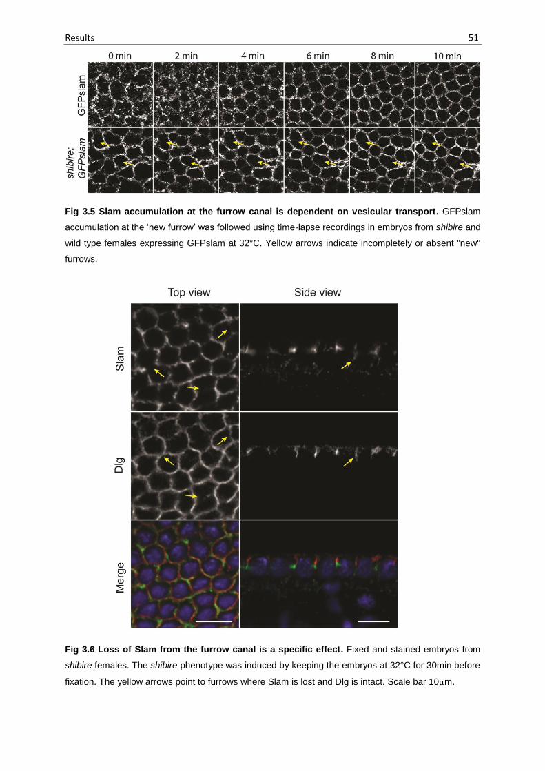
Results 51
Fig 3.5 Slam accumulation at the furrow canal is dependent on vesicular transport. GFPslam
accumulation at the ‘new furrow’ was followed using time-lapse recordings in embryos from shibire and
wild type females expressing GFPslam at 32°C. Yellow arrows indicate incompletely or absent "new"
furrows.
Fig 3.6 Loss of Slam from the furrow canal is a specific effect. Fixed and stained embryos from
shibire females. The shibire phenotype was induced by keeping the embryos at 32°C for 30min before
fixation. The yellow arrows point to furrows where Slam is lost and Dlg is intact. Scale bar 10m.

Results 52
Slam at the furrow canal therefore could also be due to absence of a membrane
structure. To check this, we shifted the shi conditional mutants to the restrictive
temperature, fixed and stained them for Slam and Dlg (a lateral membrane marker).
Dlg is able to localise to the furrow in the absence of Slam (Acharya et al., 2014; Dr.
Philip Laupsien, PhD dissertion). It was found that Slam was lost from the ‘new border’
before the loss of Dlg (Fig 3.6). Therefore it can be concluded that loss of Slam from
the furrow canal was not a consequence of disintegration of the membrane structure.
To investigate the specific role of recycling endosome in Slam protein transport,
we stained wild-type embryos during cellularisation for Slam and Rab11 to see whether
we find a colocalisation of Slam protein with a subset of Rab11 vesicles (Fig 3.7 A).
Rab11 was observed to be primarily localising at the pericentriolar region defining the
recycling endosome, as described in Riggs et al., 2003. Slam was not found in this
region. When we focussed at the furrow canal where Slam is known to localise, Rab11
did not show any specific localisation.
Fig 3.7 Rab11 is associated with the perinuclear recycling endosome and the recycling
endosome vesicles. Rab11 shows a different localisation near the furrow canal in fixed (A) and live
embryos (B). All images represent the top view of the embryo. The red, dashed line in the illustration
indicates the plane of intersection of the Z-stack.
We observed embryos expressing Rab11YFP to see if additional Rab11
vesicles were detectable via live imaging (Fig 3.7 B). Rab11 showed similar
accumulation at the recycling endosome as seen in the immunostaining pictures.
However, additional Rab11 punctae were observed around the furrow canal, which
were not visible via immunostaining.

Results 53
We followed GFPslam and Rab11YFP live to see whether there is any
colocalisation at any stage with the additional Rab11 punctae around the furrow canal.
No colocalisation was observed at any time point. Rab11YFP punctae were distributed
in the cytoplasm whereas GFPslam was always found associated with the membrane
(Fig 3.8). This suggests that Slam is not transported to the site of invagination on
Rab11 vesicles.
Fig 3.8 Slam is not transported on Rab11 vesicles. Embryos expressing GFPslam (red) and
Rab11YFP (green) were followed from late cycle 13 to the onset of cellularsation using time-lapse
recordings. (Imaging and image processing by Prof. Jörg Großhans).
Rab11 transheterozygote mutants however, show a reduced accumulation of
Slam at the furrow canal (Dr. Philip Laupsien, PhD dissertation). In the absence of
direct transport of Slam via the recycling endosome, we examined a functional link. It
has been previously reported that in embryos derived from nuf homozygous females
RhoGEF2 localisation at the furrow canal is disturbed (Cao et al., 2008). Previously in
our lab it had been shown that Slam is sufficient and necessary for the localisation of
RhoGEF2 to the furrow canal (Wenzl et al., 2010). Hence we wondered whether the
mislocalisation of RhoGEF2 in embryos from nuf females (henceforth referred to as
nuf embryos) is due to mislocalisation of Slam.
nuf embryos show a variable phenotype where the furrow canal morphology and
invagination rate ranges from normal to completely disrupted. At least 50% of the
embryos showed a strong or delayed phenotype (n=14). Fig 3.9 and 3.10 represent
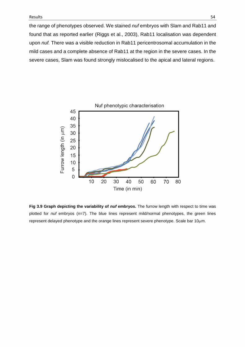
Results 54
the range of phenotypes observed. We stained nuf embryos with Slam and Rab11 and
found that as reported earlier (Riggs et al., 2003), Rab11 localisation was dependent
upon nuf. There was a visible reduction in Rab11 pericentrosomal accumulation in the
mild cases and a complete absence of Rab11 at the region in the severe cases. In the
severe cases, Slam was found strongly mislocalised to the apical and lateral regions.
Fig 3.9 Graph depicting the variability of nuf embryos. The furrow length with respect to time was
plotted for nuf embryos (n=7). The blue lines represent mild/normal phenotypes, the green lines
represent delayed phenotype and the orange lines represent severe phenotype. Scale bar 10m.

Fig 3.10 nuf embryos show variable phenotype ranging from mild to severe. Time-lapse movies of nuf embryos were obtained by Differential Interference
Contrast (DIC) microscopy. Representive cases are depicted. Arrows point to the cellularisation front. Scale bar 10 m.
Res
ult
s
55

Results 56
We further analysed the cases with delayed invagination and found that often
Slam was mislocalised into the lateral membrane domain as shown by colocalisation
with Dlg (Fig 3.11 A, yellow arrows). During cellularisation, slam, Rhogef2, nullo and
dia control separation of lateral and basal domains (Sokac and Wieschaus, 2008b;
Wenzl et al., 2010). So it is expected that in nuf embryos where Slam and therefore
RhoGEF2 are mislocalised, the membrane polarisation would be disrupted. This was
observed in the form of Dlg spreading into the basal domain (Fig 3.11 A, red arrows).
Fig 3.11 Slam is not restricted to the basal domain in nuf embryos. (A) Fixed nuf embryos were
stained for Slam (green) and Dlg (red). Slam is found mislocalised into the lateral domain (yellow arrows)
and Dlg is occasionally spead into the basal domain (red arrows). (B) GFPslam was followed in WT and
nuf embryos using time-lapse recordings. Red arrowheads point to the accumulations of GFPslam that
persist much longer than in the WT.

Results 57
Fig 3.12 Slam mislocalisation corresponds to the severity of nuf phenotype. In mild nuf
phenotypes, Slam is mislocalised to the cortex in regions of nuclear fallout (Red arrow). Occasionally,
an ectopic furrow between the two centrosomes is formed due to greater centrosomal separation in nuf
embryos (Orange arrow). In severe nuf phenotypes, Slam is mislocalised to the cortex and the lateral
domain of the furrow (Yellow arrows). Scale bar 10 m.
The mislocalisation of Slam was found even in situations where Dlg was not spread
into the basal domain, indicating an intact membrane polarity. This suggests that Slam
mislocalisation was the cause and not a consequence of membrane polarity disruption.
To further examine the role of nuf in the restriction of Slam to the furrow canal,
we observed GFPslam dynamics in nuf embryos (Fig 3.11B). In wild-type GFPslam
embryos, we observed that at the onset of cellularisation, GFPslam was found around
the centrosomal exclusion area at the prospective site of invagination. This state,
however, persisted only briefly as GFPslam was seen to arrange itself in a pseudo-

Results 58
hexagonal array in a few minutes. In nuf embryos, these accumulations persisted for
a much longer time and sometimes failed to resolve into the pseudo-hexagonal array.
Eventually, nuclei begin to fall out and Slam accumulates at the cortex. Together, these
data suggest that a functional recycling endosome is necessary for the restriction of
Slam to the basal domain.
We found that the extent of Slam mislocalisation in nuf embryos was dependent
upon the severity of the nuf phenotype. The severity of phenotype was evident by
abnormal nuclear morphology and loss of Rab11 staining (Fig 3.12). Another
interesting observation was that due to nuclear fallout and spreading of the
neighbouring nuclei, the centrosomes were found more apart from each other as
compared to the wild-type (as indicated by Rab11 staining). Due to this, occasionally
an extra furrow was formed between the centrosomes (Fig 3.12, orange arrow), further
Fig 3.13 Interference with Rab11 function in the embryo causes delay in Slam restriction to the
furrow canal. (A) Images from time lapse recordings of wild type embryos injected with purified GST or
GST-Rab11S25N (dominant negative allele) protein. Arrows point to the cellularisation front. (B)
Fluorescent images from time lapse recording of embryo expressing GFPslam and injected with GST
(10 embryos) or Rab11S25N (at least 3 out of 10 embryos showed delayed Slam restriction).

Results 59
confirming our earlier conclusion that centrosomes determined the site of membrane
invagination.
Since Nuf and Rab11 form a physical complex and are mutually dependent on
their localisation (Riggs et al., 2003), we tested whether GFPslam restriction at the
furrow canal is affected also when Rab11 function is disturbed. To accomplish this, we
injected a dominant negative Rab11 allele, Rab11S25N (Serine 25 mutated to
Asparagine) in GFPslam embryos and followed it by live imaging. It has been reported
earlier that injection of purified Rab11S25N protein delays furrow invagination
(Pelissier et al., 2003). About 40% of the injected embryos (n=5) showed delayed
furrow invagination (Fig 3.13 A) and GFPslam restriction at the furrow canal was also
delayed by several minutes (Fig 3.13 B).
3.2 Dynamics of Slam protein, mobility and life-time
3.2.1 A fraction of Slam is membrane-associated
Structured Illumination Microscopy has revealed that Slam is found at the
membrane in a punctate form rather than being evenly distributed (Fig 3.14 A).
Previously, it has been shown that Slam is present in both cytoplasmic and membrane
fractions in the embryo (Lecuit et al., 2002). We confirmed this result using an antibody
against endogenous Slam as opposed to the anti-HA antibody which was used by
Lecuit and colleagues. Additionally, we found out that Slam can be solubilised from the
membranes using a high-salt membrane extraction which means that Slam is merely
associated with the membrane and is not a transmembrane protein (Fig 3.14 B). This
is in accordance with the fact that domain prediction of Slam does not reveal any
transmembrane-domains in the protein.

Results 60
Fig 3.14 Slam is a membrane-associated protein. (A) Structured-illumination microscopy (SIM)
reveals that Slam is distributed at the furrow canal in a punctate manner (Imaging carried out by Olaf
Schulz) (B) Fractionation of WT embryos into cytoplasmic and membrane fractions was carried out and
the membrane fraction solubilised with 1M NaCl. Slam is solubilised by the high-salt treatment indicating
that it is a membrane-associated protein.
3.2.2 Slam protein is stable during cellularisation
We asked whether Slam protein turnover is fast during cellularisation or whether
the protein is stable. To determine the half-life time of Slam protein, we first utilised
embryos expressing Eos-slam. Eos is a photoactivatable fluorescent protein that
undergoes an irreversible photoconversion from green emission peak to a red
emission peak when exposed to a wavelength of 390 nm (Wiedenmann et al., 2004).
We photoconverted an area of an embryo expressing Eos-slam during cycle 14 and
measured fluorescence decay over time (Fig 3.16 A). However, it was realised that this
was not an optimal system because a) the photoconversion was not efficient and it
was not possible to attain a 100% conversion b) Photoconversion of a large area was
not feasible and hence the effects of diffusion masked the actual degradation rate of
Slam.

Results 61
Therefore, we switched to another assay to calculate the half-life time of Slam.
Cycloheximide is a potent translation inhibitor and injecting it into syncitial Drosophila
embryos blocks cell cycle in the G2 phase (Zalokar and Erk, 1976; Edgar and
Schubiger, 1986). To first determine the effect of blocking protein translation in different
stages of cycle 13 and cycle 14, cycloheximide was injected in different stages of
embryos expressing GFPslam (Fig 3.15). As a positive control, we injected
cycloheximide in histone-RFP embryos. It was seen that injection of cycloheximide
prevented the embryo from entering the next mitosis as reported earlier (Fig 3.15 A).
Injections were carried out ten minutes before the stage of cellularisation we intended
to perturb. When cycloheximide was injected during anaphase of cycle 13 (Fig 3.15 B)
to block protein synthesis at early cycle 14, cellularisation was severely affected.
Membrane morphology was disturbed and the embryos displayed loss of membrane
Fig 3.15 Effect of blocking protein translation in different stages of embryo. (A) Cycloheximide
injected in syncytial embryo expressing his-RFP; the embryo is unable to finish mitosis. (B,C,D)
Cycloheximide injected during Anaphase of cycle 13, onset of cycle 14 and mid-cycle 14 respectively.
Scale bar 10m.

Results 62
between cells. The loss of membrane was not biased towards ‘old’ or the ‘new’
membranes, suggesting that protein synthesis during early cellularisation is essential
for the formation of the new membrane as well as the maintenance of the old
membrane. The severity of phenotype was reduced when the injection was carried out
at the onset of cellularisation (to induce protein synthesis block at mid-cellularisation;
Fig 3.15 C) and was even milder when injected during mid-cellularisation (to induce
protein synthesis block at late-cellularisation; Fig 3.15 D). The loss of membranes
between the cells depended on how early the injection was carried out. In the later
stages, cycloheximide only induced thickening of membrane but loss of membranes
was not noticed. This implies that synthesis of new proteins is most crucial at the onset
of cellularisation and once the embryo starts cellularising, the need for new protein
synthesis is diminished.
Fig 3.16 Slam protein is stable during cellularisation. (A) EOSslam was converted from its green
emission to red emission peak and was followed using time-lapse recordings. Yellow arrows point to the
diffusion of EOSslam signal thus interfering with protein half-life calculation. (B) GFPslam embryos were
injected with buffer or Cycloheximide and the reduction of fluorescence signal was followed to calculate
the protein half-life.
We measured the fluorescence of embryos injected with cycloheximide during
mid-cellularisation. Half-life of Slam was determined by using the half-life equation:-

Results 63
𝑡1/2 = 𝑡 ×ln(2)
ln (𝑁𝑜
𝑁𝑡)
where t1/2 = half-life time, t = time, No = initial fluorescence and Nt = final fluorescence.
The half-life time of Slam is about 42 min (coefficient of determination, R2 = ~97%),
which is almost 70% of the duration of cellularisation process. This indicates that Slam
remains stable without being degraded for most part of cellularisation.
3.2.3 Mobility of Slam is independent of new translation
slam protein and mRNA colocalise with each other at the furrow canal (Wenzl
et al., 2010). Therefore it is plausible that the mRNA is necessary for localised
translation at the furrow canal or anchoring of the protein at the furrow canal or both.
We asked whether preventing new synthesis of Slam would affect the mobility of Slam
at the furrow canal. To test this, we carried out FRAP experiments to check for any
changes in Slam mobility in mid to late cellularisation, in the absence of new translation
(Fig 3.17). Cycloheximide was injected in embryos expressing GFP-slam and
bleaching of a defined area was carried out after 10 min. It was found that the mobility
at this stage was unaffected in cycloheximide-injected embryos. It implies that
impairing new translation has no effect on the mobility of Slam during mid to late
cellularisation.
Fig 3.17 Mobility of Slam during cellularisation is independent of new translation. FRAP was
carried out during cellularisation in embryos expressing GFPslam and injected with buffer or
Cycloheximide. Fluorescence recovery of the bleached region was measured over time.

Results 64
This experiment, however, does not rule out the possibility that Slam mobility
could be affected at the onset of cellularisation. FRAP for GFPslam at the onset is not
feasible since injecting cycloheximide in cycle 13 to attain a protein synthesis block at
the onset of cellularisation leads to a block of cellularisation. Therefore, it is only
possible to induce the phenotype after the onset during early cellularisation. However,
even at this stage rapid loss of membranes and change in membrane morphology
makes the analysis difficult.
3.2.4 Mobility of Slam is not directly affected by the recycling endosome
Slam shows a very unique behaviour in terms of mobility at the furrow canal.
FRAP experiments have revealed that it shows a very stable association at the furrow
canal during interphase of cell cycle 13 and 14. However, at the onset of cellularisation,
it becomes very mobile and exchanges very rapidly (Acharya et al., 2014; Dr. Philip
Laupsien, PhD dissertation). This is in contrast with the behaviour of other furrow canal
markers such as Amphiphysin and 4XPDZ-RFP (four PDZ domains of RhoGEF2 fused
with RFP) which show a high mobility at all times. We asked whether this change in
mobility of Slam is achieved gradually or whether it is a switch-like behaviour. To
answer this, FRAP was carried out in GFPslam embryos at defined time-points and
fluorescent recovery was observed. It was observed that during the onset of
cellularisation, the rate of recovery was above or equal to 10% per minute whereas
once the ‘new furrow’ was formed completely, the rate of recovery dropped to 3% per
minute. The rate of recovery was determined by calculating the linear slope of
fluorescent recovery per minute (Fig 3.18). No intermediate recovery rates were found,
suggesting a switch-like change in Slam mobility once the ‘new furrow’ is formed.
Since Slam restriction to the furrow canal is dependent on the recycling
endosome, we looked at possible changes in Slam mobility in nuf embryos (Fig 3.19).
Out of the five embryos that we observed, three showed normal behaviour of Slam,
which can be attributed to the variable phenotype of nuf mutants. Two embryos,
however, showed slightly increased mobility (about 6% per minute) (Fig 3.19 C). Apart
from the slight increase in mobility, it was observed that the unbleached region in these
embryos showed an increase in fluorescence, which was never observed in wild-type

Results 65
Fig 3.18 Slam is stably associated to the membrane during interphase 13 and 14 and switches
to a high-mobility state at the onset of cellularisation. FRAP was carried out at different stages of
cellularisation. Time point ‘0’ is immediately after the onset of cellularisation right after the formation of
the new furrow. The fluorescence recovery rate was calculated by measuring the linear slope of
fluorescence recovery. The behaviour of Slam changes in a switch-like manner from a very highly mobile
state before the onset of cellularisation to a stable, low mobility state after the onset of cellularisation.
Fig 3.19 Slam mobility is slightly increased in a subset of nuf embryos. (A,B) GFPslam FRAP in
WT background (C,D) GFPslam FRAP in nuf embryos showing recovery like WT (E,F) GFPslam FRAP
in nuf embroys showing increased recovery and excessive GFPslam accumulation in the unbleached
region.
embryos. This can be explained by the fact that Slam spreads into the lateral
membrane due to decreased restriction at the furrow canal in nuf mutants. Thus, less

Results 66
spacial constraints allow more Slam to accumulate at/near the furrow canal, causing
increase in fluorescence. To further investigate whether the change in mobility of Slam
during cellularisation is dependent on the recycling endosome, we carried out FRAP
experiment in shibire mutant embryos. FRAP of GFPslam during mid-cellularisation
showed no difference in wild-type and shibire mutants (Fig 3.20). Therefore the slight
increase in mobility of Slam in nuf embryos could be the due to the mistargeted Slam
population at the apical and lateral regions being more mobile. Taken together, these
data show that the recycling endosome plays a role in targeting Slam to the furrow
canal and maintaining its spacial restriction but the mobility of Slam during
cellularisation is not directly dependent on vesicular trafficking.
Fig 3.20 The mobility of Slam during cellularisation is not dependent on vesicular trafficking.
FRAP was carried out during cellularisation in WT and shibire embryos expressing GFPslam.
Fluorescence recovery of the ‘old furrows’ was measured over time.
3.3 Role of Slam during cellularisation
3.1.1 Additional factors apart from Slam are needed for furrow specification
Different alleles of slam were analysed to see whether the effect on furrow invagination
is variable depending on the mutation (Fig 3.21). The alleles 35.16 and 20.89 are
slamwaldo1 and slamwaldo2 respectively and have already been analysed and reported in
Stein et al., 2002. They discovered that these alleles showed a germ cell migration
defect in the presence of maternal slamwaldo contribution whereas M-Z- slamwaldo
embryos failed to cellularise. We analysed the mutant alleles 9.35 and B4.1 in
comparison with wild-type and embryos derived from slam germline clones (slam glc)
which completely lack both maternal and zygotic Slam. Allele 9.35 has a point mutation
at the 91st amino acid where Phenylalanine is changed to Serine while allele B4.1 has
a deletion from nucleotide 517 to 874, causing a frame-shift that gives rise to a
premature stop codon. Allele B4.1 produces a peptide product containing the first 173

Results 67
amino acids of Slam sequence fused to an additional 24 amino-acid long peptide that
doesn’t correspond to Slam.
Fig 3.21 Schematic representation of alleles of slam. The peptide sequence refers to the B4.1 allele
that contains the first 172 amino acids of Slam and an additional 24 amino-acid long peptide, the first of
which is (T) is also present in the Slam sequence.
Live imaging of embryos was carried out using differential interference contrast
(DIC) microscopy and the furrow invagination was compared between the alleles (Fig
3.22). It was found that furrow invagination in allele 9.35 was largely unaffected, though
in some cases it was slightly delayed. In allele B4.1, furrow was formed and also
invaginated, though only to a certain extent. Later nuclear morphology was disrupted
and the embryos failed to cellularise. In comparison, the most severe phenotype was
seen in slam glc embryos where there was no furrow invagination (Fig 3.22 ; Dr. Philip
Laupsien, PhD dissertation). It is known that Slam is necessary, at least in part, for
furrow specification (Lecuit et al., 2002). However, a vague furrow seemed to be
specified despite Slam’s absence (Fig 3.22, slam glc panel). When slam glc embryos
were stained for various furrow markers, it was found that F-actin, Dia, Nullo were
deposited at the furrow site, suggesting that there are additional signals for the
specification of furrow apart from Slam (Acharya et al., 2014; Dr. Philip Laupsien, PhD
dissertation).
3.3.1 nullo and slam together control the specification of the cleavage furrow
As mentioned earlier, furrow specification takes place even in the absence of slam.

Fig 3.22 Furrow specification requires factors additional to slam. Time-lapse movies of embryos of different slam alleles were obtained by Differential
Interference Contrast (DIC) microscopy. The yellow arrows mark the invaginating furrow. Allele 9.35 is a weak allele, usually showing a normal cellularisation
but sometimes showing a delay in furrow invagination. Allele B4.1 shows invagination until about half of the length of the nuclei after which it fails to invaginate
further. Slam glc (germline-clones) lack both maternal and zygotic Slam and fail to cellularise. However, the specification of a regressed furrow is visible. Yellow
arrow represents the cellularisation front.Res
ult
s
68

Results 69
This might either mean that slam is not involved in furrow specification or that there
are additional redundant factors that are responsible for furrow specification. The
zygotic gene nullo was a likely candidate that could act redundantly to slam because
nullo has been shown to act redundantly to RhoGEF2 (Grosshans et al., 2005) while
localisation of RhoGEF2 is dependent on slam (Wenzl et al., 2010). To test this, we
generated embryos lacking zygotic nullo and both maternal and zygotic slam. The
furrow was visualised by Dia staining. We found that in nullo;slam double mutants, Dia
formed large accumulations in contrast to the single mutants where one could see a
proper furrow structure (Fig 3.23). This shows that nullo and slam act redundant to
each other to specify the furrow canal.
Fig 3.23 nullo and slam together control the specification of the furrow. Embryos
from nullo heterozygous females with slam germline-clones crossed with slam heterozygous males.
Genotype of embryos was determined by staining for Slam and Nullo. The furrow array was visualised
by Dia (green) staining.

Results 70
3.3.2 spire – an interactor of slam in yeast two-hybrid screen
spire was identified as an interactor of slam in a yeast two-hybrid screen
(Hybrigenics services; Dr. Philip Laupsien, PhD dissertation). Full-length Slam as well
as its N-terminal fragment (amino acid 1 to 657) was found to interact with the WH2-
domain (actin-nucleation domain) of Spire (Fig 3.24). Spire is a maternal-effect gene
that is required for proper dorso-ventral and antero-posterior axis determination of the
embryo. spire mutant oocytes display premature cytoplasmic streaming and the
embryos lack pole cells (Theurkauf, 1994). Role of spire in cellularisation hadn’t been
explored yet and therefore we set out to characterise spire mutant phenotype in the
embryos in further detail.
Fig 3.24 Schematic representation of interaction of Slam and Spire in a Yeast two-hybrid screen.
The N-terminal part of Slam interacts with the WH2 (actin-binding) domains of the Spire protein.
(Representation of Spire protein modified from Vizcarra et al., 2011).
We generated spire transheterozygote embryos (spir1/Df(2L)Exel6046
embryos) and analysed the phenotype using Differential Interference Contrast
microscopy. None of the embryos displayed a cellularisation defect though many
embryos had a cell cycle defect where either whole or part of the embryo would
undergo an extra cell cycle, leading to a delay in membrane invagination in that region.
We refer to this phenotype as the ‘cell cycle phenotype’. Furthermore, we analysed a
number of so far uncharacterised mutant alleles of spire. Some of them displayed a
‘cellularisation phenotype’ where furrow was formed and also invaginated but failed to
complete the invagination.

Results 71
Fig 3.25 Spire alleles show cell-cycle defects and occasional cellularisation defects. Time-lapse
movies of embryos of different spire alleles were obtained by Differential Interference Contrast (DIC)
microscopy. The shown examples are representatives of the cell-cycle defect and the cellularisation
defect.
Table 3.1 Penetrance of cell-cycle and cellularisation defects in different spire alleles
spir transheterozygotes embryos (spir1/Df(2L)Exel6046) display only cell-cycle defects. Cellularisation
defect is most penetrant in the temperature-sensitive allele of spire (spir2L-210-2). n = number of embryos
observed.
Allele Phenotype n spir1/Df(2L)Exel6046
60% cell cycle defect
10
spir2L-62-29
33.3% cell cycle defect
6
spir2L-75-28
7.7% cell cycle defect
13
spir2L-133-31
33.3% cell cycle defect
12
spir2L-146-30
7% cellularisation defect 7% cell cycle defect
14
spir2L-210-2
44.4% cellularisation defect 11% cell cycle defect
9
spir2L-216-18
30% cell cycle defect
10
spir2L-244-35
21% cell cycle defect 21% cellularisation defect
14

Results 72
Typical examples of the ‘cell cycle phenotype’ and cellularisation phenotype’ have
been depicted in Fig 3.25. The percentage of embryos displaying these phenotypes in
the alleles analysed has been mentioned in Table 3.1.
Next, we generated an antibody against the KIND domain of Spire. KIND
domain was purified using an N-terminal His tag under denaturing conditions (Fig 3.26
A) Antibodies were generated in Guinea Pig and Rabbit. Affinity purification of the
antibodies was carried out and the purified antibodies were used for western blot and
immunostaining. The results from the antibodies derived from guinea pig have been
shown here. Western blot against KIND in staged embryos (1.5 to 2.5 hours) displayed
several missing bands in the spir transheterozygote embryos when compared to wild-
Fig 3.26 Spire antibody against the KIND domain detects several isoforms of Spire. (A) The KIND-
domain was purified under denaturing conditions using a GST tag at the N-terminal part. (B) Western
blot using the affinity-purified antibody developed against the KIND domain of Spire in WT and Spire
transheterozygote embryos.
type. There was an additional band just below 100 kDa (Fig 3.26 B, marked by *) which
does not correspond to the size of the truncated Spir fragment arising from the spir1
allele. It is unclear as to what this band represents. There were also additional
background bands suggesting that the antibody is not entirely specific.

Results 73
Nevertheless, we utilised this antibody to carry out immunostaining in embryos
to determine the localisation of Spire during cellularisation. A strong apical staining
reminiscent of actin caps was noticed in cellularising embryos (Fig 3.27). However, we
found a similar staining pattern in spir transheterozygote embryos (not shown). Since
the spir1 allele leads to a truncated fragment that includes about 85% of the KIND
domain, it is likely that this fragment is still detected in the transheterozygote embryos,
provided the fragment still retains localisation ability. A complete null mutant of spir
would clarify the correct localisation pattern of Spire in the embryos.
Fig 3.27 Immunostaining against Spire shows apical staining during cellularisation. WT embryos
in early and late cellularisation stained with Spire (red) and Dlg (green). Spire accumulates at the apical
domain of the cellularising embryos, in a pattern similar to Actin caps in the earlier cell cycles.
However, we did not proceed to generate a complete null of spir because we
could not find a common phenotype between slam and spir. Cellularisation phenotype
in spir was milder and significantly different from the slam phenotype and slam mutants
do not display cell-cycle defects. Also the pole cell formation is normal in slam mutants.
Therefore, we concluded that Spire might have a redundant role in cellularisation as
the phenotype is neither fully penetrant, nor very severe.
3.3.3 Other interactors of Slam
In the above mentioned yeast two-hybrid screen, Slam was also found as an
interactor. This could mean that Slam might form a dimer/oligomer. To test this, we
carried out pulldown of GFPslam using GFP-binder magnetic beads from staged

Results 74
embryos. Endogenous Slam was not coprecipitated with it (Fig 3.28 A). Therefore we
did not find any evidence of dimerisation of Slam.
In an attempt to discover more interactors of Slam, we planned to carry out
pulldown of Slam by immunoprecipitation, followed by Mass Spectrometry. For this
purpose, antibody against Slam raised in Rabbit was affinity purified (Fig 3.28 B). We
were able to reduce the background via affinity purification, though there were still
some unspecific bands detected. Endogenous Slam and GFPslam were
immunoprecipitated using this antibody (Fig 3.28 C) and we could show that this
method can be utilised in the future for large scale immunoprecipitation followed by
mass spectrometry to obtain more interactors.
Fig 3.28 Pulldown of Slam and GFPslam. (A) GFPslam pulldown using Streptavidin-magnetic beads.
Only GFPslam is detected in the bound fraction (Courtesy: Stephanie Gröning) (B) Affinity purification
of rabbit anti-slam antibody (C) Immunoprecipitation using affinity-purified antibody can precipitate both
GFPslam and endogenous Slam. Input and Unbound ~ 10 embryos/lane, Bound ~ 500 embroyos/lane.
3.4 Slam protein properties and the RNP complex
3.4.1 Slam has a predicted structured N-terminal half
Slam is a non-conserved protein with no known functional domains. Secondary
structure prediction of Slam shows that it has a highly disordered structure in the middle

Results 75
region from amino acid ~450 to 850 (Fig 3.29). The N-terminus and the C-terminus on
the other hand, contain more predicted ordered regions.
Fig 3.29 Schematic representation of disordered regions in Slam protein. The gray areas represent
disordered regions. The blue regions show putative coiled-coil regions (Prof. Jörg Großhans).
3.4.2 GFP tag at the N-terminus of Slam partially interferes with its function
We determined whether the genomic GFPslam was able to rescue viability of
flies (Section 2.2.3.7, paragraph 3). We could not obtain flies that contained only
Fig. 3.30 GFP tag partially interferes with Slam function. (A) DIC microscopy showing development
of rescued embryos and GFPslam embryos; yellow arrows indicate the cellularisation front (B) GFPslam
embryos can proceed into cellularisation to a certain extent but unable to finish the process.

Results 76
GFPslam in the absence of endogenous slam. We further looked for rescue of the
cellularisation phenotype (Section 2.2.3.7, paragraph 4) and found that embryos
containing only GFPslam were able to cellularise to a large extent but were unable to
complete cellularisation (Fig 3.30). This suggests that the N-terminal GFP tag causes
interference with Slam function in some way.
To identify any difference in localisation behaviour between GFPslam and Slam,
fractionation of embryos expressing both GFPslam and endogenous slam was carried
out. It was found that the population of GFPslam at the membrane was lesser
compared to endogenous Slam (Fig 3.31). This reduced localisation efficiency could
explain the inability of GFPslam embryos to finish cellularisation.
Fig 3.31 GFPslam is less efficiently bound to the membrane. Fractionation of staged embryos (1.5-
2.5 hr) shows that the efficiency of GFPslam localisation is less in comparison to the endogenous Slam.
3.4.3 Slam protein and slam mRNA are present in a complex
It was shown earlier that Slam mRNA and protein colocalise at the furrow canal and
basal particles (Wenzl et al., 2010). It was found in our lab that the protein and the
mRNA are present in a complex. When Slam protein was immunopreciptated, the
mRNA was coprecipitated with it (Fig 3.32; Dr. Shuling Yan, unpublished). We wanted
to find out whether slam mRNA was enriched at the membrane when compared to a
control mRNA. To determine this, fractionation of staged embryos was carried out and
protein and mRNA were detected using western blot and qRT-PCR respectively (Fig

Results 77
3.33). As a reference we quantified actin mRNA. We found that actin mRNA is enriched
in the cytoplasmic fraction (~4 folds) and slightly depleted in the membrane fraction. In
contrast, slam mRNA was slightly enriched in the membrane fraction (at least ~1.4 fold,
and on an average ~3 fold in three independent experiments). This shows that a large
pool of slam mRNA is membrane-associated indicating that the mRNA could possess
some non-coding functions. The variation in the qPCR Ct numbers in different
experiments (Table 3.2) is most likely due to loss of membranes during the
fractionation procedure. Indeed the Ct numbers of actin and slam mRNA correspond
to each other in all experiments.
Fig 3.32 slam RNA and protein are present in a complex. (A) Slam mRNA (red) and protein (green)
colocalise at the furrow canal and the basal particles. (B) Slam protein was immunoprecipitated and
detected by a western bot and slam mRNA was detected by qRT-PCR. The mRNA was found to be
enriched in the immunoprecipitated sample when compared to a control mRNA. (Experiments carried
out by Dr. Shuling Yan).

Results 78
Fig 3.33 slam mRNA is enriched at the membrane. Fractionation of embryos was followed by western
blot to detect the protein (with -tubulin as a control) and qRT-PCR (with actin as a control) to detect
the mRNA. n refers to the number of independent experiments.
Table 3.2 qRT-PCR threshold cycle (Ct) numbers for three independent fractionation experiments
Experiment 1 Experiment 2
Experiment 3
actin slam actin slam actin slam
Total (n)
16.42
25.76
14.00
23.27
14.16
24.28
Cytoplasm (n)
16.88
29.44
15.89
28.87
15.91
29.31
Membrane (n)
18.41
26.46
18.94
27.53
17.31
26.16
M/C Ratio
0.35
7.9
0.12
2.48
0.38
8.7
The changes in slam M/C (Membrane/Cytoplasm) ratios in different experiments are comparable to
changes in the respective Actin M/C ratios.
Next we wondered whether the RNP complex is maintained in a situation where
the protein is mistargeted. To test this, we carried out in-situ hybridisation for slam
mRNA, followed by immunostaining for Slam protein in nuf embryos. We observed
embryos displaying severe nuf phenotype where Slam protein was mislocalised to the
cortex and to the lateral membrane and noticed that the mRNA and protein retained

Results 79
their colocalisation (Fig 3.34). Thus, we could further confirm that the protein and
mRNA are part of the same complex.
Fig 3.34 slam mRNA and protein also colocalise ectopically. In-situ hybridisation against slam
mRNA followed by immunostaining for Slam protein in nuf embryos shows that the complex is retained
even when mistargeted.
We wondered whether Slam protein can directly bind its mRNA. In preliminary
assays with rabbit reticulocyte lysate in-vitro translation system, it was observed that
Slam full-length protein was able to bind to an mRNA localising element on the slam
mRNA (Dr. Shuling Yan, unpublished). We carried out electrophoretic mobility-shift
assay (EMSA) to determine if Slam protein can directly bind to its localising elements
(Fig 3.35).
We used two localising elements of the slam mRNA called ‘slam2-1’ and ‘slam2-
2’ which are located in the 5’ half of the mRNA. Both the mRNAs had a 5’ GFP
sequence. The mRNAs were in-vitro transcribed and labelled with Cy3. The protein
fragment SlamC651 was used, as repeated attempts to purify a soluble recombinant
full-length Slam protein were unsuccessful. GST protein and GFP mRNA were used
as a negative control. Purification of SlamC651 caused another band of a lower
molecular weight to co-purify. Analysis of this band using mass spectrometry revealed
that this band corresponds to DnaK, a chaperone protein. When we used this
preparation for EMSA, it was found that it caused both ‘slam2-1’ and ‘slam2-2’
fragments to concentrate into a dark single band from an initial smear. Even the GFP
RNA which was initially found as a single band was resolved into two bands at a high
SlamC651 concentration.

Results 80
Fig 3.35 Binding assay with SlamC651 protein and slam mRNA localising elements. (A) Purified
SlamC651 contains a second band corresponding to DnaK (B) EMSA carried out on 1% Agarose gels.
At a higher concentration, SlamC651 appears to induce/stabilise a second band with the GFP
sequence (green arrow) and coalesce the GFPslam2-1 and GFPslam2-2 smears into a prominent band
(red arrows).
From the result it appears as if a general ‘helicase’ activity might be present in
the preparation that leads to stabilisation of certain secondary structures of the RNA,
which may or may not be due to SlamC651. Further experiments are required to
clarify whether Slam protein and the mRNA show a direct binding to each other.

81
4. DISCUSSION
4.1 Centrosomes specify the site of cleavage furrow and restrict Slam to the
furrow canal
The first step in cellularisation is the definition of the site of a cleavage furrow.
The signals that are required for furrow-specification are as yet unclear. In this study,
we were able to confirm previous reports and further define that centrosomes are the
source of the initial signal for furrow specification. We found that ectopic centrosomes
are able to specify a furrow around themselves even in the absence of nuclei. The
involvement of nuclei in furrow specification was ruled out by following SAS6GFP and
GFPslam live from cycle 13. It was found that the ectopic centrosomes were a result
of additional replication and not nuclear fallout. It was observed that even single
centrosomes were able to specify a furrow. However, it was seen that after the initial
specification of furrow, GFPslam was not maintained at the furrow in a restricted
manner. This can be explained by the fact that the ectopic centrosomes might be only
partially functional. This is quite likely since it has been shown that despite having the
ability to organise microtubules these ectopic structures lack centrioles (Rodrigues-
Martins et al., 2007). Another possibility is that in the absence of the nuclei, there is a
premature basal closure. However, this possibility is unlikely because in wild-type
embryos, basal closure does not occur as soon as the membrane invagination front is
past the nuclei. The membrane invaginates several micrometres further before
expanding laterally. Also, in bnk mutants premature basal closure is observed despite
the presence of the nuclei. It suggests that basal closure is a regulated process that
takes place independent of the nuclei and the maintenance of the specified furrow is a
centrosome-associated function.
We carried out ablation of regular centrosomes in the embryo and were able to
identify that Slam restriction to the prospective furrow canal is dependent on the initial
signal originating from the centrosomes. Although we were not able to completely
ablate the centrosomes, functional ablation could be achieved as the centrosomes lost
the ability to specify the furrow. GFPslam was found dispersed and was not maintained
at the furrow. Even though it is likely that the ablation resulted in a nuclear-fallout, it
does not obscure our interpretation because we have shown that centrosomes are
able to specify the furrow in the absence of nuclei. Therefore we conclude that even if
ablation caused nuclear-fallout, the resultant effect on furrow-specification was in fact
due to ablation of centrosome function.

Discussion 82
The signals deposited by the centrosomes at the prospective site of cleavage
furrow are not understood yet. They could be factors needed for vesicle targeting such
as exocyst components and/or for vesicle fusion such as SNARE proteins. It has been
shown that the recruitment of exocyst complex to the midbody in mammalian cells is
mediated by the microtubule motor protein MKLP (Mitotic Kinesin-like protein) and the
centrosomal protein centriolin (Gromley et al., 2005). Centrosomal proteins could play
similar roles during cleavage furrow specification during Drosophila cellularisation.
These vesicle-tethering factors at the plasma membrane could then act as target sites
for recycling endosome-derived vesicles.
4.2 Slam restriction at the furrow canal is dependent upon the recycling
endosome
We aimed to further determine the mechanism through which Slam
accumulation and maintenance at the furrow canal takes place. In the absence of
evidence for microtubule-based transport of Slam, we looked into the role of vesicular
transport in Slam accumulation. When vesicular transport was perturbed by induction
of shi phenotype at the onset of cellularisation, we discovered that the behaviour of
Slam at the ‘old’ furrows (metaphase furrows) and ‘new’ furrows (cellularisation
cleavage furrows) was different. While the localisation of Slam at the ‘old’ furrows was
unaffected, it was either not deposited at the ‘new’ furrows, or was weakly deposited
followed by a rapid loss. The loss of Slam preceded loss of membranes as Dlg
localisation to the lateral membrane was intact. This differential behaviour of Slam at
the ‘old’ and ‘new’ border suggests the existence of two different mechanisms for the
accumulation of Slam at the prospective furrow canal – one that utilises existing signals
from the previous cell cycle while the other that requires fresh signals for the de novo
accumulation of Slam.
Since shi inhibits all clathrin-coated vesicular budding, we further investigated
the role of specific vesicular trafficking pathways for their role in Slam accumulation at
the prospective furrow. Slam is necessary and sufficient for RhoGEF2 localisation at
the furrow (Wenzl et al., 2010) and since RhoGEF2 had been shown to be dependent
on the recycling endosome function for its accumulation at the furrow (Cao et al., 2008),
we looked into the possible role of recycling endosome in Slam localisation. Live-
imaging and immunostaing in fixed samples both proved that Slam was not transported
on Rab11 positive vesicles. Therefore, we looked for a functional dependence of Slam

Discussion 83
localisation on recycling endosome. We observed that restriction of Slam to the furrow
canal was significantly delayed and/or impaired in nuf embryos. Interfering with Rab11
function by injection of a dominant negative Rab11 protein into embryos also caused
less efficient restriction of Slam at the furrows. Additionally in nuf mutants, Slam was
also found to be mistargeted to the lateral membrane and the cortex. This indicates
that restriction of Slam to the basal domain of the invaginating furrow is dependent on
the recycling endosome. Since Slam is not transported on recycling endosome
vesicles, we concluded that a receptor for Slam must be deposited via the recycling
endosome and Slam is subsequently recruited to the membrane through the receptor.
Mislocalisation of Slam to the lateral or apical domains was, however, not
noticed in shi mutants. This could be due to the different roles of Shibire and Nuf
proteins in the vesicular trafficking pathways. Shi promotes vesicular budding and
therefore in shi mutants, it is likely that Slam receptor does not reach the site. However,
Nuf/Rab11 complex is involved in targeting of the vesicles (Wilson et al., 2005) and
therefore, nuf mutants probably are unable to restrict the Slam receptor to the basal
domain of both ‘old’ and ‘new’ furrows, thus resulting in mislocalisation of Slam that is
unbiased between ‘old’ and ‘new’ furrows.
A minimal distance is necessary between the two centrosomes to be able to
induce a furrow between them (Rappaport, 1986). Due to nuclear fallout, we observed
an increased distance between the two centrosome pairs above each nucleus during
the interphase of cycle 14 and occasionally noticed an extra furrow formed between
them. This observation further confirms that centrosomes are the source of the initial
furrow-specifying signals. We showed that singular centrosomes are also able to
specify a furrow, which suggests that the increased distance between centrosomes of
a pair in nuf mutants seems to cause the individual centrosome to act as an
independent furrow-specifying unit.
The roles of centrosomes and the recycling endosome are not mutually
exclusive. It has been shown that centrosome regulates recycling endosome
organisation and function through its role in regulation of centriolar Rab11 localisation
(Hehnly et al., 2012). During syncytial divisions in the early Drosophila embryo, Rab11
maintains a reasonably constant localisation at the MTOC whereas Nuf is recruited to
the MTOC only during the furrow invagination phases of cell cycle (Riggs et al., 2007).

Discussion 84
Fig 4.1 Model for the role of centrosomes in targeting of recycling endosome vesicles to the site
of cleavage furrow. (A) Centrosomes relay the initial signals to the cleavage furrow site at the region
of overlapping asters probably through microtubule-dependent transport (B) Recycling endosome
vesicles are targeted to the site of cleavage furrow via the initial cues at the site and deposit a receptor
for Slam to which the Slam RNP is recruited from the cytoplasm
Both Rab11 and Nuf localisation has been shown to be dependent on microtubules
organised by the centrosomes (Riggs et al., 2007). Therefore, it is conceivable that the
mechanism through which centrosomes relay the furrow-specifying signals involves
the recycling endosome. Indeed, both in case of centrosome ablation and in nuf
embryos we notice lack of restriction of Slam at the furrow canal. Centrosomal proteins
such as CNN and CEN disrupt the organisation of pericentriolar material and analysis

Discussion 85
of mutant alleles of their genes have shown that they are needed for cleavage furrow
formation even though the microtubular organisation is undisturbed in their mutants
(Kao and Megraw, 2009 and references therein). It is plausible that centrosomal
proteins contribute to the proper organisation and function of the pericentriolar
recycling endosome and consequently exert their effect on the furrows via a
microtubule-independent pathway. Further studies are required to clarify the
relationship between centrosomal proteins and the recycling endosome.
4.3 Role of Slam during cellularisation
Previous work had found that slam is a zygotic gene that has a role in
acceleration of membrane invagination (Lecuit et al., 2002). In our lab, it was shown
that slam is not just necessary for acceleration of membrane invagination but that it is
essential. However, even in the absence of Slam, furrow specification takes place, as
demonstrated by the localisation of furrow markers such as Nullo, Diaphanous and F-
actin (Acharya et al., 2014; Dr. Philip Laupsien, PhD dissertation). This suggests that
slam is either not involved in the specification of the site of membrane invagination or
that it acts redundantly to other factors. One of the likely candidates for a redundant
factor is the zygotic gene nullo, which has been shown to act redundantly to RhoGEF2
(Grosshans et al., 2005). Embryos maternally and zygotically deficient for slam and
zygotically deficient for nullo displayed a severely disrupted furrow, as marked by Dia
staining. Dia accumulations might either indicate a tendency to aggregate in the
absence of a furrow determinant or it might indicate the presence of a regressed
furrow. In the latter case it could mean that there is a third independent pathway that
controls the furrow specification. In any case however, we could show that nullo and
slam act redundant to each other to specify the furrow, even if they might not be the
only factors responsible. Therefore, at least one of the functions of Slam during
cellularisation is to specify the furrow together with Nullo, via regulation of F-actin at
the furrow.
It is unknown how Slam mediates membrane invagination. We tried to uncover
a possible mechanism via the actin nucleator Spire which is maternally provided to the
embryo. Slam and Spire were shown to interact in a yeast two-hybrid screen
(Hybrigenics services; Dr. Philip Laupsien, PhD dissertation). Spire, like formins such
as Dia, nucleates unbranched actin filaments though with a different mechanism

Discussion 86
(Quinlan et al., 2005). Dia plays an essential role in cellularisation (Afshar et al., 2000)
and therefore we looked into the role of Spire in furrow specification/invagination. spir
transheterozygote embryos, however, showed normal cellularisation. Even though
some alleles of spir showed a cellularisation defect, we could not establish any link
between slam and spir and believe that Spire either has no significant role to play
during cellularisation or acts redundantly to other actin nucleators.
We aimed to pulldown the Slam RNP complex in order to obtain more interactors
that could reveal the function of Slam. For this purpose we used two approaches –
immunoprecipitation and GFPtrap. Both methods have their pros and cons.
Immunoprecipitation shows higher background than GFPtrap, but tagging slam with
an N-terminal GFP causes partial impairment in Slam functionality which might result
in loss of certain Slam interactors, especially in the fast phase of cellularisation. It would
be best to employ both techniques in the future for pulldown and subsequent Mass
spectrometry analysis.
4.4 Mobility of Slam at the furrow canal
We confirmed previous reports that Slam is present in both cytoplasmic and
membrane fractions in the embryo and were further able to show that it can be
dissociated from the membrane using high salt treatment. FRAP experiments have
shown that Slam is highly mobile at the onset of cellularisation but is quite stably
associated with the membrane during cycle 13 and cycle 14 interphases (Acharya et
al., 2014; Dr. Philip Laupsien, PhD dissertation). Slam undergoes a switch-like change
from a low mobility to high mobility and vice versa as the embryo undergoes transition
from cycle 13 to the onset of cellularisation and moves on to cycle 14 interphase.
The recovery of GFPslam fluorescence at any stage could be due to either of
the following mechanisms – a) lateral diffusion of Slam from the unbleached plasma
membrane b) exchange of bleached molecules at the furrow canal with unbleached
molecules from the cytoplasm c) active transport of slam receptor via vesicles leading
to Slam accumulation d) localised translation at the furrow canal. Lateral diffusion was
ruled out as we didn’t notice significant reduction in the diameter of the bleached area.
GFPslam FRAP in shi mutant background revealed that the recovery of the ‘old’
borders was not dependent on vesicular transport. This is not surprising as GFPslam
accumulation at the ‘old’ borders was shown to be unaffected in shi mutants compared

Discussion 87
to the ‘new’ borders. Since ‘new’ borders could not be analysed in shi mutants, we
carried out FRAP in nuf mutants which shows unbiased mislocalisation of Slam. The
first striking observation was the constant accumulation of Slam in the region outside
the bleached area in a fraction of nuf embryos. This was likely due to the lack of spacial
restriction of Slam. Apart from that, a slight increase in mobility was noticed. This
suggests that the recycling endosome might be involved in stabilising Slam mobility
after the onset of cellularisation.
In the absence of a polarised membrane, Slam seems to consist of a general
membrane-affinity as Slam expressed in S2 cells enriches at the cortex (Wenzl et al.,
2010). Even in syncytial cycles, expression of GFPslam shows that it associates in a
pseudohexagonal array showing that the receptor is maternally provided. It is unlikely
that Slam is able to bind directly to the membrane without a receptor because in shi
mutants Slam fails to associate to the membrane (apical, lateral or basal) possibly due
to the failure of receptor transport. This suggests that Slam receptor might be a general
membrane-bound protein that is expressed in varied cell types.
It is conceivable that at the onset of cellularisation when membrane is not polarised,
Slam receptor/Slam is generally associated with the membrane and is highly mobile.
As cellularisation progresses, the spacial restriction of the Slam receptor by the
recycling endosome to the prospective basal domain leads to Slam stability. It is known
that the basal domain of the invaginating membrane becomes more stable as
cellularisation progresses, probably due to F-actin-mediated prevention of endocytic
events (Sokac and Wieschaus, 2008a). It is likely that the basal domain is more stable
than the apical or the lateral membrane. Therefore in nuf mutants, it could be that the
population of Slam that is mistargeted to the apical and the lateral membrane is the
more mobile population. This would also mean that when the shi phenotype was
induced at the onset of cellularisation, Slam at the ‘old’ borders was already restricted
to the basal domain, which is why Slam does not show mislocalisation to the apical or
lateral domain, nor does it show any change in mobility. Indeed the high mobility phase
of Slam lasts very shortly at the onset of cellularisation and possibly the rapid
stabilisation of Slam to the basal domain is one reason for its switch to low-mobility.
However, this mobility-switch behaviour is not seen in other basal domain markers
such as Amphiphysin or PDZ domain of RhoGEF2, which retain high mobility at all
stages (Acharya et al., 2014; Dr. Philip Laupsien, PhD dissertation). Therefore,
restriction of Slam to the basal domain cannot be accounted as the only reason for its

Discussion 88
unique behaviour. It is not known what additionally determines this switch in the
mobility of Slam. Temporal posttranslational modifications affecting Slam conformation
and mobility could be worth exploring.
Fig 4.2 Mobility of Slam and the role of recycling endosome in its restriction (A) Slam is highly
mobile at the onset of cellularisation (B) During mid-cellularisation, mobility of Slam switches to a less-
mobile state (C) In nuf mutants, Slam is mistargeted and fails to be restricted to the prospective basal
domain (D) During mid-cellularisation in nuf embryos, the population of Slam associated with the apical
and lateral domains of the membrane possibly display higher mobility.
The possibility of localised translation was also explored by injection of
cycloheximide into GFPslam embryos at mid-cellularisation followed by FRAP. Slam

Discussion 89
mRNA and protein colocalise with each other at the furrow canal (Wenzl et al., 2010)
and therefore it is a plausible mechanism through which Slam localisation is regulated.
There was no significant change in the mobility of Slam when new translation was
blocked in the embryo during mid-cellularisation. This shows that the (low) mobility of
Slam at this stage is due to association-dissociation kinetics and not due to active
translation at the furrow canal (or in the entire embryo). The half-life time of Slam was
found to be about 42 min which is about two-thirds of the time that cellularisation lasts.
Therefore, it is quite likely that new translation of Slam does not play a significant role
during mid-cellularisation. However, we could not rule out the role of local translation
in the high-mobility phase of Slam at the onset of cellularisation because injection of
cycloheximide at an early stage leads to a cell-cycle block. A more efficient
photoconvertible tag than the Eos tag could help determine the role of local translation
of Slam at the onset of cellularisation.
4.5 Slam protein and mRNA form a complex
As mentioned earlier, Slam protein and mRNA colocalise with each other at the
furrow canal and basal particles (Wenzl et al., 2010) and have been shown to be part
of a complex (Dr. Shuling Yan, unpublished). The mRNA and protein are essential for
each other’s localisation and function at the furrow canal (Dr. Shuling Yan,
unpublished). The protein and the mRNA colocalise even at ectopic regions in nuf
embryos further confirming that the complex is a unit. Fractionation of embryos
followed by detection of protein and the mRNA revealed that a large part of slam mRNA
was enriched at the membrane while about a fifth of the protein population was
associated with the membrane.
The association of slam mRNA with its protein raises the likelihood that it carries
out some non-coding function at the furrow canal. It could be involved in a) local
translation b) local protection c) anchoring d) function. As mentioned earlier, it remains
to be seen whether it is involved in local translation at the onset of cellularisation. The
possibility that it protects Slam protein from degradation at the furrow canal is ruled out
by the fact that a large proportion of Slam is cytoplasmic. The third possibility that it
has a role in anchoring Slam protein to the furrow canal could be tested by co-injecting
an mRNA that cannot localise but makes a functional Slam protein (mRNA with
alternative codons for Slam) and an mRNA that can localise but cannot make a

Discussion 90
functional Slam protein (mRNA with a frame-shift) into a Slam mutant embryo to see
whether the mRNA can anchor the protein to the furrow canal. Targeting Slam protein
to the furrow canal independent of the mRNA would reveal whether the mRNA is
required for the function of Slam. Uncovering the non-coding function of the mRNA
would be very interesting as it could lead to a better understanding of the role of
localised RNAs in mediating cleavage furrow specification and polarised membrane
invagination.
Two redundant localisation elements in the coding region of the mRNA were
found in mapping experiments (Dr. Shuling Yan, unpublished). Preliminary results
indicated that at least one of them was able to be bound by Slam full-length protein
directly in an in-vitro translation system (Dr. Shuling Yan, unpublished). In an attempt
to map the region of the protein that is likely to bind to the mRNA, we used a fragment
consisting of the first 545 amino acids (SlamC651) to test for in-vitro binding potential
to its mRNA localising elements. EMSA results were however, inconclusive. The Slam
protein preparation seemed to contain some kind of general RNA secondary-structure
altering/stabilising property, which can either be attributed to an intrinsic property of
the protein or due to impurities in the preparation. Further experiments with purer
preparations are required to map the RNA-binding region of the protein. Since Slam
protein has no known functional domains, this could reveal a novel RNA-binding
domain.

91
5. REFERENCES
Acharya, S., Laupsien, P., Wenzl, C., Yan, S., and Großhans, J. (2014). Function and dynamics of slam in furrow formation in early Drosophila embryo. Dev. Biol. 386, 371–384.
Adam, J.C., Pringle, J.R., and Peifer, M. (2000). Evidence for functional differentiation among Drosophila septins in cytokinesis and cellularization. Mol. Biol. Cell 11, 3123–3135.
Afshar, K., Stuart, B., and Wasserman, S.A. (2000). Functional analysis of the Drosophila diaphanous FH protein in early embryonic development. Dev. Camb. Engl. 127, 1887–1897.
Allen, W.E., Jones, G.E., Pollard, J.W., and Ridley, A.J. (1997). Rho, Rac and Cdc42 regulate actin organization and cell adhesion in macrophages. J. Cell Sci. 110 ( Pt 6), 707–720.
Balasubramanian, M.K., Helfman, D.M., and Hemmingsen, S.M. (1992). A new tropomyosin essential for cytokinesis in the fission yeast S. pombe. Nature 360, 84–87.
Barrett, K., Leptin, M., and Settleman, J. (1997). The Rho GTPase and a putative RhoGEF mediate a signaling pathway for the cell shape changes in Drosophila gastrulation. Cell 91, 905–915.
Baruni, J.K., Munro, E.M., and von Dassow, G. (2008). Cytokinetic furrowing in toroidal, binucleate and anucleate cells in C. elegans embryos. J. Cell Sci. 121, 306–316.
Bear, J.E., Svitkina, T.M., Krause, M., Schafer, D.A., Loureiro, J.J., Strasser, G.A., Maly, I.V., Chaga, O.Y., Cooper, J.A., Borisy, G.G., et al. (2002). Antagonism between Ena/VASP proteins and actin filament capping regulates fibroblast motility. Cell 109, 509–521.
Bi, E., Maddox, P., Lew, D.J., Salmon, E.D., McMillan, J.N., Yeh, E., and Pringle, J.R. (1998). Involvement of an actomyosin contractile ring in Saccharomyces cerevisiae cytokinesis. J. Cell Biol. 142, 1301–1312.
Bringmann, H., and Hyman, A.A. (2005). A cytokinesis furrow is positioned by two consecutive signals. Nature 436, 731–734.
Cao, J., Albertson, R., Riggs, B., Field, C.M., and Sullivan, W. (2008). Nuf, a Rab11 effector, maintains cytokinetic furrow integrity by promoting local actin polymerization. J. Cell Biol. 182, 301–313.
Chesarone, M.A., DuPage, A.G., and Goode, B.L. (2010). Unleashing formins to remodel the actin and microtubule cytoskeletons. Nat. Rev. Mol. Cell Biol. 11, 62–74.
Daumke, O., Roux, A., and Haucke, V. (2014). BAR domain scaffolds in dynamin-mediated membrane fission. Cell 156, 882–892.

References 92
Edgar, B.A., and Schubiger, G. (1986). Parameters controlling transcriptional activation during early Drosophila development. Cell 44, 871–877.
Van Etten, R.A. (1999). Cycling, stressed-out and nervous: cellular functions of c-Abl. Trends Cell Biol. 9, 179–186.
Field, C.M., Coughlin, M., Doberstein, S., Marty, T., and Sullivan, W. (2005). Characterization of anillin mutants reveals essential roles in septin localization and plasma membrane integrity. Dev. Camb. Engl. 132, 2849–2860.
Figard, L., and Sokac, A.M. (2014). A membrane reservoir at the cell surface: unfolding the plasma membrane to fuel cell shape change. Bioarchitecture 4, 39–46.
Figard, L., Xu, H., Garcia, H.G., Golding, I., and Sokac, A.M. (2013). The plasma membrane flattens out to fuel cell-surface growth during Drosophila cellularization. Dev. Cell 27, 648–655.
Finger, F.P., Hughes, T.E., and Novick, P. (1998). Sec3p is a spatial landmark for polarized secretion in budding yeast. Cell 92, 559–571.
Foe, V.E., and Alberts, B.M. (1983). Studies of nuclear and cytoplasmic behaviour during the five mitotic cycles that precede gastrulation in Drosophila embryogenesis. J. Cell Sci. 61, 31–70.
Foe, V.E., Odell, G.M., and Edgar, B.A. (1993). Mitosis and morphogenesis in the Drosophila embryo: Point and counterpoint. Dev. Drosoph. Melanogaster 149–300.
Fox, D.T., and Peifer, M. (2007). Abelson kinase (Abl) and RhoGEF2 regulate actin organization during cell constriction in Drosophila. Development 134, 567–578.
Fujiwara, K., and Pollard, T.D. (1976). Fluorescent antibody localization of myosin in the cytoplasm, cleavage furrow, and mitotic spindle of human cells. J. Cell Biol. 71, 848–875.
Fullilove, S.L., and Jacobson, A.G. (1971). Nuclear elongation and cytokinesis in Drosophila montana. Dev. Biol. 26, 560–577.
Grevengoed, E.E., Fox, D.T., Gates, J., and Peifer, M. (2003). Balancing different types of actin polymerization at distinct sites: roles for Abelson kinase and Enabled. J. Cell Biol. 163, 1267–1279.
Gromley, A., Yeaman, C., Rosa, J., Redick, S., Chen, C.-T., Mirabelle, S., Guha, M., Sillibourne, J., and Doxsey, S.J. (2005). Centriolin anchoring of exocyst and SNARE complexes at the midbody is required for secretory-vesicle-mediated abscission. Cell 123, 75–87.
Grosshans, J., Wenzl, C., Herz, H.-M., Bartoszewski, S., Schnorrer, F., Vogt, N., Schwarz, H., and Müller, H.-A. (2005). RhoGEF2 and the formin Dia control the formation of the furrow canal by directed actin assembly during Drosophila cellularisation. Dev. Camb. Engl. 132, 1009–1020.
Hain, D., Langlands, A., Sonnenberg, H.C., Bailey, C., Bullock, S.L., and Müller, H.-A.J. (2014). The Drosophila MAST kinase Drop out is required to initiate membrane

References 93
compartmentalisation during cellularisation and regulates dynein-based transport. Dev. Camb. Engl. 141, 2119–2130.
Hehnly, H., Chen, C.-T., Powers, C.M., Liu, H.-L., and Doxsey, S. (2012). The centrosome regulates the Rab11- dependent recycling endosome pathway at appendages of the mother centriole. Curr. Biol. CB 22, 1944–1950.
Hickson, G.R.X., Matheson, J., Riggs, B., Maier, V.H., Fielding, A.B., Prekeris, R., Sullivan, W., Barr, F.A., and Gould, G.W. (2003). Arfophilins are dual Arf/Rab 11 binding proteins that regulate recycling endosome distribution and are related to Drosophila nuclear fallout. Mol. Biol. Cell 14, 2908–2920.
Hinshaw, J.E., and Schmid, S.L. (1995). Dynamin self-assembles into rings suggesting a mechanism for coated vesicle budding. Nature 374, 190–192.
Horgan, C.P., Oleksy, A., Zhdanov, A.V., Lall, P.Y., White, I.J., Khan, A.R., Futter, C.E., McCaffrey, J.G., and McCaffrey, M.W. (2007). Rab11-FIP3 is critical for the structural integrity of the endosomal recycling compartment. Traffic Cph. Den. 8, 414–430.
Hunter, C., and Wieschaus, E. (2000). Regulated expression of nullo is required for the formation of distinct apical and basal adherens junctions in the Drosophila blastoderm. J. Cell Biol. 150, 391–401.
Hunter, C., Sung, P., Schejter, E.D., and Wieschaus, E. (2002). Conserved Domains of the Nullo Protein Required for Cell-Surface Localization and Formation of Adherens Junctions. Mol. Biol. Cell 13, 146–157.
Ishizaki, T., Morishima, Y., Okamoto, M., Furuyashiki, T., Kato, T., and Narumiya, S. (2001). Coordination of microtubules and the actin cytoskeleton by the Rho effector mDia1. Nat. Cell Biol. 3, 8–14.
Kao, L.-R., and Megraw, T.L. (2009). Centrocortin cooperates with centrosomin to organize Drosophila embryonic cleavage furrows. Curr. Biol. CB 19, 937–942.
Knoblich, J.A. (2000). Epithelial polarity: The ins and outs of the fly epidermis. Curr. Biol. 10, R791–R794.
Krause, M., Bear, J.E., Loureiro, J.J., and Gertler, F.B. (2002). The Ena/VASP enigma. J. Cell Sci. 115, 4721–4726.
Lecuit, T., and Wieschaus, E. (2000). Polarized insertion of new membrane from a cytoplasmic reservoir during cleavage of the Drosophila embryo. J. Cell Biol. 150, 849–860.
Lecuit, T., Samanta, R., and Wieschaus, E. (2002). slam encodes a developmental regulator of polarized membrane growth during cleavage of the Drosophila embryo. Dev. Cell 2, 425–436.
Lee, D.M., and Harris, T.J.C. (2013). An Arf-GEF regulates antagonism between endocytosis and the cytoskeleton for Drosophila blastoderm development. Curr. Biol. CB 23, 2110–2120.

References 94
Li, K., and Kaufman, T.C. (1996). The homeotic target gene centrosomin encodes an essential centrosomal component. Cell 85, 585–596.
Mabuchi, I., and Okuno, M. (1977). The effect of myosin antibody on the division of starfish blastomeres. J. Cell Biol. 74, 251–263.
Mavrakis, M., Rikhy, R., and Lippincott-Schwartz, J. (2009). Plasma Membrane Polarity and Compartmentalization are Established Before Cellularization in the Fly Embryo. Dev. Cell 16, 93–104.
McCleland, M.L., and O’Farrell, P.H. (2008). RNAi of mitotic cyclins in Drosophila uncouples the nuclear and centrosome cycle. Curr. Biol. CB 18, 245–254.
Megraw, T.L., Li, K., Kao, L.R., and Kaufman, T.C. (1999). The centrosomin protein is required for centrosome assembly and function during cleavage in Drosophila. Dev. Camb. Engl. 126, 2829–2839.
Merrill, P.T., Sweeton, D., and Wieschaus, E. (1988). Requirements for autosomal gene activity during precellular stages of Drosophila melanogaster. Dev. Camb. Engl. 104, 495–509.
Meyer, W.J., Schreiber, S., Guo, Y., Volkmann, T., Welte, M.A., and Müller, H.A.J. (2006). Overlapping functions of argonaute proteins in patterning and morphogenesis of Drosophila embryos. PLoS Genet. 2, e134.
Motegi, F., Velarde, N.V., Piano, F., and Sugimoto, A. (2006). Two phases of astral microtubule activity during cytokinesis in C. elegans embryos. Dev. Cell 10, 509–520.
Murthy, M., Teodoro, R.O., Miller, T.P., and Schwarz, T.L. (2010). Sec5, a member of the exocyst complex, mediates Drosophila embryo cellularization. Dev. Camb. Engl. 137, 2773–2783.
Padash Barmchi, M., Rogers, S., and Häcker, U. (2005). DRhoGEF2 regulates actin organization and contractility in the Drosophila blastoderm embryo. J. Cell Biol. 168, 575–585.
Palazzo, A.F., Cook, T.A., Alberts, A.S., and Gundersen, G.G. (2001). mDia mediates Rho-regulated formation and orientation of stable microtubules. Nat. Cell Biol. 3, 723–729.
Papoulas, O., Hays, T.S., and Sisson, J.C. (2005). The golgin Lava lamp mediates dynein-based Golgi movements during Drosophila cellularization. Nat. Cell Biol. 7, 612–618.
Peel, N., Stevens, N.R., Basto, R., and Raff, J.W. (2007). Overexpressing centriole-replication proteins in vivo induces centriole overduplication and de novo formation. Curr. Biol. CB 17, 834–843.
Pelissier, A., Chauvin, J.-P., and Lecuit, T. (2003). Trafficking through Rab11 Endosomes Is Required for Cellularization during Drosophila Embryogenesis. Curr. Biol. 13, 1848–1857.

References 95
Postner, M.A., and Wieschaus, E.F. (1994). The nullo protein is a component of the actin-myosin network that mediates cellularization in Drosophila melanogaster embryos. J. Cell Sci. 107 ( Pt 7), 1863–1873.
Quinlan, M.E., Heuser, J.E., Kerkhoff, E., and Mullins, R.D. (2005). Drosophila Spire is an actin nucleation factor. Nature 433, 382–388.
Raff, J.W., and Glover, D.M. (1989). Centrosomes, and not nuclei, initiate pole cell formation in Drosophila embryos. Cell 57, 611–619.
Rappaport, R. (1961). Experiments concerning the cleavage stimulus in sand dollar eggs. J. Exp. Zool. 148, 81–89.
Rappaport, R. (1985). Repeated furrow formation from a single mitotic apparatus in cylindrical sand dollar eggs. J. Exp. Zool. 234, 167–171.
Rappaport, R. (1986). Establishment of the mechanism of cytokinesis in animal cells. Int. Rev. Cytol. 105, 245–281.
Riggs, B., Rothwell, W., Mische, S., Hickson, G.R.X., Matheson, J., Hays, T.S., Gould, G.W., and Sullivan, W. (2003). Actin cytoskeleton remodeling during early Drosophila furrow formation requires recycling endosomal components Nuclear-fallout and Rab11. J. Cell Biol. 163, 143–154.
Riggs, B., Fasulo, B., Royou, A., Mische, S., Cao, J., Hays, T.S., and Sullivan, W. (2007). The concentration of Nuf, a Rab11 effector, at the microtubule-organizing center is cell cycle regulated, dynein-dependent, and coincides with furrow formation. Mol. Biol. Cell 18, 3313–3322.
Robinson, J.T., Wojcik, E.J., Sanders, M.A., McGrail, M., and Hays, T.S. (1999). Cytoplasmic dynein is required for the nuclear attachment and migration of centrosomes during mitosis in Drosophila. J. Cell Biol. 146, 597–608.
Rodrigues-Martins, A., Bettencourt-Dias, M., Riparbelli, M., Ferreira, C., Ferreira, I., Callaini, G., and Glover, D.M. (2007). DSAS-6 organizes a tube-like centriole precursor, and its absence suggests modularity in centriole assembly. Curr. Biol. CB 17, 1465–1472.
Rose, L.S., and Wieschaus, E. (1992). The Drosophila cellularization gene nullo produces a blastoderm-specific transcript whose levels respond to the nucleocytoplasmic ratio. Genes Dev. 6, 1255–1268.
Rothwell, W.F., Zhang, C.X., Zelano, C., Hsieh, T.S., and Sullivan, W. (1999). The Drosophila centrosomal protein Nuf is required for recruiting Dah, a membrane associated protein, to furrows in the early embryo. J. Cell Sci. 112, 2885–2893.
Royou, A., Field, C., Sisson, J.C., Sullivan, W., and Karess, R. (2004). Reassessing the Role and Dynamics of Nonmuscle Myosin II during Furrow Formation in Early Drosophila Embryos. Mol. Biol. Cell 15, 838–850.
Schejter, E.D., and Wieschaus, E. (1993). bottleneck acts as a regulator of the microfilament network governing cellularization of the Drosophila embryo. Cell 75, 373–385.
References 96
Schweisguth, F., Lepesant, J.A., and Vincent, A. (1990). The serendipity alpha gene encodes a membrane-associated protein required for the cellularization of the Drosophila embryo. Genes Dev. 4, 922–931.
Shubeita, G.T., Tran, S.L., Xu, J., Vershinin, M., Cermelli, S., Cotton, S.L., Welte, M.A., and Gross, S.P. (2008). Consequences of motor copy number on the intracellular transport of kinesin-1-driven lipid droplets. Cell 135, 1098–1107.
Simpson, L., and Wieschaus, E. (1990). Zygotic activity of the nullo locus is required to stabilize the actin-myosin network during cellularization in Drosophila. Dev. Camb. Engl. 110, 851–863.
Sisson, J.C., Field, C., Ventura, R., Royou, A., and Sullivan, W. (2000). Lava lamp, a novel peripheral golgi protein, is required for Drosophila melanogaster cellularization. J. Cell Biol. 151, 905–918.
Sokac, A.M., and Wieschaus, E. (2008a). Local actin-dependent endocytosis is zygotically controlled to initiate Drosophila cellularization. Dev. Cell 14, 775–786.
Sokac, A.M., and Wieschaus, E. (2008b). Zygotically controlled F-actin establishes cortical compartments to stabilize furrows during Drosophila cellularization. J. Cell Sci. 121, 1815–1824.
Sommi, P., Ananthakrishnan, R., Cheerambathur, D.K., Kwon, M., Morales-Mulia, S., Brust-Mascher, I., and Mogilner, A. (2010). A mitotic kinesin-6, Pav-KLP, mediates interdependent cortical reorganization and spindle dynamics in Drosophila embryos. J. Cell Sci. 123, 1862–1872.
Stein, J.A., Broihier, H.T., Moore, L.A., and Lehmann, R. (2002). Slow as molasses is required for polarized membrane growth and germ cell migration in Drosophila. Dev. Camb. Engl. 129, 3925–3934.
Stevenson, V., Hudson, A., Cooley, L., and Theurkauf, W.E. (2002). Arp2/3-Dependent Psuedocleavage Furrow Assembly in Syncytial Drosophila Embryos. Curr. Biol. 12, 705–711.
Sullivan, W., Fogarty, P., and Theurkauf, W. (1993). Mutations affecting the cytoskeletal organization of syncytial Drosophila embryos. Development 118, 1245–1254.
Swanson, M.M., and Poodry, C.A. (1981). The shibire(ts) mutant of Drosophila: a probe for the study of embryonic development. Dev. Biol. 84, 465–470.
Theurkauf, W.E. (1994). Premature microtubule-dependent cytoplasmic streaming in cappuccino and spire mutant oocytes. Science 265, 2093–2096.
Thomas, J.H., and Wieschaus, E. (2004). src64 and tec29 are required for microfilament contraction during Drosophila cellularization. Dev. Camb. Engl. 131, 863–871.

References 97
Vizcarra, C.L., Kreutz, B., Rodal, A.A., Toms, A.V., Lu, J., Zheng, W., Quinlan, M.E., Eck, M.J. (2011). Structure and function of the interacting domains of Spire and Fmn-family formins. Proc. Natl. Acad. Sci. U. S. A. 108, 11884-11889.
Wen, Y., Eng, C.H., Schmoranzer, J., Cabrera-Poch, N., Morris, E.J.S., Chen, M., Wallar, B.J., Alberts, A.S., and Gundersen, G.G. (2004). EB1 and APC bind to mDia to stabilize microtubules downstream of Rho and promote cell migration. Nat. Cell Biol. 6, 820–830.
Wenzl, C., Yan, S., Laupsien, P., and Grosshans, J. (2010). Localization of RhoGEF2 during Drosophila cellularization is developmentally controlled by Slam. Mech. Dev. 127, 371–384.
Wheatley, S.P., and Wang, Y. (1996). Midzone microtubule bundles are continuously required for cytokinesis in cultured epithelial cells. J. Cell Biol. 135, 981–989.
Wiedenmann, J., Ivanchenko, S., Oswald, F., Schmitt, F., Röcker, C., Salih, A., Spindler, K.-D., and Nienhaus, G.U. (2004). EosFP, a fluorescent marker protein with UV-inducible green-to-red fluorescence conversion. Proc. Natl. Acad. Sci. U. S. A. 101, 15905–15910.
Wieschaus, E., and Sweeton, D. (1988). Requirements for X-linked zygotic gene activity during cellularization of early Drosophila embryos. Dev. Camb. Engl. 104, 483–493.
Williams, B.C., Riedy, M.F., Williams, E.V., Gatti, M., and Goldberg, M.L. (1995). The Drosophila kinesin-like protein KLP3A is a midbody component required for central spindle assembly and initiation of cytokinesis. J. Cell Biol. 129, 709–723.
Wilson, G.M., Fielding, A.B., Simon, G.C., Yu, X., Andrews, P.D., Hames, R.S., Frey, A.M., Peden, A.A., Gould, G.W., and Prekeris, R. (2005). The FIP3-Rab11 Protein Complex Regulates Recycling Endosome Targeting to the Cleavage Furrow during Late Cytokinesis. Mol. Biol. Cell 16, 849–860.
Yan, S., Lv, Z., Winterhoff, M., Wenzl, C., Zobel, T., Faix, J., Bogdan, S., and Grosshans, J. (2013). The F-BAR protein Cip4/Toca-1 antagonizes the formin Diaphanous in membrane stabilization and compartmentalization. J. Cell Sci. 126, 1796–1805.
Zalokar, M., and Erk, I. (1976). Division and migration of nuclei during early
embryogenesis of Drosophila melanogaster. J. Microbiol. Cell. 25, 97-106.