Embed Size (px)
Citation preview
www.sciencedirect.com
c o r t e x x x x ( 2 0 1 2 ) 1e2 6
Available online at
Journal homepage: www.elsevier.com/locate/cortex
Research report
Beyond cortical localisation in clinico-anatomical correlation
Marco Catani a,b,*, Flavio Dell’Acqua a,b, Alberto Bizzi d, Stephanie J. Forkel a,b,Steve C. Williams b, Andrew Simmons b, Declan G. Murphy a andMichel Thiebaut de Schotten a,b,c,*aNatbrainlab, Department of Forensic and Neurodevelopmental Sciences, Institute of Psychiatry, King’s College London, UKbDepartment of Neuroimaging, Institute of Psychiatry, King’s College London, UKc Inserm-UPMC UMR S 975, G.H. Pitie-Salpetriere, Paris, FrancedNeuroradiology Unit, Istituto Clinico Humanitas IRCCS, Rozzano, Milan, Italy
a r t i c l e i n f o
Article history:
Received 13 June 2011
Reviewed 9 August 2011
Revised 31 July 2012
Accepted 31 July 2012
Action editor Roberto Cubelli
Published online xxx
Keywords:
Behavioural neurology
Diffusion tensor tractography
White matter anatomy
Clinico-anatomical correlation
Disconnection syndromes
Hodology
* Corresponding authors. Natbrainlab, DepaDe Crespigny Park, SE5 8AF London, UK.
E-mail addresses: [email protected]
Please cite this article in press as: Catanihttp://dx.doi.org/10.1016/j.cortex.2012.07
0010-9452/$ e see front matter ª 2012 Elsevhttp://dx.doi.org/10.1016/j.cortex.2012.07.001
a b s t r a c t
Last year was the 150th anniversary of Paul Broca’s landmark case report on speech
disorder that paved the way for subsequent studies of cortical localisation of higher
cognitive functions. However, many complex functions rely on the activity of distributed
networks rather than single cortical areas. Hence, it is important to understand how brain
regions are linked within large-scale networks and to map lesions onto connecting white
matter tracts. To facilitate this network approach we provide a synopsis of classical
neurological syndromes associated with frontal, parietal, occipital, temporal and limbic
lesions. A review of tractography studies in a variety of neuropsychiatric disorders is also
included. The synopsis is accompanied by a new atlas of the human white matter
connections based on diffusion tensor tractography freely downloadable on http://www.
natbrainlab.com. Clinicians can use the maps to accurately identify the tract affected by
lesions visible on conventional CT or MRI. The atlas will also assist researchers to interpret
their group analysis results. We hope that the synopsis and the atlas by allowing a precise
localisation of white matter lesions and associated symptoms will facilitate future work on
the functional correlates of human neural networks as derived from the study of clinical
populations. Our goal is to stimulate clinicians to develop a critical approach to clinico-
anatomical correlative studies and broaden their view of clinical anatomy beyond the
cortical surface in order to encompass the dysfunction related to connecting pathways.
ª 2012 Elsevier Srl. All rights reserved.
1. Introduction reasoning that allows brain function to be inferred by studying
The clinico-anatomical correlation method celebrates 150
years since Paul Broca brought it to existence in his seminal
publication (Broca, 1861; Cubelli and De Bastiani, 2011; Lorch,
2011). To this day the method is still based on the circular
rtment of Forensic and
(M. Catani), michel.thieb
M, et al., Beyond cortica.001
ier Srl. All rights reserved
the correspondence between clinical manifestations and
lesion location. The validity of the method depends on: (i) the
theoretical constructs and hypotheses being tested (e.g.,
psychological models); (ii) the level of sophistication of the
methodology used for the patient’s clinical characterisation,
Neurodevelopmental Sciences, PO50 Institute of Psychiatry, 16
[email protected] (M. Thiebaut de Schotten).
l localisation in clinico-anatomical correlation, Cortex (2012),
.
c o r t e x x x x ( 2 0 1 2 ) 1e2 62
and (iii) the resolution of the brain mapping methods
(Damasio and Damasio, 1989; Catani and ffytche, 2010). In
what follows we consider the evolution of the clinical-
anatomical correlation method from the work of the
pioneers to more recent neuroimaging approaches based on
magnetic resonance imaging. The first part of our contribution
is a historical introduction to the origin of themethod, the ebb
and flow of its fortune and the theoretical constructs derived
from its application to neurological patients. In the second
part we highlight the advantages of recent MRI methods to
improve lesion localisation and propose an atlas approach to
extend the clinico-anatomical correlation to disorders
affecting white matter tracts. In the final section we present
a synopsis of themain clinical manifestations associated with
lobe and tract lesions as a useful reference to guide the clinical
and neuropsychological assessment of patients presenting
with lesions to specific tracts.
2. Cortical localizationism and the origin ofthe clinico-anatomical correlation method
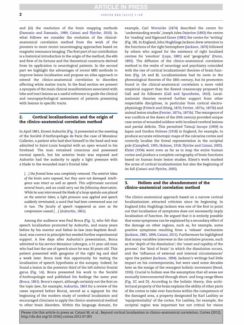
In April 1861, Ernest Auburtin (Fig. 1) presented at themeeting
of the Societe d’Anthropologie de Paris the case of Monsieur
Cullerier, a patient who had shot himself in the head and was
admitted to Saint-Louis hospital with an open wound in his
forehead. The man remained conscious and possessed
normal speech, but his anterior brain was exposed and
Auburtin had the audacity to apply a light pressure with
a blade to the wounded man’s frontal lobe:
[.] the frontal bone was completely removed. The anterior lobes
of the brain were exposed, but they were not damaged. Intelli-
gence was intact as well as speech. This unfortunate survived
several hours, and we could carry out the following observation.
While he was interviewed the blade of a large spatula was placed
on the anterior lobes; a light pressure was applied and speech
suddenly terminated; a word that had been commenced was cut
in two. The faculty of speech reappeared as soon as the
compression ceased [.] (Auburtin, 1861).
Among the audience was Paul Broca (Fig. 1), who felt that
speech localisation promoted by Auburtin, and many years
before by his mentor and father-in-law Jean-Baptiste Bouil-
laud, was correct in principle but needed further experimental
support. A few days after Auburtin’s presentation, Broca
admitted to his service Monsieur Leborgne, a 51 year-old man
who had lost the use of speech since he was 30 years old. This
patient presented with gangrene of the right leg and died
a week later. Broca took this opportunity for testing the
localisation of speech hypothesis at the autopsy. Indeed, he
found a lesion in the posterior third of the left inferior frontal
gyrus (Fig. 1A). Broca presented his work to the Societe
d’Anthropologie and published his findings the same year
(Broca, 1861). Broca’s report, although certainly not the first on
the topic (see, for example; Auburtin, 1863 for a review of the
cases reported before Broca), served as a signpost for the
beginning of the modern study of cerebral localisation and
encouraged clinicians to apply the clinico-anatomical method
to other brain disorders. Within the language domain, for
Please cite this article in press as: Catani M, et al., Beyond corticahttp://dx.doi.org/10.1016/j.cortex.2012.07.001
example, Carl Wernicke (1874) described the centre for
‘understanding words’, Joseph Jules Dejerine (1892) the centre
for ‘reading’ and Sigmund Exner (1881) the centre for ‘writing’
(Fig. 1B). In England, John Hughlings Jackson drew attention to
the functions of the right hemisphere (Jackson, 1876) followed
by others who argued for the existence of right localised
centres for ‘emotion’ (Luys, 1881) and ‘geography’ (Dunn,
1895). The diffusion of the clinico-anatomical correlation
method in the realm of neurology and psychiatry coincided
with the rise of cortical localizationist theories of brain func-
tion (Fig. 2A and B). Localizationism had its roots in the
phrenological theories of the 18th century, but its promoters
found in the clinical-anatomical correlation a more valid
empirical support than the flawed cranioscopy proposed by
Gall and its followers (Gall and Spurzheim, 1810). Local-
izationist theories received further support from other
respectable disciplines, in particular from cortical electro-
physiology (Fritsch and Hitzig, 1870; Ferrier, 1875a, 1875b) and
animal lesion studies (Ferrier, 1875a, 1875b). The resurgence of
war conflicts at the dawn of the 20th century provided unique
case series of wounded soldiers with localised cerebral lesions
and partial deficits. This permitted Tatsuji Inouye (1909) in
Japan and Gordon Holmes (1918) in England, for example, to
produce accurate retinotopicmaps of the calcarine cortex and
correctly localise the foveal representation in the occipital
pole (Campbell, 1905; Holmes, 1918; ffytche and Catani, 2005).
Kleist (1934) went even as far as to map the entire human
cortex and produce a complete cartography of brain functions
based on human brain lesion studies. Kleist’s work marked
the acme of cortical localizationism but also the beginning of
its fall (Catani and ffytche, 2005).
3. Holism and the abandonment of theclinico-anatomical correlation method
The clinico-anatomical approach based on a narrow cortical
localizationism attracted criticism since its beginning. In
England John Hughlings Jackson was one of the first to point
out that localisation of symptoms does not necessarily imply
localisation of function. He argued that it is entirely possible
that some symptoms can be explained by a secondary effect of
the damage on other regions, such as, for example, some
positive symptoms resulting from a ‘release’ mechanism
(Jackson, 1881, 1894; Catani, 2011). Furthermore he highlighted
that many variables intervene in the correlative process, such
as the ‘depth of the dissolution’; the ‘onset and rapidity of the
process’; the ‘kind of brain’ in which the dissolution occurs,
and the ‘influence of external and internal circumstances’
upon the patient (Jackson, 1894). Jackson’s writings had little
impact on his contemporaries, but were used some decades
later as the ensign of the resurgent holistic movement (Head,
1926). Crucial to holism was the assumption that all areas are
mutually interconnected through short- and long-range fibres
(Fig. 2C and D). According to the holistic theory, this archi-
tectural property of the brain explains the ability of other parts
of the cortex to take over functions within the competence of
the damaged area, a property designated by Karl Lashley as
‘equipotentiality’ of the cortex. For Lashley, for example, the
occipital region was important but not critical for vision
l localisation in clinico-anatomical correlation, Cortex (2012),
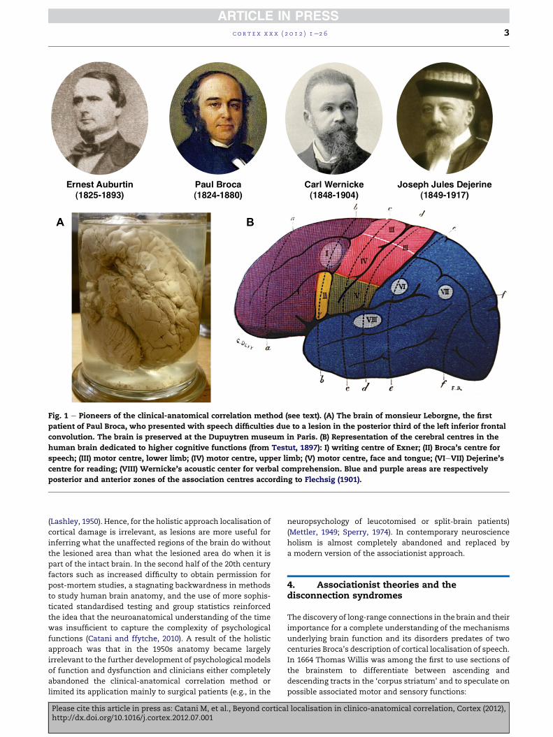
Fig. 1 e Pioneers of the clinical-anatomical correlation method (see text). (A) The brain of monsieur Leborgne, the first
patient of Paul Broca, who presented with speech difficulties due to a lesion in the posterior third of the left inferior frontal
convolution. The brain is preserved at the Dupuytren museum in Paris. (B) Representation of the cerebral centres in the
human brain dedicated to higher cognitive functions (from Testut, 1897): I) writing centre of Exner; (II) Broca’s centre for
speech; (III) motor centre, lower limb; (IV) motor centre, upper limb; (V) motor centre, face and tongue; (VIeVII) Dejerine’s
centre for reading; (VIII) Wernicke’s acoustic center for verbal comprehension. Blue and purple areas are respectively
posterior and anterior zones of the association centres according to Flechsig (1901).
c o r t e x x x x ( 2 0 1 2 ) 1e2 6 3
(Lashley, 1950). Hence, for the holistic approach localisation of
cortical damage is irrelevant, as lesions are more useful for
inferring what the unaffected regions of the brain do without
the lesioned area than what the lesioned area do when it is
part of the intact brain. In the second half of the 20th century
factors such as increased difficulty to obtain permission for
post-mortem studies, a stagnating backwardness in methods
to study human brain anatomy, and the use of more sophis-
ticated standardised testing and group statistics reinforced
the idea that the neuroanatomical understanding of the time
was insufficient to capture the complexity of psychological
functions (Catani and ffytche, 2010). A result of the holistic
approach was that in the 1950s anatomy became largely
irrelevant to the further development of psychological models
of function and dysfunction and clinicians either completely
abandoned the clinical-anatomical correlation method or
limited its application mainly to surgical patients (e.g., in the
Please cite this article in press as: Catani M, et al., Beyond corticahttp://dx.doi.org/10.1016/j.cortex.2012.07.001
neuropsychology of leucotomised or split-brain patients)
(Mettler, 1949; Sperry, 1974). In contemporary neuroscience
holism is almost completely abandoned and replaced by
a modern version of the associationist approach.
4. Associationist theories and thedisconnection syndromes
The discovery of long-range connections in the brain and their
importance for a complete understanding of the mechanisms
underlying brain function and its disorders predates of two
centuries Broca’s description of cortical localisation of speech.
In 1664 Thomas Willis was among the first to use sections of
the brainstem to differentiate between ascending and
descending tracts in the ‘corpus striatum’ and to speculate on
possible associated motor and sensory functions:
l localisation in clinico-anatomical correlation, Cortex (2012),
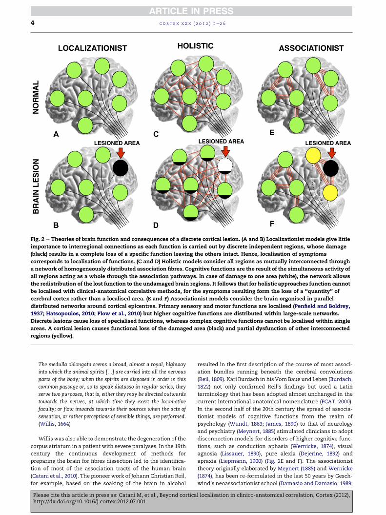
Fig. 2 e Theories of brain function and consequences of a discrete cortical lesion. (A and B) Localizationist models give little
importance to interregional connections as each function is carried out by discrete independent regions, whose damage
(black) results in a complete loss of a specific function leaving the others intact. Hence, localisation of symptoms
corresponds to localisation of functions. (C and D) Holistic models consider all regions as mutually interconnected through
a network of homogeneously distributed association fibres. Cognitive functions are the result of the simultaneous activity of
all regions acting as a whole through the association pathways. In case of damage to one area (white), the network allows
the redistribution of the lost function to the undamaged brain regions. It follows that for holistic approaches function cannot
be localised with clinical-anatomical correlative methods, for the symptoms resulting form the loss of a “quantity” of
cerebral cortex rather than a localised area. (E and F) Associationist models consider the brain organised in parallel
distributed networks around cortical epicentres. Primary sensory and motor functions are localised (Penfield and Boldrey,
1937; Hatsopoulos, 2010; Plow et al., 2010) but higher cognitive functions are distributed within large-scale networks.
Discrete lesions cause loss of specialised functions, whereas complex cognitive functions cannot be localised within single
areas. A cortical lesion causes functional loss of the damaged area (black) and partial dysfunction of other interconnected
regions (yellow).
c o r t e x x x x ( 2 0 1 2 ) 1e2 64
The medulla oblongata seems a broad, almost a royal, highway
into which the animal spirits [.] are carried into all the nervous
parts of the body; when the spirits are disposed in order in this
common passage or, so to speak diatasso in regular series, they
serve two purposes, that is, either they may be directed outwards
towards the nerves, at which time they exert the locomotive
faculty; or flow inwards towards their sources when the acts of
sensation, or rather perceptions of sensible things, are performed.
(Willis, 1664)
Willis was also able to demonstrate the degeneration of the
corpus striatum in a patient with severe paralyses. In the 19th
century the continuous development of methods for
preparing the brain for fibres dissection led to the identifica-
tion of most of the association tracts of the human brain
(Catani et al., 2010). The pioneer work of Johann Christian Reil,
for example, based on the soaking of the brain in alcohol
Please cite this article in press as: Catani M, et al., Beyond corticahttp://dx.doi.org/10.1016/j.cortex.2012.07.001
resulted in the first description of the course of most associ-
ation bundles running beneath the cerebral convolutions
(Reil, 1809). Karl Burdach in his VomBaue und Leben (Burdach,
1822) not only confirmed Reil’s findings but used a Latin
terminology that has been adopted almost unchanged in the
current international anatomical nomenclature (FCAT, 2000).
In the second half of the 20th century the spread of associa-
tionist models of cognitive functions from the realm of
psychology (Wundt, 1863; James, 1890) to that of neurology
and psychiatry (Meynert, 1885) stimulated clinicians to adopt
disconnection models for disorders of higher cognitive func-
tions, such as conduction aphasia (Wernicke, 1874), visual
agnosia (Lissauer, 1890), pure alexia (Dejerine, 1892) and
apraxia (Liepmann, 1900) (Fig. 2E and F). The associationist
theory originally elaborated by Meynert (1885) and Wernicke
(1874), has been re-formulated in the last 50 years by Gesch-
wind’s neoassociationist school (Damasio and Damasio, 1989;
l localisation in clinico-anatomical correlation, Cortex (2012),
c o r t e x x x x ( 2 0 1 2 ) 1e2 6 5
Geschwind, 1965a, 1965b; Mesulam, 1990; Ross, 2010) and has
received further support in the recent years from functional
imaging and diffusion magnetic resonance imaging tractog-
raphy (Catani and ffytche, 2005; Catani, 2006). According to
this theory large-scale networks in the human brain are
dedicated to specific functions such as language, face-and-
object recognition, executive function-comportment, spatial
attention, and memory-emotion (Fig. 3) (Mesulam, 2000). The
nodes of these networks can be divided into critical versus
participating epicentres. Critical network epicentres consti-
tute ‘relays or integration centres, hubs, nexuses, sluices for
convergence, divergence, feedback loops, feed-forward
connections, and transition points from serial to parallel
processing’ (Catani and Mesulam, 2008a,b). Cognition and
behaviour are considered as emergent properties of large-
scale neural networks (Ross, 2010; Bartolomeo, 2011), where
lesions to connections lead to the inability to transfer infor-
mation from one node to another (as in the classical discon-
nection syndromes such as conduction aphasia) and a series
of distant ‘hodological’ effects on each node of the network (as
in the case of diaschisis) (von Monakow, 1914).
Recent advances in functional (Friston et al., 2003) and
diffusion (Basser et al., 2000; Catani et al., 2002; Jones, 2008;
Fig. 3 e Large-scale networks for cognition and behaviour (Mes
network with epicentres in Wernickeʼs and Brocaʼs areas (Catan
network with epicentres in occipito-temporal and temporopola
comportment network with epicentres in lateral prefrontal cort
(Zappala et al., 2012). (D) A right hemisphere-dominant spatial at
cortex, the frontal eye fields, and the cingulate gyrus (Doricchi e
in the hippocampal-entorhinal regions and the amygdaloid com
Please cite this article in press as: Catani M, et al., Beyond corticahttp://dx.doi.org/10.1016/j.cortex.2012.07.001
Dell’Acqua and Catani, in press) magnetic resonance imaging
are important steps to gain insight into neuronal networks of
the human brain. The diffusion tractography approach, for
example, has demonstrated that the ‘arcuate fasciculus’
contains a direct and also an indirect component (Catani et al.,
2005) and so added a new layer of understanding that is more
in keepingwith the parallel processingmodels of the language
network (Mesulam, 2005). Unfortunately these advanced MRI
methods are often not available to most clinicians, who
frequently are the first to have the opportunity to study
patients with unique neurological manifestations. Further-
more the lack of normalised white matter atlases of
human brain connections based on diffusion tensor tractog-
raphy and the limitations of currently available histological
atlases in identifying association tracts hindered our ability to
correctly localise damaged white matter pathways (Thiebaut
de Schotten et al., 2011a). To fill this gap we propose in the
following section a statistical atlas of human brain connec-
tions based on diffusion tensor imaging tractography.
Together with a synopsis of the major lobe syndromes the
atlas could help clinicians to include information on
white matter anatomy in the clinico-anatomical correlative
process.
ulam, 2000). (A) A left hemisphere-dominant language
i and Mesulam, 2008). (B) A face-object identification
r cortex (Fow et al., 2008). (C) An executive function-
ex, orbitofrontal cortex, and posterior parietal cortex
tention network with epicentres in dorsal posterior parietal
t al., 2008). (E) A memory-emotion network with epicentres
plex (Park et al., 2010).
l localisation in clinico-anatomical correlation, Cortex (2012),
c o r t e x x x x ( 2 0 1 2 ) 1e2 66
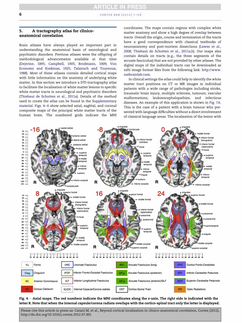
5. A tractography atlas for clinico-anatomical correlation
Brain atlases have always played an important part in
understanding the anatomical basis of neurological and
psychiatric disorders. Previous atlases were the offspring of
methodological advancements available at that time
(Dejerine, 1895; Campbell, 1905; Brodmann, 1909; Von
Economo and Koskinas, 1925; Talairach and Tournoux,
1988). Most of these atlases contain detailed cortical maps
with little information on the anatomy of underlying white
matter. In this section we introduce a DTI-tractography atlas
to facilitate the localisation of white matter lesions to specific
white matter tracts in neurological and psychiatric disorders
(Thiebaut de Schotten et al., 2011a). Details of the method
used to create the atlas can be found in the Supplementary
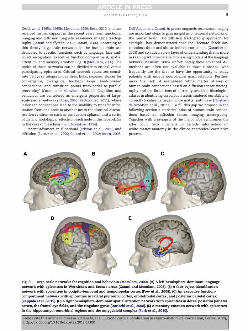
material. Figs. 4e6 show selected axial, sagittal, and coronal
composite maps of the principal white matter tracts of the
human brain. The numbered grids indicate the MNI
Fig. 4 e Axial maps. The red numbers indicate the MNI coordin
letter R. Note that when the internal capsule/corona radiata over
Please cite this article in press as: Catani M, et al., Beyond corticahttp://dx.doi.org/10.1016/j.cortex.2012.07.001
coordinates. The maps contain regions with complex white
matter anatomy and show a high degree of overlap between
tracts. Overall the origin, course and termination of the tracts
have a good correspondence with classical textbooks of
neuroanatomy and post-mortem dissections (Lawes et al.,
2008; Thiebaut de Schotten et al., 2011a,b). Our maps also
contain details on tracts (e.g., the three segments of the
arcuate fasciculus) that are not provided by other atlases. The
digital maps of the individual tracts can be downloaded as
nifti image format files from the following link: http://www.
natbrainlab.com.
In clinical settings the atlas could help to identify the white
matter tract positions on CT or MR images in individual
patients with a wide range of pathologies including stroke,
traumatic brain injury, multiple sclerosis, tumours, vascular
malformations, leukoencephalopathies, and infectious
diseases. An example of this application is shown in Fig. 7A.
This is the case of a patient with a brain tumour who pre-
sentedwith language difficulties without a direct involvement
of classical language areas. The localisation of the lesion with
ates along the z-axis. The right side is indicated with the
laps with the cortico-spinal tract only the latter is displayed.
l localisation in clinico-anatomical correlation, Cortex (2012),
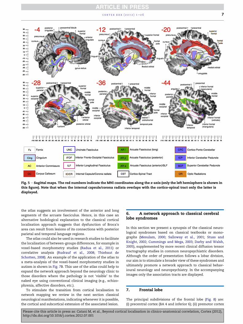
Fig. 5 e Sagittal maps. The red numbers indicate the MNI coordinates along the x-axis (only the left hemisphere is shown in
this figure). Note that when the internal capsule/corona radiata overlaps with the cortico-spinal tract only the latter is
displayed.
c o r t e x x x x ( 2 0 1 2 ) 1e2 6 7
the atlas suggests an involvement of the anterior and long
segments of the arcuate fasciculus. Hence, in this case an
alternative hodological explanation to the classical cortical
localisation approach suggests that dysfunction of Broca’s
area can result from lesions of its connections with posterior
parietal and temporal language regions.
The atlas could also be used in research studies to facilitate
the localisation of between-groups differences, for example in
voxel-based morphometry studies (Radua et al., 2011) or
correlative analysis (Rudrauf et al., 2008; Thiebaut de
Schotten, 2008). An example of the application of the atlas to
a meta-analysis of the voxel-based morphometry studies in
autism is shown in Fig. 7B. This use of the atlas could help to
expand the network approach beyond the neurology clinic to
those disorders where the pathology is not ‘visible’ to the
naked eye using conventional clinical imaging (e.g., schizo-
phrenia, affective disorders, etc.).
To stimulate the transition from cortical localisation to
network mapping we review in the next section classical
neurological manifestations, indicating whenever it is possible,
the cortical and subcortical extension of the associated lesion.
Please cite this article in press as: Catani M, et al., Beyond corticahttp://dx.doi.org/10.1016/j.cortex.2012.07.001
6. A network approach to classical cerebrallobe syndromes
In this section we present a synopsis of the classical neuro-
logical syndromes based on classical textbooks or mono-
graphs (Mesulam, 2000; Salloway et al., 2001; Stuss and
Knight, 2002; Cummings and Mega, 2003; Darby and Walsh,
2005), supplemented by more recent clinical diffusion tensor
tractography studies in common neuropsychiatric disorders.
Although the order of presentation follows a lobar division,
our aim is to stimulate a broader view of these syndromes and
ultimately promote a network approach to classical behav-
ioural neurology and neuropsychiatry. In the accompanying
images only the association tracts are displayed.
7. Frontal lobe
The principal subdivisions of the frontal lobe (Fig. 8) are:
(i) precentral cortex (BA 4 and inferior 6); (ii) premotor cortex
l localisation in clinico-anatomical correlation, Cortex (2012),
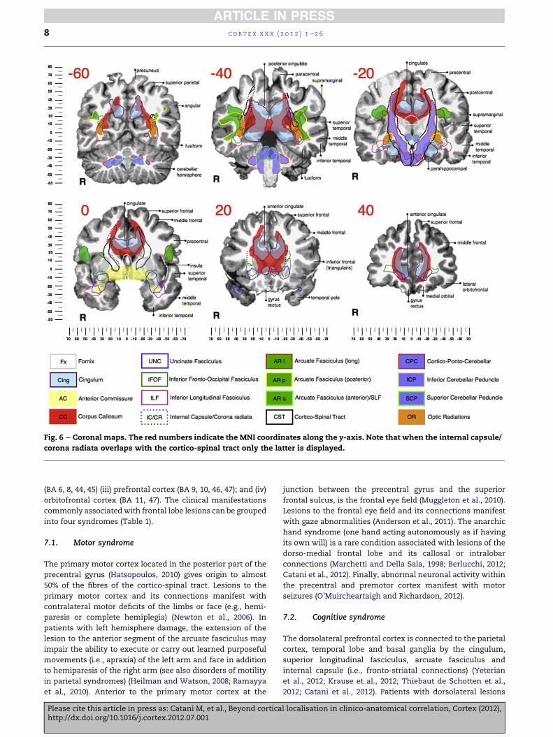
Fig. 6 e Coronal maps. The red numbers indicate the MNI coordinates along the y-axis. Note that when the internal capsule/
corona radiata overlaps with the cortico-spinal tract only the latter is displayed.
c o r t e x x x x ( 2 0 1 2 ) 1e2 68
(BA 6, 8, 44, 45) (iii) prefrontal cortex (BA 9, 10, 46, 47); and (iv)
orbitofrontal cortex (BA 11, 47). The clinical manifestations
commonly associatedwith frontal lobe lesions can be grouped
into four syndromes (Table 1).
7.1. Motor syndrome
The primary motor cortex located in the posterior part of the
precentral gyrus (Hatsopoulos, 2010) gives origin to almost
50% of the fibres of the cortico-spinal tract. Lesions to the
primary motor cortex and its connections manifest with
contralateral motor deficits of the limbs or face (e.g., hemi-
paresis or complete hemiplegia) (Newton et al., 2006). In
patients with left hemisphere damage, the extension of the
lesion to the anterior segment of the arcuate fasciculus may
impair the ability to execute or carry out learned purposeful
movements (i.e., apraxia) of the left arm and face in addition
to hemiparesis of the right arm (see also disorders of motility
in parietal syndromes) (Heilman and Watson, 2008; Ramayya
et al., 2010). Anterior to the primary motor cortex at the
Please cite this article in press as: Catani M, et al., Beyond corticahttp://dx.doi.org/10.1016/j.cortex.2012.07.001
junction between the precentral gyrus and the superior
frontal sulcus, is the frontal eye field (Muggleton et al., 2010).
Lesions to the frontal eye field and its connections manifest
with gaze abnormalities (Anderson et al., 2011). The anarchic
hand syndrome (one hand acting autonomously as if having
its own will) is a rare condition associated with lesions of the
dorso-medial frontal lobe and its callosal or intralobar
connections (Marchetti and Della Sala, 1998; Berlucchi, 2012;
Catani et al., 2012). Finally, abnormal neuronal activity within
the precentral and premotor cortex manifest with motor
seizures (O’Muircheartaigh and Richardson, 2012).
7.2. Cognitive syndrome
The dorsolateral prefrontal cortex is connected to the parietal
cortex, temporal lobe and basal ganglia by the cingulum,
superior longitudinal fasciculus, arcuate fasciculus and
internal capsule (i.e., fronto-striatal connections) (Yeterian
et al., 2012; Krause et al., 2012; Thiebaut de Schotten et al.,
2012; Catani et al., 2012). Patients with dorsolateral lesions
l localisation in clinico-anatomical correlation, Cortex (2012),
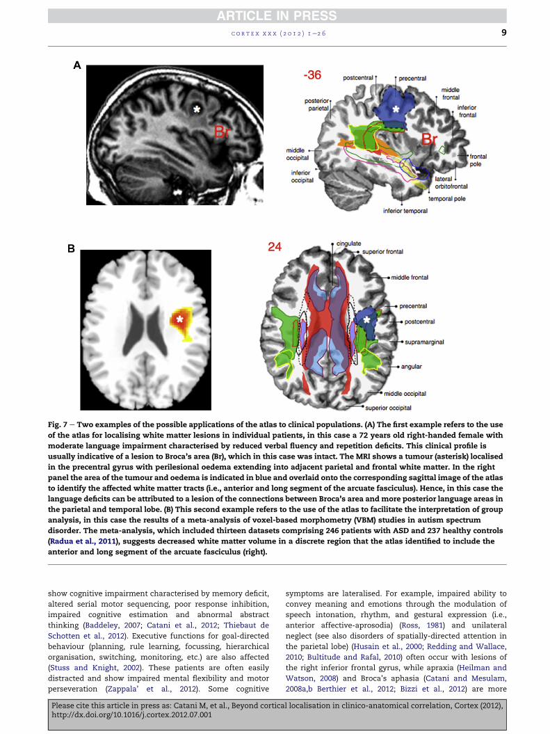
Fig. 7 e Two examples of the possible applications of the atlas to clinical populations. (A) The first example refers to the use
of the atlas for localising white matter lesions in individual patients, in this case a 72 years old right-handed female with
moderate language impairment characterised by reduced verbal fluency and repetition deficits. This clinical profile is
usually indicative of a lesion to Broca’s area (Br), which in this case was intact. The MRI shows a tumour (asterisk) localised
in the precentral gyrus with perilesional oedema extending into adjacent parietal and frontal white matter. In the right
panel the area of the tumour and oedema is indicated in blue and overlaid onto the corresponding sagittal image of the atlas
to identify the affected white matter tracts (i.e., anterior and long segment of the arcuate fasciculus). Hence, in this case the
language deficits can be attributed to a lesion of the connections between Broca’s area and more posterior language areas in
the parietal and temporal lobe. (B) This second example refers to the use of the atlas to facilitate the interpretation of group
analysis, in this case the results of a meta-analysis of voxel-based morphometry (VBM) studies in autism spectrum
disorder. The meta-analysis, which included thirteen datasets comprising 246 patients with ASD and 237 healthy controls
(Radua et al., 2011), suggests decreased white matter volume in a discrete region that the atlas identified to include the
anterior and long segment of the arcuate fasciculus (right).
c o r t e x x x x ( 2 0 1 2 ) 1e2 6 9
show cognitive impairment characterised by memory deficit,
altered serial motor sequencing, poor response inhibition,
impaired cognitive estimation and abnormal abstract
thinking (Baddeley, 2007; Catani et al., 2012; Thiebaut de
Schotten et al., 2012). Executive functions for goal-directed
behaviour (planning, rule learning, focussing, hierarchical
organisation, switching, monitoring, etc.) are also affected
(Stuss and Knight, 2002). These patients are often easily
distracted and show impaired mental flexibility and motor
perseveration (Zappala’ et al., 2012). Some cognitive
Please cite this article in press as: Catani M, et al., Beyond corticahttp://dx.doi.org/10.1016/j.cortex.2012.07.001
symptoms are lateralised. For example, impaired ability to
convey meaning and emotions through the modulation of
speech intonation, rhythm, and gestural expression (i.e.,
anterior affective-aprosodia) (Ross, 1981) and unilateral
neglect (see also disorders of spatially-directed attention in
the parietal lobe) (Husain et al., 2000; Redding and Wallace,
2010; Bultitude and Rafal, 2010) often occur with lesions of
the right inferior frontal gyrus, while apraxia (Heilman and
Watson, 2008) and Broca’s aphasia (Catani and Mesulam,
2008a,b Berthier et al., 2012; Bizzi et al., 2012) are more
l localisation in clinico-anatomical correlation, Cortex (2012),
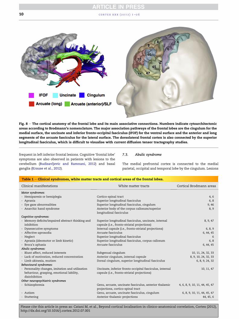
Fig. 8 e The cortical anatomy of the frontal lobe and its main associative connections. Numbers indicate cytoarchitectonic
areas according to Brodmann’s nomenclature. The major association pathways of the frontal lobes are the cingulum for the
medial surface, the uncinate and inferior fronto-occipital fasciculus (IFOF) for the ventral surface and the anterior and long
segments of the arcuate fasciculus for the lateral surface. The dorsolateral frontal cortex is also connected by the superior
longitudinal fasciculus, which is difficult to visualise with current diffusion tensor tractography studies.
c o r t e x x x x ( 2 0 1 2 ) 1e2 610
frequent in left inferior frontal lesions. Cognitive ‘frontal lobe’
symptoms are also observed in patients with lesions to the
cerebellum (Budisavljevic and Ramnani, 2012) and basal
ganglia (Krause et al., 2012).
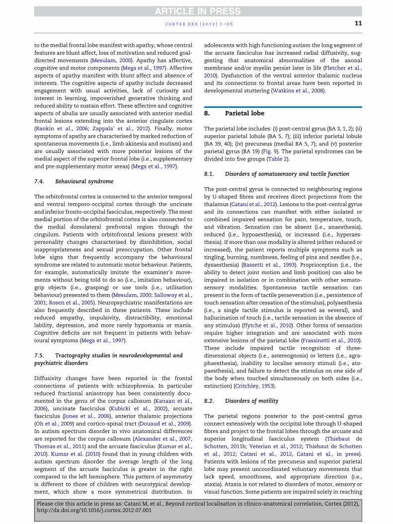
Table 1 e Clinical syndromes, white matter tracts and cortical
Clinical manifestations W
Motor syndromes:
- Hemiparesis or hemiplegia Cortico-spinal tra
- Apraxia Superior longitudi
- Eye gaze abnormalities Superior longitudi
- Anarchic hand syndrome Anterior body of t
longitudinal fascic
Cognitive syndromes:
- Memory deficits/impaired abstract thinking and
inhibition
Superior longitudi
capsule (i.e., front
- Dysexecutive symptoms Internal capsule (i
- Affective-aprosodia Arcuate fasciculus
- Neglect Superior longitudi
- Apraxia (ideomotor or limb kinetic) Superior longitudi
- Broca’s aphasia Arcuate fasciculus
Abulic syndromes:
- Blunt affect, reduced interests Subgenual cingulu
- Lack of motivation, reduced concentration Anterior cingulum
- Limb akinesia, mutism Dorsal cingulum,
Behavioural syndromes:
- Personality changes, imitation and utilisation
behaviour, grasping, emotional lability,
disinhibition
Uncinate, inferior
capsule (i.e., front
Other neuropsychiatric syndromes
- Schizophrenia Genu, arcuate, un
projections, cortic
- Autism Genu, arcuate, un
- Stuttering Anterior thalamic
Please cite this article in press as: Catani M, et al., Beyond corticahttp://dx.doi.org/10.1016/j.cortex.2012.07.001
7.3. Abulic syndrome
The medial prefrontal cortex is connected to the medial
parietal, occipital and temporal lobe by the cingulum. Lesions
areas of the frontal lobes.
hite matter tracts Cortical Brodmann areas
ct 4, 6
nal fasciculus 6, 8
nal fasciculus, cingulum 9, 46
he corpus callosum/superior
ulus
8, 9
nal fasciculus, uncinate, internal
o-striatal projections)
8, 9, 47
.e., fronto-striatal projections) 6, 8, 9
6, 44, 45
nal fasciculus 6
nal fasciculus, corpus callosum 6, 8
6, 44, 45
m 10, 11, 24, 32, 33
, internal capsule 8, 9, 10, 24, 32, 33
superior longitudinal fasciculus 6, 8, 9, 24, 32
fronto-occipital fasciculus, internal
o-striatal projections)
10, 11, 47
cinate fasciculus, anterior thalamic
o-spinal tract
4, 6, 8, 9, 10, 11, 44, 45, 47
cinate fasciculus, cingulum 6, 8, 9, 10, 11, 44, 45, 47
projections 44, 45, 6
l localisation in clinico-anatomical correlation, Cortex (2012),
c o r t e x x x x ( 2 0 1 2 ) 1e2 6 11
to themedial frontal lobemanifestwith apathy, whose central
features are blunt affect, loss of motivation and reduced goal-
directed movements (Mesulam, 2000). Apathy has affective,
cognitive and motor components (Mega et al., 1997). Affective
aspects of apathy manifest with blunt affect and absence of
interests. The cognitive aspects of apathy include decreased
engagement with usual activities, lack of curiosity and
interest in learning, impoverished generative thinking and
reduced ability to sustain effort. These affective and cognitive
aspects of abulia are usually associated with anterior medial
frontal lesions extending into the anterior cingulate cortex
(Rankin et al., 2006; Zappala’ et al., 2012). Finally, motor
symptoms of apathy are characterised bymarked reduction of
spontaneous movements (i.e., limb akinesia and mutism) and
are usually associated with more posterior lesions of the
medial aspect of the superior frontal lobe (i.e., supplementary
and pre-supplementary motor areas) (Mega et al., 1997).
7.4. Behavioural syndrome
The orbitofrontal cortex is connected to the anterior temporal
and ventral temporo-occipital cortex through the uncinate
and inferior fronto-occipital fasciculus, respectively. Themost
medial portion of the orbitofrontal cortex is also connected to
the medial dorsolateral prefrontal region through the
cingulum. Patients with orbitofrontal lesions present with
personality changes characterised by disinhibition, social
inappropriateness and sexual preoccupation. Other frontal
lobe signs that frequently accompany the behavioural
syndrome are related to automatic motor behaviour. Patients,
for example, automatically imitate the examiner’s move-
ments without being told to do so (i.e., imitation behaviour),
grip objects (i.e., grasping) or use tools (i.e., utilisation
behaviour) presented to them (Mesulam, 2000; Salloway et al.,
2001; Rosen et al., 2005). Neuropsychiatric manifestations are
also frequently described in these patients. These include
reduced empathy, impulsivity, distractibility, emotional
lability, depression, and more rarely hypomania or mania.
Cognitive deficits are not frequent in patients with behav-
ioural symptoms (Mega et al., 1997).
7.5. Tractography studies in neurodevelopmental andpsychiatric disorders
Diffusivity changes have been reported in the frontal
connections of patients with schizophrenia. In particular
reduced fractional anisotropy has been consistently docu-
mented in the genu of the corpus callosum (Kanaan et al.,
2006), uncinate fasciculus (Kubicki et al., 2002), arcuate
fasciculus (Jones et al., 2006), anterior thalamic projections
(Oh et al., 2009) and cortico-spinal tract (Douaud et al., 2009).
In autism spectrum disorder in vivo anatomical differences
are reported for the corpus callosum (Alexander et al., 2007,
Thomas et al., 2011) and the arcuate fasciculus (Kumar et al.,
2010). Kumar et al. (2010) found that in young children with
autism spectrum disorder the average length of the long
segment of the arcuate fasciculus is greater in the right
compared to the left hemisphere. This pattern of asymmetry
is different to those of children with neurotypical develop-
ment, which show a more symmetrical distribution. In
Please cite this article in press as: Catani M, et al., Beyond corticahttp://dx.doi.org/10.1016/j.cortex.2012.07.001
adolescents with high functioning autism the long segment of
the arcuate fasciculus has increased radial diffusivity, sug-
gesting that anatomical abnormalities of the axonal
membrane and/or myelin persist later in life (Fletcher et al.,
2010). Dysfunction of the ventral anterior thalamic nucleus
and its connections to frontal areas have been reported in
developmental stuttering (Watkins et al., 2008).
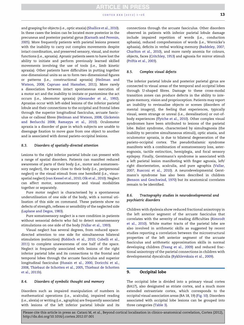
8. Parietal lobe
The parietal lobe includes: (i) post-central gyrus (BA 3, 1, 2); (ii)
superior parietal lobule (BA 5, 7); (iii) inferior parietal lobule
(BA 39, 40); (iv) precuneus (medial BA 5, 7); and (v) posterior
parietal gyrus (BA 19) (Fig. 9). The parietal syndromes can be
divided into five groups (Table 2).
8.1. Disorders of somatosensory and tactile function
The post-central gyrus is connected to neighbouring regions
by U-shaped fibres and receives direct projections from the
thalamus (Catani et al., 2012). Lesions to the post-central gyrus
and its connections can manifest with either isolated or
combined impaired sensation for pain, temperature, touch,
and vibration. Sensation can be absent (i.e., anaesthesia),
reduced (i.e., hypoaesthesia), or increased (i.e., hyperaes-
thesia). If more than onemodality is altered (either reduced or
increased), the patient reports multiple symptoms such as
tingling, burning, numbness, feeling of pins and needles (i.e.,
dysaesthesia) (Bassetti et al., 1993). Proprioception (i.e., the
ability to detect joint motion and limb position) can also be
impaired in isolation or in combination with other somato-
sensory modalities. Spontaneous tactile sensation can
present in the form of tactile perseveration (i.e., persistence of
touch sensation after cessation of the stimulus), polyaesthesia
(i.e., a single tactile stimulus is reported as several), and
hallucination of touch (i.e., tactile sensation in the absence of
any stimulus) (ffytche et al., 2010). Other forms of sensation
require higher integration and are associated with more
extensive lesions of the parietal lobe (Frassinetti et al., 2010).
These include impaired tactile recognition of three-
dimensional objects (i.e., astereognosis) or letters (i.e., agra-
phaesthesia), inability to localise sensory stimuli (i.e., ato-
paesthesia), and failure to detect the stimulus on one side of
the body when touched simultaneously on both sides (i.e.,
extinction) (Critchley, 1953).
8.2. Disorders of motility
The parietal regions posterior to the post-central gyrus
connect extensively with the occipital lobe through U-shaped
fibres and project to the frontal lobes through the arcuate and
superior longitudinal fasciculus system (Thiebaut de
Schotten, 2011b; Yeterian et al., 2012; Thiebaut de Schotten
et al., 2012; Catani et al., 2012, Catani et al., in press).
Patients with lesions of the precuneus and superior parietal
lobe may present uncoordinated voluntary movements that
lack speed, smoothness, and appropriate direction (i.e.,
ataxia). Ataxia is not related to disorders of motor, sensory or
visual function. Some patients are impaired solely in reaching
l localisation in clinico-anatomical correlation, Cortex (2012),
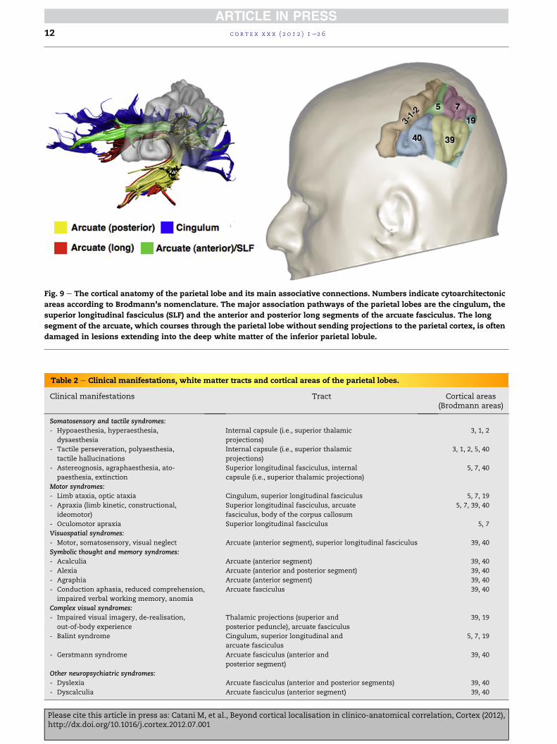
Table 2 e Clinical manifestations, white matter tracts and cortical areas of the parietal lobes.
Clinical manifestations Tract Cortical areas(Brodmann areas)
Somatosensory and tactile syndromes:
- Hypoaesthesia, hyperaesthesia,
dysaesthesia
Internal capsule (i.e., superior thalamic
projections)
3, 1, 2
- Tactile perseveration, polyaesthesia,
tactile hallucinations
Internal capsule (i.e., superior thalamic
projections)
3, 1, 2, 5, 40
- Astereognosis, agraphaesthesia, ato-
paesthesia, extinction
Superior longitudinal fasciculus, internal
capsule (i.e., superior thalamic projections)
5, 7, 40
Motor syndromes:
- Limb ataxia, optic ataxia Cingulum, superior longitudinal fasciculus 5, 7, 19
- Apraxia (limb kinetic, constructional,
ideomotor)
Superior longitudinal fasciculus, arcuate
fasciculus, body of the corpus callosum
5, 7, 39, 40
- Oculomotor apraxia Superior longitudinal fasciculus 5, 7
Visuospatial syndromes:
- Motor, somatosensory, visual neglect Arcuate (anterior segment), superior longitudinal fasciculus 39, 40
Symbolic thought and memory syndromes:
- Acalculia Arcuate (anterior segment) 39, 40
- Alexia Arcuate (anterior and posterior segment) 39, 40
- Agraphia Arcuate (anterior segment) 39, 40
- Conduction aphasia, reduced comprehension,
impaired verbal working memory, anomia
Arcuate fasciculus 39, 40
Complex visual syndromes:
- Impaired visual imagery, de-realisation,
out-of-body experience
Thalamic projections (superior and
posterior peduncle), arcuate fasciculus
39, 19
- Balint syndrome Cingulum, superior longitudinal and
arcuate fasciculus
5, 7, 19
- Gerstmann syndrome Arcuate fasciculus (anterior and
posterior segment)
39, 40
Other neuropsychiatric syndromes:
- Dyslexia Arcuate fasciculus (anterior and posterior segments) 39, 40
- Dyscalculia Arcuate fasciculus (anterior segment) 39, 40
Fig. 9 e The cortical anatomy of the parietal lobe and its main associative connections. Numbers indicate cytoarchitectonic
areas according to Brodmann’s nomenclature. The major association pathways of the parietal lobes are the cingulum, the
superior longitudinal fasciculus (SLF) and the anterior and posterior long segments of the arcuate fasciculus. The long
segment of the arcuate, which courses through the parietal lobe without sending projections to the parietal cortex, is often
damaged in lesions extending into the deep white matter of the inferior parietal lobule.
c o r t e x x x x ( 2 0 1 2 ) 1e2 612
Please cite this article in press as: Catani M, et al., Beyond cortical localisation in clinico-anatomical correlation, Cortex (2012),http://dx.doi.org/10.1016/j.cortex.2012.07.001
c o r t e x x x x ( 2 0 1 2 ) 1e2 6 13
and grasping for objects (i.e., optic ataxia) (Shallice et al., 2010).
In these cases the lesion can be located more posterior in the
precuneus and posterior parietal gyrus (Karnath and Perenin,
2005). More frequently patients with parietal lesions present
with the inability to carry out complex movements despite
intact coordination, and preserved sensory, visual, and motor
functions (i.e., apraxia). Apraxic patients seem to have lost the
ability to initiate and perform previously learned skilled
movements involving the use of tools (i.e., limb kinetic
apraxia). Other patients have difficulties in putting together
one-dimensional units so as to form two-dimensional figures
or patterns (i.e., constructional apraxia) (Heilman and
Watson, 2008; Capruso and Hamsher, 2011). More rarely
a dissociation between intact spontaneous execution of
a motor act and the inability to imitate or pantomime the act
occurs (i.e., ideomotor apraxia) (Alexander et al., 1992).
Apraxias occur with left-sided lesions of the inferior parietal
lobule and their connections to the occipital and frontal lobes
through the superior longitudinal fasciculus, arcuate fascic-
ulus or callosal fibres (Heilman and Watson, 2008; Glickstein
and Berlucchi 2008; Ramayya et al., 2010). Oculomotor
apraxia is a disorder of gaze in which subjects are unable to
disengage fixation to move gaze from one object to another
and is associated with dorsal parieto-occipital lesions.
8.3. Disorders of spatially-directed attention
Lesions to the right inferior parietal lobule can present with
a range of spatial disorders. Patients can manifest reduced
awareness of parts of their body (i.e., motor and somatosen-
sory neglect), the space close to their body (i.e., peripersonal
neglect) or the visual stimuli from one hemifield (i.e., visuo-
spatial neglect) (van Kessel et al., 2010; Olk et al., 2010). Neglect
can affect motor, somatosensory and visual modalities
together or separately.
Pure motor neglect is characterised by a spontaneous
underutilisation of one side of the body, with a normal uti-
lisation of this side on command. These patients show no
defects of strength, reflexes or sensibility of the neglected side
(Laplane and Degos, 1983).
Pure somatosensory neglect is a rare condition in patients
without sensorial defects who fail to detect somatosensory
stimulations on one side of the body (Vallar et al., 1993).
Visual neglect has several degrees, from reduced space-
directed attention to one side for simultaneous bilateral
stimulation (extinction) (Riddoch et al., 2010; Cubelli et al.,
2011) to complete unawareness of one half of the space.
Neglect is frequently associated with lesions of the right
inferior parietal lobe and its connections to the frontal and
temporal lobes through the arcuate fasciculus and superior
longitudinal fasciculus (Husain et al., 2000, Doricchi et al.,
2008, Thiebaut de Schotten et al., 2005, Thiebaut de Schotten
et al., 2011b).
8.4. Disorders of symbolic thought and memory
Disorders such as impaired manipulation of numbers in
mathematical operations (i.e., acalculia), impaired reading
(i.e., alexia) or writing (i.e., agraphia) are frequently associated
with lesions of the left inferior parietal lobule and its
Please cite this article in press as: Catani M, et al., Beyond corticahttp://dx.doi.org/10.1016/j.cortex.2012.07.001
connections through the arcuate fasciculus. Other disorders
observed in patients with inferior parietal lobule damage
include impaired repetition of words (i.e., conduction
aphasia), reduced comprehension of words (i.e., Wernicke’s
aphasia), deficits in verbal working memory (Baddeley, 2007;
Charlton et al., 2010), and more rarely anomia for colours,
objects, faces (Critchley, 1953) and agnosia for mirror stimuli
(Priftis et al., 2003).
8.5. Complex visual defects
The inferior parietal lobule and posterior parietal gyrus are
connected to visual areas of the temporal and occipital lobes
through U-shaped fibres. Damage to these cross-modal
transition zones can produce defects in the ability to inte-
gratememory, vision and proprioception. Patientsmay report
an inability to revisualise objects or scenes (disorders of
mental imagery), the feeling that experiences, typically
visual, seem strange or unreal (i.e., derealisation) or out-of-
body experiences (ffytche et al., 2010). Other complex visual
syndromes have been attributed to lesions of the parietal
lobe. Balint syndrome, characterised by simultagnosia (the
inability to perceive simultaneous stimuli), optic ataxia, and
oculomotor apraxia, is due to bilateral degeneration of the
parieto-occipital cortex. The pseudothalamic syndrome
manifests with a combination of somatosensory loss, aster-
eognosis, tactile extinction, hemiplegia, and focal sensory
epilepsy. Finally, Gerstmann’s syndrome is associated with
a left parietal lesion manifesting with finger agnosia, left/
right disorientation, acalculia, and pure agraphia (Vallar,
2007; Rusconi et al., 2010). A neurodevelopmental Gerst-
mann’s syndrome has also been described in children
(Benson and Geschwind, 1970) but its anatomical correlates
remain to be identified.
8.6. Tractography studies in neurodevelopmental andpsychiatric disorders
Childrenwith dyslexia show reduced fractional anisotropy in
the left anterior segment of the arcuate fasciculus that
correlates with the severity of reading difficulties (Rimrodt
et al., 2010). White matter tracts of the parietal lobe are
also involved in arithmetic skills as suggested by recent
studies reporting a correlation between the microstructural
properties of the left anterior segment of the arcuate
fasciculus and arithmetic approximation skills in normal
developing children (Tsang et al., 2009) and reduced frac-
tional anisotropy of the parietal connections in children with
developmental dyscalculia (Rykhlevskaia et al., 2009).
9. Occipital lobe
The occipital lobe is divided into a primary visual cortex
(BA17), also designated as striate cortex, and a much more
extended extrastriate cortex, which corresponds to the
occipital visual association areas (BA 18, 19) (Fig. 10). Disorders
associated with occipital lobe lesions can be grouped into
three categories (Table 3).
l localisation in clinico-anatomical correlation, Cortex (2012),
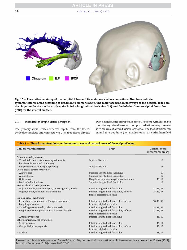
Fig. 10 e The cortical anatomy of the occipital lobes and its main associative connections. Numbers indicate
cytoarchitectonic areas according to Brodmann’s nomenclature. The major association pathways of the occipital lobes are
the cingulum for the medial surface, the inferior longitudinal fasciculus (ILF) and the inferior fronto-occipital fasciculus
(IFOF) for the ventral surface.
c o r t e x x x x ( 2 0 1 2 ) 1e2 614
9.1. Disorders of simple visual perception
The primary visual cortex receives inputs from the lateral
geniculate nucleus and connects via U-shaped fibres directly
Table 3 e Clinical manifestations, white matter tracts and cor
Clinical manifestations
Primary visual syndromes:
- Visual field deficits (scotome, quadranopia,
hemianopia, cerebral blindness)
Optic
- Simple hallucinations (phosphenes) Optic
Dorsal visual stream syndromes:
- Akinetopsia Super
- Alloaesthesia Super
- Optic ataxia Cingu
- Motion hallucinations Super
Ventral visual stream syndromes:
- Object agnosia, achromatopsia, prosopagnosia, alexia Inferi
- Object, colour, face, text hallucinations Inferi
fronto
Complex visual syndromes:
- Reduplicative phenomena (Capgras syndrome;
Fregoli syndrome)
Inferi
fronto
- Visual hypoemotionality, visual amnesia Inferi
- Fregoli syndrome, post-traumatic stress disorder Inferi
fronto
- Anton’s syndrome Inferi
Other neuropsychiatric syndromes:
- Schizophrenia Inferi
- Congenital prosopagnosia Inferi
fronto
- Autism Inferi
Please cite this article in press as: Catani M, et al., Beyond corticahttp://dx.doi.org/10.1016/j.cortex.2012.07.001
with neighbouring extrastriate cortex. Patients with lesions to
the primary visual area or the optic radiations may present
with an area of altered vision (scotoma). The loss of vision can
extend to a quadrant (i.e., quadranopia), an entire hemifield
tical areas of the occipital lobes.
Tract Cortical areas(Brodmann areas)
radiations 17
radiations 17
ior longitudinal fasciculus 19
ior longitudinal fasciculus 19
lum, superior longitudinal fasciculus 18, 19
ior longitudinal fasciculus 19
or longitudinal fasciculus 18, 19, 37
or longitudinal fasciculus, inferior
-occipital fasciculus
18, 19, 37
or longitudinal fasciculus, inferior
-occipital fasciculus
18, 19, 37
or longitudinal fasciculus 18, 19, 37
or longitudinal fasciculus, inferior
-occipital fasciculus
18, 19, 37
or longitudinal fasciculus 18, 19
or longitudinal fasciculus 18, 19
or longitudinal fasciculus, inferior
-occipital fasciculus
18, 19
or longitudinal fasciculus 18, 19
l localisation in clinico-anatomical correlation, Cortex (2012),
c o r t e x x x x ( 2 0 1 2 ) 1e2 6 15
(i.e., homonymous hemianopia) or to both hemifields
(i.e., complete cerebral blindness) if the lesion is bilateral
(Cavezian et al., 2010). Hallucinations linked to primary visual
cortex pathology are of simple featureless forms and colours
(i.e., phosphenes) (ffytche et al., 2010).
9.2. Disorders of the dorsal stream
The associative occipital cortex is composed of several regions
linked through multiple, parallel, cortico-cortical connections
divided into a dorsal and a ventral pathway stream. The dorsal
stream is related to spatial aspects of vision (the ‘where’
stream) (Ungerleider and Mishkin, 1982) including spatial
working memory, visually guided action and navigation
(Kravitz et al., 2011). Patients with dorsal stream lesions
present with selective loss of motion vision (i.e., akinetopsia).
In visual alloaesthesia the world is perceived in an incorrect
orientation, for example, inverted, tilted or right-left reversed.
Optic ataxia is a disturbance of limb guidance such that
subjects are unable to reach for objects in an otherwise intact
visual field. Increased activity in the dorsal occipital regions
can cause motion hallucinations.
9.3. Disorders of the ventral stream
The ventral stream is composed of associative interconnected
cortical areas specialised for colour, face, object, and letter
vision (the ‘what’ stream) (Ungerleider and Mishkin, 1982).
Patients with an impairment of the ventral cortex and its
connections present with selective loss of colour vision
(i.e., achromatopsia) (Zeki, 1990), face perception (i.e., proso-
pagnosia) (Fox et al., 2008; Ramon and Rossion, 2010; Tree and
Wilkie, 2010; Gruter et al., 2011; Tree, 2011), object perception
(i.e., visual object agnosia) (Catani et al., 2003, Germine et al.,
2011), and words (i.e., alexia) (Cohen et al., 2000). Similarly
colour, object, and text or letter-string hallucinations are each
linked to pathology causing hyperfunctioning of their
respective region of cortical specialisation. Face hallucina-
tions and illusions characterised by distorted facial features
(i.e., prosopometamorphopsia) are likely to relate to a region
specialised for face features on the lateral convexity of the
occipital lobe (i.e., occipital face area) while hallucinations of
normal faces or facial intermetamorphosis (a change in the
visually perceived identity of a face) are likely to relate to
activity within an area specialised for faces on the ventral
occipito-temporal surface (i.e., fusiform face area) (ffytche and
Howard, 1999).
9.4. Complex visual syndromes
In some patients the involvement of long association tracts
connecting the occipital lobe to more anterior regions lead to
complex visual syndromes. Reduplicative phenomena are
thought to relate to disconnection between visual, affective
andmemory regions due to lesions of the inferior longitudinal
fasciculus and inferior fronto-occipital fasciculus (ffytche
et al., 2010). In these disorders familiar people, places and
objects are perceived as duplicates that have replaced the real
person, place, or object. The disorder is termed Capgras
syndrome when involving a person. Similar disconnection
Please cite this article in press as: Catani M, et al., Beyond corticahttp://dx.doi.org/10.1016/j.cortex.2012.07.001
accounts are given of disorders manifesting with reduced
emotional tone to visual experience (i.e., visual hypo-
emotionality) (Bauer, 1982), impaired registering of visual
experiences in short termmemory (i.e., visual amnesia) (Ross,
1980, 2008) or feelings that experiences, typically visual, seem
strange or unreal (i.e., derealisation) (Sierra et al., 2002). The
Fregoli syndrome in which unfamiliar people are perceived as
familiar (typically as a person in disguise with malevolent
intent) can be interpreted as a hyperconnection between
visual, emotional andmemory networks. Similarly, the strong
affective and imagery components of flashbacks in post-
traumatic stress disorder suggest hyperconnection between
visual, emotional and memory regions (ffytche et al., 2010).
Patients with cortical blindness due to extensive occipital
lesions can sometime deny their visual impairment (Anton’s
syndrome) (Anton, 1889). Patients with Anton’s syndrome
may also report visual experiences, that traditionally have
been interpreted as confabulations (a false memory or false
report of visual perceptual experience) or as spontaneous
visual imagery. Anton’s syndrome can be conceptualised as
a disconnection of visual cortex from body schema repre-
sentations in the parietal lobe (ffytche et al., 2010).
9.5. Tractography studies in neurodevelopmental andneuropsychiatric disorders
Altered microstructural integrity (i.e., reduced fractional
anistotropy) of the inferior longitudinal fasciculus in adoles-
cents with schizophrenia has been reported, especially in
those subjects with a history of visual hallucinations (Ashtari
et al., 2007). In patients with congenital prosopagnosia, where
conventional structural imaging is usually normal, the frac-
tional anisotropy of the inferior longitudinal fasciculus and
inferior fronto-occipital fasciculus is reduced and this reduc-
tion correlates significantly with performances on face pro-
cessing tasks (Thomas et al., 2009). Conversely an increased
number of streamlines in the inferior longitudinal fasciculus
has been reported in subjects with high functioning autism
(Thomas et al., 2011) and Asperger syndrome (Pugliese et al.,
2009).
10. Temporal lobe
Themain divisions of the temporal lobe include the: (i) primary
auditory cortex (BA41); (ii) auditory association cortex (BA42,
22); (iii) visual association cortex (BA20, 21, 37); (iv) tempor-
opolar cortex (BA38) (Fig. 11). Lesions to each of the above
regions cause four distinct temporal lobe syndromes (Table 4).
10.1. Cortical deafness for sounds and words
The primary auditory cortex receives projections of the
acoustic radiations from both medial geniculate nuclei and
connects to adjacent areas through U-shaped fibres. Lesions
to the left primary auditory area impair the ability to recognise
words (i.e., ‘word deafness’ or sensory aphasia), while right-
sided lesions impair recognition of non-verbal sounds
(i.e., ‘sound deafness’ or acoustic agnosia) (Suarez et al., 2010).
l localisation in clinico-anatomical correlation, Cortex (2012),
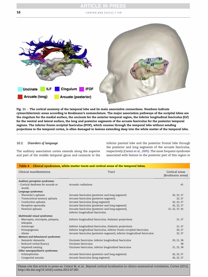
Fig. 11 e The cortical anatomy of the temporal lobe and its main associative connections. Numbers indicate
cytoarchitectonic areas according to Brodmann’s nomenclature. The major association pathways of the occipital lobes are
the cingulum for the medial surface, the uncinate for the anterior temporal region, the inferior longitudinal fasciculus (ILF)
for the ventral and lateral surface, the long and posterior segments of the arcuate fasciculus for the posterior temporal
regions. The inferior fronto-occipital fasciculus (IFOF), which courses through the temporal lobe without sending
projections to the temporal cortex, is often damaged in lesions extending deep into the white matter of the temporal lobe.
c o r t e x x x x ( 2 0 1 2 ) 1e2 616
10.2. Disorders of language
The auditory association cortex extends along the superior
and part of the middle temporal gyrus and connects to the
Table 4 e Clinical syndromes, white matter tracts and cortical
Clinical manifestations
Auditory perception syndromes:
- Cortical deafness for sounds or
words
Acoustic radiations
Language syndromes:
- Wernicke’s aphasia Arcuate fasciculus (posterior a
- Transcortical sensory aphasia Arcuate fasciculus (posterior s
- Conduction aphasia Arcuate fasciculus (long segme
- Receptive aprosodia Arcuate fasciculus (posterior a
- Nominal aphasia Arcuate fasciculus (posterior a
inferior longitudinal fasciculus
Multimodal visual syndromes:
- Macropsia, micropsia, pelopsia,
teleopsia
Inferior longitudinal fasciculus
- Autoscopy Inferior longitudinal fasciculus
- Prosopagnosia Inferior longitudinal fasciculus
- Alexia Arcuate fasciculus (posterior s
Memory and behavioural syndromes:
- Semantic dementia Uncinate fasciculus, inferior lo
- Reduced verbal fluency Uncinate fasciculus
- Impaired naming Uncinate fasciculus, inferior lo
Other neuropsychiatric syndromes:
- Schizophrenia Arcuate fasciculus (posterior a
- Congenital amusia Arcuate fasciculus (long segme
Please cite this article in press as: Catani M, et al., Beyond corticahttp://dx.doi.org/10.1016/j.cortex.2012.07.001
inferior parietal lobe and the posterior frontal lobe through
the posterior and long segments of the arcuate fasciculus,
respectively (Catani et al., 2005). The most frequent syndrome
associated with lesions to the posterior part of this region in
areas of the temporal lobes.
Tract Cortical areas(Brodmann areas)
41
nd long segment) 42, 22, 37
egment) 22, 37
nt) 42, 22, 37
nd long segment) 42, 22, 37
nd long segment), 21, 22, 37, 42
, thalamic projections 21, 37
, thalamic projections 21, 37
, inferior fronto-occipital fasciculus 20, 37
egment), inferior longitudinal fasciculus 22, 37
ngitudinal fasciculus 20, 21, 38
38
ngitudinal fasciculus 21, 22, 38
nd long segment) 42, 22, 37
nt) 42, 22, 37
l localisation in clinico-anatomical correlation, Cortex (2012),
c o r t e x x x x ( 2 0 1 2 ) 1e2 6 17
the left hemisphere is Wernicke’s aphasia, characterised by
impaired auditory comprehension and repetitionwith normal
verbal fluency (Hillis et al., 2002). Equivalent lesions in the
right hemisphere may cause impairment in understanding
emotional aspects of language (i.e., receptive aprosodia) (Ross,
1981) or music (Dellacherie et al., 2011; Gosselin et al., 2011).
The auditory association cortex is also connected to more
posterior visual occipital regions and more anterior temporal
areas through the inferior longitudinal fasciculus and U-sha-
ped fibres. Patients with lesions to the above connections may
show difficulty in using words to name objects (i.e., nominal
aphasia), or inability to repeat a series of words with normal
repetition of single words (i.e., auditory amnesic aphasia).
Auditory hallucinations are also frequently observed with
lesions or stimulation (e.g., intraoperative cortical stimula-
tion) of the auditory association cortex.
10.3. Disorders of multimodal visual processing
The visual association cortex is located in the middle and
inferior temporal gyrus and connects to the occipital lobes
through the inferior longitudinal fasciculus and the frontal
lobe through the long segment of the arcuate fasciculus and
the uncinate fasciculus. Its most posterior part is connected to
the inferior parietal through the posterior segment of the
arcuate fasciculus. Objects appearing larger (i.e., macropsia),
smaller (i.e., micropsia), nearer (i.e., pelopsia) or further
(i.e., teleopsia) have been attributed to a dysfunction of object
constancy within this posterior region of the temporal lobe.
Autoscopic phenomena describe a range of experiences in
which the self is duplicated in external space. In autoscopy,
visual perspective remains in the physical body (the duplicate
self is seen in the external world). In out-of-body experience
the physical body is seen from the perspective of the external
self (Blanke et al., 2002). In the autoscopy perspective changes
between the external self and physical self in rapid alterna-
tion. These phenomena are thought to relate to the disinte-
gration of visual, proprioceptive, tactile and vestibular
modalities and have been linked to transient dysfunction in
the region of the temporoparietal junction (ffytche et al., 2010).
Lesions to the ventral aspect of the temporo-occipital (fusi-
form) gyrus cause prosopagnosia (Fox et al., 2008), mainly for
lesions in the right hemisphere, and pure alexia (Epelbaum
et al., 2008; Starrfelt et al., 2010) or pathological orthographic
processing (Tsapkini and Rapp, 2010) in the left hemisphere.
10.4. Disorders of memory and behaviour
The temporopolar region morphologically belongs to the
temporal lobe but functionally is considered as part of the
limbic system. It is connected to more posterior temporal and
occipital regions through the inferior longitudinal fasciculus
and U-shaped fibres and to the frontal lobe via the uncinate
fasciculus. Semantic dementia is a memory disorder charac-
terised by a progressive inability to associate meaning to
sensorial perception (Laisney M, 2011). These patients show
marked atrophy of the anterior temporal lobe and its main
connections (Agosta et al., 2010). Lesions to the uncinate
fasciculus may also present with reduced verbal fluency and
naming deficits, especially for famous faces (Papagno et al.,
Please cite this article in press as: Catani M, et al., Beyond corticahttp://dx.doi.org/10.1016/j.cortex.2012.07.001
2011). Other complex syndromes associated with anterior
temporal dysfunction are discussed in the section on limbic
disorders.
10.5. Tractography studies in neurodevelopmental andneuropsychiatric disorders
In schizophrenia altered microstructural integrity of the
arcuate segments originating or projecting to the posterior
temporal auditory regions have been reported using diffusion
tensor tractography (Jones et al., 2005). These findings are
bilateral and more significant in patients with auditory
hallucinations (Catani et al., 2011). In subjects with tone-
deafness (i.e., congenital amusia) a reduced fractional
anisotropy and volume of the right long segment of the
arcuate fasciculus has been reported using diffusion tensor
imaging tractography (Loui et al., 2009). Disorders of the
anterior temporal lobe are reviewed in the next section.
11. Limbic lobe
The limbic system includes a core subcortical network (cen-
tred around the hippocampus and thalamus) formed by the
fornix and mammillo-thalamic tract, and a group of para-
limbic cortical areas connected through the cingulum and
uncinate fasciculus (Fig. 12). Other tracts, such as the inferior
longitudinal fasciculus and inferior fronto-occipital fasciculus
connect the limbic regions to visual and auditory areas. The
limbic lobe syndromes are divided into three distinct groups
(Table 5).
11.1. Disorders of the hippocampal-hypothalamicdivision
Lesions to the hippocampal-thalamic centred division of the
limbic system cause severe amnesia for events before (retro-
grade) and after (anterograde) the onset of the lesion
(Markowitsch, 2000). Retrograde amnesia is more severe for
recent than remote events, while anterograde amnesia affects
explicit learning of new events but leaves implicit procedural
memory for motor and perceptual tasks intact. Amnesia is
more often associated to other symptoms. Degeneration of
themedial temporal lobe structures is a feature of Alzheimer’s
disease, a form of dementia where memory deficits are
accompanied by at least two other cognitive problems (e.g.
language deficits, apraxia, etc.). Korsakoff disease is a severe
amnesia with confabulation commonly due to alcoholic
degeneration of the mammillo-thalamic tracts (Markowitsch,
2000). Some patients with amnesia may also present difficul-
ties in spatial orientation due to the inability to derive direc-
tional information from landmark cues in familiar and new
environments (Aguirre and D’Esposito, 1999). This form of
spatial memory dysfunction (i.e. topographical amnesia) can
also present as an isolated problem and is often associated
with stroke lesions to the posterior parahippocampal, retro-
splenial cingulate cortex and posterior precuneus (Vann et al.,
2009, Valenstein et al., 1987).
l localisation in clinico-anatomical correlation, Cortex (2012),
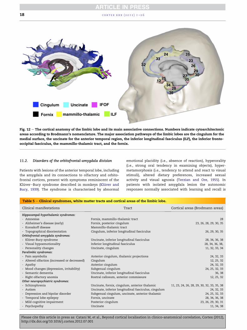
Fig. 12 e The cortical anatomy of the limbic lobe and its main associative connections. Numbers indicate cytoarchitectonic
areas according to Brodmann’s nomenclature. The major association pathways of the limbic lobes are the cingulum for the
medial surface, the uncinate for the anterior temporal region, the inferior longitudinal fasciculus (ILF), the inferior fronto-
occipital fasciculus, the mammillo-thalamic tract, and the fornix.
c o r t e x x x x ( 2 0 1 2 ) 1e2 618
11.2. Disorders of the orbitofrontal-amygdala division
Patients with lesions of the anterior temporal lobe, including
the amygdala and its connections to olfactory and orbito-
frontal cortices, present with symptoms reminiscent of the
KluvereBucy syndrome described in monkeys (Kluver and
Bucy, 1939). The syndrome is characterised by abnormal
Table 5 e Clinical syndromes, white matter tracts and cortical
Clinical manifestations T
Hippocampal-hypothalamic syndromes:
- Amnesias Fornix, mammillo-thala
- Alzheimer’s disease (early) Fornix, posterior cingulu
- Korsakoff disease Mammillo-thalamic trac
- Topographical disorientation Cingulum, inferior longi
Orbitofrontal-amygdala syndromes:
- Kluver-Bucy syndrome Uncinate, inferior longit
- Visual hypoemotionality Inferior longitudinal fasc
- Personality changes Uncinate, cingulum
Paralimbic syndromes:
- Pain asymbolia Anterior cingulum, thala
- Altered olfaction (increased or decreased) Cingulum
- Apathy Anterior cingulum
- Mood changes (depression, irritability) Subgenual cingulum
- Semantic dementia Uncinate, inferior longit
- Right olfactory anomia Rostral callosum, anterio
Other neuropsychiatric syndromes:
- Schizophrenia Uncinate, fornix, cingulu
- Autism Uncinate, inferior longit
- Depression and bipolar disorder Subgenual cingulum, un
- Temporal lobe epilepsy Fornix, uncinate
- Mild cognitive impairment Posterior cingulum
- Psychopathy Uncinate
Please cite this article in press as: Catani M, et al., Beyond corticahttp://dx.doi.org/10.1016/j.cortex.2012.07.001
emotional placidity (i.e., absence of reaction), hyperorality
(i.e., strong oral tendency in examining objects), hyper-
metamorphosis (i.e., tendency to attend and react to visual
stimuli), altered dietary preferences, increased sexual
activity and visual agnosia (Terzian and Ore, 1955). In
patients with isolated amygdala lesion the autonomic
responses normally associated with learning and recall is
areas of the limbic lobe.
ract Cortical areas (Brodmann areas)
mic tract 28
m 23, 26, 28, 29, 30, 35
t
tudinal fasciculus 26, 29, 30, 35
udinal fasciculus 28, 34, 36, 38
iculus 28, 34, 36, 38,
11, 32, 33, 34
mic projections 24, 32, 33
12, 25, 32
24, 32, 33
24, 25, 32, 33
udinal fasciculus 36, 38
r commissure 12, 25, 32
m, anterior thalamic 11, 23, 24, 26, 28, 29, 30, 32, 33, 35, 38
udinal fasciculus, cingulum 24, 32, 33
cinate, anterior thalamic 24, 25, 32, 33
28, 34, 36, 38
23, 26, 29, 30, 31
11, 34, 38
l localisation in clinico-anatomical correlation, Cortex (2012),
c o r t e x x x x ( 2 0 1 2 ) 1e2 6 19
abolished. Lesions confined to the amygdalae are extremely
rare in humans and result in mildly impaired modulation of
social behaviour and abnormalities in the visual examination
of faces (Adolphs et al., 1994). In patients with temporolimbic
epilepsy a wide range of acute and chronic psychiatric
symptoms have been observed, from panic attacks to
aggressive outbursts, depression, and psychosis (Waxman
and Geschwind, 1974; Flugel et al., 2006). Personality
changes are often associated with abnormalities in the
orbitofrontal cortex, medial anterior cingulate, amygdala and
their reciprocal connections through the uncinate fasciculus,
corpus callosum and anterior commissure (Zappala’ et al.,
2012; Berlucchi, 2012).
11.3. Disorders of the paralimbic areas
The paralimbic areas include the temporopolar region, insula,
cingulate gyrus, orbitofrontal cortex, and parahippocampal
gyrus. Lesions to paralimbic regions may present with
emotional indifference to pain (i.e., pain asymbolia), altered
olfaction, impaired ability to express emotions, reduced
attention and motivation (Sprengelmeyer et al., 2010; Reker
et al., 2010). Severe apathy and personality changes are
frequently associated with orbitofrontal and anterior cingu-
late pathology (Shamay-Tsoory et al., 2010). Degeneration of
the anterior temporal and posterior orbitofrontal cortex is
frequently observed in patients with semantic dementia,
alteration in personality and behaviour, and mood changes.
The anterior temporal and orbitofrontal cortex are commonly
affected in traumatic brain injury (Zappala’ et al., 2012).
Stimulation of the cingulate and parahippocampal cortices
causes memory flashbacks, dreamlike states, and mood
alterations. Obsessive-compulsive disorder has been associ-
ated with increased activity in the cingulate cortex causing
‘rigid hyperattentiveness’ to thoughts and emotions.
Reduced olfactory sensation is commonly observed in
patients with Alzheimer’s disease and Parkinson’s disease.
Anomia for olfactory stimuli presented to the right nostril
(right olfactory anomia) (Gordon and Sperry, 1969) and
memory deficits (Zaidel and Sperry, 1974) have been described
in split-brain patients with complete commissurotomy
(complete severing of the corpus callosum and anterior and
posterior commissure) but not in those with intact anterior
commissure (Ledoux et al., 1977; Risse et al., 1978). The right
olfactory anomia in these patients is due to a disconnection
between the right olfactory cortex and the left fronto-
temporal language areas.
11.4. Tractography studies in neurodevelopmental andneuropsychiatric disorders
Limbic dysfunction underlie many symptoms of other
psychiatric conditions, including schizophrenia, affective
disorders, psychopathy and autism. Voxel-based and trac-
tography diffusion tensor imaging approaches have been used
to examine the integrity of limbic whitematter connections in
schizophenia, with microstructural differences reported in
the cingulum (Fujiwara et al., 2007), uncinate fasciculus
(McIntosh et al., 2008), fornix (Kuroki et al., 2006, Takei et al.,
2008), and the anterior thalamic radiations (McIntosh et al.,
Please cite this article in press as: Catani M, et al., Beyond corticahttp://dx.doi.org/10.1016/j.cortex.2012.07.001
2008). A recent tractography study in adolescents with major
depressive disorder reported lower fractional anisotropy in
the white matter tract connecting the subgenual cingulate
region to the amygdala in the right hemisphere (Cullen et al.,
2010). In adults with bipolar affective disorder tractography
showed increased tract volume of the subgenual-amygdala
connections (i.e., subpopulation of uncinate fibres) (Houenou
et al., 2007) and reduced fractional anisotropy in the unci-
nate and anterior thalamic projections (McIntosh et al., 2008).
Diffusion imaging tractography studies in patients with
unilateral temporal lobe epilepsy reported diffuse damage to
the limbic white matter tracts such as the fornix (Concha
et al., 2005) and the uncinate fasciculus (Diehl et al., 2008)
often extending contralaterally from the side of the suspected
seizure. In temporal lobe epilepsy patients with mesial
hippocampal sclerosis, decrease in fractional anisotropy of
the fornix fibres is probably due to reduced axonal diameter
and myelin content (Concha et al., 2010). Such changes have
an impact on cognitive abilities as demonstrated by the
correlation between the diffusivity measures of the uncinate
fasciculus and the severity of delayed recall deficits (Diehl
et al., 2008). Preliminary data also suggest, that in patients
with temporal lobe epilepsy undergoing surgery, the pre-
operative tractography assessment of the temporal tracts
can help in predicting naming deficits after the operation (i.e.,
patients with more leftward asymmetry showed worse post-
operative deficits) (Powell et al., 2008). Correlations between
fractional anisotropy levels and verbal fluency performances
before and after anterior temporal resection have also been
reported (Yogarajah et al., 2010).
In patients with mild cognitive impairment a combined
cortical morphometry and diffusion tensor imaging study
found reduced cortical thickness and white matter abnor-
malities of the retrosplenial regions (Acosta-Cabronero et al.,
2010). Craig et al. (2009) reported a significantly reduced frac-
tional anisotropy in the uncinate fasciculus of psychopaths
compared to healthy subjects with similar age and intelli-
gence. A correlation between measures of antisocial behav-
iour and anatomical differences in the uncinate fasciculus
was also described. A tractography study showed that
compared to healthy controls, adults with Asperger’s
syndrome had a significantly higher number of streamlines in
the cingulum bilaterally and a lower number of streamlines in
the right uncinate (Pugliese et al., 2009). Diffusivity changes in
the inferior longitudinal fasciculus have been reported in
autistic subjects with impaired ability to recognise faces
(Conturo et al., 2008).
12. Discussion and conclusions
In this study we provide a new digital atlas of white matter
tracts based on diffusion tensor imaging tractography to facil-
itate the anatomical localisation of deep white matter lesions.
The atlas is accompanied by a synopsis of the major lobe
syndromes tohelp correlatingwhitematter lesionswith clinical
manifestations. The atlas has threemain features: (i) it provides
normalisedmaps in a reference space (i.e., MNI); (ii) it is derived
from a statistical analysis at a group level and, therefore, it is
l localisation in clinico-anatomical correlation, Cortex (2012),
c o r t e x x x x ( 2 0 1 2 ) 1e2 620
representative of the average anatomy; (iii) the maps of the
single tracts are provided in a digital format (i.e., nifti).
The atlas reference space (MNI) will facilitate the overlay of
the tracts onto normalised neuroradiological images. Some of
the white matter tracts reported in the atlas have been
described only in recent years and are not available in previ-
ously published histological or tractography atlases (e.g., the
three segments of the arcuate fasciculus). For other tracts,
such as the corpus callosum, uncinate and cingulum, the atlas
shows anatomical features that are close to classic post-
mortem blunt dissections and post-mortem histology
(Thiebaut de Schotten et al., 2011a).
Our presentation of the white matter tracts as a single
composite map may help explain findings that in clinico-
anatomical correlation studies may sometimes appear incon-
gruent. For example, it has been reported that hemianopia,
a symptom commonly associated with lesions to the optic
radiations, occur in patients with lesions of the parietal lobe
(Critchley, 1953). Although inputs from the visual thalamus
(i.e., pulvinar) reach the parietal cortex whose damage has
been suggested as a possible mechanism for the visual deficits
in these patients, in our opinion a more likely explanation
would be an involvement of the optic radiations that run
deeply in the white matter of the parietal lobe, which can be
easily appreciated on the coronal slices of our atlas (e.g., slice
�40 of Fig. 6 shows in orange the optic radiations deep in the
white matter of the parietal lobe). Lesions to the parietal lobe
extending deep into the white matter could therefore affect
the optic radiations and cause hemianopia. Furthermore
a network approach in clinico-anatomical correlation studies
offers the advantage of explaining how symptoms classically
attributed to damage of a cortical area (e.g., non fluent aphasia
with lesions to Broca’s area) canmanifest also in patients with
lesions outside that area. For example, empirical evidence
suggests that in 15% of patientswith Broca’s aphasia the lesion
is outside Broca’s area, often encompassing post-Rolandic
parietal regions (Basso et al., 2000) or deep white matter
tracts (Bizzi et al., 2012). This is the case of the patient with
brain tumour presented in Fig. 7A, where the atlas suggests an
involvement of the anterior and long segment of the arcuate
fasciculus without direct damage to Broca’s area. In this case
an alternative hodological explanation to the classical cortical
localisation approach suggests that dysfunction of Broca’s area
can result from lesions of its connections with posterior pari-
etal and temporal language regions.
We realise, of course, that mapping symptoms onto single
tracts is subject to the same criticisms directed to narrow
cortical localizationism and that often complex syndromes
clearly seem to result from a dysfunction of an extended
network of cortical and subcortical areas connected by several
tracts. Unilateral neglect, for example, has been described in
association with lesions of the superior longitudinal fascic-
ulus, inferior longitudinal fasciculus, inferior fronto-occipital
fasciculus and the overlying parietal, temporal, occipital and
frontal cortex (Doricchi et al., 2008). Hence, a hodological
approach to clinico-anatomical correlation should not
underestimate the role of the cortex and it should certainly
refrain from considering the effects of cortical lesions as
equivalent to those of lesions to subcortical tracts. Further-
more different mechanisms act for cortical and subcortical
Please cite this article in press as: Catani M, et al., Beyond corticahttp://dx.doi.org/10.1016/j.cortex.2012.07.001
lesions, which may influence the severity of symptoms and
their recovery. The clinical manifestations commonly seen in
patients with multiple sclerosis are a clear example. Here,
even with extensive subcortical lesions, the observation of
classical syndromes commonly seen with other pathologies
(e.g., Wernicke’s aphasia in stroke patients) is extremely rare.
For some tracts (e.g., inferior occipito-frontal fasciculus and
arcuate fasciculus) the atlas has an advantage over histological
approaches by providing more accurate anatomical recon-
structions (Thiebaut de Schotten et al., 2011a). Thismay reflect
the difficulty of using post-mortem myelin staining methods
to trace tracts with a longitudinal course (Burgel et al., 2006). In
contrast, compared to histological maps, the atlas presents
only a partial reconstruction of the lateral projections of the
cortico-spinal tract and the terminal projections ofmost of the
tracts. This is most likely due to the limitations of current
diffusion tensor model in resolving fibre crossing. Future
in vivo studies of tracts such as these may be aided by other
diffusion methods such as diffusion spectrum imaging (DSI)
(Schmahmann et al., 2007) or spherical deconvolution
(Tournier et al., 2004, 2008; Dell’Acqua et al., 2007, 2010, 2012;
Dell’Acqua and Catani, 2012; Catani, Bode and Dell’Acqua, in
press) that can, in part, overcome the limitations of the tensor
model. This is also the reason for the absence of the first and
second branch of the superior longitudinal fasciculus (Catani
et al., in press) and many short U-shaped fibres that are often
difficult to dissect reliably with DTI tractography (Catani et al.,
2012).
An atlas-based approach can be problematic in disorders
where the presence of oedema or mass effect alters the
anatomy of the tracts (Clark et al., 2003). In these cases find-
ings from the atlas could be difficult to interpret or could
sometimes bemisleading. Finally, our atlas is based on people
from a relatively young and narrow age range, and therefore
the results of our study may not be representative of white
matter anatomy across the lifespan. Future atases will need to
take into account age-related changes in diffusion measure-
ments (Verhoeven et al., 2010, Lebel et al., 2008, 2010) and how
these changes affect the results of fibre tracking.
In conclusion after 150 years the clinical-anatomical
correlation method still occupies a prominent role in
contemporary neuroscience. Anatomical atlases have always
played an important part in the process of correlating lesions
to symptoms and understanding the neuroanatomical basis of
neurological and psychiatric disorders. We hope that our new
atlas will help to capture the excitement of 21st century
anatomy and will help clinicians to understand the role of
many tracts whose functions remain unknown empirical
contributions to come.
Acknowledgements
This project was generously supported by Guy’s and St
Thomas’ Charity (GSST), The Wellcome Trust, the Medical
Research Council, UK Autism Multi-Centre Imaging Study
network (AIMS), the French Agence Nationale de la Recherche
(project CAFORPFC, no. ANR-09-RPDOC-004-01 and project
HM-TC, no. ANR-09-EMER-006) and the National Division of
l localisation in clinico-anatomical correlation, Cortex (2012),
c o r t e x x x x ( 2 0 1 2 ) 1e2 6 21
the South London and Maudsley NHS Foundation Trust.
AS, SW, DM and Fd’A were supported by the NIHR Biomedical
Research Centre for Mental Health at South London and
Maudsley NHS Foundation Trust and Institute of Psychiatry,
King’s College London. AS was also supported by the NIHR
Biomedical Research Unit for Dementia and Alzheimer’s
Research UK.
Appendix A. Supplementary material
Supplementary data related to this article can be found online
at http://dx.doi.org/10.1016/j.cortex.2012.07.001.
r e f e r e n c e s
Acosta-Cabronero J, Williams GB, Pengas G, and Nestor PJ.Absolute diffusivities define the landscape of white matterdegeneration in Alzheimer’s disease. Brain, 133(2): 529e539,2010.
Adolphs R, Tranel D, Damasio H, and Damasion A. Impairedrecognition of emotion in facial expressions followingbilateral damage to the human amydgala. Nature, 372(6507):669e672, 1994.
Agosta F, Henry RG, Migliaccio R, Neuhaus J, Miller BL,Dronkers NF, et al. Language networks in semantic dementia.Brain, 133(1): 286e299, 2010.
Aguirre GK and D’Esposito M. Topographical disorientation: Asynthesis and taxonomy. Brain, 122(9): 1613e1628, 1999.
Alexander AL, Lee JE, Lazar M, Boudos R, DuBray MB,Oakes TR, et al. Diffusion tensor imaging of the corpuscallosum in autism. NeuroImage, 34(1): 61e73, 2007.
Alexander MP, Baker E, Naeser MA, Kaplan E, and Palumbo C.Neuropsychological and neuroanatomical dimensions ofideomotor apraxia. Brain, 115(1): 87e107, 1992.
Anderson EJ, Jones DK, O’Gorman RL, Leemans A, Catani M, andHusain M. Cortical network for gaze control in humansrevealed using multimodal MRI. Cerebral Cortex, 22(4): 765e775,2012.
Anton G. Ueber die Selbstwahrnehmung der Herderkrankungendes Gehirns durch den Kranken bei Rindenblindheit undRindentaubheit. Archiv fur Psychiatrie, 32: 86e127, 1889.
Ashtari M, Cottone J, Ardekani BA, Cervellione K, Szeszko PR,Wu J, et al. Disruption of white matter integrity in the inferiorlongitudinal fasciculus in adolescents with schizophrenia asrevealed by fiber tractography. Archives of General Psychiatry,64(11): 1270e1280, 2007.
Auburtin SAE. Considerations sur les localisations cerebrales et enparticulier sur le siege de la faculte du language articule. Paris:Masson, 1863.
Auburtin SAE. Reprise de la discussion sur la forme et le volumedu cerveau. Bulletins de la Societe d’Anthropologie de Paris, 2:209e220, 1861.
Baddeley AD. Working memory, thought and action. Oxford: OxfordUniversity Press, 2007.
Bartolomeo P. The quest for the ‘critical lesion site’ in cognitivedeficits: Problems and perspectives. Cortex, 47(8): 1010e1012,2011.
Basser PJ, Pajevic S, Pierpaoli C, Duda J, and Aldroubi A. In vivofiber tractography using DT-MRI data. Magnetic Resonance inMedicine, 44(4): 625e632, 2000.
Bassetti C, Bogousslavsky J, and Regli F. Sensory syndromes inparietal stroke. Neurology, 43(10): 1942e1949, 1993.
Please cite this article in press as: Catani M, et al., Beyond corticahttp://dx.doi.org/10.1016/j.cortex.2012.07.001
Basso A, Burgio F, and Caporali A. Acalculia, aphasia and spatialdisorders in left and right brain-damaged patients. Cortex,36(2): 265e280, 2000.
Bauer RM. Visual hypoemotionality as a symptom of visual-limbic disconnection in man. Archives of Neurology, 39(11):702e708, 1982.
Berlucchi G. Frontal callosal disconnection syndromes. Cortex,48(1): 35e44, 2012.
Berthier ML, Lambon Ralph MA, Pujol J, and Green C. Arcuatefasciculus variability and repetition: The left sometimes canbe right. Cortex, 48(2): 133e143, 2012.
Benson DF and Geschwind N. Developmental Gerstmannsyndrome. Neurology, 20(3): 293e298, 1970.
Bizzi A, Nava S, Ferre‘ F, Castelli G, Aquino D, Ciaraffa F, et al.Gliomas infiltrating the ventrolateral frontal region: Clinicalanatomo-functional language assessment withneuropsychology, diffusion MR tractography and functionalMR imaging. Cortex, 48(2): 255e272, 2012.
Blanke O, Ortigue S, Landis T, and Seeck M. Stimulatingillusory own-body perceptions. Nature, 419(6904): 269e270,2002.
Broca P. Perte de la parole, ramollissement chronique etdestruction partielle du lobe anterieur gauche du cerveau.Bulletins de la Societe d’Anthropologie de Paris, 2. 235e8 and301e321 1861.
Brodmann K. Vergleichende Lokalisationslehre der Großhirnrinde: inihren Prinzipien. Leipzig: JA Barth, 1909.
Budisavljevic S and Ramnani N. Cognitive deficits froma cerebellar tumour: A historical case report from Luria’slaboratory. Cortex, 48(1): 26e34, 2012.
Bultitude JH and Rafal RD. Amelioration of right spatial neglectafter visuo-motor adaptation to leftward-shifting prisms.Cortex, 46(3): 404e406, 2010.
Burdach K. Vom Baue und Leben des Gehirns. Leipzig: In derDyk’schen Buchhandlung, 1822.
Burgel U, Amunts K, Hoemke L, Mohlberg H, Gilsbach JM, andZilles K. White matter fiber tracts of the human brain: three-dimensional mapping at microscopic resolution, topographyand intersubject variability. NeuroImage, 29(4): 1092e1105,2006.
Campbell AW. Histological studies on the localisation of cerebralfunction. Cambridge: Cambridge University Press, 1905.
Capruso DX and Hamsher KdS. Constructional ability in two-versus three-dimensions: Relationship to spatial vision andlocus of cerebrovascular lesion. Cortex, 47(6): 698e705, 2011.
Catani M, Howard RJ, Pajevic S, and Jones DK. Virtual in vivointeractive dissection of white matter fasciculi in the humanbrain. NeuroImage, 17(1): 77e94, 2002.
Catani M, Jones DK, Donato R, and ffytche DH. Occipito-temporalconnections in the human brain. Brain, 126(9): 2093e2107,2003.
Catani M, Jones DK, and ffytche DH. Perisylvian languagenetworks of the human brain. Annals of Neurology, 57(1): 8e16,2005.
Catani M and ffytche D. The rise and fall of disconnectionsyndromes. Brain, 128: 2224e2239, 2005.
Catani M. Diffusion tensor magnetic resonance imagingtractography in cognitive disorders. Current Opinion InNeurology, 19(6): 599e606, 2006.
Catani M and Mesulam M. The arcuate fasciculus and thedisconnection theme in language and aphasia: History andcurrent state. Cortex, 44(8): 953e961, 2008a.
Catani M and Mesulam MM. What is a disconnection syndrome?Cortex, 44(8): 911e913, 2008b.
Catani M and ffytche DH. On ’the study of the nervous systemand behaviour’. Cortex, 46(1): 106e109, 2010.
Catani M, Forkel S, and Thiebaut de Schotten M. Asymmetry ofhuman brain pathways. In Hugdahl. Kenneth (Ed), The Two
l localisation in clinico-anatomical correlation, Cortex (2012),
c o r t e x x x x ( 2 0 1 2 ) 1e2 622
Halves of the Brain: Information Processing in the CerebralHemispheres. Cambridge: MIT Press, 2010.
Catani M. John Hughlings Jackson and the clinico-anatomicalcorrelation method. Cortex, 47: 905e907, 2011.
Catani M, Dell’Acqua F, Vergani F, Malik F, Hodge H, Roy P, et al.Short frontal lobe connections of the human brain. Cortex, 48:273e291, 2012.
Catani M, Craig MC, Forkel SJ, Kanaan R, Picchioni M,Toulopoulou T, et al. Altered integrity of perisylvian languagepathways in schizophrenia: Relationship to auditoryhallucinations. Biological Psychiatry, 70(12): 1143e1150, 2011.
Catani M, Bode I, and Dell’Acqua F. Comment on “The Geometricstructure of the brain fiber pathways”. Science, in press.
Cavezian C, Gaudry I, Perez C, Coubard O, Doucet G, Peyrin C,et al. Specific impairments in visual processing followinglesion side in hemianopic patients. Cortex, 46(9): 1123e1131,2010.
Charlton RA, Barrick TR, Lawes INC, Markus HS, Robin G, andMorris RG. White matter pathways associated with workingmemory in normal aging. Cortex, 46(4): 474e489, 2010.
Clark CA, Barrick TR, Murphy MM, and Bell BA. White matter fibertracking in patients with space-occupying lesions of the brain:A new technique for neurosurgical planning? NeuroImage,20(3): 1601e1608, 2003.
Cohen L, Dehaene S, Naccache L, Lehericy S, Dehaene-Lambertz G, Henaff MA, et al. The visual word form area:Spatial and temporal characterization of an initial stage ofreading in normal subjects and posterior split-brain patients.Brain, 123(2): 291e307, 2000.
Concha L, Beaulieu C, and Gross DW. Bilateral limbic diffusionabnormalities in unilateral temporal lobe epilepsy. Annals ofNeurology, 57: 188e196, 2005.
Concha L, Livy DJ, Beaulieu C, Wheatley BM, and Gross DW.In vivo diffusion tensor imaging and histopathology of thefimbria-fornix in temporal lobe epilepsy. Journal ofNeuroscience, 30(3): 996e1002, 2010.
Conturo TE, Williams DL, Smith CD, Gultepe E, Akbudak E, andMinshew NJ. Neuronal fiber pathway abnormalities in autism:An initial MRI diffusion tensor tracking study of hippocampo-fusiform and amygdalo-fusiform pathways. Journal of theInternational Neuropsychological Society, 14(6): 933e946, 2008.
Craig MC, Catani M, Deeley Q, Latham R, Daly E, Kanaan R, et al.Altered connections on the road to psychopathy. MolecularPsychiatry, 14(10): 946e953, 2009.
Critchley M. The Parietal Lobes. London: Edward Arnold, 1953.Cubelli R, Beschin N, and Della Sala S. Ipsilateral neglect for non-
verbal stimuli following left brain damage. Cortex, 47(2):146e147, 2011.
Cubelli R and De Bastiani P. 150 years after Leborgne: Why is PaulBroca so important in the history of neuropsychology? Cortex,47(7): 899e901, 2011.
Cullen KR, Klimes-Dougan B, Muetzel R, Mueller BA, Camchong J,Houri A, et al. Altered white matter microstructure inadolescents with major depression: A preliminary study.Journal of the American Academy of Child and AdolescentPsychiatry, 49(2): 173e183, 2010.
Cummings JL and Mega MS. Neuropsychiatry and BehavioralNeuroscience. New York: Oxford University Press, 2003.
Damasio H and Damasio A. Lesion Analysis in Neuropsychology.New York: Oxford University Press, 1989.
Darby D and Walsh K. Walsh’s Neuropsychology. 5 ed. Amsterdam:Elsevier, 2005.
Dejerine J. Anatomie des Centres Nerveux. Paris: Rueff et Cie, 1895.Dejerine J. Contribution a l’etude anatomo-pathologique et
clinique des differentes varietes de cecite-verbale. Memoires dela Societe de Biologie, 44: 61e90, 1892.
Dellacherie D, Bigand E, Molin P, Baulac M, and Samson S.Multidimensional scaling of emotional responses to music in
Please cite this article in press as: Catani M, et al., Beyond corticahttp://dx.doi.org/10.1016/j.cortex.2012.07.001
patients with temporal lobe resection. Cortex, 47(9):1107e1115, 2011.
Dell’Acqua F, Rizzo G, Scifo P, Clarke RA, Scotti G, and Fazio F. Amodel-based deconvolution approach to solve fiber crossingin diffusion-weighted MR imaging. IEEE Transactions on Bio-medical Engineering, 54(3): 462e472, 2007.
Dell’Acqua F, Scifo P, Rizzo G, Catani M, Simmons A, Scotti G,et al. A modified damped Richardson-Lucy algorithm toreduce isotropic background effects in sphericaldeconvolution. NeuroImage, 49(2): 1446e1458, 2010.
Dell’Acqua F, Simmons A, Williams SC, and Catani M. Canspherical deconvolution provide more information than fiberorientations? Hindrance modulated orientational anisotropy,a true-tract specific index to characterize white matterdiffusion. Human Brain Mapping, http://dx.doi.org/10.1002/hbm.22080 [Epub ahead of print] 2012.
Dell’Acqua F and Catani M. Structural human brain networks: Hottopics in diffusion tractography. Current Opinion Neurology,25(4): 375e383, 2012.
Diehl B, Busch RM, Duncan JS, Piao Z, Tkach J, and Luders HO.Abnormalities in diffusion tensor imaging of the uncinatefasciculus relate to reduced memory in temporal lobeepilepsy. Epilepsia, 49(8): 1409e1418, 2008.
Doricchi F, Thiebaut de Schotten M, Tomaiuolo F, andBartolomeo P. White matter (dis)connections and gray matter(dys)functions in visual neglect: Gaining insights into thebrain networks of spatial awareness. Cortex, 44(8): 983e995,2008.
Douaud G, Mackay C, Andersson J, James S, Quested D, Ray MK,et al. Schizophrenia delays and alters maturation of the brainin adolescence. Brain, 132(9): 2437e2448, 2009.
Dunn TD. Double hemiplegia with double hemianopsia and lossof geographical center. Transactions of the College of Physicians ofPhiladelphia, 17: 45e55, 1895.
Epelbaum S, Pinel P, Gaillard R, Delmaire C, Perrin M, Dupont S,et al. Pure alexia as a disconnection syndrome: New diffusionimaging evidence for an old concept. Cortex, 44(8): 962e974,2008.
Exner S. Untersuchungen uber die Localization der Functionen in derGrosshirnrinde des Menschen. Wien: Wilhelm Braumuller, 1881.
FCAT Terminologia anatomica: international anatomical classification.New York: Thieme Medical Publisher, 2000.
Ferrier D. Experiments on the brain of monkeys.-No. I. Proceedingsof the Royal Society of London, Series B, 23: 409e432, 1875a.
Ferrier D. The Croonian Lecture: Experiments on the brain ofmonkeys (second series). Philosophical Transactions of the RoyalSociety, 165: 433e488, 1875b.
ffytche DH and Catani M. Beyond localization: From hodology tofunction. Philosophical Transactions of the Royal Society B,360(1456): 767e779, 2005.
ffytche DH, Blom JD, and Catani M. Disorders of visual perception.Journal of Neurology, Neurosurgery & Psychiatry, 81(11):1280e1287, 2010.
ffytche DH and Howard RJ. The perceptual consequences of visualloss: ’positive’ pathologies of vision. Brain, 122(7): 1247e1260,1999.
Flechsig P. Developmental (myelogenetic) localization of thecerebral cortex in the human subject. Lancet, 158(4077):1027e1030, 1901.
Fletcher PT, Whitaker RT, Tao R, DuBray MB, Froehlich A,Ravichandran C, et al. Microstructural connectivity of thearcuate fasciculus in adolescents with high-functioningautism. NeuroImage, 51(3): 1117e1125, 2010.
Flugel D, Cercignani M, Symms MR, O’Toole A, Thompson PJ,Koepp MJ, et al. Diffusion tensor imaging findings and theircorrelation with neuropsychological deficits in patients withtemporal lobe epilepsy and interictal psychosis. Epilepsia,47(5): 941e944, 2006.
l localisation in clinico-anatomical correlation, Cortex (2012),
c o r t e x x x x ( 2 0 1 2 ) 1e2 6 23
Fox C, Iaria G, and Barton J. Disconnection in prosopagnosia andface processing. Cortex, 44(8): 996e1009, 2008.
Frassinetti F, Maini M, Benassi M, Avanzi S, Cantagallo A, andFarne A. Selective impairment of self body-parts processing inright brain-damaged patients. Cortex, 46(3): 322e328, 2010.
Friston KJ, Harrison L, and Penny W. Dynamic causal modelling.NeuroImage, 19(4): 1273e1302, 2003.
Fritsch G and Hitzig E. Uber die elektrische Erregbarkeit desGrosshirns. Archivfuer Anatomie. Physiologie undwissenschaftliche Medicin, 37: 300e332, 1870.
Fujiwara H, Hirao K, Namiki C, Yamada M, Shimizu M,Fukuyama H, et al. Anterior cingulate pathology and socialcognition in schizophrenia: A study of gray matter, whitematter and sulcal morphometry. NeuroImage, 36(4): 1236e1245,2007.
Gall FJ and Spurzheim JC. Anatomie et physiologie du systemenerveux en general, et du cerveau en particulier. Paris: Schoell,1810e19.
Germine L, Cashdollar N, Duzel E, and Duchaine B. A newselective developmental deficit: Impaired object recognitionwith normal face recognition. Cortex, 47(5): 598e607, 2011.
Geschwind N. Disconnexion syndromes in animals and man. I.Brain, 88(2): 237e294, 1965a.
Geschwind N. Disconnexion syndromes in animals and man. II.Brain, 88(3): 585e644, 1965b.
Glickstein M and Berlucchi G. Classical disconnection studies ofthe corpus callosum. Cortex, 44(8): 914e927, 2008.
Gordon HW and Sperry RW. Lateralization of olfactory perceptionin the surgically separated hemispheres of man.Neuropsychologia, 7(2): 111e120, 1969.
Gosselin N, Peretz I, Hasboun D, Baulac M, and Samson S.Impaired recognition of musical emotions and facialexpressions following anteromedial temporal lobe excision.Cortex, 47(9): 1116e1125, 2011.
Gruter T, Gruter M, and Carbon CC. Congenital prosopagnosia.Diagnosis and mental imagery: Commentary on “Tree JJ, andWilkie J. Face and object imagery in congenital prosopagnosia:A case series.”. Cortex, 46(4): 460e472, 2011.
Hatsopoulos NG. Columnar organization in the motor cortex.Cortex, 46(2): 242e255, 2010.
Head H. Aphasia and kindred Disorders of Speech. London:Cambridge University Press, 1926.
Heilman KM and Watson RT. The disconnection apraxias. Cortex,44(8): 975e982, 2008.
Hillis AE, Wityk RJ, Barker PB, Beauchamp NJ, Gailloud P,Murphy K, et al. Subcortical aphasia and neglect in acutestroke: The role of cortical hypoperfusion. Brain, 125(5):1094e1104, 2002.
Holmes G. Disturbance of visual orientation. British Journal ofOphthalmology, 2(9): 1e21, 1918.
Houenou J, Wessa M, Douaud G, Leboyer M, Chanraud S, Perrin M,et al. Increased white matter connectivity in euthymic bipolarpatients: Diffusion tensor tractography between thesubgenual cingulate and the amygdalo-hippocampal complex.Molecular Psychiatry, 12(11): 1001e1010, 2007.
Husain M, Mattingley JB, Rorden C, Kennard C, and Driver J.Distinguishing sensory and motor biases in parietal andfrontal neglect. Brain, 123(8): 1643e1659, 2000.
Inouye T. Die Sehstorungen bei Schussverletzungen der kortikalenSehsphare nach Beobachtungen an Versundeten der letztenJapanische Kriege. Leipzig: Wilhelm Engelmann, 1909.
Jackson H. Case of large cerebral tumour without optic neuritisand with left hemiplegia and imperception. In Taylor J (Ed),Selected Writings of John Hughlings Jackson. London: Hodden andStoughton, 1876.
Jackson JH. Remarks on dissolution of the nervous system asexemplified by certain post-epileptic conditions. Medical Pressand Circular, 35: 399e402, 1881.
Please cite this article in press as: Catani M, et al., Beyond corticahttp://dx.doi.org/10.1016/j.cortex.2012.07.001
Jackson JH. The factors of insanities. Medical Press and Circular,615e619, 1894.
James W. The Principles of Psychology. New York: Henry Holt, 1890.Jones DK, Catani M, Pierpaoli C, Reeves SJ, Shergill SS,
O’Sullivan M, et al. Age effects on diffusion tensor magneticresonance imaging tractography measures of frontal cortexconnections in schizophrenia. Human Brain Mapping, 27(3):230e238, 2006.
Jones DK, Catani M, Pierpaoli C, Reeves SJ, Shergill SS,O’Sullivan M, et al. A diffusion tensor magnetic resonanceimaging study of frontal cortex connections in very-late-onsetschizophrenia-like psychosis. American Journal of GeriatricPsychiatry, 13: 1092e1099, 2005.
Jones DK. Studying connections in the living human brain withdiffusion MRI. Cortex, 44(8): 936e952, 2008.
Kanaan RA, Shergill SS, Barker GJ, Catani M, Ng VW, Howard R,et al. Tract-specific anisotropy measurements in diffusiontensor imaging. Psychiatry Research, 146(1): 73e82, 2006.
Karnath HO and Perenin MT. Cortical control of visually guidedreaching: Evidence from patients with optic ataxia. CerebralCortex, 15(10): 1561e1569, 2005.
Kleist K. Gehirnpathologie. Leipzig: Barth, 1934.Kluver H and Bucy PC. Preliminary analysis of functions of the
temporal lobes in monkeys. Archives of Neurology & Psychiatry,42(6): 979e1000, 1939.
Krause M, Mahant N, Kotschet K, Fung VS, Vagg D, Wong CH,et al. Dysexecutive behaviour following deep brain lesions e Adifferent type of disconnection syndrome. Cortex, 48(1):96e117, 2012.
Kravitz DJ, Saleem KS, Baker CI, and Mishkin M. A new neuralframework for visuospatial processing. Nature ReviewsNeuroscience, 12: 217e230, 2011.
Kubicki M, Westin CF, Maier SE, Frumin M, Nestor PG,Salisbury DF, et al. Uncinate fasciculus findings inschizophrenia: Amagnetic resonance diffusion tensor imagingstudy. American Journal of Psychiatry, 159(5): 813e820, 2002.
Kumar A, Sundaram SK, Sivaswamy L, Behen ME, Makki MI,Ager J, et al. Alterations in frontal lobe tracts and corpuscallosum in young children with autism spectrum disorder.Cerebral Cortex, 20(9): 2103e2113, 2010.
Kuroki N, Kubicki M, Nestor PG, Salisbury DF, Park H-J, Levitt JJ,et al. Fornix integrity and hippocampal volume in maleschizophrenic patients. Biological Psychiatry, 60(1): 22e31, 2006.
Laisney M, Giffard B, Belliard S, de la Sayette V, Desgranges B, andEustache F. When the zebra loses its stripes: Semantic primingin early Alzheimer’s disease and semantic dementia. Cortex,47(1): 35e46, 2011.
Laplane D and Degos JD. Motor neglect. Journal of Neurology,Neurosurgery & Psychiatry, 46(2): 152e158, 1983.
Lashley KS. In search of engram. Symposia Society for ExperimentalBiology, 4: 454e482, 1950.
Lawes IN, Barrick TR, Murugam V, Spierings N, Evans DR, Song M,et al. Atlas-based segmentation of white matter tracts of thehuman brain using diffusion tensor tractography andcomparison with classical dissection. NeuroImage, 39(1): 62e79,2008.
Lebel C, Caverhill-Godkewitsch S, and Beaulieu C. Age-relatedregional variations of the corpus callosum identified bydiffusion tensor tractography. NeuroImage, 52(1): 20e31, 2010.
Lebel C, Walker L, Leemans A, Phillips L, and Beaulieu C.Microstructural maturation of the human brain fromchildhood to adulthood. NeuroImage, 40(3): 1044e1055, 2008.
Ledoux JE, Risse GL, Springer SP, Wilson DH, and Gazzaniga MS.Cognition and commissurotomy. Brain, 100(1): 87e104, 1977.
Liepmann H. Das Krankheitsbild der Apraxie (motorischeAsymbolie) auf Grund eines Falles von einseitiger Apraxie.Monatsschrift fur Psychiatrie und Neurologie, 8. 15e44, 102e32,182e97 1900.
l localisation in clinico-anatomical correlation, Cortex (2012),
c o r t e x x x x ( 2 0 1 2 ) 1e2 624
Lissauer H. Ein Fall Von Seelenblindheit Nebst Einim Beitrage ZurTheori Derselben. Archiv fur Psychiatrie und Nervenkrankheit, 21:222e270, 1890.
Lorch M. Re-examining Paul Broca’s initial presentation of M.Leborgne: Understanding the impetus for brain and languageresearch. Cortex, 47(10): 1228e1235, 2011.
Loui P, Alsop D, and Schlaug G. Tone deafness: A newdisconnection syndrome? Journal of Neuroscience, 29(33):10215e10220, 2009.
Luys JB. Traite clinique et pratique des maladies mentales. Paris: J.B.Luys, Delahaye et Lecrosnier, 1881.
Marchetti C and Della Sala S. Disentangling the alien andanarchic hand. Cognitive Neuropsychiatry, 3(3): 191e207, 1998.
Markowitsch H. Memory and amnesia. In Mesulam M (Ed),Principles of Behavioural and Cognitive Neurology. 2 ed. New York:Oxford University Press, 2000.
McIntosh AM, Maniega SM, Lymer GKS, McKirdy J, Hall J,Sussmann JE, et al. White matter tractography in bipolardisorder and schizophrenia. Biological Psychiatry, 64(12):1088e1092, 2008.
Mega MS, Cummings JL, Salloway S, and Malloy P. The limbicsystem: An anatomic, phylogenetic, and clinical perspective.The Journal of Neuropsychiatry and Clinical Neurosciences, 9:315e330, 1997.
Mesulam M. Behavioural neuroanatomy: large-scale networks,association cortex, frontal syndromes, the limbic system, andthe hemispheric specializations. In Mesulam M (Ed), Principlesof Behavioral and Cognitive Neurology. Oxford: Oxford UniversityPress, 2000.
Mesulam M. Imaging connectivity in the human cerebral cortex:The next frontier? Annals of Neurology, 57: 5e7, 2005.
Mesulam MM. Large-scale neurocognitive networks anddistributed processing for attention, language, and memory.Annals of Neurology, 28(5): 597e613, 1990.
Mettler F. Selective Partial Ablation of the Frontal Cortex. New York:Paul B Hoeber, 1949.
Meynert TA. Clinical Treatise on Diseases of the Fore-brain Based Upona Study of Its Structure, Functions, and Nutrition. Translated byBernard Sachs. New York: G.P. Putnam’s Sons, 1885.
Muggleton NG, Juan CH, Cowey A, Walsh V, and O’Breathnach U.Human frontal eye fields and target switching. Cortex, 46(2):178e184, 2010.
Newton JM, Ward NS, Parker GJ, Deichmann R, Alexander DC,Friston KJ, et al. Non-invasive mapping of corticofugal fibresfrom multiple motor areaserelevance to stroke recovery.Brain, 129(7): 1844e1858, 2006.
O’Muircheartaigh J and Richardson MP. Epilepsy and the frontallobes. Cortex, 48(1): 144e155, 2012.
Oh JS, Kubicki M, Rosenberger G, Bouix S, Levitt JJ, McCarley RW,et al. Thalamo-frontal white matter alterations in chronicschizophrenia: A quantitative diffusion tractography study.Human Brain Mapping, 30(11): 3812e3825, 2009.
Olk B, Hildebrandt H, and Kingstone A. Involuntary but notvoluntary orienting contributes to a disengage deficit in visualneglect. Cortex, 46(9): 1149e1164, 2010.
Papagno C, Miracapillo C, Casarotti A, Romero Lauro LJ,Castellano A, Falini A, et al. What is the role of the uncinatefasciculus? Surgical removal and proper name retrieval. Brain,134(2): 405e414, 2011.
Park JY, Gu B-M, Kang D-H, Shin Y-W, Choi C-H, Lee J-M, et al.Integration of cross-modal emotional information in thehuman brain: An fMRI study. Cortex, 46(2): 161e169, 2010.
Penfield W and Boldrey E. Somatic motor and sensoryrepresentation in the cerebral cortex of man as studied byelectrical stimulation. Brain, 60: 389e440, 1937.
Plow EB, Arora P, Pline MT, Binenstock MT, and Carey JR. Within-limb somatotopy in primary motor cortex e revealed usingfMRI. Cortex, 46(3): 310e321, 2010.
Please cite this article in press as: Catani M, et al., Beyond corticahttp://dx.doi.org/10.1016/j.cortex.2012.07.001
Powell HWR, Parker GJM, Alexander DC, Symms MR, Boulby PA,Barker GJ, et al. Imaging language pathways predictspostoperative naming deficits. Journal of Neurology,Neurosurgery & Psychiatry, 79(3): 327e330, 2008.
Priftis K, Rusconi E, Umilta C, and Zorzi M. Pure agnosia for mirrorstimuli after right inferior parietal lesion. Brain, 126(4):908e919, 2003.
Pugliese L, CataniM,Ameis S, Dell’Acqua F, Thiebaut de SchottenM,Murphy C, et al. The anatomy of extended limbic pathways inAsperger syndrome: A preliminary diffusion tensor imagingtractography study. NeuroImage, 47(2): 427e434, 2009.
Radua J, Via E, Catani M, and Mataix-Cols D. Voxel-based meta-analysis of regional white-matter volume differences inautism spectrum disorder versus healthy controls.Psychological Medicine, 41: 1539e1550, 2011.
Ramayya AG, Glasser MF, and Rilling JK. A DTI investigation ofneural substrates supporting tool use. Cerebral Cortex, 20(3):507e516, 2010.
Ramon M and Rossion B. Impaired processing of relativedistances between features and of the eye region in acquiredprosopagnosiadTwo sides of the same holistic coin? Cortex,46(3): 374e389, 2010.
Rankin KP, Gorno-Tempini ML, Allison SC, Stanley CM, Glenn S,Weiner MW, et al. Structural anatomy of empathy inneurodegenerative disease. Brain, 129(11): 2945e2956, 2006.
Redding GM and Wallace B. Impaired processing of relativedistances between features and of the eye region in acquiredprosopagnosia - Two sides of the same holistic coin? Cortex,46(3): 390e396, 2010.
Reil J. Untersuchungen uber den Bau des grossen Gehirns imMenschen. Archiv fur die Physiologie, 9: 136e208, 1809.
Reker M, Ohrmann P, Rauch AV, Kugel H, Bauer J, Dannlowski U,et al. Individual differences in alexithymia and brain responseto masked emotion faces. Cortex, 46(5): 658e667, 2010.
Riddoch MJ, Scott SB, and Humphreys GW. No direction home:Extinction is affected by implicit motion. Cortex, 46(5):678e684, 2010.
Rimrodt SL, Peterson DJ, Denckla MB, Kaufmann WE, andCutting LE. White matter microstructural differences linked toleft perisylvian language network in children with dyslexia.Cortex, 46(6): 739e749, 2010.
Risse GL, Ledoux JE, Springer SP, Wilson DH, and Gazzaniga MS.The anterior commissure in man: Functional variation ina multisensory system. Neuropsychologia, 16(1): 23e31, 1978.
Rosen HJ, Allison SC, Schauer GF, Gorno-Tempini ML,Weiner MW, and Miller MI. Neuroanatomical correlates ofbehavioural disorders in dementia. Brain, 128(11): 2612e2625,2005.
Ross ED. Cerebral localization of functions and the neurology oflanguage: Fact versus fiction or is it something else?Neuroscientist, 16(3): 222e243, 2010.
Ross ED. Sensory-specific amnesia and hypoemotionality inhumans and monkeys: Gateway for developing a hodology ofmemory. Cortex, 44(8): 1010e1022, 2008.
Ross ED. Sensory-specific and fractional disorders of recentmemory in man. I. Isolated loss of visual recent memory.Archives of Neurology, 37(4): 193e200, 1980.
Ross ED. The aprosodias. Functional-anatomic organization ofthe affective components of language in the right hemisphere.Archives of Neurology, 38(9): 561e569, 1981.
Rudrauf D, Mehta S, and Grabowski TJ. Disconnection’srenaissance takes shape: Formal incorporation in group-levellesion studies. Cortex, 44(8): 1084e1096, 2008.
Rusconi E, Pinel P, Dehaene S, and Kleinschmidt A. The enigma ofGerstmann’s syndrome revisited: A telling tale of thevicissitudes of neuropsychology. Brain, 133(2): 320e332, 2010.
Rykhlevskaia E, Uddin LQ, Kondos L, and Menon V.Neuroanatomical correlates of developmental dyscalculia:
l localisation in clinico-anatomical correlation, Cortex (2012),
c o r t e x x x x ( 2 0 1 2 ) 1e2 6 25
Combined evidence from morphometry and tractography.Frontiers in Human Neuroscience, 3(51): 1e13, 2009.
Salloway S, Malloy P, and Duffy J. The Frontal Lobes andNeuropsychiatric Illness. Washington, DC: Am Psychiatr Press,2001.
Schmahmann JD, Pandya DN, Wang RP, Dai G, D’arceuil HE, DeCrespigny AJ, et al. Association fibre pathways of the brain:Parallel observations from diffusion spectrum imaging andautoradiography. Brain, 130(3): 630e653, 2007.
Sierra M, Lopera F, Lambert MV, Phillips ML, and David AS.Separating depersonalisation and derealisation: The relevanceof the "lesion method". Journal of Neurology, Neurosurgery &Psychiatry, 72(4): 530e532, 2002.
Shallice T, Mussoni A, D’Agostino S, and Skrap M. Right posteriorcortical functions in a tumour patient series. Cortex, 46(9):1178e1188, 2010.
Shamay-Tsoory SG, Harari H, Aharon-Peretz J, and Levkovitz Y.The role of the orbitofrontal cortex in affective theory of minddeficits in criminal offenders with psychopathic tendencies.Cortex, 46(5): 668e677, 2010.
Sperry RW. Lateral Specialization in the Surgically SeparatedHemispheres. New York: Rockefeller University Press, 1974.
Sprengelmeyer R, Atkinson AP, Sprengelmeyer A, Mair-Walther J,Jacobi C, Wildemann B, et al. Disgust and fear recognition inparaneoplastic limbic encephalitis. Cortex, 46(5): 650e657, 2010.
Starrfelt R, Habekost T, and Gerlach C. Visual processing in purealexia: A case study. Cortex, 46(2): 242e255, 2010.
Stuss DT and Knight RT. Principles of Frontal Lobe Function. Oxford:Oxford University Press, 2002.
Suarez RO, Golby A, Whalen S, Sato S, Theodore WH, Kufta CV,et al. Contributions to singing ability by the posteriorportion of the superior temporal gyrus of the non-language-dominant hemisphere: First evidence from subdural corticalstimulation, Wada testing, and fMRI. Cortex, 46(3): 343e353,2010.
Takei K, Yamasue H, Abe O, Yamada H, Inoue H, Suga M, et al.Disrupted integrity of the fornix is associated with impairedmemory organization in schizophrenia. Schizophrenia Research,103(1e3): 52e61, 2008.
Talairach J and Tournoux P. Co-planar Stereotaxic Atlas of theHuman Brain: 3-Dimensional Proportional System e an Approach toCerebral Imaging. New York: Thieme Medical Publishers, 1988.
Terzian H and Ore GD. Syndrome of Kluver and Bucy; reproducedin man by bilateral removal of the temporal lobes. Neurology, 5:373e380, 1955.
Testut L. Traite d’anatomie humaine. Paris: O. Doin, 1897.Thiebaut de Schotten M, Dell’Acqua F, Forkel SJ, Simmons A,
Vergani F, Murphy DGM, et al. A lateralized brain network forvisuospatial attention. Nature Neuroscience, 14: 1245e1246,2011a.
Thiebaut de Schotten M, Kinkingnehun S, Delmaire C,Lehericy S, Duffau H, Thivard L, et al. Visualization ofdisconnection syndromes in humans. Cortex, 44(8):1097e1103, 2008.
Thiebaut de Schotten M, ffytche DH, Bizzi A, Dell’Acqua F,Allin M, Walshe M, et al. Atlasing location, asymmetry andinter-subject variability of white matter tracts in the humanbrain with MR diffusion tractography. NeuroImage, 54(1):49e59, 2011b.
Thiebaut de Schotten M, Urbanski M, Duffau H, Volle E, Levy R,Dubois B, et al. Direct evidence for a parietal-frontal pathwaysubserving spatial awareness in humans. Science, 309(5744):2226e2228, 2005.
Thiebaut de Schotten M, Dell’Acqua F, Valabregue R, andCatani M. Monkey to human comparative anatomy of thefrontal lobe association tracts. Cortex, 48(1): 82e96, 2012.
Thomas C, Avidan G, Humphreys K, Jung KJ, Gao F, andBehrmann M. Reduced structural connectivity in ventral
Please cite this article in press as: Catani M, et al., Beyond corticahttp://dx.doi.org/10.1016/j.cortex.2012.07.001
visual cortex in congenital prosopagnosia. Nature Neuroscience,12(1): 29e31, 2009.
Thomas C, Humphreys K, Jung K-J, Minshew N, and Behrmann M.The anatomy of the callosal and visual-association pathwaysin high-functioning autism: A DTI tractography study. Cortex,47(7): 863e873, 2011.
Tournier JD, Calamante F, Gadian DG, and Connelly A. Directestimation of the fiber orientation density function fromdiffusion-weighted MRI data using spherical deconvolution.NeuroImage, 23(3): 1176e1185, 2004.
Tree JJ. Mental imagery in congenital prosopagnosia: A reply toGruter et al. Cortex, 47(4): 514e518, 2011.
Tree JJ and Wilkie J. Face and object imagery in congenitalprosopagnosia: A case series. Cortex, 46(9): 1189e1198,2010.
Tournier JD, Yeh CH, Calamante F, Cho KH, Connelly A, andLin CP. Resolving crossing fibres using constrained sphericaldeconvolution: Validation using diffusion-weighted imagingphantom data. NeuroImage, 42(2): 617e625, 2008.
Tsang JM, Dougherty RF, Deutsch GK, Wandell BA, and Ben-Shachar M. Frontoparietal white matter diffusion propertiespredict mental arithmetic skills in children. Proceedings of theNationalAcadamy of Science of the USA, 106(52): 22546e22551, 2009.
Tsapkini K and Rapp B. The orthography-specific functions of theleft fusiform gyrus: Evidence of modality and categoryspecificity. Cortex, 46(2): 185e205, 2010.
Ungerleider LG and Mishkin M. Two cortical visual systems. InIngle DJ, Goodale MA, and Mansfield RJW (Eds), Analysis ofVisual Behavior. Cambridge, Massachusetts: The MIT Press,1982.
Valenstein E, Bowers D, Verfaellie M, Heilman KM, Day A, andWatson RT. Retrosplenial amnesia. Brain, 110(6): 1631e1646,1987.
Vallar G, Bottini G, Rusconi ML, and Sterzi R. Exploringsomatosensory hemineglect by vestibular stimulation. Brain,116(1): 71e86, 1993.
Vallar G. Spatial neglect, Balint-Homes’ and Gerstmann’ssyndrome, and other spatial disorders. CNS Spectrums, 12(7):527e536, 2007.
van Kessel ME, van Nes IJW, Brouwer HW, Geurts ACH, andFasotti L. Visuospatial asymmetry and non-spatial attentionin subacute stroke patients with and without neglect. Cortex,46(5): 602e612, 2010.
Vann SD, Aggleton JP, and Maguire EA. What does theretrosplenial cortex do? Nature Review Neuroscience, 10(11):792e802, 2009.
Verhoeven JS, Sage CA, Leemans A, Van Hecke W, Callaert D,Peeters R, et al. Construction of a stereotaxic DTI atlas withfull diffusion tensor information for studying white mattermaturation from childhood to adolescence usingtractography-based segmentations. Human Brain Mapping,31(3): 470e486, 2010.
Von Economo C and Koskinas GN. Die Cytoarchitektonik derHirnrinde des erwachsenen Menschen. Berlin: Springer-Verlag,1925.
von Monakow C. Die Lokalisation im Grosshirn und der Abbau derFunktion durch kortikale Herde. Wiesbaden: JF Bergmann,1914.
Watkins KE, Smith SM, Davis S, and Howell P. Structural andfunctional abnormalities of the motor system indevelopmental stuttering. Brain, 131(1): 50e59, 2008.
Waxman SG and Geschwind N. Hypergraphia in temporal lobeepilepsy. Neurology, 24(7): 629e636, 1974.
Wernicke C. Der Aphasische Symptomencomplex. Ein psychologischestudie auf anatomischer Basis. Breslau: Cohn & Weigert, 1874.
Willis T. Cerebri anatome. London: Martyn & Allestry, 1664.Wundt W. Vorlesungen uber die Menschen- und Thierseele. Leipzig:
Leopold Voss, 1863.
l localisation in clinico-anatomical correlation, Cortex (2012),
c o r t e x x x x ( 2 0 1 2 ) 1e2 626
Yeterian EH, Pandya DN, Tomaiuolo F, and Petrides M. Thecortical connectivity of the prefrontal cortex in the monkeybrain. Cortex, 48(1): 57e80, 2012.
Yogarajah M, Focke NK, Bonelli SB, Thompson P, Vollmar C,McEvoy AW, et al. The structural plasticity of white matternetworks following anterior temporal lobe resection. Brain,133(8): 2348e2364, 2010.
Please cite this article in press as: Catani M, et al., Beyond corticahttp://dx.doi.org/10.1016/j.cortex.2012.07.001
Zaidel D and Sperry RW. Memory impairment aftercommissurotomy in man. Brain, 97(2): 263e272, 1974.
Zappala’ G, Thiebaut de Schotten M, and Eslinger PJ. What can wegain with diffusion tensor imaging. Cortex, 48(2): 156e165,2012.
Zeki S. A century of cerebral achromatopsia. Brain, 113(6):1721e1777, 1990.
l localisation in clinico-anatomical correlation, Cortex (2012),