Embed Size (px)
Citation preview

LIVER FUNCTION TESTS:
HEPATICMegan Chan, PGY-1
UHCMC 2015

Bile synthesis & secretionBilirubin production and excretionDetoxification: e.g. converts ammonia into urea• “First pass metabolism”• Phase 1 reaction via
cytochrome P450 enzymes• Phase 2 reaction—
conjugation of substances• Kupffer cells—liver
macrophages
LIVER FUNCTIONMetabolic function• Gluconeogenesis,
glycogen storage• Synthesis of plasma
proteins, albumin, clotting factors, non-essential amino acids
• Fatty acid oxidation, synthesis of cholesterol, lipoproteins

LIVER ANATOMY
Afferent vessels• Hepatic artery—30% of blood flow, oxygenated• Portal vein—70% of blood flow
Efferent vessels• Bile duct• Central vein (aka Terminal hepatic vein)
Portal Triad• Bile duct + Hepatic artery + Portal vein
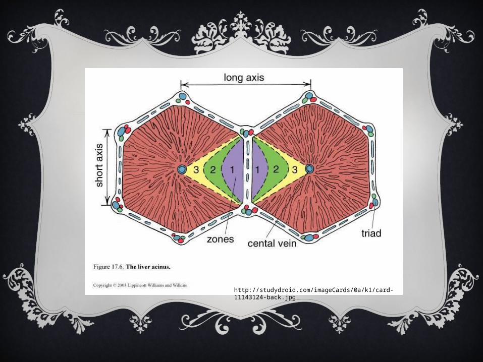
http://studydroid.com/imageCards/0a/k1/card-11143124-back.jpg
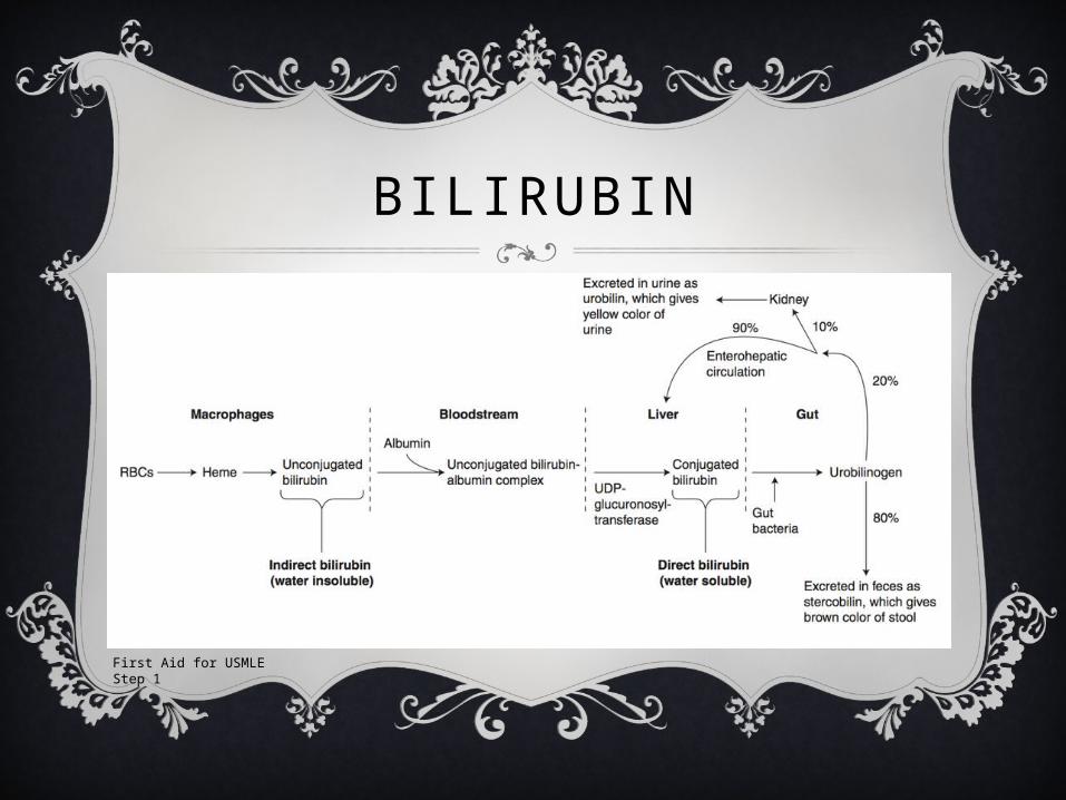
BILIRUBIN
First Aid for USMLE Step 1

GUESS THE LFTS

ACUTE HEPATITISAST
• Elevated
ALT• Elevated
Alk Phos• Normal
T bili• Normal
http://www.atsu.edu/faculty/chamberlain/Website/lectures/lecture/hepatit2.htm
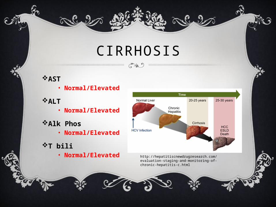
CIRRHOSISAST
• Normal/Elevated
ALT• Normal/Elevated
Alk Phos• Normal/Elevated
T bili• Normal/Elevated
http://hepatitiscnewdrugresearch.com/evaluation-staging-and-monitoring-of-chronic-hepatitis-c.html

CIRRHOSIS
As cirrhosis progresses, Total Bili increases because the liver can still conjugate bilirubin but can’t excrete it. MELD Score for 3 month mortality:
• Total bilirubin• Serum creatinine• INR• ± Dialysis
40 + --71.3% mortality30-39– 52.6% mortality20-29– 19.6% mortality10-19 – 6.0% mortality<9 – 1.9% mortality
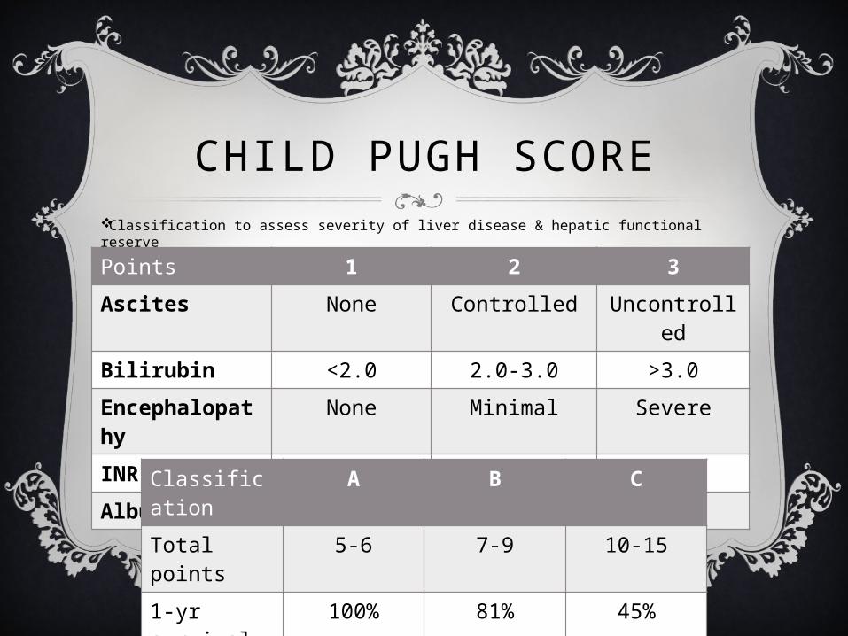
CHILD PUGH SCOREClassification to assess severity of liver disease & hepatic functional reserve
Points 1 2 3Ascites None Controlled Uncontrolle
dBilirubin <2.0 2.0-3.0 >3.0Encephalopathy
None Minimal Severe
INR <1.7 1.7-2.2 >2.2Albumin >3.5 2.8-3.5 <32.8Classificati
onA B C
Total points
5-6 7-9 10-15
1-yr survival
100% 81% 45%
2-yr survival
85% 57% 35%

LIVER TRANSPLANTEvaluate when Child Class B or MELD ≥ 10Indications:
• Recurrent/severe encephalopathy• Refractory ascites• SBP• Recurrent variceal bleeding• Hepatorenal or Hepatopulmonary syndrome• HCC if no single lesion > 5cm or ≤ 3 lesions w/ largest ≤ 3 cm• Fulminant hepatic failure
Contraindications:• Advanced HIV, active substance abuse (ETOH w/in 6 mo),
sepsis, extrahepatic malignancy, severe comorbidity (esp cardiopulm), persistent non-compliance
PRACTICE CASES
CASE 1
65 y/o male with 25 year history of alcohol and tobacco abuse who presents with abdominal swelling and confusion. Pt reports an unintentional 15 lbs weight gain and frequent forgetfulness. On exam, pt is A&O x1 (only to person), is slow to answer questions and often answers inappropriately. Pt has scleral icterus, distended abdomen with + fluid wave, and several ecchymoses on his lower extremities. Slight asterixis is observed.
What is the most likely diagnosis?
Alcoholic Cirrhosis
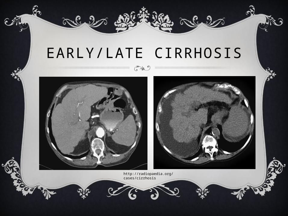
EARLY/LATE CIRRHOSIS
http://radiopaedia.org/cases/cirrhosis
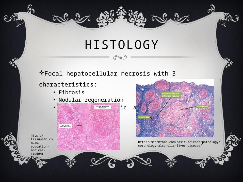
HISTOLOGY
Focal hepatocellular necrosis with 3 characteristics:
• Fibrosis• Nodular regeneration• Distortion of hepatic architecture
http://medchrome.com/basic-science/pathology/morphology-alcoholic-liver-disease/
http://tissupath.com.au/education-medical-student-liver/
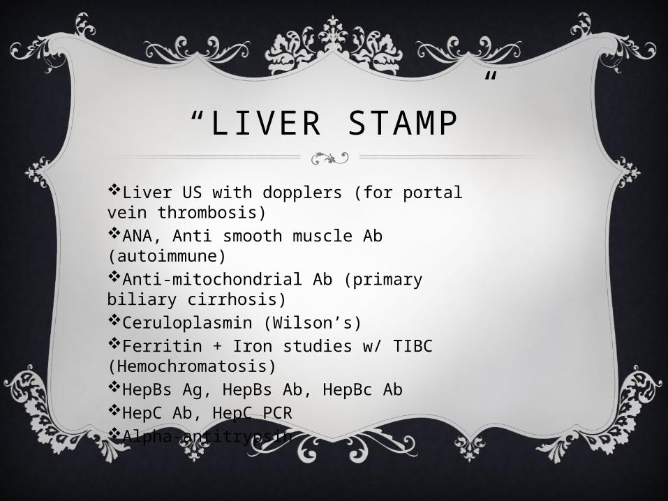
“LIVER STAMP”Liver US with dopplers (for portal vein thrombosis)ANA, Anti smooth muscle Ab (autoimmune)Anti-mitochondrial Ab (primary biliary cirrhosis)Ceruloplasmin (Wilson’s)Ferritin + Iron studies w/ TIBC (Hemochromatosis)HepBs Ag, HepBs Ab, HepBc AbHepC Ab, HepC PCRAlpha-antitrypsin
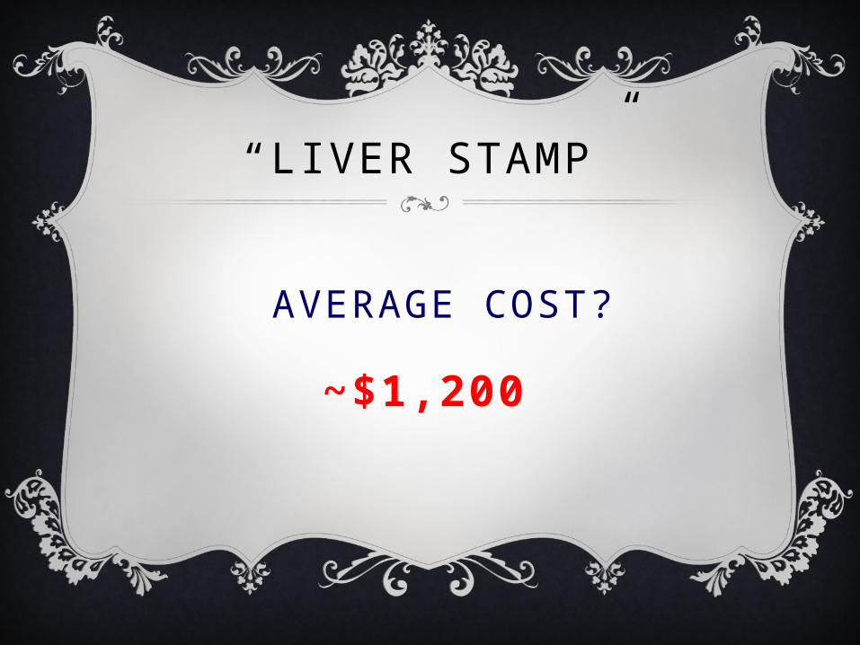
“LIVER STAMP”
AVERAGE COST?
~$1,200
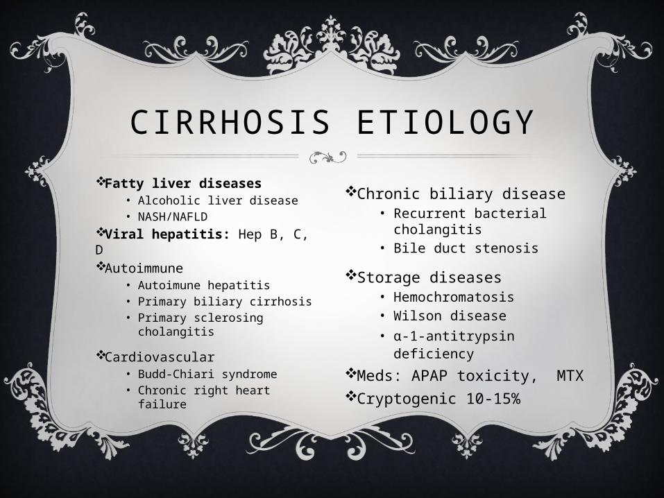
Fatty liver diseases• Alcoholic liver disease• NASH/NAFLD
Viral hepatitis: Hep B, C, DAutoimmune
• Autoimune hepatitis• Primary biliary cirrhosis• Primary sclerosing
cholangitis
Cardiovascular• Budd-Chiari syndrome• Chronic right heart failure
CIRRHOSIS ETIOLOGY
Chronic biliary disease• Recurrent bacterial
cholangitis• Bile duct stenosis
Storage diseases• Hemochromatosis• Wilson disease• α-1-antitrypsin deficiency
Meds: APAP toxicity, MTXCryptogenic 10-15%

DIAGNOSTIC IMAGING
Ultrasound• Surface nodularity: 88% sensitive, 82-95% specific
(1)• Coarse & heterogeneous echotexture• Signs of portal HTN:
• Portal vein >13mm: 42% sensitive, 95-100% specific (2)• Splenomegaly, ascites
CT insensitive in early cirrhosisMRI also insensitive in early cirrhosis, but significant role in assessing small hepatocellular carcinoma (HCC)—develops in 10-25%Liver biopsy = gold standard for diagnosis

TREATMENTAscites
• Furosemide + Spironolactone with goal negative ~1L/day (~80% effective)• Lasix: Aldactone ratio of 2:5 helps maintain K+ (thus Lasix 40mg qday, Aldactone 100mg qday
initially)• Low-sodium diet (1-2 g/day)
Refractory Ascites= no response on max doses of Lasix (160mg) & Aldactone (400mg)• LVP 4-6L (does not improve mortality)
• Albumin replacement controversial. AASLD 2009 guidelines recommend if >5L removed, provide 6-8 g/L of albumin 25% (IIA, Grade C)
• If >5L removed, can have post-paracentesis circulatory dysfxn via RAAS activation• TIPS (↓ ascites in 75%, improves mortality but ↑ HE, 40% need revision for stent stenosis)
Hepatic encephalopathy• Lactulose
Hepatorenal syndrome• Transplantation
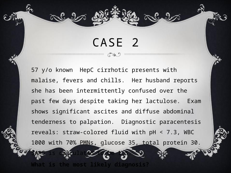
CASE 2
57 y/o known HepC cirrhotic presents with malaise, fevers and chills. Her husband reports she has been intermittently confused over the past few days despite taking her lactulose. Exam shows significant ascites and diffuse abdominal tenderness to palpation. Diagnostic paracentesis reveals: straw-colored fluid with pH < 7.3, WBC 1000 with 70% PMNs, glucose 35, total protein 30. SAAG is calculated to be 1.5.
What is the most likely diagnosis?
Spontaneous Bacterial Peritonitis (SBP)
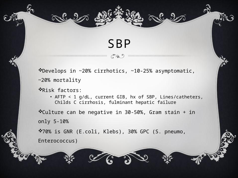
SBP
Develops in ~20% cirrhotics, ~10-25% asymptomatic, ~20% mortality
Risk factors: • AFTP < 1 g/dL, current GIB, hx of SBP, Lines/catheters, Childs C
cirrhosis, fulminant hepatic failure
Culture can be negative in 30-50%, Gram stain + in only 5-10%
70% is GNR (E.coli, Klebs), 30% GPC (S. pneumo, Enterococcus)
SBP
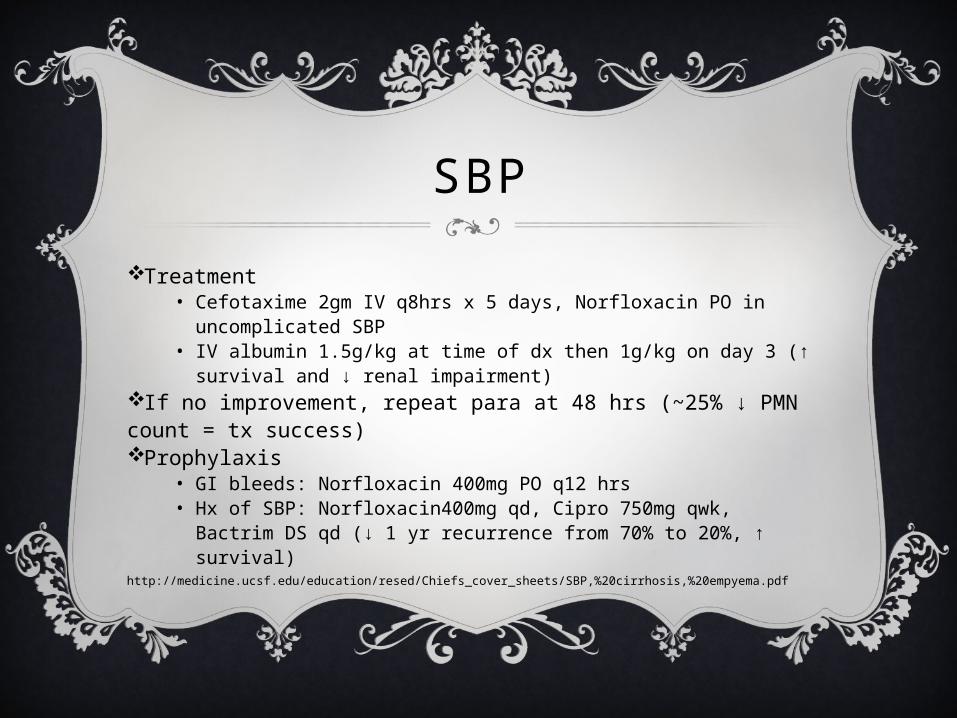
Treatment• Cefotaxime 2gm IV q8hrs x 5 days, Norfloxacin PO in
uncomplicated SBP• IV albumin 1.5g/kg at time of dx then 1g/kg on day 3 (↑
survival and ↓ renal impairment)If no improvement, repeat para at 48 hrs (~25% ↓ PMN count = tx success)Prophylaxis
• GI bleeds: Norfloxacin 400mg PO q12 hrs• Hx of SBP: Norfloxacin400mg qd, Cipro 750mg qwk, Bactrim
DS qd (↓ 1 yr recurrence from 70% to 20%, ↑ survival)http://medicine.ucsf.edu/education/resed/Chiefs_cover_sheets/SBP,%20cirrhosis,%20empyema.pdf

ASCITES: PATHOPHYSIOLOGY
Also: 1. Hypoalbuminemia ↓ serum oncotic pressure2. ↑ hepatic lymph ↑ splanchnic pressure
http://medical-dictionary.thefreedictionary.com/ascites
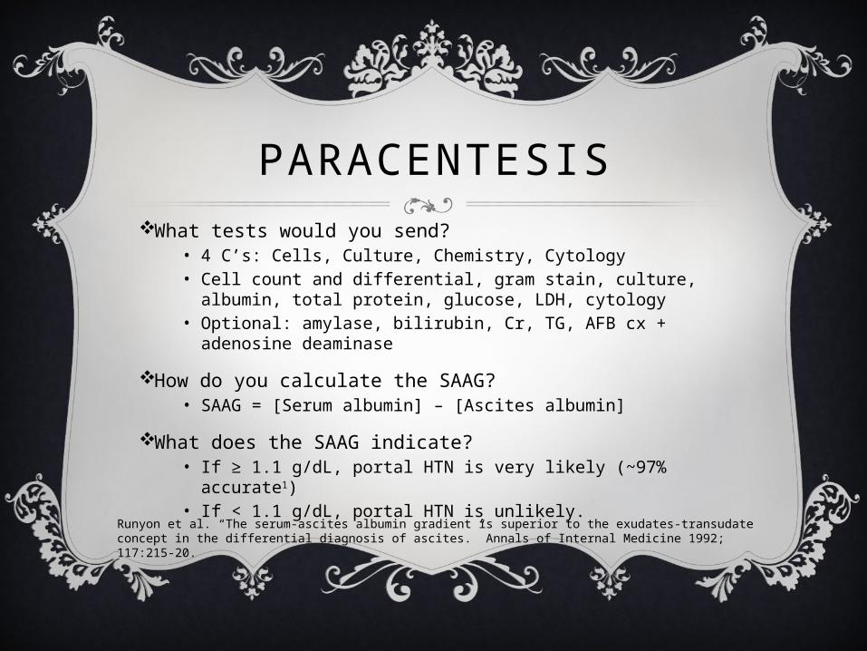
PARACENTESISWhat tests would you send?
• 4 C’s: Cells, Culture, Chemistry, Cytology• Cell count and differential, gram stain, culture, albumin,
total protein, glucose, LDH, cytology• Optional: amylase, bilirubin, Cr, TG, AFB cx + adenosine
deaminase
How do you calculate the SAAG?• SAAG = [Serum albumin] – [Ascites albumin]
What does the SAAG indicate?• If ≥ 1.1 g/dL, portal HTN is very likely (~97% accurate1)• If < 1.1 g/dL, portal HTN is unlikely.
Runyon et al. “The serum-ascites albumin gradient is superior to the exudates-transudate concept in the differential diagnosis of ascites.” Annals of Internal Medicine 1992; 117:215-20.
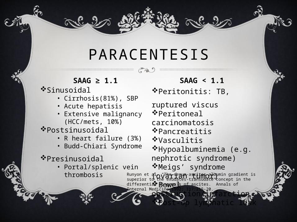
Sinusoidal• Cirrhosis(81%), SBP• Acute hepatisis• Extensive malignancy
(HCC/mets, 10%)Postsinusoidal
• R heart failure (3%)• Budd-Chiari Syndrome
Presinusoidal• Portal/splenic vein
thrombosis
Peritonitis: TB, ruptured viscusPeritoneal carcinomatosisPancreatitisVasculitisHypoalbuminemia (e.g. nephrotic syndrome)Meigs’ syndrome (ovarian tumor)Bowel obstruction/infarctionPost-op lymphatic leak
PARACENTESISSAAG ≥ 1.1 SAAG < 1.1
Runyon et al. “The serum-ascites albumin gradient is superior to the exudates-transudate concept in the differential diagnosis of ascites.” Annals of Internal Medicine 1992; 117:215-20.
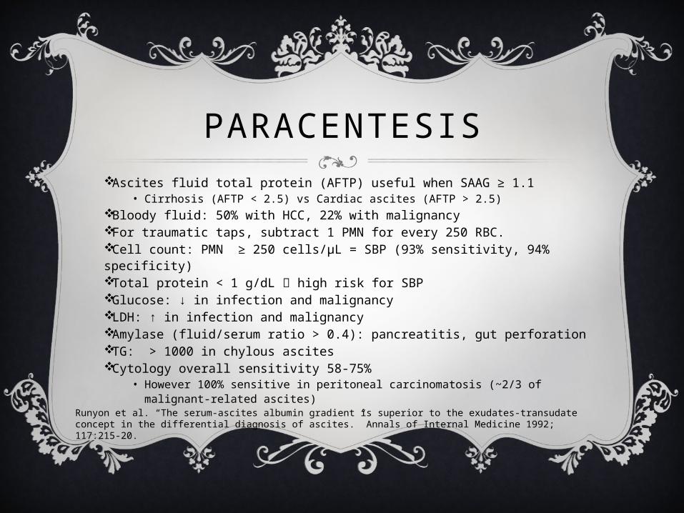
PARACENTESISAscites fluid total protein (AFTP) useful when SAAG ≥ 1.1
• Cirrhosis (AFTP < 2.5) vs Cardiac ascites (AFTP > 2.5)Bloody fluid: 50% with HCC, 22% with malignancyFor traumatic taps, subtract 1 PMN for every 250 RBC.Cell count: PMN ≥ 250 cells/μL = SBP (93% sensitivity, 94% specificity)Total protein < 1 g/dL high risk for SBPGlucose: ↓ in infection and malignancyLDH: ↑ in infection and malignancyAmylase (fluid/serum ratio > 0.4): pancreatitis, gut perforationTG: > 1000 in chylous ascitesCytology overall sensitivity 58-75%
• However 100% sensitive in peritoneal carcinomatosis (~2/3 of malignant-related ascites)
Runyon et al. “The serum-ascites albumin gradient is superior to the exudates-transudate concept in the differential diagnosis of ascites.” Annals of Internal Medicine 1992; 117:215-20.
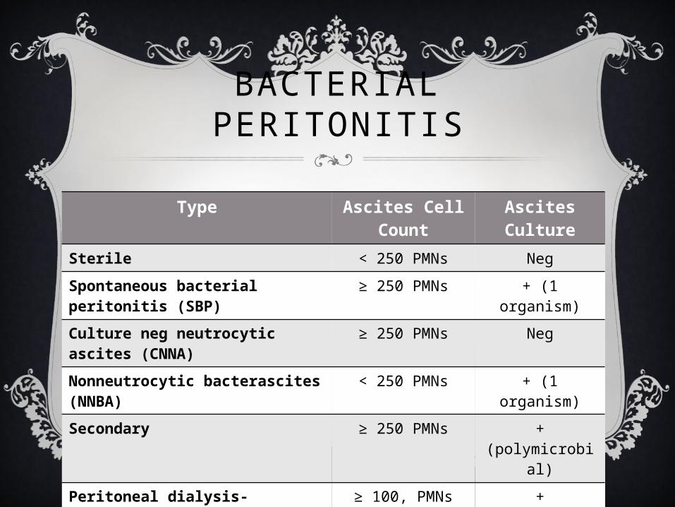
BACTERIAL PERITONITIS
Type Ascites Cell Count
Ascites Culture
Sterile < 250 PMNs Neg
Spontaneous bacterial peritonitis (SBP)
≥ 250 PMNs + (1 organism)
Culture neg neutrocytic ascites (CNNA)
≥ 250 PMNs Neg
Nonneutrocytic bacterascites (NNBA)
< 250 PMNs + (1 organism)
Secondary ≥ 250 PMNs + (polymicrobial)
Peritoneal dialysis-associated ≥ 100, PMNs predom
+
CASE 3
35 y/o male presents with fatigue and tea-colored urine for 5 days. Exam reveals jaundice and tender heaptomegaly but is otherwise unremarkable. Labs are significant for AST 2400, ALT 2640, Alk Phos 210, and Total Bilirubin 8.6.
Which of the following is least likely to cause this clinical picture?A. Acute hepatitis A infectionB. Acute hepatitis B infectionC. Acute hepatitis C infectionD. Acetaminophen ingestionE. Budd-Chiari Syndrome
CASE 3
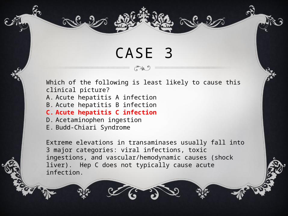
Which of the following is least likely to cause this clinical picture?A. Acute hepatitis A infectionB. Acute hepatitis B infectionC. Acute hepatitis C infectionD. Acetaminophen ingestionE. Budd-Chiari Syndrome
Extreme elevations in transaminases usually fall into 3 major categories: viral infections, toxic ingestions, and vascular/hemodynamic causes (shock liver). Hep C does not typically cause acute infection.

CASE 4
24 y/o patient is admitted to the MICU with obtundation and jaundice over 1-2 days. No further history is available. The following labs are obtained:Total Bili 7.2, Direct Bili 4.0, AST 1478, ALT 1056, Alk Phos 132, INR 3.1, Albumin 3.6.
All of the following tests are indicated except?A. Antinuclear Ab (ANA)B. CeruloplasminC. Hepatitis B surface AgD. ERCPE. Toxicology screen
CASE 4
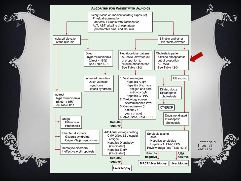
All of the following tests are indicated except?A. Antinuclear Ab (ANA)B. CeruloplasminC. Hepatitis B surface AgD. ERCPE. Toxicology screenWhen evaluating a patient with jaundice, initial steps include determining whether the hyperbilirubinemia is predominantly unconjugated or conjugated and whether there is any other evidence for hepatobiliary dysfxn. Next is to discriminate into a predominantly cholestatic or hepatocellular pattern. In this case, the pt has a hepatocellular pattern with AST/ALT elevated out of proportion to Alk Phos.

Harrison’s Internal Medicine
CASE 5
41 y/o male who presents to your clinic with a week of jaundice. He notes pruritus, icterus, and dark urine. He denies fever or abdominal pain. Exam is unremarkable except for jaundice. Labs: Total bili 6.0 , direct bili 5.1, AST 84 , ALT 92, Alk phos 662.CT scan of abdomen is unremarkable. RUQ ultrasound shows a normal bile duct but does not visualize the common bile duct.
What is the most appropriate next management step?A. Antibiotics and observationB. ERCPC. Hepatic serologiesD. HIDA scanE. Serologies for antimitochondrial Ab
CASE 5What is the most appropriate next management step?A. Antibiotics and observationB. ERCPC. Hepatic serologiesD. HIDA scanE. Serologies for antimitochondrial Ab
Anatomic abnormalities are more common when there is a cholestatic pattern of injury (Alk Phos elevated out of proportion to AST/ALT). Painless jaundice always requires extensive workup with concern for malignant causes (e.g. cholangiocarcinoma, tumor of ampulla of vater) vs nonmalignant causes (e.g. primary sclerosing cholangitis), which may only be detected by direct visualization with ERCP. Negative CT does not rule out source of cholestatis in biliary tree. Furthermore, ERCP is useful therapeutically with stenting to alleviate the obstruction.
Harrison’s Internal Medicine
CASE 6
61 y/o male is admitted to your service for new onset ascites. You perform a paracentesis with the following results of the non-bloody peritoneal fluid:WBC 300 with 35% PMNs, albumin 1.2, protein 2.6, TG 320Peritoneal cultures are pending. Serum albumin 2.7.
Which of the following is the most likely diagnosis?A. Peritoneal tuberculosisB. Peritoneal carcinomatosisC. Congestive heart failureD. Bacterial peritonitisE. Chylous ascites
CASE 6
Which of the following is the most likely diagnosis?A. Peritoneal tuberculosisB. Peritoneal carcinomatosisC. Congestive heart failureD. Bacterial peritonitisE. Chylous ascites
SAAG 1.5, AFTP 2.6 Cardiac ascitesLow WBC and PMNs make SBP and TB less likely
CASE 7
An alcoholic cirrhosis patient has increasing ascites despite dietary sodium control and diuretics. A paracentesis shows clear, turbid fluid. There are 2300 WBCs and 150 RBC. Differential shows 75% lymphocytes. Fluid protein is 3.2 and SAAG is 1.0.
What is the most appropriate next test?
A. Adenosine deaminase activity of ascitic fluid
B. CT scan of liver
C. Peritoneal biopsy
D. None; consider transplant evaluation

CASE 7What is the most appropriate next test?A. Adenosine deaminase activity of ascitic fluidB. CT scan of liverC. Peritoneal biopsyD. None; consider transplant evaluation
In pts with chronic cirrhosis who develop new or worsening ascites without dietary or medication nonadherence, consider an occult disorder (e.g. peritoneal TB, HCC, portal vein thrombosis). ↑ WBC is more common in neoplasm, bacterial peritonitis, or TB. Predominance of lymphocytes raises the suspicion for TB. SAAG is classically low in TB peritonitis but can be elevated in concomitant cirrhosis/transudative ascites. The sensitivity of ADA is poor in those with cirrhosis 2/2 poor T cell-mediated response. Thus peritoneal biopsy or visual diagnosis during laparoscopy is likely needed to confirm the diagnosis.

Sinusoidal• Cirrhosis(81%), SBP• Acute hepatisis• Extensive malignancy
(HCC/mets, 10%)Postsinusoidal
• R heart failure (3%)• Budd-Chiari Syndrome
Presinusoidal• Portal/splenic vein
thrombosis
Peritonitis: TB, ruptured viscusPeritoneal carcinomatosisPancreatitisVasculitisHypoalbuminemia (e.g. nephrotic syndrome)Meigs’ syndrome (ovarian tumor)Bowel obstruction/infarctionPost-op lymphatic leak
PARACENTESISSAAG ≥ 1.1 SAAG < 1.1
Runyon et al. “The serum-ascites albumin gradient is superior to the exudates-transudate concept in the differential diagnosis of ascites.” Annals of Internal Medicine 1992; 117:215-20.
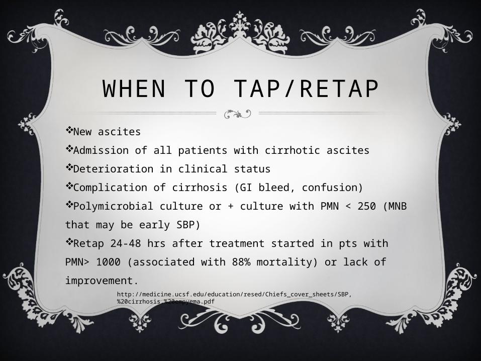
WHEN TO TAP/RETAPNew ascitesAdmission of all patients with cirrhotic ascitesDeterioration in clinical statusComplication of cirrhosis (GI bleed, confusion)Polymicrobial culture or + culture with PMN < 250 (MNB that may be early SBP)Retap 24-48 hrs after treatment started in pts with PMN> 1000 (associated with 88% mortality) or lack of improvement.
http://medicine.ucsf.edu/education/resed/Chiefs_cover_sheets/SBP,%20cirrhosis,%20empyema.pdf
CASE 8
When evaluating a patient with chronic ascites, a SAAG > 1.1 is consistent with all of the following diagnoses except?
A. Cirrhosis
B. Congestive heart failure
C. Constrictive pericarditis
D. Hepatic vein thrombosis
E. Nephrosis
CASE 8
When evaluating a patient with chronic ascites, a SAAG > 1.1 is consistent with all of the following diagnoses except?
A. Cirrhosis
B. Congestive heart failure
C. Constrictive pericarditis
D. Hepatic vein thrombosis
E. Nephrosis

Sinusoidal• Cirrhosis(81%), SBP• Acute hepatisis• Extensive malignancy
(HCC/mets, 10%)Postsinusoidal
• R heart failure (3%)• Budd-Chiari Syndrome
Presinusoidal• Portal/splenic vein
thrombosis
Peritonitis: TB, ruptured viscusPeritoneal carcinomatosisPancreatitisVasculitisHypoalbuminemia (e.g. nephrotic syndrome)Meigs’ syndrome (ovarian tumor)Bowel obstruction/infarctionPost-op lymphatic leak
PARACENTESISSAAG ≥ 1.1 SAAG < 1.1
Runyon et al. “The serum-ascites albumin gradient is superior to the exudates-transudate concept in the differential diagnosis of ascites.” Annals of Internal Medicine 1992; 117:215-20.
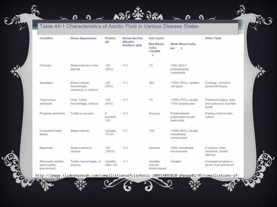
http://image.slidesharecdn.com/complicationsofcirrhosis-100914093820-phpapp02/95/complications-of-cirrhosis-18-728.jpg?cb=1284460955
CASE 9
28 y/o woman who is 30 weeks pregnant presents with 2 week history of pruritus and scleral icterus. It is her first pregnancy, and she has no significant medical hx. She does not drink alcohol and takes only a prenatal vitamin. Vitals are stable. Exam reveals a gravid uterus, mild scleral icterus and linear excoriations on the skin. There is no ascites or lower extremity edema.
CASE 9
Labs reveal:Hb 13.4 Platelet 275.000 AST 44, ALT 38, Total Bili 4.2, Direct Bili 2.3, Alk Phos 180LDH 82, INR 1.0Hep Bs Ag Neg, Hep Bs Ab Positive,Hep C Ab Neg, Hep A Ab (IgG) PositiveANA negative, Anti-smooth muscle Ab neg
Ultrasound of the liver is normal.

CASE 9
Which of the following is the most likely diagnosis?
A. Acute fatty liver of pregnancy
B. Acute hepatitis A infection
C. Cholestasis of pregnancy
D. HELLP syndrome

CASE 9Which of the following is the most likely diagnosis?A. Acute fatty liver of pregnancyB. Acute hepatitis A infectionC. Cholestasis of pregnancyD. HELLP syndrome
Cholestasis of pregnancy is the most common pregnancy-related liver disorder that is benign for the mother but increases risk for pre-term delivery and fetal loss if untreated. It often presents in the 2nd or 3rd trimester of pregnancy and treatment is with ursodeoxycholic acid for symptomatic treatment. In contrast acute fatty liver occurs in the 3rd trimester and is associated with high AST/ALT, high bilirubin and fat on liver US.HELLP syndrome is part of spectrum of eclampsia/pre-eclampsia and presents with HTN, hemolytic anemia, proteinuria high AST/ALT, & thrombocytopenia. It occurs during 3rd trimester and up to 48 hrs postpartum. Tx is delivery of the baby.
CASE 1045y/o male admitted for 2 day hx of fever and abdominal pain. Medical hx is notable for HepC cirrhosis and esophageal varices. Medications include furosemide, spironolactone, nadolol, and lactulose. Pt is afebrile, BP 100/50, HR 84. Abdominal exam is consistent with ascites and is nontender to palpation. Labs: Hb 10, WBC 3500, Plt 70,000, INR 1.5, Albumin 2.5, Alk Phos 220, AST 40, ALT 30, T bili 4, Cr 1.8, UA normal.Abdominal US shows cirrhosis, spenomegaly, & ascites. Portal & hepatic veins are patent. Diagnostic paracentesis shows WBC 2000 with 20% PMNs, Total protein 1, Albumin 0.7.
Which of the following is the most appropriate treatment?A. CefotaximeB. Cefotaxime and albuminC. Furosemide and spironolactoneD. LVPE. Observation

CASE 10Which of the following is the most appropriate treatment?A. CefotaximeB. Cefotaxime and albuminC. Furosemide and spironolactoneD. LVPE. Observation
In patients with SBP, the concomitant use of IV albumin with antibiotic therapy is associated with a survival benefit compared with antibiotic therapy alone.
REFERENCESAdamek HE, Albert J, Weitz M et-al. A prospective evaluation of magnetic resonance cholangiopancreatography in patients with suspected bile duct obstruction. Gut. 1998;43 (5): 680-3.Agabegi SS, Agabegi ED. Step –Up to Medicine, 3rd ed. 2013. Lippincott Williams & Wilkins. Philadelphia, PA.Caoili EM, Paulson EK, Heyneman LE et-al. Helical CT cholangiography with three-dimensional volume rendering using an oral biliary contrast agent: feasibility of a novel technique. AJR Am J Roentgenol. 2000;174 (2): 487-92.Cronan JJ. US diagnosis of choledocholithiasis: a reappraisal. Radiology. 1986;161 (1): 133-4.Guardino JM. Primo Gastro. 2008. Lippincott Williams & Wilkins. Philadelphia, PA.Miller FH, Hwang CM, Gabriel H et-al. Contrast-enhanced helical CT of choledocholithiasis. AJR Am J Roentgenol. 2003;181 (1): 125-30.Sabatine, MS. Pocket Medicine, 4th ed. 2011. Lippincott Williams & Wilkins. Philadelphia, PA.Sugiyama M, Suzuki Y, Abe N et-al. Endoscopic retreatment of recurrent choledocholithiasis after sphincterotomy. Gut. 2004;53 (12): 1856-9.Wiener C, Fauci AS, Braunwald E, et al. Harrison’s Principles of Internal Medicine Self-Assessment & Board Review, 17th ed. 2008. McGraw Hill. New York, NY.http://medicine.ucsf.edu/education/resed/Chiefs_cover_sheets/SBP,%20cirrhosis,%20empyema.pdfhttp://radiopaedia.orgSpecial thanks to Dr. Caroline Soyka for the inspiration!