Embed Size (px)
Citation preview
Session 11: The ABCs of LFTs
Learning Objectives
1. Define 3 key components of the patient history that should be further evaluated when liver function testing reveals elevated aminotransferases.
2. Identify at least 3 laboratory tests that should be considered in a patient with an ALT value that is 3 times the upper normal limit.

Session 11 The ABCs of LFTs Faculty
Dr John Russell is a graduate of Temple University and the Pennsylvania State University College of Medicine. He completed his family medicine training at Abington Memorial Hospital, where he served as chief resident, eventually joining the faculty in 1993. Dr Russell is co-editor of LearningLink Clinical Update, a twice-monthly literature review journal for primary care physicians through the American Academy of Family Physicians, which also features a twice-monthly podcast of Update highlights. Dr Russell has worked on creating palm-based guidelines for the American Diabetes Association, the Centers for Disease Control and Prevention, and the Infectious Diseases Society of America. In 2005, Dr Russell served as contributing editor to Patient Care magazine and co-authored the textbook Dermatology Skills for Primary Care. He has written articles and textbook chapters on a variety of topics and is currently a contributor to and reviewer for American Family Physician. Dr Russell lectures extensively to primary care physicians on a national level, and has won several resident teaching awards. He has been named several times to Philadelphia magazine’s list of “Top Doctors” in family medicine. Dr Russell’s special interests include pediatrics, dermatology, medical history, and bioethics. Faculty Financial Disclosure Statement The presenting faculty reports the following:
Dr Russell receives honoraria as a speaker for Sanofi Pasteur.
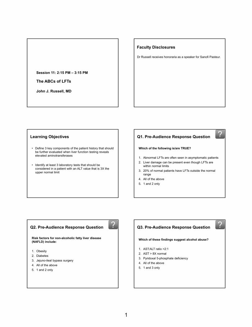
John J. Russell, MDClinical Associate Professor Family and Community Medicine Temple University School of Medicine Associate Director, Family Medicine Residency Program Abington Memorial Hosptial Abington, Pennsylvania
1
The ABCs of LFTs
John J. Russell, MD
Session 11: 2:15 PM – 3:15 PM
Faculty Disclosures
Dr Russell receives honoraria as a speaker for Sanofi Pasteur.
Learning Objectives
• Define 3 key components of the patient history that should be further evaluated when liver function testing reveals elevated aminotransferases
• Identify at least 3 laboratory tests that should be considered in a patient with an ALT value that is 3X the upper normal limit
Q1. Pre-Audience Response Question
Which of the following is/are TRUE?
1. Abnormal LFTs are often seen in asymptomatic patients
2. Liver damage can be present even though LFTs are within normal limits
3. 20% of normal patients have LFTs outside the normal range
4. All of the above
5. 1 and 2 only
Q2. Pre-Audience Response Question
Risk factors for non-alcoholic fatty liver disease (NAFLD) include:
1. Obesity
2. Diabetes
3. Jejuno-ileal bypass surgery
4. All of the above
5. 1 and 2 only
Q3. Pre-Audience Response Question
Which of these findings suggest alcohol abuse?
1. AST:ALT ratio >2:1
2. AST > 8X normal
3. Pyridoxal 5-phosphate deficiency
4. All of the above
5. 1 and 3 only
2
Case 1: Debra, 48-year-old female
• Comes to the office for routine annual checkup
• No current complaints
• Past medical history:
– Hypertension
– Dyslipidemia
– Obesity
• No allergies
• Medications: HCTZ, simvastatin, acetaminophen prn joint pain
Case 1: Debra, 48-year-old female
• Exam:
– 144/86 mm Hg, 72 regular, 14
– BMI: 36
• HEENT: no icterus
• Lungs: clear
• Heart: regular, no murmurs
• Abdomen: soft, nontender, no organomegaly
• Extremities: no edema
Debra
• Labs:
– ALT: 74 U/L (normal 9-52)
– AST: 48 U/L (normal 14-36)
– Total bilirubin: 0.3 mg/dL (normal 0.2-1.2)
– Total cholesterol: 202 mg/dL (normal 120-199)
– LDL cholesterol: 112 mg/dL (normal 60-129)
– HDL cholesterol: 31 mg/dL (normal >39)
– Triglycerides: 245 mg/dL (normal <150)
– Fasting glucose: 119 mg/dL (normal 70-99)
Question
Which of the following causes of abnormal LFTs should be initially considered?
1. Statin hepatotoxicity
2. Alcohol use
3. Acetaminophen use
4. NASH
5. All of the Above
Ioannou GN, et al. Am J Gastroenterol. 2006;101:76-82.
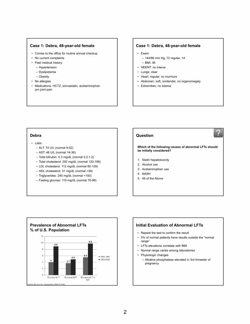
Prevalence of Abnormal LFTs% of U.S. Population
Initial Evaluation of Abnormal LFTs
• Repeat the test to confirm the result
• 5% of normal patients have results outside the “normal range”
• LFTs elevations correlate with BMI
• Normal range varies among laboratories
• Physiologic changes
– Alkaline phosphatase elevated in 3rd trimester of pregnancy
3
Commonly reported LFTs
• Enzymes
– Aspartate transaminase or aminotransferase (AST, SGOT)
– alanine transaminase or aminotransferase (ALT, SGPT)
– Alkaline phosphatase (ALP)
– Gamma-glutamyl transpeptidase (GGT)
• Synthetic Function
– Albumin
– Prothrombin time
• Bilirubin
Initial Evaluation of Abnormal LFTs
• Physical Examination
– Muscle wasting
– Stigmata of chronic liver disease
• Spider nevi, palmar erythema, gynecomastia, caput medusae
– Lymphadenopathy
– Jugular venous distension
– Pleural effusion
Audience Response Question
What is the most common cause of abnormal LFTs in the U.S. primary care population?
1. Viral hepatitis
2. Fatty liver
3. Statin use
4. Strenuous exercise
Abnormal LFTs—Most Common Causes in U.S.
• Fatty liver
– Nonalcoholic fatty liver disease (NAFLD)
Prevalence ~30%
– Nonalcoholic steatohepatitis (NASH)
• Alcoholic liver disease
• Viral hepatitis (chronic HBV and HCV)
http://digestive.niddk.nih.gov/ddiseases/pubs/nash/
Causes of Chronically Elevated ALT Levels
Hepatic causes
– NASH
– Alcohol abuse
– Infectious hepatitis
– Autoimmune hepatitis
– Hemochromatosis
– Wilson’s disease
– Alpha1-antitrypsin deficiency
Nonhepatic causes
– Celiac disease
– Inherited disorders of muscle metabolism
– Acquired muscle diseases
– Strenuous exercise
Pratt DS. NEJM. 2000;342 (17):1266-71.
Evaluation of Chronic ALT Elevation
Step 1
Review medications, herbal therapies, or recreational drugs
Screen for alcohol abuse
Obtain serology for Hepatitis B and C
Screen for hemochromatosis
Evaluate for fatty liver
4
Evaluation of Chronic ALT Elevation
Step 2
Exclude muscle disorders
Obtain thyroid function tests
Consider celiac disease
Consider adrenal insufficiency
Evaluation of Chronic ALT Elevation
Step 3
Consider autoimmune hepatitis
Consider Wilson’s disease
Consider α1-antitrypsin deficiency
Evaluation of Chronic ALT Elevation
Step 4
Obtain a liver biopsy
Consider observation
• if ALT, AST only mildly elevated
Transaminase Levels in Alcohol Abuse
• Diagnosis supported by AST:ALT of at least 2:1
– Alcohol-related deficiency of pyridoxal 5-phosphate
– Low serum activity of ALT
• Gamma-glutamyltransferase twice normal with AST:ALT of at least 2:1 strongly suggests diagnosis
– GGT not specific
• Rare for AST > 8X normal value
• Rare for ALT > 5X normal value
Pratt DS. NEJM. 2000;342(17):1266-71.
Medications and Elevated LFTs
Selected Drugs/Substances Associated With LFT Elevations
acetaminophen omeprazole
antifungals, INH lisinopril, losartan
glipizide NSAIDs
allopurinol risperidone
buproprion SSRIs, trazodone
kava kava; germander, ephedra, shark cartilage, senna
PCP, anabolic steroids, cocaine, ecstasy, glues/solvents
DPH, valproate, carbamazepine statins
Almost any drug can be a causeStop medication and determine if LFTs normalize
Oh RC, et al. Am Fam Phys.2011;84(9):1003-1008. Pratt DS. NEJM. 2000;342 (17):1266-71.
Drug-Related Transaminase Elevations
Acetaminophen
• 4 gm/day for 5-10 days caused elevated transaminases in >50% healthy non-drinkers
• Alcohol can potentiate hepatotoxic effects
Statins
• Frequently cause elevated transaminases, but significant hepatoxicity is rare
• Transaminase elevations usually resolve spontaneously
• May be used safely in persons with chronic liver disease
Pratt DS. NEJM. 2000;342 (17):1266-71.
5
Testing for Other Causes of Abnormal Transaminases
AutoimmuneHepatitis
Serum Protein ElectrophoresisANA
Anti-Smooth Muscle ABsLiver-Kidney Microsomal ABs
HemochromatosisSerum Iron
TIBCHFE Genetic Testing
Wilson’s DiseaseSerum CeruloplasminOphthalmology Exam
24-hour Urine Copper Excretion
α1-Antitrypsin Deficiency
Serum α1-Antitrypsin Level
Pratt DS. NEJM. 2000;342(17):1266-71. Oh RC, et al. Am Fam Phys. 2011;84(9):1003-1008.
Nonhepatic Causes of Abnormal Transaminases
• Celiac disease
• Muscle disorders
– Inborn errors of metabolism
– Polymyositis
– Strenuous exercise
• Thyroid disorders
• Adrenal insufficiency
• Anorexia nervosa
Back to Debra
• Denies significant alcohol or acetaminophen use
• Viral serologies are negative
• Repeat LFTs after statin discontinued: unchanged
• Ultrasound: fatty liver
Diagnosis: NASH
Oh RC, et al. Am Fam Phys. 2011;84(9):1003-1008.
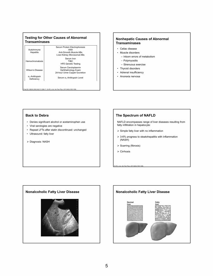
The Spectrum of NAFLD
NAFLD encompasses range of liver diseases resulting from fatty infiltration in hepatocyte:
Simple fatty liver with no inflammation
3-6% progress to steatohepatitis with inflammation (NASH)
Scarring (fibrosis)
Cirrhosis
Nonalcoholic Fatty Liver Disease Nonalcoholic Fatty Liver Disease
6
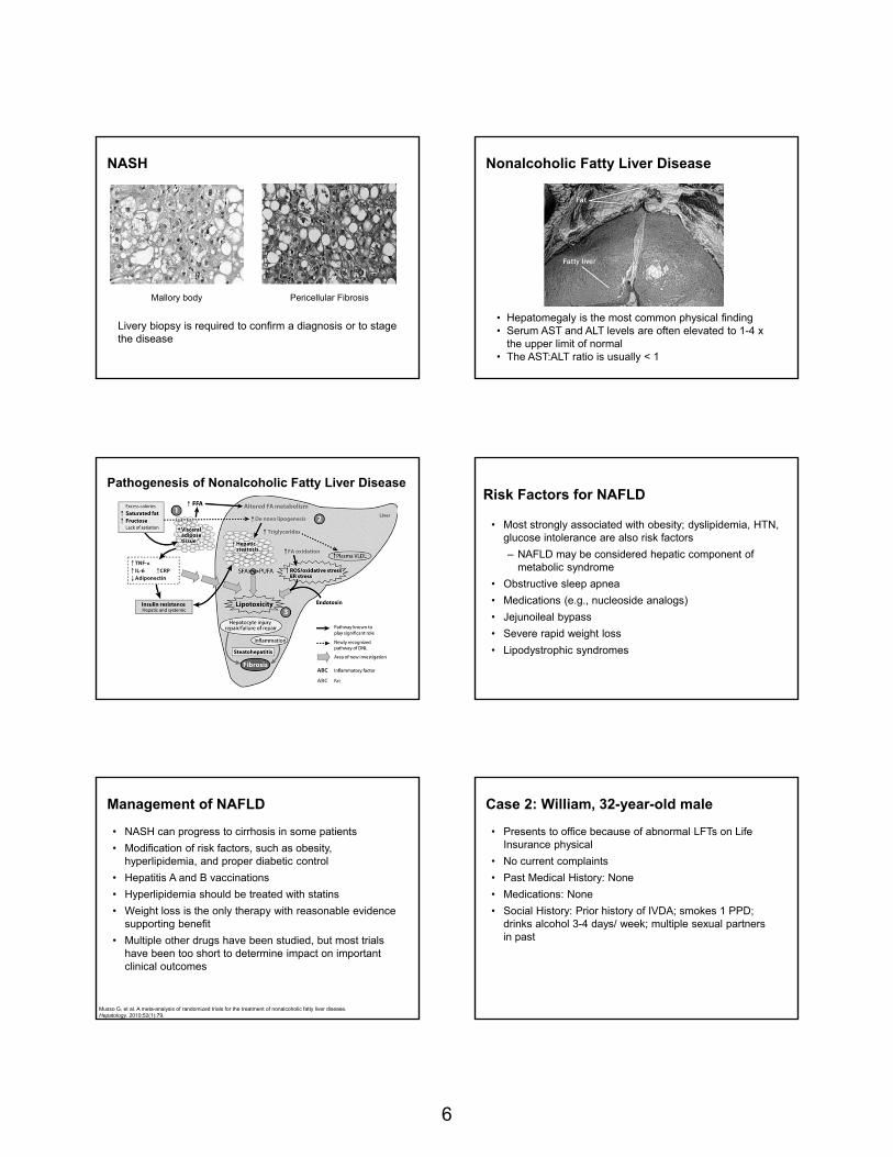
NASH
Mallory body Pericellular Fibrosis
Livery biopsy is required to confirm a diagnosis or to stage the disease
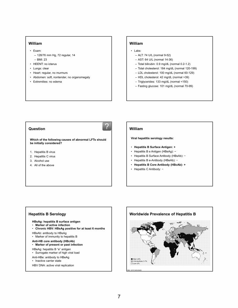
Nonalcoholic Fatty Liver Disease
• Hepatomegaly is the most common physical finding• Serum AST and ALT levels are often elevated to 1-4 x
the upper limit of normal• The AST:ALT ratio is usually < 1
Pathogenesis of Nonalcoholic Fatty Liver Disease Risk Factors for NAFLD
• Most strongly associated with obesity; dyslipidemia, HTN, glucose intolerance are also risk factors
– NAFLD may be considered hepatic component of metabolic syndrome
• Obstructive sleep apnea
• Medications (e.g., nucleoside analogs)
• Jejunoileal bypass
• Severe rapid weight loss
• Lipodystrophic syndromes
Management of NAFLD
• NASH can progress to cirrhosis in some patients
• Modification of risk factors, such as obesity, hyperlipidemia, and proper diabetic control
• Hepatitis A and B vaccinations
• Hyperlipidemia should be treated with statins
• Weight loss is the only therapy with reasonable evidence supporting benefit
• Multiple other drugs have been studied, but most trials have been too short to determine impact on important clinical outcomes
Musso G, et al. A meta-analysis of randomized trials for the treatment of nonalcoholic fatty liver disease. Hepatology. 2010;52(1):79.
Case 2: William, 32-year-old male
• Presents to office because of abnormal LFTs on Life Insurance physical
• No current complaints
• Past Medical History: None
• Medications: None
• Social History: Prior history of IVDA; smokes 1 PPD; drinks alcohol 3-4 days/ week; multiple sexual partners in past
7
William
• Exam:
– 128/76 mm Hg, 72 regular, 14
– BMI: 23
• HEENT: no icterus
• Lungs: clear
• Heart: regular, no murmurs
• Abdomen: soft, nontender, no organomegaly
• Extremities: no edema
William
• Labs:
– ALT: 74 U/L (normal 9-52)
– AST: 64 U/L (normal 14-36)
– Total bilirubin: 0.9 mg/dL (normal 0.2-1.2)
– Total cholesterol: 184 mg/dL (normal 120-199)
– LDL cholesterol: 100 mg/dL (normal 60-129)
– HDL cholesterol: 42 mg/dL (normal >39)
– Triglycerides: 133 mg/dL (normal <150)
– Fasting glucose: 101 mg/dL (normal 70-99)
Question
Which of the following causes of abnormal LFTs should be initially considered?
1. Hepatitis B virus
2. Hepatitis C virus
3. Alcohol use
4. All of the above
William
Viral hepatitis serology results:
• Hepatitis B Surface Antigen: +
• Hepatitis B e Antigen (HBeAg): −
• Hepatitis B Surface Antibody (HBsAb): −
• Hepatitis B e-Antibody (HBeAb): −
• Hepatitis B Core Antibody (HBcAb): +
• Hepatitis C Antibody: −
Hepatitis B Serology
HBsAg: hepatitis B surface antigen• Marker of active infection• Chronic HBV: HBsAg positive for at least 6 months
HBsAb: antibody to HBsAg• Marker of immunity to hepatitis B
Anti-HB core antibody (HBcAb)• Marker of present or past infection
HBeAg: hepatitis B “e” antigen• Surrogate marker of high viral load
Anti-HBe: antibody to HBeAg• Inactive carrier state
HBV DNA: active viral replication
Worldwide Prevalence of Hepatitis B
BMJ. 2010;340:b5429.
8
16
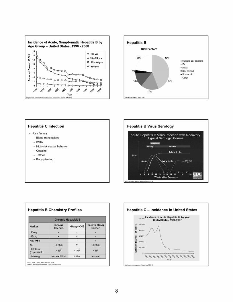
14
12
10
8
6
4
2
0
Year
Re
po
rted
Cas
es/1
00,0
00
<15 yrs
15 – 24 yrs
25 – 44 yrs
45+ yrs
Adapted from National Notifiable Diseases Surveillance System (NNDSS)
Incidence of Acute, Symptomatic Hepatitis B by Age Group – United States, 1990 - 2008
CDC Sentinel Sites. 2001 data.
Hepatitis B
Hepatitis C Infection
• Risk factors
– Blood transfusions
– IVDA
– High-risk sexual behavior
– Cocaine
– Tattoos
– Body piercing
Hepatitis B Virus Serology
http://pathmicro.med.sc.edu/virol/hepb-cd1.gif
Hepatitis B Chemistry Profiles
Lai CL, et al. Lancet. 2003;362:2089-2094.Lok AS, et al. Gastroenterology. 2001;120:1828-1853.
Hepatitis C – Incidence in United States
http://www.medscape.com/viewarticle/735146
9
Hepatitis C - Diagnostic Testing
• Serologic Testing
– 92-97% sensitivity
• Serum Hepatitis C Virus RNA for confirmation
• If positive – consider liver biopsy to assess severity
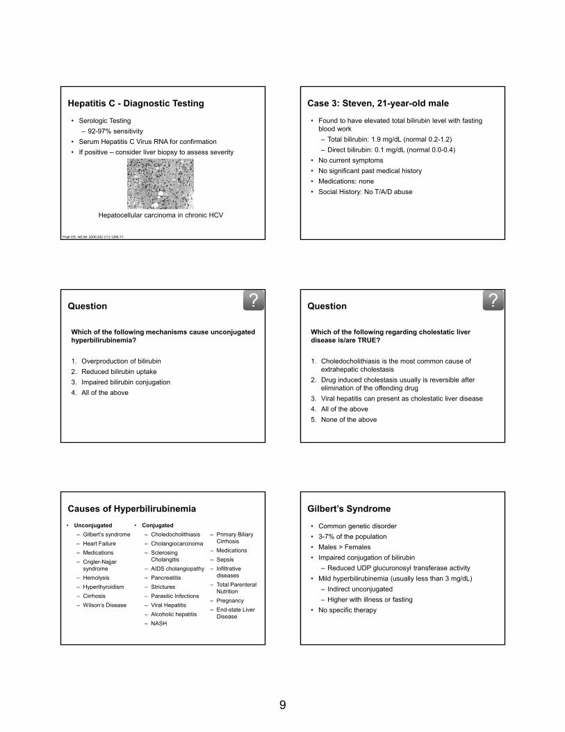
Hepatocellular carcinoma in chronic HCV
Pratt DS. NEJM. 2000;342 (17):1266-71.
Case 3: Steven, 21-year-old male
• Found to have elevated total bilirubin level with fasting blood work
– Total bilirubin: 1.9 mg/dL (normal 0.2-1.2)
– Direct bilirubin: 0.1 mg/dL (normal 0.0-0.4)
• No current symptoms
• No significant past medical history
• Medications: none
• Social History: No T/A/D abuse
Question
Which of the following mechanisms cause unconjugated hyperbilirubinemia?
1. Overproduction of bilirubin
2. Reduced bilirubin uptake
3. Impaired bilirubin conjugation
4. All of the above
Question
Which of the following regarding cholestatic liver disease is/are TRUE?
1. Choledocholithiasis is the most common cause of extrahepatic cholestasis
2. Drug induced cholestasis usually is reversible after elimination of the offending drug
3. Viral hepatitis can present as cholestatic liver disease
4. All of the above
5. None of the above
Causes of Hyperbilirubinemia
• Unconjugated
– Gilbert’s syndrome
– Heart Failure
– Medications
– Crigler-Najjarsyndrome
– Hemolysis
– Hyperthyroidism
– Cirrhosis
– Wilson’s Disease
• Conjugated
– Choledocholithiasis
– Cholangiocarcinoma
– SclerosingCholangitis
– AIDS cholangiopathy
– Pancreatitis
– Strictures
– Parasitic Infections
– Viral Hepatitis
– Alcoholic hepatitis
– NASH
– Primary Biliary Cirrhosis
– Medications
– Sepsis
– Infiltrative diseases
– Total Parenteral Nutrition
– Pregnancy
– End-state Liver Disease
Gilbert’s Syndrome
• Common genetic disorder
• 3-7% of the population
• Males > Females
• Impaired conjugation of bilirubin
– Reduced UDP glucuronosyl transferase activity
• Mild hyperbilirubinemia (usually less than 3 mg/dL)
– Indirect unconjugated
– Higher with illness or fasting
• No specific therapy
10
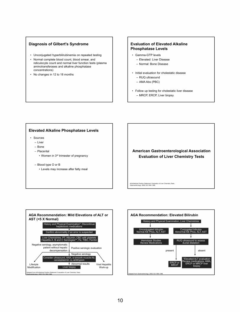
Diagnosis of Gilbert's Syndrome
• Unconjugated hyperbilirubinemia on repeated testing
• Normal complete blood count, blood smear, and reticulocyte count and normal liver function tests (plasma aminotransferases and alkaline phosphatase concentrations)
• No changes in 12 to 18 months
Evaluation of Elevated Alkaline Phosphatase Levels
• Gamma-GTP levels
– Elevated: Liver Disease
– Normal: Bone Disease
• Initial evaluation for cholestatic disease
– RUQ ultrasound
– AMA Abs (PBC)
• Follow up testing for cholestatic liver disease
– MRCP, ERCP, Liver biopsy
Elevated Alkaline Phosphatase Levels
• Sources
– Liver
– Bone
– Placental
• Women in 3rd trimester of pregnancy
– Blood type O or B
• Levels may increase after fatty meal
AGA Medical Position Statement: Evaluation of Liver Chemistry Tests.. Gastroenterology. 2002;123:1364-1366.
American Gastroenterological Association
Evaluation of Liver Chemistry Tests
AGA Recommendation: Mild Elevations of ALT or AST (<5 X Normal)
Adapted from AGA Medical Position Statement: Evaluation of Liver Chemistry Tests.. Gastroenterology. 2002;123:1364-1366.
LifestyleModification
Viral Hepatitis Work-up
History and Physical Examination*, Discontinue hepatotoxic medications
Confirm abnormality if an error is suspected
Liver Chemistries, PT, Albumin, CBC with platelets Hepatitis A, B and C Serologies**, Fe, TIBC, Ferritin
Consider ultrasound, ANA, α-smooth muscle Ab, ceruloplasmin, α1-antitrypsin
Liver biopsy
Negative serology, asymptomatic patient without hepatic
decompensationPositive serologic evaluation
Negative serology
Abnormal results
AGA Recommendation: Elevated Bilirubin
Adapted from Gastroenterology. 2002;123:1364-1366.
present
Hemolysis StudiesReview Medications
RUQ ultrasound to assess ductal dilatation
absent
ERCP or MRCP
*Elevated ALT evaluationReview medications, AMA,
ERCP or MRCP liver biopsy
History and Physical Examination, Liver Chemistries
Unconjugated bilirubinNormal Alk Phos, ALT, AST
Conjugated bilirubinAbnormal Alk Phos, ALT, AST
11
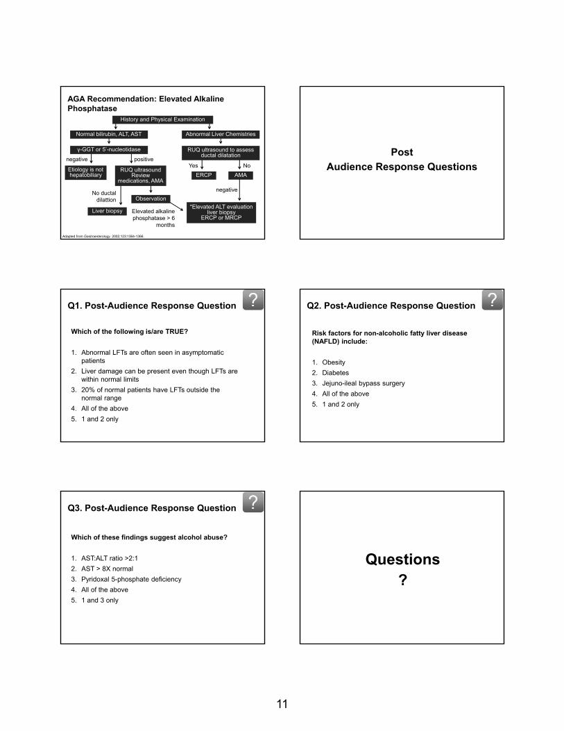
AGA Recommendation: Elevated Alkaline Phosphatase
Etiology is not hepatobiliary
*Elevated ALT evaluationliver biopsy
ERCP or MRCPLiver biopsy
Yes No
negative
Elevated alkaline phosphatase > 6
months
No ductaldilattion
History and Physical Examination
Normal bilirubin, ALT, AST Abnormal Liver Chemistries
RUQ ultrasound to assess ductal dilatation
ERCP AMARUQ ultrasound
Review medications, AMA
Observation
γ-GGT or 5’-nucleotidase
negative positive
Adapted from Gastroenterology. 2002;123:1364-1366.
Post
Audience Response Questions
Q1. Post-Audience Response Question
Which of the following is/are TRUE?
1. Abnormal LFTs are often seen in asymptomatic patients
2. Liver damage can be present even though LFTs are within normal limits
3. 20% of normal patients have LFTs outside the normal range
4. All of the above
5. 1 and 2 only
Q2. Post-Audience Response Question
Risk factors for non-alcoholic fatty liver disease (NAFLD) include:
1. Obesity
2. Diabetes
3. Jejuno-ileal bypass surgery
4. All of the above
5. 1 and 2 only
Q3. Post-Audience Response Question
Which of these findings suggest alcohol abuse?
1. AST:ALT ratio >2:1
2. AST > 8X normal
3. Pyridoxal 5-phosphate deficiency
4. All of the above
5. 1 and 3 only
Questions?