Embed Size (px)
Citation preview
LIVER FUNCTION TESTS
Megan Chan, PGY-2
UHCMC 2015
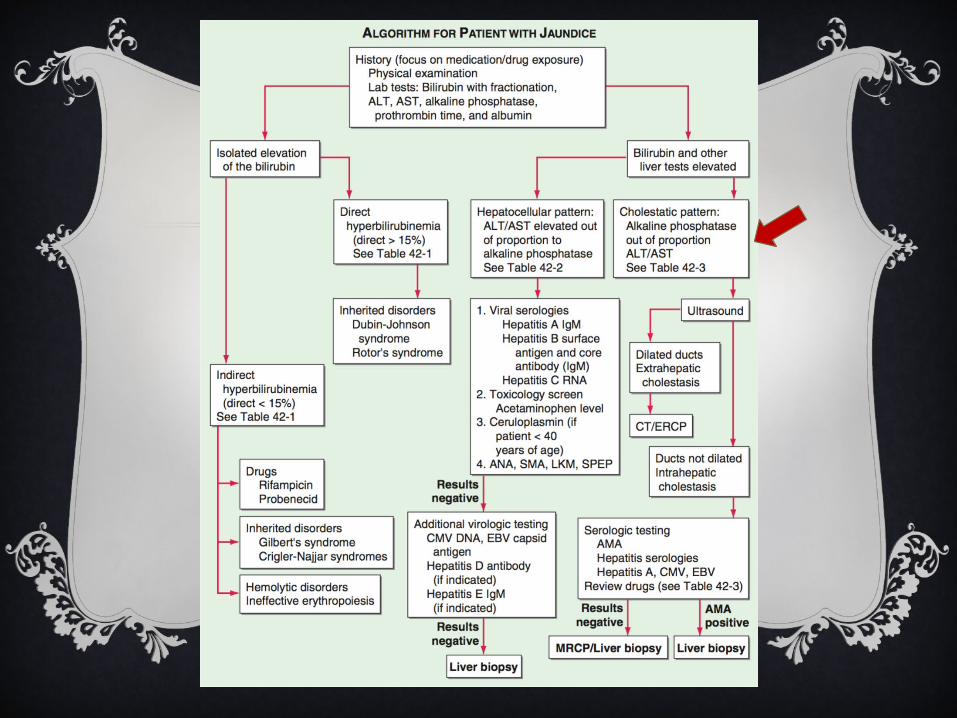
GENERAL APPROACH
Cholestatic—intrahepatic/extrahepatic biliary obstruction
Hepatocellular—hepatocyte damage (e.g. viral hepatitis, drugs/toxins, ETOH, ischemia, malignant infiltration)
Isolated hyperbilirubinemia—e.g. congestive hepatopathy

GUESS THE LFTS
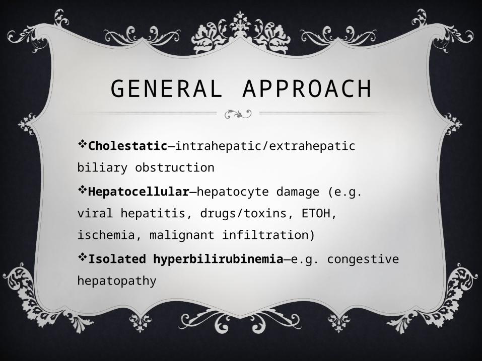
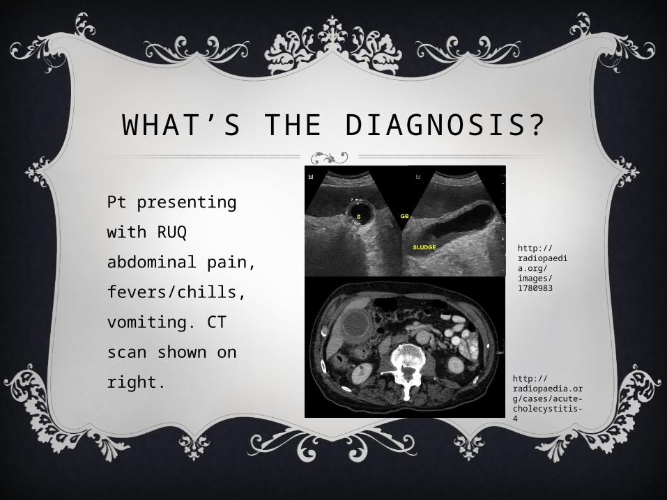
WHAT’S THE DIAGNOSIS?
http://radiopaedia.org/articles/cholelithiasis
Pt with hx of intermittent abdominal pain associated with meals undergoes RUQ US.
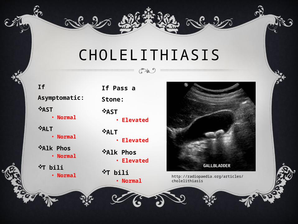
CHOLELITHIASIS
If Asymptomatic:AST
• Normal
ALT• Normal
Alk Phos• Normal
T bili• Normal http://radiopaedia.org/articles/cholelithiasis
If Pass a Stone:AST
• Elevated
ALT• Elevated
Alk Phos• Elevated
T bili• Normal
WHAT’S THE DIAGNOSIS?
http://radiopaedia.org/cases/acute-cholecystitis-4
http://radiopaedia.org/images/1780983
Pt presenting with RUQ abdominal pain, fevers/chills, vomiting. CT scan shown on right.
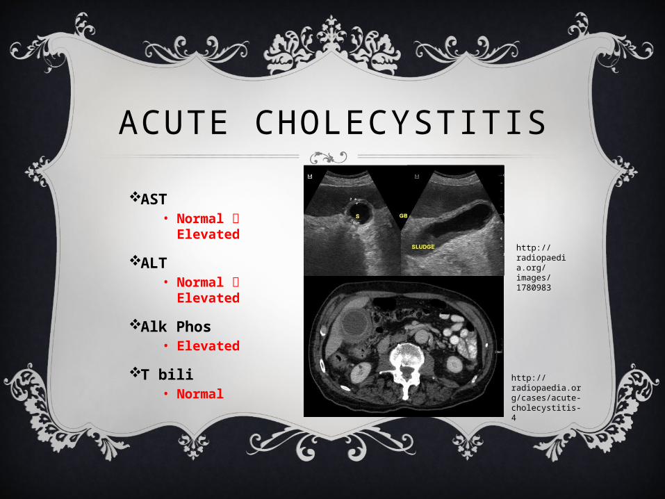
ACUTE CHOLECYSTITIS
AST• Normal
Elevated
ALT• Normal
Elevated
Alk Phos• Elevated
T bili• Normal
http://radiopaedia.org/cases/acute-cholecystitis-4
http://radiopaedia.org/images/1780983
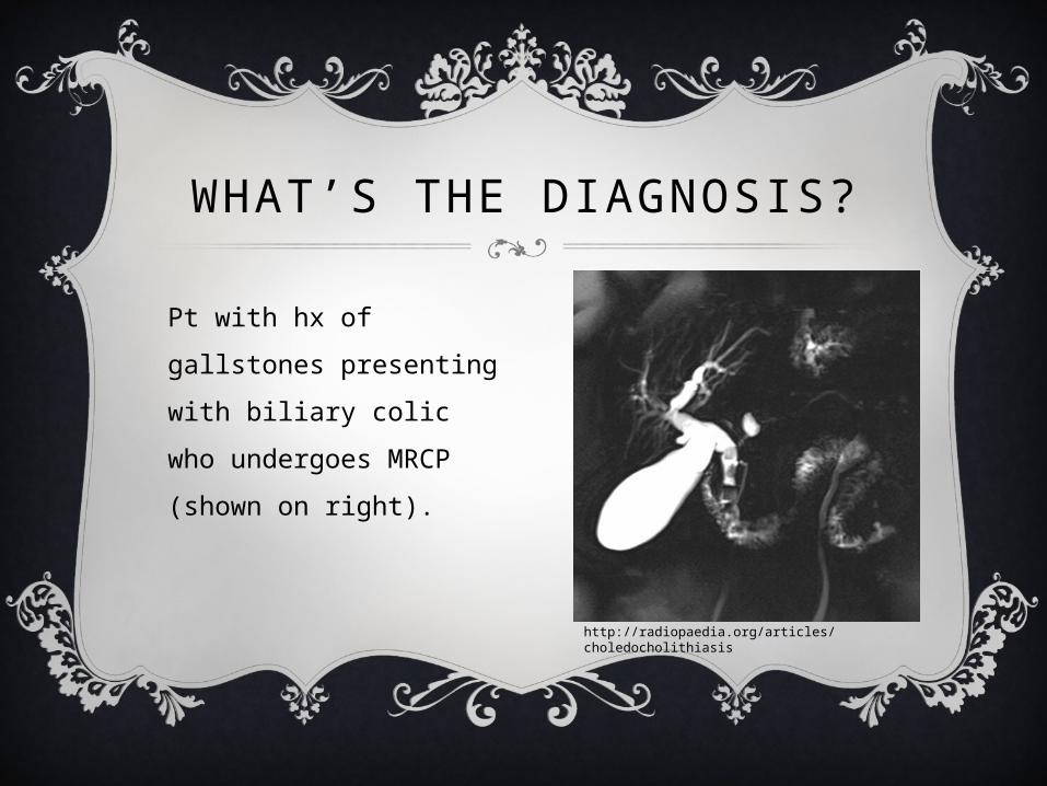
WHAT’S THE DIAGNOSIS?
http://radiopaedia.org/articles/choledocholithiasis
Pt with hx of gallstones presenting with biliary colic who undergoes MRCP (shown on right).
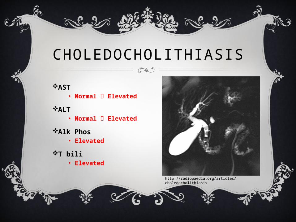
CHOLEDOCHOLITHIASIS
AST• Normal Elevated
ALT• Normal Elevated
Alk Phos• Elevated
T bili• Elevated
http://radiopaedia.org/articles/choledocholithiasis
PRACTICE CASES
CASE 1
46 y/o female presents to your clinic with intermittent RUQ pain and heartburn. Vitals are stable and exam is unremarkable. LFTs are wnl.
What is the next best imaging test to confirm your diagnosis?
RUQ ultrasoundsensitivity and specificity > 95% for stones > 2mm
Pure cholesterol stones are hypodense to bile and calcified gallstones are
hyperdense to bile and some gallstones may be isodense to bile and may therefore be
missed by CT.
CHOLELITHIASISGallstones or sludge in the gallbladder~10% population, symptomatic in only 25% of cases3 types of stones:
• Cholesterol stones—associated with obesity, DM, HLD, OCP use, multiple pregnancies, advanced age, Crohn’s disease, ileal resection, cirrhosis, CF
• Pigment stones• Black stones—hemolysis, alcoholic cirrhosis• Brown stones—biliary tract infection
• Mixed stones = 80%
CHOLELITHIASIS
Pt asks about surgical treatment. What do you tell her?Only 1-2 % of patients with asymptomatic gallstone disease will develop complications that will require surgery yearly.
4 factors should be considered in evaluation for surgery:1) Symptoms that are severe and frequent enough to necessitate surgery.2) Hx of prior complications of gallstone disease (e.g pancreatitis, acute
cholecystitis)3) Presence of anatomic factors that increase the likelihood of complications
(e.g. porcelain gallbladder, congenital biliary tract abnormalities)4) Large stones >3cm
Ursodeoxycholic acid can be used to dissolve gallstones. It decreases the cholesterol saturation of bile & allows the dispersion of cholesterol from stones. It is only effective, however, for radiolucent stones <10mm.
CASE 2
55 y/o male with PMHx of recurrent pancreatitis presents to the ED with RUQ abdominal pain and vomiting. Pt is found to be febrile and hypotensive. IV fluids are initiated and the following labs are obtained:
WBC 13,000, AST 25, ALT 30, Alk Phos 450, T bili 1.0, Lipase 20
What is the most likely diagnosis?
Acute Cholecystitis
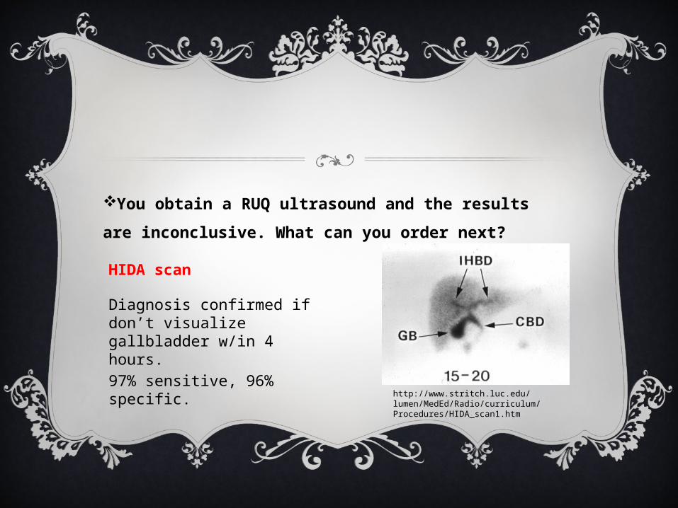
You obtain a RUQ ultrasound and the results are inconclusive. What can you order next?
http://www.stritch.luc.edu/lumen/MedEd/Radio/curriculum/Procedures/HIDA_scan1.htm
HIDA scan
Diagnosis confirmed if don’t visualize gallbladder w/in 4 hours.97% sensitive, 96% specific.
ACUTE CHOLECYSTITIS
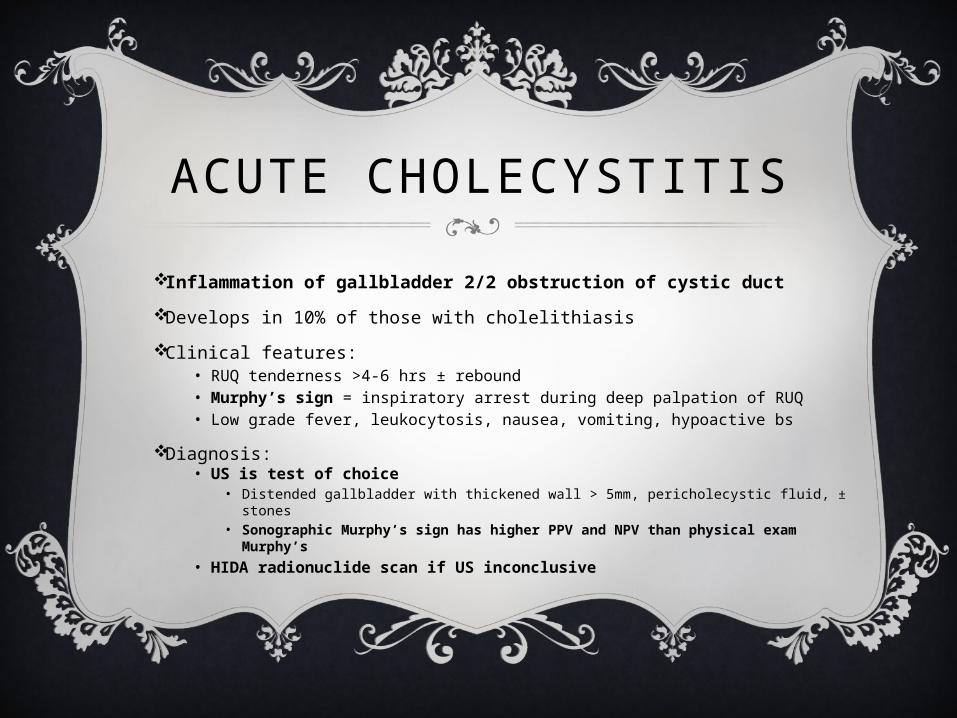
Inflammation of gallbladder 2/2 obstruction of cystic ductDevelops in 10% of those with cholelithiasis
Clinical features: • RUQ tenderness >4-6 hrs ± rebound• Murphy’s sign = inspiratory arrest during deep palpation of RUQ• Low grade fever, leukocytosis, nausea, vomiting, hypoactive bs
Diagnosis:• US is test of choice
• Distended gallbladder with thickened wall > 5mm, pericholecystic fluid, ± stones• Sonographic Murphy’s sign has higher PPV and NPV than physical exam
Murphy’s• HIDA radionuclide scan if US inconclusive
ACUTE CHOLECYSTITIS
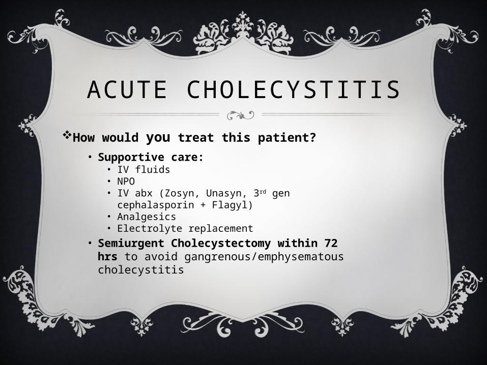
How would you treat this patient?• Supportive care:
• IV fluids• NPO• IV abx (Zosyn, Unasyn, 3rd gen cephalasporin +
Flagyl)• Analgesics• Electrolyte replacement
• Semiurgent Cholecystectomy within 72 hrs to avoid gangrenous/emphysematous cholecystitis
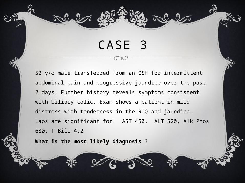
CASE 3
52 y/o male transferred from an OSH for intermittent abdominal pain and progressive jaundice over the past 2 days. Further history reveals symptoms consistent with biliary colic. Exam shows a patient in mild distress with tenderness in the RUQ and jaundice. Labs are significant for: AST 450, ALT 520, Alk Phos 630, T Bili 4.2
What is the most likely diagnosis ?
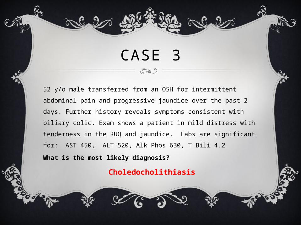
CASE 3
52 y/o male transferred from an OSH for intermittent abdominal pain and progressive jaundice over the past 2 days. Further history reveals symptoms consistent with biliary colic. Exam shows a patient in mild distress with tenderness in the RUQ and jaundice. Labs are significant for: AST 450, ALT 520, Alk Phos 630, T Bili 4.2
What is the most likely diagnosis?
Choledocholithiasis
CHOLEDOCHOLITHIASIS
Gallstones within the common bile duct or common hepatic duct, formed in situ or passed from gallbladderPresentation: asymptomatic (~50%) biliary colic ascending cholangitis, obstructive jaundice, acute pancreatitisDefinitions of dilated bile duct
• >6mm + 1mm per decade above 60 y/o• >10mm post-cholecystectomy• Dilated intrahepatic biliary tree
CHOLEDOCHOLITHIASIS
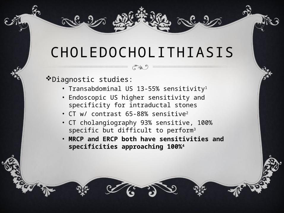
Diagnostic studies:• Transabdominal US 13-55% sensitivity1
• Endoscopic US higher sensitivity and specificity for intraductal stones
• CT w/ contrast 65-88% sensitive2
• CT cholangiography 93% sensitive, 100% specific but difficult to perform3
• MRCP and ERCP both have sensitivities and specificities approaching 100%4
CHOLEDOCHOLITHIASIS
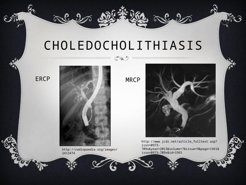
ERCP
MRCP
http://radiopaedia.org/images/2413474
http://www.jcdr.net/article_fulltext.asp?issn=0973-709x&year=2013&volume=7&issue=9&page=1941&issn=0973-709x&id=3365
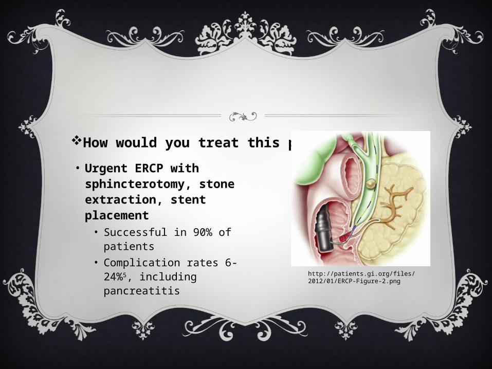
How would you treat this patient?• Urgent ERCP with
sphincterotomy, stone extraction, stent placement• Successful in 90% of
patients• Complication rates 6-
24%5, including pancreatitis
http://patients.gi.org/files/2012/01/ERCP-Figure-2.png
CASE 4
50 y/o female admitted to the MICU for AMS. Vitals include temp 39, HR 110, BP 90/60, RR 20, sat 96% on RA. Exam reveals a somnolent female with jaundice, scleral icterus, and guarding upon palpation of the RUQ. Labs reveal: WBC 16,000, AST 160, ALT 200, Alk Phos 650, T bili 8.0. Blood cultures are pending.
What is the most likely diagnosis?
CASE 4
50 y/o female admitted to the MICU for AMS. Vitals include temp 39, HR 110, BP 80/60, RR 20, sat 96% on RA. Exam reveals a somnolent female with jaundice, scleral icterus, and guarding upon palpation of the RUQ. Labs reveal: WBC 16,000, AST 160, ALT 200, Alk Phos 650, T bili 8.0. Blood cultures are pending.
What is the most likely diagnosis?
Acute Cholangitis
CHOLANGITISInfection of biliary tract 2/2 obstruction biliary stasis & bacterial overgrowth
• Ecoli & Klebsiella 70%, Enterococcus & Anaerobes (15%)Choledocholithiasis accounts for 60% of casesOther causes: pancreatic/biliary neoplasm, strictures, s/p ERCP, choledochal cystsClinical features:
• Charcot’s Triad: RUQ pain + Jaundice + Fever• Present in 60-79%
• Reynolds’ Pentad: Charcot’s triad + Hypotension + AMS• Present in ~15%
• Medical emergency if fever >40ºC, septic shock, peritoneal signs, or bilirubin > 10
CHOLANGITIS
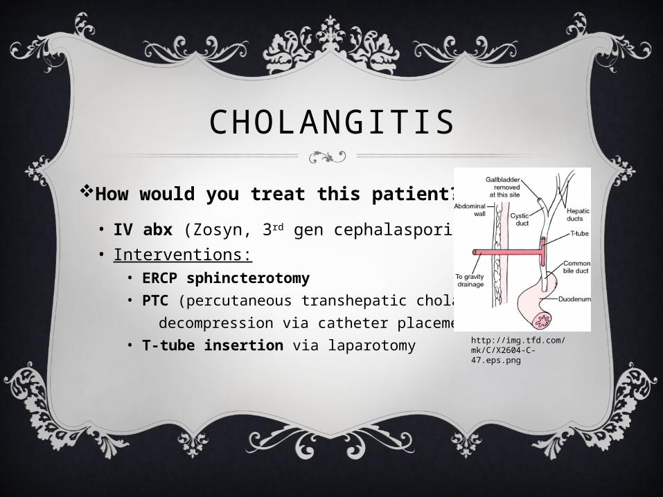
• IV abx (Zosyn, 3rd gen cephalasporin), IV fluids• Interventions:
• ERCP sphincterotomy• PTC (percutaneous transhepatic cholangiography)
decompression via catheter placement• T-tube insertion via laparotomy
How would you treat this patient?
http://img.tfd.com/mk/C/X2604-C-47.eps.png
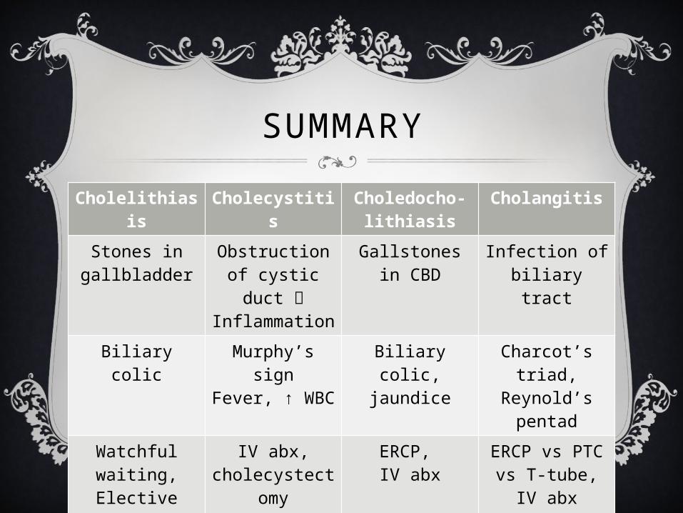
Cholelithiasis
Cholecystitis Choledocho-lithiasis
Cholangitis
Stones in gallbladder
Obstruction of cystic duct
Inflammation
Gallstones in CBD
Infection of biliary tract
Biliary colic Murphy’s sign
Fever, ↑ WBC
Biliary colic, jaundice
Charcot’s triad,
Reynold’s pentad
Watchful waiting,Elective surgery
IV abx, cholecystecto
my
ERCP, IV abx
ERCP vs PTC vs T-tube,
IV abx
SUMMARY

GUESS THE LFTS
WHAT’S THE DIAGNOSIS?
Pt presents with insidious onset of fatigue, anorexia, nausea, RUQ tenderness. He’s also noticed that his urine has been darker for the past couple of days and that his eyes have a yellow hue.
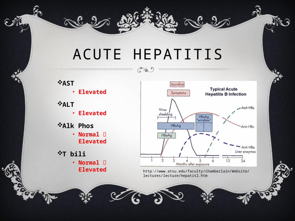
ACUTE HEPATITISAST
• Elevated
ALT• Elevated
Alk Phos• Normal
Elevated
T bili• Normal
Elevated http://www.atsu.edu/faculty/chamberlain/Website/lectures/lecture/hepatit2.htm
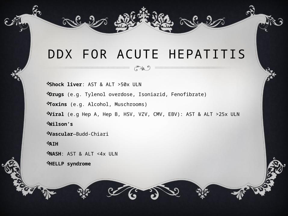
DDX FOR ACUTE HEPATITIS
Shock liver: AST & ALT >50x ULN
Drugs (e.g. Tylenol overdose, Isoniazid, Fenofibrate)
Toxins (e.g. Alcohol, Muschrooms)
Viral (e.g Hep A, Hep B, HSV, VZV, CMV, EBV): AST & ALT >25x ULN
Wilson’s Vascular—Budd-Chiari
AIHNASH: AST & ALT <4x ULN
HELLP syndrome
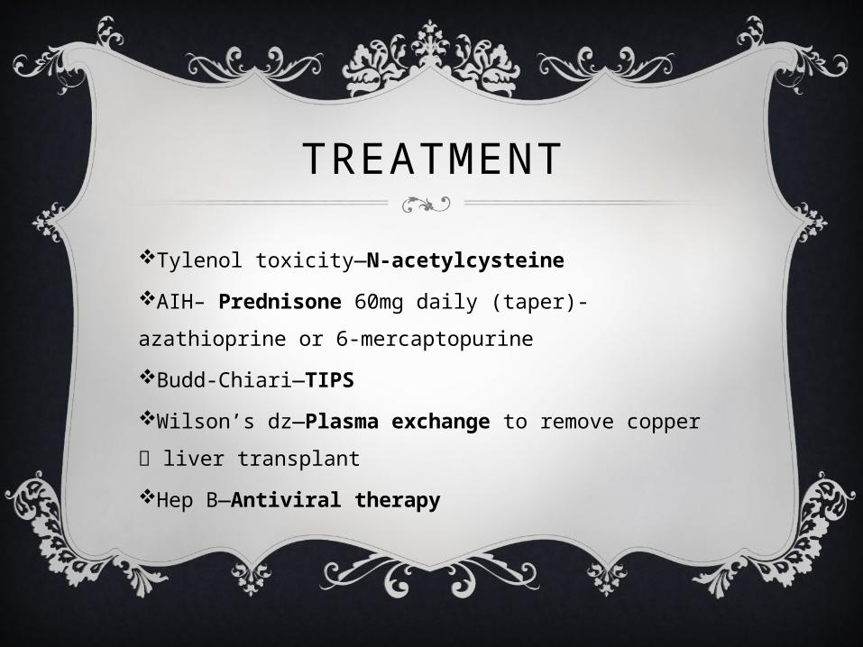
TREATMENT
Tylenol toxicity—N-acetylcysteineAIH– Prednisone 60mg daily (taper)- azathioprine or 6-mercaptopurine
Budd-Chiari—TIPSWilson’s dz—Plasma exchange to remove copper liver transplant
Hep B—Antiviral therapy
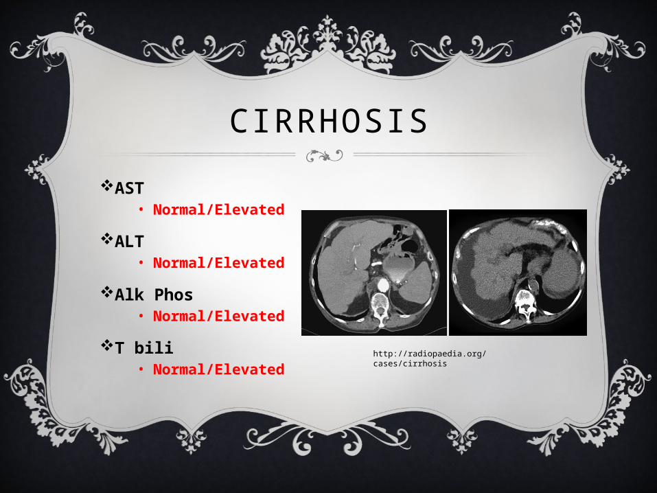
CIRRHOSISAST
• Normal/Elevated
ALT• Normal/Elevated
Alk Phos• Normal/Elevated
T bili• Normal/Elevated http://radiopaedia.org/cases/
cirrhosis
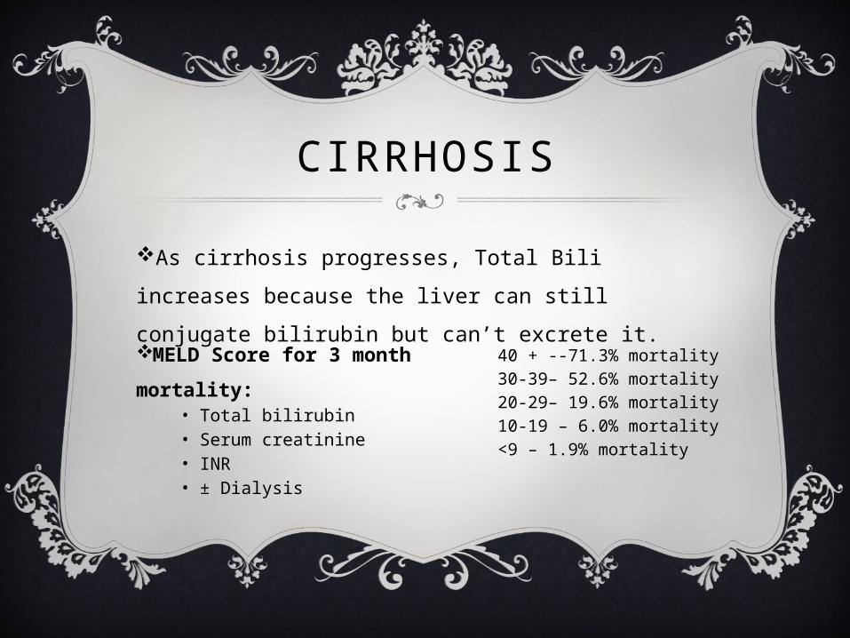
CIRRHOSIS
As cirrhosis progresses, Total Bili increases because the liver can still conjugate bilirubin but can’t excrete it. MELD Score for 3 month mortality:
• Total bilirubin• Serum creatinine• INR• ± Dialysis
40 + --71.3% mortality30-39– 52.6% mortality20-29– 19.6% mortality10-19 – 6.0% mortality<9 – 1.9% mortality
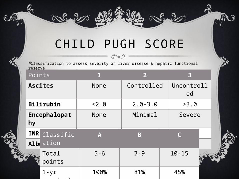
CHILD PUGH SCOREClassification to assess severity of liver disease & hepatic functional reserve
Points 1 2 3Ascites None Controlled Uncontrolle
dBilirubin <2.0 2.0-3.0 >3.0Encephalopathy
None Minimal Severe
INR <1.7 1.7-2.2 >2.2Albumin >3.5 2.8-3.5 <32.8Classificati
onA B C
Total points
5-6 7-9 10-15
1-yr survival
100% 81% 45%
2-yr survival
85% 57% 35%
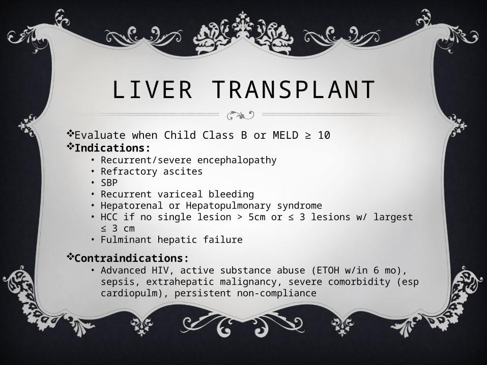
LIVER TRANSPLANTEvaluate when Child Class B or MELD ≥ 10Indications:
• Recurrent/severe encephalopathy• Refractory ascites• SBP• Recurrent variceal bleeding• Hepatorenal or Hepatopulmonary syndrome• HCC if no single lesion > 5cm or ≤ 3 lesions w/ largest ≤ 3 cm• Fulminant hepatic failure
Contraindications:• Advanced HIV, active substance abuse (ETOH w/in 6 mo),
sepsis, extrahepatic malignancy, severe comorbidity (esp cardiopulm), persistent non-compliance
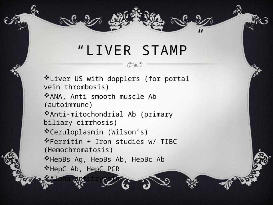
“LIVER STAMP”Liver US with dopplers (for portal vein thrombosis)ANA, Anti smooth muscle Ab (autoimmune)Anti-mitochondrial Ab (primary biliary cirrhosis)Ceruloplasmin (Wilson’s)Ferritin + Iron studies w/ TIBC (Hemochromatosis)HepBs Ag, HepBs Ab, HepBc AbHepC Ab, HepC PCRAlpha-antitrypsin
“LIVER STAMP”
AVERAGE COST?
~$1,200
Fatty liver diseases• Alcoholic liver disease• NASH/NAFLD
Viral hepatitis: Hep B, C, DAutoimmune
• Autoimune hepatitis• Primary biliary cirrhosis• Primary sclerosing
cholangitis
Cardiovascular• Budd-Chiari syndrome• Chronic right heart failure
CIRRHOSIS ETIOLOGY
Chronic biliary disease• Recurrent bacterial
cholangitis• Bile duct stenosis
Storage diseases• Hemochromatosis• Wilson disease• α-1-antitrypsin deficiency
Meds: APAP toxicity, MTXCryptogenic 10-15%
DIAGNOSTIC IMAGING
Ultrasound• Surface nodularity: 88% sensitive, 82-95% specific
(1)CT insensitive in early cirrhosisMRI also insensitive in early cirrhosis, but significant role in assessing small hepatocellular carcinoma (HCC)—develops in 10-25%Liver biopsy = gold standard for diagnosis
TREATMENTAscites
• Furosemide + Spironolactone with goal negative ~1L/day (~80% effective)• Lasix: Aldactone ratio of 2:5 helps maintain K+ (thus Lasix 40mg qday, Aldactone
100mg qday initially)
• Low-sodium diet (1-2 g/day)Refractory Ascites= no response on max doses of Lasix (160mg) & Aldactone (400mg)
• LVP 4-6L (does not improve mortality)• Albumin replacement controversial. AASLD 2009 guidelines recommend if >5L
removed, provide 6-8 g/L of albumin 25% (IIA, Grade C)• If >5L removed, can have post-paracentesis circulatory dysfxn via RAAS activation
• TIPS (↓ ascites in 75%, improves mortality but ↑ HE, 40% need revision for stent stenosis)
LAST BUT NOT LEAST…
ASCITES FLUID
THE SPECIAL “LFT”
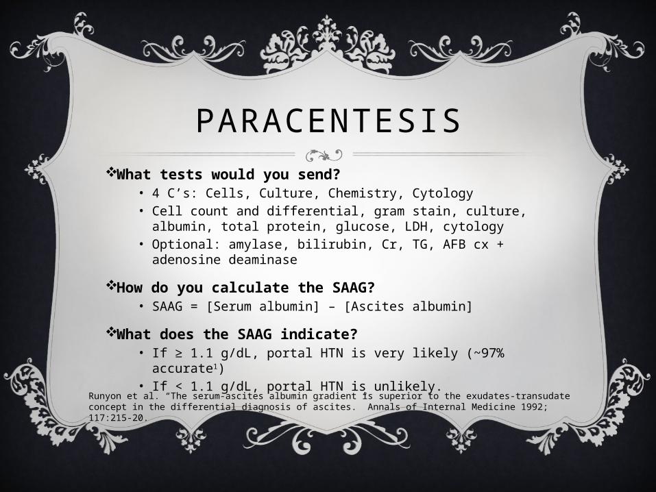
PARACENTESISWhat tests would you send?
• 4 C’s: Cells, Culture, Chemistry, Cytology• Cell count and differential, gram stain, culture, albumin,
total protein, glucose, LDH, cytology• Optional: amylase, bilirubin, Cr, TG, AFB cx + adenosine
deaminase
How do you calculate the SAAG?• SAAG = [Serum albumin] – [Ascites albumin]
What does the SAAG indicate?• If ≥ 1.1 g/dL, portal HTN is very likely (~97% accurate1)• If < 1.1 g/dL, portal HTN is unlikely.
Runyon et al. “The serum-ascites albumin gradient is superior to the exudates-transudate concept in the differential diagnosis of ascites.” Annals of Internal Medicine 1992; 117:215-20.
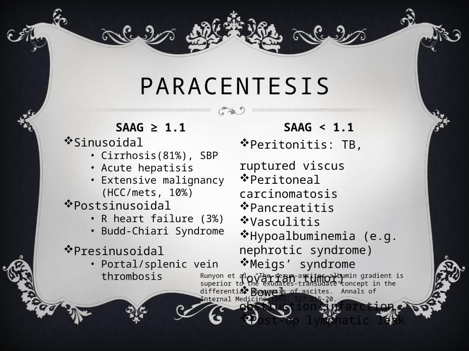
Sinusoidal• Cirrhosis(81%), SBP• Acute hepatisis• Extensive malignancy
(HCC/mets, 10%)Postsinusoidal
• R heart failure (3%)• Budd-Chiari Syndrome
Presinusoidal• Portal/splenic vein
thrombosis
Peritonitis: TB, ruptured viscusPeritoneal carcinomatosisPancreatitisVasculitisHypoalbuminemia (e.g. nephrotic syndrome)Meigs’ syndrome (ovarian tumor)Bowel obstruction/infarctionPost-op lymphatic leak
PARACENTESISSAAG ≥ 1.1 SAAG < 1.1
Runyon et al. “The serum-ascites albumin gradient is superior to the exudates-transudate concept in the differential diagnosis of ascites.” Annals of Internal Medicine 1992; 117:215-20.
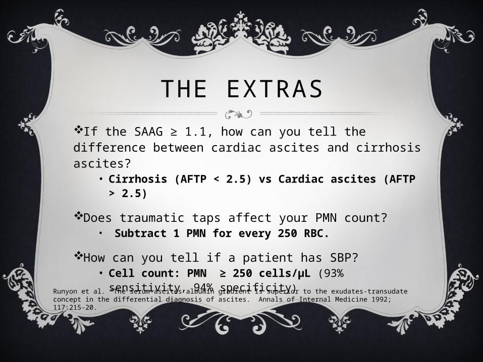
THE EXTRASIf the SAAG ≥ 1.1, how can you tell the difference between cardiac ascites and cirrhosis ascites?
• Cirrhosis (AFTP < 2.5) vs Cardiac ascites (AFTP > 2.5)
Does traumatic taps affect your PMN count?• Subtract 1 PMN for every 250 RBC.
How can you tell if a patient has SBP?• Cell count: PMN ≥ 250 cells/μL (93%
sensitivity, 94% specificity)Runyon et al. “The serum-ascites albumin gradient is superior to the exudates-transudate concept in the differential diagnosis of ascites.” Annals of Internal Medicine 1992; 117:215-20.
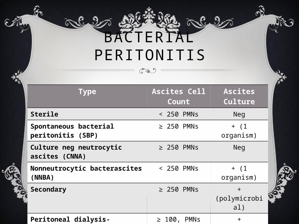
BACTERIAL PERITONITIS
Type Ascites Cell Count
Ascites Culture
Sterile < 250 PMNs Neg
Spontaneous bacterial peritonitis (SBP)
≥ 250 PMNs + (1 organism)
Culture neg neutrocytic ascites (CNNA)
≥ 250 PMNs Neg
Nonneutrocytic bacterascites (NNBA)
< 250 PMNs + (1 organism)
Secondary ≥ 250 PMNs + (polymicrobial)
Peritoneal dialysis-associated ≥ 100, PMNs predom
+
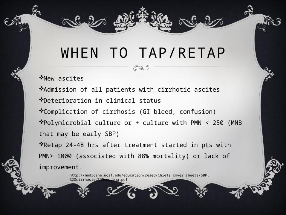
WHEN TO TAP/RETAPNew ascitesAdmission of all patients with cirrhotic ascitesDeterioration in clinical statusComplication of cirrhosis (GI bleed, confusion)Polymicrobial culture or + culture with PMN < 250 (MNB that may be early SBP)Retap 24-48 hrs after treatment started in pts with PMN> 1000 (associated with 88% mortality) or lack of improvement.
http://medicine.ucsf.edu/education/resed/Chiefs_cover_sheets/SBP,%20cirrhosis,%20empyema.pdf
REFERENCESAdamek HE, Albert J, Weitz M et-al. A prospective evaluation of magnetic resonance cholangiopancreatography in patients with suspected bile duct obstruction. Gut. 1998;43 (5): 680-3.Agabegi SS, Agabegi ED. Step –Up to Medicine, 3rd ed. 2013. Lippincott Williams & Wilkins. Philadelphia, PA.Caoili EM, Paulson EK, Heyneman LE et-al. Helical CT cholangiography with three-dimensional volume rendering using an oral biliary contrast agent: feasibility of a novel technique. AJR Am J Roentgenol. 2000;174 (2): 487-92.Cronan JJ. US diagnosis of choledocholithiasis: a reappraisal. Radiology. 1986;161 (1): 133-4.Guardino JM. Primo Gastro. 2008. Lippincott Williams & Wilkins. Philadelphia, PA.Miller FH, Hwang CM, Gabriel H et-al. Contrast-enhanced helical CT of choledocholithiasis. AJR Am J Roentgenol. 2003;181 (1): 125-30.Sabatine, MS. Pocket Medicine, 4th ed. 2011. Lippincott Williams & Wilkins. Philadelphia, PA.Sugiyama M, Suzuki Y, Abe N et-al. Endoscopic retreatment of recurrent choledocholithiasis after sphincterotomy. Gut. 2004;53 (12): 1856-9.Wiener C, Fauci AS, Braunwald E, et al. Harrison’s Principles of Internal Medicine Self-Assessment & Board Review, 17th ed. 2008. McGraw Hill. New York, NY.http://medicine.ucsf.edu/education/resed/Chiefs_cover_sheets/SBP,%20cirrhosis,%20empyema.pdfhttp://radiopaedia.orgSpecial thanks to Dr. Caroline Soyka for the inspiration!