Embed Size (px)
Citation preview
2263
INTRODUCTIONLight can play a number of roles in coordinating the temporal activity
of an organism. It has been most extensively studied as a signal for
entraining circadian rhythms. However, these same light signals
often act to modify or mask aspects of circadian output (Aschoff,
1960; Mrosovsky, 1999). Classic examples of masking by changes
in light include the initiation of singing in sparrows at lights-on
(Binkley et al., 1983) and the onset of hamster running behavior at
lights-off (Redlin and Mrosovsky, 1999). Although numerous
examples of masking have been identified, their underlying cellular
and molecular mechanisms and how they interact with circadian
systems are not well understood.
For many insects, the emergence of the adult (eclosion) is
under circadian regulation (Helfrich-Förster, 2006; Saunders,
1976; Saunders, 1982; Saunders, 2002). In Drosophilamelanogaster Meigen, the circadian rhythm of eclosion is based
on a central clock that depends on the interaction of a number
of proteins encoded by genes including Clock, cycle, period and
timeless (reviewed by Helfrich-Förster, 2006). The circadian
clock restricts the timing of Drosophila eclosion to a ‘gate’ that
occurs during the early part of the day (Engelmann and Honegger,
1966; Jackson, 1983; Lorenz et al., 1989). Gating results in a
discontinuous eclosion pattern in which entrained pharate adults
that complete adult development at night wait for the opening
of the gate at around dawn in order to eclose (Pittendrigh and
Skopik, 1970). Release of the neuropeptide eclosion hormone
(EH), a key regulator of ecdysis (reviewed by Truman, 2005),
is gated in the moth Manduca sexta, suggesting that eclosion
gating may result from circadian regulation of EH release. This
could occur directly as the result of declining ecdysone titers
that are associated with the activation of ecdysis (Hewes and
Truman, 1991). Alternatively, it could result from the ecdysone-
responsive release of ecdysis triggering hormone (ETH) (Kingan
and Adams, 2000; Zitnan et al., 1999; Zitnanova et al., 2001),
which stimulates EH release (Ewer et al., 1997; Kingan et al.,
1997; Zitnan et al., 1996). Pigment dispersing factor (PDF)
produced by the lateral neurons may influence the gate by
modulating ecdysteroid release from the prothoracic gland
(Myers et al., 2003).
The circadian pattern of Drosophila eclosion can be masked by
light. When administered close to the eclosion gate, a lights-on
(LOn) signal shifts the distribution of flies emerging within the gate.
This is manifest as a burst of eclosion soon after the LOn signal.
The pathway of light reception for this LOn response is distinct
from those utilized for circadian entrainment. Drosophila mutants
that lack both the ocelli and compound eyes lack the lights-on
response but nevertheless show normal circadian entrainment of their
ecdysis clock (Engelmann and Honegger, 1966). For circadian
regulation of locomotion and eclosion, the central clock resides in
lateral neurons of the brain that express central clock proteins, the
neuropeptide pigment-dispersing factor (PDF) and a cryptochrome
photoreceptor (reviewed by Helfrich-Förster, 2006; Nitabach and
Taghert, 2008).
Eclosion is regulated by a cascade of peptide hormones. These
peptides include EH from the brain, pre-ecdysis triggering hormone
(PETH) and ecdysis triggering hormone (ETH) from the epitracheal
glands (Park et al., 2002; Zitnan et al., 1996), and crustacean
cardioactive peptide (CCAP) from the ventral central nervous system
(CNS) (reviewed by Truman, 2005). To test the requirement for
EH in eclosion, molecular genetic tools were used to target the
ablation of the EH-expressing neurons in Drosophila (McNabb et
al., 1997). Surprisingly, these experiments showed that EH is not
strictly required for eclosion. However, the EH cell knockouts had
significant defects. A third died at larval ecdyses with defects in
tracheal filling, and the two thirds that eclosed as adults had defects
in eclosion and post-eclosion behaviors. Interestingly, the EH cell
knockouts had normal circadian eclosion rhythms but lacked the
LOn response. Thus, like the retinal photoreceptors, the EH neurons
appear to be components of the LOn pathway. In this paper, we
define the basic characteristics of the LOn response and examine
the way this signal interacts with EH release, eclosion, and post-
ecdysial wing spreading.
The Journal of Experimental Biology 211, 2263-2274Published by The Company of Biologists 2008doi:10.1242/jeb.015818
Light and peptidergic eclosion hormone neurons stimulate a rapid eclosion responsethat masks circadian emergence in Drosophila
Susan L. McNabb* and James W. TrumanDepartment of Zoology, Box 351800, University of Washington, Seattle, WA 98195-1800, USA
*Author for correspondence (e-mail: [email protected])
Accepted 12 May 2008
SUMMARYLight signals can entrain circadian clocks, but they can also mask aspects of the circadian output. We have analyzed the maskingeffects of a lights-on (LOn) signal on Drosophila eclosion. The LOn response results in 12–21% of the flies that emerge on a givenday eclosing within 10min of the LOn signal. Flies that lack the neuropeptide eclosion hormone (EH), or in which its release isinhibited by the tetanus toxin light chain, lack the response. Optic photoreceptors in both the ocelli and the compound eyesappear to be required for the response. The LOn signal has two effects: (1) it drastically reduces the interval between EH releaseand eclosion, presumably by suppressing a transient descending inhibition that immediately follows EH release, and (2) itstimulates premature EH release. The LOn signal does not influence the latency of wing spreading, an EH-regulated post-ecdysisbehavior.
THE JOURNAL OF EXPERIMENTAL BIOLOGY
2264
MATERIALS AND METHODSDrosophila strains
The w1118�UAS-reaper (UAS-rpr; see below) control flies were
generated by crossing males from the w1118 strain to females of the
UAS-rpr strain (McNabb et al., 1997). The w1118 strain was the
recipient strain for the transposon that contains the GAL4transcription factor under the regulation of EH gene upstream
sequences (see below). When crossed to the UAS-rpr strain, it is
the control for the EHups-Gal4�UAS-rpr flies. Since the UAS-rprinsertion is carried on a y w67 c23 X chromosome, the hemizygous
male progeny of this cross have yellow cuticle and pale apricot eyes.
Female progeny are heterozygous w1118/y w67 c2 UAS-rpr, with wild-
type cuticle and eyes that are slightly paler than those of the males.
Despite these differences, no difference in LOn responsiveness or
wing spreading latency (see below) was detected between these
males and females.
The EH cell knockout flies (McNabb et al., 1997) were generated
by using the GAL4-UAS system (Brand and Perrimon, 1993). EH
gene upstream sequences (EHups) fused to Gal4 (EHups-Gal4;P{GAL4-Eh2.4}) were used to drive expression of the cell
death gene reaper (rpr) in the neurons that produce EH. Male flies
from the C21 EHups-Gal4 strain were crossed to females of the
UAS-rpr strain. Their progeny are referred to as EHups�UAS-rprthroughout this paper. Since the C21 transposon is located on the
second chromosome, the progeny possess the X chromosome
genotype described for the w1118�UAS-rpr strain above, which
results in male progeny with yellow cuticle and females with wild-
type cuticle. All knockouts were heterozygous for the second
chromosome, i.e. C21/+. The EHups transposon conferred bright
orange-red eyes on all progeny, although the eyes of females were
slightly paler than those of the males. Despite differences in cuticle
and eye pigmentation, no differences between males and females
were detected in the LOn response or wing spreading latency.
Canton-S (CS), a standard wild-type lab strain, was used as a
control for the ocelliless and eyeless strains. Although the progenitor
strains for these mutants is unknown, it is likely to be CS. Strains
that lacked ocelli were ocelliless (oc1) (Flybase, 1999; Lindsley and
Zimm, 1992) and sine oculis (so+2). Strains that lacked compound
eyes were alleles of eyes absent [eya2; also called clifteya-2 (Bonini
et al., 1993)] and eya1 [also called clieya-1 (Eissenberg and Ryerse,
1991; Sved, 1986)]. To ensure that the lack of a LOn response
observed for the ocelliless and eyeless strains were not due to
locomotor defects, they were tested for geotaxis using a
countercurrent assay (Benzer, 1967). All exhibited positive geotaxis.
These strains were also tested by immunocytochemistry for normal
levels and release of EH. The CNS of flies that were staged at
approximately 6h prior to eclosion and those that had just eclosed
were labeled with anti-EH and analyzed as described below. These
strains appeared to synthesize normal levels of EH and to release
it at eclosion as expected.
The w1118�UAS-TNT-L and EHups�UAS-TNT-L flies were
generated by crossing males from the w1118 strain or the C21 EHups-Gal4 strain, respectively, to females of the UAS-TNT-L strain
(Sweeney et al., 1995). We detected no sex-specific differences in
cuticle or eye pigmentation, LOn response or wing spreading latency.
Lights-on (LOn) assaysFlies were cultured in half-pint culture bottles containing cornmeal
agar food, at 25°C. Cultures were raised continuously under a
14 h:10 h light:dark (14L:10D) cycle at approximately 750 lx.
Embryos were collected over 24h to an experimentally determined
optimum density that depended on both parental strain fecundity
and mortality of the progeny. Food was removed from the bottles
on the afternoon prior to assay. Five bottles of flies were reared for
each assay condition and the eclosing flies for each treatment were
pooled upon collection. Flies that emerged before the day of assay
were removed just prior to lights-off of the preceding night.
Emerging flies were collected at 10min intervals, except as noted,
and subsequently counted. One final collection of adults was made
just prior to the end of the photoperiod and the total number of flies
that emerged between the first and last collection periods used to
generate eclosion rates as a percentage of the day’s total eclosion.
Each treatment group was reared and tested in parallel.
For the LOn shift paradigm, entrained adults were subjected to
normal lighting or to a LOn that was shifted either earlier (–1h or
–2h) or later (+2h) than normal. Each strain was tested at least
twice and each test yielded qualitatively similar results.
ImmunocytochemistryFly CNSs were dissected into Ca2+-free Ikeda’s saline (Ashburner,
1989) then fixed in 3.7% formaldehyde in phosphate-buffered saline
(PBS) overnight at 4°C. Samples were washed extensively
(5�10min) with PBS containing 0.3% Triton X-100 (PBST) and
blocked in 1% normal donkey serum (Jackson ImmunoResearch
Laboratories, Inc., West Grove, Pennsylvania, USA). They were
then incubated overnight at 4°C in a rabbit anti-EH antiserum
(Copenhaver and Truman, 1986) at 1:100 in PBST. Samples were
washed extensively in PBST, and then incubated in donkey anti-
rabbit Cy5 (Jackson ImmunoResearch Laboratories, Inc.) at 1:1000
overnight at 4°C. Samples were then washed extensively, mounted
on poly-lysine-coated coverslips, passed through an ethanol
dehydration series, cleared in xylene, and mounted in DPX
histological mountant (Fluka BioChemika, Sigma Aldrich Chemie,
Steinheim, Germany). Preparations were examined on a Bio-Rad
MRC-600 (Hercules, California, USA) confocal microscope. Image
stacks were scored visually and representative z-series were
collapsed to provide two-dimensional images.
To examine EH release in w1118�UAS-TNT-L and EHups�UAS-TNT-L strains, CNSs were collected either 8–11h prior to eclosion
before meconium transport (Kimura and Truman, 1990) or within
1min post-eclosion.
To examine the effects of light on EH release, we used
w1118�UAS-rpr flies. On the night before eclosion, 0–2h before
lights-off, late pharate adults with anterior meconia were removed
from their puparial cases. One group was dissected immediately as
a no-treatment control and the rest were returned to 14L:10D
conditions. One group was dissected 1h before normal LOn to verify
that EH release had not occurred; another was dissected 10–20min
after LOn. Two groups served as controls: one was maintained in
darkness and dissected at the same time as the post-LOn set, the
other was observed for time of eclosion. The latter control showed
that the flies selected at this stage emerged approximately 1h after
the normal LOn signal. The CNSs of flies in the no-treatment control
group were dissected into saline and fixed overnight. All other
groups were dissected directly into fix to preserve their in vivorelease state and maintained in fix overnight at 4°C. They were
subsequently treated as described above. Samples were examined
on a confocal microscope (as above) and graded according to
intensity of labeling, on a scale of 0–4. Z-series were collapsed to
provide two-dimensional images.
Wing spreading assayTo determine if light influences the interval between eclosion and
the completion of wing spreading (referred to as the wing spreading
S. L. McNabb and J. W. Truman
THE JOURNAL OF EXPERIMENTAL BIOLOGY

2265A lights-on eclosion response in Drosophila
latency; WSL), newly emerged flies from LOn shift experiments
were collected, placed four to a vial, held in the light at 25°C and
scored for wing spreading at 10 min intervals. It was important to
avoid disturbing the flies during this time as physical agitation
delayed the wing spreading process. For assessing the effect of
the LOn signal, we examined only the flies that eclosed within
the first 20 min after LOn. Each experiment was performed at least
twice.
RESULTSThe lights-on eclosion response is independent of the rate of
developmentPopulations of newly emerging adult Drosophila exhibit a LOn
response when entrained to a 14L:10D cycle. As shown in Fig.1,
the LOn response is independent of the development rate of these
flies. This was demonstrated using the w1118�UAS-rpr strain, a strain
that showed a robust LOn response in previous studies (McNabb
et al., 1997). Eggs collected over a 24h period and subsequently
maintained under a 14L:10D cycle emerged as adults over a 3–4
day period. Since previous experiments demonstrated that the
largest peak of the day’s eclosion takes place within 1h of LOn,
we focused on the distribution of eclosion around the beginning of
the LOn signal, collecting newly emerged flies at 10min intervals.
We also counted the total number of flies that emerged each day
and have expressed the emergence observed within a particular time
bin as a percentage of the day’s total eclosion. This experiment was
repeated three times and yielded very similar results each time. Very
few flies eclosed during the 9h of darkness between lights-off and
–1h, on average 7±2% of the day’s eclosion whereas 42±4% of the
day’s eclosion took place within 2h of light onset on each day
sampled.
Most of the adults emerged during the first 2 days of the
distribution, then eclosion declined on days3 and 4. A few flies
from this experiment (85 of 2654; 3%) had previously eclosed during
the afternoon of day 0, within the distribution expected for normal
entrained eclosion. A strong LOn response was observed for each
day’s eclosion irrespective of the numbers eclosing. This response
resulted in 12–21% of the flies that eclosed throughout the entire
day emerging in the 10min interval immediately following LOn.
The effect of the LOn signal was short-lived; in the second 10min
following LOn, there was a 53–78% decline in eclosion rate, and
often it decreased to the level seen for the next few hours.
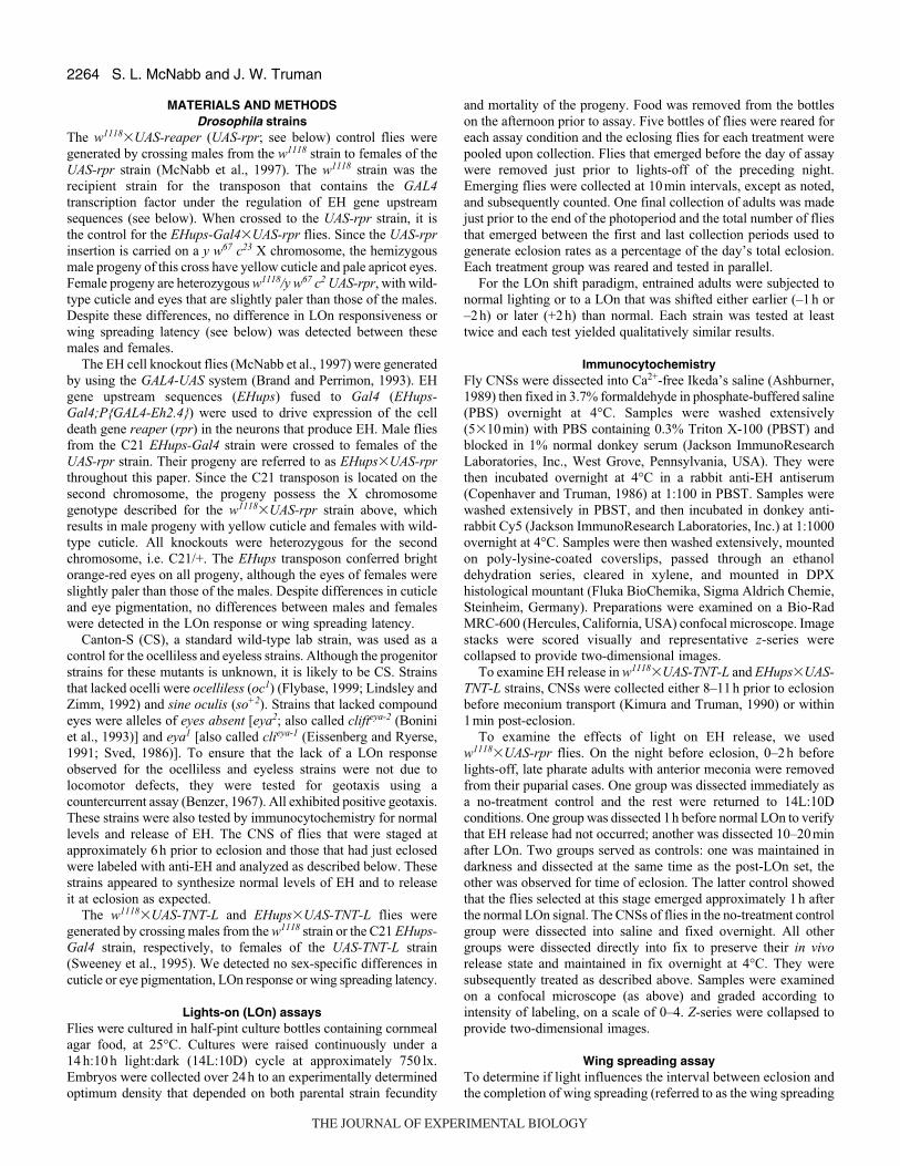
The effects of a light pulse on stimulating eclosionTo separate the effects of light as a LOn signal from its role as an
entraining signal, we used a pulse as a LOn signal. Entrained
w1118�UAS-rpr flies were subjected in parallel to either light at the
normal time, extended darkness or extended darkness except for a
brief (20min) pulse of light. Fig.2 summarizes the results of four
independent replicate experiments for which sample sizes were
between 993 and 1316. For flies that received light at the normal
LOn time, 14% of the day’s total eclosion occurred within 20min
of the signal (Fig.2A). This ‘burst’ of eclosion was not seen for
flies maintained in darkness (Fig.2B). Instead, those flies showed
a normal distribution of eclosion that ranged up to a maximum of
5% of the daily emergence per 10min collection window. When a
20min light pulse was delayed to 1h after the expected LOn signal
(Fig.2C), it also resulted in a massive eclosion burst (25% of the
day’s eclosions in 20min) that rapidly tailed off. Despite the
differences in eclosion distributions, the proportion of the day’s total
eclosion that ensued in the first 4h after normal LOn was very similar
under each of the three conditions: 60% for flies that received the
normal LOn, 62% for flies that received the pulse, and 59% for
flies held in the dark.
Effects of varying the time of the LOn signal on control andEH cell knockout flies
Experiments were performed to determine the effects of early or
late LOn signals on eclosion in w1118�UAS-rpr controls and in the
eclosion hormone (EH) cell knockout strain. Entrained adults were
N=350
N=498
N=837
N=834
w1118�UAS-rpr
Hours from lights-on
Per
cent
age
of d
ay’s
ecl
osio
n
Day 1
Day 2
Day 3
Day 4
12
25
20
15
16
12
8
4
16
12
8
–1 0 1 2
4
10
5
8
4
Fig. 1. Effects of developmental age on the rapid eclosion response to light.Representative data for a population of w1118�UAS-rpr flies that resultedfrom a 1 day egg collection, monitored every morning for 4 days. Emergingflies were collected every 10 min between –1 h and +2 h relative to lights-on(LOn, 0 h). The amount of eclosion is normalized to the day’s total eclosion.The horizontal bar below the day 4 panel represents the time relative tolights-on; black for the dark, white for the light. N, the total number of fliescollected each day.
THE JOURNAL OF EXPERIMENTAL BIOLOGY
2266
subjected to normal lighting or to a LOn signal that was either
advanced (–1h or –2h) or delayed (+2h) relative to the expected
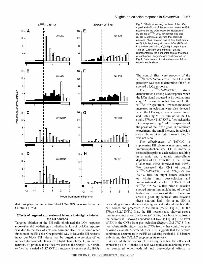
LOn signal. Representative results are shown in Fig.3. For the
w1118�UAS-rpr flies, the normal LOn signal resulted in a robust
burst of eclosion, with 29% of the day’s emergence occurring during
the first 20min (Fig.3A). Delaying the light signal by 2h resulted
in a corresponding delay in the abrupt eclosion burst, with about
14% of the flies emerging during the 20 min following LOn
(Fig.3B). This delayed LOn peak was smaller than that observed
at normal LOn because the majority of flies that would eclose during
this gate had already emerged. However, this peak represented about
41% of the flies that had not yet eclosed at the time of the shifted
LOn signal. This percentage is similar to that seen in Fig.3A, where
36% of the flies emerged within 20min of the LOn signal. When
the LOn signal was advanced by 1h there was no immediate burst
of eclosion (Fig.3C), but a substantial increase in eclosion was
observed during the next hour as compared to flies that were still
in the dark (compare with Fig.3B). Similar results were obtained
when the LOn signal was advanced by 2h (Fig.3D).
The LOn response observed for the w1118�UAS-rpr controls was
absent in the EH cell knockout (KO) flies (Fig.3E–H). The EH cell
KOs did not show an eclosion burst in response to any of the
conditions tested. Advancing the LOn signal failed to swell the
leading edge of the eclosion distribution (compare Fig.3G,H with
dark region of Fig.3F). Overall, the phase of eclosion relative to
the L:D cycle is the same in controls and the EH cell KOs (McNabb
et al., 1997). Hence, entrainment of the circadian clock for eclosion
is normal. It is only the masking of the LOn signal that is missing
upon removal of the EH neurons.
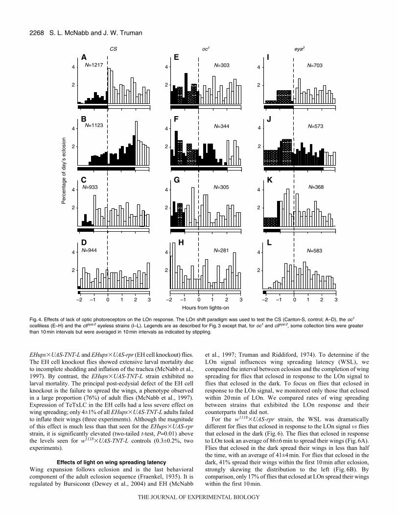
The role of the compound eyes and ocelli in the LOnresponse
A previous report showed that flies that lack both compound eyes
and ocelli due to a mutant allele of the sine oculis (so) locus, so1,fail to show a LOn response (Engelmann and Honegger, 1966). To
determine if the LOn response requires only one or both of the sets
of optic photoreceptors, we tested mutant strains that lack either the
compound eyes or the ocelli. The strains that had no ocelli were
mutant for the oc1 allele of ocelliless (Flybase, 1999; Lindsley and
Zimm, 1992) and the so+2 (Heitzler et al., 1993) allele of sine oculis.
The strains that lacked compound eyes were mutant for alleles of
eyes absent, eya2 (clieya-2; Bonini et al., 1993) and eya1 (clieya-1;Eissenberg and Ryerse, 1991; Sved, 1986). The Canton-S (CS) wild-
type strain was used as a control. Preliminary results obtained with
oc1 and so+2 were essentially identical. Similarly, results obtained
with eya2 and eya1 were essentially identical to each other. It was
difficult to obtain large cultures of each of these mutant strains
because of their reduced viability, so we focused on oc1 and eya2
for the detailed studies described below.
The eclosion patterns of the CS and w1118�UAS-rpr strains
differed in a few aspects. First, for CS, the normal entrained eclosion
distribution started about an hour earlier and a substantial number
emerged prior to LOn. Secondly, the CS LOn response was not as
robust (Fig.4A–D). In addition, the phase of the CS eclosion gate
was much broader; by 3h after normal LOn, only 32% of the flies
had eclosed vs 69% of w1118�UAS-rpr flies (χ2-test, P<0.0001).
The observation that the CS strain showed a definite LOn response
when the LOn signal was advanced to –1h and –2h suggests that
the CS strain is competent to respond to light earlier in the day than
is the w1118�UAS-rpr strain. We assume that this difference is due
to the phase of the eclosion gate being earlier for the CS than the
w1118�UAS-rpr strain (compare Fig.3B with Fig.4B).
Fly strains that lacked either compound eyes or ocelli lacked the
LOn response (Fig. 4E–L) despite normal circadian locomotor
rhythms (Vosshall and Young, 1995) and geotaxis. For both the oc1
and the eya2 strains, no significant increases in eclosion rate were
observed regardless of when the flies were exposed to a LOn signal
(χ2-test, P<0.0001). The apparent increase seen in Fig.4F was not
seen upon repetition of this test and thus appears to be due to random
fluctuation, much like the variation observed throughout the day.
In both mutant strains, particularly oc1, the eclosion rate was variable
and substantial eclosion preceded the LOn signal. However, overall
oc1 eclosion appeared circadian. Few flies emerged during the night
(7% for oc1 vs 8% for CS) and the proportion of the day’s eclosion
S. L. McNabb and J. W. Truman
A
B
C
w1118�UAS-rpr
Hours from normal lights-on
Per
cent
age
of d
ay’s
ecl
osio
n
25
20
15
10
5
25
20
15
10
5
25
20
15
10
5
0 1 2 3 4
Fig. 2. Effects of a light pulse on the rapid eclosion response. Flies of thew1118�UAS-rpr strain (A) received light continuously from the time ofnormal LOn, (B) were held in the dark or (C) received a 20 min pulse oflight beginning at +1 h from normal LOn. Axes and coloring are asdescribed for Fig. 1. Data bars give the means ± s.e.m. for four trials(N=993–1316 per test condition per trial).
THE JOURNAL OF EXPERIMENTAL BIOLOGY
2267A lights-on eclosion response in Drosophila
that took place within the first 3h of LOn (28%) was similar to the
CS strain (32%).
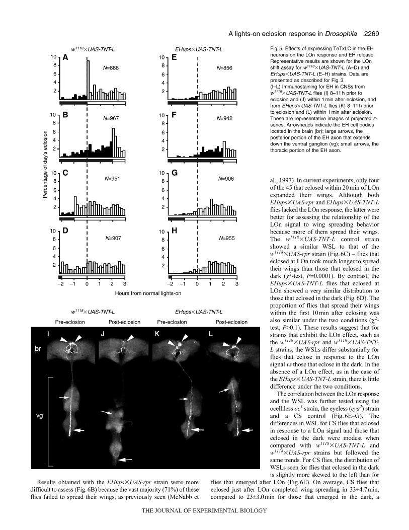
Effects of targeted expression of tetanus toxin light chain inthe EH neurons
Targeted ablation of the EH cells eliminated the LOn response
(above) but did not distinguish whether the loss of the LOn response
was due to the lack of eclosion hormone itself or to some other
function of the EH cells. One potential way to leave the EH neurons
intact but block EH release was by targeting expression of an
intracellular form of tetanus toxin light chain (TeTxLC) in the EH
neurons. To produce these flies, we crossed the EHups-Gal4 strain
to flies that carried a UAS-TNT-L transgene (Sweeney et al., 1995).
The control flies were progeny of the
w1118�UAS-TNT-L cross. The LOn shift
paradigm was used to determine if the flies
showed a LOn response.
The w1118�UAS-TNT-L strain
demonstrated a strong LOn response when
the LOn signal occurred at its normal time
(Fig.5A,B), similar to that observed for the
w1118�UAS-rpr strain. However, moderate
increases in eclosion were also detected
when the LOn signal was advanced to –1
and –2h (Fig.5C,D), similar to the CS
strain. EHups�UAS-TNT-L flies lacked the
LOn response (Fig.5E–H) irrespective of
the phase of the LOn signal. In a replicate
experiment, the small increase in eclosion
rate at the onset of light shown in Fig.5F
was not seen.
The effectiveness of TeTxLC in
suppressing EH release was assessed using
immunocytochemistry. EH is normally
released just prior to each ecdysis, resulting
in a rapid and dramatic intracellular
depletion of EH from the EH cell axons
(Baker et al., 1999; Horodyski et al., 1993).
We harvested the CNS of control
w1118�UAS-TNT-L and EHups�UAS-TNT-L flies the night before eclosion
or within 1 min post-eclosion and
immunostained them for EH. The CNS of
w1118�UAS-TNT-L flies prior to eclosion
showed strong immunolabeling of the cell
bodies and processes of the EH neurons
(N=4; Fig.5I). By contrast, after eclosion
these neurons had little or no EH in
descending axons in the ventral ganglion and reduced levels in the
cell bodies and processes in the brain (N=12; Fig.5J). In the
EHups�UAS-TNT-L flies, the EH neurons also showed strong
immunostaining prior to eclosion (N=5; Fig.5K), but after eclosion
the neurons still showed abundant EH (N=14; Fig.5L). The level
of EH in the CNSs from post-eclosion EHups�UAS-TNT-L flies
was substantially higher than in CNSs from either control or pre-
eclosion EHups�UAS-TNT-L flies. This suggests that the peptide
continues to accumulate in the EH cells during the final 8–11h before
ecdysis and that TeTxLC suppresses its release.
As an additional means of assessing whether the effects of
expressing TeTxLC in the EH cells was equivalent to ablating them,
we compared other ecdysial and post-ecdysial effects in
N=481
N=959 N=429
N=922
N=1250
N=1042
N=443
N=458
w1118�UAS-rpr EHups�UAS-rpr
Hours from normal lights-on
Per
cent
age
of d
ay’s
ecl
osio
n
16
12
8
4
16
12
8
4
16
12
8
4
16
12
8
4
16
12
8
4
16
12
8
4
16
12
8
4
16
12
8
4
–2 –1 0 1 2 3 –2 –1 0 1 2 3
D H
C G
B F
A E
Fig. 3. Effects of varying the time of the LOnsignal and of loss of the eclosion hormone (EH)neurons on the LOn response. Eclosion of(A–D) the w1118�UAS-rpr control flies and(E–H) EHups�UAS-rpr flies that lack EHneurons. Flies received one of four treatments:(A,E) light beginning at normal LOn, (B,F) heldin the dark until +2 h, (C,G) light beginning at–1 h or (D,H) light beginning at –2 h, asrepresented by the horizontal bars at the baseof each panel. Legends are as described forFig. 1. Data from an individual representativeexperiment is shown.
THE JOURNAL OF EXPERIMENTAL BIOLOGY
2268
EHups�UAS-TNT-L and EHups�UAS-rpr (EH cell knockout) flies.
The EH cell knockout flies showed extensive larval mortality due
to incomplete shedding and inflation of the trachea (McNabb et al.,
1997). By contrast, the EHups�UAS-TNT-L strain exhibited no
larval mortality. The principal post-ecdysial defect of the EH cell
knockout is the failure to spread the wings, a phenotype observed
in a large proportion (76%) of adult flies (McNabb et al., 1997).
Expression of TeTxLC in the EH cells had a less severe effect on
wing spreading; only 4±1% of all EHups�UAS-TNT-L adults failed
to inflate their wings (three experiments). Although the magnitude
of this effect is much less than that seen for the EHups�UAS-rprstrain, it is significantly elevated (two-tailed t-test, P=0.01) above
the levels seen for w1118�UAS-TNT-L controls (0.3±0.2%, two
experiments).
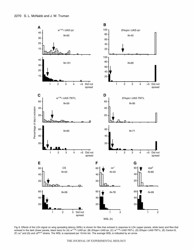
Effects of light on wing spreading latency Wing expansion follows eclosion and is the last behavioral
component of the adult eclosion sequence (Fraenkel, 1935). It is
regulated by Bursiconα (Dewey et al., 2004) and EH (McNabb
et al., 1997; Truman and Riddiford, 1974). To determine if the
LOn signal influences wing spreading latency (WSL), we
compared the interval between eclosion and the completion of wing
spreading for flies that eclosed in response to the LOn signal to
flies that eclosed in the dark. To focus on flies that eclosed in
response to the LOn signal, we monitored only those that eclosed
within 20 min of LOn. We compared rates of wing spreading
between strains that exhibited the LOn response and their
counterparts that did not.
For the w1118�UAS-rpr strain, the WSL was dramatically
different for flies that eclosed in response to the LOn signal vs flies
that eclosed in the dark (Fig.6). The flies that eclosed in response
to LOn took an average of 86±6min to spread their wings (Fig.6A).
Flies that eclosed in the dark spread their wings in less than half
the time, with an average of 41±4min. For flies that eclosed in the
dark, 41% spread their wings within the first 10min after eclosion,
strongly skewing the distribution to the left (Fig. 6B). By
comparison, only 17% of flies that eclosed at LOn spread their wings
within the first 10min.
S. L. McNabb and J. W. Truman
N=933
N=1123
N=1217
N=944
N=305
N=344
N=303
N=281
N=368
N=573
N=703
N=583
CS oc1 eya2
Hours from lights-on
Per
cent
age
of d
ay’s
ecl
osio
n4
2
4
2
4
2
4
2
4
2
4
2
4
2
4
2
4
2
4
2
4
2
4
2
–2 –1 0 1 2 3 –2 –1 0 1 2 3 –2 –1 0 1 2 3
EA I
F J
G
B
C K
HD L
Fig. 4. Effects of lack of optic photoreceptors on the LOn response. The LOn shift paradigm was used to test the CS (Canton-S, control; A–D), the oc1
ocelliless (E–H) and the clieya-2 eyeless strains (I–L). Legends are as described for Fig. 3 except that, for oc1 and clieya-2, some collection bins were greaterthan 10 min intervals but were averaged in 10 min intervals as indicated by stippling.
THE JOURNAL OF EXPERIMENTAL BIOLOGY
2269A lights-on eclosion response in Drosophila
Results obtained with the EHups�UAS-rpr strain were more
difficult to assess (Fig.6B) because the vast majority (71%) of these
flies failed to spread their wings, as previously seen (McNabb et
al., 1997). In current experiments, only four
of the 45 that eclosed within 20min of LOn
expanded their wings. Although both
EHups�UAS-rpr and EHups�UAS-TNT-Lflies lacked the LOn response, the latter were
better for assessing the relationship of the
LOn signal to wing spreading behavior
because more of them spread their wings.
The w1118�UAS-TNT-L control strain
showed a similar WSL to that of the
w1118�UAS-rpr strain (Fig.6C) – flies that
eclosed at LOn took much longer to spread
their wings than those that eclosed in the
dark (χ2-test, P=0.0001). By contrast, the
EHups�UAS-TNT-L flies that eclosed at
LOn showed a very similar distribution to
those that eclosed in the dark (Fig.6D). The
proportion of flies that spread their wings
within the first 10min after eclosing was
also similar under the two conditions (χ2-
test, P>0.1). These results suggest that for
strains that exhibit the LOn effect, such as
the w1118�UAS-rpr and w1118�UAS-TNT-L strains, the WSLs differ substantially for
flies that eclose in response to the LOn
signal vs those that eclose in the dark. In the
absence of a LOn effect, as in the case of
the EHups�UAS-TNT-L strain, there is little
difference under the two conditions.
The correlation between the LOn response
and the WSL was further tested using the
ocelliless oc1 strain, the eyeless (eya2) strain
and a CS control (Fig. 6E–G). The
differences in WSL for CS flies that eclosed
in response to a LOn signal and those that
eclosed in the dark were modest when
compared with w1118�UAS-TNT-L and
w1118�UAS-rpr strains but followed the
same trends. For CS flies, the distribution of
WSLs seen for flies that eclosed in the dark
is slightly more skewed to the left than for
flies that emerged after LOn (Fig.6E). On average, CS flies that
eclosed just after LOn completed wing spreading in 33±4.7min,
compared to 23±3.0min for those that emerged in the dark, a
w1118�UAS-TNT-L EHups�UAS-TNT-L
w1118�UAS-TNT-L EHups�UAS-TNT-L
Hours from normal lights-on
Per
cent
age
of d
ay’s
ecl
osio
n
10
8
6
4
2
10
8
6
4
2
10
8
6
4
2
10
8
6
4
2
10
8
6
4
2
10
8
6
4
2
10
8
6
4
2
10
8
6
4
2
–2 –1 0 1 2 3 –2 –1 0 1 2 3
D H
C G
B F
A E
Pre-eclosion Pre-eclosionPost-eclosion Post-eclosion
N=888
N=967
N=951
N=907
N=856
N=942
N=906
N=955
Fig. 5. Effects of expressing TeTxLC in the EHneurons on the LOn response and EH release.Representative results are shown for the LOnshift assay for w1118�UAS-TNT-L (A–D) andEHups�UAS-TNT-L (E–H) strains. Data arepresented as described for Fig. 3.(I–L) Immunostaining for EH in CNSs fromw1118�UAS-TNT-L flies (I) 8–11 h prior toeclosion and (J) within 1 min after eclosion, andfrom EHups�UAS-TNT-L flies (K) 8–11 h priorto eclosion and (L) within 1 min after eclosion.These are representative images of projected z-series. Arrowheads indicate the EH cell bodieslocated in the brain (br); large arrows, theposterior portion of the EH axon that extendsdown the ventral ganglion (vg); small arrows, thethoracic portion of the EH axon.
THE JOURNAL OF EXPERIMENTAL BIOLOGY
2270 S. L. McNabb and J. W. Truman
w118�UAS-rpr EHups�UAS-rpr
N=45
N=69
N=93
N=101
w118�UAS-TNT-L
N=59
N=60
CS
N=52
N=58
EHups�UAS-TNT-L
N=90
N=71
WSL (h)
Per
cent
age
of d
ay’s
ecl
osio
n40
100
80
60
40
20
100
80
60
40
20
30
20
10
40
30
20
60
40
20
60
40
20
60
40
20
60
40
20
60
40
20
60
40
20
10
1 2 3 4 >5 Did notspread
1 2 3 Did notspread
oc1
N=33
N=78
60
40
20
60
40
20
1 2
eya2
N=80
N=99
60
40
20
60
40
20
1 2
1 2 3 4 >5 Did notspread
1 2 3 4 >5 Did notspread
1 2 3 4 >5 Did notspread
B
D
FE G
C
A
Fig. 6. Effects of the LOn signal on wing spreading latency (WSL) is shown for flies that eclosed in response to LOn (upper panels, white bars) and flies thateclosed in the dark (lower panels, black bars) for (A) w1118�UAS-rpr, (B) EHups�UAS-rpr, (C) w1118�UAS-TNT-L, (D) EHups�UAS-TNT-L, (E) Canton-S,(F) oc1 and (G) and clieya-2 strains. The WSL is expressed per 10 min bin. The average WSL is indicated by an arrow.
THE JOURNAL OF EXPERIMENTAL BIOLOGY
2271A lights-on eclosion response in Drosophila
statistically significant difference (χ2-test, P<0.01). In both cases,
almost half of the CS flies spread their wings within the first 10min
of eclosion. For both the oc1 and eya2 strains (Fig.6F,G), flies that
eclosed at LOn and those that eclosed in the dark showed similar
WSLs and proportions of flies that spread their wings within 10min
of eclosion. These results are consistent with those of the two previous
sets of experiments. Strains such as CS that show a LOn response
complete wing expansion more rapidly when they emerge in the dark
than after a LOn signal. By contrast, those that lack a LOn response,
oc1 and eya2, show no difference under the two conditions.
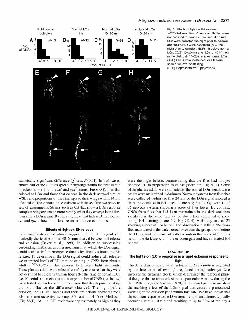
Effects of light on EH releaseExperiments described above suggest that a LOn signal can
markedly shorten the normal 40–60min interval between EH release
and eclosion (Baker et al., 1999). In addition to suppressing
descending inhibition, another mechanism by which the LOn signal
could cause a shift in emergence time is by directly stimulating EH
release. To determine if the LOn signal could induce EH release,
we examined levels of EH immunostaining in CNSs from pharate
adult w1118�UAS-rpr flies exposed to different light treatments.
These pharate adults were selected carefully to ensure that they were
not destined to eclose within an hour after the time of normal LOn
(see Materials and methods) and a large number of CNSs (see below)
were tested for each condition to ensure that developmental stage
did not influence the differences observed. The night before
eclosion, the EH cell bodies and their projections showed strong
EH immunoreactivity, scoring 3.7 out of 4 (see Methods)
(Fig.7A,E). At –1h, EH levels were approximately as high as they
were the night before, demonstrating that the flies had not yet
released EH in preparation to eclose (score 3.5; Fig.7B,F). Some
of the pharate adults were subjected to the normal LOn signal, while
others were maintained in darkness. Nervous systems from flies that
were collected within the first 20min of the LOn signal showed a
dramatic decrease in EH levels (score 0.5; Fig.7C,G), with 14 of
36 nervous systems showing a score of 1 or lower. By contrast,
CNSs from flies that had been maintained in the dark and then
sacrificed at the same time as the above flies continued to show
strong EH staining (score 2.9; Fig.7D,H), with only one of 25
showing a score of 1 or below. The observation that the CNSs from
flies maintained in the dark scored lower than the groups from before
the LOn signal is consistent with the notion that some of the flies
held in the dark are within the eclosion gate and have initiated EH
release.
DISCUSSIONThe lights-on (LOn) response is a rapid eclosion response to
lightThe daily distribution of adult eclosion in Drosophila is regulated
by the interaction of two light-regulated timing pathways. One
involves the circadian clock, which determines the temporal phase
of the gate that restricts eclosion to a particular window during the
day (Pittendrigh and Skopik, 1970). The second pathway involves
the masking effect of the LOn signal that causes a pronounced
skewing of the eclosion peak within this gate. We have shown that
the eclosion response to the LOn signal is rapid and strong, typically
occurring within 10min and resulting in up to 22% of the day’s
86
4
284
1216
84
16
12 8642
A DCB
Level of EH-IR
Night before eclosion
Normal LOn –1 h
Normal LOn +10–20 min
In dark at LOn +10–20 min
No. of CNSs
N=13 N=32 N=36 N=25
4 3 2 1 0.5 04 3 2 1 0.5 04 3 2 1 0.5 04 3 2 1 0.5 0
Fig. 7. Effects of light on EH release inw1118�UAS-rpr flies. Pharate adults that werenot destined to eclose at the time of normalLOn were collected the night prior to eclosionand their CNSs were harvested (A,E) thenight prior to eclosion, (B,F) 1 h before normalLOn, (C,G) 10–20 min after LOn or (D,H) heldin the dark until 10–20 min after normal LOn.(A–D) CNSs immunostained for EH werescored for level of staining.(E–H) Representative Z projections.
THE JOURNAL OF EXPERIMENTAL BIOLOGY

2272
eclosion within that 10min interval. The LOn response can occur
either earlier or later in the eclosion gate but it can only take place
after the normal gate has opened. The response is exhibited in the
absence of a normal circadian rhythm in per and tim mutants
(S.L.M., unpublished data). A temperature transition may also exert
a masking effect on eclosion via the EH neurons (Jackson et al.,
2005).
The role of the EH neurons in the LOn responseThe EH neurons are essential for the LOn response as shown by
the results of specifically expressing either cell death genes to ablate
them (Fig.3; McNabb et al., 1997) or tetanus toxin light chain to
suppress synaptic release (Fig.5). How do the EH neurons mediate
the LOn response? As seen in Fig.7, one effect of the LOn signal
appears to be the stimulation of EH release. Our
immunocytochemical studies show that the LOn signal caused a
rapid depletion of EH in pharate adult flies that were expected to
release EH later in the day.
A second effect of the LOn signal appears to be in decreasing
the latency of eclosion relative to EH action. In Drosophila, the
normal latency from EH release to eclosion is 40–60min (Baker et
al., 1999) but the LOn response occurs within 10min (e.g. Fig.1).
Experiments in both Manduca and Drosophila suggest that the delay
between EH release and ecdysis is due to a descending inhibition
that is set up as a consequence of EH release. EH release normally
takes place well in advance of ecdysis. In Manduca, the delay period
is about 20–25min for larval ecdysis (Ewer et al., 1994), and 2–3h
for adult eclosion (Ewer and Truman, 1996). Decapitation of
Manduca pharate adults during the delay period leads to the rapid
onset of eclosion behavior (Ewer et al., 1997; Reynolds, 1980),
suggesting that the delay is due to descending inhibition from the
head. Transection of the CNS at different levels shows that neurons
from the subesophageal and thoracic ganglia contribute to this
descending inhibition (Fuse and Truman, 2002; Zitnan and Adams,
2000). Similarly in Drosophila, decapitating pharate adults that have
released EH results in their rapid eclosion (Baker et al., 1999). This
rapid behavioral response to decapitation is not seen in EH cell
knockout flies (Baker et al., 1999), suggesting that EH release is
required for the inhibition to be set in place.
A LOn signal appears to be another way to elicit early eclosion
in flies that have released EH. The simplest interpretation is that
the LOn signal acts by suppressing the descending inhibition. This
mechanism is favored by the rapid nature of the LOn response and
by evidence from the pulse experiment with the w1118�UAS-rprstrain. If the LOn response results from the removal of the
descending inhibition, flies that eclose during the LOn peak should
include all of the pharate adults that have released EH and are in
the delay period at the time of the pulse. This can be seen by
comparing the results of the pulse and dark experiments (Fig.8A).
In the dark-maintained group, about 21% of the flies that eclosed
during the day emerged between 1 and 2h after the time of normal
LOn. Given a latency of 40–60min between EH release and ecdysis
(Baker et al., 1999), most of these flies would have been expected
to have released EH by the time the LOn pulse was given at 1h. In
this example, in the group that received the pulse, the LOn signal
was followed by 20% of the flies emerging within the first 10min,
similar to the 21% predicted to be in the waiting period. This is
consistent with the interpretation that flies that demonstrate the LOn
response have already released EH, are competent to respond to the
LOn signal, and are rapidly released from the inhibition that causes
the normal delay (Fig.8B). If the LOn signal acted only to suppress
the descending inhibition and hence reduce the eclosion latency in
S. L. McNabb and J. W. Truman
Light
Minutes from lights-on
–30 0 30 60 90
Per
cent
age
of d
ay’s
ecl
osio
n
Per
cent
age
of d
ay’s
ecl
osio
n
20%
0 1 2 3
Hours from normal lights-on
21%
A
B
25
20
15
10
5
25
20
15
10
20
15
10
5
5
Fig. 8. Light stimulates rapid eclosion in competent flies, but also appearsto have a second, delayed, effect. (A) Data from the w1118�UAS-rpr pulseexperiment (Fig. 2B,C) show that the proportion of flies that eclosed duringthe first 10 min of the 20 min light pulse (white bars) was roughly equivalentto the proportion that eclosed over the corresponding 60 min interval in thedark (bracketed by dashed lines). (B) Summary of the effects of light oneclosion, adapted from the w1118�UAS-rpr pulse experiment (Fig. 2C).Dashed arrows indicate the intervals over which flies that have releasedEH are recruited to eclose by a LOn signal. Pharate adults that hadreleased EH and were competent to eclose within approximately 60 minwere recruited to eclose in the first 10 min after light exposure (gray arrow).If the pool of competent flies all eclosed at or shortly after LOn, the amountof eclosion after the light pulse would be expected to diminish to 0 (thickblack line). Instead, the proportion of flies eclosing went down to onlyapproximately 2% (thin black line) possibly as a result of light stimulatingEH release in a group that was developmentally mature and ready toeclose.
THE JOURNAL OF EXPERIMENTAL BIOLOGY

2273A lights-on eclosion response in Drosophila
pharate adults that had released EH, we would expect eclosion to
drop down to zero for 40–60min after the LOn peak. The fact that
we do not observe this severe depression is most likely explained
by our observation that the LOn signal can induce premature EH
release in addition to reducing the eclosion latency.
Photoreceptors and the LOn responseUnlike the circadian entrainment of eclosion, the LOn response
requires a signal transduced from the eyes. Previous research
demonstrated that the LOn response is mediated by retinal
photoreceptors (Engelmann and Honegger, 1966). Using mutations
that eliminate either the compound eyes or the ocelli, we have shown
that both sets of photoreceptors are required for the response under
our assay conditions. The reason that both sets of photoreceptors
are required is unknown, but one possibility is that they act, either
in series or in parallel, to amplify the response. We also cannot rule
out the possibility that the mutants we tested had defects in other
components of the ecdysis pathway that may mediate the LOn
response. For example, eyes absent mutations affect the development
of additional regions of the brain (Bonini et al., 1998; Boyle et al.,
1997), and ocelliless and sine oculis also affect other developing
tissues (Finkelstein et al., 1990; Serikaku and O’Tousa, 1994).
We postulate a mechanism in which a signal from the
photoreceptors acts on the eclosion inhibitory neurons to suppress
the descending inhibition (Fig.9). Before the cellular pathway and
the mechanism of the release of the inhibition can be elucidated,
the source of the inhibition must be identified. Based on the results
of head ligations described above, it must reside in the head. In
Manduca, neurons of the cell 27/704 group are EH targets (Ewer
et al., 1994; Ewer et al., 1997) and some that are located in the
subesophagheal ganglion are candidate sources of the inhibition
(Fuse and Truman, 2002; Zitnan and Adams, 2000). The identity
of the Drosophila inhibitory neurons is less clear. Efforts to identify
the Drosophila homologs of the cell 27/704 group as EH targets
have been unsuccessful because they do not show a cGMP response
following EH release (Ewer and Truman, 1996). Currently, the
function of the Drosophila CCAP-expressing neurons in eclosion
is uncertain, as their ablation does not prevent eclosion (Park et al.,
2003). However, several other sets of neurons demonstrate calcium
transients in response to ETH, suggesting candidate neuropeptide-
expressing neurons as downstream activators (Kim et al., 2006).
The pathway by which the optic photoreceptors signal the ecdysis
inhibitory neurons also remains to be identified.
Differences in magnitude of the LOn response were observed
between the control strains. The w1118�UAS-rpr flies have pale apricot
eyes and are highly responsive to the LOn signal. The Canton-S and
w1118�UAS-TNT-L flies have red eyes and show a weaker LOn
response. The magnitude of the response may be a function of light
sensitivity and thus be higher in strains with less pigmentation since
screening pigments are associated with decreased sensitivity of the
eyes to light (Ostroy and Pak, 1974; Zimmerman and Ives, 1971).
Expression of tetanus toxin light chain in the EH neuronssuppresses the LOn response and EH release
We have demonstrated three different ways of eliminating the LOn
response: (1) removal of the EH neurons by targeted expression of
the cell death gene rpr (i.e. making an EH cell knockout), (2) removal
of either ocelli or compound eyes by the use of previously identified
mutants, and (3) alteration of EH cell function by targeting expression
of an intracellular form of tetanus toxin light chain (TeTxLC) to the
EH cells. The ability of EH neuron-targeted TeTxLC to block the
LOn response shows that the elimination of this response in the EH
cell knockouts is not due to death of EH cells, but rather to loss of
EH cell function. Since TeTxLC, which acts by inhibiting
synaptobrevin-mediated docking (reviewed by Humeau et al., 2000),
inhibits the release of EH (Fig.5K,L), synaptobrevin appears to play
a role in the release of neuropeptides. Flies that express TeTxLC in
their EH neurons also show a partial failure to spread their wings, a
post-ecdysial effect that is characteristic of the EH cell knockouts
(McNabb et al., 1997). We do not yet understand the difference in
penetrance of this phenotype between these two strains. It may reflect
a low level of EH release by the flies that express targeted TeTxLC,
interstrain variation (as seen previously in McNabb et al., 1997) or
a combination of these factors.
Wing spreading, a post-ecdysial behavior that is regulated byEH, is not accelerated by the LOn signal
For strains that exhibited a strong LOn response, flies that eclosed
in response to LOn took substantially longer to spread their wings
than flies that eclosed in the dark. For w1118�UAS-rpr flies, it took
Descending inhibitory neurons
EH neurons
Light
Optic photoreceptors
CCAP neurons and/or other
effectors
Eclosion
EH
EH
Fig. 9. A model of effects of light on eclosion. EH activates both an eclosionactivation pathway and a set of inhibitory neurons that repress eclosionbehavior. The activation pathway may include the CCAP neurons andadditional EH-downstream neurons. We postulate that the release of CCAPand other eclosion activators is inhibited by EH action. Light suppressesthe inhibitory pathway to allow the release of CCAP and other factors andpermit subsequent eclosion. In addition, light acts on the EH cells tostimulate EH release, probably via the retinal photoreceptors. Althougheclosion can be accelerated by light, wing spreading cannot, suggestingthat EH stimulates these behaviors via distinct pathways. Arrow and linethickness indicate the strengths of the different responses.
THE JOURNAL OF EXPERIMENTAL BIOLOGY

2274
an average of 47min longer; for w1118�UAS-TNT-L flies, about
40min longer. This time difference is within the 40–60min latency
range between EH release and eclosion (Baker et al., 1999). Our
findings are consistent with EH playing roles in initiating both
eclosion and wing spreading, and with these two behaviors having
separate downstream pathways with distinct latencies. The LOn
signal interacts with the ecdysis delay pathway to result in early
ecdysis, but does not affect the wing spreading circuit. Hence, the
LOn signal causes early ecdysis and a corresponding increase in
latency between eclosion and wing expansion.
The authors thank Dr Ray Huey for advice on statistical analysis and thereviewers for their helpful comments. This research was supported by NSF grantIOS-0452009 to J.W.T.
REFERENCESAschoff, J. (1960). Exogenous and endogenous components in circadian rhythms.
Cold Spring Harbor Symp. Quant. Biol. 25, 11-28.Ashburner, M. (1989). Drosophila: A Laboratory Manual. Cold Spring Harbor, NY:
Cold Spring Harbor Laboratory Press.Baker, J. W., McNabb, S. L. and Truman, J. W. (1999). The hormonal coordination
of behavior and physiology at adult ecdysis in Drosophila melanogaster. J. Exp. Biol.202, 3037-3048.
Benzer, S. (1967). Behavioral mutants of Drosophila isolated by countercurrentdistribution. Proc. Natl. Acad. Sci. USA 58, 1112-1119.
Binkley, S., Mosher, K. and Reilly, K. (1983). Circadian rhythms in house sparrows:Lighting ad lib. Physiol. Behav. 31, 829-893.
Bonini, N. M., Leiserson, W. M. and Benzer, S. (1993). The eyes absent gene:genetic control of cell survival and differentiation in the developing Drosophila eye.Cell 72, 379-395.
Bonini, N. M., Leiserson, W. M. and Benzer, S. (1998). Multiple roles of the eyesabsent gene in Drosophila. Dev. Biol. 196, 42-57.
Boyle, M., Bonini, N. and DiNardo, S. (1997). Expression and function of clift in thedevelopment of somatic gonadal precursors within the Drosophila mesoderm.Development 124, 971-982.
Brand, A. and Perrimon, N. (1993). Targeting gene expression as a means of alteringcell fates and generating dominant phenotypes. Development 118, 401-415.
Copenhaver, P. F. and Truman, J. W. (1986). Identification of the cerebralneurosecretory cells that contain eclosion hormone in the moth Manduca sexta. J.Neurosci. 6, 1738-1747.
Dewey, E. M., McNabb, S. L., Ewer, J., Kuo, G. R., Takanishi, C. L., Truman, J. W.and Honegger, H.-W. (2004). Identification of the gene encoding bursicon, an insectneuropeptide responsible for cuticle sclerotization and wing spreading. Curr. Biol. 14,1208-1213.
Eissenberg, J. C. and Ryerse, J. S. (1991). eya-2: A recessive eyeless mutation onthe second chromosome of Drosophila melanogaster. DIS 70, 266-268.
Engelmann, W. and Honegger, H. W. (1966). Tagesperiodische Schlupfrhythmikeiner augenlosen Drosophila melanogaster-Mutante. Naturwissenschaften 53, 588.
Ewer, J. and Truman, J. W. (1996). Increases in cyclic GMP occur at ecdysis in anevolutionarily conserved insect neuronal network. J. Comp. Neurol. 370, 330-341.
Ewer, J., De Vente, J. and Truman, J. W. (1994). Neuropeptide induction of cyclicGMP increases in the insect CNS: resolution at the level of single identifiableneurons. J. Neurosci. 14, 7704-7712.
Ewer, J., Gammie, S. C. and Truman, J. W. (1997). Control of insect ecdysis by apositive feedback endocrine system: roles of eclosion hormone and ecdysistriggering hormone. J. Exp. Biol. 200, 869-881.
Finkelstein, R., Smouse, D., Capaci, T. M., Spradling, A. C. and Perrimon, N.(1990). The orthodenticle gene encodes a novel homeo domain protein involved inthe development of the Drosophila nervous system and ocellar visual structures.Genes Dev. 4, 1516-1527.
Flybase (1999). The FlyBase database of the Drosophila genome projects andcommunity literature. Nucl. Acids Res. 27, 85-88.
Fraenkel, G. (1935). Observations and experiments on the blow-fly (Calliphoraerythrocephala) during the first day after emergence. Proc. Zool. Soc. Lond. Part 2,893-901.
Fuse, M. and Truman, J. W. (2002). Modulation of ecdysis in the moth Manducasexta: the roles of the subesophageal and thoracic ganglia. J. Exp. Biol. 205, 1047-1058.
Heitzler, P., Coulson, D., Saenz-Robles, M. T., Ashburner, M., Roote, J., Simpson,P. and Gubb, D. (1993). Genetic and cytogenetic analysis of the 43A-E regioncontaining the segment polarity gene costa and the cellular polarity genes prickleand spiny-legs in Drosophila melanogaster. Genetics 135, 105-115.
Helfrich-Förster, C. (2006). Neurobiology of the fruit fly’s circadian clock. Genes,Brain Behav. 4, 65-76.
Hewes, R. S. and Truman, J. W. (1991). The roles of central and peripheral eclosionhormone release in the control of ecdysis behavior in Manduca sexta. J. Comp.Physiol. A 168, 697-707.
Horodyski, F. M., Ewer, J., Riddiford, L. M. and Truman, J. W. (1993). Isolation,characterization and expression of the eclosion hormone gene of Drosophilamelanogaster. Eur. J. Biochem. 215, 221-228.
Humeau, Y., Doussau, F., Grant, N. J. and Poulain, B. (2000). How botulinum andtetanus neurotoxins block neurotransmitter release. Biochimie 82, 427-446.
Jackson, F. R. (1983). The isolation of biological rhythm mutations on the autosomesof Drosophila melanogaster. J. Neurogenet. 1, 3-15.
Jackson, F. R., Genova, G. K., Huang, Y., Kleyner, Y., Suh, J., Roberts, M. A.,Sundram, V. and Akten, B. (2005). Genetic and biochemical strategies foridentifying Drosophila genes that function in circadian control. Methods Enzymol.398, 663-682.
Kim, Y.-J., Zitnan, D., Cho, K.-H., Schooley, D. A., Mizoguchi, A. and Adams, M.E. (2006). Central peptidergic ensembles associated with organization of an innatebehavior. Proc. Natl. Acad. Sci. USA 103, 14211-14214.
Kimura, K. and Truman, J. W. (1990). Postmetamorphic cell death in the nervousand muscular systems of Drosophila melanogaster. J. Neurosci. 10, 403-411.
Kingan, T. G. and Adams, M. E. (2000). Ecdysteroids regulate secretory competencein Inka cells. J. Exp. Biol. 203, 3011-3018.
Kingan, T. G., Gray, W., Zitnan, D. and Adams, M. E. (1997). Regulation ofecdysis-triggering hormone release by eclosion hormone. J. Exp. Biol. 200, 3245-3256.
Lindsley, D. L. and Zimm, G. G. (1992). The Genome of Drosophila melanogaster.San Diego, NY, Boston, London, Sydney, Tokyo, Toronto: Academic Press.
Lorenz, L. J., Hall, J. C. and Rosbash, M. (1989). Expression of a Drosophila mRNAis under circadian clock control during pupation. Development 107, 869-880.
McNabb, S. L., Baker, J. D., Agapite, J., Steller, H., Riddiford, L. M. and Truman,J. W. (1997). Disruption of a behavioral sequence by targeted death of peptidergicneurons in Drosophila. Neuron 19, 813-823.
Mrosovsky, N. (1999). Masking: history, definitions, and measurement. Chronobiol.Int. 16, 415-429.
Myers, E. M., Yu, J. and Sehgal, A. (2003). Circadian Control of Eclosion: Interactionbetween and central and peripheral clock in Drosophila melanogaster. Curr. Biol. 13,526-533.
Nitabach, M. N. and Taghert, P. H. (2008). Organization of the Drosophila circadiancontrol circuit. Curr. Biol. 18, R84-R93.
Ostroy, S. E. and Pak, W. L. (1974). Protein and electroretinogram changes in thealleles of the norp AP12 Drosophila phototransduction mutant. Biochim. Biophys.Acta 368, 259-268.
Park, J. H., Schroeder, A. J., Helfrich-Forster, C., Jackson, F. R. and Ewer, J.(2003). Targeted ablation of CCAP neuropeptide-containing neurons of Drosophilacauses specific defects in execution and circadian timing of ecdysis behavior.Development 130, 2645-2656.
Park, Y., Filippov, V., Gill, S. S. and Adams, M. E. (2002). Deletion of the ecdysis-triggering hormone gene leads to lethal ecdysis deficiency. Development 129, 493-503.
Pittendrigh, C. S. and Skopik, S. D. (1970). Circadian systems, V. The drivingoscillation and the temporal sequence of development. Proc. Nat. Acad. Sci. USA65, 500-507.
Redlin, U. and Mrosovsky, N. (1999). Masking of locomotor activity in hamsters. J.Comp. Physiol. A 184, 429-437.
Reynolds, S. E. (1980). Integration of behaviour and physiology in ecdysis. Adv.Insect Physiol. 15, 475-595.
Saunders, D. S. (1976). Insect Clocks. Oxford; New York: Pergamon Press.Saunders, D. S. (1982). Insect Clocks. Oxford and N.Y.: Pergamon Press.Saunders, D. S. (2002). Insect Clocks, 3rd Edition. Amsterdam; Boston: Elsevier.Serikaku, M. A. and O’Tousa, J. E. (1994). sine oculis is a homeobox gene required
for Drosophila visual system development. Genetics 138, 1137-1150.Sved, J. (1986). Report of new mutants. DIS 63, 169.Sweeney, S. T., Broadie, K., Keane, J., Niemann, H. and O’Kane, C. J. (1995).
Targeted expression of tetanus toxin light chain in Drosophila specifically eliminatessynaptic transmission and causes behavioral defects. Neuron 14, 351-361.
Truman, J. W. (2005). Hormonal control of insect ecdysis: endocrine cascades forcoordinating behavior with physiology. Vitam. Horm. 73, 1-30.
Truman, J. W. and Riddiford, L. M. (1974). Physiology of insect rhythms, III. Thetemporal organization of the endocrine events underlying pupation of the tobaccohornworm. J. Exp. Biol. 60, 371-382.
Vosshall, L. B. and Young, M. W. (1995). Circadian rhythms in Drosophila can bedriven by period expression in a restricted group of central brain cells. Neuron 15,345-360.
Zimmerman, W. F. and Ives, D. (1971). Some photophysiological aspects of circadianrhythmicity in Drosophila. In Biochronometry (ed. M. Menaker), pp. 381-391.Washington D.C.: National Academy of Sciences.
Zitnan, D. and Adams, M. E. (2000). Excitatory and inhibitory roles of central gangliain initiation of the insect ecdysis behavioural sequence. J. Exp. Biol. 203, 1329-1340.
Zitnan, D., Kingan, T. G., Hermesman, J. L. and Adams, M. E. (1996). Identificationof ecdysis-triggering hormone from an epitracheal endocrine system. Science 271,88-91.
Zitnan, D., Ross, L. S., Zitnanova, I., Hermesman, J. L., Gill, S. S. and Adams, M.E. (1999). Steroid induction of a peptide hormone gene leads to orchestration of adefined behavioral sequence. Neuron 23, 523-535.
Zitnanova, I., Adams, M. E. and Zitnan, D. (2001). Dual ecdysteroid action on theepitracheal glands and central nervous system preceding ecdysis of Manduca sexta.J. Exp. Biol. 204, 3483-3495.
S. L. McNabb and J. W. Truman
THE JOURNAL OF EXPERIMENTAL BIOLOGY
![RESEARCH ARTICLE Open Access The essential role of bursicon … · 2017. 8. 27. · eclosion hormone (EH) and CCAP (reviewed extensively in [1-3]). The canonical model proposes that](https://img.pdfslide.us/doc/110x75/60d1226e6a672903243c90a9/research-article-open-access-the-essential-role-of-bursicon-2017-8-27-eclosion.jpg)


![A Caenorhabditis elegans Mass Spectrometric Resource for ...been favored for such studies [5, 29–32]. Caenorhabditis elegans has emerged as a powerful model to study peptidergic](https://img.pdfslide.us/doc/110x75/5e9f3464f963cc16d66b513b/a-caenorhabditis-elegans-mass-spectrometric-resource-for-been-favored-for-such.jpg)