Embed Size (px)
Citation preview
29
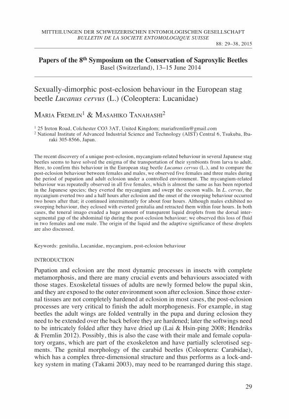
Sexually-dimorphic post-eclosion behaviour in the European stagbeetle Lucanus cervus (L.) (Coleoptera: Lucanidae)MARIA FREMLIN1 & MASAHIKO TANAHASHI2
1 25 Ireton Road, Colchester CO3 3AT, United Kingdom; [email protected] National Institute of Advanced Industrial Science and Technology (AIST) Central 6, Tsukuba, Iba-
raki 305-8566, Japan.
The recent discovery of a unique post-eclosion, mycangium-related behaviour in several Japanese stagbeetles seems to have solved the enigma of the transportation of their symbionts from larva to adult.Here, to confirm this behaviour in the European stag beetle Lucanus cervus (L.), and to compare thepost-eclosion behaviour between females and males, we observed five females and three males duringthe period of pupation and adult eclosion under a controlled environment. The mycangium-relatedbehaviour was repeatedly observed in all five females, which is almost the same as has been reportedin the Japanese species; they everted the mycangium and swept the cocoon walls. In L. cervus, themycangium everted two and a half hours after eclosion and the onset of the sweeping behaviour occurredtwo hours after that; it continued intermittently for about four hours. Although males ex hibited nosweeping behaviour, they eclosed with everted genitalia and retracted them within four hours. In bothcases, the teneral imago exuded a huge amount of transparent liquid droplets from the dorsal inter-segmental gap of the abdominal tip during the post-eclosion behaviour; we observed this loss of fluidin two females and one male. The origin of the liquid and the adaptive significance of these dropletsare also discussed.
Keywords: genitalia, Lucanidae, mycangium, post-eclosion behaviour
INTRODUCTIONPupation and eclosion are the most dynamic processes in insects with completemetamorphosis, and there are many crucial events and behaviours associated withthose stages. Exoskeletal tissues of adults are newly formed below the pupal skin,and they are exposed to the outer environment soon after eclosion. Since those exter-nal tissues are not completely hardened at eclosion in most cases, the post-eclosionprocesses are very critical to finish the adult morphogenesis. For example, in stagbeetles the adult wings are folded ventrally in the pupa and during eclosion theyneed to be extended over the back before they are hardened; later the softwings needto be intricately folded after they have dried up (Lai & Hsin-ping 2008; Hendriks& Fremlin 2012). Possibly, this is also the case with their male and female copula-tory organs, which are part of the exoskeleton and have partially sclerotised seg-ments. The genital morphology of the carabid beetles (Coleoptera: Carabidae),which has a complex three-dimensional structure and thus performs as a lock-and-key system in mating (Takami 2003), may need to be rearranged during this stage.
MITTEILUNGEN DER SCHWEIZERISCHEN ENTOMOLOGISCHEN GESELLSCHAFTBULLETIN DE LA SOCIETE ENTOMOLOGIQUE SUISSE
88: 29–38, 2015
Papers of the 8th Symposium on the Conservation of Saproxylic BeetlesBasel (Switzerland), 13–15 June 2014
Fremlin & Tanahashi.qxp_Start 2013.qxd 23.04.15 07:55 Seite 29
30
MARIA FREMLIN & MASAHIKO TANAHASHI
However, the post-eclosion behaviour associated with the morphogenesis of geni-talia has not been fully investigated in stag beetles.
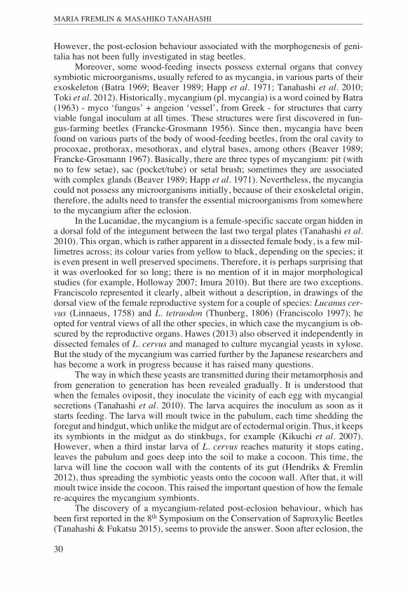
Moreover, some wood-feeding insects possess external organs that conveysymbiotic microorganisms, usually refered to as mycangia, in various parts of theirexoskeleton (Batra 1969; Beaver 1989; Happ et al. 1971; Tanahashi et al. 2010;Toki et al. 2012). Historically, mycangium (pl. mycangia) is a word coined by Batra(1963) - myco ‘fungus’ + angeion ‘vessel’, from Greek - for structures that carryviable fungal inoculum at all times. These structures were first discovered in fun-gus-farming beetles (Francke-Grosmann 1956). Since then, mycangia have beenfound on various parts of the body of wood-feeding beetles, from the oral cavity toprocoxae, prothorax, mesothorax, and elytral bases, among others (Beaver 1989;Francke-Grosmann 1967). Basically, there are three types of mycangium: pit (withno to few setae), sac (pocket/tube) or setal brush; sometimes they are associatedwith complex glands (Beaver 1989; Happ et al. 1971). Nevertheless, the mycangiacould not possess any microorganisms initially, because of their exoskeletal origin,therefore, the adults need to transfer the essential microorganisms from somewhereto the mycangium after the eclosion.
In the Lucanidae, the mycangium is a female-specific saccate organ hidden ina dorsal fold of the integument between the last two tergal plates (Tanahashi et al.2010). This organ, which is rather apparent in a dissected female body, is a few mil-limetres across; its colour varies from yellow to black, depending on the species; itis even present in well preserved specimens. Therefore, it is perhaps surprising thatit was overlooked for so long; there is no mention of it in major morphologicalstud ies (for example, Holloway 2007; Imura 2010). But there are two exceptions.Franciscolo represented it clearly, albeit without a description, in drawings of thedorsal view of the female reproductive system for a couple of species: Lucanus cer-vus (Linnaeus, 1758) and L. tetraodon (Thunberg, 1806) (Franciscolo 1997); heopted for ventral views of all the other species, in which case the mycangium is ob -scured by the reproductive organs. Hawes (2013) also observed it independently indissected females of L. cervus and managed to culture mycangial yeasts in xylose.But the study of the mycangium was carried further by the Japanese researchers andhas become a work in progress because it has raised many questions.
The way in which these yeasts are transmitted during their metamorphosis andfrom generation to generation has been revealed gradually. It is understood thatwhen the females oviposit, they inoculate the vicinity of each egg with mycangialsecretions (Tanahashi et al. 2010). The larva acquires the inoculum as soon as itstarts feeding. The larva will moult twice in the pabulum, each time shedding theforegut and hindgut, which unlike the midgut are of ectodermal origin. Thus, it keepsits symbionts in the midgut as do stinkbugs, for example (Kikuchi et al. 2007).However, when a third instar larva of L. cervus reaches maturity it stops eating,leaves the pabulum and goes deep into the soil to make a cocoon. This time, thelarva will line the cocoon wall with the contents of its gut (Hendriks & Fremlin2012), thus spreading the symbiotic yeasts onto the cocoon wall. After that, it willmoult twice inside the cocoon. This raised the important question of how the femalere-acquires the mycangium symbionts.
The discovery of a mycangium-related post-eclosion behaviour, which hasbeen first reported in the 8th Symposium on the Conservation of Saproxylic Beetles(Tanahashi & Fukatsu 2015), seems to provide the answer. Soon after eclosion, the
Fremlin & Tanahashi.qxp_Start 2013.qxd 23.04.15 07:55 Seite 30
31
ON POST-ECLOSION BEHAVIOUR OF LUCANUS CERVUS (L.) (COLEOPTERA: LUCANIDAE)
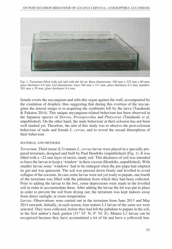
female everts the mycangium and rubs this organ against the wall, accompanied bythe exudation of droplets; thus suggesting that during this evertion of the mycan-gium the teneral imago is re-acquiring the symbionts left by the larva (Tanahashi& Fukatsu 2014). This unique mycangium-related behaviour has been observed inthe Japanese species of Dorcus, Prosopocoilus and Platycerus (Tanahashi et al.unpublished). On the other hand, the male behaviour at their eclosion has not beenwell studied yet. Therefore, the aim of this study was to observe the post-eclosionbehaviour of male and female L. cervus, and to reveal the sexual dimorphism oftheir behaviour. MATERIAL AND METHODSTerrarium. Third instar (L3) mature L. cervus larvae were placed in a specially pre-pared terrarium, designed and built by Paul Hendriks (unpublished) (Fig. 1). It wasfilled with a ~22 mm layer of moist, sandy soil. This thickness of soil was intendedto force the larvae to keep a ‘window’ in their cocoon (Hendriks, unpublished). Withsmaller larvae some ‘windows’ had to be enlarged when the pre-pupa had emptiedits gut and was quiescent. The soil was pressed down firmly and levelled to avoidcollapse of the cocoons. In case some larvae were not yet ready to pupate, one fourthof the terrarium was filled with the pabulum from which they had been collected.Prior to adding the larvae to the box, some depressions were made in the levelledsoil in order to accommodate them. After adding the larvae the lid was put in placein order to prevent the soil from drying out; the terrarium was kept indoors awayfrom direct sunlight, at room temperature.Larvae. Observations were carried out in the terrarium from June 2013 and May2014 onwards. Initially, in each season, four mature L3 larvae of the same sex wereselected. They were collected, before they had left the pabulum to pupate in the soil,in the first author’s back garden (51° 54’ N; 0° 54’ E). Mature L3 larvae can berecognized because they have accumulated a lot of fat and have a yellowish hue.
Fig. 1. Terrarium filled with soil and with the lid on. Base dimensions: 360 mm x 325 mm x 60 mm,glass thickness 4.6 mm. Lid dimensions: base 346 mm x 311 mm, glass thickness 6.2 mm; handles:301 mm x 29 mm, glass thickness 8.4 mm.
Fremlin & Tanahashi.qxp_Start 2013.qxd 23.04.15 07:55 Seite 31
32
MARIA FREMLIN & MASAHIKO TANAHASHI
Sexing was done by examining the medio-ventral region of the larva's 9th ab -dominal segment with a hand lens for the presence of the terminal ampulla (or,Herold organ) (Herold 1815; Martinez & Lumaret 2005). It is absent in the females;their ovaries are visible through gaps in the fat body between the 7th and 8th seg-ments (Fremlin & Hendriks 2014). At a later stage, more individuals were movedinto vacated cocoons: one pre-pupa larva and one pupa, July 2013, and one pre-pupa, August 2014; they were sourced from another terrarium. In 2013 a femalepupa was later moved out and placed on a rudimentary cocoon in order to observerear views during post-eclosion.
In total, eleven individuals were selected for observations during this study.Wherever possible, some biometric data was collected from these individuals eitherat the beginning or during their development: head capsule width, body lengthexcluding the mandibles, and body weight. The instruments used were: digital cal-lipers, to 0.01 mm accuracy, and a Scalix CB-310 electronic scale, to 0.01 g accur -acy.Photographic equipment. The equipment used was an EOS 550D SLR Canoncamera fitted with a Sigma 105 mm objective; a Canon Speedlight 430EXII wassometimes used. The camera was either set on a Jessops copystand or a Velbon tri-pod (PH-248 Head). Photography in the terrarium was with the lid removed.RESULTS
Pupation and eclosion behaviourOf the eleven individuals selected for this study, eight reached the final moult: fourfemales in the first year and three males and one female in the following one. Inboth years, one of the four larvae, which were initially introduced in the terrarium,took very long to settle down and did not survive for unknown reasons; one died inthe pre-pupal stage and the other soon after it pupated. Another individual was killedby a centipede soon after pupation.
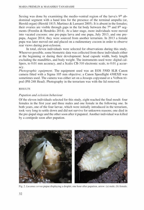
Fig. 2. Lucanus cervus pupae displaying a droplet, one hour after pupation, arrow: (a) male; (b) female.a b
Fremlin & Tanahashi.qxp_Start 2013.qxd 23.04.15 07:55 Seite 32
33
ON POST-ECLOSION BEHAVIOUR OF LUCANUS CERVUS (L.) (COLEOPTERA: LUCANIDAE)
The larvae, which settled well, took 36 days to pupate (n=4); the final moulttook an additional 34 days (n=4), at room temperature. Placing the larvae in a shal-low terrarium allowed for close-up observations of what was going on inside thetruncated cocoons, but after the hindwings expanded, they obscured the view of thelast abdominal segment and this hampered observations. Therefore, good rear viewswere only possible with a female, which moulted in a rudimentary cocoon, 2013,and a couple of imagos which eclosed in a boomerang shaped cocoon, 2014.
The metamorphosis of male and female larvae was identical. During eachmoult both lost a fair amount of fluid; this was apparent because of the way in whichsoil clung to the tip of their body and the wetness on the base of the cocoon.
The pre-pupa moulted on its back; the exuvia was left at the end of the cocoonaway from the pupa, which also reclined on its back. Soon after ecdysis, a dropletwas observed in the pupa’s abdomen; in the male it hung from the dorsal side ofthe pupa’s last abdominal segment (Fig. 2a). This was only observed in one malebecause the pupation of the other two occurred while the observer was absent fromthe country. In the female the droplet was at the very tip of the pupa’s abdomen(n=4) (Fig. 2b), it was visible for a few hours.
The pre-imago moulted on its front, but this time the exuvia was tucked under-neath the body. During ecdysis the elytra extended and moved over the back of theabdomen, which was very distended. The tips of the hindwings appeared soon after.The elytra were very pale but from then on melanised, gradually. This moult wasvideoed with two individuals, which eclosed in the boomerang shaped cocoon, rightfrom the beginning for several hours (Fremlin 2014a, 2014b).
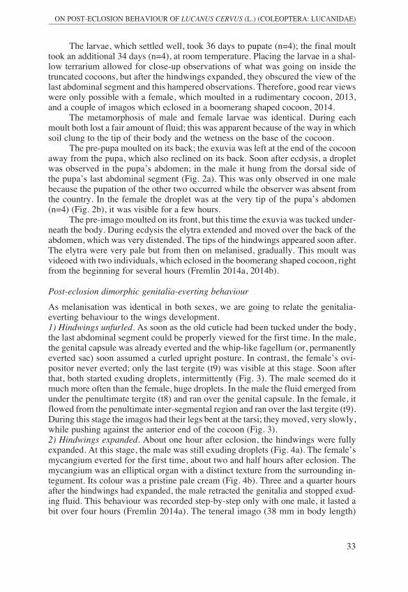
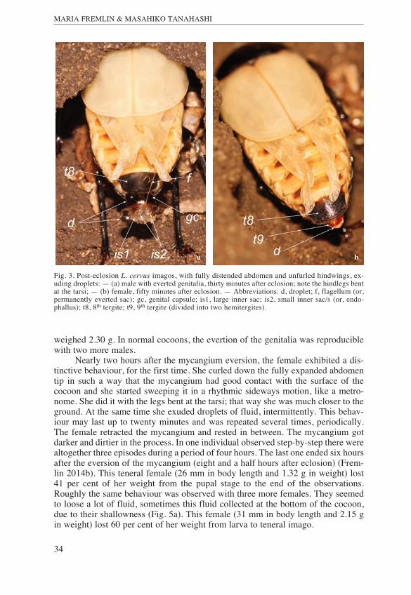
Post-eclosion dimorphic genitalia-everting behaviourAs melanisation was identical in both sexes, we are going to relate the genitalia-everting behaviour to the wings development.1) Hindwings unfurled. As soon as the old cuticle had been tucked under the body,the last abdominal segment could be properly viewed for the first time. In the male,the genital capsule was already everted and the whip-like fagellum (or, permanentlyeverted sac) soon assumed a curled upright posture. In contrast, the female’s ovi-positor never everted; only the last tergite (t9) was visible at this stage. Soon afterthat, both started exuding droplets, intermittently (Fig. 3). The male seemed do itmuch more often than the female, huge droplets. In the male the fluid emerged fromunder the penultimate tergite (t8) and ran over the genital capsule. In the female, itflowed from the penultimate inter-segmental region and ran over the last tergite (t9).During this stage the imagos had their legs bent at the tarsi; they moved, very slowly,while pushing against the anterior end of the cocoon (Fig. 3).2) Hindwings expanded. About one hour after eclosion, the hindwings were fullyexpanded. At this stage, the male was still exuding droplets (Fig. 4a). The female’smycangium everted for the first time, about two and half hours after eclosion. Themycangium was an elliptical organ with a distinct texture from the surrounding in -tegument. Its colour was a pristine pale cream (Fig. 4b). Three and a quarter hoursafter the hindwings had expanded, the male retracted the genitalia and stopped exud -ing fluid. This behaviour was recorded step-by-step only with one male, it lasted abit over four hours (Fremlin 2014a). The teneral imago (38 mm in body length)
Fremlin & Tanahashi.qxp_Start 2013.qxd 23.04.15 07:55 Seite 33
34
MARIA FREMLIN & MASAHIKO TANAHASHI
weighed 2.30 g. In normal cocoons, the evertion of the genitalia was reproduciblewith two more males.
Nearly two hours after the mycangium eversion, the female exhibited a dis-tinctive behaviour, for the first time. She curled down the fully expanded abdomentip in such a way that the mycangium had good contact with the surface of thecocoon and she started sweeping it in a rhythmic sideways motion, like a metro-nome. She did it with the legs bent at the tarsi; that way she was much closer to theground. At the same time she exuded droplets of fluid, intermittently. This behav -iour may last up to twenty minutes and was repeated several times, periodically.The female retracted the mycangium and rested in between. The mycangium gotdarker and dirtier in the process. In one individual observed step-by-step there werealtogether three episodes during a period of four hours. The last one ended six hoursafter the eversion of the mycangium (eight and a half hours after eclosion) (Frem-lin 2014b). This teneral female (26 mm in body length and 1.32 g in weight) lost41 per cent of her weight from the pupal stage to the end of the observations.Roughly the same behaviour was observed with three more females. They seemedto loose a lot of fluid, sometimes this fluid collected at the bottom of the cocoon,due to their shallowness (Fig. 5a). This female (31 mm in body length and 2.15 gin weight) lost 60 per cent of her weight from larva to teneral imago.
Fig. 3. Post-eclosion L. cervus imagos, with fully distended abdomen and unfurled hindwings, ex -uding droplets: — (a) male with everted genitalia, thirty minutes after eclosion; note the hindlegs bentat the tarsi; — (b) female, fifty minutes after eclosion. — Abbreviations: d, droplet; f, flagellum (or,permanently everted sac); gc, genital capsule; is1, large inner sac; is2, small inner sac/s (or, endo-phallus); t8, 8th tergite; t9, 9th tergite (divided into two hemitergites).
ba
Fremlin & Tanahashi.qxp_Start 2013.qxd 23.04.15 07:55 Seite 34
35
ON POST-ECLOSION BEHAVIOUR OF LUCANUS CERVUS (L.) (COLEOPTERA: LUCANIDAE)
Serendipitously, an interesting behaviour was observed with the female whosepupa was placed on the rudimentary cocoon. This female was found on her backand, judging by the dark colour of her elytra, must have eclosed in the previoushours during the night; she could not turn round. Consequently, her hindwings didnot develop properly, and were sticking out in an unnatural manner. However,shortly after she was placed on her feet, she started exuding droplets and within abit over two hours she exhibited the normal ‘sweeping’ behaviour. At one time sheflipped on her back and started retracting the mycangium (Fig. 5b); then she re -sumed that behaviour once back on her feet. She carried on for about three and ahalf hours, intermittently, exuding droplets and sweeping, exactly like a normalfemale would have. This female (27 mm in body length and 1.54 g in weight) lost 42 %of her weight from the pupal stage till the end of these observations.3) Hindwings folded, elytra chestnut brown. After the hindwings were folded, thefemales were observed only sporadically and no more sweeping episodes wererecorded.
Both sexes remained with a distended abdomen until their cuticle hardenedup, but their last abdominal segment was no longer telescoped. This lasted until thenext day. After that, they no longer presented a distended abdomen, hence the 9thtergite and the 8th and 9th sternites were no longer everted. From then on the adults
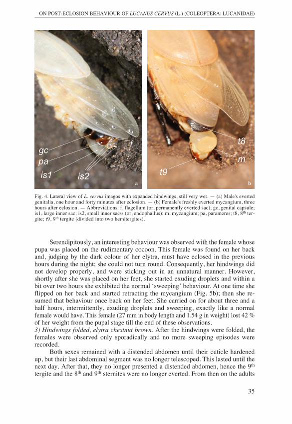
Fig. 4. Lateral view of L. cervus imagos with expanded hindwings, still very wet. — (a) Male's evertedgenitalia, one hour and forty minutes after eclosion. — (b) Female's freshly everted mycangium, threehours after eclosion. — Abbreviations: f, flagellum (or, permanently everted sac); gc, genital capsule;is1, large inner sac; is2, small inner sac/s (or, endophallus); m, mycangium; pa, parameres; t8, 8th ter-gite; t9, 9th tergite (divided into two hemitergites).
ba
Fremlin & Tanahashi.qxp_Start 2013.qxd 23.04.15 07:55 Seite 35
36
MARIA FREMLIN & MASAHIKO TANAHASHI
remained quiescent in situ for eight to nine months until their emergence the follow -ing year. DISCUSSIONOverall, their metamorphosis was reproducible and predictable. Some researchershave reported on this (Harvey & Gange 2003; Lai & Hsin-ping 2008; Hendriks &Fremlin 2012). Regardless of their size, there was no difference between their moult -ing times; speed of melanisation; hindwings development or straightening of thehead; but their post-eclosion behaviour seems to have been unreported in the past.As expected, L. cervus females behaved virtually the same way as the previouslytested Japanese species: Dorcus, Prosopocoilus and Platycerus (Tanahashi et al.,unpublished); but the latter exhibited their mycangium-related behaviour after thehindwings were folded and we have probably missed that. A female, which had anabnormal eclosion, resumed the mycangium-related behaviour as soon as she wasturned on her feet, and it lasted for a good length of time, indicating that this behav -iour is switched on by some other external stimulus or antecedent behaviour.
The male’s genitalia-everting behaviour came as a total surprise. The uniquemale’s post-eclosion everted genitalia has been captured by Lai & Hsin-ping (2008)in a post-eclosion series with Dorcus curvidens binodulosus (Waterhouse, 1874)and is evident in photos of other stag beetle species posted in the Internet by hob-byists; but, to our knowledge, it has has not been described. What we observedduring post-eclosion is rather different from what occurs during copulation. Thenthe genitalia rotate sideways as they evert from the genital capsule (Fremlin pers.obs.); but this rotation mechanism has also not been described.
The abdomen of a hardened imago also telescopes when gentle pressure isapplied (Franciscolo 1997), or when one is trodden on (Fremlin pers. obs.); in bothcases the male’s genital capsule everts but usually only a bit of the parameres andthe flagellum are visible. The description of L. cervus genitalia by Franciscolo(1997) fits with our observations regarding the flagellum and the parameres; theendophallus (is2) is cognate with the penis, which is a semi-transparent membra-nous organ. The complex structure of the endophallus when manually inflated has
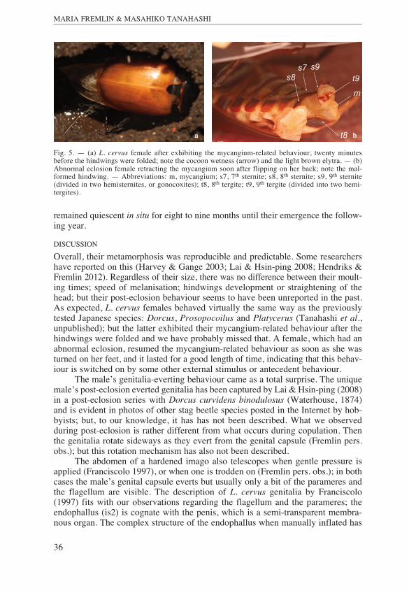
Fig. 5. — (a) L. cervus female after exhibiting the mycangium-related behaviour, twenty minutesbefore the hindwings were folded; note the cocoon wetness (arrow) and the light brown elytra. — (b)Abnormal eclosion female retracting the mycangium soon after flipping on her back; note the mal-formed hindwing. — Abbreviations: m, mycangium; s7, 7th sternite; s8, 8th sternite; s9, 9th sternite(divided in two hemisternites, or gonocoxites); t8, 8th tergite; t9, 9th tergite (divided into two hemi-tergites).
a b
Fremlin & Tanahashi.qxp_Start 2013.qxd 23.04.15 07:55 Seite 36
37
ON POST-ECLOSION BEHAVIOUR OF LUCANUS CERVUS (L.) (COLEOPTERA: LUCANIDAE)
been extensively studied for Platycerus species (Imura 2007, 2010), but not yet forthis species. The bigger sac (is1) is probably related to the integument, whichconnects the genitalia with the genital capsule, but its eversion needs further inves-tigation.
There also seem to be no reports about the exudation of liquid either by thepupa or the imago. Even though both male and female exuded droplets soon afterthe eclosion, it is interesting to note that the male seemed to do it more liberallythan the female; but this could be linked to the fact that the observed females weresmaller than the male. The duration of the male genitalia-related behaviour was muchshort er than in the female and it started right after eclosion. The female took a goodtwo hours to evert the mycangium, and then a couple of hours to start sweeping;then she exuded droplets with greater frequency. We have scant data of their weightloss from pupa to imago, about forty percent, but this needs to be investigated andcompared with that of the male.
However, the male genitalia-everting behaviour raises many questions. Per-haps this behaviour is necessary for the male to arrange the inner sacs of his geni-talia? Why both exuded droplets right after the eclosion? It is clear that in the femalethis loss of fluid is associated with the mycangium-related behaviour. The femalewould have a vested interest in maintaining a high level of humidity in her cocoon;it would have a direct influence in the survival of the purged larval gut symbiontsand the efficiency of symbiont uptake from the cocoon. However, its importance tothe male is not so clear. The fluid is probably from the haemolymph, but it needsto be analysed. Also its source needs to be located accurately; the fact that there wasloss of fluid soon after pupation suggests that the morphology of this exocrine sys-tem, like the genitalia, could already be fairly developed at that stage; this needs tobe investigated further. From our observations in the teneral imago the fluid seemsto originate from the same integument in both sexes: the dorsal region of the penul-timate inter-segmental membrane. In the male, this membrane was not everted thesame way, but the liquid dripped from above the genital capsule; in the female itdripped below the mycangium. In either case, it is not being discarded via the gut.The anal opening in the female is in the ventral area (Franciscolo 1997; Holloway2007); in the male the rectum goes through the genital capsule and opens above theflagellum (Wanat 2007; Fremlin pers. obsv.).CONCLUSIONThe post-eclosion behaviour of the stag beetle, Lucanus cervus, was recorded inboth male and female. The female exhibited the mycangium-related behaviour,which is almost the same as has been known in several Japanese stag beetle speciesof different genera, suggesting that this kind of behaviour is common in the Luca-nidae, as well as the universality of the female-specific mycangium. The genitalia-everting behaviour in the male was observed for the first time. We also discoveredthat both male and female exuded a huge amount of liquid from the abdominal tip,not via the gut, along with these sexually dimorphic post-eclosion behaviours. Themechanism and the adaptive significance of such exudation of liquid should beinvestigated in future studies.
Fremlin & Tanahashi.qxp_Start 2013.qxd 23.04.15 07:55 Seite 37
38
MARIA FREMLIN & MASAHIKO TANAHASHI
ACKNOWLEDGEMENTSThe first author wishes to thank Paul Hendriks for the gift of his terrarium and for his expert adviceabout rearing the larvae; also John Fremlin and Kristin Bloom for the gift of photographic equipment.Without their generous support this work would not have been possible. Many thanks to Darren Mannfor help with the genitalia morphology; to Luca Bartolozzi, Carlos Fletchmann, M. J. Paulsen andMarek Wanat for helpful discussion; to Claire Hengeveld and David Fremlin for the English correc-tions. Finally, we are very grateful to a couple of reviewers for their constructive comments.
REFERENCESBatra, L.R. 1963. Ecology of ambrosia fungi and their dissemination by beetles. — Transactions of
the Kansas Academy of Science 66: 213–236. Beaver, R.A. 1989. Insect-fungus relationship in the bark and ambrosia beetles. In: Insect fungus inter-
actions. Wilding N., Collins N. M., Hammond P. M. & Webber J. F. (eds), pp. 121–143. —London: Academic Press.
Franciscolo, M.E. 1997. Coleoptera: Lucanidae, Fauna d’Italia. Volume 35, 228 pp. — Bologna, Cal-derini.
Francke-Grosmann, H. 1956. Grundlagen der Symbiose bei pilzzüchtenden Holzinsekten. —Verhand-lungen der Deutschen Zoologischen Gesellschaft 1956: 112–118.
Fremlin, M. 2014a. Male stag beetle (Lucanus cervus) post eclosion behaviour. URL: https://youtu.be/MuqimTli1sk as accessed on 20 March 2015.
Fremlin, M. 2014b. Female stag beetle (Lucanus cervus) post-eclosion behaviour. URL: http://youtu.be/56cnR1FnEXY as accessed on 20 March 2015.
Fremlin, M. & Hendriks, P. 2014. Number of instars of!Lucanus cervus (Coleoptera: Lucanidae) lar-vae. — Entomologische Berichten 74 (3): 115–120.
Happ, G.M., Happ, C.M. & Barras, S.J. 1971. Fine structure of the prothoracic mycangium, a cham-ber for the culture of symbiotic fungi, in the southern pine beetle, Dendroctonus frontalis. —Tissue Cell 3 (2): 295–308.
Harvey, D.J. & Gange, A.C.!2003.! The Private Life of the Stag Beetle (Lucanus cervus). — The Bul-letin of the Amateur Entomologists’ Society 62: 240–244.
Hawes, C. 2013. Discovery of a mycangium and associated yeasts in the stag beetle Lucanus cervus(Coleoptera: Lucanidae). — White Admiral, 85: 22–23.
Herold, J.M.D. 1815. Entwicklungsgeschichte der Schmetterlinge anatomisch und physiologisch bear-beitet, 167 pp. – Cassel und Marburg.
Hendriks, P.!& Fremlin, M. 2012.! How stag beetles pupate. URL: http://maria.fremlin.de/stagbeetles/pupation/pupation_captivity.html as accessed on 20March 2015.
Holloway, B.A. 2007. Lucanidae (Insecta: Coleoptera). Fauna of New Zealand 61, 254 pp. — ManaakiWhenua Press, Lincoln, Canterbury, New Zealand.
Imura, Y. 2007. Endophallic structure of the genus Platycerus (Coleoptera, Lucanidae) of Japan, withdescriptions of two new species. — Elytra 35: 471–489.
Imura, Y. 2010. The Genus Platycerus of East Asia, pp. 240. — Roppon-Ashi Entomological Books.Kikuchi, Y., Hosokawa, T. & Fukatsu, T. 2007. Insect-Microbe Mutualism without Vertical Trans-
mission: a Stinkbug Acquires a Beneficial Gut Symbiont from the Environment Every Gen -eration. — Applied and Environmental Microbiology 73: 4308–4316.
Lai, J. & Hsin-ping, K. 2008. For the Love of Rhinoceros and Stag Beetles. 2nd Ed . Volume 2, 250–450 pp. — Morning Star Publisher Inc.
Martínez, M.I. & Lumaret, J.P. 2005. Structure of the terminal ampulla in male larvae of Canthon cya-nellus LeConte (Coleoptera: Scarabaeidae: Scarabaeinae). — Coleopterists Bulletin 59: 35–39.
Takami, Y. (2003) Experimental analysis of the effect of genital morphology on insemination successin the ground beetle Carabus insulicola (Coleoptera: Carabidae). — Ethology, Ecology & Evo-lution 15: 51–61.
Tanahashi, M., Kubota, K., Matsushita, N. & Togashi, K. 2010. Discovery of mycangia and associat edxylose-fermenting yeasts in stag beetles (Coleoptera: Lucanidae). — Naturwissenschaften 97:311–317.
Tanahashi, T. & Fukatsu, T. 2015. Specialized mycangial structure and host behavior for vertical trans-mission of symbiotic yeasts in stag beetles. — Mitteilung der Schweizerischen Entomologi-schen Gesellschaft 88: 1.
Toki, W., Tanahashi, M., Togashi, K. & Fukatsu, T. 2012. Fungal farming in a non-social beetle. —PLoS One, 7, e41893.
Wanat, M. 2007. Alignment and homology of male terminalia in!Curculionoidea and other Coleo -ptera. — Invertebrate Systematics 21:!147–171.
(received January 20, 2015; accepted March 31, 2015; published June 30, 2015)
Fremlin & Tanahashi.qxp_Start 2013.qxd 23.04.15 07:55 Seite 38
Errata, corrected links
Fremlin, M. 2014a. Male stag beetle (Lucanus cervus) post eclosion behaviour. URL: https://youtu.be/FgiPDq1FWpc as accessed on 7 June 2015.
Fremlin, M. 2014b. Female stag beetle (Lucanus cervus) post-eclosion behaviour. URL: https://youtu.be/szzHzZjsXfc as accessed on 6 June 2015.