Embed Size (px)
Citation preview
Light Absorption and Energy Transfer in the Antenna Complexes ofPhotosynthetic OrganismsTihana Mirkovic,† Evgeny E. Ostroumov,‡ Jessica M. Anna,§ Rienk van Grondelle,∥ Govindjee,⊥
and Gregory D. Scholes*,†,‡
†Department of Chemistry, University of Toronto, 80 St. George Street, Toronto, Ontario M5S 3H6, Canada‡Department of Chemistry, Princeton University, Washington Road, Princeton, New Jersey 08544, United States§Department of Chemistry, University of Pennsylvania, 231 S. 34th Street, Philadelphia, Pennsylvania 19104, United States∥Department of Physics and Astronomy, Faculty of Sciences, VU University Amsterdam, De Boelelaan 1081, 1081 HV, Amsterdam,The Netherlands⊥Department of Biochemistry, Center of Biophysics & Quantitative Biology, and Department of Plant Biology, University of Illinoisat Urbana−Champaign, 265 Morrill Hall, 505 South Goodwin Avenue, Urbana, Illinois 61801, United States
ABSTRACT: The process of photosynthesis is initiated by the capture of sunlight by a network oflight-absorbing molecules (chromophores), which are also responsible for the subsequent funnelingof the excitation energy to the reaction centers. Through evolution, genetic drift, and speciation,photosynthetic organisms have discovered many solutions for light harvesting. In this review, wedescribe the underlying photophysical principles by which this energy is absorbed, as well as themechanisms of electronic excitation energy transfer (EET). First, optical properties of the individualpigment chromophores present in light-harvesting antenna complexes are introduced, and then weexamine the collective behavior of pigment−pigment and pigment−protein interactions. Thedescription of energy transfer, in particular multichromophoric antenna structures, is shown to varydepending on the spatial and energetic landscape, which dictates the relative coupling strengthbetween constituent pigment molecules. In the latter half of the article, we focus on the light-harvesting complexes of purple bacteria as a model to illustrate the present understanding of thesynergetic effects leading to EET optimization of light-harvesting antenna systems while exploringthe structure and function of the integral chromophores. We end this review with a brief overview ofthe energy-transfer dynamics and pathways in the light-harvesting antennas of various photosynthetic organisms.
CONTENTS
1. Introduction B2. Spectral Composition of Light and the Pigments D3. Light-Harvesting Systems and Their Efficiency F4. Physical Principles of Antenna Architecture G5. Mechanism of Forster Excitation Energy Transfer J6. Beyond Forster Theory of Excitation Energy
Transfer M6.1. Electronic Coupling and Orbital Overlap M6.2. Breakdown of the Dipole Approximation N6.3. Solvent Screening N
7. Molecular Excitons Q8. Structural and Spectral Considerations S9. Excitonics and Generalized Forster Theory (GFT) S
9.1. Excitons and Electronic Couplings U9.2. Formulating GFT V
10. Structure and Photophysics of Light-HarvestingComplexes of Purple Bacteria V
11. Structure and Absorption of Light-HarvestingComplexes X11.1. LH2 Model: Electronic Coupling and Ex-
cition Formation Y
11.2. LH2 Model: Disorder and Exciton Delocal-ization AA
12. Energy-Transfer Time Scales AB12.1. LH2 of Purple Bacteria AB12.2. LHCII of Higher Plants AC12.3. FCP and PCP of Brown and Dinoflagellate
Algae AD12.4. Chlorosomes of Green Sulfur Bacteria AE12.5. Phycobilisomes of Cyanobacteria AE12.6. Phycobiliproteins of Cryptophyte Algae AE
13. Carotenoids and Photoprotection AF14. Trapping of Energy AG15. Concluding Remarks AHAuthor Information AH
Corresponding Author AHNotes AHBiographies AH
Acknowledgments AIReferences AI
Special Issue: Light Harvesting
Received: January 8, 2016
Review
pubs.acs.org/CR
© XXXX American Chemical Society A DOI: 10.1021/acs.chemrev.6b00002Chem. Rev. XXXX, XXX, XXX−XXX
1. INTRODUCTION
The development, nourishment, and regulation of all forms oflife on our planet are directed by sunlight. Photosynthesis, themost important light-induced process, allows plants, algae,cyanobacteria, and anoxygenic photosynthetic bacteria toconvert energy harvested from light into a chemical form.1,2
It is initiated by a sequence of remarkable and finely tunedphotophysical and photochemical reactions (see Scheme 1,top). The process of photosynthesis, which takes place over ahierarchy of time scales and distances, ultimately powers,directly or indirectly, all living cells on the planet.3 Planet Earthreflects 30% of the incident 166 PW (1 PW = 1015 W) of solarpower back into space; 19% is absorbed by the clouds, leaving85 PW available for terrestrial energy harvesting. Of this 85 PWof solar radiation, only a small fraction, 75 TW (1 TW = 1012
W), is utilized for products of terrestrial photosynthesis or thenet global primary production through photosynthesis.4 To putthese numbers in perspective, the average total powerconsumption of the human world in 2010 was 16 TW.
In the past century, curiosity-driven research led to thediscovery of the intricate structural and functional organizationof the photosynthetic apparatus, which has more recently alsobeen fueled by the opportunity to mimic these naturalprocesses in man-made energy-harvesting systems. A numberof reviews have discussed the development of artificial systemsbased on molecular and supramolecular architectures, andprospective redesigns of photosynthetic plant systems onvarious scales have been presented as plausible solutions toglobal food and bioenergy demands.5−11 We have exciting daysahead.There are two types of photosynthesis: oxygenic photosyn-
thesis and anoxygenic photosynthesis. Plants, algae, andcyanobacteria carry out oxygenic photosynthesis, wherebycarbon dioxide is reduced to carbohydrate and the oxidationof water delivers the necessary electrons and eventually leads tooxygen production.12 However, in bacteria, other thancyanobacteria, water is not used as the primary electrondonor, and no oxygen is produced during anoxygenic
Scheme 1. (Top) Fine Tuning of the Function of a Light-Harvesting Apparatus Occurs through Photophysical Properties of ItsConstituent Chromophores and Synergetic Effects Resulting from Their Collective Interactions and (Bottom) the Light-Harvesting Complexes of Purple Bacteria Illustrate Phenomena Affecting Excitation Energy Transfer
Chemical Reviews Review
DOI: 10.1021/acs.chemrev.6b00002Chem. Rev. XXXX, XXX, XXX−XXX
B
photosynthesis.13,14 For example, purple sulfur bacteria utilizehydrogen sulfide or thiosulfate as the electron donor, whereaspurple nonsulfur bacteria consume organic compounds, such asfatty and amino acids.15 In fact, many studies have concludedthat oxygenic photosynthesis appeared later, having evolvedfrom anoxygenic photosynthesis, because geochemical evidencepoints strongly to a largely anoxic atmosphere up until the“Great Oxidation Event”, which occurred about 2.4 billion yearsago.16
The three stages of photosynthesis take place in the presenceof light: (1) light harvesting from sunlight; (2) use of thatenergy for the production of ATP and reducing power, reducedferredoxin, and NADPH; and (3) capture and conversion ofCO2 into carbohydrates and other cell constituents. However,the only true light reactions are over when charge separationhas ended at the reaction centers (see, e.g., Govindjee andGovindjee, 1974).17 During the third stage, namely, the carbonreactions, long incorrectly designated as the “dark reactions” or“light-independent reactions”, the energy-rich products of thelight reactions are used to reduce CO2. Certain enzymes of thecarbon reactions require light for regulation (see, e.g., Wolosiukand Buchanan, 2015).18 Thus, the division of even “light-
dependent” and “light-independent” reactions is hazy, to saythe least.Robert Emerson and William Arnold performed pioneering
experiments, in 1932, exposing a suspension of the green algaChlorella pyrenoidosa to a series of light flashes and measuringthe maximum oxygen evolution.19 Their surprising findingssuggested that about 2500 chlorophyll (Chl) molecules areinvolved in the production of a single molecule of oxygen. In1934, Arnold and Henry Kohn, after examining several otherphotosynthetic systems, confirmed the existence of a “unit” of∼2400 Chl molecules per oxygen molecule, calling it a“chlorophyll unit”.20 In 1936, Kohn concluded that the physicalnumber of chlorophylls in a chlorophyll unit was closer to 500,a number consistent with the idea developed by Warburg andNegelein (1922) that four photons were required for theproduction of oxygen.21,22 The controversy over the minimumquantum requirement for oxygen evolution and the opposingviews of Warburg and Emerson were reviewed in a nicehistorical perspective by Nickelsen and Govindjee (2011)23 andsummarized by Karin Hill and Govindjee (2014).24 Also in1936, Hans Gaffron and Kurt Wohl expanded on Emerson andArnold’s 1932 experiment, where exposure of a Chlorella
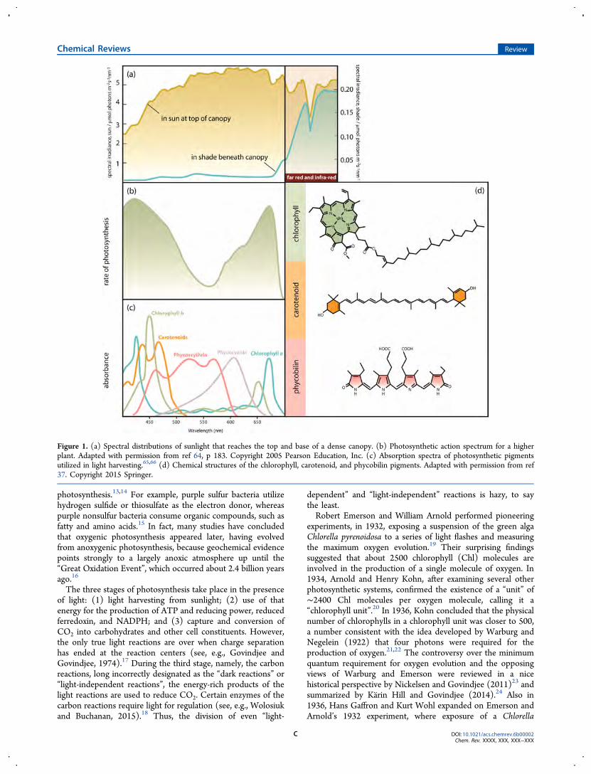
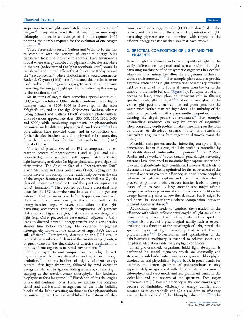
Figure 1. (a) Spectral distributions of sunlight that reaches the top and base of a dense canopy. (b) Photosynthetic action spectrum for a higherplant. Adapted with permission from ref 64, p 183. Copyright 2005 Pearson Education, Inc. (c) Absorption spectra of photosynthetic pigmentsutilized in light harvesting.65,66 (d) Chemical structures of the chlorophyll, carotenoid, and phycobilin pigments. Adapted with permission from ref37. Copyright 2015 Springer.
Chemical Reviews Review
DOI: 10.1021/acs.chemrev.6b00002Chem. Rev. XXXX, XXX, XXX−XXX
C
suspension to weak light immediately initiated the evolution ofoxygen.25 They determined that it would take one singlechlorophyll molecule an average of 1 h to capture 4−12photons, the number required for the evolution of one oxygenmolecule.26
These observations forced Gaffron and Wohl to be the firstto come up with the concept of quantum energy beingtransferred from one molecule to another. They envisioned amodel where energy absorbed by pigment molecules anywherein the unit (today termed the “photosynthetic unit”) would betransferred and utilized efficiently at the center (today termedthe “reaction center”) where photochemistry would commence.Roderick Clayton (1965) later formulated this model in termsused today: “The pigment aggregate acts as an antenna,harvesting the energy of light quanta and delivering this energyto the reaction center”.27
So, in terms of size, is there something special about 2400Chl/oxygen evolution? Other studies confirmed even highernumbers, such as 3200−5000 in Lemna sp., in the mossSelaginella sp., and in the alga Stichococcus bacillaris, whereasGeorg Schmid and Gaffron (1968) observed photosyntheticunits of various approximate sizes (300, 600, 1200, 1800, 2400,and 5000) while conducting experiments on photosyntheticorganisms under different physiological conditions.20,28 Theseobservations have provided clues, and in conjunction withfurther detailed biochemical and biophysical information, theyform the physical basis for the photosynthetic unit (PSU)model of today.The typical physical size of the PSU encompasses the two
reaction centers of photosystems I and II (PSI and PSII,respectively), each associated with approximately 200−400light-harvesting molecules (in higher plants and green algae). Intheir review “The Absolute Size of a Photosynthetic Unit”,David Mauzerall and Elias Greenbaum (1989) highlighted theimportance of this concept in the relationship between the sizeof the oxygen forming unit, the total chlorophyll per O2 (theclassical Emerson−Arnold unit), and the quantum requirementfor O2 formation.29 They pointed out that a theoretical limitexists for the PSU sizethe same limit as in a homogeneousantennathat the mean time before trapping increases withthe size of the antenna, owing to the random walk of theenergy-transfer steps. However, modulation of the light-harvesting architecture through incorporation of pigmentsthat absorb at higher energies, that is, shorter wavelengths oflight (e.g., Chl b, phycobilins, carotenoids), adjacent to Chl aleads to directed downhill energy transfer and a considerablyshorter time before trapping. The existence of pigmentheterogeneity allows for the existence of larger PSUs that arestill efficient.30 Furthermore, determining the PSU size, interms of the numbers and classes of the constituent pigments, isof great value for the elucidation of adaptive mechanisms ofphotosynthetic organisms in varied environments.31
The photosynthetic unit comprises numerous light-harvest-ing complexes that have diversified and optimized throughevolution.32 The mechanism of highly efficient energycapturefirst light absorption, followed by rapid excitationenergy transfer within light-harvesting antennas, culminating intrapping at the reaction-center chlorophyllshas fascinatedbiophysicists for a long time.33,34 Elucidation of this challengingpuzzle still continues today. Here, we examine the composi-tional and architectural arrangement of the main buildingblocks of the light-harvesting machineries that photosyntheticorganisms utilize. The well-established foundations of elec-
tronic excitation energy transfer (EET) are described in thisreview, and the effects of the structural organization of light-harvesting pigments are also examined with respect to theefficient energy-transfer mechanisms that have evolved.
2. SPECTRAL COMPOSITION OF LIGHT AND THEPIGMENTS
Even though the intensity and spectral quality of light can bevastly different on temporal and spatial scales, the light-harvesting machinery of photosynthetic organisms has evolvedadaptation mechanisms that allow these organisms to thrive indiverse environments.35−37 For example, plant canopies providea vertical gradient of sunlight, attenuating the intensity of visiblelight by a factor of up to 100 as it passes from the top of thecanopy to the shade beneath (Figure 1a). For algae growing inoceans or lakes, water plays an important role in filteringspecific wavelengths of light.38,39 Short wavelengths of thevisible light spectrum, such as blue and green, penetrate thewater much farther than red light does. The turbidity of thewater from particulate matter plays another important role indefining the depth profile of irradiance.40 For example,downwelling irradiance can vary by orders of magnitudewhen comparing depth profiles in lakes, depending on the localconditions of dissolved organic matter and scatteringparticulates (e.g., humus from vegetation distinctly stains thewater).41
Microbial mats present another interesting example of lightpenetration, but in this case, the light profile is controlled bythe stratification of photosynthetic organisms.42 In 2012, ZoeePerrine and co-workers43 noted that, in general, light-harvestingantennas have developed to maximize light capture under bothlow- and high-intensity light conditions. However, that leads tothe antenna size not being optimized for the achievement of themaximal apparent quantum efficiency, as poor kinetic couplingbetween fast photon capture and the slower downstreamprocess of photosynthetic electron transfer leads to energylosses of up to 50%. A large antenna size might offer acompetitive advantage in mixed cultures when competition forenergy harvesting arises at low flux densities, but it might beredundant in monocultures where competition betweendifferent species is absent.43
Additionally, one needs to consider the variation in theefficiency with which different wavelengths of light are able todrive photosynthesis. The photosynthetic action spectrum(Figure 1b), a plot of a physiological activity such as oxygenevolution as a function of the wavelength of light, reveals thespectral region of light harvesting that is effective inphotosynthesis.44,45 Diversification and optimization of thelight-harvesting machinery is essential to achieve short- andlong-term adaptation under varying light conditions.In all photosynthetic organisms, initial light absorption is
performed by special pigments, which are chemically andstructurally subdivided into three major groups: chlorophylls,carotenoids, and phycobilins (Figure 1c,d). In green plants, forexample, the action spectrum of photosynthesis is onlyapproximately in agreement with the absorption spectrum ofchlorophylls and carotenoids and has prominent bands in theviolet-blue and red regions of the spectrum. Two majordifferences are (1) lowered efficiency in the carotenoid regionbecause of diminished efficiency of energy transfer fromcarotenoids to chlorophylls and (2) a red drop in efficiencyeven in the far-red end of the chlorophyll absorption.46,47 The
Chemical Reviews Review
DOI: 10.1021/acs.chemrev.6b00002Chem. Rev. XXXX, XXX, XXX−XXX
D
reflection and/or transmission of the central part of thespectrum causes leaves to appear green.One might wonder why plants evolved to reflect green
light.48 One suggestion is that chlorophyll absorption iscomplementary to that of bacteriorhodopsin, a purplechromophore that was employed as a light-driven protonpump in the earliest aquatic organisms (e.g., halobacteria),which relied on light-driven energy generation while inhabitingoceanic surface waters. Further, organisms that evolved lateroptimized their light-harvesting apparatus based on chlorophyllsystems to maximize absorption of available sunlight after it wasattenuated by bacteriorhodopsin. Electron transport mediatedby metal porphyrins already existed before that in photo-synthetic transport.49 Mauzerall (1973)50 and Bjorn et al.(2009)51 have speculated that biosynthetic pathways for metalporphyrins and implementation of the existing precursor for theproduction of chlorins through porphyrins was a clearevolutionary advantage.The optimal absorption wavelength range for the light-
harvesting pigment has been suggested to be in the red region(680−690 nm), as chlorophylls utilize that part of the spectrumfor the energy required to split water and to reduceferredoxin.52 The evolution of chlorophyll a as the most widelyutilized photosynthetic pigment can be attributed perhaps to itsefficient absorption of red light and also, perhaps, its chemistry(such as redox potential).53,51 Particularly in land plants, forwhich light was abundant, there was an absence of evolutionarypressure to generate innovative light-harvesting architecturesthat would utilize other parts of the solar spectrum. See Bjornand Govindjee (2015)47 for a discussion of the evolution ofphotosynthesis.The photosynthetic action spectra of cyanobacteria and red
algae show strong activity in the blue-green region, attributed tothe specific class of accessory pigments called phycobilins(Figure 1c).54−56 Green light penetrates through great waterdepths, and utilization of green-light-absorbing pigments allowsred algae and cyanobacteria to live at greater depths thanorganisms that primarily use chlorophyll, such as green algaeand sea grasses, which tend to grow in shallow waters where thevisible spectrum is similar to the spectrum of incident sunlight.However, all photosynthesis in these organisms occurs throughchlorophyll a, because energy absorbed by phycobilins istransferred very efficiently to chlorophyll a.57−59 Phycobilipro-teins exhibit a high nitrogen content, and under conditions oflimited nitrogen supply, their synthesis tends to be unfavorable.In cyanobacteria, rhodophytes, and cryptophytes, phycobilipro-teins are selectively lost during nutrient stress conditions,because photosynthesis can function in the absence ofphycobiliproteins but chlorophyll is essential not only for thelight-harvesting apparatus but also for the reaction centers.60−63
Consequently, for higher plants that grow on land under anabundance of sunlight, it is energetically inefficient to utilizephycobiliproteins for harvesting of green light. Thus, in theinterest of energy conservation, higher plants, which areexposed to an abundance of light when growing on land, donot utilize phycobiliproteins for the capture of green light.51
One reason why “red” carotenoids are not utilized is possiblythat red carotenoids have their S1 transition degenerate or evenbelow the Chl lowest excitonic transitions, which wouldtherefore quench the excited state of chlorophyll fluorescenceand thus make light harvesting inefficient.The variety of chromophores employed in light harvesting is
considerably smaller than the enormous diversity of photo-
synthetic organisms that exists. Certainly, the types of LHC“designs” outnumber chromophore types by a lot. So whatoptimizations in their structure and function have led to thedominance of these pigments in light-harvesting systems?Chlorophyll molecules are based on a cyclic tetrapyrrole ring,chlorin, coordinated to a central atom, a structure very similarto that found in the heme group of hemoglobin, with thedifference being that, in chlorophyll, magnesium is the centralatom, whereas heme contains iron (Figure 1d). Attachment ofdifferent side chains to the chlorin ring allows for structuraldiversification of the chlorophyll family and production ofchlorophylls (a, b, c, d, e, and f). The different side chains onthe chlorophylls are responsible for tuning the absorptionspectra of the pigment molecules.67 Carotenoids, yellow-orangechromophores, exhibit a characteristic triple peak absorbance inthe range of 400−500 nm, more or less coinciding with the BXand BY Soret bands of chlorophyll.Carotenes occur naturally in a number of isomeric forms; α-
carotene and β-carotene are the primary isomers, differing onlyin the position of the double bonds in the cyclic group at theend of the molecule (Figure 1d). Phycobilins are linear open-chain tetrapyrroles that bear a resemblance to a porphyrin thathas been split open (Figure 1d). Photosynthetic pigments arecyclic or linear examples of conjugated π-electron systems withexceptional molar extinction coefficients, ∼105 M−1 cm−1. Thescaling laws for linear chromophores, such as carotenoids andπ-conjugated polymers, predict that the dipole strength of theirlowest allowed electronic transition will depend on the lengthsquared.68 However, the scaling plateaus at lengths of about10−15 double bonds are due to effects such as conformationaldisorder that twists bonds and breaks conjugation.69
Up to this point, we have treated pigment chromophoresonly as individual entities characterized by a large absorptionstrength, but in the following sections, we reveal that theirsynergetic interactions as constituent elements of light-harvesting machineries play a crucial role. They have clearlydifferent biochemical and biophysical properties when they areassociated with different amino acids within specific proteins.70
The path toward realization of artificial light-harvestingmodel systems has, thus far, faced two major challenges:synthesis of well-suited chromophores and construction of thescaffolding that would avoid the difficulties of assembling largenumbers of integral pigment molecules. Recently, a synergeticcombination of bioinspired and synthetic building blocks led tothe development of a series of multichromophore biohybridcomplexes that can be utilized in the realization of multifunc-tional light-harvesting assemblies.71,72 The rationale for thedesign of these biohybrid architectures is to overcomelimitations faced by synthetic chemists, as it is an extremelychallenging task to fabricate a framework structure that wouldallow for an organized assembly of a large number of pigmentmolecules. The model is based on creating a framework fromnative photosynthetic peptide analogues. The native chromo-phores, bacteriochlorophylls and their derivatives, on the otherhand, have often limited synthetic malleability. However,recently developed bacteriochlorins exhibit good stability, andtheir structural tailoring enables wavelength tuning, giving thesestructures an advantage over naturally occurring systems thatface limitations in terms of the extent of spectral coverage.72
Through static and time-resolved optical studies on theseoligomeric biohybrid antenna, Reddy et al. (2013)71 observedefficient (90%) excitation energy transfer from the attachedbacteriochlorin to the BChl a target.
Chemical Reviews Review
DOI: 10.1021/acs.chemrev.6b00002Chem. Rev. XXXX, XXX, XXX−XXX
E
Realization of artificial photosynthetic model systems iscontingent on the awareness that the role of constituentpigment molecules in these large light-harvesting assemblies ismanifold, as they can act as initial light absorbers, efficientenergy conduits, or facilitators of charge separation. Further-more, the photophysical properties and spectral features ofpigment molecules within these light-harvesting assemblies ishighly affected by the synergetic interactions of all of theconstituent elements. Some aspects of the photophysicsgoverning these constraints are discussed in the followingsections.In summary, the main pigment groups are chlorophylls,
carotenoids, and phycobilins. Pigments are examples ofconjugated π-electron systems and have exceptionally highmolar extinction coefficients (∼1 × 10⁵ M−1 cm−1).
3. LIGHT-HARVESTING SYSTEMS AND THEIREFFICIENCY
Pigments in pigment−protein (antenna) complexes, usuallycalled light-harvesting complexes (LHCs), are responsible formost of the absorption of sunlight. Excitation energy issubsequently funnelled, first among the other surroundingmolecules of the same complex, then from one light-harvestingcomplex to another before being trapped at a photochemicalreaction center (RC), where it is converted into a chargeseparated state with >90% quantum efficiency.73
Although many measurements of photosynthetic quantumyield, or the efficiency of energy conversion, are available, westress the importance of the measurement techniques andconditions when comparing the values reported in differentecophysiological studies.74 In early days, evaluation of absolutequantum efficiency was most often performed on intact algae orbacteria, but the respiratory activity complicated thoseinvestigations of the primary photochemical act.75 Usually,the internal (intrinsic) quantum efficiency of the primaryreactions of photosynthesis is close to unity when onecompares the percentage of absorbed photons converted tocharge carriers.76 The absolute quantum yield of primaryphotochemistry, measured in the reaction centers of Rhodop-seudomonas sphaeroides73,77 and Rhodospirillum rubrum,78 hasbeen found to be near unity. Further, measurements onsinglet−singlet energy transfer from carotenoid to BChl in LH2showed typical efficiencies ranging from 50% to 100%, with astrong dependence on both the type of carotenoid(s) and thetype of LH complex. In Rhodopseudomonas (Rps.) acidophila[now Rhodobastus (Rbl.) acidophilus] LH2, carotenoid to BChlenergy transfer is about 55% efficient,79 whereas in Rhodobacter(Rb.) sphaeroides 2.4.1 LH2, it is about 95% efficient.80
Wientjes et al. (2012)81 determined the PSII efficiency inArabidopsis thaliana by studying the thylakoid membrane of theplant under varying light conditions. In high light, the plant hada smaller PSII antenna size and an efficiency of 91%, whereas anincrease of the antenna size in low light led to an increase in theabsorption cross section but at the cost of a lowered PSIIefficiency (84%). Time-resolved Chl a fluorescence experi-ments yielded higher values of quantum efficiency than thoseobtained from the ratio of the variable to maximum Chlfluorescence, Fv/Fm (∼80%), a parameter that is widely used toindicate the PSII quantum efficiency.81 (Note: Fv = Fm − Fo,where Fo is the minimal fluorescence.)In the case of green sulfur bacteria, the energy from absorbed
photons is transferred down an energy gradient from BChl c(absorbing at 742 nm) in the chlorosome antenna, to BChl a
(absorbing at 792 nm) in the chlorosome baseplate, throughthe membrane-bound Fenna−Matthews−Olson (FMO) com-plex (BChl a, ∼805 nm), before finally reaching the reactioncenter (BChl a, ∼865 nm).82 The energy transfer from BChl cto the BChl a component of the chlorosomes occurs with anefficiency of about 55%, whereas the energy transfer within thepigment−protein complex proceeds with an efficiency close to100%.83,84 For the determination of the overall efficiency in thebaseplate and the reaction-center environment of the Fenna−Matthews−Olson (FMO) protein, which connects the outerantenna system (chlorosome/baseplate) with the reaction-center complex in green sulfur bacteria, much more detailedstructural information is needed.85 On the other hand, in redalgae, such as Porphyridium cruentum, the efficiency of energytransfer from the phycobiliproteins to chlorophyll a is higherthan 80−90% .56,57,86
The reason for the development of an elaborate energy-collecting system is that the reaction-center chlorophylls(present in 1 of ∼300 antenna molecules) cannot absorbsunlight at a rate anywhere near high enough for efficientphotosynthesis to occur. [See its first discussion by Gaffron andWohl (1936), where the concepts of “antenna” and “reactioncenter” were born.25] The inefficiency of the system is due tothe fact that chlorophyll molecules absorb only a few photonseach second, which would be completely insufficient to drivethe multielectron process of photosynthesis if only reactioncenters were found in the membrane.25 The solution to theshortcoming of independently functioning reaction centers isthe association of intricate antenna pigment−protein assem-blies, light-harvesting complexes, with reaction centers. Theseproteins associate approximately 100−800 additional chlor-ophylls (or other pigments) with each reaction center.29
Electronic excitation resulting from the absorption of sunlightby pigments in light-harvesting complexes is transferred veryefficiently to reactions centers, thus increasing their effectiveabsorption cross section. Light-harvesting complexes are vitalfor photosynthetic organisms in that they ensure, through acombination an increase in effective absorption and regulation,a steady supply of excitation to each reaction center.2,69,87
The diversity in antenna systems is remarkable, differing inthe number of pigments they associate and their compositionand structural organization at the nanoscale, in addition to thelocation relative to the reaction center, emphasizing theimportance and necessity of the light-gathering mechanism inphotosynthesis. The existence of such a variety is alsosuggestive of independent evolutionary origins that have ledto the development of optimized multichromophoric energycollector systems. Specific examples will be examined in latersections, with a more focused look at the antenna complexes ofpurple bacteria such as Rhodobastus (Rbl.) acidophilus, which isa model case for studying the physical principles that governlight absorption and energy transfer.88
The sites in which key photosynthetic processes, includinglight harvesting, subsequent charge separation, and electrontransport, occur are multifunctional membrane systems, whichvary greatly both in their architecture and composition. Ingreen sulfur bacteria, the antenna complexes, chlorosomes, areassociated with the plasma membrane, whereas purplephototrophic bacteria have intracytoplasmic membranes.14,89
Cyanobacteria are quite different from these anoxygenicbacteria in that their membrane system comprises stacks ofparallel sheets of thylakoids, which are closely positioned to thecytoplasmic membrane.13,14,90,91 Other photosynthetic organ-
Chemical Reviews Review
DOI: 10.1021/acs.chemrev.6b00002Chem. Rev. XXXX, XXX, XXX−XXX
F
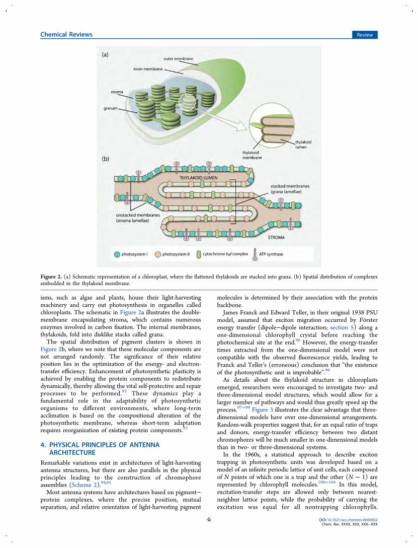
isms, such as algae and plants, house their light-harvestingmachinery and carry out photosynthesis in organelles calledchloroplasts. The schematic in Figure 2a illustrates the double-membrane encapsulating stroma, which contains numerousenzymes involved in carbon fixation. The internal membranes,thylakoids, fold into disklike stacks called grana.The spatial distribution of pigment clusters is shown in
Figure 2b, where we note that these molecular components arenot arranged randomly. The significance of their relativeposition lies in the optimization of the energy- and electron-transfer efficiency. Enhancement of photosynthetic plasticity isachieved by enabling the protein components to redistributedynamically, thereby allowing the vital self-protective and repairprocesses to be performed.92 These dynamics play afundamental role in the adaptability of photosyntheticorganisms to different environments, where long-termacclimation is based on the compositional alteration of thephotosynthetic membrane, whereas short-term adaptationrequires reorganization of existing protein components.93
4. PHYSICAL PRINCIPLES OF ANTENNAARCHITECTURE
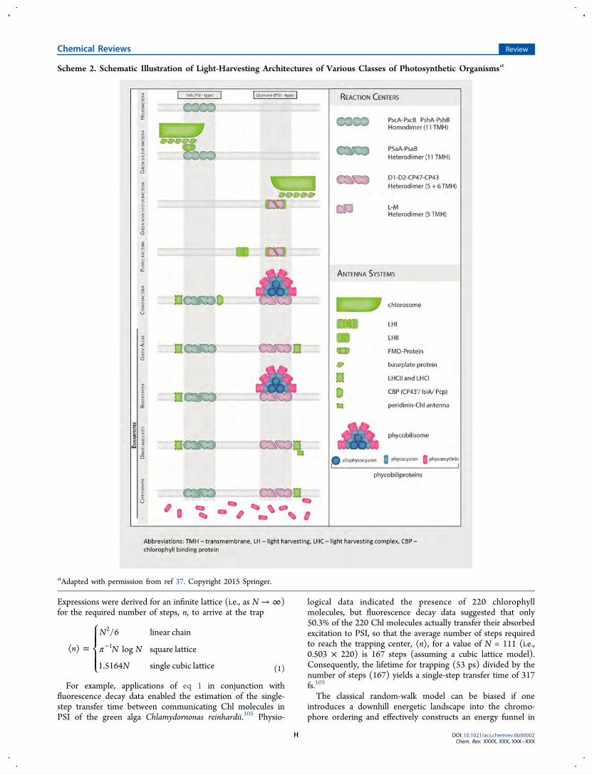
Remarkable variations exist in architectures of light-harvestingantenna structures, but there are also parallels in the physicalprinciples leading to the construction of chromophoreassemblies (Scheme 2).94,95
Most antenna systems have architectures based on pigment−protein complexes, where the precise position, mutualseparation, and relative orientation of light-harvesting pigment
molecules is determined by their association with the proteinbackbone.James Franck and Edward Teller, in their original 1938 PSU
model, assumed that exciton migration occurred by Forsterenergy transfer (dipole−dipole interaction; section 5) along aone-dimensional chlorophyll crystal before reaching thephotochemical site at the end.96 However, the energy-transfertimes extracted from the one-dimensional model were notcompatible with the observed fluorescence yields, leading toFranck and Teller’s (erroneous) conclusion that “the existenceof the photosynthetic unit is improbable”.96
As details about the thylakoid structure in chloroplastsemerged, researchers were encouraged to investigate two- andthree-dimensional model structures, which would allow for alarger number of pathways and would thus greatly speed up theprocess.97−99 Figure 3 illustrates the clear advantage that three-dimensional models have over one-dimensional arrangements.Random-walk properties suggest that, for an equal ratio of trapsand donors, energy-transfer efficiency between two distantchromophores will be much smaller in one-dimensional modelsthan in two- or three-dimensional systems.In the 1960s, a statistical approach to describe exciton
trapping in photosynthetic units was developed based on amodel of an infinite periodic lattice of unit cells, each composedof N points of which one is a trap and the other (N − 1) arerepresented by chlorophyll molecules.100−104 In this model,excitation-transfer steps are allowed only between nearest-neighbor lattice points, while the probability of carrying theexcitation was equal for all nontrapping chlorophylls.
Figure 2. (a) Schematic representation of a chloroplast, where the flattened thylakoids are stacked into grana. (b) Spatial distribution of complexesembedded in the thylakoid membrane.
Chemical Reviews Review
DOI: 10.1021/acs.chemrev.6b00002Chem. Rev. XXXX, XXX, XXX−XXX
G
Expressions were derived for an infinite lattice (i.e., as N → ∞)for the required number of steps, n, to arrive at the trap
π⟨ ⟩ = −
⎧⎨⎪⎪
⎩⎪⎪
n
N
N N
N
/6 linear chain
log square lattice
1.5164 single cubic lattice
2
1
(1)
For example, applications of eq 1 in conjunction withfluorescence decay data enabled the estimation of the single-step transfer time between communicating Chl molecules inPSI of the green alga Chlamydornonas reinhardii.105 Physio-
logical data indicated the presence of 220 chlorophyllmolecules, but fluorescence decay data suggested that only50.3% of the 220 Chl molecules actually transfer their absorbedexcitation to PSI, so that the average number of steps requiredto reach the trapping center, ⟨n⟩, for a value of N = 111 (i.e.,0.503 × 220) is 167 steps (assuming a cubic lattice model).Consequently, the lifetime for trapping (53 ps) divided by thenumber of steps (167) yields a single-step transfer time of 317fs.105
The classical random-walk model can be biased if oneintroduces a downhill energetic landscape into the chromo-phore ordering and effectively constructs an energy funnel in
Scheme 2. Schematic Illustration of Light-Harvesting Architectures of Various Classes of Photosynthetic Organismsa
aAdapted with permission from ref 37. Copyright 2015 Springer.
Chemical Reviews Review
DOI: 10.1021/acs.chemrev.6b00002Chem. Rev. XXXX, XXX, XXX−XXX
H
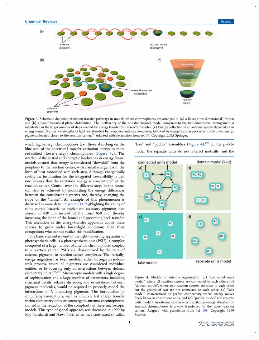
which high-energy chromophores (i.e., those absorbing on theblue side of the spectrum) transfer excitation energy to morered-shifted (lower-energy) chromophores (Figure 3c). Theoverlap of the spatial and energetic landscapes in energy-funnelmodels ensures that energy is transferred “downhill” from theperiphery to the reaction center, with a small energy loss in theform of heat associated with each step. Although energeticallycostly, the justification for the integrated irreversibility is thatone ensures that the excitation energy is concentrated at thereaction center. Control over the different steps in the funnelcan also be achieved by modulating the energy differencesbetween the constituent pigments and, thereby, changing theslope of the “funnel”. An example of this phenomenon isdiscussed in more detail in section 11, highlighting the ability ofsome purple bacteria to implement accessory pigments thatabsorb at 820 nm instead of the usual 850 nm, therebyincreasing the slope of the funnel and preventing back transfer.This alteration in the energy-transfer apparatus allows thesespecies to grow under lower-light conditions than theircompetitors who cannot realize this modification.The basic elementary unit of the light-harvesting apparatus of
photosynthetic cells is a photosynthetic unit (PSU), a complexcomposed of a large number of antenna chromophores coupledto a reaction center. PSUs are characterized by the ratio ofantenna pigments to reaction-center complexes. Theoretically,energy migration has been modeled either through a random-walk process, where all pigments are considered individualentities, or by focusing only on interactions between definedelementary units.106,107 Microscopic models with a high degreeof sophistication and a large number of parameters, includingstructural details, relative distances, and orientations betweenpigment molecules, would be required to precisely model theinteractions of N interacting pigments. The introduction ofsimplifying assumptions, such as infinitely fast energy transferwithin elementary units or isoenergetic antenna chromophores,can aid in the reduction of the complexity of these microscopicmodels. This type of global approach was discussed in 1999 byKay Bernhardt and Hawi Trissl when they contrasted so-called
“lake” and “puddle” assemblies (Figure 4).108 In the puddle
model, the separate units do not interact mutually, and the
Figure 3. Schematic depicting excitation-transfer pathways in models where chromophores are arranged in (a) a linear (one-dimensional) formatand (b) a two-dimensional planar distribution. The inefficiency of the one-dimensional model compared to the two-dimensional arrangement ismanifested in the larger number of steps needed for energy transfer to the reaction center. (c) Energy collection in an antenna system depicted as anenergy funnel. Shorter wavelengths of light are absorbed by peripheral antenna complexes, followed by energy-transfer processes to the lower-energypigments located closer to the reaction center.66 Adapted with permission from ref 37. Copyright 2015 Springer.
Figure 4. Models of antenna organization: (a) “connected unitsmodel”, where all reaction centers are connected to each other; (b)“domain model”, where two reaction centers are close to each otherbut the groups of two are not connected to each other; (c) “lakemodel”, characterized by perfect connectivity where energy movesfreely between constituent units; and (d) “puddle model” (or separateunits model), an extreme case in which excitation energy absorbed byantenna chromophores is always transferred to the same reactioncenters. Adapted with permission from ref 108. Copyright 1999Elsevier.
Chemical Reviews Review
DOI: 10.1021/acs.chemrev.6b00002Chem. Rev. XXXX, XXX, XXX−XXX
I
excitation energy absorbed by antenna chromophores is alwaystransferred to the same reaction center within the specificphotosynthetic unit. The lake model allows for unrestrictedexciton transfer as antenna chromophores form a matrix withembedded reaction centers (Figure 4c).103 In the latter model,which is often used for PSII and purple bacteria, excitationenergy can visit multiple reaction centers before eventuallybeing trapped at a reaction-center complex that is open forphotochemistry.2,109−111
Most photosynthetic organisms fall somewhere between thetwo extremes in terms of their antenna organizations and thedegree of connectivity between different PSUs. The basicproperties of such an intermediate case were first suggestedwithin the framework of the “connected units model”developed by Pierre Joliot and Anne Joliot (1964). In thismodel, a partial connectivity between puddles exists, but energytransfer among pigments within a specific puddle is moreprobable than energy transfer between chromophores locatedin distinct puddles.112Alternatively, limited excitation transfercan be taken into account by dividing the photosyntheticmembrane into domains (mini-lakes) comprising clusters ofphotosynthetic units. The domain model allows for unrestrictedexcitation migration within a domain but restricts excitonexchange between the separate units.113,114 This model is well-suited for scenarios in which dimeric aggregation of reactioncenters exist, as in the case of the chlorosome antenna complexof green photosynthetic bacteria.2,108
Today, our understanding of the structure and function ofPSUs has become much more sophisticated throughinformation obtained from studies employing statistical modelsof PSUs in conjunction with a number of fluorescencetechniques. Elucidation of structural details of a number oflight-harvesting complexes through high-resolution crystallog-raphy has fueled the generation of more sophisticated energy-transfer models.115−118 Even models that contain chromo-phore−protein interactions treated with atomistic detail haverecently been reported.119−122 See the basic discussion byAmarnath et al. (2016)123 and the review by Stirbet (2013).124
In summary, light-harvesting antennas exhibit a largevariation in the architectural assembly of their constituentchromophores. Pigment−protein associations allow for controlover the separation and mutual orientation of light-harvestingmolecules. Spatially, a three-dimensional arrangement ofchromophores is statistically preferred for efficient energytransfer. The principle of an energy funnel biases the random
walk but contributes to the irreversibility of the concentrationof energy at the reaction center. Variations in the models ofantenna organization are based on photosynthetic units (PSUs)on the macroscopic level. High-resolution crystal structures andmodels with atomistic details allow for sophisticated models ofenergy transfer on a microscopic level.
5. MECHANISM OF FORSTER EXCITATION ENERGYTRANSFER
During 1927−1929, Jean Perrin and Francis Perrin observedenergy transfer as they researched fluorescence quenching offluorophores in solution.125−127 They noted that molecules insolution could interact without collisions and at distancesexceeding their molecular diameters. It was then postulated thatthis observed phenomenon, which leads to electronic energytransfer, derives from an inductive resonance interactionbetween transition dipole moments of the molecules. Inother words, the semiclassical oscillation of the electrons on thedonor, during de-excitation, induces oscillations of the acceptorelectrons, causing electronic excitation. This interaction is aCoulombic dipole−dipole interaction, which varies as theinverse of the cube of the center-to-center intermoleculardistance between donor and acceptor. It can thus be effective atdistances on the order of several nanometers.The semiclassical idea of a classical inductive resonance that
transfers energy from donor to acceptor in the same way asmechanical energy can be transferred among oscillators isconceptually appealing. Classical analogues are well-known inclassical mechanics, such as the transfer of oscillations from onetuning fork to another by mechanical coupling of the tuningforks to sound waves through the intervening medium (air).Technically, however, the coupling V is a quantum mechanicalinteract ion between the reactant wave funct ion|ψD,exc i t edψA,g round⟩ and the product wave function|ψD,groundψA,excited⟩, where D and A stand for donor andacceptor, respectively. In shorthand, we can write these wavefunctions equivalently as |D′A⟩ and |DA′⟩. These wavefunctions are written as product states of one excited-statemolecule (initially D) and a ground-state molecule (initially A).Physically, what one need to ascertain is the interaction thatcauses de-excitation of the donor, D′ → D, synchronously withexcitation of the acceptor, A → A′. This picture is worthkeeping in mind because it indicates that, to understand energytransfer, one must explicitly include four electronic states in the
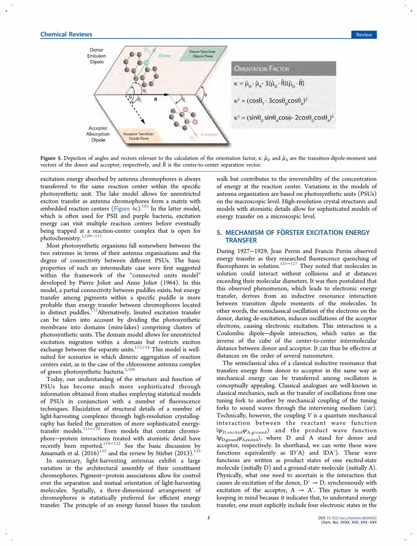
Figure 5. Depiction of angles and vectors relevant to the calculation of the orientation factor, κ. μD and μA are the transition-dipole-moment unitvectors of the donor and acceptor, respectively, and R is the center-to-center separation vector.
Chemical Reviews Review
DOI: 10.1021/acs.chemrev.6b00002Chem. Rev. XXXX, XXX, XXX−XXX
J
model: the ground and excited states of each of thechromophores.128
The coupling that promotes excitation to jump from donorto acceptor is primarily a Coulombic interaction between“transition densities” (vide infra).129 In the dipole approx-imation, the interaction potential between these transitiondensities is expanded as an infinite sum of transitionmultipole−multipole interactions.130 As long as the donorand acceptor molecules are widely separated compared to theirphysical size, one can simply take the first (leading) term of thisexpansion, which is the transition dipole−dipole interaction
πεμ μ μ μ
πεκ μ μ
= ·
− · ·
≡| || |⎡
⎣⎢⎢
⎤⎦⎥⎥V
R
R R
R R1
4
3( )( ) 140
D A3
D A5
0
D A3
(2)
Here, μD and μA are the transition dipole moments (SI units ofthe coulomb-meter) of the donor and acceptor, respectively,and R is the center-to-center separation between the donor andacceptor molecules. The orientation factor, κ, is given by
κ μ μ μ μ= · − · · R R3( )( )D A D A (3)
where μD and μA represent unit vectors of the donor andacceptor, respectively, in the direction of the appropriate
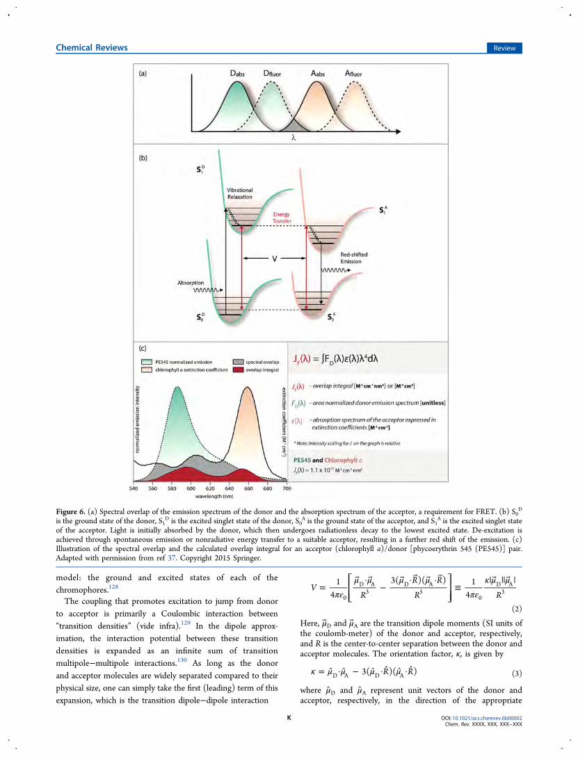
Figure 6. (a) Spectral overlap of the emission spectrum of the donor and the absorption spectrum of the acceptor, a requirement for FRET. (b) S0D
is the ground state of the donor, S1D is the excited singlet state of the donor, S0
A is the ground state of the acceptor, and S1A is the excited singlet state
of the acceptor. Light is initially absorbed by the donor, which then undergoes radiationless decay to the lowest excited state. De-excitation isachieved through spontaneous emission or nonradiative energy transfer to a suitable acceptor, resulting in a further red shift of the emission. (c)Illustration of the spectral overlap and the calculated overlap integral for an acceptor (chlorophyll a)/donor [phycoerythrin 545 (PE545)] pair.Adapted with permission from ref 37. Copyright 2015 Springer.
Chemical Reviews Review
DOI: 10.1021/acs.chemrev.6b00002Chem. Rev. XXXX, XXX, XXX−XXX
K
transition dipole moment and R is their mutual displacementunit vector pointing from D to A. The schematic in Figure 5illustrates the vectors and angles relevant to the orientationfactor, and the following expression describes the dependenceof the orientation factor on the relative orientation between thedonor and acceptor transition-dipole-moment vectors
κ θ θ θ
θ θ ϕ θ θ
= −
= −
(cos 3 cos cos )
(sin sin cos 2 cos cos )
2T D A
2
D A D A2
(4)
When the orientations of D and A are independent andrandom, either dynamically or in the ensemble average, theisotropic average of the dipole orientation factor equals 2/3. Inphotosynthetic light harvesting, transition dipoles have fixedrelative positions, in which case κ2 can range from 0 to 4. For adetailed discussion of κ2, see van der Meer.131
In 1946, Theodor Forster provided a notable advance byrelating the predicted energy-transfer rate to the spectra of thedonor and acceptor molecules. He first became interested inthe subject following the realization that, in photosynthesis, theefficiency of energy collection is much greater than if oneassumes that reaction centers are responsible for direct photoncapture. Forster’s “hopping” energy-transfer mechanismdescribed this process of exciton migration as a kind of randomwalk in which a series of energy-transfer steps shuttle theexcitation from chromophore to chromophore. Each hop isinduced by the weak point-dipole−point-dipole interactionbetween chromophore transition dipole moments.Forster first showed that the rate of energy transfer (kForster)
between a donor and acceptor chromophore is determined byseveral parameters: the donor lifetime (τD), the quantum yieldof the donor fluorescence (ϕD), the interchromophore distanceR (cm), the relative orientation of the donor−acceptor pair (κ)(Figure 5), and the overlap integral (JF) (Figure 6c). The rate isexpressed as
τκ ϕ
π=k
J
N n R1 9(ln 10)
1281Forster
D
2D F
5A
4 6(5)
where n is the medium index of refraction and NA is Avogadro’snumber.132 The Forster spectral overlap (JF) (M
−1 cm3 or M−1
cm−1 nm4) measures the overlap of the donor emissionspectrum and the acceptor absorption spectrum and ensuresenergy conservation. The expression for JF is a function of thearea-normalized spectrum of the donor emission, FD(λ), andεA(λ), the extinction coefficient spectrum of the acceptor inunits of M−1 cm−1
∫ λ ε λ λ λ=∞
J F ( ) ( ) dF 0D A
4(6)
An example of Forster overlap JF(λ) is illustrated in Figure 6cfor an acceptor (chlorophyll a)/donor [phycoerythrin 545(PE545)] pair. The two y axes on the plot represent thespectral intensities of the donor and acceptor, and thus, thescaling for JF in the graph is arbitrary.In Forster theory, energy conservation is determined by the
overlap of the fluorescence spectrum of the donor and theabsorption spectrum of the acceptor. Part of the electroniccoupling (the quantum mechanical inductive resonanceinteraction) comes from the magnitude of the donor transitiondipole moment and is provided by the radiative rate offluorescence, krad = ϕD/τD ∝ |μD|
2, where ϕD is the fluorescencequantum yield, τD is the fluorescence lifetime. and μD is the
transition dipole moment of the donor. The magnitude of theacceptor dipole moment is encoded in the molar extinction ofthe acceptor absorption spectrum. Deconvolving the requiredinformation from the experimental spectra and discardinginformation that is not needed for the energy-transfer theory iswhat clutters Forster’s equation with so many constants. Seereferences by Silvia Braslavsky et al. (2008)132 and Robert Knoxand Herbert van Amerongen (2002)133 for further details onthis matter.Forster realized that spectral line broadening for molecules in
solution leads to phase decoherence before incoherentexcitation energy transfer occurs.134 This means that a modelbased on the Fermi golden rule rate expression is sufficient. Inthat framework, the rate of energy transfer scales as the squareof the electronic coupling. Hence, the 1/R3 distance depend-ence of the dipole−dipole interaction translates to a 1/R6
distance dependence for the rate of energy transfer.To conserve energy during the excitation transfer from donor
to acceptor, the fluorescence emission spectrum of the donormolecule should overlap to some degree with the absorptionspectrum of the acceptor molecule, illustrated by the gray areain Figure 6a. This is the basis of Forster’s famous spectraloverlap integral. The larger the spectral overlap, the higher theenergy-transfer rate. Notably, the spectral overlap integraldepends not only on the donor fluorescence being coincidentwith frequencies at which the acceptor can absorb light, but alsoon the line broadening.135 Details on how and why spectrallines are broadened can be neglected in Forster theory;however, they can be important for the understanding of themodern, more advanced treatments of coherent energytransfer.136,137 We do not discuss the topic here, but somebackground reading on line broadening can be found in articlesby Clegg et al. (2010)33 and Fleming and Cho (1996)138 and insection 2 of the article by Oh et al. (2011),139 as well as thereferences cited therein.At low temperature, spectral overlap can be much smaller, as
molecules in the gas phase have very sharp vibronic transitionsand, therefore, need to be close to degenerate to overlap.Nevertheless, by examining a schematic diagram of the donorand acceptor vibronic transitions in the gas phase (Figure 6b),one can see most clearly an important aspect of Forster’stheory. That is, vibronic progressions in the donor fluorescencespectrum and acceptor absorption spectrum provide importantcontributions to the spectral overlap (energy conservationduring energy transfer), especially when the two chromophoresare different. The Forster spectral overlap sums over possiblecombinations of these energy-conserving coupled transitions.This vibronic overlap is a powerful attribute of Forster theorythat is often neglected in contemporary theories. Indeed, it isbecause of this aspect that Forster theory remains one of theforemost quantitatively predictive theories.The sensitivity of the rate of energy transfer and the critical
distance range also correspond to a number of biologicallysignificant dimensions, including the thickness of cellmembranes and the separations between chromophore siteson different protein subunits. Therefore, EET measurementscan be employed as an effective molecular ruler, resolving thespatial relationships between molecules, as it is capable ofquantitatively determining distances between chromophores(10−100 Å), thereby providing more insight into the structuraland dynamic aspects of macromolecules.140−142
Chemical Reviews Review
DOI: 10.1021/acs.chemrev.6b00002Chem. Rev. XXXX, XXX, XXX−XXX
L
6. BEYOND FORSTER THEORY OF EXCITATIONENERGY TRANSFER
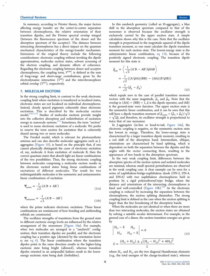
Despite the general success of conventional Forster theory,especially for predicting energy transfer from phycocyanins tochlorophyll a in cyanobacteria and elsewhere, it can provide acomplete description of energy transfer for only a few cases ofphotosynthetic light-harvesting complexes. Typically, light-harvesting antenna structures contain chromophores at veryhigh concentrations, reaching levels of up to 0.6 M in somepigment−protein assemblies. Interchromophoric distancebetween neighboring chlorophyll molecules in light-harvestingsystems can vary between 5 and 20 Å, consequently resulting invariations in the strength of intermolecular coupling, whichdirectly influences the quantum mechanical nature of theenergy-transfer mechanism. This has inspired advances thathave extended Forster’s original theory.115,117,136,143,144
Four principle modifications to the energy-transfer theoriesare needed to predict energy transfer in light-harvestingcomplexes.First, electronic coupling must be calculated without
invoking the dipole approximation, because of the closeintermolecular separation mentioned previously. Second,solvent screening of the electronic coupling needs to bereconsidered hand-in-hand while dealing with a breakdown inthe dipole−dipole approximation.122,145 Third, the presenceand role of molecular exciton states as excitation donors andacceptors needs to be considered. Typically, the generalizedForster theory (GFT) or the modified Redfield theory areemployed to do this.115,146−149 We introduce GFT in a sectionbelow after describing molecular excitons. Fourth, quantum-mechanical corrections need to be introduced into energy-transfer dynamics to account for coherence ef-fects.136,144,150−152 In models beyond Forster theory, it isessential to know and account for details about the bath,especially the time scales of fluctuations that produce linebroadeningand correlations of these fluctuations. Emphasiz-ing this point was an important contribution of Akito Ishizakiand Graham Fleming (2009), who introduced the hierarchicalequations of motion (HEOM) approach for calculating energy-transfer dynamics.153 The HEOM method provides an accurateprediction of the dynamics of a reduced system coupled to aquantum bath, regardless of the relative strength of electroniccoupling and coupling of the system to the bath (i.e., theintermediate coupling regime). It takes advantage of theGaussian property of the phonon operators in the exciton−phonon interaction Hamiltonian. This approach has beenhighly useful for many cases.136
6.1. Electronic Coupling and Orbital Overlap
In photosynthetic light harvesting, energy-transfer processescan be driven by different interaction mechanisms: the long-range dipole−dipole Coulombic interactions (electrodynamicinteractions154), Ved, and interactions due to intermolecularorbital overlap, Vioo, which operate at short range and becomecritical at distances of less than 5 Å..135,155 The coupling term,Vtotal, is defined as the sum of the long-range and short-rangecontributions, which is worth emphasizing because it is oftenmisunderstood that the dipole−dipole mechanism (Ved term)and the Dexter mechanism (Vioo term) are mutuallyexclusive128
= +V V Vtotal ed ioo (7)
The electronic coupling is strongly influenced by inter-chromophore orbital overlap effects at small intermoleculardistances. For example, these effects play an important rolewhen transitions involving simultaneous donor de-excitationand acceptor excitation are spin-forbidden.156 In photosyn-thesis, processes that are mediated by Vioo include triplet−triplet energy transfer, as well as the chlorophyll-sensitizedgeneration of singlet oxygen. This topic is described later in thisreview.In molecular systems, the part of the electronic coupling that
depends on orbital overlap, Vioo, comes from the fact thatelectrons are not definitively associated with a particularmolecule when orbitals overlap. It is most convenient toinclude this effect by rendering the model for the reactant andproduct wave functions more flexible by including so-calledcharge-transfer (or ionic) configurations in addition to thelocally excited configurations that mediate Ved.157 In this model,Vioo appears as an intuitive double-step electron transferbetween donor and acceptor that effectively exchangeselectronic excitation.The formal derivation157 takes a bit more work than the
intuition suggests, but the net result is that the primaryelectronic coupling for EET that is mediated by orbital overlapis not the exchange interaction but the product of two one-electron transfers, each quantified by a matrix element (eq 8),called a bond integral (β) in the old literature. The βET termaccounts for transfer of an electron from D′ to A, and the βHTterm moves a hole from D′ to A (equivalently, an electron fromthe highest occupied molecular orbital of A to D′). The neteffect of these two virtual and synchronous electron transfers isthat electronic excitation is transferred from D to A. See ref 157for details and ref 135 for a review. Note that the electrontransfers are conceptual, stemming from the classical valence-bond formulation of the problem; they are not a real sequenceof one-electron-transfer events because they do not separatelyand sequentially induce solvent reorganization.The part of the electronic coupling that promotes EET
through explicit orbital overlap effects, therefore, has a steepdistance dependence. Each of the β terms in eq 8 dependsexponentially on donor−acceptor separation. Therefore, themagnitude of Vioo depends exponentially on R and increasesexponentially twice as steeply as the rate of a hypotheticalelectron transfer between the same molecules with a decreaseof R. The sign of Vioo can be positive or negative (it depends onhow the donor is oriented with respect to the acceptor), andthe sign of Vioo does not have to correlate with the sign of theCoulombic interaction Ved. The specific form of Vioo is
β β≈V A2 /iooET HT (8)
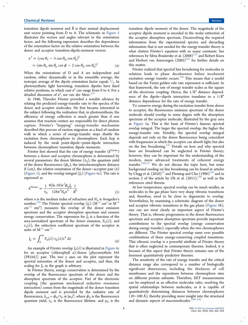
where A is the energy difference between charge-transferconfigurations and the locally excited donor configuration(corresponding to ΔECT in Figure 9). The distance dependenceof Vioo is directly proportional to exp(−2αR) given that theelectron and hole transfer matrix elements are βET ∝ exp(−αR)and βHT ∝ exp(−αR).158,135 Owing to this very steep scaling ofVioo with R, Vioo dominates at close separations when themolecules are in van der Waals contact. Figure 7 shows acalculation of the electronic coupling between naphthalenechromophores at various separations. The distinction betweenthe electrodynamic (approximately dipole−dipole) couplingregime and the regime where orbital overlap effects matter isquite clear.
Chemical Reviews Review
DOI: 10.1021/acs.chemrev.6b00002Chem. Rev. XXXX, XXX, XXX−XXX
M
In Forster theory, the weakly coupled chromophores areassumed to be well-separated compared to their size, so that theshort-range term, Vioo, is neglected and the Coulombic couplingcan be approximated as a point-dipole−dipole interaction. Sucha model based on the localized donor−acceptor states isreasonable for the weakly coupled B800 ring of purple bacterialLH2. (For readers unfamiliar with LH2, structural detailspresented in section 10 would be helpful in a further discussionof this topic.129) The main problem with the dipole−dipoleapproximation is that it works well only when the separationbetween the chromophores is large compared to the size ofthose molecules.6.2. Breakdown of the Dipole Approximation
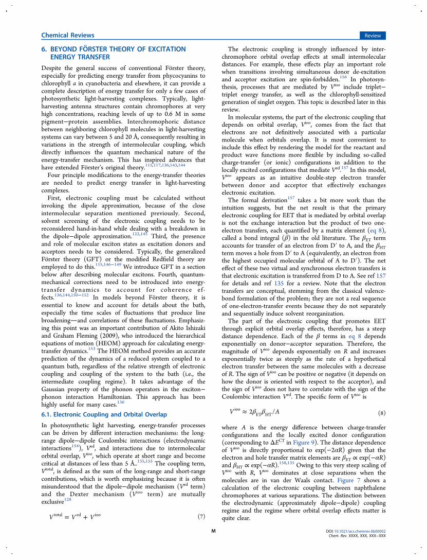
At small separations, the dipole−dipole approximation fails, andthe calculation of the Coulombic coupling between chromo-phores requires a more realistic account of the shape of thetransition densities. A straightforward method is to use thetransition density cube (TDC) method developed by BrentKrueger et al.129 How and why this method is useful is reviewedin detail elsewhere.116,155 In section 9 of this review, we discussthe TDC method further. The essential picture of the TDCmethod is illustrated in Figure 8.The first concept to understand when thinking about Ved and
any accurate way to calculate it, including the TDC method, isthe transition density. The properties of an electronic transition
are determined by the transition density: it is a virtual chargedistribution that captures the way a molecule’s electronic wavefunction jumps from one state to another as a result of theaction of a resonant electromagnetic field. One can calculatetransition densities and plot them as if they were a real(classical) charge distribution, as shown in Figure 8. Forexample, it is a common procedure in freshman chemistry toplot charge distributions to illustrate the shapes of atomicorbitals; for example, the shape of the 2px orbital wave functionψ2px is indicated by the one-electron spinless density ρ(x,y,z) =
|ψ2px(x,y,z)|2. This is the real (quantum mechanical) charge
distribution for an electron in the 2px atomic orbital. Thetransition density is not quite the same because it is constructedfrom wave functions of two different electronic states. Thetransition density connecting electronic state Ψ0 to state Ψ1 isgiven by
∫= Ψ Ψ* ′ ′ ′ ′ ′δP r N x x x x x x x x x x s( ) ( , , ..., ) ( , , ..., ) d ...d d ...d dkN N N N0 1 0 1 2 1 1 2 2 2 1
(9)
where N is a normalization constant, xi includes both the spatialand spin coordinates of each electron i, and s1 is the spin ofelectron 1. In other words, the wave-function outer product hasbeen integrated over all space and spin coordinates of allelectrons except electron one.When light interacts with a molecule to instigate the
transition from state Ψ0 to state Ψ1, it averages over the detailof the transition density, because the wavelength of the lightbeing absorbed or emitted is very large compared to the size ofthe molecule. This averaging means that absorption andemission observables, such as extinction coefficients andradiative rates, are well-quantified by a simple vector, thetransition dipole moment. Just as a dipole moment can becalculated for any real charge distribution, it can be calculatedfor the transition density simply by applying the dipoleoperator. The resulting quantity is called the transition dipolemoment.There is a difference between how light interacts with
molecules and how molecules, such as those shown in Figure 8,interact with each other through their transition densities. Themolecules and the distance separating them are comparable insize. Thus, the Coulombic interaction, proportional to 1/r,effectively traces out the shapes of the molecules. 1/r is quite asteep interaction, so it weights closely separated parts of thetransition density more than distantly separated parts. Forinstance, in Figure 8, the interaction between transition chargeelements i and j with separation rij is stronger than theinteraction between transition charge elements k and j withseparation rkj by the ratio rkj/rij, if the charge elements are all ofsimilar magnitude.The net effect of averaging over the interactions, instead of
first averaging over the transition densities and then calculatingthe interaction, can be significant and is the reason for thefailure of the dipole−dipole approximation. Failure of thedipole−dipole approximation is related to the shapes ofmolecules, and therefore, when the dipole approximationfails, so does the multipole expansion (taken to reasonable, low,order).
6.3. Solvent Screening
It is known that the solvent environment, or the host mediumsurrounding the donor and acceptor molecules, modifies theelectronic coupling. For example, the EET rate in a polarizable
Figure 7. Total electronic coupling between the S1 states ofnaphthalene and the S2 states as a function of interchromophoreseparation. The steep rise at short separations indicates the onset ofinteractions depending on interchromophore orbital overlap. Repro-duced with permission from ref 128. Copyright 1994 AmericanChemical Society.
Figure 8. Plot of transition densities calculated for (right) achlorophyll molecule and (left) a carotenoid molecule. TheCoulombic interaction Ved is determined by the integral over allinteractions between the molecules in a way that maps over the shapesof the donor and acceptor. Reprinted with permission from ref 156.Copyright 2011 Wiley-VCH.
Chemical Reviews Review
DOI: 10.1021/acs.chemrev.6b00002Chem. Rev. XXXX, XXX, XXX−XXX
N
medium is reduced by the factor 1/n4 according to Forstertheory, where n is the refractive index of the medium at opticalfrequency. This factor (often somewhat misleadingly referred toas screening; see below) comes from a modification of thedipole−dipole electronic coupling, Vdip−dip(solv) = Vdip−dip(vacuum)/n2. A similar equation is easily derived for the interaction energybetween charge dipoles in a polarizable medium, but there is animportant distinction for transition dipole: The medium effectscome only from the high-frequency dielectric response of themedium, not from the low-frequency dielectric response.Hence, the refractive index and not the dielectric constant ofthe solvent is the parameter that matters. The other confusingpoint is that Forster theory is formulated in terms of spectra,and the donor emission and acceptor absorption spectra eachalso contain refractive index factors. Knox and van Amerongen(2002)133 provided a very clear account disentangling thedifferent refractive index terms that appear in the derivation ofForster theory and showed how only the solvent “screening”term ends up in the final equation.
The Forster treatment of the dielectric medium is adequateonly for two chromophores found at large distances comparedto their molecular dimensions in a nondispersive, isotropic hostmedium and for which the dipole approximation holds.159
These conditions are quite restrictive. Given that the EET isstrongly affected by solvent effects, for example, if n = 1.4, thencompared to unscreened electronic couplings, the rate differs bya factor of almost 4. It therefore makes sense that estimations ofelectronic couplings using more accurate methods such as TDCshould also incorporate more sophisticated treatments ofsolvent effects. It turns out that quantum chemical theory isneeded to provide a realistic solution, as was worked out byIozzi et al. (2004)160 and Jurinovich et al. (2015).161 JohannesNeugebauer described how to calculate solvent screening in theframework of time-dependent density functional theory.162
Another important contribution was from Julia Adolphs andThomas Renger, who developed ways to calculate electro-chromic shifts of transition energies in protein environments.163
In that work, an electrostatic method, now known as the
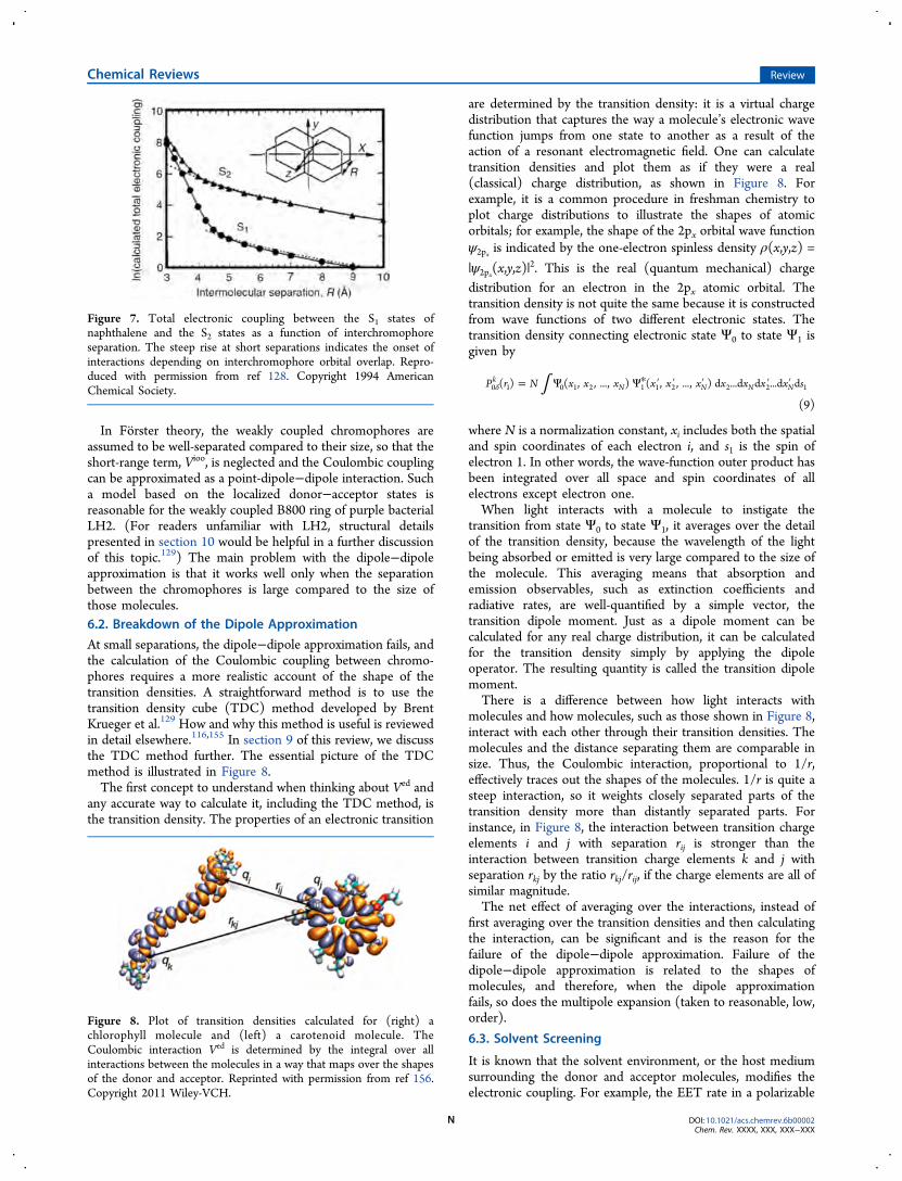
Figure 9. (a) Schematic depiction of the interaction between two transition dipoles (arrows) immersed within cavities in a polarizable medium. Theeffective charges representing the response of the medium to one transition dipole are drawn. As the cavities converge, the effective charges remainheld away from the transition dipoles. (b) On the basis of quantum mechanical calculations, the dependence of s on the separation between theChl602−Chl607 molecules in the dimer structure from LHCII is explained by the formation of a common cavity, physically representing exclusion ofsolvent from the intermolecular region. The result is represented by a progressive spread of the effective surface charges over the acceptormicroenvironment, as shown for three Chl−Chl separations. (c) The spread of the surface charges over the cavities changes the magnitude andfunctional form of the electronic coupling. The dipole−dipole coupling (screened by n2), Vdip−dip/n
2 (squares), is compared here to sVdip−dip. Thedifference corresponds to a factor of at least 2 in the calculated rate of EET. (d) The origin of the distance dependence of s is seen by inspecting theratio of the direct electronic coupling, Vs, to the explicit solvent contribution, Vexplicit. As the intermolecular separation decreases, V explicit assumesdiminishing significance, and thus, s = Vs/Vtot → 1 because the total coupling Vtot ≈ Vs. Figure reproduced with permission from ref 145. Copyright2007 American Chemical Society.
Chemical Reviews Review
DOI: 10.1021/acs.chemrev.6b00002Chem. Rev. XXXX, XXX, XXX−XXX
O
Poisson-TrEsp method,164,165 was introduced for the parame-ter-free calculation of excitonic couplings including screeningeffects. It was applied to calculate electronic couplings in theFMO protein and, thereby, provided a microscopic explanationof the small effective oscillator strength inferred fromspectroscopic studies, reported by Louwe et al. (1997).166
Using the polarizable continuum model (PCM), themolecular system under study (the donor−acceptor pair) isrepresented as a quantum mechanical charge distribution withina molecular cavity, characterized by a realistic shape, whereasthe complex host environment comprising the protein medium,intrinsic water, and surrounding medium is collectivelymodeled as a structureless polarizable continuum. This modelcan more precisely elucidate solvent effects for realistic systemsbecause it accounts for the shapes of interacting moleculartransition densities. The calculation includes the way solventresponds to the interaction between transition densities andhow the transition densities in turn respond, through a reactionfield, to the polarization of the solvent (Figure 9).145 Combinedwith TDC calculations, one thereby finds the general form ofthe screened (or, better, solvent-corrected) electronic coupling:VCoul(solv) = sVCoul(vacuum). In the limit of the dipole−dipoleapproximation, s = 1/n2.As an example, calculations of electronic couplings were
performed for more than 100 pairs of molecules taken fromdifferent photosynthetic proteins, with variations in themolecular dimensions, shapes, and orientations.145 By analyzingthese results, a functional form of the solvent screening factor s,was evaluated
β= − +s A R sexp( ) 0 (10)
where the pre-exponential function A = 2.68 and theseparation-dependent dielectric screening is fitted by theexponential term. Two contributions to the electronic couplinginterplay: the direct coupling, implicitly altered by the medium(Vs), and the coupling involving the explicit solvent effect(Vexplicit), such that s = Vtot/Vs = (Vs + Vexplicit)/Vs. At decreasingintermolecular separations, the difference of the dipole−dipolecoupling (screened by n2), Vdip−dip/n
2, compared to sVdip−dipincreases dramatically (Figure 9c). Additionally, at diminishingintermolecular separations, the explicit-solvent contribution,Vexplicit, becomes increasingly less significant compared to thedirect electronic coupling Vs (Figure 9d).In contrast to these results, Renger and Muh (2012)164
reported calculations of screening of electronic couplings inphotosystem I trimers. In their work, no notable distancedependence of the screening was predicted. Instead, thescreening depended on the mutual orientation of pigmentsand the local protein environment.163,164,167−169 For example,in photosystem I, the screening factor was found to bedominated by the protein itself, leading to red-shifted siteenergies, whereas smaller contributions from chlorophyllmolecules, lipids, and water molecules gave rise to blue-shiftedsite energies.167 In these works by Renger and colleagues,inclusion of large number of amino acids was required toproduce screening factors that led to calculated spectra thatwere in good agreement with experimental results. Theseresults indicate that long-range electrostatic interactions areimportant for determining site energies for reproducingexperimental spectra.163,167,168
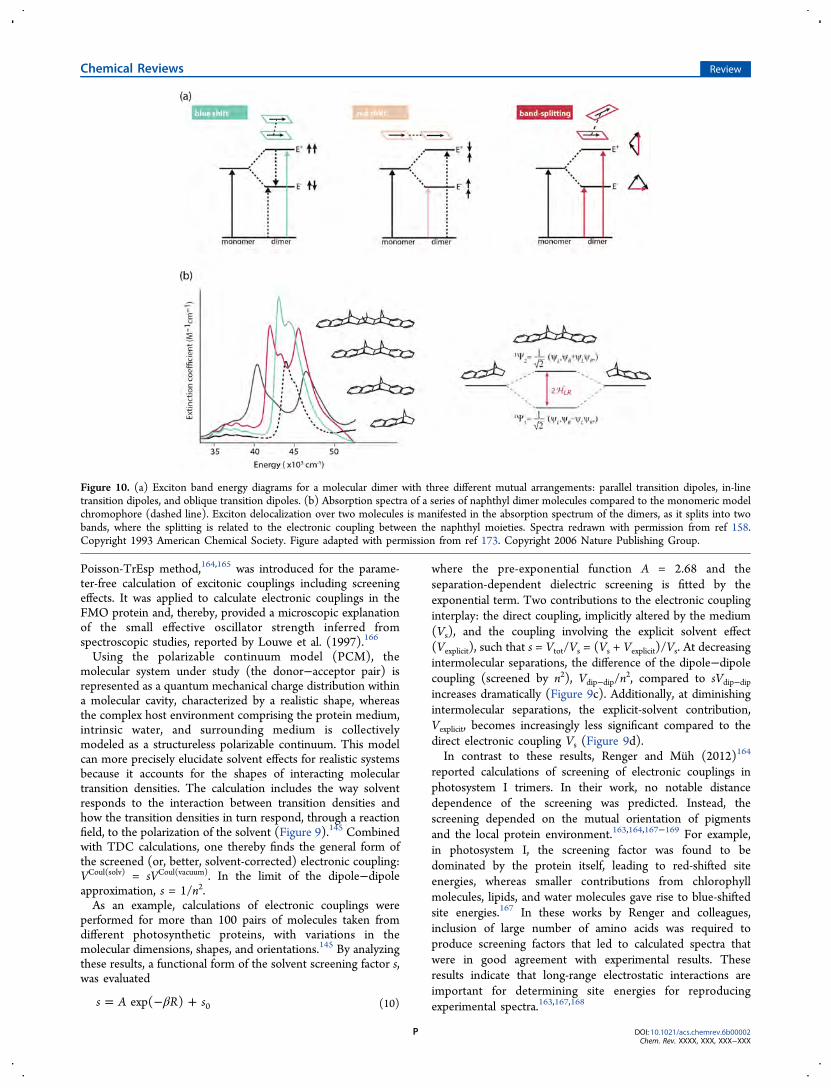
Figure 10. (a) Exciton band energy diagrams for a molecular dimer with three different mutual arrangements: parallel transition dipoles, in-linetransition dipoles, and oblique transition dipoles. (b) Absorption spectra of a series of naphthyl dimer molecules compared to the monomeric modelchromophore (dashed line). Exciton delocalization over two molecules is manifested in the absorption spectrum of the dimers, as it splits into twobands, where the splitting is related to the electronic coupling between the naphthyl moieties. Spectra redrawn with permission from ref 158.Copyright 1993 American Chemical Society. Figure adapted with permission from ref 173. Copyright 2006 Nature Publishing Group.
Chemical Reviews Review
DOI: 10.1021/acs.chemrev.6b00002Chem. Rev. XXXX, XXX, XXX−XXX
P
In summary, according to Forster theory, the major factorsaffecting energy transfer are the center-to-center separationbetween chromophores, the relative orientation of theirtransition dipoles, and the Forster spectral overlap integral(between the fluorescence spectrum of the donor and theabsorption spectrum of the acceptor). The distance betweeninteracting chromophores has a direct impact on the quantummechanical characteristics of the energy-transfer mechanism.Extensions of the original theory include the followingconsiderations: electronic coupling without invoking the dipoleapproximation, molecular exciton states, solvent screening ofthe electron coupling, and dynamic effects of coherence.Regarding the electronic coupling between donor and acceptorchromophores, the coupling term, Vtotal, is defined as the sumof long-range and short-range contributions, given by theelectrodynamic interaction (Ved) and the interchromophoreorbital overlap (Vioo), respectively.
7. MOLECULAR EXCITONSIn the strong coupling limit, in contrast to the weak electroniccoupling limit where excitation is transferred as localized states,electronic states are not localized on individual chromophores.Instead, closely spaced pigments coherently share electronicexcitation. This is described by the molecular excitonmodel.170−172 Studies of molecular excitons provide insightinto the collective absorption and redistribution of excitationenergy in nanoscale systems.173 Sometimes, the term “exciton”is used to mean electronic excitation of a molecule. We preferto reserve the term exciton for excitation that is coherentlyshared among two or more molecules.The Frenkel model, which is relevant for photosynthetic
antenna and can also describe molecular aggregates such as J-aggregates (Figure 10), is based on the principle that, if onecannot physically distinguish the cases of electronic excitationof, say, molecule A from excitation of molecule B, then thecorrect quantum states that absorb light are linear combinationsof the two possibilities. Thus, the strong electronic couplingbetween molecules comprising a molecular exciton results inthe electronic excited states being linear combinations ofexcitations of different molecules. The result for twoindistinguishable molecules is the symmetric and antisymmetriclinear combinations of excitation
Ψ = ′ + ′12
(A B AB )symm (11a)
Ψ = ′ − ′12
(A B AB )antisymm (11b)
where the prime indicates electronic excitation. These linearcombinations are reminiscent of how bonding and antibondingorbitals are constructed.The oscillator strengths of transitions from the ground state
to different excitonic energy levels are influenced by the relativearrangement of the monomers (Figure 10a). For example,when two molecules are arranged in a “sandwich” config-uration, their transition dipoles are parallel, and the electroniccoupling has a positive sign (dictated by the orientation factorκ; see eq 4). The linear combination where the transitiondipoles point in the same direction results in the higher-lyingexcitonic state being bright (allowed), whereas transitiondipoles oriented in an antiparallel fashion result in the lower-energy excitonic state being dark (forbidden).
In this sandwich geometry (called an H-aggregate), a blueshift in the absorption spectrum compared to that of themonomer is observed because the oscillator strength isexclusively carried by the upper exciton state. A simplecalculation shows why this is the case. Note that the oscillatorstrength is proportional to the magnitude squared of the dipoletransition moment, so one must calculate the dipole transitionmoment for each exciton state. The lowest-energy state is theantisymmetric linear combination, eq 11b, because of thepositively signed electronic coupling. The transition dipolemoment for this state is
μ μ
μ μ
μ μ
= ′ − ′ | |
= ⟨ ′| | ⟩⟨ | ⟩ − ⟨ | ⟩⟨ ′| | ⟩
= −
12
(A B AB ) AB
12
( A A B B A A B B )
12
( )
anti
A B (12)
which equals zero in the case of parallel transition momentvectors with the same magnitude, μA and μB. Note that theoverlap is ⟨A|A⟩ = ⟨B|B⟩ = 1, μ is the dipole operator, and |AB⟩is the ground-state wave function. The upper exciton state isthe symmetric linear combination, and it can be seen that thiswill have a dipole transition moment of μsymm = (μA + μB)/√2= √2μ, and therefore, its oscillator strength is proportional totwice that of one monomer.In J-aggregates (in-line or head-to-tail; Figure 10a), the
electronic coupling is negative, so the symmetric exciton statelies lowest in energy. Therefore, the lower-energy state ischaracterized by a larger transition dipole moment, resulting ina red shift of the absorption band. Intermediate, oblique,orientations are characterized by band splitting, which isdependent on both the separation between the dipoles and theangles with the vector connecting them, resulting in theappearance of two bands in the absorption spectrum.In the very weak coupling limit, differences between the
absorption spectra of the exciton system and isolated moleculesare minimal, whereas small spectral perturbations are observedin the weak coupling case. A clear example can be seen for aseries of naphthalene-bridge-naphthalene dyads (DN-2, DN-4,and DN-6) with two naphthalene chromophores held inposition by a rigid polynorbornyl-type bridge, where thedistance and orientations of the interacting chromophores isfixed and well-controlled (Figure 10b).158 As the electroniccoupling is reduced by increasing the separation between thechromophores, the exciton splitting diminishes. The strongcoupling limit is defined as the case when the exciton splitting islarger than the line broadening of the absorption bands.When the molecules are not identical or when there are more
than two interacting molecules, the exciton states are obtainedby solving a suitable secular determinant. For example, in thegeneral case of a dimer, the exciton transition energies are givenby
−
−=
H H
H H
E
E0
11 12
21 22 (13)
=+ ± − + | |
EH H H H H( ) ( ) 4
211 22 11 22
212
2
where H11 and H22 are the two diagonal Hamiltonian elements(e.g., the total energies of the charge-localized state), whereas
Chemical Reviews Review
DOI: 10.1021/acs.chemrev.6b00002Chem. Rev. XXXX, XXX, XXX−XXX
Q
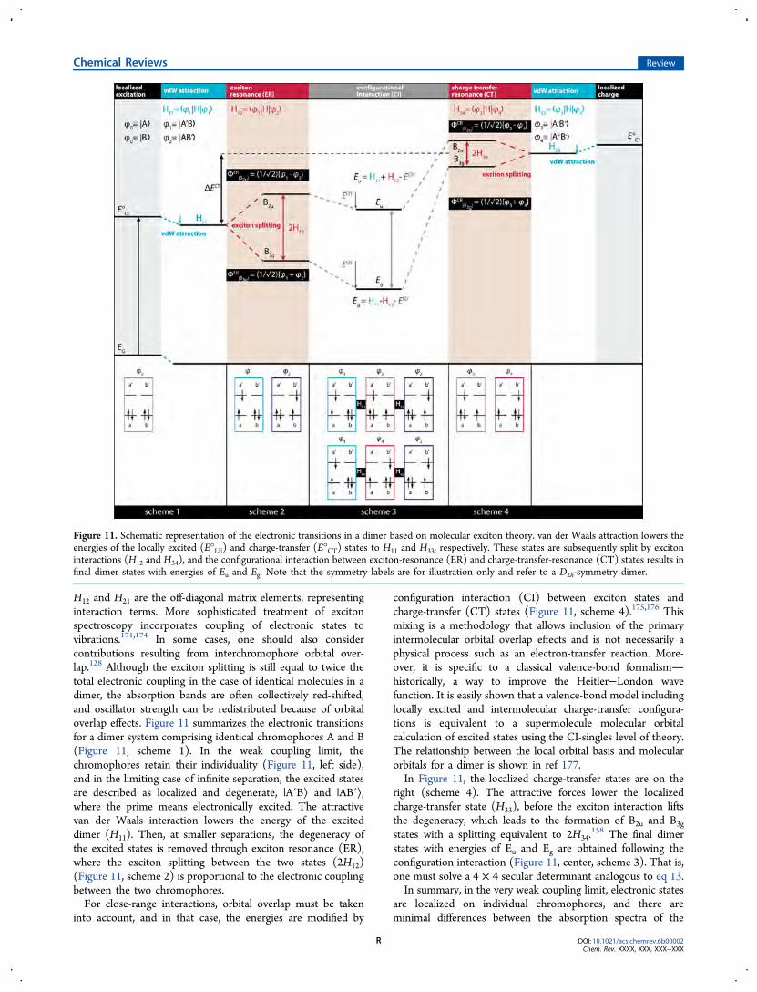
H12 and H21 are the off-diagonal matrix elements, representinginteraction terms. More sophisticated treatment of excitonspectroscopy incorporates coupling of electronic states tovibrations.171,174 In some cases, one should also considercontributions resulting from interchromophore orbital over-lap.128 Although the exciton splitting is still equal to twice thetotal electronic coupling in the case of identical molecules in adimer, the absorption bands are often collectively red-shifted,and oscillator strength can be redistributed because of orbitaloverlap effects. Figure 11 summarizes the electronic transitionsfor a dimer system comprising identical chromophores A and B(Figure 11, scheme 1). In the weak coupling limit, thechromophores retain their individuality (Figure 11, left side),and in the limiting case of infinite separation, the excited statesare described as localized and degenerate, |A′B⟩ and |AB′⟩,where the prime means electronically excited. The attractivevan der Waals interaction lowers the energy of the exciteddimer (H11). Then, at smaller separations, the degeneracy ofthe excited states is removed through exciton resonance (ER),where the exciton splitting between the two states (2H12)(Figure 11, scheme 2) is proportional to the electronic couplingbetween the two chromophores.For close-range interactions, orbital overlap must be taken
into account, and in that case, the energies are modified by
configuration interaction (CI) between exciton states andcharge-transfer (CT) states (Figure 11, scheme 4).175,176 Thismixing is a methodology that allows inclusion of the primaryintermolecular orbital overlap effects and is not necessarily aphysical process such as an electron-transfer reaction. More-over, it is specific to a classical valence-bond formalismhistorically, a way to improve the Heitler−London wavefunction. It is easily shown that a valence-bond model includinglocally excited and intermolecular charge-transfer configura-tions is equivalent to a supermolecule molecular orbitalcalculation of excited states using the CI-singles level of theory.The relationship between the local orbital basis and molecularorbitals for a dimer is shown in ref 177.In Figure 11, the localized charge-transfer states are on the
right (scheme 4). The attractive forces lower the localizedcharge-transfer state (H33), before the exciton interaction liftsthe degeneracy, which leads to the formation of B2u and B3g
states with a splitting equivalent to 2H34.158 The final dimer
states with energies of Eu and Eg are obtained following theconfiguration interaction (Figure 11, center, scheme 3). That is,one must solve a 4 × 4 secular determinant analogous to eq 13.In summary, in the very weak coupling limit, electronic states
are localized on individual chromophores, and there areminimal differences between the absorption spectra of the
Figure 11. Schematic representation of the electronic transitions in a dimer based on molecular exciton theory. van der Waals attraction lowers theenergies of the locally excited (E°LE) and charge-transfer (E°CT) states to H11 and H33, respectively. These states are subsequently split by excitoninteractions (H12 and H34), and the configurational interaction between exciton-resonance (ER) and charge-transfer-resonance (CT) states results infinal dimer states with energies of Eu and Eg. Note that the symmetry labels are for illustration only and refer to a D2h-symmetry dimer.
Chemical Reviews Review
DOI: 10.1021/acs.chemrev.6b00002Chem. Rev. XXXX, XXX, XXX−XXX
R
exciton system and the isolated molecules. In the weak couplinglimit, small spectral perturbations are observed. In the strongcoupling limit, closely spaced pigments coherently shareelectronic excitation, as described by the molecular excitonmodel. The oscillator strengths of excitonic energy levelsdepend on the geometry and mutual arrangement ofmonomers. Interchromophore orbital-overlap-dependent inter-actions become significant at small separations.
8. STRUCTURAL AND SPECTRAL CONSIDERATIONSWith variations in the interchromophoric interactions (asdiscussed above), it is important to discuss the principlesbehind the organizational structure in multichromophoricsystems. By modulating the structure and arrangement ofchromophores, organisms are able to maximize the collection oflight and the subsequent funneling of that energy to thereaction centers. The optimization of the spatial and energeticlandscapes of the antenna complex involves balancing theconcentrations of the constituent chromophores, their mutualarrangement, and the integration of pigments of variousabsorption energy gaps.The phenomenon of concentration quenching, originally
observed in solutions of organic dyes, describes the rapiddecrease in fluorescence quantum yield when the fluorophore ispresent in a high concentration. Fluorescence emission ofdissolved Chl a was found to be completely quenched at aconcentration of 0.3 M.178,179 Light-harvesting antennas havesucceeded in circumventing this phenomenon, and a very highpacking density of pigments can therefore be attained beforequenching is observed. For example, chlorophyll in LHCII hasan effective concentration of 0.25 M, yet its lifetime is hardlyquenched.180 One of the classic examples is the aggregatedbacteriochlorophylls (BChls) in chlorosomes, the light-harvest-ing organelles of green sulfur bacteria. BChl a protein in theFenna−Matthews−Olson (FMO) complex in green sulfurbacteria is found at a concentration of 0.1 M.181
These examples serve as evidence that, although high localconcentrations of chromophores in these supramolecularpigment structures are achieved, concentration quenching isgenerally avoided. This suggests a highly optimized arrange-ment of the constituent light-harvesting elements. It alsoemphasizes that the chromophores in light-harvesting com-plexes can be closely packed and, therefore, electronic couplingcan be strong in many instances. This has profoundimplications for energy-transfer mechanisms, as we discuss inthe following section. Note that there is quenching of excitationin chlorosomes by quinones, but this redox-activated quenchingis not concentration quenching.182
We argue that pigment separations have been optimized tominimize electron transfer, a process enhanced proportional tothe overlap of the molecular wave functions. A recent study183
of light harvesting by a purple bacterium, Rhodopseudomonaspalustris, mutant showed the quenching role of charge-transferstates. This mutant has a blue-shifted B850 Bchl band in theLH2 ring such that the LH2 absorption spectrum peaks solelyat about 800 nm (unlike the wild-type LH2, which shows thetypical B800−B850 absorption spectrum). Upon excitation ofLH2, a red-shifted charge-transfer (CT) state, hidden in thewild-type bacterium by spectral overlap, was observed to bepopulated in <100 fs, quenching the emissive excited state. Thisindicates that CT states are close in energy to the excitonicstates. The light-harvesting system “protects” itself by redshifting the excitonic manifold, which results in a uniform
mixing of the CT state with many of the exciton states. Thiswill still quench the states, but the effect is far less dramatic thanhaving a CT state below the exciton manifold. This mixing ofthe red states with CT states will, of course, strongly enhancecharge separation in RCs this is a major point of manyarticles on PSII charge separation.184−187 CT states areemployed by the RC-antenna supercomplex to (i) escapequenching by unavoidable CT states, (ii) red shift thespectrum, and (iii) provide a fast and effective pathway in theRC for charge separation.
9. EXCITONICS AND GENERALIZED FORSTER THEORY(GFT)
Strong electronic coupling produces new chromophoresbecause light is absorbed and emitted collectively (andnonadditively) by two or more coupled molecules. This hasimplications for light harvesting because now the donor and/oracceptor of excitation energy are/is not a single molecule, as inthe case treated by normal Forster theory, but instead theexciton states shared between strongly interacting chromo-phores. In other words, new effective chromophores can beformed based on pigments already present in the organism. Amodified version of Forster theory, called generalized Forstertheory (GFT), can be used to account for excitonic donors andacceptors.188
Klaus Schulten and co-workers modeled exciton energytransfer in LH2 between exciton states,189 which was aprecursor to the GFT formalism. Mukai et al. (1999),147
Sumi (1999),190 and Scholes and Fleming (2000)146
independently developed the GFT method for treating energytransfer in excitonic systems using a model with most of theForster theory attributes. The key insight was the wayelectronic coupling must be taken into account, which wedescribe below. Scholes and Fleming delayed publication oftheir work because they were working (unsuccessfully) on howto include dynamic localization in the donor state and theywere trying to formulate the theory in terms of a spectraloverlap analogous to that of Forster. This “coupling-weightedspectral overlap” turned out to be interesting because it explainshow averaging over the measured optical line shapes hidesinformation about how donor and acceptor excitons couple. Afew years later, Seogjoo Jang and co-workers (2004) reported atheory very similar to GFT that they called MC-FRET(multichromophoric Forster resonance energy transfer).148
There are three essential elements in the rate theory forexcitation energy transfer that need to be addressed.188 First,the electronic coupling is broken down into a linearcombination of molecule-to-molecule electronic couplings,where the coefficients in the linear combination are simplythose defining the exciton wave functions of donor andacceptor. Second, instead of one electronic state associated onlywith the donor and one associated only with the acceptor, thereare two or more excitonic states, and one must sum over allpermutations of donor and acceptor interactions. The key is toassociate with each pair of excitonic donor and acceptor statecouplings a corresponding spectral overlap. This leads to therate expression containing a sum over coupling-weightedspectral overlaps in place of the simple Forster spectraloverlap.146 Third, when there is significant inhomogeneousbroadening (disorder), one must perform the averaging“outside” this sum of coupling-weighted spectral overlapsbecause disorder makes each realization of exciton states (their
Chemical Reviews Review
DOI: 10.1021/acs.chemrev.6b00002Chem. Rev. XXXX, XXX, XXX−XXX
S
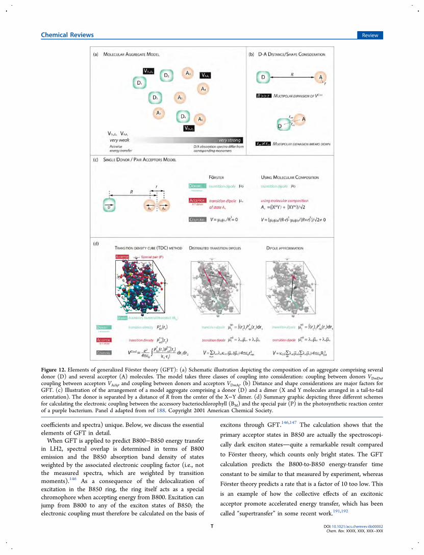
coefficients and spectra) unique. Below, we discuss the essentialelements of GFT in detail.When GFT is applied to predict B800−B850 energy transfer
in LH2, spectral overlap is determined in terms of B800emission and the B850 absorption band density of statesweighted by the associated electronic coupling factor (i.e., notthe measured spectra, which are weighted by transitionmoments).146 As a consequence of the delocalization ofexcitation in the B850 ring, the ring itself acts as a specialchromophore when accepting energy from B800. Excitation canjump from B800 to any of the exciton states of B850; theelectronic coupling must therefore be calculated on the basis of
excitons through GFT.146,147 The calculation shows that the
primary acceptor states in B850 are actually the spectroscopi-
cally dark exciton statesquite a remarkable result compared
to Forster theory, which counts only bright states. The GFT
calculation predicts the B800-to-B850 energy-transfer time
constant to be similar to that measured by experiment, whereas
Forster theory predicts a rate that is a factor of 10 too low. This
is an example of how the collective effects of an excitonic
acceptor promote accelerated energy transfer, which has been
called “supertransfer” in some recent work.191,192
Figure 12. Elements of generalized Forster theory (GFT): (a) Schematic illustration depicting the composition of an aggregate comprising severaldonor (D) and several acceptor (A) molecules. The model takes three classes of coupling into consideration: coupling between donors VDmDn,coupling between acceptors VAiAj, and coupling between donors and acceptors VDmAj. (b) Distance and shape considerations are major factors forGFT. (c) Illustration of the arrangement of a model aggregate comprising a donor (D) and a dimer (X and Y molecules arranged in a tail-to-tailorientation). The donor is separated by a distance of R from the center of the X−Y dimer. (d) Summary graphic depicting three different schemesfor calculating the electronic coupling between the accessory bacteriochlorophyll (BM) and the special pair (P) in the photosynthetic reaction centerof a purple bacterium. Panel d adapted from ref 188. Copyright 2001 American Chemical Society.
Chemical Reviews Review
DOI: 10.1021/acs.chemrev.6b00002Chem. Rev. XXXX, XXX, XXX−XXX
T
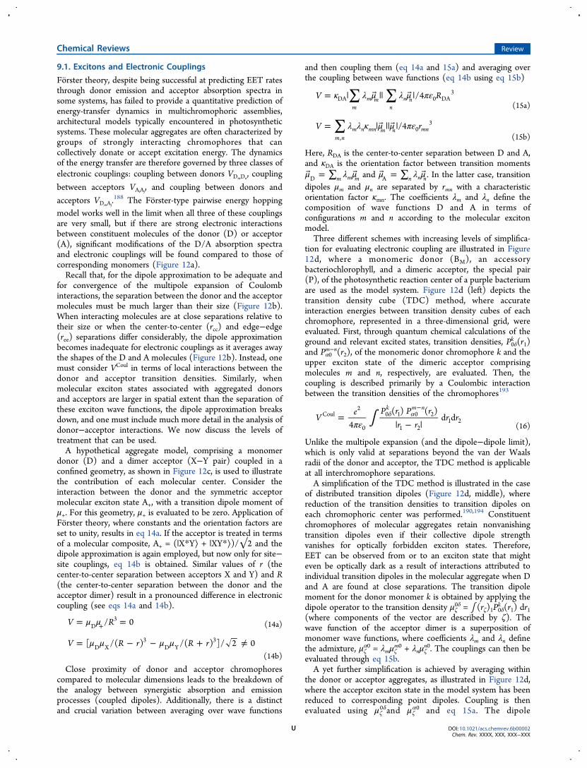
9.1. Excitons and Electronic Couplings
Forster theory, despite being successful at predicting EET ratesthrough donor emission and acceptor absorption spectra insome systems, has failed to provide a quantitative prediction ofenergy-transfer dynamics in multichromophoric assemblies,architectural models typically encountered in photosyntheticsystems. These molecular aggregates are often characterized bygroups of strongly interacting chromophores that cancollectively donate or accept excitation energy. The dynamicsof the energy transfer are therefore governed by three classes ofelectronic couplings: coupling between donors VDmDn
, coupling
between acceptors VAiAj, and coupling between donors and
acceptors VDmAj.188 The Forster-type pairwise energy hopping
model works well in the limit when all three of these couplingsare very small, but if there are strong electronic interactionsbetween constituent molecules of the donor (D) or acceptor(A), significant modifications of the D/A absorption spectraand electronic couplings will be found compared to those ofcorresponding monomers (Figure 12a).Recall that, for the dipole approximation to be adequate and
for convergence of the multipole expansion of Coulombinteractions, the separation between the donor and the acceptormolecules must be much larger than their size (Figure 12b).When interacting molecules are at close separations relative totheir size or when the center-to-center (rcc) and edge−edge(ree) separations differ considerably, the dipole approximationbecomes inadequate for electronic couplings as it averages awaythe shapes of the D and A molecules (Figure 12b). Instead, onemust consider VCoul in terms of local interactions between thedonor and acceptor transition densities. Similarly, whenmolecular exciton states associated with aggregated donorsand acceptors are larger in spatial extent than the separation ofthese exciton wave functions, the dipole approximation breaksdown, and one must include much more detail in the analysis ofdonor−acceptor interactions. We now discuss the levels oftreatment that can be used.A hypothetical aggregate model, comprising a monomer
donor (D) and a dimer acceptor (X−Y pair) coupled in aconfined geometry, as shown in Figure 12c, is used to illustratethe contribution of each molecular center. Consider theinteraction between the donor and the symmetric acceptormolecular exciton state A+, with a transition dipole moment ofμ+. For this geometry, μ+ is evaluated to be zero. Application ofForster theory, where constants and the orientation factors areset to unity, results in eq 14a. If the acceptor is treated in termsof a molecular composite, A+ = (|X*Y⟩ + |XY*⟩)/√2 and thedipole approximation is again employed, but now only for site−site couplings, eq 14b is obtained. Similar values of r (thecenter-to-center separation between acceptors X and Y) and R(the center-to-center separation between the donor and theacceptor dimer) result in a pronounced difference in electroniccoupling (see eqs 14a and 14b).
μ μ= =+V R/ 0D3
(14a)
μ μ μ μ= − − + ≠V R r R r[ /( ) /( ) ]/ 2 0D X3
D Y3
(14b)
Close proximity of donor and acceptor chromophorescompared to molecular dimensions leads to the breakdown ofthe analogy between synergistic absorption and emissionprocesses (coupled dipoles). Additionally, there is a distinctand crucial variation between averaging over wave functions
and then coupling them (eq 14a and 15a) and averaging overthe coupling between wave functions (eq 14b using eq 15b)
∑ ∑κ λ μ λ μ πε= | || |V R/4m
m mn
n nDA 0 DA3
(15a)
∑ λ λ κ μ μ πε= | || |V r/4m n
m n mn m n mn,
03
(15b)
Here, RDA is the center-to-center separation between D and A,and κDA is the orientation factor between transition momentsμ λ μ = ∑ m m mD and μ λ μ = ∑ n n nA . In the latter case, transitiondipoles μm and μn are separated by rmn with a characteristicorientation factor κmn. The coefficients λm and λn define thecomposition of wave functions D and A in terms ofconfigurations m and n according to the molecular excitonmodel.Three different schemes with increasing levels of simplifica-
tion for evaluating electronic coupling are illustrated in Figure12d, where a monomeric donor (BM), an accessorybacteriochlorophyll, and a dimeric acceptor, the special pair(P), of the photosynthetic reaction center of a purple bacteriumare used as the model system. Figure 12d (left) depicts thetransition density cube (TDC) method, where accurateinteraction energies between transition density cubes of eachchromophore, represented in a three-dimensional grid, wereevaluated. First, through quantum chemical calculations of theground and relevant excited states, transition densities, P0δ
k (r1)and Pα0
m−n(r2), of the monomeric donor chromophore k and theupper exciton state of the dimeric acceptor comprisingmolecules m and n, respectively, are evaluated. Then, thecoupling is described primarily by a Coulombic interactionbetween the transition densities of the chromophores193
∫πε=
| − |δ α
−V
e P r P rr r
r r4
( ) ( )d d
k m nCoul
2
0
0 1 0 2
1 21 2
(16)
Unlike the multipole expansion (and the dipole−dipole limit),which is only valid at separations beyond the van der Waalsradii of the donor and acceptor, the TDC method is applicableat all interchromophore separations.A simplification of the TDC method is illustrated in the case
of distributed transition dipoles (Figure 12d, middle), wherereduction of the transition densities to transition dipoles oneach chromophoric center was performed.190,194 Constituentchromophores of molecular aggregates retain nonvanishingtransition dipoles even if their collective dipole strengthvanishes for optically forbidden exciton states. Therefore,EET can be observed from or to an exciton state that mighteven be optically dark as a result of interactions attributed toindividual transition dipoles in the molecular aggregate when Dand A are found at close separations. The transition dipolemoment for the donor monomer k is obtained by applying thedipole operator to the transition density μζ
0δ = ∫ (rζ)1P0δk (r1) dr1(where components of the vector are described by ζ). Thewave function of the acceptor dimer is a superposition ofmonomer wave functions, where coefficients λm and λn definethe admixture, μζ
α0 = λmμζm0 + λnμζ
n0. The couplings can then beevaluated through eq 15b.A yet further simplification is achieved by averaging within
the donor or acceptor aggregates, as illustrated in Figure 12d,where the acceptor exciton state in the model system has beenreduced to corresponding point dipoles. Coupling is thenevaluated using μζ
0δand μζα0 and eq 15a. The dipole
Chemical Reviews Review
DOI: 10.1021/acs.chemrev.6b00002Chem. Rev. XXXX, XXX, XXX−XXX
U
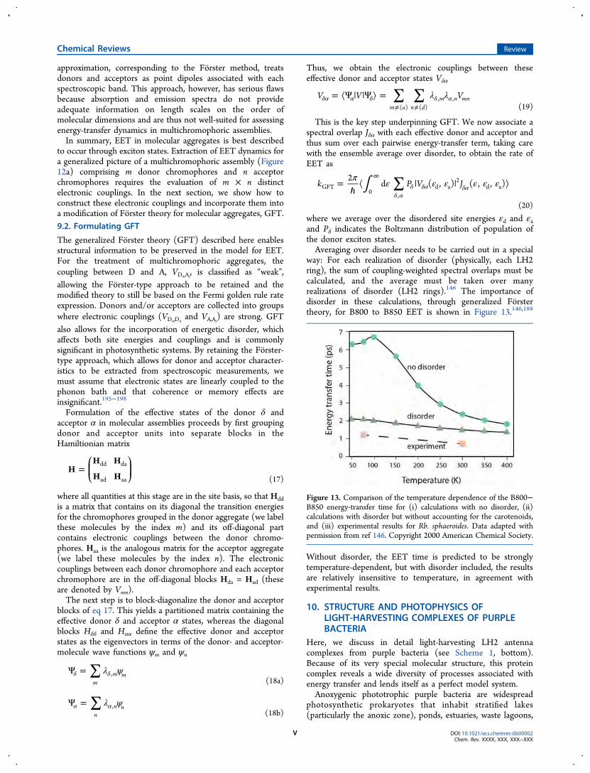
approximation, corresponding to the Forster method, treatsdonors and acceptors as point dipoles associated with eachspectroscopic band. This approach, however, has serious flawsbecause absorption and emission spectra do not provideadequate information on length scales on the order ofmolecular dimensions and are thus not well-suited for assessingenergy-transfer dynamics in multichromophoric assemblies.In summary, EET in molecular aggregates is best described
to occur through exciton states. Extraction of EET dynamics fora generalized picture of a multichromophoric assembly (Figure12a) comprising m donor chromophores and n acceptorchromophores requires the evaluation of m × n distinctelectronic couplings. In the next section, we show how toconstruct these electronic couplings and incorporate them intoa modification of Forster theory for molecular aggregates, GFT.9.2. Formulating GFT
The generalized Forster theory (GFT) described here enablesstructural information to be preserved in the model for EET.For the treatment of multichromophoric aggregates, thecoupling between D and A, VDmAj
, is classified as “weak”,allowing the Forster-type approach to be retained and themodified theory to still be based on the Fermi golden rule rateexpression. Donors and/or acceptors are collected into groupswhere electronic couplings (VDmDn
and VAiAj) are strong. GFT
also allows for the incorporation of energetic disorder, whichaffects both site energies and couplings and is commonlysignificant in photosynthetic systems. By retaining the Forster-type approach, which allows for donor and acceptor character-istics to be extracted from spectroscopic measurements, wemust assume that electronic states are linearly coupled to thephonon bath and that coherence or memory effects areinsignificant.195−198
Formulation of the effective states of the donor δ andacceptor α in molecular assemblies proceeds by first groupingdonor and acceptor units into separate blocks in theHamiltionian matrix
=⎛⎝⎜
⎞⎠⎟H
H H
H Hdd da
ad aa (17)
where all quantities at this stage are in the site basis, so that Hddis a matrix that contains on its diagonal the transition energiesfor the chromophores grouped in the donor aggregate (we labelthese molecules by the index m) and its off-diagonal partcontains electronic couplings between the donor chromo-phores. Haa is the analogous matrix for the acceptor aggregate(we label these molecules by the index n). The electroniccouplings between each donor chromophore and each acceptorchromophore are in the off-diagonal blocks Hda = Had (theseare denoted by Vmn).The next step is to block-diagonalize the donor and acceptor
blocks of eq 17. This yields a partitioned matrix containing theeffective donor δ and acceptor α states, whereas the diagonalblocks Hδδ and Hαα define the effective donor and acceptorstates as the eigenvectors in terms of the donor- and acceptor-molecule wave functions ψm and ψn
∑ λ ψΨ =δ δm
m m,(18a)
∑ λ ψΨ =α αn
n n,(18b)
Thus, we obtain the electronic couplings between theseeffective donor and acceptor states Vδα
∑ ∑ λ λ= ⟨Ψ | |Ψ ⟩ =δα α δ δ α≠ ≠
V V Vm a n d
m n mn( ) ( )
, ,(19)
This is the key step underpinning GFT. We now associate aspectral overlap Jδα with each effective donor and acceptor andthus sum over each pairwise energy-transfer term, taking carewith the ensemble average over disorder, to obtain the rate ofEET as
∫ ∑π ε ε ε ε ε ε=ℏ
⟨ | | ⟩δ α
δ δα δα
∞k P V J
2d ( , ) ( , , )GFT
0 ,d a
2d a
(20)
where we average over the disordered site energies εd and εaand Pδ indicates the Boltzmann distribution of population ofthe donor exciton states.Averaging over disorder needs to be carried out in a special
way: For each realization of disorder (physically, each LH2ring), the sum of coupling-weighted spectral overlaps must becalculated, and the average must be taken over manyrealizations of disorder (LH2 rings).146 The importance ofdisorder in these calculations, through generalized Forstertheory, for B800 to B850 EET is shown in Figure 13.146,188
Without disorder, the EET time is predicted to be stronglytemperature-dependent, but with disorder included, the resultsare relatively insensitive to temperature, in agreement withexperimental results.
10. STRUCTURE AND PHOTOPHYSICS OFLIGHT-HARVESTING COMPLEXES OF PURPLEBACTERIA
Here, we discuss in detail light-harvesting LH2 antennacomplexes from purple bacteria (see Scheme 1, bottom).Because of its very special molecular structure, this proteincomplex reveals a wide diversity of processes associated withenergy transfer and lends itself as a perfect model system.Anoxygenic phototrophic purple bacteria are widespread
photosynthetic prokaryotes that inhabit stratified lakes(particularly the anoxic zone), ponds, estuaries, waste lagoons,
Figure 13. Comparison of the temperature dependence of the B800−B850 energy-transfer time for (i) calculations with no disorder, (ii)calculations with disorder but without accounting for the carotenoids,and (iii) experimental results for Rb. sphaeroides. Data adapted withpermission from ref 146. Copyright 2000 American Chemical Society.
Chemical Reviews Review
DOI: 10.1021/acs.chemrev.6b00002Chem. Rev. XXXX, XXX, XXX−XXX
V
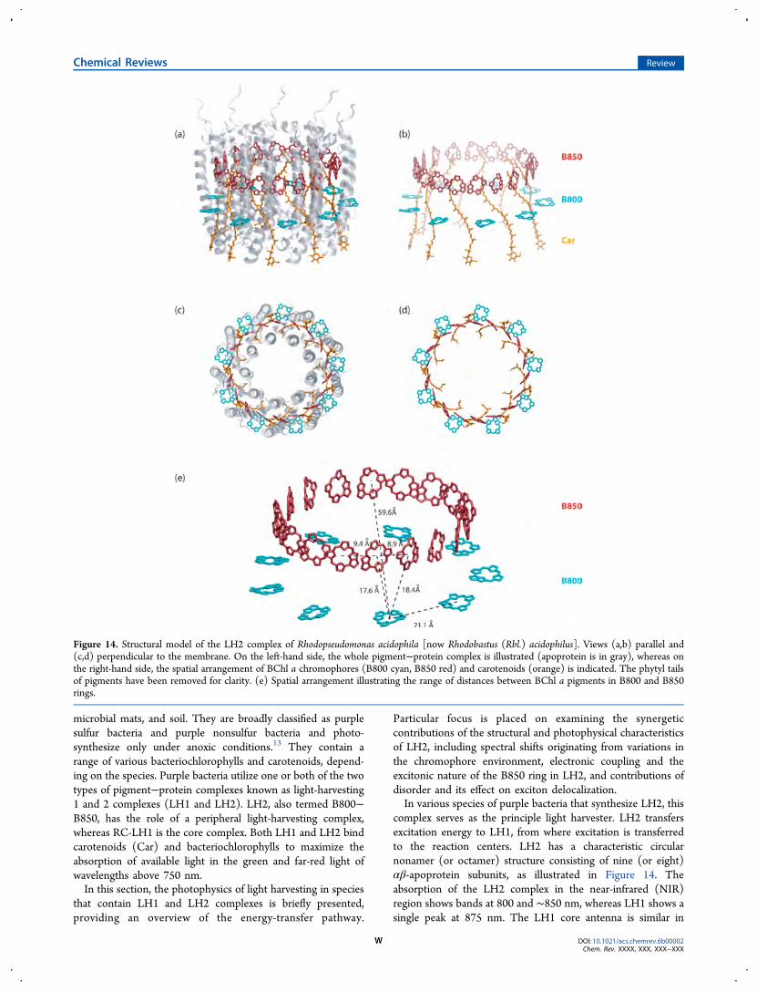
microbial mats, and soil. They are broadly classified as purplesulfur bacteria and purple nonsulfur bacteria and photo-synthesize only under anoxic conditions.13 They contain arange of various bacteriochlorophylls and carotenoids, depend-ing on the species. Purple bacteria utilize one or both of the twotypes of pigment−protein complexes known as light-harvesting1 and 2 complexes (LH1 and LH2). LH2, also termed B800−B850, has the role of a peripheral light-harvesting complex,whereas RC-LH1 is the core complex. Both LH1 and LH2 bindcarotenoids (Car) and bacteriochlorophylls to maximize theabsorption of available light in the green and far-red light ofwavelengths above 750 nm.In this section, the photophysics of light harvesting in species
that contain LH1 and LH2 complexes is briefly presented,providing an overview of the energy-transfer pathway.
Particular focus is placed on examining the synergeticcontributions of the structural and photophysical characteristicsof LH2, including spectral shifts originating from variations inthe chromophore environment, electronic coupling and theexcitonic nature of the B850 ring in LH2, and contributions ofdisorder and its effect on exciton delocalization.In various species of purple bacteria that synthesize LH2, this
complex serves as the principle light harvester. LH2 transfersexcitation energy to LH1, from where excitation is transferredto the reaction centers. LH2 has a characteristic circularnonamer (or octamer) structure consisting of nine (or eight)αβ-apoprotein subunits, as illustrated in Figure 14. Theabsorption of the LH2 complex in the near-infrared (NIR)region shows bands at 800 and ∼850 nm, whereas LH1 shows asingle peak at 875 nm. The LH1 core antenna is similar in
Figure 14. Structural model of the LH2 complex of Rhodopseudomonas acidophila [now Rhodobastus (Rbl.) acidophilus]. Views (a,b) parallel and(c,d) perpendicular to the membrane. On the left-hand side, the whole pigment−protein complex is illustrated (apoprotein is in gray), whereas onthe right-hand side, the spatial arrangement of BChl a chromophores (B800 cyan, B850 red) and carotenoids (orange) is indicated. The phytyl tailsof pigments have been removed for clarity. (e) Spatial arrangement illustrating the range of distances between BChl a pigments in B800 and B850rings.
Chemical Reviews Review
DOI: 10.1021/acs.chemrev.6b00002Chem. Rev. XXXX, XXX, XXX−XXX
W
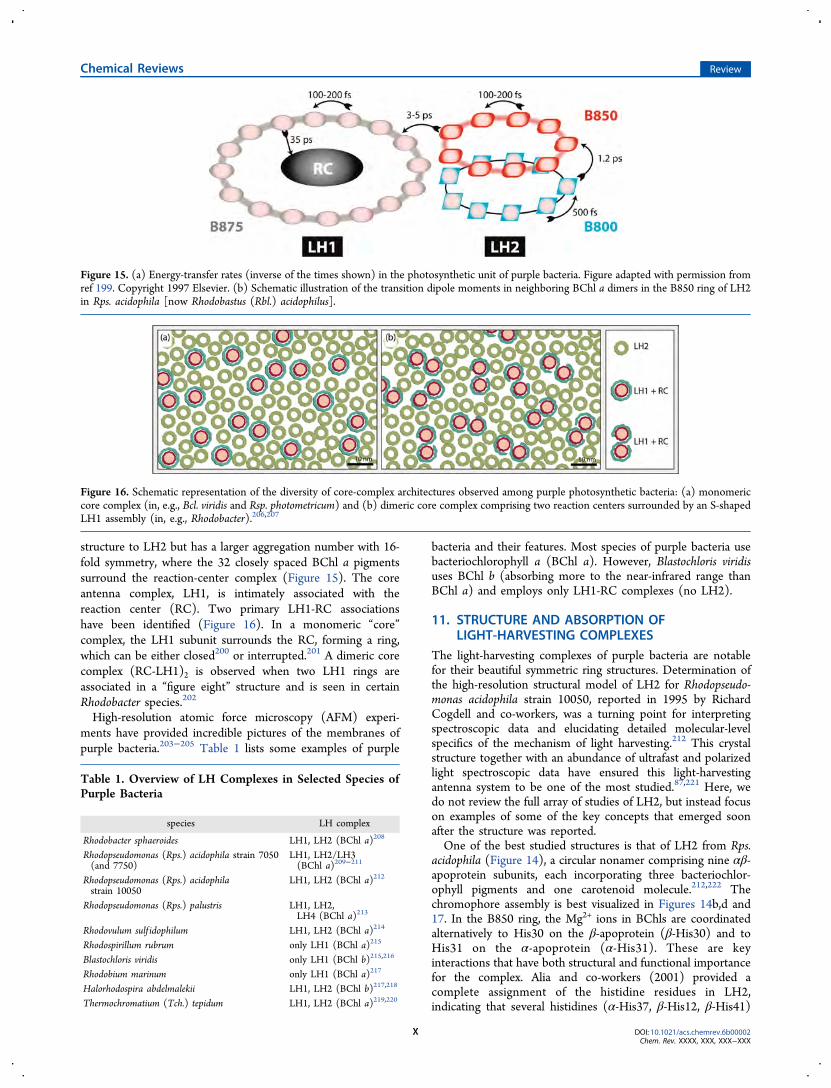
structure to LH2 but has a larger aggregation number with 16-fold symmetry, where the 32 closely spaced BChl a pigmentssurround the reaction-center complex (Figure 15). The coreantenna complex, LH1, is intimately associated with thereaction center (RC). Two primary LH1-RC associationshave been identified (Figure 16). In a monomeric “core”complex, the LH1 subunit surrounds the RC, forming a ring,which can be either closed200 or interrupted.201 A dimeric corecomplex (RC-LH1)2 is observed when two LH1 rings areassociated in a “figure eight” structure and is seen in certainRhodobacter species.202
High-resolution atomic force microscopy (AFM) experi-ments have provided incredible pictures of the membranes ofpurple bacteria.203−205 Table 1 lists some examples of purple
bacteria and their features. Most species of purple bacteria usebacteriochlorophyll a (BChl a). However, Blastochloris viridisuses BChl b (absorbing more to the near-infrared range thanBChl a) and employs only LH1-RC complexes (no LH2).
11. STRUCTURE AND ABSORPTION OFLIGHT-HARVESTING COMPLEXES
The light-harvesting complexes of purple bacteria are notablefor their beautiful symmetric ring structures. Determination ofthe high-resolution structural model of LH2 for Rhodopseudo-monas acidophila strain 10050, reported in 1995 by RichardCogdell and co-workers, was a turning point for interpretingspectroscopic data and elucidating detailed molecular-levelspecifics of the mechanism of light harvesting.212 This crystalstructure together with an abundance of ultrafast and polarizedlight spectroscopic data have ensured this light-harvestingantenna system to be one of the most studied.87,221 Here, wedo not review the full array of studies of LH2, but instead focuson examples of some of the key concepts that emerged soonafter the structure was reported.One of the best studied structures is that of LH2 from Rps.
acidophila (Figure 14), a circular nonamer comprising nine αβ-apoprotein subunits, each incorporating three bacteriochlor-ophyll pigments and one carotenoid molecule.212,222 Thechromophore assembly is best visualized in Figures 14b,d and17. In the B850 ring, the Mg2+ ions in BChls are coordinatedalternatively to His30 on the β-apoprotein (β-His30) and toHis31 on the α-apoprotein (α-His31). These are keyinteractions that have both structural and functional importancefor the complex. Alia and co-workers (2001) provided acomplete assignment of the histidine residues in LH2,indicating that several histidines (α-His37, β-His12, β-His41)
Figure 15. (a) Energy-transfer rates (inverse of the times shown) in the photosynthetic unit of purple bacteria. Figure adapted with permission fromref 199. Copyright 1997 Elsevier. (b) Schematic illustration of the transition dipole moments in neighboring BChl a dimers in the B850 ring of LH2in Rps. acidophila [now Rhodobastus (Rbl.) acidophilus].
Figure 16. Schematic representation of the diversity of core-complex architectures observed among purple photosynthetic bacteria: (a) monomericcore complex (in, e.g., Bcl. viridis and Rsp. photometricum) and (b) dimeric core complex comprising two reaction centers surrounded by an S-shapedLH1 assembly (in, e.g., Rhodobacter).206,207
Table 1. Overview of LH Complexes in Selected Species ofPurple Bacteria
species LH complex
Rhodobacter sphaeroides LH1, LH2 (BChl a)208
Rhodopseudomonas (Rps.) acidophila strain 7050(and 7750)
LH1, LH2/LH3(BChl a)209−211
Rhodopseudomonas (Rps.) acidophilastrain 10050
LH1, LH2 (BChl a)212
Rhodopseudomonas (Rps.) palustris LH1, LH2,LH4 (BChl a)213
Rhodovulum sulf idophilum LH1, LH2 (BChl a)214
Rhodospirillum rubrum only LH1 (BChl a)215
Blastochloris viridis only LH1 (BChl b)215,216
Rhodobium marinum only LH1 (BChl a)217
Halorhodospira abdelmalekii LH1, LH2 (BChl b)217,218
Thermochromatium (Tch.) tepidum LH1, LH2 (BChl a)219,220
Chemical Reviews Review
DOI: 10.1021/acs.chemrev.6b00002Chem. Rev. XXXX, XXX, XXX−XXX
X
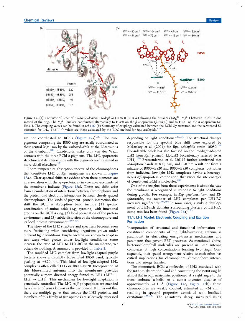
are not coordinated to BChls (Figure 17a).223 The ninepigments comprising the B800 ring are axially coordinated attheir central Mg2+ ion by the carboxyl-αM1 at the N-terminusof the α-subunit.224 Carotenoids make only van der Waalscontacts with the three BChl a pigments. The LH2 apoproteinstructure and its interactions with the pigments are presented inmore detail elsewhere.225
Room-temperature absorption spectra of the chromophoresthat constitute LH2 of Rps. acidophila are shown in Figure18a,b. Clear spectral shifts are evident when these pigments arein association with the apoprotein, as in vivo measurements ofthe membrane indicate (Figure 18c). These red shifts arisefrom a combination of interactions between chromophores andthe protein and electronic interactions between closely spacedchromophores. The kinds of pigment−protein interaction thatshift the BChl a absorption band include (1) specificcoordination of amino acids (e.g., tyrosine) with functionalgroups on the BChl a ring, (2) local polarization of the proteinenvironment, and (3) subtle distortion of the chromophore andits local protein environment.226−228
The story of the LH2 structure and spectrum becomes evenmore fascinating when considering organisms grown underdifferent light conditions. Purple bacteria are known to adapt intwo ways when grown under low-light conditions: Someincrease the ratio of LH2 to LH1-RC in the membrane, yetothers do nothing. A summary is provided in Table 2.The modified LH2 complex from low-light-adapted purple
bacteria shows a distinctly blue-shifted B850 band, typicallypeaking at ∼820 nm. This kind of low-light-adapted LH2complex is often called LH3 or B800−B820. Incorporation ofthis blue-shifted antenna into the membrane providespotentially a more directed energy funnel to LH1 (LH3 →LH2 → LH1). This mechanism for low-light adaptation isgenetically controlled. The LH2 α/β polypeptides are encodedby a cluster of genes known as the puc operon. It turns out thatthere are multiple genes that encode the LH2 proteins, andmembers of this family of puc operons are selectively expressed
depending on light conditions.229,230 The structural changesresponsible for the spectral blue shift were explored byMcLuskey et al. (2001) for Rps. acidophila strain 10050.211
Considerable work has also focused on the low-light-adaptedLH2 from Rps. palustris, LL-LH2 (occasionally referred to asLH4).230 Brotosudarmo et al. (2011) further confirmed thatabsorption bands at 800, 820, and 850 nm result not from amixture of B800−B820 and B800−B850 complexes, but ratherfrom individual low-light LH2 complexes having a heteroge-neous αβ-apoprotein composition that varies the site energiesof constituent BChl a molecules.230
One of the insights from these experiments is about the waythe membrane is reorganized in response to light conditionsduring growth. For example, in Rsp. photometricum and Rb.sphaeroides, the number of LH2 complexes per LH1-RCincreases significantly.204,205 In some cases, a striking develop-ment of LH2-rich domains surrounding clusters of LH1-RCcomplexes has been found (Figure 16a).235
11.1. LH2 Model: Electronic Coupling and ExcitionFormation
Incorporation of structural and functional information onconstituent components of the light-harvesting antenna isparamount in elucidating energy-transfer mechanisms andparameters that govern EET processes. As mentioned above,bacteriochlorophyll molecules are present in LH2 antennacomplexes at high concentrations forming two rings. Con-sequently, their spatial arrangement relative to each other hascritical implications for chromophore−chromophore interac-tions and energy transfer.The monomeric BChl a molecules of LH2 associated with
the 800-nm absorption band and constituting the B800 ring liealmost flat in Rsp. acidophila, positioned at a right angle to thetransmembrane α-helix. At a center-to-center distance ofapproximately 21.1 Å (Figure 14e, Figure 17b), thesechromophores are weakly coupled, estimated at ∼24 cm−1,resulting in spectral properties associated with localizedexcitations.236,237 The anisotropy decay, measured using
Figure 17. (a) Top view of B850 of Rhodopseudomonas acidophila (PDB ID 2FKW) showing the distances (Mg2+−Mg2+) between BChls in onesection of the ring. The Mg2+ ions are coordinated alternatively to His30 on the β apoprotein (β-His30) and to His31 on the α apoprotein (α-His31). The coupling values can be found in ref 116. (b) Summary of couplings calculated between the BChl Qy transition and the carotenoid S2transition for LH2. The VTDC values are those calculated by the TDC method for Rps. acidophila.129
Chemical Reviews Review
DOI: 10.1021/acs.chemrev.6b00002Chem. Rev. XXXX, XXX, XXX−XXX
Y
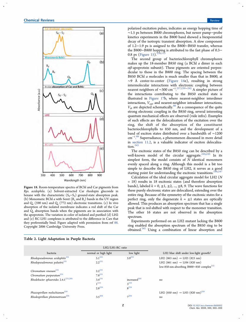
polarized excitation pulses, indicates an energy hopping time of∼1.5 ps between B800 chromophores, but newer pump−probekinetics experiments in the B800 band showed a biexponentialdecay of the isotropic transient absorption. A slow componentof 1.2−1.9 ps is assigned to the B800−B850 transfer, whereasthe B800−B800 hopping is attributed to the fast phase of 0.3−0.8 ps (Figure 15).238,115
The second group of bacteriochlorophyll chromophoresmakes up the 18-member B850 ring (a BChl a dimer in eachαβ-apoprotein subunit). These pigments are oriented perpen-dicular to those in the B800 ring. The spacing between theB850 BChl a molecules is much smaller than that in B800, at∼9 Å center-to-center (Figure 14e), resulting in strongintermolecular interactions with electronic coupling betweennearest neighbors of ∼300 cm−1.237,239−242 A simpler picture ofthe interactions contributing to the B850 excited state isillustrated in Figure 17b, where nearest-neighbor interdimerinteractions, Vext, and nearest-neighbor intradimer interactions,Vd, are depicted schematically.88 As a consequence of the quitestrong electronic coupling in the B850 ring, several interestingquantum mechanical effects are observed (vide infra). Examplesof such effects are the delocalization of the excitation over thering, the shift of the absorption of the constituentbacteriochlorophylls to 850 nm, and the development of aband of exciton states distributed over a bandwidth of ∼1200cm−1.88 Superradiance, a phenomenon discussed in more detailin section 11.2, is a valuable indicator of exciton delocaliza-tion.243
The excitonic states of the B850 ring can be described by awell-known model of the circular aggregate.244,245 In itssimplest form, the model consists of N identical monomersevenly spaced along a ring. Although this model is a bit toosimple to describe the B850 ring of LH2, it serves as a goodstarting point for understanding the excitonic transitions.246,247
Calculation of the ideal circular aggregate model for LH2 (N= 18) results in 18 excitonic states (and therefore absorptionbands), labeled k = 0, ±1, ±2, ..., ±8, 9. The wave functions forthese purely electronic states are delocalized, extending over theentire ring. Because of the symmetry of the excitonic states for aperfect ring, only the degenerate k = ±1 states are opticallyallowed. This produces an absorption spectrum that has a singlepeak that is red-shifted with respect to the monomer transition.The other 16 states are not observed in the absorptionspectrum.Experiments performed on an LH2 mutant lacking the B800
ring enabled the absorption spectrum of the B850 ring to beobtained.242 Using a combination of linear absorption and
Figure 18. Room-temperature spectra of BChl and Car pigments fromRps. acidophila. (a) Solvent-extracted Car rhodopin glucoside inhexane with the characteristic (S0−S2) ground-state absorption peak.(b) Monomeric BChl a with Soret (Bx and By) bands in the UV regionand Qx (590 nm) and Qy (772 nm) electronic transitions. (c) In vivoabsorption of the isolated membrane indicates a red shift of the Carand Qy absorption bands when the pigments are in association withthe apoprotein. The variation in color of isolated and purified (d) LH2and (e) RC-LH1 complexes is attributed to the difference in Cars thatthey preferentially bind. Figure adapted with permission from ref 88.Copyright 2006 Cambridge University Press.
Table 2. Light Adaptation in Purple Bacteria
LH2/LH1-RC ratio
bacteria normal or high light low light LH2 blue shift under low-light growth?
Rhodopseudomonas acidophila231 5.1231 5.9231 LH2 (863 nm) → LH3 (823 nm)Rhodopseudomonas palustris231 2.2231 LH2 (861 nm) → LH4 (820 nm)
low-850-nm-absorbing B800−850 complex232
Chromatium vinosum231 2.2231
Chromatium purpuratum231 7.8231
Rhodobacter sphaeroides 2.4.1231,205 3.4231 increased205 no1233 3233
2.8234 8234
Phaeospirillum molischianum229 LH2 (850 nm) → LH3 (820 nm)229
Rhodospirillum photometricum204 3.5204 7204 no
Chemical Reviews Review
DOI: 10.1021/acs.chemrev.6b00002Chem. Rev. XXXX, XXX, XXX−XXX
Z
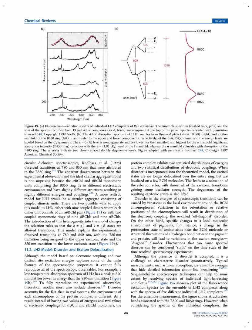
circular dichroism spectroscopies, Koolhaas et al. (1998)observed transitions at 780 and 850 nm that were attributedto the B850 ring.242 The apparent disagreement between thisexperimental observation and the ideal circular aggregate modelis not surprising because the αBChl and βBChl monomericunits comprising the B850 ring lie in different electrostaticenvironments and have slightly different structures resulting inslightly different energies and couplings.226 A more realisticmodel for LH2 would be a circular aggregate consisting ofcoupled dimeric units. There are two possible ways to applythis model to LH2, either with nine coupled dimers where eachdimer unit consists of an αβBChl pair (Figure 17) or with twocoupled monomeric rings of nine βBChls and nine αBChls.The introduction of this heterogeneity into the model changesthe selection rules so that the k = ±1 and k = ±8 states areallowed transitions. This model explains the experimentallyobserved transitions at 780 and 850 nm, with the 780-nmtransition being assigned to the upper excitonic state and the850-nm transition to the lower excitonic state (Figure 19b).
11.2. LH2 Model: Disorder and Exciton Delocalization
Although the model based on electronic coupling and twodistinct site excitation energies captures some of the mainfeatures of the spectroscopic properties, it cannot fullyreproduce all of the spectroscopic observables. For example, alow-temperature absorption spectrum of LH2 has a peak at 870nm that lies lower in energy than the 850-nm transition (Figure19b).249 To fully reproduce the experimental observables,theoretical models must also include disorder.115 Disorderaccounts for the fact that, in real systems, the environment ofeach chromophore of the protein complex is different. As aresult, instead of having two values of energies and two valuesof electronic couplings for αBChl and βBChl monomers, the
protein complex exhibits two statistical distributions of energiesand two statistical distributions of electronic couplings. Whendisorder is incorporated into the theoretical model, the excitedstates are no longer delocalized over the entire ring, but arelocalized on a few BChl molecules. This leads to a relaxation ofthe selection rules, with almost all of the excitonic transitionsgaining some oscillator strength. The degeneracy of theresulting excitonic states is also lifted.Disorder in the energies of spectroscopic transitions can be
caused by variations in the local environment around the BChlchromophores. Variations in the orientation or relativepositions of the chromophores will result in distribution ofthe electronic coupling, the so-called “off-diagonal” disorder.On the other hand, specific changes in a local molecularenvironment of pigments, for instance, a change in theprotonation state of amino acids near the BChl molecule orstructural fluctuations of a hydrogen bond between the pigmentand protein, will lead to variations in the exciton energies“diagonal” disorder. Fluctuations that can cause spectraldisorder can be considered “static” on the time scale of thetime-resolved spectroscopy experiments.Although the presence of disorder is accepted, it is a
challenge to characterize disorder quantitatively. Typicalmeasurements, such as linear absorption, are ensemble averagesthat hide detailed information about line broadening.250,251
Single-molecule spectroscopic techniques can help to someextent by resolving spectra of individual light-harvestingcomplexes.248,252 Figure 19a shows a plot of the fluorescenceexcitation spectra for the ensemble of LH2 complexes alongwith the spectra of five different individual LH2 complexes.248
For the ensemble measurement, the figure shows structurelessbands associated with the B800 and B850 rings. However, whenconsidering the spectra of the individual complexes, the
Figure 19. (a) Fluorescence−excitation spectra of individual LH2 complexes of Rps. acidophila. The ensemble spectrum (dashed trace, pink) and thesum of the spectra recorded from 19 individual complexes (solid, black) are compared at the top of the panel. Spectra reprinted with permissionfrom ref 248. Copyright 1999 AAAS. (b) The 4.2 K absorption spectrum of LH2 complex from Rps. acidophila (strain 10050) (right) and excitonmanifold of the B850 ring (left). u and l refer to the upper and lower components, respectively, of the basic B850 dimer, and the energy levels arelabeled based on the C9 symmetry. The k = 0 (A) level is nondegenerate and lies lowest for the l manifold and highest for the u manifold. Significantabsorption intensity (B850 ring) coincides with the k = {1,8} (E1) level of the l manifold, whereas the u manifold coincides with absorption of theB800 ring. The asterisks indicate two closely spaced doubly degenerate levels. Figure adapted with permission from ref 249. Copyright 1997American Chemical Society.
Chemical Reviews Review
DOI: 10.1021/acs.chemrev.6b00002Chem. Rev. XXXX, XXX, XXX−XXX
AA
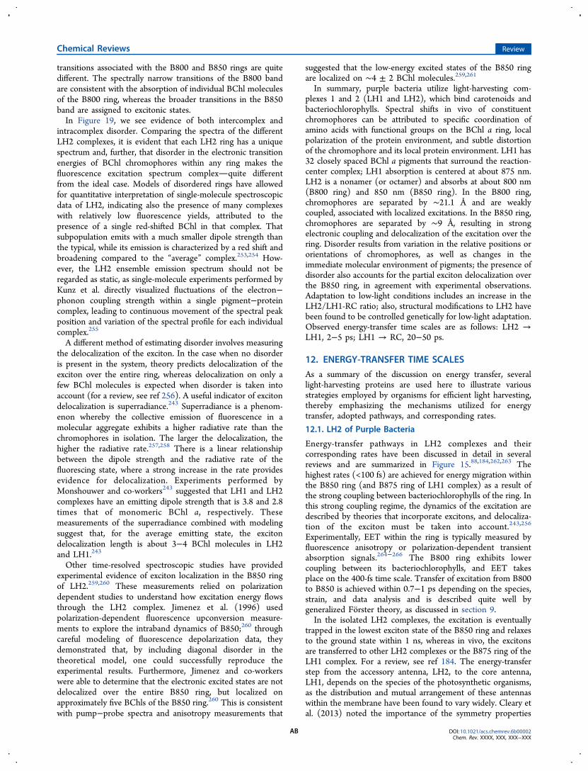
transitions associated with the B800 and B850 rings are quitedifferent. The spectrally narrow transitions of the B800 bandare consistent with the absorption of individual BChl moleculesof the B800 ring, whereas the broader transitions in the B850band are assigned to excitonic states.In Figure 19, we see evidence of both intercomplex and
intracomplex disorder. Comparing the spectra of the differentLH2 complexes, it is evident that each LH2 ring has a uniquespectrum and, further, that disorder in the electronic transitionenergies of BChl chromophores within any ring makes thefluorescence excitation spectrum complexquite differentfrom the ideal case. Models of disordered rings have allowedfor quantitative interpretation of single-molecule spectroscopicdata of LH2, indicating also the presence of many complexeswith relatively low fluorescence yields, attributed to thepresence of a single red-shifted BChl in that complex. Thatsubpopulation emits with a much smaller dipole strength thanthe typical, while its emission is characterized by a red shift andbroadening compared to the “average” complex.253,254 How-ever, the LH2 ensemble emission spectrum should not beregarded as static, as single-molecule experiments performed byKunz et al. directly visualized fluctuations of the electron−phonon coupling strength within a single pigment−proteincomplex, leading to continuous movement of the spectral peakposition and variation of the spectral profile for each individualcomplex.255
A different method of estimating disorder involves measuringthe delocalization of the exciton. In the case when no disorderis present in the system, theory predicts delocalization of theexciton over the entire ring, whereas delocalization on only afew BChl molecules is expected when disorder is taken intoaccount (for a review, see ref 256). A useful indicator of excitondelocalization is superradiance.243 Superradiance is a phenom-enon whereby the collective emission of fluorescence in amolecular aggregate exhibits a higher radiative rate than thechromophores in isolation. The larger the delocalization, thehigher the radiative rate.257,258 There is a linear relationshipbetween the dipole strength and the radiative rate of thefluorescing state, where a strong increase in the rate providesevidence for delocalization. Experiments performed byMonshouwer and co-workers243 suggested that LH1 and LH2complexes have an emitting dipole strength that is 3.8 and 2.8times that of monomeric BChl a, respectively. Thesemeasurements of the superradiance combined with modelingsuggest that, for the average emitting state, the excitondelocalization length is about 3−4 BChl molecules in LH2and LH1.243
Other time-resolved spectroscopic studies have providedexperimental evidence of exciton localization in the B850 ringof LH2.259,260 These measurements relied on polarizationdependent studies to understand how excitation energy flowsthrough the LH2 complex. Jimenez et al. (1996) usedpolarization-dependent fluorescence upconversion measure-ments to explore the intraband dynamics of B850;260 throughcareful modeling of fluorescence depolarization data, theydemonstrated that, by including diagonal disorder in thetheoretical model, one could successfully reproduce theexperimental results. Furthermore, Jimenez and co-workerswere able to determine that the electronic excited states are notdelocalized over the entire B850 ring, but localized onapproximately five BChls of the B850 ring.260 This is consistentwith pump−probe spectra and anisotropy measurements that
suggested that the low-energy excited states of the B850 ringare localized on ∼4 ± 2 BChl molecules.259,261
In summary, purple bacteria utilize light-harvesting com-plexes 1 and 2 (LH1 and LH2), which bind carotenoids andbacteriochlorophylls. Spectral shifts in vivo of constituentchromophores can be attributed to specific coordination ofamino acids with functional groups on the BChl a ring, localpolarization of the protein environment, and subtle distortionof the chromophore and its local protein environment. LH1 has32 closely spaced BChl a pigments that surround the reaction-center complex; LH1 absorption is centered at about 875 nm.LH2 is a nonamer (or octamer) and absorbs at about 800 nm(B800 ring) and 850 nm (B850 ring). In the B800 ring,chromophores are separated by ∼21.1 Å and are weaklycoupled, associated with localized excitations. In the B850 ring,chromophores are separated by ∼9 Å, resulting in strongelectronic coupling and delocalization of the excitation over thering. Disorder results from variation in the relative positions ororientations of chromophores, as well as changes in theimmediate molecular environment of pigments; the presence ofdisorder also accounts for the partial exciton delocalization overthe B850 ring, in agreement with experimental observations.Adaptation to low-light conditions includes an increase in theLH2/LH1-RC ratio; also, structural modifications to LH2 havebeen found to be controlled genetically for low-light adaptation.Observed energy-transfer time scales are as follows: LH2 →LH1, 2−5 ps; LH1 → RC, 20−50 ps.
12. ENERGY-TRANSFER TIME SCALES
As a summary of the discussion on energy transfer, severallight-harvesting proteins are used here to illustrate variousstrategies employed by organisms for efficient light harvesting,thereby emphasizing the mechanisms utilized for energytransfer, adopted pathways, and corresponding rates.
12.1. LH2 of Purple Bacteria
Energy-transfer pathways in LH2 complexes and theircorresponding rates have been discussed in detail in severalreviews and are summarized in Figure 15.88,184,262,263 Thehighest rates (<100 fs) are achieved for energy migration withinthe B850 ring (and B875 ring of LH1 complex) as a result ofthe strong coupling between bacteriochlorophylls of the ring. Inthis strong coupling regime, the dynamics of the excitation aredescribed by theories that incorporate excitons, and delocaliza-tion of the exciton must be taken into account.243,256
Experimentally, EET within the ring is typically measured byfluorescence anisotropy or polarization-dependent transientabsorption signals.264−266 The B800 ring exhibits lowercoupling between its bacteriochlorophylls, and EET takesplace on the 400-fs time scale. Transfer of excitation from B800to B850 is achieved within 0.7−1 ps depending on the species,strain, and data analysis and is described quite well bygeneralized Forster theory, as discussed in section 9.In the isolated LH2 complexes, the excitation is eventually
trapped in the lowest exciton state of the B850 ring and relaxesto the ground state within 1 ns, whereas in vivo, the excitonsare transferred to other LH2 complexes or the B875 ring of theLH1 complex. For a review, see ref 184. The energy-transferstep from the accessory antenna, LH2, to the core antenna,LH1, depends on the species of the photosynthetic organisms,as the distribution and mutual arrangement of these antennaswithin the membrane have been found to vary widely. Cleary etal. (2013) noted the importance of the symmetry properties
Chemical Reviews Review
DOI: 10.1021/acs.chemrev.6b00002Chem. Rev. XXXX, XXX, XXX−XXX
AB
both of LH2 rings on the individual complex level, and of theirorganization on a supramolecular scale, as well as their impacton the efficiency of excitation energy transfer.267
An average time scale for energy transfer in LH2 is ∼2−5 ps(Figure 15).221 A consequence of having circular light-harvesting complexes is that excitation energy migration forLH2 → LH2 or LH2 → LH1 is independent of the relativeorganization of the rings. In the LH1 complex, the finaltrapping of the harvested energy by the reaction-center specialpair occurs as the energy is transferred from the B875 ring tothe RC on a time scale of 20−50 ps.268−270 This energy-migration step into the RC can be an uphill transfer, as theexcited state of the LH1 antenna is lower in energy than thespecial BChl a pair in the reaction center. The slowest EET stepoccurs over a relatively larger distance because of the separationof B875 chromophores and the RC core (around 40 Å), but theefficiency of light harvesting is not diminished, as the singletexcited-state lifetime of LH1 is fairly long (1−2 ns) andprovides more than enough time for efficient EET.
12.2. LHCII of Higher Plants
Higher plants and green algae contain a very heterogeneouslight-harvesting apparatus comprising two RCs and numerousantenna proteins. Here, we have photosystem I, photosystem II,and several accessory antenna complexes. Each of thephotosystems contains an RC and several inner antennacomplexes bound to the RC. In addition, the apparatus containsan accessory antenna complex, LHCII, that can migrate withinthe membrane. Depending on physiological conditions LHCII
associates with either of the photosystems to increase light-harvesting efficiency under low light conditions, or it isdetached under excess light conditions. These are so-called“state transitions”.271−273 Excitation dynamics in LHCII attractsignificant attention because of its role in the nonphotochem-ical quenching of excited state of Chl a, which is discussedbriefly below.274
In contrast to the LH2 complex of purple bacteria, theLHCII trimer does not have a highly symmetric structure, butrather consists of three monomer subunits, with eachcontaining 13−15 chlorophylls a and b and 3−4 carotenoids,depending on the species.275 Coupling between the chlor-ophylls varies, and thus, both Forster theory and Redfieldtheory have been used to model the excitation dynamics.276−278
Carotenoids transfer their energy to chlorophylls on the timescale of <100 fs to the Qx state and <1 ps to the Qy state.
279,280
Chlorophyll b molecules typically serve as accessory pigments,but in the LHCII complex, some of them are strongly coupledto chlorophyll a molecules and, therefore, act as a single“exciton unit”. The equilibration time scales between differentchlorophyll exciton units vary significantly from <100 fs to >20ps, as dictated by the coupling strength and the distancebetween the chlorophylls.277,279−281
As an example of possible EET pathways and rates, theresults of the combined Forster−Redfield modeling of theLHCII are shown in Figure 20a. For details of the completecalculation based on a quantitative fit of the experimental linearspectra and transient absorption kinetics, see ref 277. It is worth
Figure 20. (a) Illustration of pigment arrangement in the LHCII trimer at the stromal side. Green, Chl a; blue, Chl b; yellow, lutein; orange,neoxanthin; pink, xanthophyll-cycle carotenoids (left). Schematic illustration of the energy-transfer pathways between Chl a (green) and Chl b(blue) molecules within the stromal-side layer of the LHCII trimer. Figure adapted from ref 277. (b) (Left) Structure of the peridinin-chlorophyllprotein. (Right) Energy levels and energy-transfer pathways between peridinin and Chl a. For peridinin, the orange lines represent electronic levels,whereas the black lines denote vibrational levels. Figure adapted with permission from ref 297. Copyright 2002 National Academy of Sciences.
Chemical Reviews Review
DOI: 10.1021/acs.chemrev.6b00002Chem. Rev. XXXX, XXX, XXX−XXX
AC
noting that, in a 2011 study,282 the slow exciton transfer timeswere explained through the implicit treatment of the dynamiclocalization of the excitons in LHCII. The time scale of thetransfer process became clear when the dynamical localizationof the components was treated explicitly with a nonperturbativehierarchical equation of motion (HEOM) method, demonstrat-ing that a Redfield treatment that neglects such localizationeffects can give transfer times that are an order of magnitudetoo fast.283
After equilibration in LHCII, the lifetime of the excitationdepends significantly on the environment. In the case ofisolated proteins (in a buffer solution), relaxation to the groundstate takes place on a time scale of 4−5 ns; the LHCIIembedded in the membrane has a shorter lifetime of ∼2 ns.284
However, under high light conditions, additional excitationtraps are created for fast quenching of the excess excitation inthe antenna. The relaxation time to the ground state in thatcase can decrease to 200 ps.285,286 The mechanisms of thenonphotochemical quenching of the excited state of Chl a aredescribed in section 13.12.3. FCP and PCP of Brown and Dinoflagellate Algae
A number of eukaryotic organisms, such as diatoms anddinoflagellates, contain antenna complexes with a highcarotenoid-to-chlorophyll ratio (4:1). These carotenoids,fucoxanthin in fucoxanthin-chlorophyll complex (FCP) andperidinin in peridinin-chlorophyll protein (PCP), contain acarbonyl group that significantly alters the electronic propertiesof the molecule; that is, it results in a new intramolecular
Figure 21. (a) Schematic representation of chlorosome from green bacteria, illustrating the energy-transfer pathway. (b) Architectural organizationin a phycobilisome illustrating the energy-transfer steps as the excitation travels through a number of antenna molecules before it reaches thereaction-center chlorophyll (P680) in photosystem II. Figure adapted with permission from ref 300. Copyright 2011 Frontiers Media S.A. (c)Structure of the PE545 phycobiliprotein (left) detailing the time constants of energy transfer (picoseconds) among the eight light-absorbing bilinmolecules (middle). Electronic absorption spectrum of the isolated PE545 protein with approximate absorption peaks corresponding to the bilinmolecules. Figure adapted with permission from ref 327. Copyright 2013 American Chemical Society.
Chemical Reviews Review
DOI: 10.1021/acs.chemrev.6b00002Chem. Rev. XXXX, XXX, XXX−XXX
AD
charge-transfer (ICT) state and shifts the absorption spectrumof the carotenoid more to the visible range (500−600nm).287,288 Both FCP and PCP are the best examples ofcomplexes where carotenoids make a significant contribution tolight harvesting.289
Additionally, FCP utilizes chlorophyll c as an accessorypigment, which has a porphyrin structure in contrast to thechlorin-type chlorophyll a, and the diadinoxanthin carotenoid,which is involved in nonphotochemical quenching of excited-state Chl a.290 The network of energy-transfer pathways hasbeen studied by a number of investigators.291−294 The fastestEET, ∼100 fs, occurs from the S2 excited state of fucoxanthin tothe Qx state of chlorophyll a. Slower EET, 300−600 fs and 1−2ps, was assigned to EET from the fucoxanthin S1/ICT state tothe Qy state of chlorophyll a. Finally, the time constants of ∼5−10 ps and 1−3 ns are assigned to chlorophyll a Qy-to-ground-state radiationless relaxation, and a ∼20-ps time constant isassigned to the S1-to-ground-state radiationless relaxation of thecarotenoid (diadinoxanthin and fucoxanthin). The EET fromchlorophyll c to chlorophyll a was found to occur on a <100-fstime scale (below the temporal resolution of the experi-ment).291,295 In contrast, Akimoto et al. (2014) assigned aslower EET of ∼600 fs to this pathway.292
The excitation dynamics in PCP is similar, but simpler,because of a more homogeneous pigment content (Figure20b).296 Again, EET from the peridinin S2 state to thechlorophyll a Qy state occurs on a <200-fs time scale, whereas0.7- and 3-ps time constants were reported for peridinin S1/ICT-to-chlorophyll Qy energy transfer.
297−299 The radiationlessrelaxations to the ground states of peridinin and chlorophyll awere observed on a 16-ps time scale and a ∼1−3-ns time scale,respectively. It is important to note that the conventional dipoleapproximation often fails to describe the ultrafast energytransfer from carotenoid to neighboring chlorophyll because ofthe close proximity of these molecules and the long polyenebackbone of the carotenoid. We suggest that the transitiondensity cube (TDC) method, mentioned above, should beemployed in this.129
12.4. Chlorosomes of Green Sulfur Bacteria
Another unique type of light-harvesting complex, thechlorosome, is present in green sulfur bacteria (Figure 21a).In contrast to most other light-harvesting complexes,chlorosomes do not contain a protein scaffold. Instead, anextremely large number of self-aggregated bacteriochlorophylls(∼200000) are organized in rods, packed in a lipid “sack”. Inaddition to bacteriochlorophylls, chlorosomes contain smallamounts of carotenoids, quinones, proteins, and lipids.301
Detailed description of chlorosomes and bacteriochlorophyllarrangement within it can be found in papers by Ganapathy etal. (2009) and Oostergetel et al. (2010).302,303 Thisorganization of the pigments allows green sulfur bacteria toharvest light under extremely low-light conditions, possiblyeven including the tail of blackbody radiation from deep-seahydrothermal vents.304
More typically, green sulfur bacteria are found in environ-ments such as hot sulfur springs and aquatic microbial mats andcommunities. They photosynthesize only under very low light,at dawn and dusk. During the day, photosynthetic activity islikely suppressed by quenching excitations in the chlorosome.Chlorosomes from green sulfur bacteria are known to exhibit aredox-dependent quenching; this quenching appears to beactivated under oxidizing conditions, suppressing energy
transfer to the reaction centers.305−310 We consider it likelythat this unusual quenching might serve as a protectionmechanism against the formation of toxic reactive oxygenspecies that might otherwise be a problem for the bacteria whenthey are exposed to oxygen. This quenching possibly involvesthe quinione chlorobiumquinone, found uniquely in greensulfur bacteria.The light energy absorbed by chlorosomes (∼700−800-nm
absorption band of bacteriochlorophyll c/e) is funnelled to theso-called “baseplate”, a protein connecting the chlorosome tothe membrane, and then, through the Fenna−Matthews−Olson (FMO) complex, to the reaction center. The excitationdynamics within the chlorosomes of various organisms hasbeen a topic of numerous studies.311−315 Typically, threelifetime components are resolved. The equilibration within asingle bacteriochlorophyll rod occurs within 200−500 fs.Slower processes of 2−5 ps are assigned to inter-rod EET,and finally, the energy transfer from bacteriochlorophyll c/e ofthe chlorosome to bacteriochlorophyll a of the baseplate isknown to occur within 20−50 ps. The chlorosomes haveinspired research in the field of artificial photosynthesis, wherethe principles of self-organized chromophores are utilized forincreasing the efficiency of solar devices.316
12.5. Phycobilisomes of Cyanobacteria
Phycobilisomes are peripheral membrane light-harvestingcomplexes in the thylakoid membrane (Figure 21b). Thiscomplex is a signature of cyanobacteria, the oldest photo-synthetic organisms performing oxygenic photosynthesis, andincorporates several types of bilins, open-chain tetrapyrrolepigments. Structurally, phycobilisomes consist of protein rodsattached radially to a core protein.317,318 The elemental units ofthe phycobilisome are trimers, which are organized into disks.The rods are built of two types of disks, phycoerythrins (PEs)and phycocyanins (PCs). The trimers of the PCs and PEscontain three bilin pigments per trimer. The core, similarly, isbuilt of allophycocyanin (APC) disks with six bilin pigmentsper monomer. All units (disks within a rod and rod to core) areconnected by linker proteins.The phycobilisome can migrate on the surface of the
membrane and attach to either photosystem I or II. Excitationenergy is funneled along the rods to the core and through thelinker protein to the reaction center. The three types of theproteins, PE, PC and APC, absorb light in the spectral rangebetween 550 and 650 nm, allowing cyanobacteria to increasethe absorption cross section compared to those of thechlorophyll-containing antenna complexes of other organisms.The excitation dynamics in phycobilisomes have been
studied for more than 30 years.319−323 A cascade of energy-transfer processes has typically been described by fourcomponents, with the fastest, describing equilibration in thePC complexes, being 6−8 ps, followed by EET from PC toAPC on a ∼20−40-ps time scale. The last two steps describedynamics within the core: EET from APC to the final emitterprotein of the phycobilisome (∼60−90 ps) and energy transferfrom the terminal emitter to the reaction center, which typicallyoccurs on a 150−200-ps time scale. Over the past decade, themechanism of photoprotection in cyanobacteria, which differsfrom nonphotochemical quenching in higher plants, has beenof particular interest to many.324
12.6. Phycobiliproteins of Cryptophyte Algae
Cryptophytes emerged from cyanobacteria during evolution,which explains why phycobiliproteins are the main antenna
Chemical Reviews Review
DOI: 10.1021/acs.chemrev.6b00002Chem. Rev. XXXX, XXX, XXX−XXX
AE
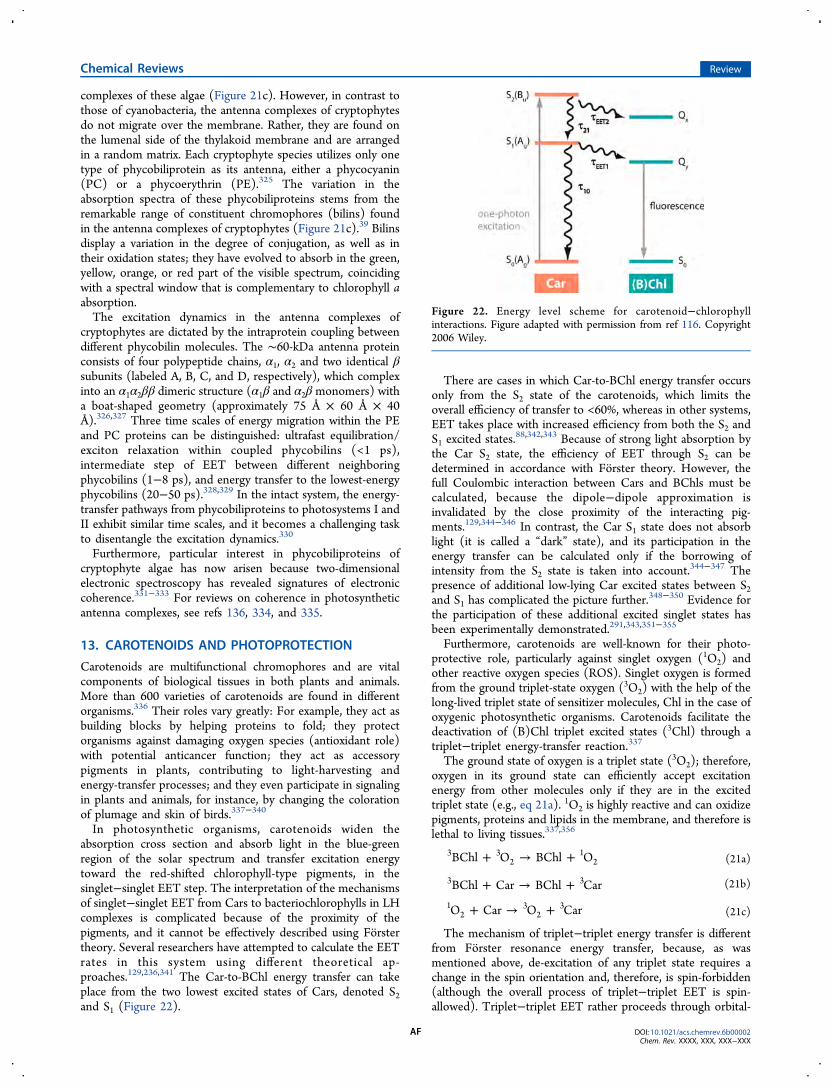
complexes of these algae (Figure 21c). However, in contrast tothose of cyanobacteria, the antenna complexes of cryptophytesdo not migrate over the membrane. Rather, they are found onthe lumenal side of the thylakoid membrane and are arrangedin a random matrix. Each cryptophyte species utilizes only onetype of phycobiliprotein as its antenna, either a phycocyanin(PC) or a phycoerythrin (PE).325 The variation in theabsorption spectra of these phycobiliproteins stems from theremarkable range of constituent chromophores (bilins) foundin the antenna complexes of cryptophytes (Figure 21c).39 Bilinsdisplay a variation in the degree of conjugation, as well as intheir oxidation states; they have evolved to absorb in the green,yellow, orange, or red part of the visible spectrum, coincidingwith a spectral window that is complementary to chlorophyll aabsorption.The excitation dynamics in the antenna complexes of
cryptophytes are dictated by the intraprotein coupling betweendifferent phycobilin molecules. The ∼60-kDa antenna proteinconsists of four polypeptide chains, α1, α2 and two identical βsubunits (labeled A, B, C, and D, respectively), which complexinto an α1α2ββ dimeric structure (α1β and α2β monomers) witha boat-shaped geometry (approximately 75 Å × 60 Å × 40Å).326,327 Three time scales of energy migration within the PEand PC proteins can be distinguished: ultrafast equilibration/exciton relaxation within coupled phycobilins (<1 ps),intermediate step of EET between different neighboringphycobilins (1−8 ps), and energy transfer to the lowest-energyphycobilins (20−50 ps).328,329 In the intact system, the energy-transfer pathways from phycobiliproteins to photosystems I andII exhibit similar time scales, and it becomes a challenging taskto disentangle the excitation dynamics.330
Furthermore, particular interest in phycobiliproteins ofcryptophyte algae has now arisen because two-dimensionalelectronic spectroscopy has revealed signatures of electroniccoherence.331−333 For reviews on coherence in photosyntheticantenna complexes, see refs 136, 334, and 335.
13. CAROTENOIDS AND PHOTOPROTECTION
Carotenoids are multifunctional chromophores and are vitalcomponents of biological tissues in both plants and animals.More than 600 varieties of carotenoids are found in differentorganisms.336 Their roles vary greatly: For example, they act asbuilding blocks by helping proteins to fold; they protectorganisms against damaging oxygen species (antioxidant role)with potential anticancer function; they act as accessorypigments in plants, contributing to light-harvesting andenergy-transfer processes; and they even participate in signalingin plants and animals, for instance, by changing the colorationof plumage and skin of birds.337−340
In photosynthetic organisms, carotenoids widen theabsorption cross section and absorb light in the blue-greenregion of the solar spectrum and transfer excitation energytoward the red-shifted chlorophyll-type pigments, in thesinglet−singlet EET step. The interpretation of the mechanismsof singlet−singlet EET from Cars to bacteriochlorophylls in LHcomplexes is complicated because of the proximity of thepigments, and it cannot be effectively described using Forstertheory. Several researchers have attempted to calculate the EETrates in this system using different theoretical ap-proaches.129,236,341 The Car-to-BChl energy transfer can takeplace from the two lowest excited states of Cars, denoted S2and S1 (Figure 22).
There are cases in which Car-to-BChl energy transfer occursonly from the S2 state of the carotenoids, which limits theoverall efficiency of transfer to <60%, whereas in other systems,EET takes place with increased efficiency from both the S2 andS1 excited states.88,342,343 Because of strong light absorption bythe Car S2 state, the efficiency of EET through S2 can bedetermined in accordance with Forster theory. However, thefull Coulombic interaction between Cars and BChls must becalculated, because the dipole−dipole approximation isinvalidated by the close proximity of the interacting pig-ments.129,344−346 In contrast, the Car S1 state does not absorblight (it is called a “dark” state), and its participation in theenergy transfer can be calculated only if the borrowing ofintensity from the S2 state is taken into account.344−347 Thepresence of additional low-lying Car excited states between S2and S1 has complicated the picture further.348−350 Evidence forthe participation of these additional excited singlet states hasbeen experimentally demonstrated.291,343,351−355
Furthermore, carotenoids are well-known for their photo-protective role, particularly against singlet oxygen (1O2) andother reactive oxygen species (ROS). Singlet oxygen is formedfrom the ground triplet-state oxygen (3O2) with the help of thelong-lived triplet state of sensitizer molecules, Chl in the case ofoxygenic photosynthetic organisms. Carotenoids facilitate thedeactivation of (B)Chl triplet excited states (3Chl) through atriplet−triplet energy-transfer reaction.337The ground state of oxygen is a triplet state (3O2); therefore,
oxygen in its ground state can efficiently accept excitationenergy from other molecules only if they are in the excitedtriplet state (e.g., eq 21a). 1O2 is highly reactive and can oxidizepigments, proteins and lipids in the membrane, and therefore islethal to living tissues.337,356
+ → +BChl O BChl O3 32
12 (21a)
+ → +BChl Car BChl Car3 3 (21b)
+ → +O Car O Car12
32
3(21c)
The mechanism of triplet−triplet energy transfer is differentfrom Forster resonance energy transfer, because, as wasmentioned above, de-excitation of any triplet state requires achange in the spin orientation and, therefore, is spin-forbidden(although the overall process of triplet−triplet EET is spin-allowed). Triplet−triplet EET rather proceeds through orbital-
Figure 22. Energy level scheme for carotenoid−chlorophyllinteractions. Figure adapted with permission from ref 116. Copyright2006 Wiley.
Chemical Reviews Review
DOI: 10.1021/acs.chemrev.6b00002Chem. Rev. XXXX, XXX, XXX−XXX
AF
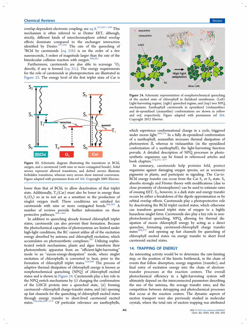
overlap-dependent electronic coupling; see eq 8.157,357−359 Thismechanism is often referred to as Dexter EET, although,strictly, different kinds of interchromophore orbital overlapeffects dominate compared to the exchanger interactionidentified by Dexter.157,358 The rate of the quenching of3BChl by carotenoids (eq 21b) is on the order of a fewnanoseconds, 3 orders of magnitude larger than the rate of thebimolecular collision reaction with oxygen.360,361
Furthermore, carotenoids are also able to scavenge 1O2directly, if any is formed (eq 21c). The energy requirementsfor the role of carotenoids in photoprotection are illustrated inFigure 23. The energy level of the first triplet state of Car is
lower than that of BChl, to allow deactivation of that tripletstate. Additionally, T1(Car) must also be lower in energy thanS1(O2) so as to not act as a sensitizer in the production ofsinglet oxygen itself. These conditions are satisfied forcarotenoids with nine or more conjugated bonds.362,363 Anumber of reviews provide further information on theseprotective pathways.363−365
In addition to quenching already formed chlorophyll tripletstates, carotenoids can also prevent their formation. Becausethe photochemical capacities of photosystems are limited underhigh-light conditions, the RC cannot utilize all of the excitationenergy absorbed by antenna and chlorophyll excitation, whichaccumulates on photosynthetic complexes.367 Utilizing sophis-ticated switch mechanisms, plants and algae transform theirphotosynthetic apparatus from an “efficient-light-harvesting”mode to an “excess-energy-dissipation” mode, where singletexcitation of chlorophylls is converted to heat, prior to theformation of chlorophyll triplet states.128,368 The process ofadaptive thermal dissipation of chlorophyll energy is known asnonphotochemical quenching (NPQ) of chlorophyll excitedstates and is shown in Figure 24. Carotenoids play a key role inthe NPQ switch mechanisms by (i) changing the conformationof the LHCII protein into a quenched state, (ii) formingcarotenoid−chlorophyll charge-transfer states, and (iii) openingup fast channels for the quenching of chlorophyll excited statesthrough energy transfer to short-lived carotenoid excitedstates.285,286,369−373 Of particular relevance are xanthophylls,
which experience conformational change in a cycle, triggeredunder excess light.368,374 In a fully de-epoxidized conformationof a xanthophyll, zeaxanthin increases thermal dissipation ofphotosystem II, whereas in violaxanthin (in the epoxidizedconformation of a xanthophyll), the light-harvesting functionprevails. A detailed description of NPQ processes in photo-synthetic organisms can be found in referenced articles andbook chapters.274,375−377
In summary, carotenoids help proteins fold, protectorganisms against damaging oxygen species, act as accessorypigments in plants, and participate in signaling. The Car-to-BChl energy transfer can occur from the Car S1 or S2 state. S2absorbs strongly and Forster theory with modifications (due toclose proximity of chromophores) can be used to estimate ratesof ensuing EET. S1, however, is a dark state and energy transferoccurs by either a breakdown of the dipole approximation or byorbital overlap effects. Carotenoids play a photoprotective roleby deactivating the BChl triplet excited states, which otherwisecan transform ground triplet state of oxygen to a highlyhazardous singlet form. Carotenoids also play a key role in non-photochemical quenching, NPQ, allowing for thermal dis-sipation of excess chlorophyll energy by acting as a directquencher, formating carotenoid-chlorophyll charge transferstates284,372 and opening up fast channels for quenching ofchlorophyll excited states via energy transfer to short livedcarotenoid excited states.
14. TRAPPING OF ENERGYAn interesting activity would be to determine the rate-limitingstep, or the position of the kinetic bottleneck, in the chain ofevents that follow absorption, energy migration (transfer), andfinal entry of excitation energy into the chain of electron-transfer processes at the reaction centers. The overallphotochemical efficiency in a light-harvesting system willultimately depend on the interconnected parameters describingthe size of the antenna, the average transfer rates, and thecompetition between detrapping and photochemical processesthat occur at the reaction centers. The dynamic aspects ofexciton transport were also previously studied in molecularcrystals, where the total rate of exciton trapping was attributed
Figure 23. Schematic diagram illustrating the transitions in BChl,oxygen, and a carotenoid (with nine or more conjugated bonds). Solidarrows represent allowed transitions, and dotted arrows illustrateforbidden transitions, whereas wavy arrows show internal conversion.Figure adapted with permission from ref 366. Copyright 2005 Elsevier.
Figure 24. Schematic representation of nonphotochemical quenchingof the excited state of chlorophyll in thylakoid membranes. (Left)Light-harvesting regime, (right) quenched regime, and (top) two NPQmechanisms. Xanthophyll carotenoids in epoxidized (violaxanthin)and de-epoxidized (zeaxanthin) conformations are shown in yellowand red, respectively. Figure adapted with permission ref 284.Copyright 2012 Elsevier.
Chemical Reviews Review
DOI: 10.1021/acs.chemrev.6b00002Chem. Rev. XXXX, XXX, XXX−XXX
AG
to both the rate at which the excitons diffused into areasoccupied by trap molecules and the rate of capture at whichexcitons decay to trap states once they enter the “sphere ofinfluence” of the traps.378,379 In analogy, in photosyntheticsystems, following the original formation of an exciton, itslifetime can be subdivided into three stages: (a) excitonmigration through transfer steps between constituent pigmentsof the light-harvesting assembly (⟨τmigr⟩; i.e., the time requiredto reach the photoactive pigment of the reaction center for thefirst time after its generation), (b) delivery of the excitationenergy to the reaction center (⟨τdel⟩), and (c) trapping time inthe reaction center (⟨τtrap⟩)
380−382
τ τ τ τ⟨ ⟩ = ⟨ ⟩+⟨ ⟩+⟨ ⟩migr del trap (22)
The value of the first passage time depends on the spatial andenergetic landscapes of the integral antenna pigments, whichdictate the number of transfer steps and their respectivekinetics. Typical models describe the trapping time asdependent on the photochemical quenching parameter at thereaction centers, as well as the rate constants associated withthe forward and backward transfer times of the exciton to thereaction-center chromophores, kt and k−t.
380 The relative valuesof the rate constants, kt and k−t, determine the classification ofthe trap. A deep trap is defined for kt/k−t ≫ 1, where themigration of the excitation from the reaction center back to theantenna system is fairly low and the excitation is oftenirreversibly quenched as kinetics of photochemistry dominateover the time scale of detrapping. The irreversibility of thetrapping essentially describes this model as “diffusion-limited”,as the rate of trapping equals the rate of diffusion of theexcitation energy to the trap, with ⟨τmigr⟩ as the dominatingterm in eq 22.In the case of shallow traps, upon localization of the exciton
at the reaction centers, the excitation is allowed to escape, sothat excitons can visit a shallow trap a number of times beforethe excited state is photochemically quenched. This type ofsystem is termed “trap-limited”, as the excited-state lifetime isdominated by the term describing the ability of the reactioncenter to capture the excitation energy from the antennapigments, ⟨τtrap⟩. The observed overall excited-state lifetimeincreases with the size of the antenna for both models. For thetrap-limited model, the increase is linear and independent ofthe excitation wavelength. Physically, this implies that excitationof any pigment, independent of its energy and position, willresult in energy-migration pathways that involve a number ofdetrapping steps and high probability of repopulation of theantenna system, so that location of the initial excitation is of nomajor importance.Schatz and co-workers (1988) reported an increase in the
overall lifetime for PSII complexes with different antenna sizesand suggested that PSII kinetics might follow a trap-limitedmodel.383 A special case involving a shallow trap was alsodescribed, where, even upon photochemical quenching, reverseelectron transfer can lead to the re-creation of the excited stateof the reaction-center chlorophyll, which can potentially diffuseback into the antenna system.2 The simplicity of the originalexciton-radical-pair-equilibrium (ERPE) model is attributed tothe observed scaling law of the fluorescence decay time with N,but its main drawback is that it does not take into accountcrystal structural information.87,383 The distances between thereaction center and the antenna pigments are relatively large,possibly necessary to prevent oxidation of antenna pigments,and these large distances were shown to create a bottleneck for
the excitation energy transfer in structure-based calculations.384
More recent studies consistent with structure-based calculationshave revealed a more complex picture with slower fluorescencedecay times than predicted by the original ERPE model.385,386
15. CONCLUDING REMARKSSubstantial advancements have been made toward elucidatingthe mechanistic details of excitation energy transfer in light-harvesting systems of photosynthetic organisms. Currentresearch goes beyond the scope of deciphering the contribu-tions of the photosynthetic organisms to optimizing this vitalprocess, as the major goal is now to apply these fundamentalconcepts and employ mimicry for the realization of highlyefficient artificial light harvestors. There are, however, somemajor concepts embedded in the blueprint of naturallyoccurring antenna complexes that have been revealed andfrom which researchers can learn to build efficient artificialsystems. The total number, their mutual arrangement, theirconcentration, as well as the variety of pigments all provide vitalinformation for the optimization of light-harvesting systems tobe used in artificial systems. The architectural design of theenergy landscape of the antenna depends both on thephotophysics of the individual components and on the natureof their assembly, which, for example, leads to the variety ofcoupling regimes that were introduced in this review.
AUTHOR INFORMATIONCorresponding Author
*Tel.: +1 609-258-0729. E-mail: [email protected]
The authors declare no competing financial interest.
Biographies
Tihana Mirkovic received her B.Sc. in Chemistry and Mathematics in2003 from the University of Toronto. In 2009, she obtained her Ph.D.in Physical Chemistry, also from the University of Toronto, with Prof.G. D. Scholes and Prof G. A. Ozin, where her interdisciplinary researchencompassed the development of nanomaterials and studies of thephotophysical and dynamical aspects of nanoscale systems. As aPostdoctoral Fellow and Research Associate (2010−2014), sheworked on elucidating the principles of light harvesting in cryptophytealgae. Currently, she is focusing on chemical education and works as aSessional Lecturer at the University of Toronto in areas of materialsand inorganic and physical chemistry.
Evgeny E. Ostroumov received his Ph.D. in Physics from University ofDusseldorf and Max-Planck-Institute for Bioinorganic Chemistry,where he worked with Prof. Alfred Holzwarth exploring the electronicproperties of isolated chromophores and photoprotection mechanismsin high plants. He is currently an Associate Specialist at PrincetonUniversity, and his research interests include the development andapplication of nonlinear multidimensional ultrafast spectroscopy tostudy dynamics in natural and artificial photoactive systems with theaim of elucidating mechanisms of energy transfer and photoprotectionand applications in diagnostics.
Jessica M. Anna is an Assistant Professor and Elliman Faculty Fellowin the Department of Chemistry at the University of Pennsylvania.Jessica joined the faculty at the University of Pennsylvania in July of2014. Before this, she was a postdoctoral fellow in the Scholes group atthe University of Toronto from 2011 to 2014. Before joining theScholes group, Jessica received her Ph.D. in Physical Chemistry fromthe University of Michigan in 2011 under the guidance of Kevin
Chemical Reviews Review
DOI: 10.1021/acs.chemrev.6b00002Chem. Rev. XXXX, XXX, XXX−XXX
AH
Kubarych and her B.S. from the University of Pittsburgh in 2006,where she worked with David Pratt. Her current research focuses oninvestigating electronic energy transfer and electron-transfer reactionsin natural and artificial light-harvesting complexes and photocatalysts.
Rienk van Grondelle studied physics at VU University Amsterdam. Heobtained his Ph.D. under the supervision of Prof. Dr. Lou Duysens. In1982, he returned to VU University, and in 1987, he was appointed fullprofessor. At VU University, he has built a large research groupstudying the early events in photosynthesis. R.v.G. has made majorcontributions to elucidating the fundamental physical mechanisms thatunderlie light harvesting and charge separation. He has developedtheoretical tools for understanding complex spectroscopic data. Usingmultidimensional electronic spectroscopy, he recently showed thatultrafast charge separation is driven by specific molecular vibrationsthat allow electronic coherences to stay alive. He proposed a molecularmodel for photoprotection and demonstrated that the major plantlight-harvesting complex operates as a nanoswitch, controlled by itsbiological environment. These results, of utmost importance forunderstanding photosynthesis, have inspired technological solutionsfor artificial and/or redesigned photosynthesis, as a route towardsustainable energy production.
Govindjee is Professor Emeritus of Biophysics, Biochemistry and PlantBiology at the University of Illinois at Urbana−Champaign (UIUC),Urbana, Illinois. He is a Series Editor of “Advances in Photosynthesisand Respiration” (Springer). He holds a B.Sc. (1952) and an M.Sc.(1954) from the University of Allahabad and a Ph.D. (1960; inBiophysics, under Eugene Rabinowitch) from UIUC. He is a fellow ofthe AAAS; recipient of a Lifetime Achievement Award from the RebeizFoundation for Basic Biology, a Liberal Arts and Sciences AlumniAchievement Award from UIUC, a Communication Award from theInternational Society of Photosynthesis Research, and a B. M. JohriMemorial Award (India) for Excellence in Plant Biology. Govindjee isan author of “Photosynthesis” (1969), has edited over 12 books, andhas published over 400 research (and review) articles on photosyn-thesis. His research has focused on the primary photochemistry ofoxygenic photosynthesis, excitation energy transfer in cyanobacteriaand algae, and electron transfer from water to NADP. One of his majordiscoveries was on the unique role of bicarbonate in protonation of areduced plastoquinone (QB
2−) on the electron-acceptor side ofphotosystem II of photosynthesis.
Greg Scholes is the William S. Tod Professor of Chemistry atPrinceton University. Originally from Melbourne, Australia, heundertook postdoctoral training at Imperial College London andUniversity of California, Berkeley. He started his independent career atthe University of Toronto (2000−2014), where he was the D. J.LeRoy Distinguished Professor. Dr. Scholes is the Deputy Editor ofthe Journal of Physical Chemistry Letters; a Fellow of the Royal Societyof Canada; a Senior Fellow in the Canadian Institute for AdvancedResearch program Biology, Energy, Technology; and a ProfessorialFellow at the University of Melbourne. Dr. Scholes has had a long-standing interest in the mechanisms of electronic energy transfer andthe photophysics of molecular excitons. Current research concernsworking out the design principles for directing and regulating light-initiated energy flow in man-made and natural systems, such asproteins involved in photosynthesis.
ACKNOWLEDGMENTS
G.D.S. and R.v.G. acknowledge CIFAR, the Canadian Institutefor Advanced Research, through its Bio-Inspired Solar Energyprogram. This work was supported partially as part of thePhotosynthetic Antenna Research Center (PARC), an Energy
Frontier Research Center funded by the U.S. Department ofEnergy, Office of Science, Basic Energy Sciences, under AwardDE-SC0001035 and partially by the Natural Sciences andEngineering Research Council of Canada (G.D.S.). Govindjeethanks the Departments of Biochemistry and Plant Biology atthe University of Illinois at Urbana−Champaign (UIUC) forsupporting him during his retirement since 1999. R.v.G. wassupported by VU University, by an Advanced Investigator grantfrom the European Research Council (No. 267333, PHOT-PROT), and by EU FP7 project PAPETS (GA 323901). Hegratefully acknowledges his Academy Professor grant from TheNetherlands Royal Academy of Sciences (KNAW).
REFERENCES(1) Rabinowitch, E.; Govindjee. Photosynthesis; Wiley: New York,1969; available at http://www.life.illinois.edu/govindjee/g/Books.html(accessed July 1, 2016).(2) Blankenship, R. E. Molecular Mechanisms of Photosynthesis; Wiley-Blackwell: Hoboken, NJ, 2014.(3) Allen, J. F. Light, Time and Micro-Organisms. In MicrobialResponses to Light and Time; Caddick, M. X., Baumberg, S., Hodgson,D. A., Phillips-Jones, M. K., Eds.; Cambridge University Press, 1998;pp 1−31. Available at http://www.jfallen.org/~john/webstar/ltm/ltm.pdf (accessed July 1, 2016).(4) Abbott, D. Keeping the Energy Debate Clean: How Do WeSupply the World’s Energy Needs? Proc. IEEE 2010, 98, 42−66.(5) Alstrum-Acevedo, J. H.; Brennaman, M. K.; Meyer, T. J.Chemical Approaches to Artificial Photosynthesis. 2. Inorg. Chem.2005, 44, 6802−6827.(6) Balzani, V.; Credi, A.; Venturi, M. Photochemical Conversion ofSolar Energy. ChemSusChem 2008, 1, 26−58.(7) Barber, J. Photosynthetic Energy Conversion: Natural andArtificial. Chem. Soc. Rev. 2009, 38, 185−196.(8) Hambourger, M.; Moore, G. F.; Kramer, D. M.; Gust, D.; Moore,A. L.; Moore, T. A. Biology and Technology for Photochemical FuelProduction. Chem. Soc. Rev. 2009, 38, 25−35.(9) Herrero, C.; Lassalle-Kaiser, B.; Leibl, W.; Rutherford, A. W.;Aukauloo, A. Artificial Systems Related to Light Driven ElectronTransfer Processes in PSII. Coord. Chem. Rev. 2008, 252, 456−468.(10) McConnell, I.; Li, G.; Brudvig, G. W. Energy Conversion inNatural and Artificial Photosynthesis. Chem. Biol. 2010, 17, 434−447.(11) Ort, D. R.; Merchant, S. S.; Alric, J.; Barkan, A.; Blankenship, R.E.; Bock, R.; Croce, R.; Hanson, M. R.; Hibberd, J. M.; Long, S. P.;Moore, T. A.; et al. Redesigning Photosynthesis to Sustainably MeetGlobal Food and Bioenergy Demand. Proc. Natl. Acad. Sci. U. S. A.2015, 112, 8529−8536.(12) Eaton-Rye, J. J., Tripathy, B. C., Sharkey, T. D., Eds.Photosynthesis: Plastid Biology, Energy Conversion and CarbonAssimilation; Advances in Photosynthesis and Respiration; Springer:Dordrecht, The Netherlands, 2012; Vol. 34.(13) Hunter, C. N., Daldal, F., Thurnauer, M. C., Beatty, J. T., Eds.The Purple Phototrophic Bacteria; Advances in Photosynthesis andRespiration; Springer: Dordrecht, The Netherlands, 2009; Vol. 28.(14) Drews, G.; Golecki, J. R. Structure, Molecular Organization, andBiosynthesis of Membranes by Purple Bacteria. In AnoxygenicPhotosynthetic Bacteria; Blankenship, R. E., Madigan, M. T., Bauer,C. E., Eds.; Advances in Photosynthesis and Respiration; KluwerAcademic Publishers: Dordrecht, The Netherlands, 1995; Vol. 2,Chapter 12, pp 231−257.(15) Overmann, J. Diversity and Ecology of Phototrophic SulfurBacteria. Microbiol. Today 2001, 28, 116−118.(16) Nisbet, E. G.; Sleep, N. H. The Habitat and Nature of Early Life.Nature 2001, 409, 1083−1091.(17) Govindjee; Govindjee, R. Primary Events in Photosynthesis. Sci.Am. 1974, 231, 68−82.(18) Wolosiuk, R. A.; Buchanan, B. B. Photosynthesis: The CarbonReactions. In Plant Physiology and Development, 6th ed.; Taiz, L.,
Chemical Reviews Review
DOI: 10.1021/acs.chemrev.6b00002Chem. Rev. XXXX, XXX, XXX−XXX
AI
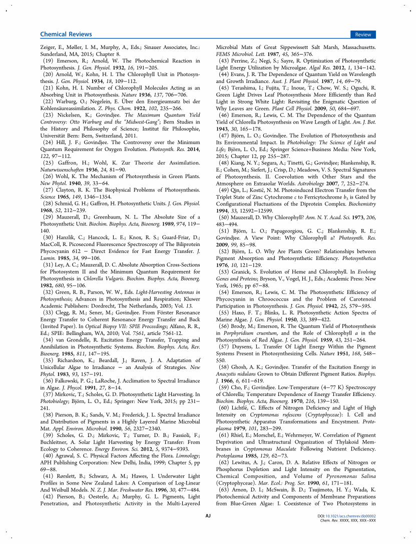
Zeiger, E., Møller, I. M., Murphy, A., Eds.; Sinauer Associates, Inc.:Sunderland, MA, 2015; Chapter 8.(19) Emerson, R.; Arnold, W. The Photochemical Reaction inPhotosynthesis. J. Gen. Physiol. 1932, 16, 191−205.(20) Arnold, W.; Kohn, H. I. The Chlorophyll Unit in Photosyn-thesis. J. Gen. Physiol. 1934, 18, 109−112.(21) Kohn, H. I. Number of Chlorophyll Molecules Acting as anAbsorbing Unit in Photosynthesis. Nature 1936, 137, 706−706.(22) Warburg, O.; Negelein, E. Uber den Energieumsatz bei derKohlensaureassimilation. Z. Phys. Chem. 1922, 102, 235−266.(23) Nickelsen, K.; Govindjee. The Maximum Quantum YieldControversy: Otto Warburg and the “Midwest-Gang”; Bern Studies inthe History and Philosophy of Science; Institut fur Philosophie,Universitat Bern: Bern, Switzerland, 2011.(24) Hill, J. F.; Govindjee. The Controversy over the MinimumQuantum Requirement for Oxygen Evolution. Photosynth. Res. 2014,122, 97−112.(25) Gaffron, H.; Wohl, K. Zur Theorie der Assimilation.Naturwissenschaften 1936, 24, 81−90.(26) Wohl, K. The Mechanism of Photosynthesis in Green Plants.New Phytol. 1940, 39, 33−64.(27) Clayton, R. K. The Biophysical Problems of Photosynthesis.Science 1965, 149, 1346−1354.(28) Schmid, G. H.; Gaffron, H. Photosynthetic Units. J. Gen. Physiol.1968, 52, 212−239.(29) Mauzerall, D.; Greenbaum, N. L. The Absolute Size of aPhotosynthetic Unit. Biochim. Biophys. Acta, Bioenerg. 1989, 974, 119−140.(30) Hanzlik, C.; Hancock, L. E.; Knox, R. S.; Guard-Friar, D.;MacColl, R. Picosecond Fluorescence Spectroscopy of The BiliproteinPhycocyanin 612 − Direct Evidence for Fast Energy Transfer. J.Lumin. 1985, 34, 99−106.(31) Ley, A. C.; Mauzerall, D. C. Absolute Absorption Cross-Sectionsfor Photosystem II and the Minimum Quantum Requirement forPhotosynthesis in Chlorella Vulgaris. Biochim. Biophys. Acta, Bioenerg.1982, 680, 95−106.(32) Green, R. B., Parson, W. W., Eds. Light-Harvesting Antennas inPhotosynthesis; Advances in Photosynthesis and Respiration; KluwerAcademic Publishers: Dordrecht, The Netherlands, 2003; Vol. 13.(33) Clegg, R. M.; Sener, M.; Govindjee. From Forster ResonanceEnergy Transfer to Coherent Resonance Energy Transfer and Back(Invited Paper). In Optical Biopsy VII: SPIE Proceedings; Alfano, R. R.,Ed.; SPIE: Bellingham, WA, 2010; Vol. 7561, article 7561-12.(34) van Grondelle, R. Excitation Energy Transfer, Trapping andAnnihilation in Photosynthetic Systems. Biochim. Biophys. Acta, Rev.Bioenerg. 1985, 811, 147−195.(35) Richardson, K.; Beardall, J.; Raven, J. A. Adaptation ofUnicellular Algae to Irradiance − an Analysis of Strategies. NewPhytol. 1983, 93, 157−191.(36) Falkowski, P. G.; LaRoche, J. Acclimation to Spectral Irradiancein Algae. J. Phycol. 1991, 27, 8−14.(37) Mirkovic, T.; Scholes, G. D. Photosynthetic Light Harvesting. InPhotobiology; Bjorn, L. O., Ed.; Springer: New York, 2015; pp 231−241.(38) Pierson, B. K.; Sands, V. M.; Frederick, J. L. Spectral Irradianceand Distribution of Pigments in a Highly Layered Marine MicrobialMat. Appl. Environ. Microbiol. 1990, 56, 2327−2340.(39) Scholes, G. D.; Mirkovic, T.; Turner, D. B.; Fassioli, F.;Buchleitner, A. Solar Light Harvesting by Energy Transfer: FromEcology to Coherence. Energy Environ. Sci. 2012, 5, 9374−9393.(40) Agrawal, S. C. Physical Factors Affecting the Flora. Limnology;APH Publishing Corporation: New Delhi, India, 1999; Chapter 5, pp69−88.(41) Rørslett, B.; Schwarz, A. M.; Hawes, I. Underwater LightProfiles in Some New Zealand Lakes: A Comparison of Log-LinearAnd Weibull Models. N. Z. J. Mar. Freshwater Res. 1996, 30, 477−484.(42) Pierson, B.; Oesterle, A.; Murphy, G. L. Pigments, LightPenetration, and Photosynthetic Activity in the Multi-Layered
Microbial Mats of Great Sippewissett Salt Marsh, Massachusetts.FEMS Microbiol. Lett. 1987, 45, 365−376.(43) Perrine, Z.; Negi, S.; Sayre, R. Optimization of PhotosyntheticLight Energy Utilization by Microalgae. Algal Res. 2012, 1, 134−142.(44) Evans, J. R. The Dependence of Quantum Yield on Wavelengthand Growth Irradiance. Aust. J. Plant Physiol. 1987, 14, 69−79.(45) Terashima, I.; Fujita, T.; Inoue, T.; Chow, W. S.; Oguchi, R.Green Light Drives Leaf Photosynthesis More Efficiently than RedLight in Strong White Light: Revisiting the Enigmatic Question ofWhy Leaves are Green. Plant Cell Physiol. 2009, 50, 684−697.(46) Emerson, R.; Lewis, C. M. The Dependence of the QuantumYield of Chlorella Photosynthesis on Wave Length of Light. Am. J. Bot.1943, 30, 165−178.(47) Bjorn, L. O.; Govindjee. The Evolution of Photosynthesis andIts Environmental Impact. In Photobiology: The Science of Light andLife; Bjorn, L. O., Ed.; Springer Science+Business Media: New York,2015; Chapter 12, pp 255−287.(48) Kiang, N. Y.; Segura, A.; Tinetti, G.; Govindjee; Blankenship, R.E.; Cohen, M.; Siefert, J.; Crisp, D.; Meadows, V. S. Spectral Signaturesof Photosynthesis. II. Coevolution with Other Stars and theAtmosphere on Extrasolar Worlds. Astrobiology 2007, 7, 252−274.(49) Qin, L.; Kostic, N. M. Photoinduced Electron Transfer from theTriplet State of Zinc Cytochrome c to Ferricytochrome b5 is Gated byConfigurational Fluctuations of the Diprotein Complex. Biochemistry1994, 33, 12592−12599.(50) Mauzerall, D. Why Chlorophyll? Ann. N. Y. Acad. Sci. 1973, 206,483−494.(51) Bjorn, L. O.; Papageorgiou, G. C.; Blankenship, R. E.;Govindjee. A View Point: Why Chlorophyll a? Photosynth. Res.2009, 99, 85−98.(52) Bjorn, L. O. Why Are Plants Green? Relationships betweenPigment Absorption and Photosynthetic Efficiency. Photosynthetica1976, 10, 121−129.(53) Granick, S. Evolution of Heme and Chlorophyll. In EvolvingGenes and Proteins; Bryson, V., Vogel, H. J., Eds.; Academic Press: NewYork, 1965; pp 67−88.(54) Emerson, R.; Lewis, C. M. The Photosynthetic Efficiency ofPhycocyanin in Chroococcus and the Problem of CarotenoidParticipation in Photosynthesis. J. Gen. Physiol. 1942, 25, 579−595.(55) Haxo, F. T.; Blinks, L. R. Photosynthetic Action Spectra ofMarine Algae. J. Gen. Physiol. 1950, 33, 389−422.(56) Brody, M.; Emerson, R. The Quantum Yield of Photosynthesisin Porphyridium cruentum, and the Role of Chlorophyll a in thePhotosynthesis of Red Algae. J. Gen. Physiol. 1959, 43, 251−264.(57) Duysens, L. Transfer Of Light Energy Within the PigmentSystems Present in Photosynthesizing Cells. Nature 1951, 168, 548−550.(58) Ghosh, A. K.; Govindjee. Transfer of the Excitation Energy inAnacystis nidulans Grown to Obtain Different Pigment Ratios. Biophys.J. 1966, 6, 611−619.(59) Cho, F.; Govindjee. Low-Temperature (4−77 K) Spectroscopyof Chlorella; Temperature Dependence of Energy Transfer Efficiency.Biochim. Biophys. Acta, Bioenerg. 1970, 216, 139−150.(60) Lichtle, C. Effects of Nitrogen Deficiency and Light of HighIntensity on Cryptomonas rufescens (Cryptophyceae): I. Cell andPhotosynthetic Apparatus Transformations and Encystment. Proto-plasma 1979, 101, 283−299.(61) Rhiel, E.; Morschel, E.; Wehrmeyer, W. Correlation of PigmentDeprivation and Ultrastructural Organization of Thylakoid Mem-branes in Cryptomonas Maculate Following Nutrient Deficiency.Protoplasma 1985, 129, 62−73.(62) Lewitus, A. J.; Caron, D. A. Relative Effects of Nitrogen orPhosphorus Depletion and Light Intensity on the Pigmentation,Chemical Composition, and Volume of Pyrenomonas Salina(Cryptophyceae). Mar. Ecol.: Prog. Ser. 1990, 61, 171−181.(63) Arnon, D. I.; McSwain, B. D.; Tsujimoto, H. Y.; Wada, K.Photochemical Activity and Components of Membrane Preparationsfrom Blue-Green Algae: I. Coexistence of Two Photosystems in
Chemical Reviews Review
DOI: 10.1021/acs.chemrev.6b00002Chem. Rev. XXXX, XXX, XXX−XXX
AJ
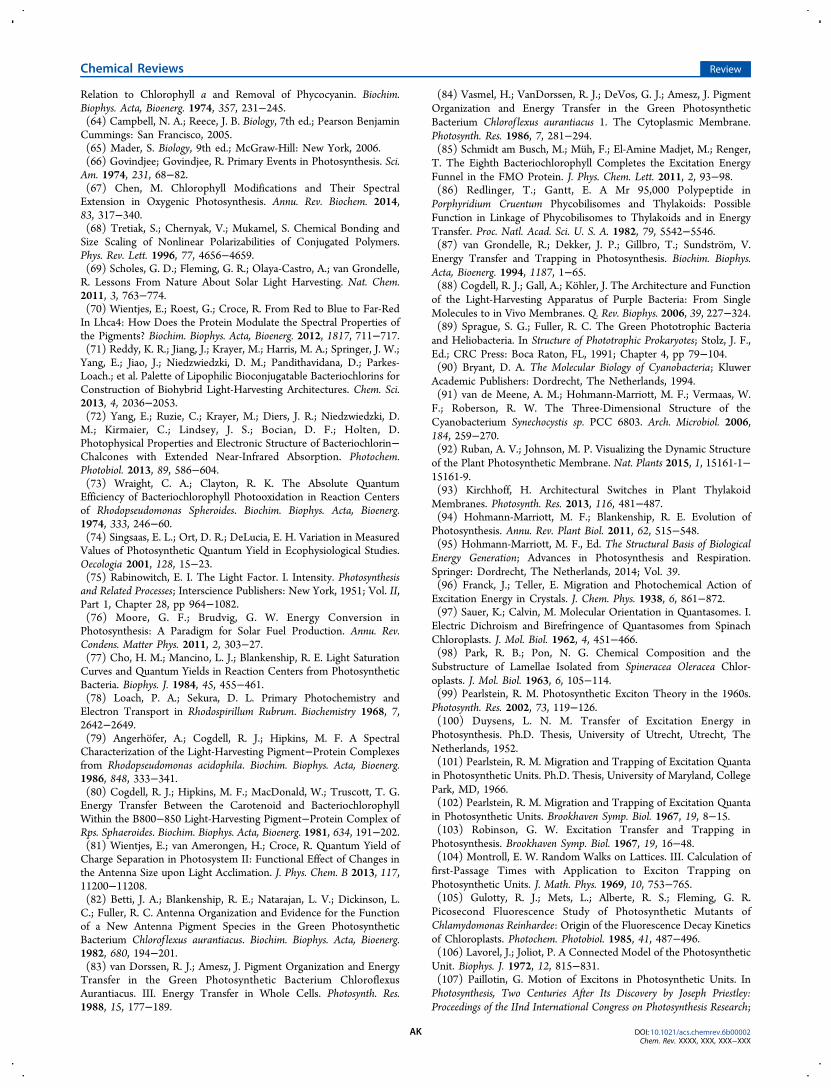
Relation to Chlorophyll a and Removal of Phycocyanin. Biochim.Biophys. Acta, Bioenerg. 1974, 357, 231−245.(64) Campbell, N. A.; Reece, J. B. Biology, 7th ed.; Pearson BenjaminCummings: San Francisco, 2005.(65) Mader, S. Biology, 9th ed.; McGraw-Hill: New York, 2006.(66) Govindjee; Govindjee, R. Primary Events in Photosynthesis. Sci.Am. 1974, 231, 68−82.(67) Chen, M. Chlorophyll Modifications and Their SpectralExtension in Oxygenic Photosynthesis. Annu. Rev. Biochem. 2014,83, 317−340.(68) Tretiak, S.; Chernyak, V.; Mukamel, S. Chemical Bonding andSize Scaling of Nonlinear Polarizabilities of Conjugated Polymers.Phys. Rev. Lett. 1996, 77, 4656−4659.(69) Scholes, G. D.; Fleming, G. R.; Olaya-Castro, A.; van Grondelle,R. Lessons From Nature About Solar Light Harvesting. Nat. Chem.2011, 3, 763−774.(70) Wientjes, E.; Roest, G.; Croce, R. From Red to Blue to Far-RedIn Lhca4: How Does the Protein Modulate the Spectral Properties ofthe Pigments? Biochim. Biophys. Acta, Bioenerg. 2012, 1817, 711−717.(71) Reddy, K. R.; Jiang, J.; Krayer, M.; Harris, M. A.; Springer, J. W.;Yang, E.; Jiao, J.; Niedzwiedzki, D. M.; Pandithavidana, D.; Parkes-Loach.; et al. Palette of Lipophilic Bioconjugatable Bacteriochlorins forConstruction of Biohybrid Light-Harvesting Architectures. Chem. Sci.2013, 4, 2036−2053.(72) Yang, E.; Ruzie, C.; Krayer, M.; Diers, J. R.; Niedzwiedzki, D.M.; Kirmaier, C.; Lindsey, J. S.; Bocian, D. F.; Holten, D.Photophysical Properties and Electronic Structure of Bacteriochlorin−Chalcones with Extended Near-Infrared Absorption. Photochem.Photobiol. 2013, 89, 586−604.(73) Wraight, C. A.; Clayton, R. K. The Absolute QuantumEfficiency of Bacteriochlorophyll Photooxidation in Reaction Centersof Rhodopseudomonas Spheroides. Biochim. Biophys. Acta, Bioenerg.1974, 333, 246−60.(74) Singsaas, E. L.; Ort, D. R.; DeLucia, E. H. Variation in MeasuredValues of Photosynthetic Quantum Yield in Ecophysiological Studies.Oecologia 2001, 128, 15−23.(75) Rabinowitch, E. I. The Light Factor. I. Intensity. Photosynthesisand Related Processes; Interscience Publishers: New York, 1951; Vol. II,Part 1, Chapter 28, pp 964−1082.(76) Moore, G. F.; Brudvig, G. W. Energy Conversion inPhotosynthesis: A Paradigm for Solar Fuel Production. Annu. Rev.Condens. Matter Phys. 2011, 2, 303−27.(77) Cho, H. M.; Mancino, L. J.; Blankenship, R. E. Light SaturationCurves and Quantum Yields in Reaction Centers from PhotosyntheticBacteria. Biophys. J. 1984, 45, 455−461.(78) Loach, P. A.; Sekura, D. L. Primary Photochemistry andElectron Transport in Rhodospirillum Rubrum. Biochemistry 1968, 7,2642−2649.(79) Angerhofer, A.; Cogdell, R. J.; Hipkins, M. F. A SpectralCharacterization of the Light-Harvesting Pigment−Protein Complexesfrom Rhodopseudomonas acidophila. Biochim. Biophys. Acta, Bioenerg.1986, 848, 333−341.(80) Cogdell, R. J.; Hipkins, M. F.; MacDonald, W.; Truscott, T. G.Energy Transfer Between the Carotenoid and BacteriochlorophyllWithin the B800−850 Light-Harvesting Pigment−Protein Complex ofRps. Sphaeroides. Biochim. Biophys. Acta, Bioenerg. 1981, 634, 191−202.(81) Wientjes, E.; van Amerongen, H.; Croce, R. Quantum Yield ofCharge Separation in Photosystem II: Functional Effect of Changes inthe Antenna Size upon Light Acclimation. J. Phys. Chem. B 2013, 117,11200−11208.(82) Betti, J. A.; Blankenship, R. E.; Natarajan, L. V.; Dickinson, L.C.; Fuller, R. C. Antenna Organization and Evidence for the Functionof a New Antenna Pigment Species in the Green PhotosyntheticBacterium Chlorof lexus aurantiacus. Biochim. Biophys. Acta, Bioenerg.1982, 680, 194−201.(83) van Dorssen, R. J.; Amesz, J. Pigment Organization and EnergyTransfer in the Green Photosynthetic Bacterium ChloroflexusAurantiacus. III. Energy Transfer in Whole Cells. Photosynth. Res.1988, 15, 177−189.
(84) Vasmel, H.; VanDorssen, R. J.; DeVos, G. J.; Amesz, J. PigmentOrganization and Energy Transfer in the Green PhotosyntheticBacterium Chlorof lexus aurantiacus 1. The Cytoplasmic Membrane.Photosynth. Res. 1986, 7, 281−294.(85) Schmidt am Busch, M.; Muh, F.; El-Amine Madjet, M.; Renger,T. The Eighth Bacteriochlorophyll Completes the Excitation EnergyFunnel in the FMO Protein. J. Phys. Chem. Lett. 2011, 2, 93−98.(86) Redlinger, T.; Gantt, E. A Mr 95,000 Polypeptide inPorphyridium Cruentum Phycobilisomes and Thylakoids: PossibleFunction in Linkage of Phycobilisomes to Thylakoids and in EnergyTransfer. Proc. Natl. Acad. Sci. U. S. A. 1982, 79, 5542−5546.(87) van Grondelle, R.; Dekker, J. P.; Gillbro, T.; Sundstrom, V.Energy Transfer and Trapping in Photosynthesis. Biochim. Biophys.Acta, Bioenerg. 1994, 1187, 1−65.(88) Cogdell, R. J.; Gall, A.; Kohler, J. The Architecture and Functionof the Light-Harvesting Apparatus of Purple Bacteria: From SingleMolecules to in Vivo Membranes. Q. Rev. Biophys. 2006, 39, 227−324.(89) Sprague, S. G.; Fuller, R. C. The Green Phototrophic Bacteriaand Heliobacteria. In Structure of Phototrophic Prokaryotes; Stolz, J. F.,Ed.; CRC Press: Boca Raton, FL, 1991; Chapter 4, pp 79−104.(90) Bryant, D. A. The Molecular Biology of Cyanobacteria; KluwerAcademic Publishers: Dordrecht, The Netherlands, 1994.(91) van de Meene, A. M.; Hohmann-Marriott, M. F.; Vermaas, W.F.; Roberson, R. W. The Three-Dimensional Structure of theCyanobacterium Synechocystis sp. PCC 6803. Arch. Microbiol. 2006,184, 259−270.(92) Ruban, A. V.; Johnson, M. P. Visualizing the Dynamic Structureof the Plant Photosynthetic Membrane. Nat. Plants 2015, 1, 15161-1−15161-9.(93) Kirchhoff, H. Architectural Switches in Plant ThylakoidMembranes. Photosynth. Res. 2013, 116, 481−487.(94) Hohmann-Marriott, M. F.; Blankenship, R. E. Evolution ofPhotosynthesis. Annu. Rev. Plant Biol. 2011, 62, 515−548.(95) Hohmann-Marriott, M. F., Ed. The Structural Basis of BiologicalEnergy Generation; Advances in Photosynthesis and Respiration.Springer: Dordrecht, The Netherlands, 2014; Vol. 39.(96) Franck, J.; Teller, E. Migration and Photochemical Action ofExcitation Energy in Crystals. J. Chem. Phys. 1938, 6, 861−872.(97) Sauer, K.; Calvin, M. Molecular Orientation in Quantasomes. I.Electric Dichroism and Birefringence of Quantasomes from SpinachChloroplasts. J. Mol. Biol. 1962, 4, 451−466.(98) Park, R. B.; Pon, N. G. Chemical Composition and theSubstructure of Lamellae Isolated from Spineracea Oleracea Chlor-oplasts. J. Mol. Biol. 1963, 6, 105−114.(99) Pearlstein, R. M. Photosynthetic Exciton Theory in the 1960s.Photosynth. Res. 2002, 73, 119−126.(100) Duysens, L. N. M. Transfer of Excitation Energy inPhotosynthesis. Ph.D. Thesis, University of Utrecht, Utrecht, TheNetherlands, 1952.(101) Pearlstein, R. M. Migration and Trapping of Excitation Quantain Photosynthetic Units. Ph.D. Thesis, University of Maryland, CollegePark, MD, 1966.(102) Pearlstein, R. M. Migration and Trapping of Excitation Quantain Photosynthetic Units. Brookhaven Symp. Biol. 1967, 19, 8−15.(103) Robinson, G. W. Excitation Transfer and Trapping inPhotosynthesis. Brookhaven Symp. Biol. 1967, 19, 16−48.(104) Montroll, E. W. Random Walks on Lattices. III. Calculation offirst-Passage Times with Application to Exciton Trapping onPhotosynthetic Units. J. Math. Phys. 1969, 10, 753−765.(105) Gulotty, R. J.; Mets, L.; Alberte, R. S.; Fleming, G. R.Picosecond Fluorescence Study of Photosynthetic Mutants ofChlamydomonas Reinhardee: Origin of the Fluorescence Decay Kineticsof Chloroplasts. Photochem. Photobiol. 1985, 41, 487−496.(106) Lavorel, J.; Joliot, P. A Connected Model of the PhotosyntheticUnit. Biophys. J. 1972, 12, 815−831.(107) Paillotin, G. Motion of Excitons in Photosynthetic Units. InPhotosynthesis, Two Centuries After Its Discovery by Joseph Priestley:Proceedings of the IInd International Congress on Photosynthesis Research;
Chemical Reviews Review
DOI: 10.1021/acs.chemrev.6b00002Chem. Rev. XXXX, XXX, XXX−XXX
AK
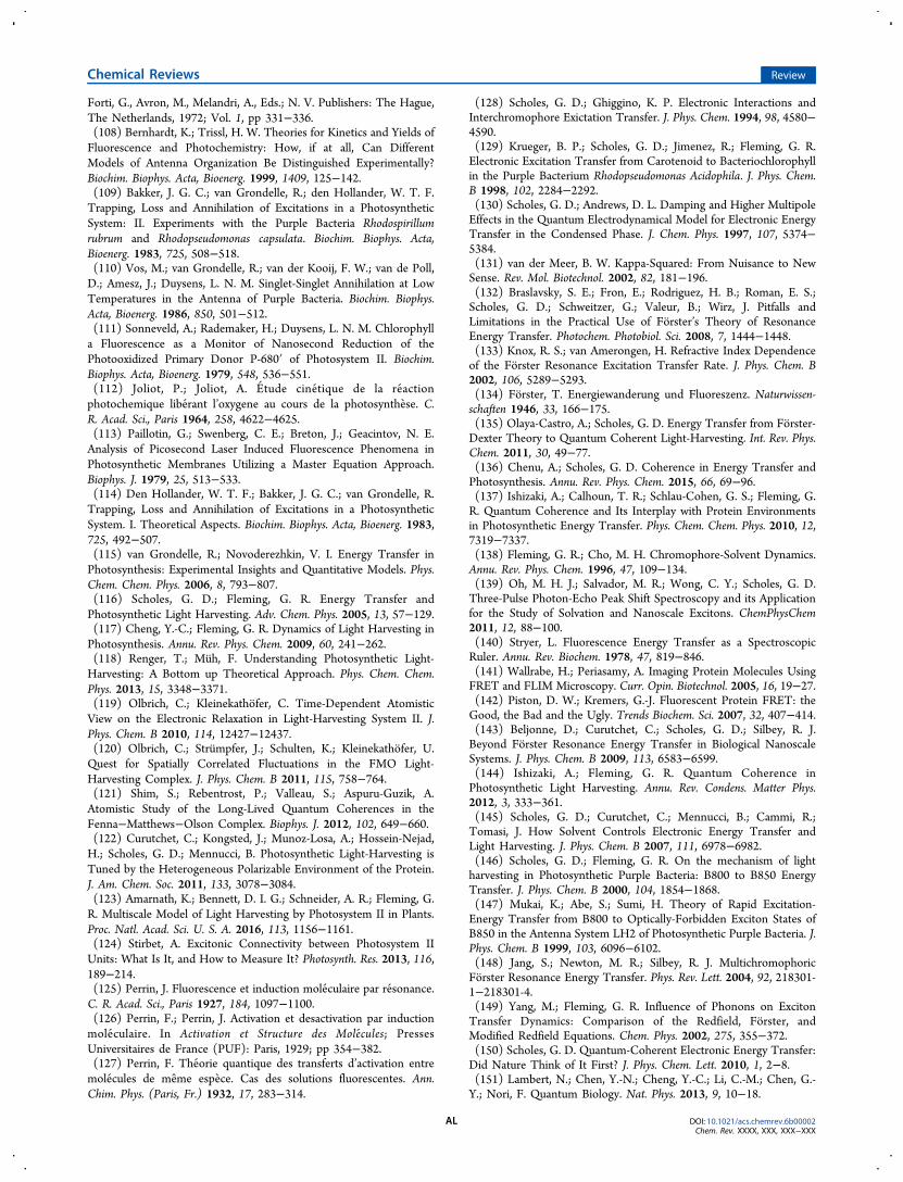
Forti, G., Avron, M., Melandri, A., Eds.; N. V. Publishers: The Hague,The Netherlands, 1972; Vol. 1, pp 331−336.(108) Bernhardt, K.; Trissl, H. W. Theories for Kinetics and Yields ofFluorescence and Photochemistry: How, if at all, Can DifferentModels of Antenna Organization Be Distinguished Experimentally?Biochim. Biophys. Acta, Bioenerg. 1999, 1409, 125−142.(109) Bakker, J. G. C.; van Grondelle, R.; den Hollander, W. T. F.Trapping, Loss and Annihilation of Excitations in a PhotosyntheticSystem: II. Experiments with the Purple Bacteria Rhodospirillumrubrum and Rhodopseudomonas capsulata. Biochim. Biophys. Acta,Bioenerg. 1983, 725, 508−518.(110) Vos, M.; van Grondelle, R.; van der Kooij, F. W.; van de Poll,D.; Amesz, J.; Duysens, L. N. M. Singlet-Singlet Annihilation at LowTemperatures in the Antenna of Purple Bacteria. Biochim. Biophys.Acta, Bioenerg. 1986, 850, 501−512.(111) Sonneveld, A.; Rademaker, H.; Duysens, L. N. M. Chlorophylla Fluorescence as a Monitor of Nanosecond Reduction of thePhotooxidized Primary Donor P-680′ of Photosystem II. Biochim.Biophys. Acta, Bioenerg. 1979, 548, 536−551.(112) Joliot, P.; Joliot, A. Etude cinetique de la reactionphotochemique liberant l’oxygene au cours de la photosynthese. C.R. Acad. Sci., Paris 1964, 258, 4622−4625.(113) Paillotin, G.; Swenberg, C. E.; Breton, J.; Geacintov, N. E.Analysis of Picosecond Laser Induced Fluorescence Phenomena inPhotosynthetic Membranes Utilizing a Master Equation Approach.Biophys. J. 1979, 25, 513−533.(114) Den Hollander, W. T. F.; Bakker, J. G. C.; van Grondelle, R.Trapping, Loss and Annihilation of Excitations in a PhotosyntheticSystem. I. Theoretical Aspects. Biochim. Biophys. Acta, Bioenerg. 1983,725, 492−507.(115) van Grondelle, R.; Novoderezhkin, V. I. Energy Transfer inPhotosynthesis: Experimental Insights and Quantitative Models. Phys.Chem. Chem. Phys. 2006, 8, 793−807.(116) Scholes, G. D.; Fleming, G. R. Energy Transfer andPhotosynthetic Light Harvesting. Adv. Chem. Phys. 2005, 13, 57−129.(117) Cheng, Y.-C.; Fleming, G. R. Dynamics of Light Harvesting inPhotosynthesis. Annu. Rev. Phys. Chem. 2009, 60, 241−262.(118) Renger, T.; Muh, F. Understanding Photosynthetic Light-Harvesting: A Bottom up Theoretical Approach. Phys. Chem. Chem.Phys. 2013, 15, 3348−3371.(119) Olbrich, C.; Kleinekathofer, C. Time-Dependent AtomisticView on the Electronic Relaxation in Light-Harvesting System II. J.Phys. Chem. B 2010, 114, 12427−12437.(120) Olbrich, C.; Strumpfer, J.; Schulten, K.; Kleinekathofer, U.Quest for Spatially Correlated Fluctuations in the FMO Light-Harvesting Complex. J. Phys. Chem. B 2011, 115, 758−764.(121) Shim, S.; Rebentrost, P.; Valleau, S.; Aspuru-Guzik, A.Atomistic Study of the Long-Lived Quantum Coherences in theFenna−Matthews−Olson Complex. Biophys. J. 2012, 102, 649−660.(122) Curutchet, C.; Kongsted, J.; Munoz-Losa, A.; Hossein-Nejad,H.; Scholes, G. D.; Mennucci, B. Photosynthetic Light-Harvesting isTuned by the Heterogeneous Polarizable Environment of the Protein.J. Am. Chem. Soc. 2011, 133, 3078−3084.(123) Amarnath, K.; Bennett, D. I. G.; Schneider, A. R.; Fleming, G.R. Multiscale Model of Light Harvesting by Photosystem II in Plants.Proc. Natl. Acad. Sci. U. S. A. 2016, 113, 1156−1161.(124) Stirbet, A. Excitonic Connectivity between Photosystem IIUnits: What Is It, and How to Measure It? Photosynth. Res. 2013, 116,189−214.(125) Perrin, J. Fluorescence et induction moleculaire par resonance.C. R. Acad. Sci., Paris 1927, 184, 1097−1100.(126) Perrin, F.; Perrin, J. Activation et desactivation par inductionmoleculaire. In Activation et Structure des Molecules; PressesUniversitaires de France (PUF): Paris, 1929; pp 354−382.(127) Perrin, F. Theorie quantique des transferts d’activation entremolecules de meme espece. Cas des solutions fluorescentes. Ann.Chim. Phys. (Paris, Fr.) 1932, 17, 283−314.
(128) Scholes, G. D.; Ghiggino, K. P. Electronic Interactions andInterchromophore Exictation Transfer. J. Phys. Chem. 1994, 98, 4580−4590.(129) Krueger, B. P.; Scholes, G. D.; Jimenez, R.; Fleming, G. R.Electronic Excitation Transfer from Carotenoid to Bacteriochlorophyllin the Purple Bacterium Rhodopseudomonas Acidophila. J. Phys. Chem.B 1998, 102, 2284−2292.(130) Scholes, G. D.; Andrews, D. L. Damping and Higher MultipoleEffects in the Quantum Electrodynamical Model for Electronic EnergyTransfer in the Condensed Phase. J. Chem. Phys. 1997, 107, 5374−5384.(131) van der Meer, B. W. Kappa-Squared: From Nuisance to NewSense. Rev. Mol. Biotechnol. 2002, 82, 181−196.(132) Braslavsky, S. E.; Fron, E.; Rodriguez, H. B.; Roman, E. S.;Scholes, G. D.; Schweitzer, G.; Valeur, B.; Wirz, J. Pitfalls andLimitations in the Practical Use of Forster’s Theory of ResonanceEnergy Transfer. Photochem. Photobiol. Sci. 2008, 7, 1444−1448.(133) Knox, R. S.; van Amerongen, H. Refractive Index Dependenceof the Forster Resonance Excitation Transfer Rate. J. Phys. Chem. B2002, 106, 5289−5293.(134) Forster, T. Energiewanderung und Fluoreszenz. Naturwissen-schaften 1946, 33, 166−175.(135) Olaya-Castro, A.; Scholes, G. D. Energy Transfer from Forster-Dexter Theory to Quantum Coherent Light-Harvesting. Int. Rev. Phys.Chem. 2011, 30, 49−77.(136) Chenu, A.; Scholes, G. D. Coherence in Energy Transfer andPhotosynthesis. Annu. Rev. Phys. Chem. 2015, 66, 69−96.(137) Ishizaki, A.; Calhoun, T. R.; Schlau-Cohen, G. S.; Fleming, G.R. Quantum Coherence and Its Interplay with Protein Environmentsin Photosynthetic Energy Transfer. Phys. Chem. Chem. Phys. 2010, 12,7319−7337.(138) Fleming, G. R.; Cho, M. H. Chromophore-Solvent Dynamics.Annu. Rev. Phys. Chem. 1996, 47, 109−134.(139) Oh, M. H. J.; Salvador, M. R.; Wong, C. Y.; Scholes, G. D.Three-Pulse Photon-Echo Peak Shift Spectroscopy and its Applicationfor the Study of Solvation and Nanoscale Excitons. ChemPhysChem2011, 12, 88−100.(140) Stryer, L. Fluorescence Energy Transfer as a SpectroscopicRuler. Annu. Rev. Biochem. 1978, 47, 819−846.(141) Wallrabe, H.; Periasamy, A. Imaging Protein Molecules UsingFRET and FLIM Microscopy. Curr. Opin. Biotechnol. 2005, 16, 19−27.(142) Piston, D. W.; Kremers, G.-J. Fluorescent Protein FRET: theGood, the Bad and the Ugly. Trends Biochem. Sci. 2007, 32, 407−414.(143) Beljonne, D.; Curutchet, C.; Scholes, G. D.; Silbey, R. J.Beyond Forster Resonance Energy Transfer in Biological NanoscaleSystems. J. Phys. Chem. B 2009, 113, 6583−6599.(144) Ishizaki, A.; Fleming, G. R. Quantum Coherence inPhotosynthetic Light Harvesting. Annu. Rev. Condens. Matter Phys.2012, 3, 333−361.(145) Scholes, G. D.; Curutchet, C.; Mennucci, B.; Cammi, R.;Tomasi, J. How Solvent Controls Electronic Energy Transfer andLight Harvesting. J. Phys. Chem. B 2007, 111, 6978−6982.(146) Scholes, G. D.; Fleming, G. R. On the mechanism of lightharvesting in Photosynthetic Purple Bacteria: B800 to B850 EnergyTransfer. J. Phys. Chem. B 2000, 104, 1854−1868.(147) Mukai, K.; Abe, S.; Sumi, H. Theory of Rapid Excitation-Energy Transfer from B800 to Optically-Forbidden Exciton States ofB850 in the Antenna System LH2 of Photosynthetic Purple Bacteria. J.Phys. Chem. B 1999, 103, 6096−6102.(148) Jang, S.; Newton, M. R.; Silbey, R. J. MultichromophoricForster Resonance Energy Transfer. Phys. Rev. Lett. 2004, 92, 218301-1−218301-4.(149) Yang, M.; Fleming, G. R. Influence of Phonons on ExcitonTransfer Dynamics: Comparison of the Redfield, Forster, andModified Redfield Equations. Chem. Phys. 2002, 275, 355−372.(150) Scholes, G. D. Quantum-Coherent Electronic Energy Transfer:Did Nature Think of It First? J. Phys. Chem. Lett. 2010, 1, 2−8.(151) Lambert, N.; Chen, Y.-N.; Cheng, Y.-C.; Li, C.-M.; Chen, G.-Y.; Nori, F. Quantum Biology. Nat. Phys. 2013, 9, 10−18.
Chemical Reviews Review
DOI: 10.1021/acs.chemrev.6b00002Chem. Rev. XXXX, XXX, XXX−XXX
AL
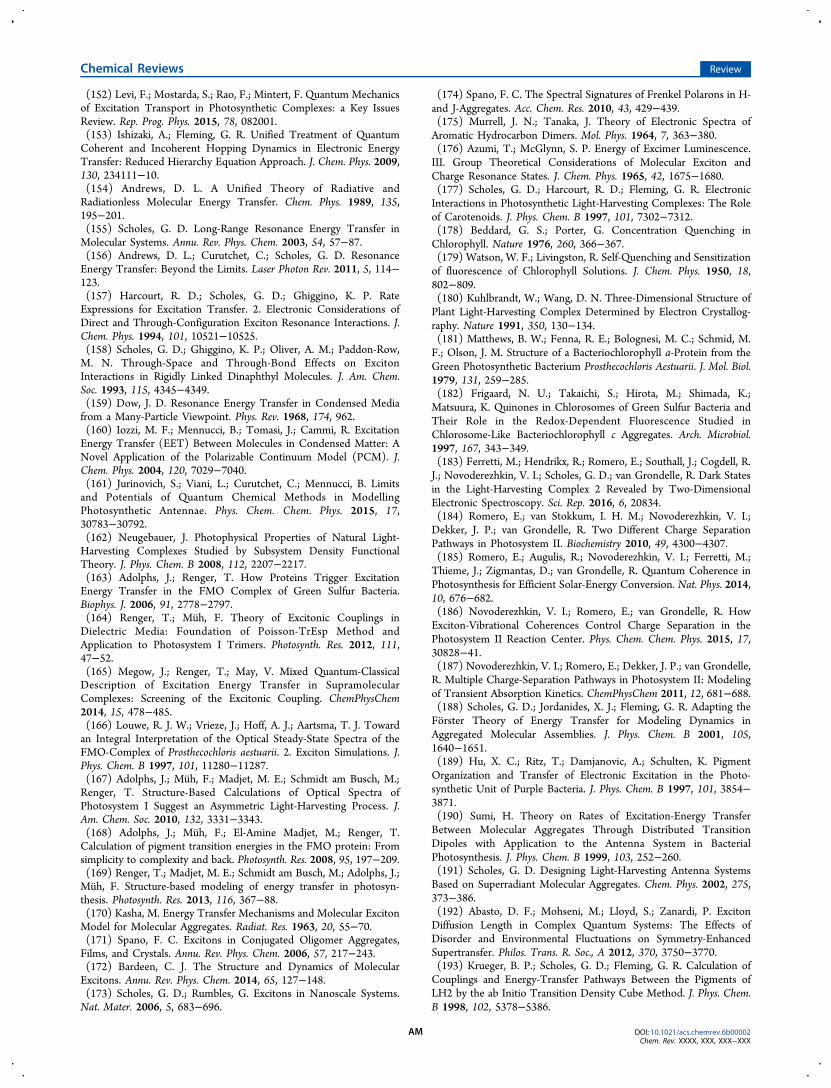
(152) Levi, F.; Mostarda, S.; Rao, F.; Mintert, F. Quantum Mechanicsof Excitation Transport in Photosynthetic Complexes: a Key IssuesReview. Rep. Prog. Phys. 2015, 78, 082001.(153) Ishizaki, A.; Fleming, G. R. Unified Treatment of QuantumCoherent and Incoherent Hopping Dynamics in Electronic EnergyTransfer: Reduced Hierarchy Equation Approach. J. Chem. Phys. 2009,130, 234111−10.(154) Andrews, D. L. A Unified Theory of Radiative andRadiationless Molecular Energy Transfer. Chem. Phys. 1989, 135,195−201.(155) Scholes, G. D. Long-Range Resonance Energy Transfer inMolecular Systems. Annu. Rev. Phys. Chem. 2003, 54, 57−87.(156) Andrews, D. L.; Curutchet, C.; Scholes, G. D. ResonanceEnergy Transfer: Beyond the Limits. Laser Photon Rev. 2011, 5, 114−123.(157) Harcourt, R. D.; Scholes, G. D.; Ghiggino, K. P. RateExpressions for Excitation Transfer. 2. Electronic Considerations ofDirect and Through-Configuration Exciton Resonance Interactions. J.Chem. Phys. 1994, 101, 10521−10525.(158) Scholes, G. D.; Ghiggino, K. P.; Oliver, A. M.; Paddon-Row,M. N. Through-Space and Through-Bond Effects on ExcitonInteractions in Rigidly Linked Dinaphthyl Molecules. J. Am. Chem.Soc. 1993, 115, 4345−4349.(159) Dow, J. D. Resonance Energy Transfer in Condensed Mediafrom a Many-Particle Viewpoint. Phys. Rev. 1968, 174, 962.(160) Iozzi, M. F.; Mennucci, B.; Tomasi, J.; Cammi, R. ExcitationEnergy Transfer (EET) Between Molecules in Condensed Matter: ANovel Application of the Polarizable Continuum Model (PCM). J.Chem. Phys. 2004, 120, 7029−7040.(161) Jurinovich, S.; Viani, L.; Curutchet, C.; Mennucci, B. Limitsand Potentials of Quantum Chemical Methods in ModellingPhotosynthetic Antennae. Phys. Chem. Chem. Phys. 2015, 17,30783−30792.(162) Neugebauer, J. Photophysical Properties of Natural Light-Harvesting Complexes Studied by Subsystem Density FunctionalTheory. J. Phys. Chem. B 2008, 112, 2207−2217.(163) Adolphs, J.; Renger, T. How Proteins Trigger ExcitationEnergy Transfer in the FMO Complex of Green Sulfur Bacteria.Biophys. J. 2006, 91, 2778−2797.(164) Renger, T.; Muh, F. Theory of Excitonic Couplings inDielectric Media: Foundation of Poisson-TrEsp Method andApplication to Photosystem I Trimers. Photosynth. Res. 2012, 111,47−52.(165) Megow, J.; Renger, T.; May, V. Mixed Quantum-ClassicalDescription of Excitation Energy Transfer in SupramolecularComplexes: Screening of the Excitonic Coupling. ChemPhysChem2014, 15, 478−485.(166) Louwe, R. J. W.; Vrieze, J.; Hoff, A. J.; Aartsma, T. J. Towardan Integral Interpretation of the Optical Steady-State Spectra of theFMO-Complex of Prosthecochloris aestuarii. 2. Exciton Simulations. J.Phys. Chem. B 1997, 101, 11280−11287.(167) Adolphs, J.; Muh, F.; Madjet, M. E.; Schmidt am Busch, M.;Renger, T. Structure-Based Calculations of Optical Spectra ofPhotosystem I Suggest an Asymmetric Light-Harvesting Process. J.Am. Chem. Soc. 2010, 132, 3331−3343.(168) Adolphs, J.; Muh, F.; El-Amine Madjet, M.; Renger, T.Calculation of pigment transition energies in the FMO protein: Fromsimplicity to complexity and back. Photosynth. Res. 2008, 95, 197−209.(169) Renger, T.; Madjet, M. E.; Schmidt am Busch, M.; Adolphs, J.;Muh, F. Structure-based modeling of energy transfer in photosyn-thesis. Photosynth. Res. 2013, 116, 367−88.(170) Kasha, M. Energy Transfer Mechanisms and Molecular ExcitonModel for Molecular Aggregates. Radiat. Res. 1963, 20, 55−70.(171) Spano, F. C. Excitons in Conjugated Oligomer Aggregates,Films, and Crystals. Annu. Rev. Phys. Chem. 2006, 57, 217−243.(172) Bardeen, C. J. The Structure and Dynamics of MolecularExcitons. Annu. Rev. Phys. Chem. 2014, 65, 127−148.(173) Scholes, G. D.; Rumbles, G. Excitons in Nanoscale Systems.Nat. Mater. 2006, 5, 683−696.
(174) Spano, F. C. The Spectral Signatures of Frenkel Polarons in H-and J-Aggregates. Acc. Chem. Res. 2010, 43, 429−439.(175) Murrell, J. N.; Tanaka, J. Theory of Electronic Spectra ofAromatic Hydrocarbon Dimers. Mol. Phys. 1964, 7, 363−380.(176) Azumi, T.; McGlynn, S. P. Energy of Excimer Luminescence.III. Group Theoretical Considerations of Molecular Exciton andCharge Resonance States. J. Chem. Phys. 1965, 42, 1675−1680.(177) Scholes, G. D.; Harcourt, R. D.; Fleming, G. R. ElectronicInteractions in Photosynthetic Light-Harvesting Complexes: The Roleof Carotenoids. J. Phys. Chem. B 1997, 101, 7302−7312.(178) Beddard, G. S.; Porter, G. Concentration Quenching inChlorophyll. Nature 1976, 260, 366−367.(179) Watson, W. F.; Livingston, R. Self-Quenching and Sensitizationof fluorescence of Chlorophyll Solutions. J. Chem. Phys. 1950, 18,802−809.(180) Kuhlbrandt, W.; Wang, D. N. Three-Dimensional Structure ofPlant Light-Harvesting Complex Determined by Electron Crystallog-raphy. Nature 1991, 350, 130−134.(181) Matthews, B. W.; Fenna, R. E.; Bolognesi, M. C.; Schmid, M.F.; Olson, J. M. Structure of a Bacteriochlorophyll a-Protein from theGreen Photosynthetic Bacterium Prosthecochloris Aestuarii. J. Mol. Biol.1979, 131, 259−285.(182) Frigaard, N. U.; Takaichi, S.; Hirota, M.; Shimada, K.;Matsuura, K. Quinones in Chlorosomes of Green Sulfur Bacteria andTheir Role in the Redox-Dependent Fluorescence Studied inChlorosome-Like Bacteriochlorophyll c Aggregates. Arch. Microbiol.1997, 167, 343−349.(183) Ferretti, M.; Hendrikx, R.; Romero, E.; Southall, J.; Cogdell, R.J.; Novoderezhkin, V. I.; Scholes, G. D.; van Grondelle, R. Dark Statesin the Light-Harvesting Complex 2 Revealed by Two-DimensionalElectronic Spectroscopy. Sci. Rep. 2016, 6, 20834.(184) Romero, E.; van Stokkum, I. H. M.; Novoderezhkin, V. I.;Dekker, J. P.; van Grondelle, R. Two Different Charge SeparationPathways in Photosystem II. Biochemistry 2010, 49, 4300−4307.(185) Romero, E.; Augulis, R.; Novoderezhkin, V. I.; Ferretti, M.;Thieme, J.; Zigmantas, D.; van Grondelle, R. Quantum Coherence inPhotosynthesis for Efficient Solar-Energy Conversion. Nat. Phys. 2014,10, 676−682.(186) Novoderezhkin, V. I.; Romero, E.; van Grondelle, R. HowExciton-Vibrational Coherences Control Charge Separation in thePhotosystem II Reaction Center. Phys. Chem. Chem. Phys. 2015, 17,30828−41.(187) Novoderezhkin, V. I.; Romero, E.; Dekker, J. P.; van Grondelle,R. Multiple Charge-Separation Pathways in Photosystem II: Modelingof Transient Absorption Kinetics. ChemPhysChem 2011, 12, 681−688.(188) Scholes, G. D.; Jordanides, X. J.; Fleming, G. R. Adapting theForster Theory of Energy Transfer for Modeling Dynamics inAggregated Molecular Assemblies. J. Phys. Chem. B 2001, 105,1640−1651.(189) Hu, X. C.; Ritz, T.; Damjanovic, A.; Schulten, K. PigmentOrganization and Transfer of Electronic Excitation in the Photo-synthetic Unit of Purple Bacteria. J. Phys. Chem. B 1997, 101, 3854−3871.(190) Sumi, H. Theory on Rates of Excitation-Energy TransferBetween Molecular Aggregates Through Distributed TransitionDipoles with Application to the Antenna System in BacterialPhotosynthesis. J. Phys. Chem. B 1999, 103, 252−260.(191) Scholes, G. D. Designing Light-Harvesting Antenna SystemsBased on Superradiant Molecular Aggregates. Chem. Phys. 2002, 275,373−386.(192) Abasto, D. F.; Mohseni, M.; Lloyd, S.; Zanardi, P. ExcitonDiffusion Length in Complex Quantum Systems: The Effects ofDisorder and Environmental Fluctuations on Symmetry-EnhancedSupertransfer. Philos. Trans. R. Soc., A 2012, 370, 3750−3770.(193) Krueger, B. P.; Scholes, G. D.; Fleming, G. R. Calculation ofCouplings and Energy-Transfer Pathways Between the Pigments ofLH2 by the ab Initio Transition Density Cube Method. J. Phys. Chem.B 1998, 102, 5378−5386.
Chemical Reviews Review
DOI: 10.1021/acs.chemrev.6b00002Chem. Rev. XXXX, XXX, XXX−XXX
AM
(194) Valku nas, L.; Kudzmauskas, S.; Juzeliu nas, G. ExcitationTransfer in Highly Concentrated Pseudoisocyanine Dye Solution. Sov.Phys.-Coll. 1985, 25, 41−47.(195) Grover, M.; Silbey, R. Exciton Migration in Molecular Crystals.J. Chem. Phys. 1971, 54, 4843−4851.(196) Jackson, B.; Silbey, R. On the Calculation of Transfer RatesBetween Impurity States in Solids. J. Chem. Phys. 1983, 78, 4193−4196.(197) Soules, T. F.; Duke, C. B. Resonant Energy Transfer betweenLocalized Electronic States in a Crystal. Phys. Rev. B 1971, 3, 262−274.(198) Kenkre, V. M. Relations Among Theories of ExcitationTransfer II: Influence of Spectral Features on Exciton Motion. Phys.Rev. B 1975, 12, 2150−2160.(199) Fleming, G. R.; van Grondelle, R. Femtosecond Spectroscopyof Photosynthetic Light-Harvesting Systems. Curr. Opin. Struct. Biol.1997, 7, 738−748.(200) Fotiadis, D.; Qian, P.; Philippsen, A.; Bullough, P. A.; Engel, A.;Hunter, C. N. Structural Analysis of the Reaction Center Light-Harvesting Complex I Photosynthetic Core Complex RhodospirillumRubrum Using Atomic Force Microscopy. J. Biol. Chem. 2004, 279,2063−2068.(201) Roszak, A. W.; Howard, T.; Southall, J.; Gardiner, A. T.; Law,C. J.; Isaacs, N. W.; Cogdell, R. J. Crystal Structure of the RC-LH1Core Complex from Rhodopseudomonas palustris. Science 2003, 302,1969−1972.(202) Jungas, C.; Ranck, J.-L.; Rigaud, J.-L.; Joliot, P.; Vermeglio, A.Supramolecular Organization of the Photosynthetic Apparatus ofRhodobacter sphaeroides. EMBO J. 1999, 18, 534−542.(203) Bahatyrova, S.; Frese, R. N.; Siebert, C. A.; Olsen, J. D.; van derWerf, K. O.; van Grondelle, R.; Niederman, R. A.; Bullough, P. A.;Otto, C.; Hunter, C. N. The Native Architecture of a PhotosyntheticMembrane. Nature 2004, 430, 1058−1062.(204) Scheuring, S.; Sturgis, J. N. Chromatic Adaptation ofPhotosynthetic Membranes. Science 2005, 309, 484−487.(205) Hunter, C. N.; Tucker, J. D.; Niederman, R. A. The Assemblyand Organisation of Photosynthetic Membranes in RhodobacterSphaeroides. Photochem. Photobiol. 2005, 4, 1023−1027.(206) Allen, J. P.; Feher, G.; Yeates, T. O.; Rees, D. C.; Deisenhofer,J.; Michel, H.; Huber, R. Structural Homology of Reaction Centersfrom Rhodopseudomonas Sphaeroides and Rhodopseudomonas Viridis asDetermined by X-Ray Diffraction. Proc. Natl. Acad. Sci. U. S. A. 1986,83, 8589−8593.(207) Scheuring, S.; Rigaud, J.-L.; Sturgis, J. N. Variable LH2Stoichiometry and Core Clustering in Native Membranes ofRhodospirillum Photometricum. EMBO J. 2004, 23, 4127−4133.(208) Jimenez, R.; van Mourik, F.; Yu, J. Y.; Fleming, G. R. Three-Pulse Photon Echo Measurements on LH1 and LH2 Complexes ofRhodobacter sphaeroides: A Nonlinear Spectroscopic Probe of EnergyTransfer. J. Phys. Chem. B 1997, 101, 7350−7359.(209) Chmeliov, J.; Songaila, E.; Rancova, O.; Gall, A.; Robert, B.;Abramavicius, D.; Valku nas, L. Excitons in the LH3 Complexes fromPurple Bacteria. J. Phys. Chem. B 2013, 117, 11058−11068.(210) Ketelaars, M.; Segura, J. M.; Oellerich, S.; de Ruijter, W. P. F.;Magis, G.; Aartsma, T. J.; Matsushita, M.; Schmidt, J.; Cogdell, R. J.;Kohler, J. Probing the Electronic Structure and ConformationalFlexibility of Individual Light-Harvesting 3 Complexes by OpticalSingle-Molecule Spectroscopy. J. Phys. Chem. B 2006, 110, 18710−18717.(211) McLuskey, K.; Prince, S. M.; Cogdell, R. J.; Isaacs, N. W. TheCrystallographic Structure of the B800−820 LH3 Light-HarvestingComplex from the Purple Bacteria Rhodopseudomonas AcidophilaStrain 7050. Biochemistry 2001, 40, 8783−8783.(212) McDermott, G.; Prince, S. M.; Freer, A. A.; Hawthornthwaite-Lawless, A. M.; Papiz, M. Z.; Cogdell, R. J.; Isaacs, N. W. CrystalStructure of an Integral Membrane Light-Harvesting Complex fromPhotosynthetic Bacteria. Nature 1995, 374, 517−521.(213) Evans, K.; Fordham-Skelton, A. P.; Mistry, H.; Reynolds, C. D.;Lawless, A. M.; Papiz, M. Z. A Bacteriophytochrome Regulates the
Synthesis of LH4 Complexes in Rhodopseudomonas Palustris. Photo-synth. Res. 2005, 85, 169−180.(214) Savage, H.; Cyrklaff, M.; Montoya, G.; Kuhlbrandt, W.;Sinning, I. Two-Dimensional Structure of Light Harvesting Complex II(LHII) from the Purple Bacterium Rhodovulum sulf idophilum andComparison with LHII from Rhodopseudomonas acidophila. Structure1996, 4, 243−252.(215) Permentier, H. P.; Neerken, S.; Overmann, J.; Amesz, J. ABacteriochlorophyll a Antenna Complex from Purple BacteriaAbsorbing at 963 nm. Biochemistry 2001, 40, 5573−5578.(216) Hoogewerf, G. J.; Jung, D. O.; Madigan, T. Evidence forLimited Species Diversity of Bacteriochlorophyll b-Containing PurpleNonsulfur Anoxygenic Phototrophs in Freshwater Habitats. FEMSMicrobiol. Lett. 2003, 218, 359−364.(217) Beatty, T. J. Genome Evolution of Photosynthetic Bacteria, 1st ed.;Advances in Botanical Research; Elsevier Inc.: New York, 2013; Vol.66.(218) Hirschler-Rea, A.; Matheron, R.; Riffaud, C.; Moune, S.;Eatock, C.; Herbert, R. A.; Willison, J. C.; Caumette, P. Isolation andCharacterization of Spirilloid Purple Phototrophic Bacteria FormingRed Layers in Microbial Mats of Mediterranean Salterns: Descriptionof Halorhodospira neutriphila sp. nov. and Emendation of the GenusHalorhodospira. Int. J. Syst. Evol. Microbiol. 2003, 53, 153−163.(219) Ma, F.; Kimura, Y.; Yu, L.-J.; Wang, P.; Ai, X.-C.; Wang, Z.-Y.;Zhang, J.-P. Specific Ca2+‑binding Motif in the LH1 Complex fromPhotosynthetic Bacterium Thermochromatium tepidum as Revealed byOptical Spectroscopy and Structural Modeling. FEBS J. 2009, 276,1739−1749.(220) Yang, F.; Yu, L.-J.; Wang, P.; Ai, X.-C.; Wang, Z.-Y.; Zhang, J.-P. Effects of Aggregation on the Excitation Dynamics of LH2 fromThermochromatium tepidum in Aqueous Phase and in Chromato-phores. J. Phys. Chem. B 2011, 115, 7906−7913.(221) (a) Hess, S.; Chachisvilis, M.; Timpmann, K.; Jones, M. R.;Fowler, G. J. S.; Hunter, C. N.; Sundstrom, V. Temporally andSpectrally Resolved Sub-Picosecond Energy Transfer within thePeripheral Antenna Complex (LH2) and from LH2 to the CoreAntenna Complex in Photosynthetic Purple Bacteria. Proc. Natl. Acad.Sci. U. S. A. 1995, 92, 12333−12337. (b) Hess, S.; Åkesson, E.;Cogdell, R.; Pullerits, T.; Sundstrom, V. Energy Transfer in SpectrallyInhomogeneous Light-Harvesting Pigment−Protein Complexes ofPurple Bacteria. Biophys. J. 1995, 69, 2211−2225.(222) Pullerits, T.; Sundstrom, V. Photosynthetic Light-HarvestingPigment−Protein Complexes: Toward Understanding How and Why.Acc. Chem. Res. 1996, 29, 381−389.(223) Alia, A.; Matysik, J.; Soede-Huijbregts, C.; Baldus, M.; Raap, J.;Lugtenburg, J.; Gast, P.; van Gorkom, H. J.; Hoff, A. J.; de Groot, H. J.M. Ultrahigh Field MAS NMR Dipolar Correlation Spectroscopy ofthe Histidine Residues in Light-Harvesting Complex II fromPhotosynthetic Bacteria Reveals Partial Internal Charge Transfer inthe B850/His Complex. J. Am. Chem. Soc. 2001, 123, 4803−4809.(224) Alia, A.; Ganapathy, S.; de Groot, H. J. M. Magic AngleSpinning (MAS) NMR: A New Tool to Study the Spatial andElectronic Structure of Photosynthetic Complexes. Photosynth. Res.2009, 102, 415−425.(225) Papiz, M. Z.; Prince, S. M.; Howard, T.; Cogdell, R. J.; Isaacs,N. W. The Structural and Thermal Motion of the B800-B850 LH2Complex from Rps. Acidophila at 2.0 Resolution and 100 K: NewStructural Features and Functionally Relevant Motions. J. Mol. Biol.2003, 326, 1523−1538.(226) Fowler, G. J.; Visschers, R. W.; Grief, G. G.; van Grondelle, R.;Hunter, C. N. Genetically Modified Photosynthetic AntennaComplexes with Blueshifted Absorbance Bands. Nature 1992, 355,848−850.(227) Madjet, M. E.; Abdurahman, A.; Renger, T. IntermolecularCoulomb Couplings from Ab Initio Electrostatic Potentials:Application to Optical Transitions of Strongly Coupled Pigments inPhotosynthetic Antennae and Reaction Centers. J. Phys. Chem. B 2006,110, 17268−17281.
Chemical Reviews Review
DOI: 10.1021/acs.chemrev.6b00002Chem. Rev. XXXX, XXX, XXX−XXX
AN
(228) Cogdell, R. J.; Howard, T. D.; Isaacs, N. W.; McLuskey, K.;Gardiner, A. T. Structural Factors Which Control the Position of theQ(Y) Absorption Band of Bacteriochlorophyll a in Purple BacterialAntenna Complexes. Photosynth. Res. 2002, 74, 135−141.(229) Mascle-Allemand, C.; Duquesne, K.; Lebrun, R.; Scheuring, S.;Sturgis, J. N. Antenna Mixing in Photosynthetic Membranes fromPhaeospirillum Molischianum. Proc. Natl. Acad. Sci. U. S. A. 2010, 107,5357−5362.(230) Brotosudarmo, T. H. P.; Collins, A. M.; Gall, A.; Roszak, A. W.;Gardiner, A. T.; Blankenship, R. E.; Cogdell, R. J. The Light IntensityUnder Which Cells Are Grown Controls the Type of Peripheral Light-Harvesting Complexes That are Assembled in a Purple PhotosyntheticBacterium. Biochem. J. 2011, 440, 51−61.(231) Trissl, H.-W.; Law, C. J.; Cogdell, R. J. Uphill Energy Transferin LH2-Containing Purple Bacteria at Room Temperature. Biochim.Biophys. Acta, Bioenerg. 1999, 1412, 149−172.(232) Gall, A.; Robert, B. Characterization of the Different PeripheralLight-Harvesting Complexes from High- and Low-Light Grown Cellsfrom Rhodopseudomonas palustris. Biochemistry 1999, 38, 5185−5190.(233) Sener, M. K.; Strumpfer, J.; Timney, J. A.; Freiberg, A.; Hunter,C. N.; Schulten, K. Photosynthetic Vesicle Architecture andConstraints on Efficient Energy Harvesting. Biophys. J. 2010, 99,67−75.(234) Sener, M. K.; Olsen, J. D.; Hunter, C. N.; Schulten, K. Atomic-Level Structural and functional model of a Bacterial PhotosyntheticMembrane Vesicle. Proc. Natl. Acad. Sci. U. S. A. 2007, 104, 15723−15728.(235) Adams, P. G.; Hunter, C. N. Adaptation of IntracytoplasmicMembranes to Altered Light Intensity in Rhodobacter Sphaeroides.Biochim. Biophys. Acta, Bioenerg. 2012, 1817, 1616−1627.(236) Hu, X.; Ritz, T.; Damjanovic, A.; Autenrieth, F.; Schulten, K.Photosynthetic Apparatus of Purple Bacteria. Q. Rev. Biophys. 2002, 35,1−62.(237) Sauer, K.; Cogdell, R. J.; Prince, S. M.; Freer, A.; Isaacs, N. W.;Scheer, H. Structure-Based Calculations of the Optical Spectra of theLH2 Bacterio-Chlorophyll-Protein Complex from RhodopseudomonasAcidophila. Photochem. Photobiol. 1996, 64, 564−576.(238) Kennis, J. T. M.; Streltsov, A. M.; Vulto, S. I. E.; Aartsma, T. J.;Nozawa, T.; Amesz, J. Femtosecond Dynamics in Isolated LH2Complexes of Various Species of Purple Bacteria. J. Phys. Chem. B1997, 101, 7827−7834.(239) Scholes, G. D.; Gould, I. R.; Cogdell, R. J.; Fleming, G. R. AbInitio Molecular Orbital Calculations of Electronic Couplings in theLH2 Bacterial Light-Harvesting Complex of Rps. Acidophila. J. Phys.Chem. B 1999, 103, 2543−2553.(240) Visschers, R. W.; Chang, M. C.; van Mourik, F.; Parkesloach, P.S.; Heller, B. A.; Loach, P. A.; van Grondelle, R. FluorescencePolarization and Low-Temperature Absorption Spectroscopy of aSubunit Form of Light-Harvesting Complex-I from Purple Photo-synthetic Bacteria. Biochemistry 1991, 30, 5734−5742.(241) van Mourik, F.; van der Oord, C. J. R.; Visscher, K. J.;Parkesloach, P. S.; Loach, P. A.; Visschers, R. W.; van Grondelle, R.Exciton Interactions in the Light-Harvesting Antenna of Photo-synthetic Bacteria Studies with Triplet-Singlet Spectroscopy andSinglet-Triplet Annihilation on the B820 Subunit Form ofRhodospirillum-Rubrum. Biochim. Biophys. Acta, Bioenerg. 1991, 1059,111−119.(242) Koolhaas, M. H. C.; Frese, R. N.; Fowler, G. J. S.; Bibby, T. S.;Georgakopoulou, S.; van der Zwan, G.; Hunter, C. N.; van Grondelle,R. Identification of the Upper Exciton Component of the B850Bacteriochlorophylls of the LH2 Antenna Complex, Using a B800-Free Mutant of Rhodobacter Sphaeroides. Biochemistry 1998, 37, 4693−4698.(243) Monshouwer, R.; Abrahamsson, M.; van Mourik, F.; vanGrondelle, R. Superradiance and Exciton Delocalization in BacterialPhotosynthetic Light-Harvesting Systems. J. Phys. Chem. B 1997, 101,7241−7248.
(244) Novoderezhkin, V. I.; Razjivin, A. P. Exciton Dynamics inCircular Aggregates: Application to Antenna of Photosynthetic PurpleBacteria. Biophys. J. 1995, 68, 1089−1100.(245) Fidder, H.; Knoester, J.; Wiersma, D. A. Optical Properties ofDisordered Molecular Aggregates: A Numerical Study. J. Chem. Phys.1991, 95, 7880−7890.(246) Bakalis, L. D.; Coca, M.; Knoester, J. Optical Line Shapes ofDynamically Disordered Ring Aggregates. J. Chem. Phys. 1999, 110,2208−2218.(247) Mostovoy, M. V.; Knoester, J. Statistics of Optical Spectra fromSingle-Ring Aggregates and Its Application to LH2. J. Phys. Chem. B2000, 104, 12355−12364.(248) van Oijen, A. M.; Ketelaars, M.; Kohler, J.; Aartsma, T. J.;Schmidt, J. Unraveling the Electronic Structure of IndividualPhotosynthetic Pigment−Protein Complexes. Science 1999, 285,400−402.(249) Wu, H.-M.; Reddy, N. R. S.; Small, G. J. Direct Observationand Hole Burning of the Lowest Exciton Level (B870) of the LH2Antenna Complex of Rhodopseudomonas Acidophila (Strain 10050). J.Phys. Chem. B 1997, 101, 651−656.(250) Berlin, Y.; Burin, A.; Friedrich, J.; Kohler, J. Low TemperatureSpectroscopy of Proteins. Part II: Experiments with Single ProteinComplexes. Phys. Life. Rev. 2007, 4, 64−89.(251) Kruger, T. P. J.; Wientjes, E.; Croce, R.; van Grondelle, R.Conformational Switching Explains the Intrinsic Multifunctionality ofPlant Light-Harvesting Complexes. Proc. Natl. Acad. Sci. U. S. A. 2011,108, 13516−13521.(252) Oellerich, S.; Kohler, J. Low-Temperature Single-MoleculeSpectroscopy on Photosynthetic Pigment−Protein Complexes fromPurple Bacteria. Photosynth. Res. 2009, 101, 171−179.(253) Novoderezhkin, V. I.; Rutkauskas, D.; van Grondelle, R.Dynamics of the Emission Spectrum of a Single LH2 Complex:Interplay of Slow and Fast Nuclear Motions. Biophys. J. 2006, 90,2890−2902.(254) Rutkauskas, D.; Novoderezkhin, V.; Cogdell, R. J.; vanGrondelle, R. Fluorescence Spectral Fluctuations of Single LH2Complexes from Rhodopseudomonas acidophila Strain 10050. Bio-chemistry 2004, 43, 4431−4438.(255) Kunz, R.; Timpmann, K.; Southall, J.; Cogdell, R. J.; Freiberg,A.; Kohler, J. Fluctuations in the Electron−Phonon Coupling of aSingle Chromoprotein. Angew. Chem., Int. Ed. 2013, 52, 8726−9730.(256) Monshouwer, R.; van Grondelle, R. Excitations and Excitons inBacterial Light-Harvesting Complexes. Biochim. Biophys. Acta, Bioenerg.1996, 1275, 70−75.(257) Meier, T.; Zhao, Y.; Chernyak, V.; Mukamel, S. Polarons,Localization and Excitonic Coherence in Superradiance of BiologicalAntenna Complexes. J. Chem. Phys. 1997, 107, 3876−3893.(258) Palacios, M. A.; de Weerd, F. L.; Ihalainen, J. A.; van Grondelle,R.; van Amerongen, H. Superradiance and Exciton (De)localization inLight-Harvesting Complex II from Green Plants? J. Phys. Chem. B2002, 106, 5782−5787.(259) Pullerits, T.; Chachisvilis, M.; Sundstrom, V. ExcitonDelocalization Length in the B850 Antenna of RhodobacterSphaeroides. J. Phys. Chem. 1996, 100, 10787−10792.(260) Jimenez, R.; Dikshit, S. N.; Bradforth, S. E.; Fleming, G. R.Electronic Excitation Transfer in the LH2 Complex of RhodobacterSphaeroides. J. Phys. Chem. 1996, 100, 6825−6834.(261) Novoderezhkin, V.; Monshouwer, R.; van Grondelle, R.Exciton (de)localization in the LH2 Antenna of RhodobacterSphaeroides as Revealed by Relative Difference Absorption Measure-ments of the LH2 Antenna and the B820 Subunit. J. Phys. Chem. B1999, 103, 10540−10548.(262) Sundstrom, V.; Pullerits, T.; van Grondelle, R. PhotosyntheticLight-Harvesting: Reconciling Dynamics and Structure of PurpleBacterial LH2 Reveals Function of Photosynthetic Unit. J. Phys. Chem.B 1999, 103, 2327−2346.(263) van Grondelle, R.; Novoderezhkin, V. I. Dynamics ofExcitation Energy Transfer in the LH1 and LH2 Light-Harvesting
Chemical Reviews Review
DOI: 10.1021/acs.chemrev.6b00002Chem. Rev. XXXX, XXX, XXX−XXX
AO
Complexes of Photosynthetic Bacteria. Biochemistry 2001, 40, 15057−15059.(264) Ma, Y. Z.; Cogdell, R. J.; Gillbro, T. Energy Transfer andExciton Annihilation in the B800−850 Antenna Complex of thePhotosynthetic Purple Bacterium Rhodopseudomonas Acidophila(Strain 10050). A Femtosecond Transient Absorption Study. J. Phys.Chem. B 1997, 101, 1087−1095.(265) Bergstrom, H.; Westerhuis, W. H. J.; Sundstrom, V.; vanGrondelle, R.; Niederman, R. A.; Gillbro, T. Energy Transfer withinthe Isolated B875 Light-Harvesting Pigment−Protein Complex ofRhodobacter sphaeroides at 77 K Studied by Picosecond AbsorptionSpectroscopy. FEBS Lett. 1988, 233, 12−16.(266) Hunter, C. N.; Bergstrom, H.; van Grondelle, R.; Sundstrom,V. Energy-Transfer Dynamics in Three Light-Harvesting Mutants ofRhodobacter sphaeroides: A Picosecond Spectroscopy Study. Biochem-istry 1990, 29, 3203−3207.(267) Cleary, L.; Chen, H.; Chuang, C.; Silbey, R. J.; Cao, J. OptimalFold Symmetry of LH2 Rings on a Photosynthetic Membrane. Proc.Natl. Acad. Sci. U. S. A. 2013, 110, 8537−8542.(268) Beekman, L. M. P.; van Mourik, F.; Jones, M. R.; Visser, H. M.;Hunter, C. N.; van Grondelle, R. Trapping Kinetics in Mutants of thePhotosynthetic Purple Bacterium Rhodobacter Sphaeroides: Influenceof the Charge Separation Rate and Consequences for the Rate-Limiting Step in the Light-Harvesting Process. Biochemistry 1994, 33,3143−3147.(269) Freiberg, A.; Allen, J. P.; Williams, J.; Woodbury, N. W. EnergyTrapping and Detrapping by Wild Type and Mutant Reaction Centersof Purple Non-Sulfur Bacteria. Photosynth. Res. 1996, 48, 309−319.(270) Visscher, K. J.; Bergstrom, H.; Sundstrom, V.; Hunter, C. N.;van Grondelle, R. Temperature Dependence of Energy Transfer fromthe Long Wavelength Antenna BChl-896 to the Reaction Center inRhodospirillum Rubrum, Rhodobacter Sphaeroides (w.t. and M21Mutant) from 77 to 177K, Studied by Picosecond AbsorptionSpectroscopy. Photosynth. Res. 1989, 22, 211−217.(271) Allen, J. F.; Mullineaux, C. W. Probing the Mechanism of StateTransitions in Oxygenic Photosynthesis by Chlorophyll FluorescenceSpectroscopy, Kinetics and Imaging. In Chlorophyll a Fluorescence: ASignature of Photosynthesis; Papageorgiou, G.C., Govindjee, Eds.;Advances in Photosynthesis and Respiration; Springer: Dordrecht,The Netherlands, 2004; Vol. 19, pp 447−461.(272) Papageorgiou, G. C.; Govindjee. Photosystem II Fluorescence:Slow Changes - Scaling from the Past. J. Photochem. Photobiol., B 2011,104, 258−70.(273) Wlodarczyk, L. M.; Snellenburg, J. J.; Ihalainen, J. A.; vanGrondelle, R.; van Stokkum, I. H. M.; Dekker, J. P. FunctionalRearrangement of the Light-Harvesting Antenna upon StateTransitions in a Green Alga. Biophys. J. 2015, 108, 261−271.(274) Demmig-Adams, B., Garab, G., Adams, W. W., III, Govindjee,Eds. Non-Photochemical Quenching and Thermal Energy Dissipation inPlants, Algae and Cyanobacteria; Springer: Dordrecht, The Nether-lands, 2014; Vol. 40.(275) Liu, Z.; Yan, H.; Wang, K.; Kuang, T.; Zhang, J.; Gui, L.; An,X.; Chang, W. Crystal Structure of Spinach Major Light-HarvestingComplex at 2.72 Å Resolution. Nature 2004, 428, 287−292.(276) Novoderezhkin, V. I.; Palacios, M. A.; van Amerongen, H.; vanGrondelle, R. Excitation Dynamics in the LHCII Complex of HigherPlants: Modeling Based on the 2.72 Å Crystal Structure. J. Phys. Chem.B 2005, 109, 10493−10504.(277) Novoderezhkin, V. I.; Marin, A.; van Grondelle, R. Intra- andInter-Monomeric Transfers in the Light Harvesting LHCII Complex:the Redfield−Forster Picture. Phys. Chem. Chem. Phys. 2011, 13,17093−17103.(278) Novoderezhkin, V. I.; Palacios, M. A.; van Amerongen, H.; vanGrondelle, R. Energy-Transfer Dynamics in the LHCII Complex ofHigher Plants: Modified Redfield Approach. J. Phys. Chem. B 2004,108, 10363−10375.(279) Croce, R.; Muller, M. G.; Bassi, R.; Holzwarth, A. R.Carotenoid-to-Chlorophyll Energy Transfer in Recombinant Major
Light-Harvesting Complex (LHCII) of Higher Plants. I. FemtosecondTransient Absorption Measurements. Biophys. J. 2001, 80, 901−915.(280) Gradinaru, C. C.; van Stokkum, I. H. M.; Pascal, A. A.; vanGrondelle, R.; van Amerongen, H. Identifying the Pathways of EnergyTransfer between Carotenoids and Chlorophylls in LHCII and CP29.A Multicolor Pump-Probe Study. J. Phys. Chem. B 2000, 104, 9330−9342.(281) Connelly, J. P.; Muller, M. G.; Hucke, M.; Gatzen, G.;Mullineaux, C. W.; Ruban, A. V.; Horton, P.; Holzwarth, A. R.Ultrafast Spectroscopy of Trimeric Light-Harvesting Complex II fromHigher Plants. J. Phys. Chem. B 1997, 101, 1902−1909.(282) Renger, T.; Madjet, M. E.; Knorr, A.; Muh, F. How theMolecular Structure Determines the Flow of Excitation Energy inPlant Light-Harvesting Complex II. J. Plant Physiol. 2011, 168, 1497−1509.(283) Kreisbeck, C.; Kramer, T.; Aspuru-Guzik, A. Scalable High-Performance Algorithm for the Simulation of Exciton-Dynamics.Application to the Light Harvesting Complex II in the Presence ofResonant Vibrational Modes. J. Chem. Theory Comput. 2014, 10,4045−4054.(284) Ruban, A. V.; Johnson, M. P.; Duffy, C. D. P. ThePhotoprotective Molecular Switch in the Photosystem II Antenna.Biochim. Biophys. Acta, Bioenerg. 2012, 1817, 167−181.(285) Ruban, A. V.; Berera, R.; Ilioaia, C.; van Stokkum, I. H. M.;Kennis, J. T. M.; Pascal, A.; van Amerongen, H.; Robert, B.; Horton,P.; van Grondelle, R. Identification of a Mechanism of PhotoprotectiveEnergy Dissipation in Higher Plants. Nature 2007, 450, 575−578.(286) Miloslavina, Y.; Wehner, A.; Lambrev, P. H.; Wientjes, E.;Reus, M.; Garab, G.; Croce, R.; Holzwarth, A. R. Far-RedFluorescence: a Direct Spectroscopic Marker for LHCII OligomerFormation in Non-Photochemical Quenching. FEBS Lett. 2008, 582,3625−3631.(287) Buchel, C. Fucoxanthin-Chlorophyll Proteins in Diatoms: 18and 19 kDa Subunits Assemble into Different Oligomeric States.Biochemistry 2003, 42, 13027−13034.(288) Hofmann, E.; Wrench, P. M.; Sharples, F. P.; Hiller, R. G.;Welte, W.; Diederichs, K. Structural Basis of Light Harvesting byCarotenoids: Peridinin-Chlorophyll-Protein from Amphidinium carter-ae. Science 1996, 272, 1788−1791.(289) Frank, H. A.; Cogdell, R. J. Carotenoids in Photosynthesis.Photochem. Photobiol. 1996, 63, 257−264.(290) Buchel, C. Fucoxanthin-Chlorophyll-Proteins and Non-Photo-chemical Fluorescence Quenching of Diatoms. In Non-PhotochemicalQuenching and Energy Dissipation in Plants, Algae and Cyanobacteria;Demmig-Adams, B., Garab, G., Adams, W. W., III, Govindjee, Eds.;Advances in Photosynthesis and Respiration; Springer: Dordrecht,The Netherlands, 2014; Vol. 40, Chapter 11, pp 259−275.(291) Papagiannakis, E.; van Stokkum, I. H. M.; Fey, H.; Buchel, C.;van Grondelle, R. Spectroscopic Characterization of the ExcitationEnergy Transfer in the Fucoxanthin−Chlorophyll Protein of Diatoms.Photosynth. Res. 2005, 86, 241−250.(292) Akimoto, S.; Teshigahara, A.; Yokono, M.; Mimuro, M.;Nagao, R.; Tomo, T. Excitation Relaxation Dynamics and EnergyTransfer in Fucoxanthin−Chlorophyll a/c-Protein Complexes, Probedby Time-Resolved Fluorescence. Biochim. Biophys. Acta, Bioenerg.2014, 1837, 1514−1521.(293) Gildenhoff, N.; Amarie, S.; Gundermann, K.; Beer, A.; Buchel,C.; Wachtveitl, J. Oligomerization and Pigmentation DependentExcitation Energy Transfer in Fucoxanthin-Chlorophyll Proteins.Biochim. Biophys. Acta, Bioenerg. 2010, 1797, 543−549.(294) Gelzinis, A.; Butkus, V.; Songaila, E.; Augulis, R.; Gall, A.;Buchel, C.; Robert, B.; Abramavicius, D.; Zigmantas, D.; Valku nas, L.Mapping Energy Transfer Channels in Fucoxanthin−ChlorophyllProtein Complex. Biochim. Biophys. Acta, Bioenerg. 2015, 1847, 241−247.(295) Songaila, E.; Augulis, R.; Gelzinis, A.; Butkus, V.; Gall, A.;Buchel, C.; Robert, B.; Zigmantas, D.; Abramavicius, D.; Valku nas, L.Ultrafast Energy Transfer from Chlorophyll c2 to Chlorophyllin
Chemical Reviews Review
DOI: 10.1021/acs.chemrev.6b00002Chem. Rev. XXXX, XXX, XXX−XXX
AP
Fucoxanthin−Chlorophyll Protein Complex. J. Phys. Chem. Lett. 2013,4, 3590−3595.(296) Polivka, T.; Frank, H. A. Molecular Factors ControllingPhotosynthetic Light Harvesting by Carotenoids. Acc. Chem. Res. 2010,43, 1125−1134.(297) Zigmantas, D.; Hiller, R. G.; Sundstrom, V.; Polivka, T.Carotenoid to Chlorophyll Energy Transfer in the Peridinin−Chlorophyll-a−Protein Complex Involves an Intramolecular ChargeTransfer State. Proc. Natl. Acad. Sci. U. S. A. 2002, 99, 16760−16765.(298) van Stokkum, I. H. M.; Papagiannakis, E.; Vengris, M.;Salverda, J. M.; Polivka, T.; Zigmantas, D.; Larsen, D. S.; Lampoura, S.S.; Hiller, R. G.; van Grondelle, R. Inter-Pigment Interactions in thePeridinin Chlorophyll Protein Studied by Global and Target Analysisof Time Resolved Absorption Spectra. Chem. Phys. 2009, 357, 70−78.(299) Bonetti, C.; Alexandre, M. T. A.; van Stokkum, I. H. M.; Hiller,R. G.; Groot, M. L.; van Grondelle, R.; Kennis, J. T. M. Identificationof Excited-State Energy Transfer and Relaxation Pathways in thePeridinin−Chlorophyll Complex: An Ultrafast Mid-Infrared Study.Phys. Chem. Chem. Phys. 2010, 12, 9256−9266.(300) Govindjee; Shevela, D. Adventures with Cyanobacteria: APersonal Perspective. Front. Plant Sci. 2011, 2, 1−17.(301) Blankenship, R. E.; Matsuura, K. Antenna Complexes fromGreen Photosynthetic Bacteria. In Light-Harvesting Antennas inPhotosynthesis; Green, B. R., Parson, W. W., Eds.; Advances inPhotosynthesis and Respiration; Kluwer Academic Publishers:Dordrecht, The Netherlands, 2003; Vol. 13, Chapter 6, pp 195−217.(302) Ganapathy, S.; Oostergetel, G. T.; Wawrzyniak, P. K.; Reus,M.; Gomez Maqueo Chew, A.; Buda, F.; Boekema, E. J.; Bryant, D. A.;Holzwarth, A. R.; de Groot, H. J. M. Alternating Syn-AntiBacteriochlorophylls form Concentric Helical Nanotubes in Chlor-osomes. Proc. Natl. Acad. Sci. U. S. A. 2009, 106, 8525−8530.(303) Oostergetel, G. T.; van Amerongen, H.; Boekema, E. J. TheChlorosome: A Prototype for Efficient Light Harvesting in Photosyn-thesis. Photosynth. Res. 2010, 104, 245−255.(304) Beatty, J. T.; Overmann, J.; Lince, M. T.; Manske, A. K.; Lang,A. S.; Blankenship, R. E.; van Dover, C. L.; Martinson, T. A.; Plumley,F. G. An Obligately Photosynthetic Bacterial Anaerobe from a Deep-Sea Hydrothermal Vent. Proc. Natl. Acad. Sci. U. S. A. 2005, 102,9306−9310.(305) van Dorssen, R. J.; Gerola, P. D.; Olson, J. M.; Amesz, J.Optical and Structural Properties of Chlorosomes of the Photo-synthetic Green Sulfur Bacterium Chlorobium Limicola. Biochim.Biophys. Acta, Bioenerg. 1986, 848, 77−82.(306) Wang, J.; Brune, D. C.; Blankenship, R. E. Ejects of Oxidantsand Reductants on the Effciency of Excitation Transfer in GreenPhotosynthetic Bacteria. Biochim. Biophys. Acta, Bioenerg. 1990, 1015,457−463.(307) Frigaard, N.-U.; Matsuura, K. Oxygen Uncouples LightAbsorption by the Chlorosome Antenna and Photosynthetic ElectronTransfer in the Green Sulfur Bacterium Chlorobium Tepidum. Biochim.Biophys. Acta, Bioenerg. 1999, 1412, 108−117.(308) Alster, J.; Zupcanova, A.; Vacha, F.; Psencik, J. Effect ofQuinones on Formation and Properties of Bacteriochlorophyll cAggregates. Photosynth. Res. 2008, 95, 183−189.(309) Frigaard, N. U.; Tokita, S.; Matsuura, K. Exogenous QuinonesInhibit Photosynthetic Electron Transfer in Chlorof lexus aurantiacus bySpecific Quenching of the Excited Bacteriochlorophyll c antenna.Biochim. Biophys. Acta, Bioenerg. 1999, 1413, 108−116.(310) Tokita, S.; Frigaard, N.-U.; Hirota, M.; Shimada, K.; Matsuura,K. Quenching of Bacteriochlorophyll Fluorescence in Chlorosomesfrom Chloroflexus aurantiacus by Exogenous Quinones. Photochem.Photobiol. 2000, 72, 345−350.(311) Psencík, J.; Polívka, T.; Nemec, P.; Dian, J.; Kudrna, J.; Maly,P.; Hala, J. Fast Energy Transfer and Exciton Dynamics inChlorosomes of the Green Sulfur Bacterium Chlorobium tepidum. J.Phys. Chem. A 1998, 102, 4392−4398.(312) Prokhorenko, V. I.; Steensgaard, D. B.; Holzwarth, A. R.Exciton Dynamics in the Chlorosomal Antennae of the Green Bacteria
Chlorof lexus aurantiacus and Chlorobium tepidum. Biophys. J. 2000, 79,2105−2120.(313) Savikhin, S.; Zhu, Y.; Lin, S.; Blankenship, R. E.; Struve, W. S.Femtosecond Spectroscopy of Chlorosome Antennas from the GreenPhotosynthetic Bacterium. J. Phys. Chem. 1994, 98, 10322−10334.(314) Savikhin, S.; Zhu, Y.; Blankenship, R. E.; Struve, W. S. UltrafastEnergy Transfer in Chlorosomes from the Green PhotosyntheticBacterium. J. Phys. Chem. 1996, 100, 3320−3322.(315) Miller, M.; Cox, R. P.; Gillbro, T. Energy Transfer Kinetics inChlorosomes from Chloroflexus Aurantiacus: Studies Using Pico-second Absorbance Spectroscopy. Biochim. Biophys. Acta, Bioenerg.1991, 1057, 187−194.(316) Huijser, A.; Marek, P.; Savenije, T.; Siebbeles, L. D. A.;Scherer, T.; Hauschild, R.; Szmytkowski, J.; Kalt, H.; Hahn, H.;Balaban, T. S. Photosensitization of TiO2 and SnO2 by Artificial Self-Assembling Mimics of the Natural Chlorosomal Bacteriochlorophylls.J. Phys. Chem. C 2007, 111, 11726−11733.(317) Grossman, A. R.; Schaefer, M. R.; Chiang, G. G.; Collier, J. L.The Phycobilisome, a Light-Harvesting Complex Responsive toEnvironmental Conditions. Microbiol. Rev. 1993, 57, 725−749.(318) MacColl, R. Cyanobacterial Phycobilisomes. J. Struct. Biol.1998, 124, 311−334.(319) Suter, G. W.; Holzwarth, A. R. A Kinetic Model for the EnergyTransfer in Phycobilisomes. Biophys. J. 1987, 52, 673−683.(320) Suter, G. W.; Mazzola, P.; Wendler, J.; Holzwarth, A. R.Fluorescence Decay Kinetics in Phycobilisomes Isolated from theBluegreen Alga Synechococcus 6301. Biochim. Biophys. Acta, Bioenerg.1984, 766, 269−276.(321) Krumova, S. B.; Laptenok, S. P.; Borst, J. W.; Ughy, B.;Gombos, Z.; Ajlani, G.; van Amerongen, H. Monitoring Photosyn-thesis in Individual Cells of Synechocystis sp. PCC 6803 on aPicosecond Timescale. Biophys. J. 2010, 99, 2006−2015.(322) Tian, L.; van Stokkum, I. H. M.; Koehorst, R. B. M.; Jongerius,A.; Kirilovsky, D.; van Amerongen, H. Site, Rate, and Mechanism ofPhotoprotective Quenching in Cyanobacteria. J. Am. Chem. Soc. 2011,133, 18304−18311.(323) Gillbro, T.; Sharkov, A. V.; Kryukov, I. V.; Khoroshilov, E. V.;Kryukov, P. G.; Fischer, R.; Scheer, H. Forster Energy TransferBetween Neighbouring Chromophoresin C-Phycocyanin Trimers.Biochim. Biophys. Acta, Bioenerg. 1993, 1140, 321−326.(324) Kirilovsky, D.; Kana, R.; Prasil, O. Mechanisms ModulatingEnergy Arriving at Reaction Centers in Cyanobacteria. In Non-Photochemical Quenching and Thermal Energy Dissipation in Plants,Algae and Cyanobacteria; Demmig-Adams, B., Garab, G., Adams, W.W., III, Govindjee, Eds.; Advances in Photosynthesis and Respiration;Springer: Dordrecht, The Netherlands, 2014; Vol. 40, Chapter 22, pp471−501.(325) Spear-Bernstein, L.; Miller, K. R. Unique Location of thePhycobiliprotein Light-Harvesting Pigment in the Cryptophyceae. J.Phycol. 1989, 25, 412−419.(326) Novoderezhkin, V. I.; van Grondelle, R. Physical Origins andModels of Energy Transfer in Photosynthetic Light-Harvesting. Phys.Chem. Chem. Phys. 2010, 12, 7352−7365.(327) Curutchet, C.; Novoderezhkin, V. I.; Kongsted, J.; Munoz-Losa, A.; van Grondelle, R.; Scholes, G. D.; Mennucci, B. Energy Flowin the Cryptophyte PE545 Antenna Is Directed by Bilin PigmentConformation. J. Phys. Chem. B 2013, 117, 4263−4273.(328) Doust, A. B.; van Stokkum, I. H.; Larsen, D. S.; Wilk, K. E.;Curmi, P. M.; van Grondelle, R.; Scholes, G. D. Mediation of UltrafastLight-Harvesting by a Central Dimer in Phycoerythrin 545 Studied byTransient Absorption and Global Analysis. J. Phys. Chem. B 2005, 109,14219−14226.(329) Marin, A.; Doust, A. B.; Scholes, G. D.; Wilk, K. E.; Curmi, P.M. G.; van Stokkum, I. H. M.; van Grondelle, R. Flow of ExcitationEnergy in the Cryptophyte Light-Harvesting Antenna Phycocyanin645. Biophys. J. 2011, 101, 1004−1013.(330) van der Weij-De Wit, C. D.; Doust, A. B.; van Stokkum, I. H.M.; Dekker, J. P.; Wilk, K. E.; Curmi, P. M. G.; Scholes, G. D.; vanGrondelle, R. How Energy Funnels from the Phycoerythrin Antenna
Chemical Reviews Review
DOI: 10.1021/acs.chemrev.6b00002Chem. Rev. XXXX, XXX, XXX−XXX
AQ
Complex to Photosystem I and Photosystem II in CryptophyteRhodomonas CS24 Cells. J. Phys. Chem. B 2006, 110, 25066−25073.(331) Collini, E.; Wong, C. Y.; Wilk, K. E.; Curmi, P. M. G.; Brumer,P.; Scholes, G. D. Coherently Wired Light-Harvesting in Photo-synthetic Marine Algae at Ambient Temperature. Nature 2010, 463,644−648.(332) Wong, C. Y.; Alvey, R. M.; Turner, D. B.; Wilk, K. E.; Bryant,D. A.; Curmi, P. M. G.; Silbey, R. J.; Scholes, G. D. ElectronicCoherence Lineshapes Reveal Hidden Excitonic Correlations inPhotosynthetic Light Harvesting. Nat. Chem. 2012, 4, 396−404.(333) Turner, D. B.; Dinshaw, R.; Lee, K.-K.; Belsley, M. S.; Wilk, K.E.; Curmi, P. M. G.; Scholes, G. D. Quantitative Investigations ofQuantum Coherence for a Light-Harvesting Protein at ConditionsSimulating Photosynthesis. Phys. Chem. Chem. Phys. 2012, 14, 4857−4874.(334) Calhoun, T. R.; Fleming, G. R. Quantum Coherence inPhotosynthetic Complexes. Phys. Status Solidi B 2011, 248, 833−838.(335) Fassioli, G.; Dinshaw, R.; Arpin, P. C.; Scholes, G. D.Photosynthetic Light Harvesting: Excitons and Coherence. J. R. Soc.,Interface 2014, 11, 20130901.(336) Britton, G., Liaaen-Jensen, S., Pfander, H., Eds. CarotenoidsHandbook; Birkhauser Verlag AG: Basel, Switzerland, 2004.(337) Britton, G.; Functions of Intact Carotenoids. In Carotenoids,Vol. 4: Natural Functions; Britton, G., Liaaen-Jensen, S., Pfander, H.,Eds.; Birkhauser Verlag AG: Basel, Switzerland, 2008; pp 189−212.(338) Truscott, G. T.; Edge, R. Carotenoid radicals and theinteraction of carotenoids with active oxygen species. In ThePhotochemistry of Carotenoids; Frank, H. A., Young, A., Britton, G.,Cogdell, R. J., Eds.; Advances in Photosynthesis and Respiration;Springer: Dordrecht, The Netherlands, 2004; Vol. 8, Chapter 12, pp223−234.(339) Telfer, A.; Pascal, A.; Gall, A. Carotenoids in Photosynthesis.In Carotenoids, Vol. 4: Natural Functions; Britton, G., Liaaen-Jensen, S.,Pfander, H., Eds.; Birkhauser Verlag AG: Basel, Switzerland, 2008;Chapter 14, pp 265−308.(340) Blount, J. D.; McGraw, K. J. Signal Functions of CarotenoidColoration. In Carotenoids, Vol 4: Natural Functions; Britton, G.,Liaaen-Jensen, S., Pfander, H., Eds.; Birkhauser Verlag AG: Basel,Switzerland, 2008; Chapter 11, pp 213−236.(341) Nagae, H.; Kakitani, T.; Katoh, T.; Mimuro, M. Calculation ofthe Excitation Transfer-Matrix Elements Between the S2 or S1 State ofCarotenoid and the S2 or S1 State of Bacteriochlorophyll. J. Chem.Phys. 1993, 98, 8012−8023.(342) Maruta, S.; Kosumi, D.; Horibe, T.; Fujii, R.; Sugisaki, M.;Cogdell, R. J.; Hashimoto, H. The Dependence of Excitation EnergyTransfer Pathways on Conjugation Length of Carotenoids in PurpleBacterial Photosynthetic Antennae. Phys. Status Solidi B 2011, 248,403−407.(343) Gradinaru, C. C.; Kennis, J. T. M.; Papagiannakis, E.; vanStokkum, I. H. M.; Cogdell, R. J.; Fleming, G. R.; Niederman, R. A.;van Grondelle, R. An Unusual Pathway of Excitation EnergyDeactivation in Carotenoids: Singlet-to-Triplet Conversion on anUltrafast Timescale in a Photosynthetic Antenna. Proc. Natl. Acad. Sci.U. S. A. 2001, 98, 2364−2369.(344) Damjanovic, A.; Ritz, T.; Schulten, K. Energy TransferBetween Carotenoids and Bacteriochlorophylls in Light-HarvestingComplex II of Purple Bacteria. Phys. Rev. E: Stat. Phys., Plasmas, Fluids,Relat. Interdiscip. Top. 1999, 59, 3293−3311.(345) Walla, P. J.; Linden, P. A.; Hsu, C.-P.; Scholes, G. D.; Fleming,G. R. Femtosecond Dynamics of the Forbidden Carotenoid S-1 Statein Light-Harvesting Complexes of Purple Bacteria Observed AfterTwo-Photon Excitation. Proc. Natl. Acad. Sci. U. S. A. 2000, 97,10808−10813.(346) Hsu, C.-P.; Walla, P. J.; Head-Gordon, M.; Fleming, G. R. TheRole of the S1 State of Carotenoids in Photosynthetic Energy Transfer:The Light-Harvesting Complex II of Purple Bacteria. J. Phys. Chem. B2001, 105, 11016−11025.(347) Zhang, J.-P.; Fujii, R.; Qian, P.; Inaba, T.; Mizoguchi, T.;Koyama, Y.; Onaka, K.; Watanabe, Y.; Nagae, H. Mechanism of the
Carotenoid-to-Bacteriochlorophyll Energy Transfer via the S1 State inthe LH2 Complex from Purple Bacteria. J. Phys. Chem. B 2000, 104,3683−3691.(348) Tavan, P.; Schulten, K. The Low-Lying Electronic Excitationsin Long Polyenes: A PPP-MRD-CI Study. J. Chem. Phys. 1986, 85,6602−6609.(349) Polívka, T.; Sundstrom, V. Dark Excited States of Carotenoids:Consensus and Controversy. Chem. Phys. Lett. 2009, 477, 1−11.(350) Gradinaru, C. C.; Kennis, J. T. M.; Papagiannakis, E.; vanStokkum, I. H. M.; Cogdell, R. J.; Fleming, G. R.; Niederman, R. A.;van Grondelle, R. An Unusual Pathway of Excitation EnergyDeactivation in Carotenoids: Singlet-to-Triplet Conversion on anUltrafast Timescale in a Photosynthetic Antenna. Proc. Natl. Acad. Sci.U. S. A. 2001, 98, 2364−2369.(351) Cerullo, G.; Polli, D.; Lanzani, G.; De Silvestri, S.; Hashimoto,H.; Cogdell, R. J. Photosynthetic Light Harvesting by Carotenoids:Detection of an Intermediate Excited State. Science 2002, 298, 2395−2398.(352) Polli, D.; Cerullo, G.; Lanzani, G.; De Silvestri, S.; Yanagi, K.;Hashimoto, H.; Cogdell, R. J. Conjugation Length Dependence ofInternal Conversion in Carotenoids: Role of the Intermediate State.Phys. Rev. Lett. 2004, 93, 163002-1−163002-4.(353) Rondonuwu, F. S.; Yokoyama, K.; Fujii, R.; Koyama, Y.;Cogdell, R. J.; Watanabe, Y. The Role of the 11Bu− State inCarotenoid-To-Bacteriochlorophyll Singlet-Energy Transfer in theLH2 Antenna Complexes from Rhodobacter Sphaeroides G1C,Rhodobacter Sphaeroides 2.4.1, Rhodospirillum Molischianum andRhodopseudomonas Acidophila. Chem. Phys. Lett. 2004, 390, 314−322.(354) Ostroumov, E. E.; Mulvaney, R. M.; Cogdell, R. J.; Scholes, G.D. Broadband 2D Electronic Spectroscopy Reveals a Carotenoid DarkState in Purple Bacteria. Science 2013, 340, 52−56.(355) Papagiannakis, E.; Kennis, J. T. M.; van Stokkum, I. H. M.;Cogdell, R. J.; van Grondelle, R. An Alternative Carotenoid-to-Bacteriochlorophyll Energy Transfer Pathway in Photosynthetic LightHarvesting. Proc. Natl. Acad. Sci. U. S. A. 2002, 99, 6017−6022.(356) Foote, C. S.; Chang, Y. C.; Denny, R. W. Chemistry of singletoxygen 10. Carotenoid Quenching Parallels Biological Protection. J.Am. Chem. Soc. 1970, 92, 5216−5218.(357) de Winter, A.; Boxer, S. G. The Mechanism of Triplet EnergyTransfer from the Special Pair to the Carotenoid in BacterialPhotosynthetic Reaction Centers. J. Phys. Chem. B 1999, 103, 8786−8789.(358) Scholes, G. D.; Harcourt, R. D.; Ghiggino, K. P. RateExpressions for Excitation Transfer. 3. An Ab-Initio Study ofElectronic Factors in Excitation Transfer and Exciton ResonanceInteractions. J. Chem. Phys. 1995, 102, 9574−9581.(359) Closs, G. L.; Piotrowiak, P.; MacInnis, J. M.; Fleming, G. R.Determination of Long Distance Intramolecular Triplet EnergyTransfer Rates. A Quantitative Comparison with Electron Transfer.J. Am. Chem. Soc. 1988, 110, 2652−2653.(360) Bittl, R.; Schlodder, E.; Geisenheimer, I.; Lubitz, W.; Cogdell,R. J. Transient EPR and Absorption Studies of Carotenoid TripletFormation in Purple Bacterial Antenna Complexes. J. Phys. Chem. B2001, 105, 5525−5535.(361) Ke, B. Role of Carotenoids in Photosynthesis. Photosynthesis:Photobiochemistry and Photobiophysics; Advances in Photosynthesis andRespiration; Kluwer Academic Publishers: Dordrecht, The Nether-lands, 2001; Vol. 10, Chapter 13, pp 229−250.(362) Foote, C. S.; Denny, R. W. Chemistry of Singlet Oxygen 7.Quenching by Beta-Carotene. J. Am. Chem. Soc. 1968, 90, 6233−6235.(363) Foote, C. S. Photosensitized Oxidation and Singlet Oxygen:Consequences in Biological Systems. In Free Radicals in Biology; Pryor,W. A., Ed.; Vol. 11. Academic Press: New York, 1976; Vol. II, Chapter3, pp 85−133.(364) Krinsky, N. I. Carotenoid Protection Against Oxidation. PureAppl. Chem. 1979, 51, 649−660.(365) Siefermann-Harms, D. The Light-Harvesting and ProtectiveFunctions of Carotenoids in Photosynthetic Membranes. Physiol.Plant. 1987, 69, 561−568.
Chemical Reviews Review
DOI: 10.1021/acs.chemrev.6b00002Chem. Rev. XXXX, XXX, XXX−XXX
AR
(366) Fiedor, J.; Fiedor, L.; Haessner, R.; Scheer, H. CyclicEndoperoxides of β-Carotene, Potential Pro-Oxidants, as Products ofChemical Quenching of Single Oxygen. Biochim. Biophys. Acta,Bioenerg. 2005, 1709, 1−4.(367) Demmig-Adams, B.; Adams, W. W. Photoprotection andOther Responses of Plants to High Light Stress. Annu. Rev. PlantPhysiol. Plant Mol. Biol. 1992, 43, 599−626.(368) Jahns, P.; Holzwarth, A. R. The Role of the Xanthophyll Cycleand of Lutein in Photoprotection of Photosystem II. Biochim. Biophys.Acta, Bioenerg. 2012, 1817, 182−193.(369) Frank, H. A.; Cua, A.; Chynwat, V.; Young, A.; Gosztola, D.;Wasielewski, M. R. Photophysics of the Carotenoids Associated withthe Xanthophyll Cycle in Photosynthesis. Photosynth. Res. 1994, 41,389−395.(370) Muller, M. G.; Lambrev, P.; Reus, M.; Wientjes, E.; Croce, R.;Holzwarth, A. R. Singlet Energy Dissipation in Photosystem II Light-Harvesting Complex Does Not Involve Energy Transfer toCarotenoids. ChemPhysChem 2010, 11, 1289−1296.(371) Wahadoszamen, M.; Berera, R.; Ara, A. M.; Romero, E.; vanGrondelle, R. Identification of Two Emitting Sites in the DissipativeState of the Major Light Harvesting Antenna. Phys. Chem. Chem. Phys.2012, 14, 759−766.(372) Avenson, T. J.; Ahn, T. K.; Zigmantas, D.; Niyogi, K. K.; Li, Z.;Ballottari, M.; Bassi, R.; Fleming, G. R. Zeaxanthin Radical CationFormation in Minor Light-Harvesting Complexes of Higher PlantAntenna. J. Biol. Chem. 2008, 283, 3550−3558.(373) Staleva, H.; Komenda, J.; Shukla, M. K.; Slouf, V.; Kana, R.;Polivka, T.; Sobotka, R. Mechanism of Photoprotection in theCyanobacterial Ancestor of Plant Antenna Proteins. Nat. Chem. Biol.2015, 11, 287−291.(374) Gilmore, A. M. Mechanistic Aspects of Xanthophyll Cycle-Dependent Photoprotection in Higher Plant Chloroplasts and Leaves.Physiol. Plant. 1997, 99, 197−209.(375) Ostroumov, E. E.; Khan, Y. R.; Scholes, G. D.; Govindjee.Photophysics of Photosynthetic Pigment−Protein Complexes. In Non-Photochemical Quenching and Energy Dissipation in Plants, Algae andCyanobacteria; Demmig-Adams, B., Garab, G., Adams, W. W., III,Govindjee, Eds.; Advances in Photosynthesis and Respiration;Springer, 2014; Vol. 40, Chapter 4, 97−128.(376) Papageorgiou, G. C.; Govindjee. The Non-PhotochemicalQuenching of the Electronically Excited State of Chlorophyll a inPlants: Definitions, Timelines, Viewpoints, Open Questions. In Non-Photochemical Quenching and Energy Dissipation in Plants, Algae andCyanobacteria; Demmig-Adams, B., Garab, G., Adams, W. W., III,Govindjee, Eds.; Advances in Photosynthesis and Respiration;Springer, 2014; Vol. 40, Chapter 1, pp 1−44.(377) Kruger, T. P. J.; Ilioaia, C.; Horton, P.; Alexandre, M. T. A.;van Grondelle, R. How Protein Disorder Controls Non-PhotochemicalFluorescence Quenching. In Non-Photochemical Quenching and EnergyDissipation in Plants, Algae and Cyanobacteria; Demmig-Adams, B.,Garab, G., Adams, W. W., III, Govindjee, Eds.; Advances inPhotosynthesis and Respiration; Springer, 2014; Vol. 40,Chapter 6,pp 157−185.(378) Kenkre, V. M.; Parris, P. E.; Schmid, D. Investigation of theAppropriateness of Sensitized Luminescence to Determine ExcitonMotion Parameters in Pure Molecular Crystals. Phys. Rev. B: Condens.Matter Mater. Phys. 1985, 32, 4946−4955.(379) Silbey, R. Electronic Energy Transfer in Molecular Crystals.Annu. Rev. Phys. Chem. 1976, 27, 203−23.(380) Pearlstein, R. M. Chlorophyll Singlet Excitons. In Photosyn-thesis, Vol. 1: Energy Conversion by Plants and Bacteria; Govindjee, Ed.;Academic Press: New York, 1982; Chapter 7, pp 293−330.(381) Pearlstein, R. M. Coupling of Exciton Motion in the CoreAntenna and Primary Charge Separation in the Reaction Center.Photosynth. Res. 1996, 48, 75−82.(382) Kudzmauskas, S.; Valku nas, L.; Borisov, A. Y. A Theory ofExcitation Transfer in Photosynthetic Units. J. Theor. Biol. 1983, 105,13−23.
(383) Schatz, G. H.; Brock, H.; Holzwarth, A. R. Kinetic andEnergetic Model for the Primary Processes in Photosystem II. Biophys.J. 1988, 54, 397−405.(384) Raszewski, G.; Renger, T. Light Harvesting in Photosystem IICore Complexes Is Limited by the Transfer to the Trap: Can the CoreComplex Turn into a Photoprotective Mode? J. Am. Chem. Soc. 2008,130, 4431−4446.(385) van der Weij-de Wit, C. D.; Dekker, J. P.; van Grondelle, R.;van Stokkum, I. H. M. Charge Separation Is Virtually Irreversible inPhotosystem II Core Complexes with Oxidized Primary QuinoneAcceptor. J. Phys. Chem. A 2011, 115, 3947−3956.(386) Renger, T.; Schlodder, E. Optical Properties, Excitation Energyand Primary Charge Transfer in Photosystem II: Theory MeetsExperiment. J. Photochem. Photobiol., B 2011, 104, 126−141.
Chemical Reviews Review
DOI: 10.1021/acs.chemrev.6b00002Chem. Rev. XXXX, XXX, XXX−XXX
AS