Embed Size (px)
Citation preview

Life Science Journal, 2011;8(4) http://www.lifesciencesite.com
http://www.sciencepub.net/life [email protected] 951
The Potential Pharmacological and Histological Benefits of Carvedilol on the Hippocampal Post- Stroke Seizures in Rats
Omnyah Ali El-Kharashi*1 and Abeer A. Abd El Samad2
Departments of Pharmacology1 and Histology2, Faculty of Medicine, Ain Shams University, Cairo, Egypt [email protected]
Abstract: Stroke is the most common cause of seizures in the elderly, and seizures are among the most common neurologic sequel of stroke. About 10% of all stroke patients experience seizures, from stroke onset until several years later. We have investigated in the current study the possible protective effects of carvedilol versus the use of carvedilol immediately post stroke in a global cerebral ischemic model in rats. Twenty six male Wistar albino rats were divided into normal control (n=6), ischemic vehicle treated group n= (8) with neck tourniquet for 7.5 minutes and preinjected with phentolamine (0.5 mg/rat), group received daily injections with carvedilol (3mg/kg) for four days before induction of ischemia and group treated with carvedilol single injection (3mg/kg) immediately after induction of ischemia. Our results demonstrated that carvedilol either pre or post treatment significantly decreased the duration and the severity of seizures and consequently the mortality of the rat. Histological examination showed that pyramidal cells had features of cell degeneration and there was significant increase in the immunohistochemical reactions for GFAP, caspase-3 and TNF- α in ischemic group. Whereas, the pre-treated group showed protection of the pyramidal cells with significant decrease in the immunoreactions, the post-treated group showed less improvement in signs and immunoreactions than that of the pre-treated group. Therefore, it is regarded that the use of carvedilol has a neuroprotective beneficial effect over the use of carvedilol just after ischemia. Weather the repeated injections with carvedilol after stroke for a given duration will give a more neurotherapeutic effect or not, this is for further evaluation. [Omnyah Ali El-Kharashi and Abeer A. Abd El Samad The Potential Pharmacological and Histological Benefits of Carvedilol on the Hippocampal Post- Stroke Seizures in Rats. Life Science Journal, 2011; 8(4):951-960] (ISSN: 1097-8135). http://www.lifesciencesite.com. 122
Keywords: carvedilol, cerebral ischemia, seizures, TNF-α, GFAP, caspase 1. Introduction
Post‐stroke seizure and post‐stroke epilepsy are common causes of hospital admissions, either as a presenting feature or as a complication after a stroke. Cerebrovascular disease is the commonest cause of epilepsy in the elderly population (Kramer, 2001). Around 45% of early onset post‐stroke seizures occur within the first 24 hours. It is described as a late onset seizure, when it occurs after two weeks of stroke onset. Late onset seizure has a peak within 6 to 12 months after the stroke and has a higher recurrence rate of up to 90% in both ischemic and haemorrhagic stroke (Myint et al., 2006).
There are several causes for early onset seizures after ischemic strokes. An increase in intracellular Ca2+ and Na+ with a resultant lower threshold for depolarization, glutamate excitotoxicity, hypoxia, metabolic dysfunction, global hypoperfusion, and hyperperfusion injury (particularly after carotid end arterectomy) have all been postulated as putative neurofunctional aetiologies. Late onset seizures are associated with the persistent changes in neuronal excitability and gliotic scarring is most probably the underlying cause (Silverman et al., 2002).
In the hippocampus, which is one of the regions most sensitive to ischemic challenge, global ischemia
induces a complete loss of Cornu Ammonis area (CA)1 pyramidal neurons, whereas the resistant CA3 pyramidal neurons display a long-term hyperexcitability several months after the insult (Epsztein et al., 2006). As regarding, pro-inflammatory cytokines play key roles in the epileptogenic cascade including seizure-related pathological changes in hippocampus, such as neuronal death, reactive gliosis and aberrant mossy fiber sprouting (Fabene et al., 2010).
Astrocytes also play a critical role in epileptogenesis (Tian et al., 2005). Reactive astrocytosis may exacerbate inflammation by inducing the migration of other leukocytes into the injured site, interrupting blood-brain-barrier function (Vezzani et al., 2010), producing reactive oxygen species (Hamby et al., 2006) and causing cytotoxic edema (Zador et al., 2009).
Glial fibrillary acidic protein (GFAP) is expressed in the central nervous system in astrocytes. It is involved in many cellular functioning processes, such as cell structure, movement, cell communication, and the functioning of the blood brain barrier. GFAP has been shown to play a role in mitosis by adjusting the filament network present in the cell and maintenance of CNS myelin integrity.

Life Science Journal, 2011;8(4) http://www.lifesciencesite.com
http://www.sciencepub.net/life [email protected] 952
GFAP is also proposed to play a role in astrocyte-neuron interactions, Purkinje cell communication and possibly many other neural cells. Moreover, GFAP levels are already used as a marker of neurologic damage in adults who suffer strokes and traumatic brain (Vos et al., 2010)
Caspases are cysteine proteases that mediate apoptotic death in a variety of cellular systems, including neurons. Caspases are activated through extrinsic or intrinsic pathways. The latter is used by most neurons in most situations. In response to harmful stresses, cells induce programmed cell death (PCD); apoptosis. Seizures can induce neural damage and activate biochemical pathways associated with PCD. Since seizures trigger intraneural calcium overload, it has been presumed that the intrinsic cell death pathway mediated by mitochondrial dysfunction would modulate cell death following seizures (Meller et al., 2006).
Kwan and Wood (2010) assessed the antiepileptic drugs (AED) for the primary and secondary prevention of seizures after stroke. They found three randomised controlled trials that have assessed the effects of several different AED for the secondary prevention of post-stroke seizures. Then, they concluded that, there is insufficient evidence to support the effectiveness of AED in the primary or secondary prevention of seizures after stroke.
Myint et al. (2006) limited the use of AED around an associated sedation. Other special considerations of AED use in older population are the possibility of drug interaction because of hepatic enzyme induction by commonly used AED such as carbamazepine and phenytoin, the higher chance of toxic effects because of the pharmacokinetic and pharmacodynamic changes associated with ageing. Drug compliance can also be an issue in older patients. They all recommended further well-conducted new strategy concerning this important clinical problem and targeting either the mechanisms or the mediators leading to development of ischemic seizures.
Carvedilol is a non-selective beta blocker/alpha-1 blocker indicated in the treatment of mild to moderate congestive heart failure (CHF). The neuroprotective efficacy of carvedilol might be related to its properties such as endothelial protection, antioxidant, anti-platelet effects and anti-inflammatory effects (Watanabe et al., 2011).
In this work, we try to find the possible effect of carvedilol on hippocamal post-stroke seizures that enabled us to model a clinically relevant scenario with behavioural and histological studies. 2. Material and Methods 2.1. Drugs and Chemicals
Carvedilol powder (GlaxoSmithKline Egypt, GSK Egypt) was dissolved in 5% DMSO (Sigma, St. Louis, MO, U.S.A.), then diluted in a 0.9% saline solution. DMSO/saline vehicle was administered as a control (Savitz et al., 2000). All the materials used in histological study will be mentioned in their condition.
2.2. Animals
Twenty six adult male Wistar rats (200–250 g) were housed in plastic Perspex cages under controlled conditions (ambient temperature of 22 ± 1°C, natural light-dark cycle) for acclimatization and classified into 4 groups. Standard laboratory chow pellets and tap water were freely available. All experiments were done at the same time of day (between 9.00 a.m. and 12.00 a.m.) to minimize circadian influences on seizure susceptibility at the department of pharmacology in Ain Shams University. Groups Group I: (Control group) n= (6) Group II: (Ischemic group) n= (8) exposed to
ischemic injury (Cizkova et al., 2000), vehicle- (DMSO and saline 9%) treated.
Group III: (Carvedilol pre-treated group) n= (6): Carvedilol (3mg/kg b. wt. subcutaneously) was injected daily, 4 days before the induction of ischemia (Savitz et al., 2000).
Group IV: (Carvedilol post ischemic treatment group) n= (6): Rats were injected with a single dose of carvedilol (3mg/Kg b. wt., subcutaneously) just after induction of cerebral ischemia.
2.3. Induction of global cerebral ischemia (Cizkova et al., 2000)
A 7.5-min period of cerebral ischemia was produced (ischemia group). This was done by inflating the neck tourniquet till cyanosis was observed, while inducing systemic hypotension for a 5 min period by giving phentolamine (0.5 mg/Rat, IV). Starting 4 hours post-ischemia convulsive behaviour responding to hot air was assessed. 2.4.The seizure severity Score (Raedt et al.,2011)
Seizure severity was assessed based on the observation of behavioral manifestations. Preconvulsive behavior in the form of initial akinesia, tremor of the whole body and/or incomplete limbic gustatory automatisms, salivation and head scratching was measured. The latency of the first seizure and the longest seizure (min.) were determined. The seizure severity score (SSS) was adapted from Racine's scale (Racine, 1972) to take into account the typical behavioral changes associated with hot air. This scale
Life Science Journal, 2011;8(4) http://www.lifesciencesite.com
http://www.sciencepub.net/life [email protected] 953
consists of six stages that correspond to the successive developmental stages of motor seizures: 0: normal non-epileptic activity; 1: snout and facial movements, hyperactivity, grooming, sniffing,
scratching, and wet dog shakes; 2: head nodding, staring, and tremor; 3: forelimb clonus and forelimb extension; 4: rearing and salivating; and 5: falling and status epilepticus. Seizure duration was the duration of limbic seizures (stage 1–2) and motor seizures (stage 3–5). Number of rats with clonic and tonic seizures was counted. Additionally animals exhibited Status Epilepticus (SE) > 120 min and animals exhibited no seizures were determined. Finally the mortality rate (%) was measured.
2.5. Histological study:
After 6 hours of induction of cerebral ischemia, the rats were sacrificed by intra-cardiac injection of 10% neutral buffered formalin under ether anesthesia. The brain was removed immediately and cut in coronal section. The specimen was fixed in 10% neutral buffered formalin and processed for light microscopic study to get paraffin sections of 5 µm thickness. They were stained with (Bancroft & Gamble, 2008): A- Haematoxylin and Eosin (H&E) B- Immunohistochemical technique for:
Glial fibrillary acidic protein (GFAP) of astrocytes and Caspase-3 to detect apoptosis (mouse monoclonal antibody purchased from Lab Vision, USA).
Tumour necrosis factor-alpha (TNF-α) (AAR33, polyclonal antibody) as pro-inflammatory marker (purchased from AbD Serotec, UK).
Serial paraffin sections were deparaffinized and dehydrated, including the positive control sections. The endogenous peroxidase activity was blocked with 0.05% hydrogen peroxide in absolute alcohol for 30 minutes. The slides were washed 5 min in phosphate buffered saline (PBS) at PH=7.4. To unmask the antigenic sites, sections were put into 0.01M citrate buffer (PH=6) in the microwave for 5 min. The slides were incubated in 1% bovin serum albumin dissolved in PBS for 30 min at 37°c in order to prevent the non specific background staining. The slides were divided to apply the three markers GFAP, caspase-3 and TNF-α. Two drops of ready to use primary antibody of GFAP and caspase-3, while dilution of 1:1000 of TNF-α were applied to sections, except for negative control. Then, they were incubated for one hour and half at room temperature. The slides were rinsed with PBS, then incubated for one hour with anti-mouse immunoglobulins (secondary antibody) conjugated to peroxidase labeled dextran polymer (DAKO, Denmark). In order to detect the reaction, the slides were incubated in
3,3-diaminobenzidene (DAB) for 15 min. The slides were counterstained by Haematoxylin, then dehydrated, cleared and mounted by DPX.
2.7. Morphometric study:
The number of the immuno-reactive star-shaped astrocytes stained by GFAP and pyramidal cells with positive brownish caspase-3 immuno-reaction were counted per high power field (HPF) in CA1 area of the hippocampus. Five fields from three serial sections of six rats per group were examined using x40 objective (final magnification x640) by Zeiss microscope in Histology Department, Faculty of Medicine, Ain Shams University. Color intensity of TNF-α was evaluated in CA1 area of the hippocampus using Olympus BX40F-3 microscope and analyzed by image analysis software at regional centre for mycology & biotechnology in Al-Azhar University.
2.8. Statistical Analysis:
Mean and standard deviation (±SD) of the values were statistically analyzed. Then, one way ANOVA with post-hoc test of SPSS 17 was used to find significance between groups. The calculations were considered significant if P < 0.05. 3. Results 3.1. Effect of pre and post treatment of carvedilol on the seizure severity Score:
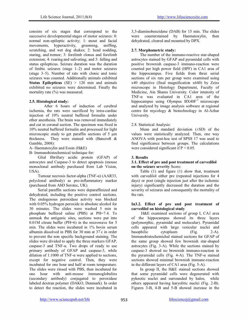
Table (1) and figure (1) show that, treatment with carvedilol either pre (repeated injections for 4 days) or post (single injection just after the ischemic injury) significantly decreased the duration and the severity of seizures and consequently the mortality of the rats.
In3.2. Effect of pre and post treatment of carvedilol on histological study
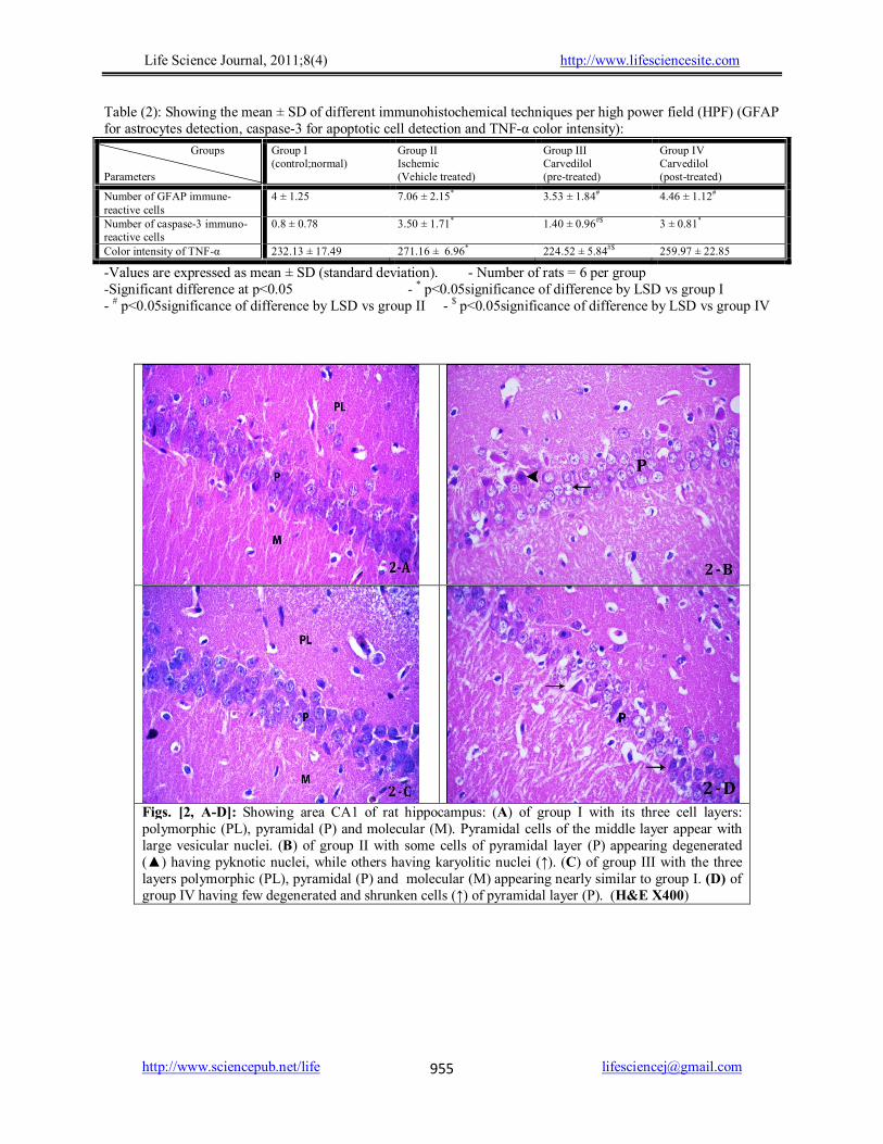
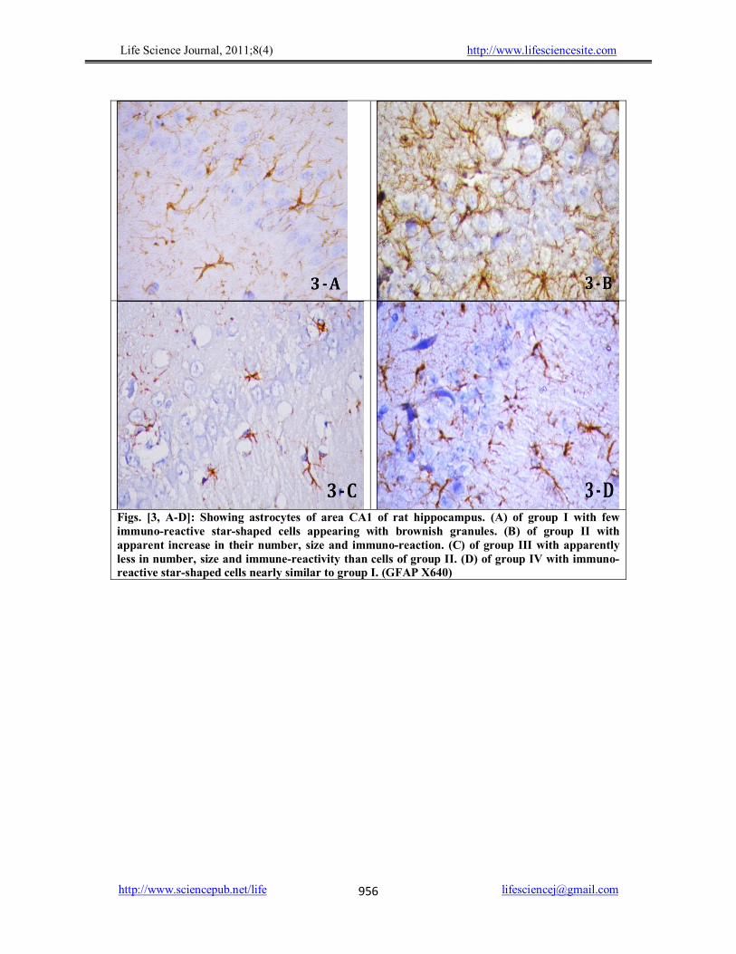
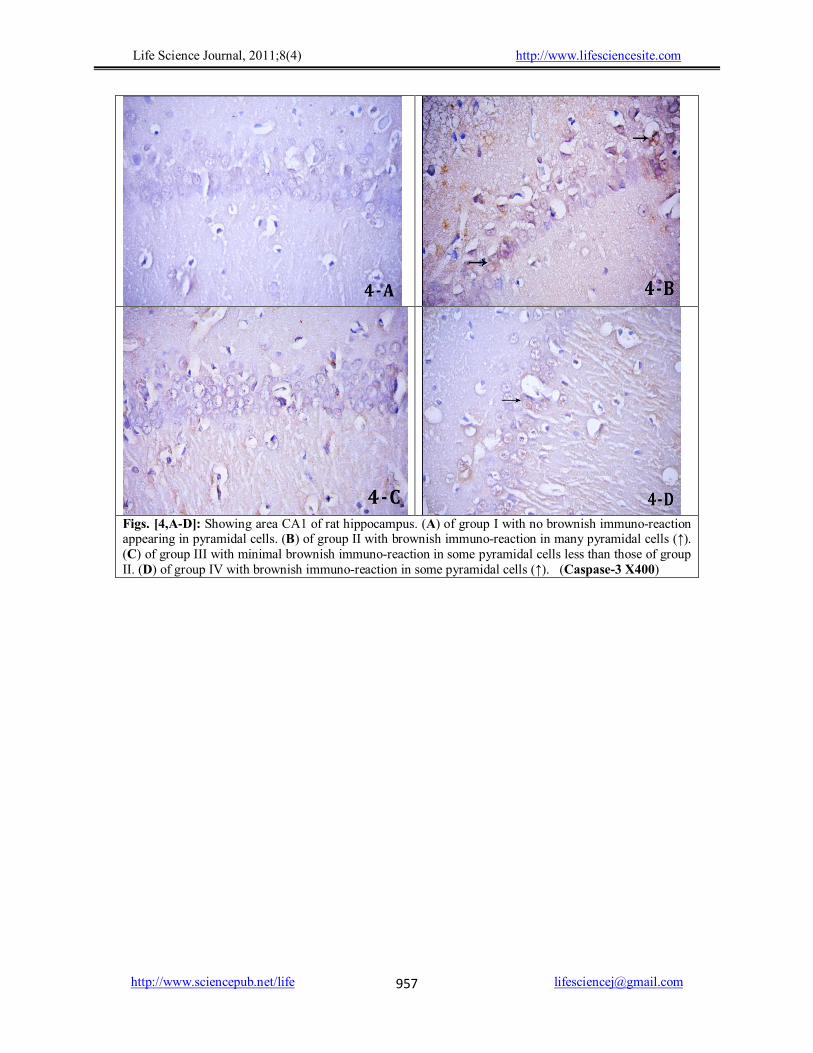
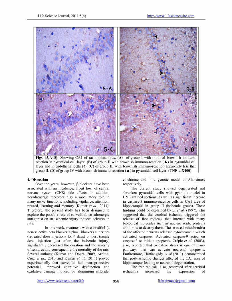
H&E examined sections of group I, CA1 area of the hippocampus showed its three layers (polymorphic, pyramidal and molecular). Pyramidal cells appeared with large vesicular nuclei and basophilic cytoplasm (Fig. 2-A). Immunohistochemichal stained sections for GFAP of the same group showed few brownish star-shaped astrocytes (Fig. 3-A). While the sections stained by caspase-3 showed no brownish immuno-reaction in the pyramidal cells (Fig. 4-A). The TNF-α stained sections showed minimal brownish immune-reaction in the different layers of CA1 area (Fig. 5-A).
In group II, the H&E stained sections showed that some pyramidal cells were degenerated with pyknotic nuclei and surrounded by halos, while others appeared having karyolitic nuclei (Fig. 2-B). Figures 3-B, 4-B and 5-B showed increase in the
Life Science Journal, 2011;8(4) http://www.lifesciencesite.com
http://www.sciencepub.net/life [email protected] 954
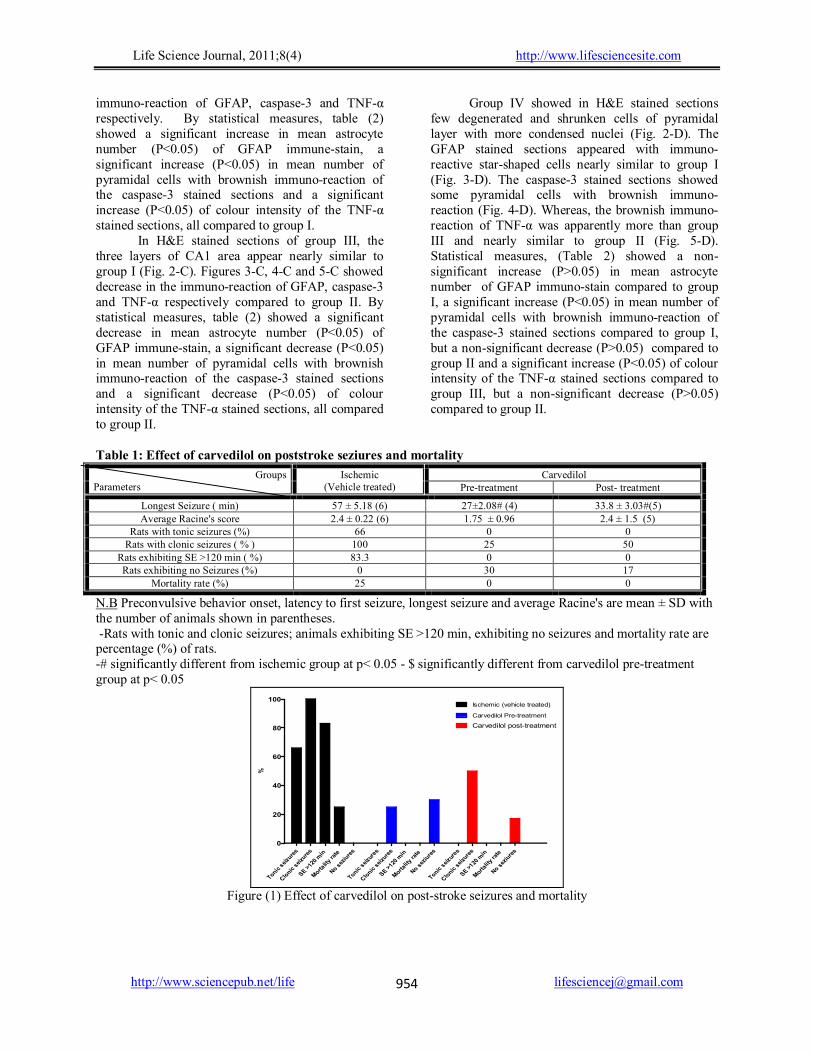
immuno-reaction of GFAP, caspase-3 and TNF-α respectively. By statistical measures, table (2) showed a significant increase in mean astrocyte number (P<0.05) of GFAP immune-stain, a significant increase (P<0.05) in mean number of pyramidal cells with brownish immuno-reaction of the caspase-3 stained sections and a significant increase (P<0.05) of colour intensity of the TNF-α stained sections, all compared to group I.
In H&E stained sections of group III, the three layers of CA1 area appear nearly similar to group I (Fig. 2-C). Figures 3-C, 4-C and 5-C showed decrease in the immuno-reaction of GFAP, caspase-3 and TNF-α respectively compared to group II. By statistical measures, table (2) showed a significant decrease in mean astrocyte number (P<0.05) of GFAP immune-stain, a significant decrease (P<0.05) in mean number of pyramidal cells with brownish immuno-reaction of the caspase-3 stained sections and a significant decrease (P<0.05) of colour intensity of the TNF-α stained sections, all compared to group II.
Group IV showed in H&E stained sections few degenerated and shrunken cells of pyramidal layer with more condensed nuclei (Fig. 2-D). The GFAP stained sections appeared with immuno-reactive star-shaped cells nearly similar to group I (Fig. 3-D). The caspase-3 stained sections showed some pyramidal cells with brownish immuno-reaction (Fig. 4-D). Whereas, the brownish immuno-reaction of TNF-α was apparently more than group III and nearly similar to group II (Fig. 5-D). Statistical measures, (Table 2) showed a non-significant increase (P>0.05) in mean astrocyte number of GFAP immuno-stain compared to group I, a significant increase (P<0.05) in mean number of pyramidal cells with brownish immuno-reaction of the caspase-3 stained sections compared to group I, but a non-significant decrease (P>0.05) compared to group II and a significant increase (P<0.05) of colour intensity of the TNF-α stained sections compared to group III, but a non-significant decrease (P>0.05) compared to group II.
Table 1: Effect of carvedilol on poststroke seziures and mortality
Groups Parameters
Ischemic (Vehicle treated)
Carvedilol Pre-treatment Post- treatment
Longest Seizure ( min) 57 ± 5.18 (6) 27±2.08# (4) 33.8 ± 3.03#(5) Average Racine's score 2.4 ± 0.22 (6) 1.75 ± 0.96 2.4 ± 1.5 (5)
Rats with tonic seizures (%) 66 0 0 Rats with clonic seizures ( % ) 100 25 50
Rats exhibiting SE >120 min ( %) 83.3 0 0 Rats exhibiting no Seizures (%) 0 30 17
Mortality rate (%) 25 0 0
N.B Preconvulsive behavior onset, latency to first seizure, longest seizure and average Racine's are mean ± SD with the number of animals shown in parentheses. -Rats with tonic and clonic seizures; animals exhibiting SE >120 min, exhibiting no seizures and mortality rate are percentage (%) of rats. -# significantly different from ischemic group at p< 0.05 - $ significantly different from carvedilol pre-treatment group at p< 0.05
Tonic
seizu
res
Clo
nic s
eizu
res
SE >
120
min
Morta
lity
rate
No sez
iure
s
Tonic
seizu
res
Clo
nic s
eizu
res
SE >
120
min
Morta
lity
rate
No sez
iure
s
Tonic
seizu
res
Clo
nic s
eizu
res
SE >
120 m
in
Morta
lity
rate
No sez
iure
s
0
20
40
60
80
100Ischemic (vehicle treated)
Carvedilol Pre-treatment
Carvedilol post-treatment
%
Figure (1) Effect of carvedilol on post-stroke seizures and mortality
Life Science Journal, 2011;8(4) http://www.lifesciencesite.com
http://www.sciencepub.net/life [email protected] 955
Table (2): Showing the mean ± SD of different immunohistochemical techniques per high power field (HPF) (GFAP for astrocytes detection, caspase-3 for apoptotic cell detection and TNF-α color intensity):
Groups Parameters
Group I (control;normal)
Group II Ischemic (Vehicle treated)
Group III Carvedilol (pre-treated)
Group IV Carvedilol (post-treated)
Number of GFAP immune-reactive cells
4 ± 1.25 7.06 ± 2.15* 3.53 ± 1.84# 4.46 ± 1.12#
Number of caspase-3 immuno-reactive cells
0.8 ± 0.78 3.50 ± 1.71* 1.40 ± 0.96#$ 3 ± 0.81*
Color intensity of TNF-α 232.13 ± 17.49 271.16 ± 6.96* 224.52 ± 5.84#$ 259.97 ± 22.85
-Values are expressed as mean ± SD (standard deviation). - Number of rats = 6 per group -Significant difference at p<0.05 - * p<0.05significance of difference by LSD vs group I - # p<0.05significance of difference by LSD vs group II - $ p<0.05significance of difference by LSD vs group IV
Figs. [2, A-D]: Showing area CA1 of rat hippocampus: (A) of group I with its three cell layers: polymorphic (PL), pyramidal (P) and molecular (M). Pyramidal cells of the middle layer appear with large vesicular nuclei. (B) of group II with some cells of pyramidal layer (P) appearing degenerated (▲) having pyknotic nuclei, while others having karyolitic nuclei (↑). (C) of group III with the three layers polymorphic (PL), pyramidal (P) and molecular (M) appearing nearly similar to group I. (D) of group IV having few degenerated and shrunken cells (↑) of pyramidal layer (P). (H&E X400)
Life Science Journal, 2011;8(4) http://www.lifesciencesite.com
http://www.sciencepub.net/life [email protected] 956
Figs. [3, A-D]: Showing astrocytes of area CA1 of rat hippocampus. (A) of group I with few immuno-reactive star-shaped cells appearing with brownish granules. (B) of group II with apparent increase in their number, size and immuno-reaction. (C) of group III with apparently less in number, size and immune-reactivity than cells of group II. (D) of group IV with immuno-reactive star-shaped cells nearly similar to group I. (GFAP X640)
Life Science Journal, 2011;8(4) http://www.lifesciencesite.com
http://www.sciencepub.net/life [email protected] 957
Figs. [4,A-D]: Showing area CA1 of rat hippocampus. (A) of group I with no brownish immuno-reaction appearing in pyramidal cells. (B) of group II with brownish immuno-reaction in many pyramidal cells (↑). (C) of group III with minimal brownish immuno-reaction in some pyramidal cells less than those of group II. (D) of group IV with brownish immuno-reaction in some pyramidal cells (↑). (Caspase-3 X400)
Life Science Journal, 2011;8(4) http://www.lifesciencesite.com
http://www.sciencepub.net/life [email protected] 958
Figs. [5,A-D]: Showing CA1 of rat hippocampus. (A) of group I with minimal brownish immuno-reaction in pyramidal cell layer. (B) of group II with brownish immuno-reaction (▲) in pyramidal cell layer and in endothelial cells (↑). (C) of group III with brownish immuno-reaction apparently less than group II. (D) of group IV with brownish immuno-reaction (▲) in pyramidal cell layer. (TNF-α X400)
4. Discussion
Over the years, however, β-blockers have been associated with an incidence, albeit low, of central nervous system (CNS) side effects. In addition, noradrenergic receptors play a modulatory role in many nerve functions, including vigilance, attention, reward, learning and memory (Kumar et al., 2011). Therefore, the present study has been designed to explore the possible role of carvedilol, an adrenergic antagonist on an ischemic injury induced seizures in rats.
In this work, treatment with carvedilol (a non-selective beta blocker/alpha-1 blocker) either pre (repeated dose injections for 4 days) or post (single dose injection just after the ischemic injury) significantly decreased the duration and the severity of seizures and consequently the mortality of the rats. Several authors; (Kumar and Dogra, 2009, Arrieta-Cruz et al., 2010 and Kumar et al., 2011) proved experimentally that carvedilol had neuroprotective potential, improved cognitive dysfunction and oxidative damage induced by aluminium chloride,
colchicine and in a genetic model of Alzheimer, respectively.
The current study showed degenerated and shrunken pyramidal cells with pyknotic nuclei in H&E stained sections, as well as significant increase in caspase-3 immuno-reactive cells in CA1 area of hippocampus in group II (ischemic group). These findings could be explained by Li et al. (1997), who suggested that the cerebral ischemia triggered the release of free radicals that interact with many biological molecules such as nucleic acids, proteins and lipids to destroy them. The stressed mitochondria of the affected neurons released cytochrome c which activated caspases. Activated caspase-9 acted on caspase-3 to initiate apoptosis. Créple et al. (2003), also, reported that oxidative stress is one of many pathways that can activate neuronal apoptosis. Furthermore, Hattiangady et al.(2011) demonstrated that post-ischemic changes affected the CA1 area of hippocampus leading to neuronal degeneration.
The free radicals, also, generated after cerebral ischaemia increased the expression of
Life Science Journal, 2011;8(4) http://www.lifesciencesite.com
http://www.sciencepub.net/life [email protected] 959
proinflammatory cytokines such as TNF-α and IL-1β (Lambersten et al., 2009). Endothelial cells, leucocytes, astrocytes and even neurons released cytokines in cerebral ischemia (Clausen et al., 2008). This explained the significant increase in the TNF-α color intensity of group II compared to group I in this study. The resident microglial cells are the main source of TNF-α (Lambersten et al., 2009). In the present study, there was a significant increase in mean number of GFAP immune-reative astrocytes in group II. This might be a response to ischemic injury as ischemia triggered secretion of TNF-α, which activated astrocytes to produce other cytokines as interleukin-6. Whereas, the activated microglia in the affected middle pyramidal layer might initiate phagocytosis with TNF-α production (Uno et al., 1997). On the other hand, some authors suggested that TNF-α might have a neuroprotective role and that the microglia acted as key regulator of neuronal survival after ischemic tissue injury (Lambersten et al., 2009).
The present study showed improvement of the histological results in H&E stained sections with significant decrease in mean number of GFAP positive astrocytes, caspase-3 positive cells and TNF-α color intensity in group III, which was pretreated with carvedilol for 4 days, compared to group II (ischemic vehicle treated group). These findings could be explained by Abreu et al. (2000) who mentioned that carvedilol, in isolated rat liver mitochondria, is an antioxidant. Several authors (Savitz et al., 2000 and Goyagi et al., 2006) reported that carvedilol versus propranalol protects the neurons after transient focal cerebral ischemia in rats, through the preservation of mitochondrial function. It has an antiapoptotic role and can downregulate the inflammatory cytokine gene expression of TNF-α and IL-1β.
Savitz et al.(2000) proved that carvedilol inhibited a number of inflammatory processes during brain damage and suppress the release of cytokines in neurological animal disease models and lastly, Kumar and Dogra (2009) stated that carvedilol had a neuroprotective effect against colchicine-induced cognitive impairment and could attenuate the oxidative damages.
In the present study, the improvement of ischemic changes in pre-treated group was more evident than that of the post ischemic carvedilol treated group. There was a non-significant decrease (P> 0.05) of mean number of caspase-3 positive cells and a non-significant decrease (P> 0.05) of TNF-α color intensity in post ischemic carvedilol treated group compared to ischemic vehicle treated group . This could be explained by the need of multiple
dosing regimens to accumulate the drug for efficacy (Lysko et al., 1992).
Strosznajder et al. (2005) revealed that carvedilol (7-70mg/kg) administered subcutaneously directly after transient (5 min) forebrain ischemia protected significant population of neurons in the hippocampal CA1 area in gerbils, thus carvedilol raises high expectations also in the therapy of ischemia.
Given our findings that carvedilol (3 mg/kg/day subcutaneous injections for 4 days) showed a significant neuroprotective effect than single carvedilol (3mg/kg) post ischemic subcutaneous injection which might find a beneficial use in the prevention of neuronal impairment, such as brain ischemia and post stroke seizures. Weather the repeated injections after stroke with carvedilol for a while will give a more neurotherapeutic effects or not, this is for further evaluation. Corresponding Author: Omnyah Ali El-Kharashi Departments of Pharmacology, Faculty of Medicine, Ain Shams University, Cairo, Egypt [email protected] Reference 1. Abreu RV, Santos D J.S.L and Moreno A J.M (2000):
Effects of carvedilol and its analog BM-910228 on mitochondrial function and oxidative stress. J Pharmacol Exp Ther., 295: 1022-1030.
2. Arrieta-Cruz I, Wang J, Pavlides C and Pasinetti GM (2010): Carvedilol reestablishes long-term potentiation in a mouse model of Alzheimer's disease. J Alzheimers Dis. ;21(2):649-54.
3. Bancroft JB and Gamble M (2008): Theory and Practice of Histological Techniques, sixth edition, Churchill Livingstone, Elsevier: 121 and 433.
4. Cizkova D, Vanicky I, Ishikawa T and Marsala M. (2000): Time course of brain neuronal degeneration and heat shock protein (72) expression following neck tourniquet-induced cerebral ischemia in the rat. Cell Mol Neurobiol.;20(3):367-81.
5. Clausen BH, Lambertsen KL, Babcock AA, Holm TH, Dagnaes-Hansen F and Finsen B (2008): Interleukin-1beta and tumor necrosis factor-alpha are expressed by different subsets of microglia and macrophages after ischemic stroke in mice. J Neuroinflammation, 5:46.
6. Crépel V, Epsztein J and Ben Ari Y (2003): Ischemia induces short- and longterm remodeling of synaptic activity in the hippocampus. J Cell Mol Med., 7:401– 407.
7. Epsztein J, Milh M, Bihi RI, Jorquera I, Ben-Ari Y, Represa A, and Crepel V.(2006): Ongoing Epileptiform Activity in the Post-Ischemic Hippocampus Is Associated with a Permanent Shift of the Excitatory–Inhibitory Synaptic Balance in CA3 Pyramidal Neurons. J Neurosci., 26(26):7082–7092.

Life Science Journal, 2011;8(4) http://www.lifesciencesite.com
http://www.sciencepub.net/life [email protected] 960
8. Fabene PF, Bramanti P and Constantin G (2010). The emerging role for chemokines in epilepsy. J Neuroimmunol., 224: 22–27.
9. Goyagi T, Kimura T, Nishikawa T, Tobe Y and Masaki Y (2006): Beta-adrenoreceptor antagonists attenuate brain injury after transient focal ischemia in rats. Anesth Analg., 103(3):658-63.
10. Hamby ME, Hewett JA and Hewett SJ (2006): TGF-beta1 potentiates astrocytic nitric oxide production by expanding the population of astrocytes that express NOS-2. Glia. ;54:566–577.
11. Hattiangady B, Kuruba R and Shetty AK (2011): Acute seizures in old age leads to a greater loss of CA1 pyramidal neurons, an increased propensity for developing chronic TLE and a severe cognitive dysfunction. Aging Dis., 2(1): 1-17.
12. Kramer G. (2001): Epilepsy in the elderly: some clinical and pharmacotherapeutic aspects. Epilepsia, 3:55-9.
13. Kumar A and Dogra S (2009): Neuroprotective effect of carvedilol, an adrenergic antagonist against colchicine induced cognitive impairment and oxidative damage in rat. Pharmacol Biochem Behav.; 92 (1):25-31.
14. Kumar A, Prakash A and Dogra S.(2011): Neuroprotective effect of carvedilol against aluminium induced toxicity: possible behavioral and biochemical alterations in rats. Pharmacol Rep.;63(4):915-23.
15. Kwan J and Wood E. (2010): Antiepileptic drugs for the primary and secondary prevention of seizures after stroke. Cochrane Database Syst Rev.;(1):CD005398
16. Lambersten KL, Clausen BH, Babcock AA, Gregersen R, Fenger C, Nielsen HH, Haugaard LS, Wirenfeldt M, Nielsen M, Dagnaes-Hansen F, Bluethmann H, Færgeman NJ, Meldgaard M, Deierborg T and Finsen B (2009): Microglia protect neurons against ischemia by synthesis of tumor necrosis factor. J Neurosci., 29(5):1319 –1330.
17. Li P, Nijhawan D, Budihardjo I, Srinivasula SM, Ahmad M, Alnemri ES and Wang X. (1997): Cytochrome c and dATP-dependent formation of Ap af-1/caspase-9 complex initiates an apoptotic protease cascade. Cell, 91(4): 479-489.
18. Lysko PG, Lysko KA, Yue TL, Webb CL, Gu JL and Feuerstein G (1992): Neuroprotective effects of carvedilol, a new antihypertensive agent, in cultured rat cerebellar neurons and in gerbil global brain ischemia. Stroke, 23:1630-1636.
19. Meller R, Clayton C, Torrey DJ, Schindler CK, Lan JQ, Cameron JA, Chu XP, Xiong ZG, Simon RP and Henshall DC.(2006): Activation of the caspase 8 pathway mediates seizure-induced cell death in
cultured hippocampal neurons. Epilepsy Res.;70(1):3-14
20. Myint PK, Staufenberg EF, and Sabanathan K.(2006); Post-stroke seizure and post-stroke epilepsy. Postgrad Med J.;82(971):568-72
21. Racine, RJ (1972): "Modification of seizure activity by electrical stimulation. II. Motor seizure.” Electroencephalography and Clin Neurophysiol., 32 (3): 281–94
22. Radt R, Clinckers R , Mollet L, Vonck K, El Tahry R, Wyckhuys T, De Herdt V, Carrette E, Wadman W, Michotte Y, Smolders I, Boon P and Meurs A (2011): Increased hippocampal noradrenaline is a biomarker for efficacy of vagus nerve stimulation in a limbic seizure model. J Neurochem., 117(3):461-9.
23. Savitz SI, Erhardt JA, Anthony JV, Gupta G, Li X, Barone FC and Rosenbaum DM (2000): The novel β-Blocker, carvedilol, provides neuroprotection intransient focal stroke.J Cereb Blood Flow Metab ; 20(8):1197-204.
24. Silverman IE, Restrepo L and Mathews GC (2002): Poststroke seizures. Arch Neurol. ;59 (2):195-201.
25. Strosznajder RP, Jesko H and Dziewulska J. (2005): Effect of carvedilol on neuronal survival and poly(ADP-ribose) polymerase activity in hippocampus after transient forebrain ischemia. Acta Neurobiol Exp.;65:137–144.
26. Tian GF, Azmi H, Takano T, Xu Q, Peng W, Lin J, Oberheim N, Lou N, Wang X, Zielke HR, Kang J, Nedergaard M (2005): An astrocytic basis of epilepsy. Nat Med., 11(9): 973–81.
27. Uno H, Matsuyama T, Akita H, Nishimura H and Sugita M (1997): Induction of tumor necrosis factor-a in the mouse hippocampus following transient forebrain ischemia. J Cereb Blood Flow and Metab., 17(5):491-99.
28. Vezzani A, Balosso S, Maroso M, Zardoni D, Noé F and Ravizza T (2010): ICE/caspase 1 inhibitors and IL-1beta receptor antagonists as potential therapeutics in epilepsy. Curr Opin Investig Drugs ;11(1):43–50.
29. Vos PE, Jacobs B, Andriessen TM, Lamers KJ, Borm GF, Beems T, Edwards M, Rosmalen CF andVissers JL (2010): GFAP and S100B are biomarkers of traumatic brain injury: an observational cohort study. Neurology; 75 (20):1786-93.
30. Watanabe K, Arozal W, Sari FR, Arumugam S, Thandavarayan RA, Suzuki K and Kodama M.(2011): The Role of Carvedilol in the Treatment of Dilated and Anthracyclines-Induced Cardiomyopathy. Pharmaceuticals, 4:770-781
31. Zador Z, Stiver S, Wang V and Manley GT (2009): Role of aquaporin-4 in cerebral edema and stroke. Handb Exp Pharmacol. ;190:159–70.
11/11/2011
Filename: 122_7733life0804_951_960.doc Directory: G:\net_fulltext2012010911222\journal\life\life0804 Template:
C:\Users\Administrator\AppData\Roaming\Microsoft\Templates\Normal.dotm
Title: 1 Subject: Author: CBA Keywords: Comments: Creation Date: 1/15/2012 9:42:00 PM Change Number: 6 Last Saved On: 1/25/2012 10:44:00 PM Last Saved By: Administrator Total Editing Time: 31 Minutes Last Printed On: 1/25/2012 10:44:00 PM As of Last Complete Printing Number of Pages: 10 Number of Words: 5,996 (approx.) Number of Characters: 34,180 (approx.)