Embed Size (px)
Citation preview
Life Imaging Center (LIC)
in the
Centre for Biological Systems Analysis (ZBSA) Albert-Ludwigs-University Freiburg
START LIC TOUR
next back 2
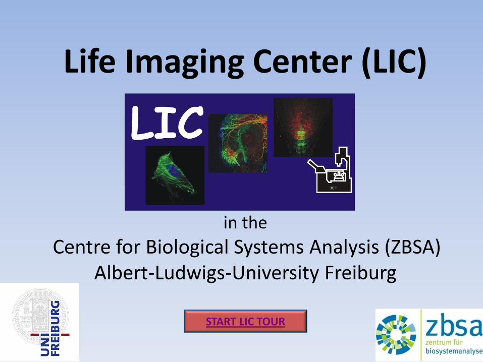
LIC Booking Calender
next back 3 Back to overview all
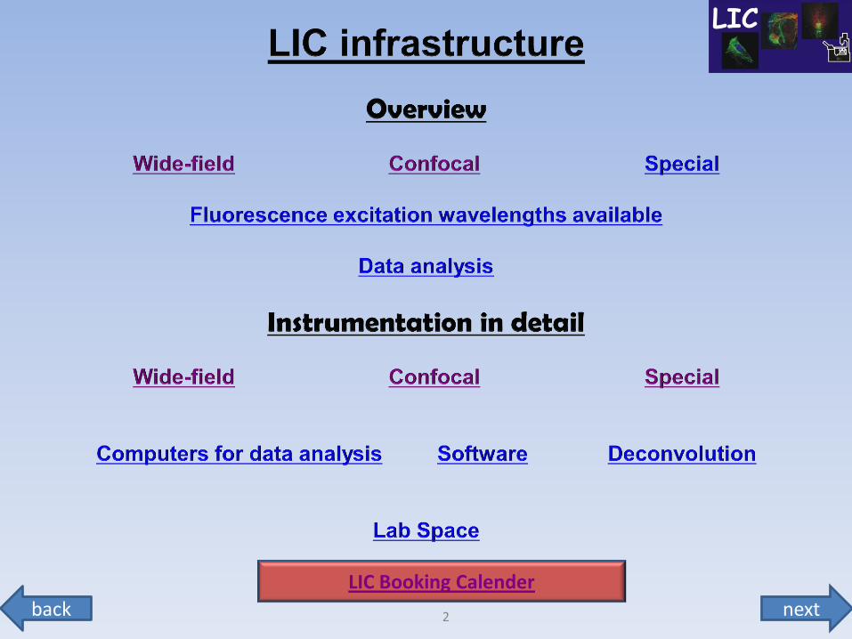
Infrastructure overview
Interactive Map of LIC (microscopes, lab and staff-offices)
The LIC in the ZBSA is app. 330 m² in 14 rooms. All lab space has biosafety level S2.
LIC – Office I LIC – Office II
Tissue Culture
room 00.025
Imaging 1
Imaging 2
LSM-U2
SD-TILL
LSM-I-2
room 00.017
LSM-I-
DuoLive
room 00.023
Leica
STED Stereo 3
room 00.024
LSM-I-
NLO2
Stereo 2
room 00.022
Develo
pm
ent
room
00.0
29
Imaging 3
room 00.030
Biostation 1 and 2
IncuCyte FLR
Imaging 4 room 00.028
LSM-I-
UV
AxioZoom
room 00.021
Wet Lab
molecular biology
preparation of
solutions
room 00.028
LIC-Office I
R. Nitschke
room 00.034
LIC-Office II Bierschenk,
Bodurova,Haxelmans, Jin,
Naumann
room 00.033 side entrance ZBSA
from Bio I
next back 4 Back to overview all
next back 5 Back to overview all Back to overview confocal
Infrastructure overview
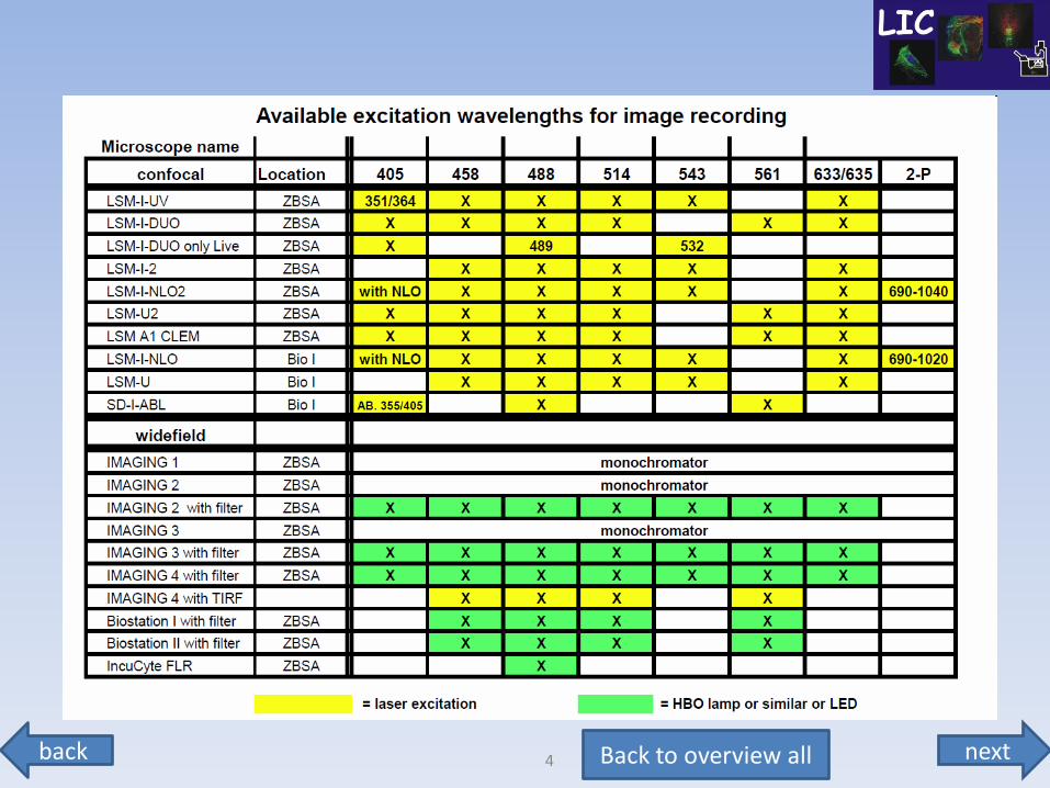
Microscopes for image recording
Inverted wide-field time-lapse / ratio imaging microscopes
Imaging 1 for calcium imaging or single color XFP, cooled CCD
Imaging 2 structured illumination (ApoTome), dualview FRET, EM-CCD
Imaging 3 with definite focus, big incubator, LED illumination, 2 cameras
Imaging 4 TIRF, FRET, multiple XFPs, dual camera
next back 6 Back to overview all Back to overview wide-field
Infrastructure overview
Microscopes for image recording
Confocal microscopes (time-lapse, spectral, multi-location rec.)
LSM-I-2 Zeiss LSM 510 (inverted)
LSM-I-UV Zeiss LSM 510 META UV (inverted)
LSM-I-NLO2 Zeiss LSM 510 META NLO (inverted) 2-P laser
LSM-I-DUO-Live Zeiss LSM 5 Live DUO (inverted) high speed confocal
LSM-U-2 Zeiss LSM 510 (upright)
A1-CLEM Nikon LSM A1 CLEM (inverted)
LSM-U Zeiss LSM 510 NLO (upright) – in Bio I
LSM-I-NLO Zeiss LSM 510 META NLO (inv.) 2P laser – in Bio I
SD-I-ABL Zeiss Spinning disk with laser ablation - (inv.) - in Bio I
STED Leica Super-Resolution gated STED Leica TCS 8ST-WS
next back 7 Back to overview all
Infrastructure overview
Microscopes for image recording
Special microscopes – long time-lapse machines up to 14 days
Biostation I Nikon Biostation IM with perfusion chamber
Biostation II Nikon Biostation IM
InCuCyte FLR Essen Instrument microscope inside incubator
Stereo fluorescence microscopes
AxioZoom Zeiss Axio Zoom V16 with B/W camera
Stereo 2 Leica MZ FL III with color camera
Stereo 3 Leica MZ FL III with color camera
next back 8 Back to overview all
Infrastructure overview
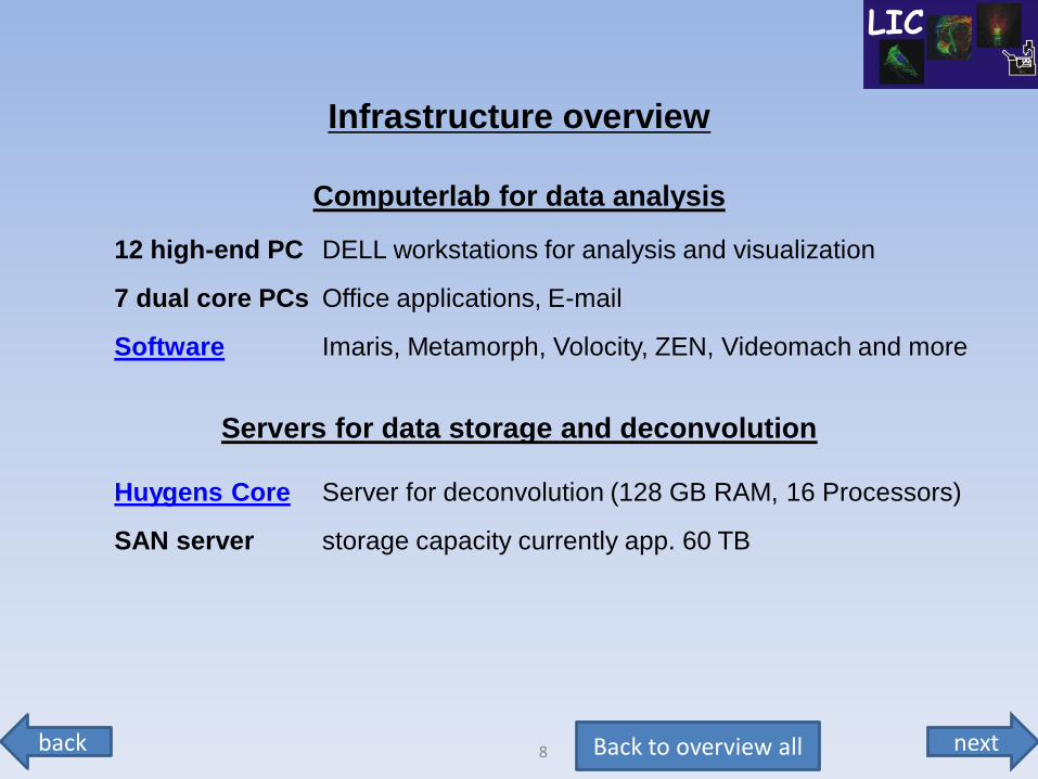
Computerlab for data analysis
12 high-end PC DELL workstations for analysis and visualization
7 dual core PCs Office applications, E-mail
Software Imaris, Metamorph, Volocity, ZEN, Videomach and more
Servers for data storage and deconvolution
Huygens Core Server for deconvolution (128 GB RAM, 16 Processors)
SAN server storage capacity currently app. 60 TB
next back 9 Back to overview all
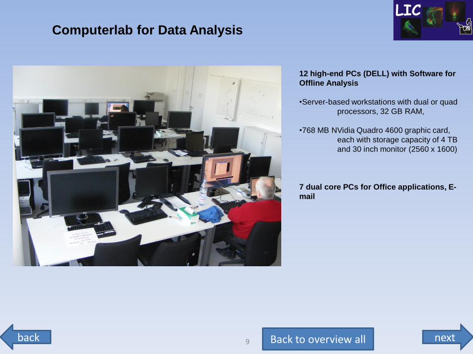
Computerlab for Data Analysis
12 high-end PCs (DELL) with Software for
Offline Analysis
•Server-based workstations with dual or quad
processors, 32 GB RAM,
•768 MB NVidia Quadro 4600 graphic card,
each with storage capacity of 4 TB
and 30 inch monitor (2560 x 1600)
7 dual core PCs for Office applications, E-
next back 10 Back to overview all
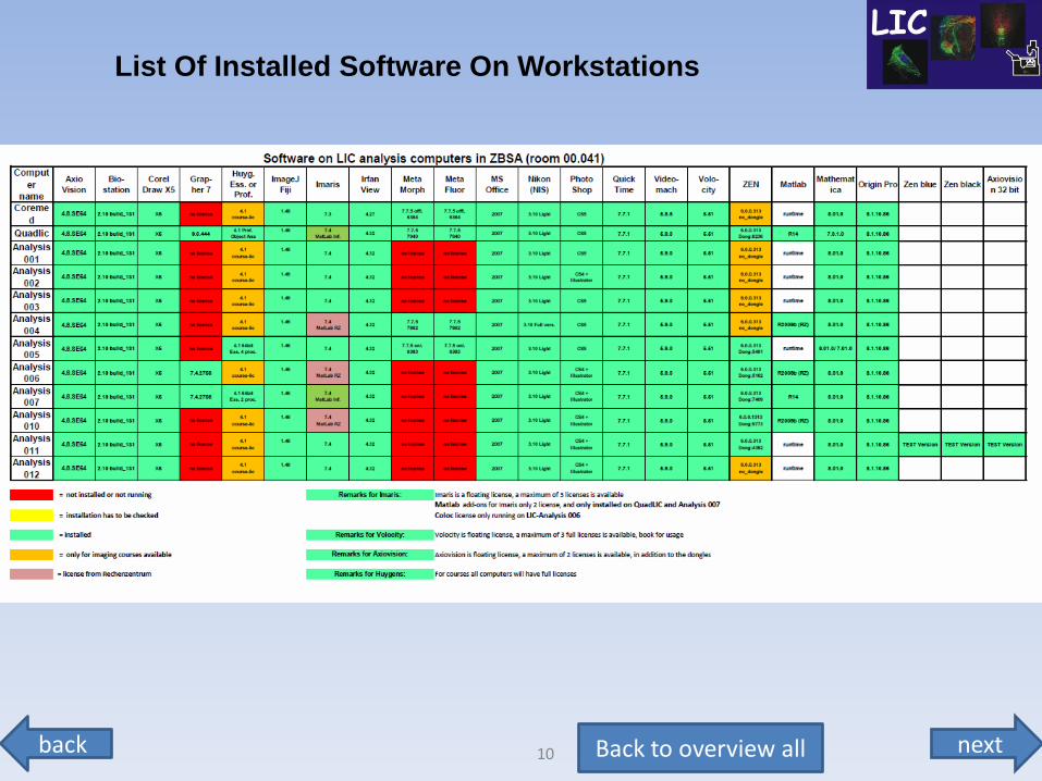
List Of Installed Software On Workstations
next back 11 Back to overview all
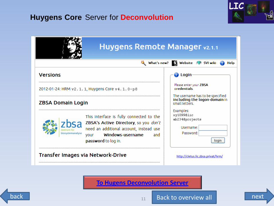
Huygens Core Server for Deconvolution
To Hugens Deconvolution Server
http://cletus.lic.zbsa.privat/hrm/
next back 12 Back to overview all Back to overview wide-field
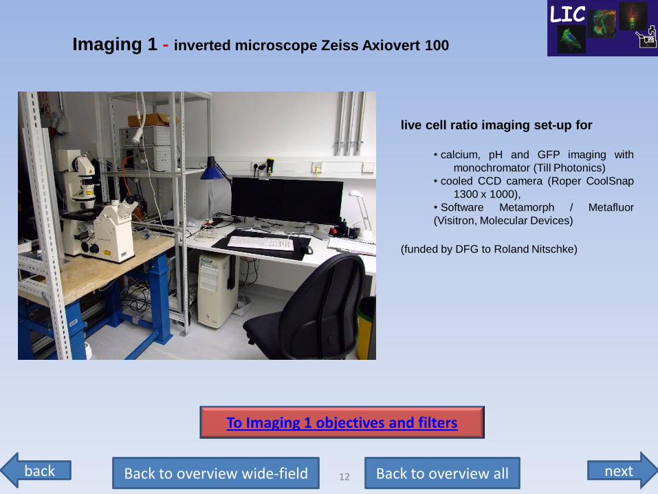
live cell ratio imaging set-up for
• calcium, pH and GFP imaging with
monochromator (Till Photonics)
• cooled CCD camera (Roper CoolSnap
1300 x 1000),
• Software Metamorph / Metafluor
(Visitron, Molecular Devices)
(funded by DFG to Roland Nitschke)
Imaging 1 - inverted microscope Zeiss Axiovert 100
To Imaging 1 objectives and filters
next back 13 Back to overview all Back to overview wide-field
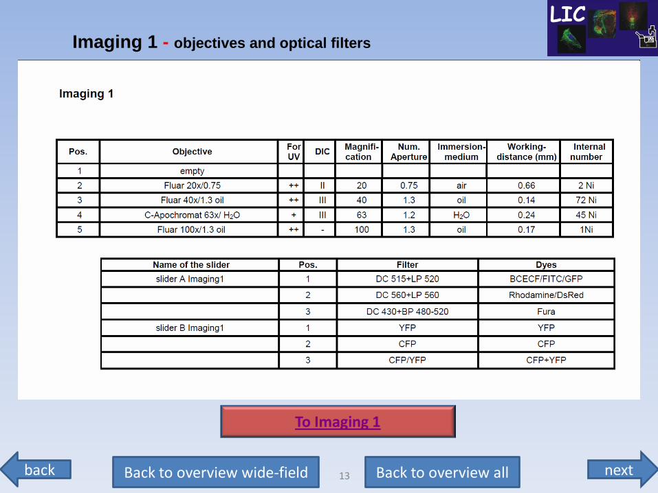
Imaging 1 - objectives and optical filters
To Imaging 1
next back 14 Back to overview all Back to overview wide-field
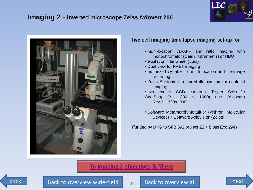
live cell imaging time-lapse imaging set-up for
• multi-location 3D-XFP and ratio imaging with
monochromator (Cairn instruments) or HBO
• excitation filter wheel (Ludl)
• Dual view for FRET imaging
• motorized xy-table for multi location and tile-image
recording
• Zeiss Apotome structured illumination for confocal
imaging
• two cooled CCD cameras (Roper Scientific
CoolSnap HQ 1300 x 1000) and (Axiocam
Rev.3, 1300x1000
• Software Metamorph/Metafluor (Visitron, Molecular
Devices) + Software Axiovision (Zeiss)
(funded by DFG to SFB 592 project Z2 + bioss Exc 294)
Imaging 2 - inverted microscope Zeiss Axiovert 200
To Imaging 2 objectives & filters

next back 15 Back to overview all Back to overview wide-field
Imaging 2 - objectives
To Imaging 2 optical filter
next back 16 Back to overview all Back to overview wide-field
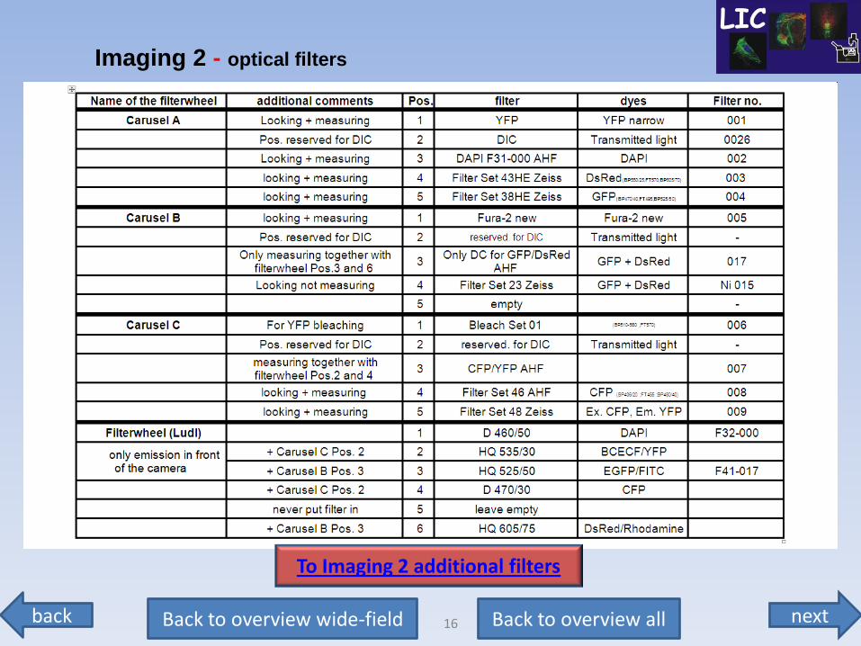
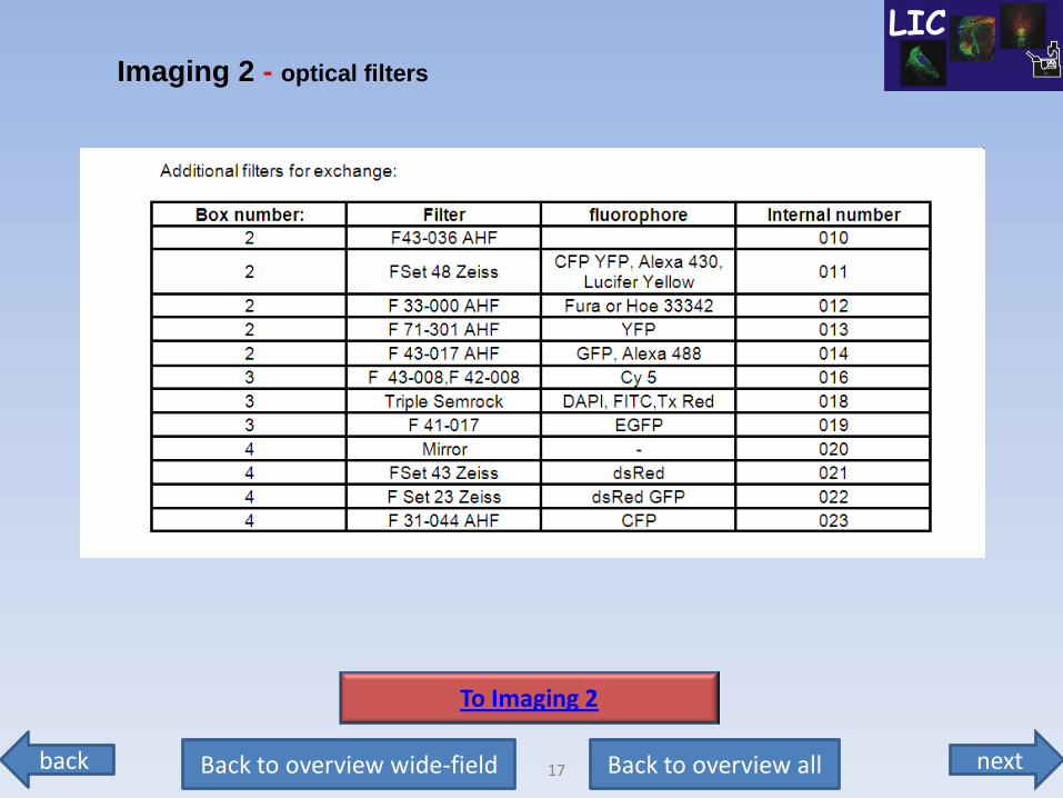
Imaging 2 - optical filters
To Imaging 2 additional filters
next back 17 Back to overview all Back to overview wide-field
Imaging 2 - optical filters
To Imaging 2
next back 18 Back to overview all Back to overview wide-field
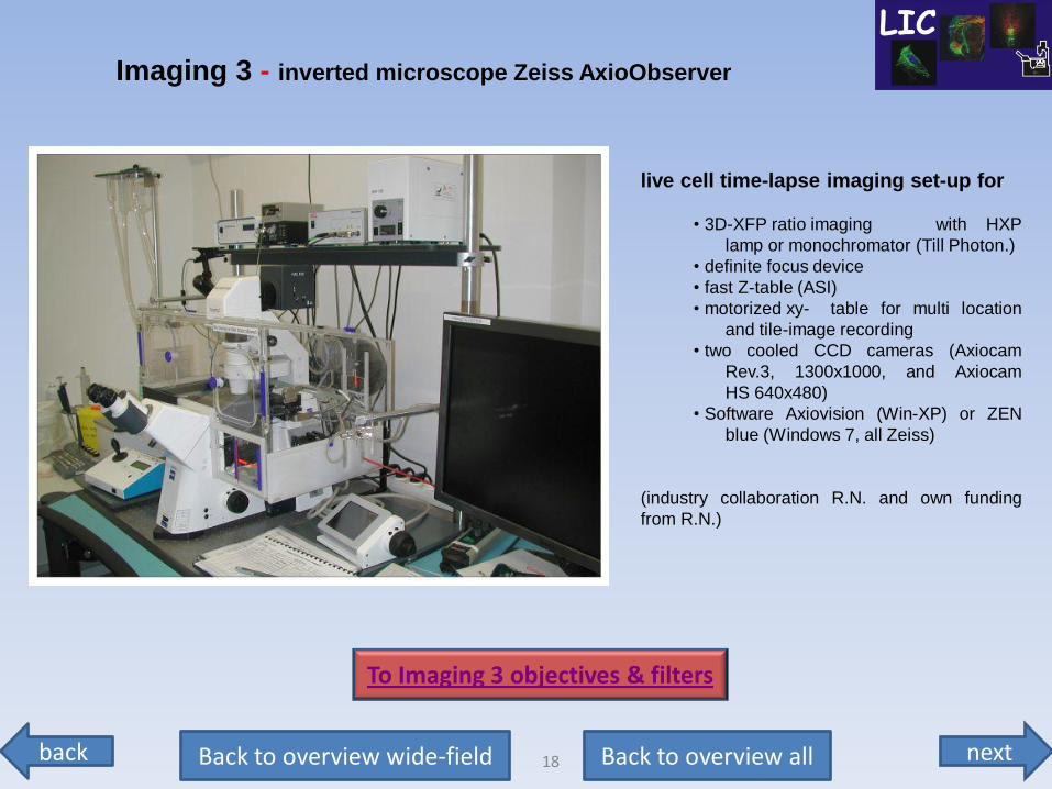
live cell time-lapse imaging set-up for
• 3D-XFP ratio imaging with HXP
lamp or monochromator (Till Photon.)
• definite focus device
• fast Z-table (ASI)
• motorized xy- table for multi location
and tile-image recording
• two cooled CCD cameras (Axiocam
Rev.3, 1300x1000, and Axiocam
HS 640x480)
• Software Axiovision (Win-XP) or ZEN
blue (Windows 7, all Zeiss)
(industry collaboration R.N. and own funding
from R.N.)
Imaging 3 - inverted microscope Zeiss AxioObserver
To Imaging 3 objectives & filters
next back 19 Back to overview all Back to overview wide-field
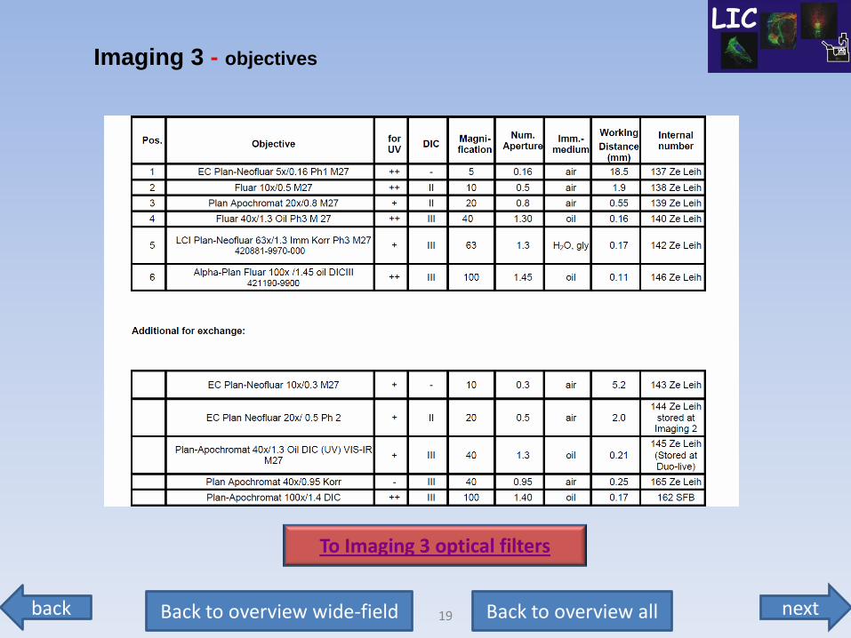
Imaging 3 - objectives
To Imaging 3 optical filters
next back 20 Back to overview all Back to overview wide-field
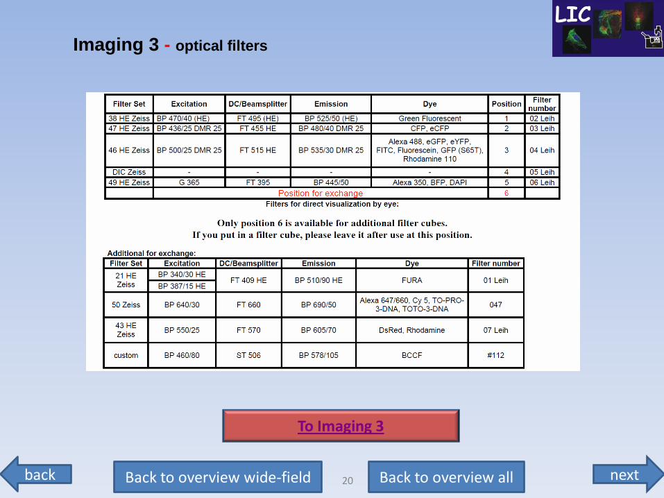
Imaging 3 - optical filters
To Imaging 3
next back 21 Back to overview all Back to overview wide-field
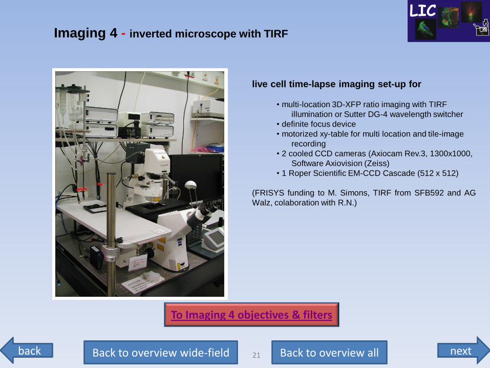
live cell time-lapse imaging set-up for
• multi-location 3D-XFP ratio imaging with TIRF
illumination or Sutter DG-4 wavelength switcher
• definite focus device
• motorized xy-table for multi location and tile-image
recording
• 2 cooled CCD cameras (Axiocam Rev.3, 1300x1000,
Software Axiovision (Zeiss)
• 1 Roper Scientific EM-CCD Cascade (512 x 512)
(FRISYS funding to M. Simons, TIRF from SFB592 and AG
Walz, colaboration with R.N.)
Imaging 4 - inverted microscope with TIRF
To Imaging 4 objectives & filters
next back 22 Back to overview all Back to overview wide-field
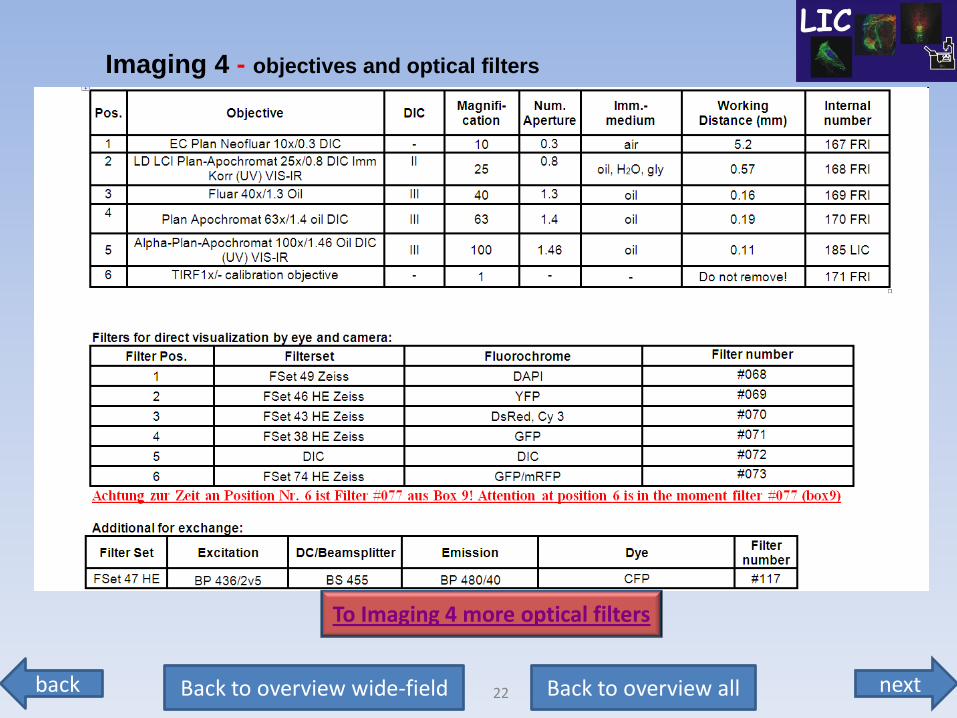
Imaging 4 - objectives and optical filters
To Imaging 4 more optical filters
next back 23 Back to overview all Back to overview wide-field
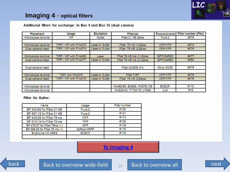
Imaging 4 - optical filters
To Imaging 4
next back 24 Back to overview all Back to overview confocal
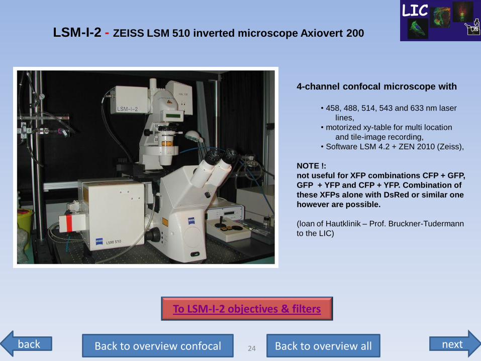
4-channel confocal microscope with
• 458, 488, 514, 543 and 633 nm laser
lines,
• motorized xy-table for multi location
and tile-image recording,
• Software LSM 4.2 + ZEN 2010 (Zeiss),
NOTE !:
not useful for XFP combinations CFP + GFP,
GFP + YFP and CFP + YFP. Combination of
these XFPs alone with DsRed or similar one
however are possible.
(loan of Hautklinik – Prof. Bruckner-Tudermann
to the LIC)
LSM-I-2 - ZEISS LSM 510 inverted microscope Axiovert 200
To LSM-I-2 objectives & filters
next back 25 Back to overview all Back to overview confocal
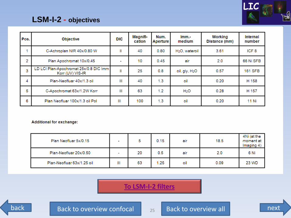
LSM-I-2 - objectives
To LSM-I-2 filters
next back 26 Back to overview all Back to overview confocal
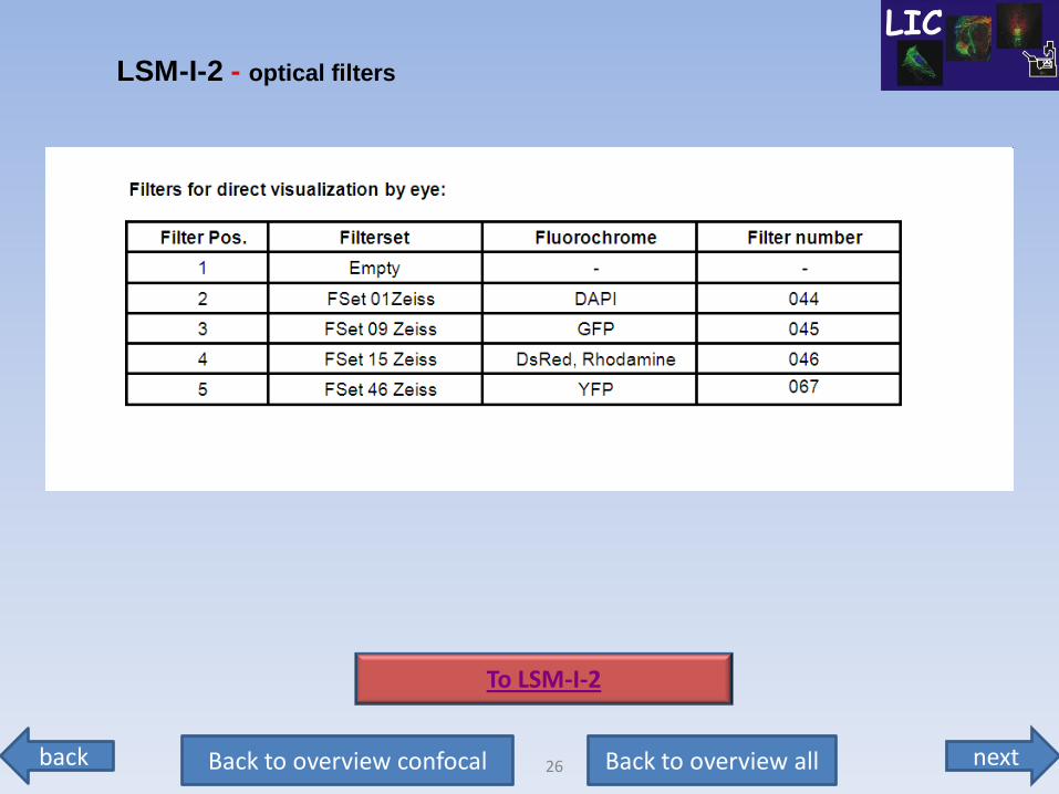
LSM-I-2 - optical filters
To LSM-I-2
next back 27 Back to overview all Back to overview confocal
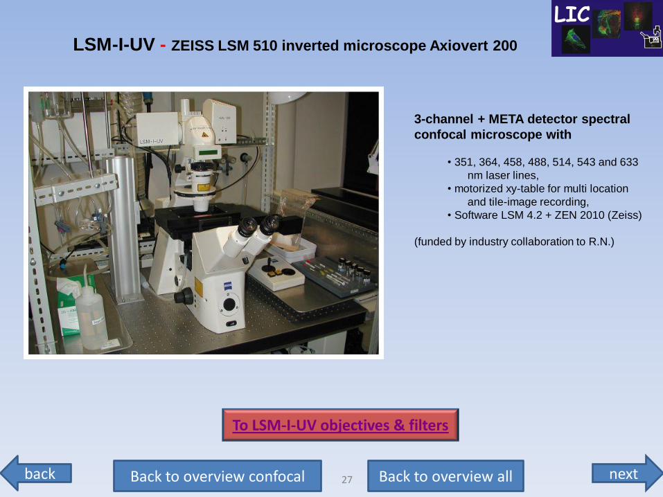
3-channel + META detector spectral
confocal microscope with
• 351, 364, 458, 488, 514, 543 and 633
nm laser lines,
• motorized xy-table for multi location
and tile-image recording,
• Software LSM 4.2 + ZEN 2010 (Zeiss)
(funded by industry collaboration to R.N.)
LSM-I-UV - ZEISS LSM 510 inverted microscope Axiovert 200
To LSM-I-UV objectives & filters
next back 28 Back to overview all Back to overview confocal
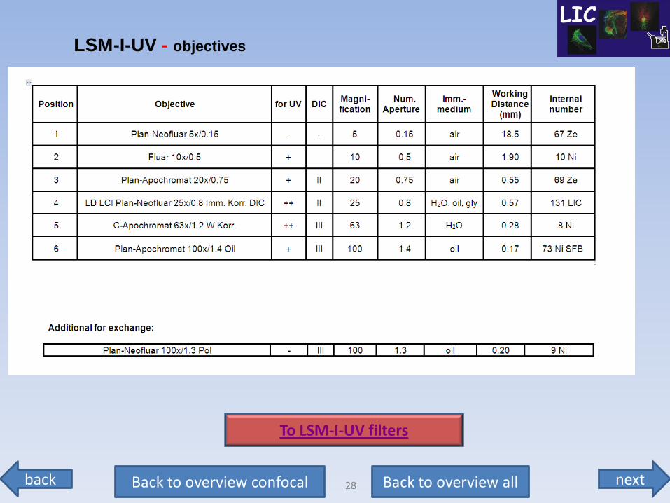
LSM-I-UV - objectives
To LSM-I-UV filters
next back 29 Back to overview all Back to overview confocal
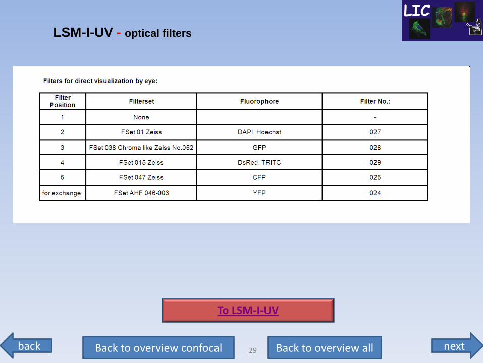
LSM-I-UV - optical filters
To LSM-I-UV
next back 30 Back to overview all Back to overview confocal
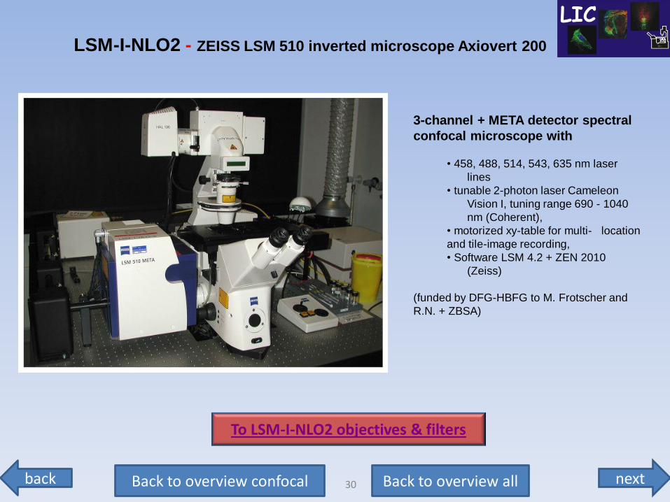
3-channel + META detector spectral
confocal microscope with
• 458, 488, 514, 543, 635 nm laser
lines
• tunable 2-photon laser Cameleon
Vision I, tuning range 690 - 1040
nm (Coherent),
• motorized xy-table for multi- location
and tile-image recording,
• Software LSM 4.2 + ZEN 2010
(Zeiss)
(funded by DFG-HBFG to M. Frotscher and
R.N. + ZBSA)
LSM-I-NLO2 - ZEISS LSM 510 inverted microscope Axiovert 200
To LSM-I-NLO2 objectives & filters
next back 31 Back to overview all Back to overview confocal
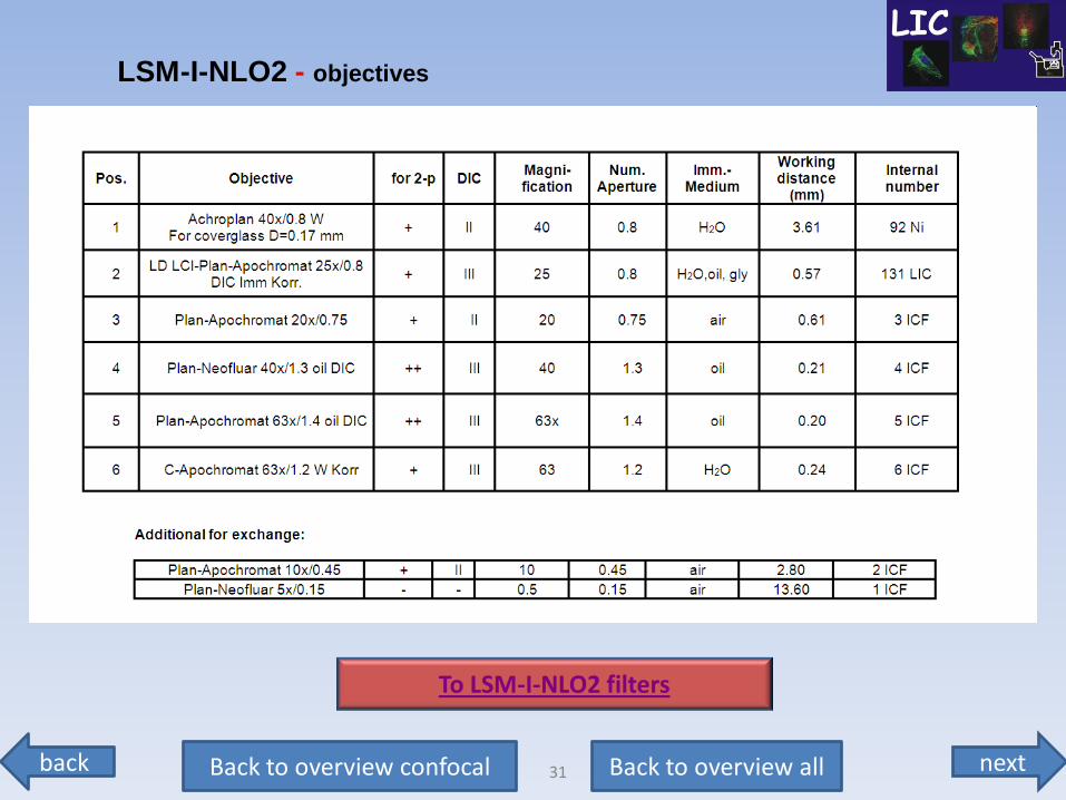
LSM-I-NLO2 - objectives
To LSM-I-NLO2 filters
next back 32 Back to overview all Back to overview confocal
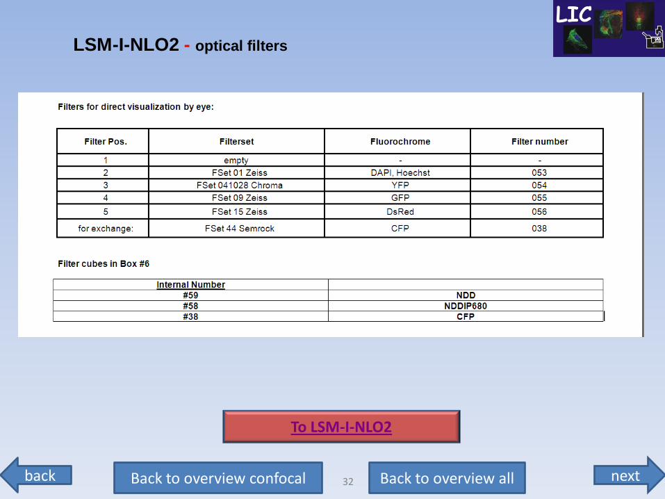
LSM-I-NLO2 - optical filters
To LSM-I-NLO2
next back 33 Back to overview all Back to overview confocal
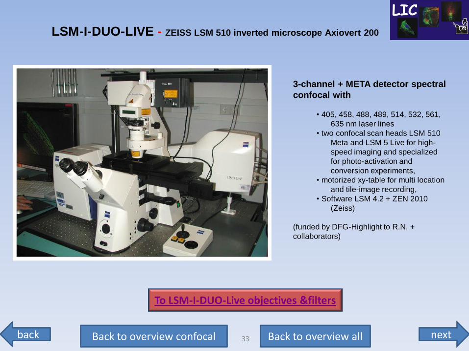
3-channel + META detector spectral
confocal with
• 405, 458, 488, 489, 514, 532, 561,
635 nm laser lines
• two confocal scan heads LSM 510
Meta and LSM 5 Live for high-
speed imaging and specialized
for photo-activation and
conversion experiments,
• motorized xy-table for multi location
and tile-image recording,
• Software LSM 4.2 + ZEN 2010
(Zeiss)
(funded by DFG-Highlight to R.N. +
collaborators)
LSM-I-DUO-LIVE - ZEISS LSM 510 inverted microscope Axiovert 200
To LSM-I-DUO-Live objectives &filters
next back 34 Back to overview all Back to overview confocal
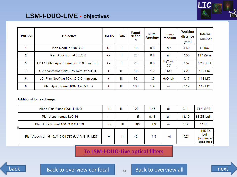
LSM-I-DUO-LIVE - objectives
To LSM-I-DUO-Live optical filters
next back 35 Back to overview all Back to overview confocal
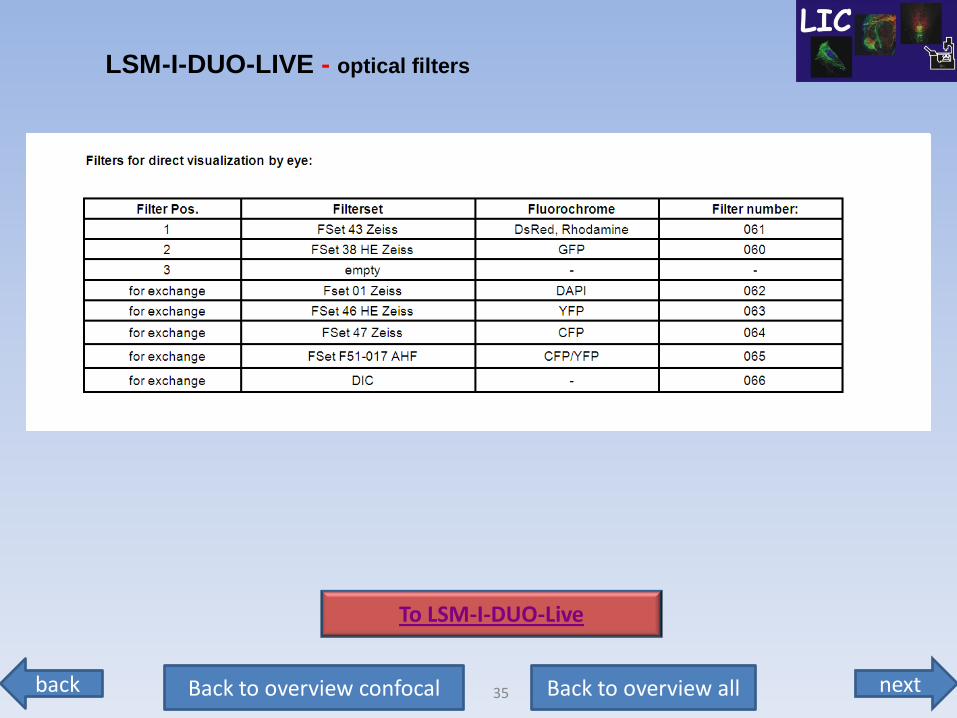
LSM-I-DUO-LIVE - optical filters
To LSM-I-DUO-Live
next back 36 Back to overview all Back to overview confocal
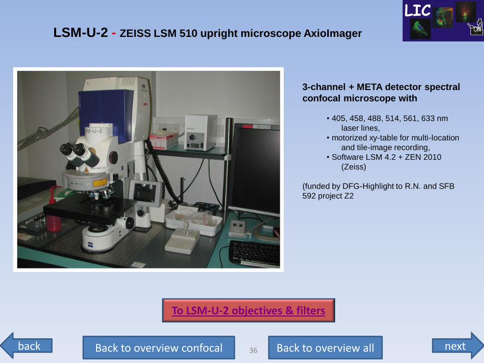
3-channel + META detector spectral
confocal microscope with
• 405, 458, 488, 514, 561, 633 nm
laser lines,
• motorized xy-table for multi-location
and tile-image recording,
• Software LSM 4.2 + ZEN 2010
(Zeiss)
(funded by DFG-Highlight to R.N. and SFB
592 project Z2
LSM-U-2 - ZEISS LSM 510 upright microscope AxioImager
To LSM-U-2 objectives & filters
next back 37 Back to overview all Back to overview confocal
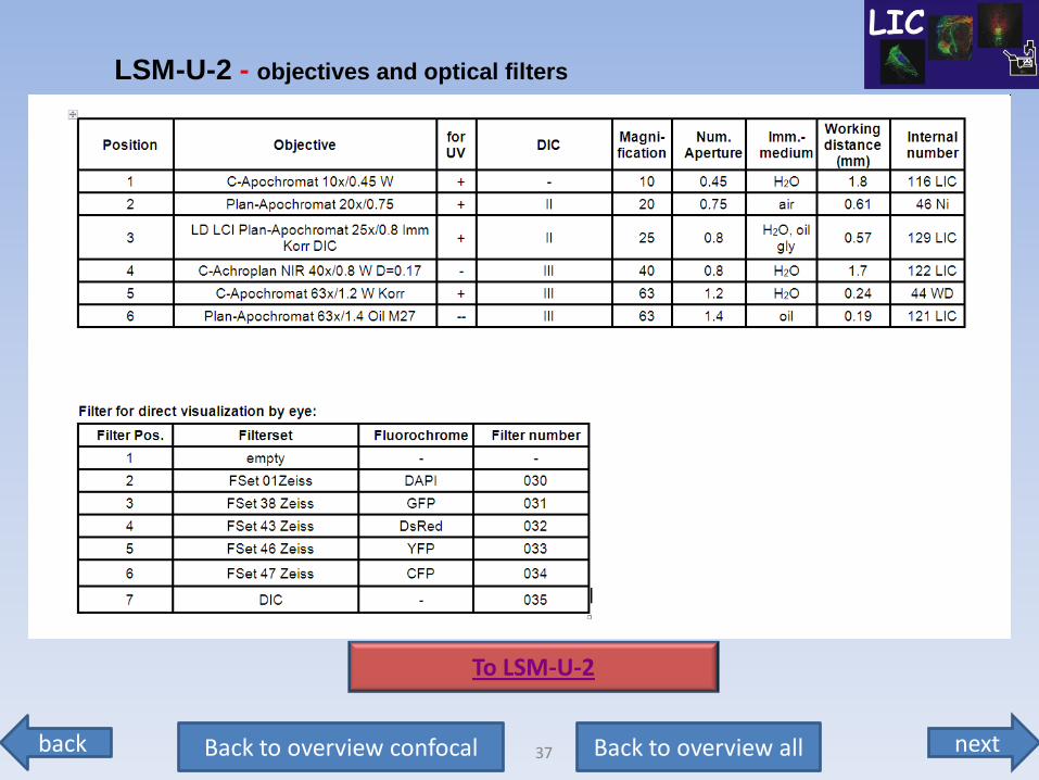
LSM-U-2 - objectives and optical filters
To LSM-U-2
next back 38 Back to overview all Back to overview confocal
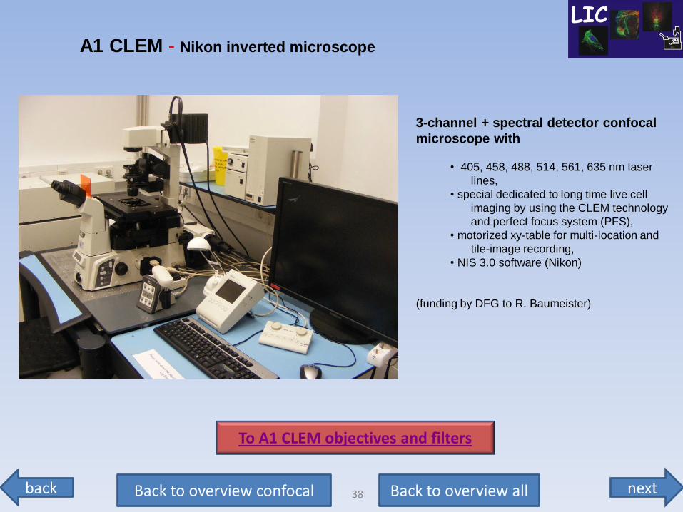
3-channel + spectral detector confocal
microscope with
• 405, 458, 488, 514, 561, 635 nm laser
lines,
• special dedicated to long time live cell
imaging by using the CLEM technology
and perfect focus system (PFS),
• motorized xy-table for multi-location and
tile-image recording,
• NIS 3.0 software (Nikon)
(funding by DFG to R. Baumeister)
A1 CLEM - Nikon inverted microscope
To A1 CLEM objectives and filters
next back 39 Back to overview all Back to overview confocal
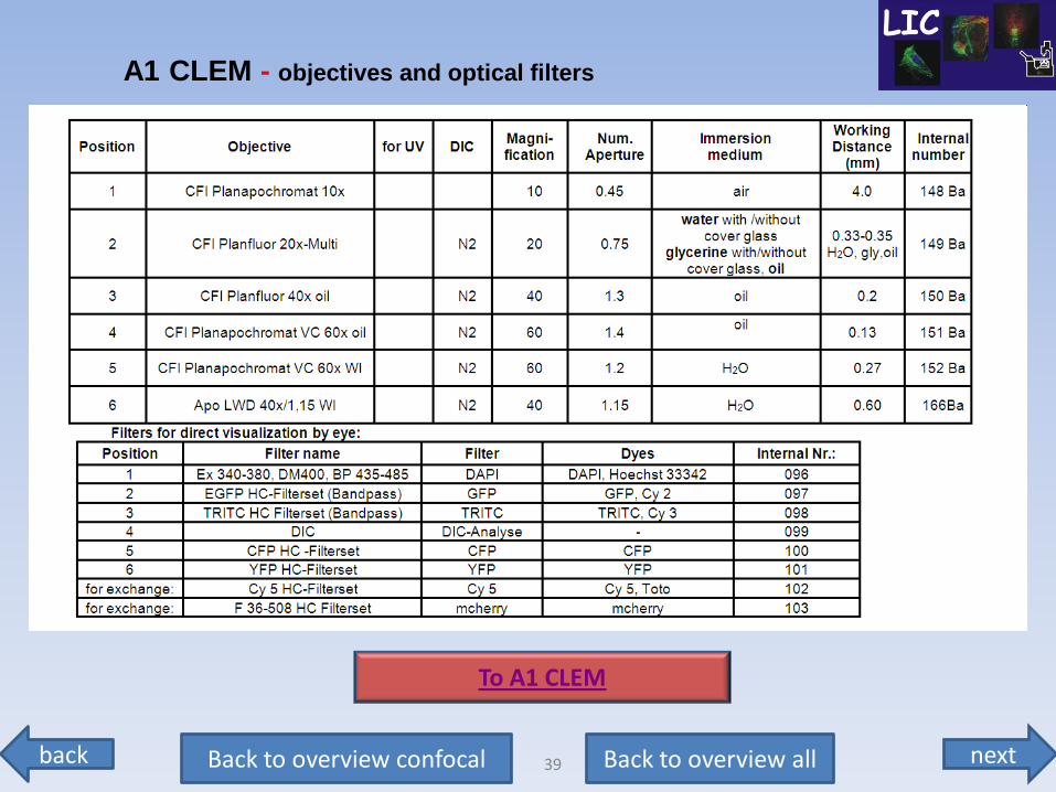
A1 CLEM - objectives and optical filters
To A1 CLEM
next back 40 Back to overview all Back to overview confocal
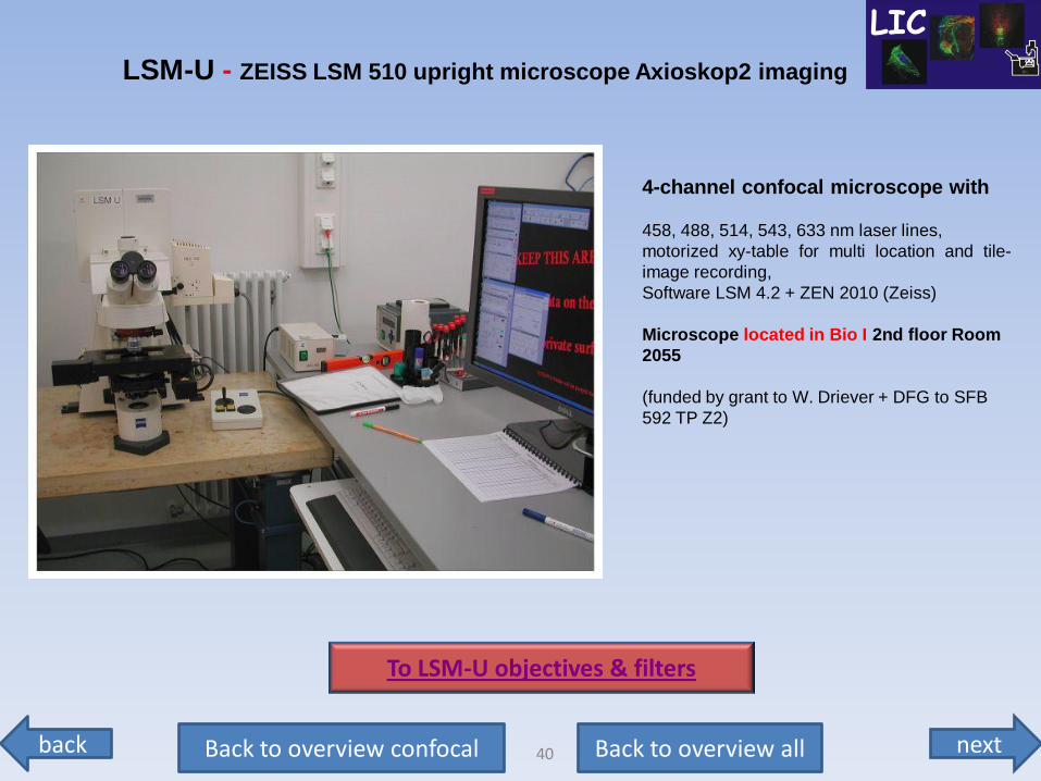
4-channel confocal microscope with
458, 488, 514, 543, 633 nm laser lines,
motorized xy-table for multi location and tile-
image recording,
Software LSM 4.2 + ZEN 2010 (Zeiss)
Microscope located in Bio I 2nd floor Room
2055
(funded by grant to W. Driever + DFG to SFB
592 TP Z2)
LSM-U - ZEISS LSM 510 upright microscope Axioskop2 imaging
To LSM-U objectives & filters
next back 41 Back to overview all Back to overview confocal
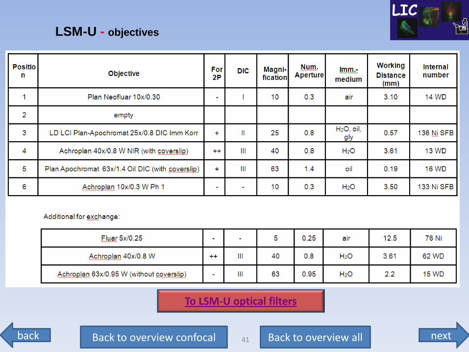
LSM-U - objectives
To LSM-U optical filters
next back 42 Back to overview all Back to overview confocal
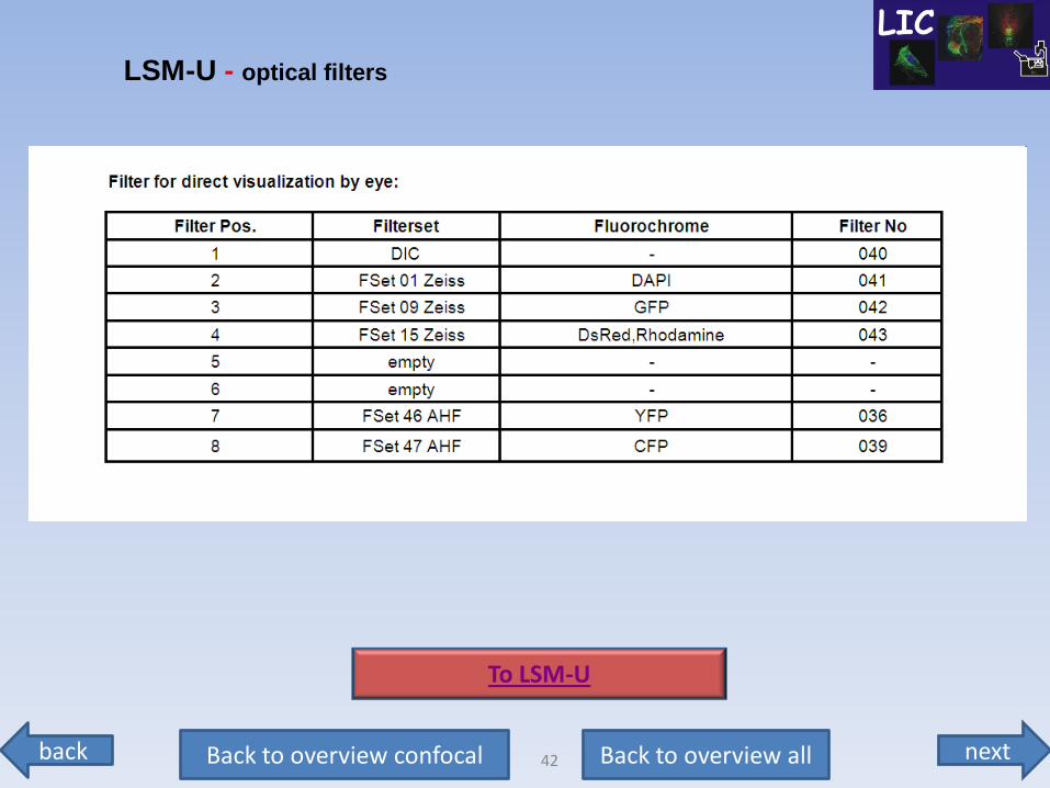
LSM-U - optical filters
To LSM-U
next back 43 Back to overview all Back to overview confocal
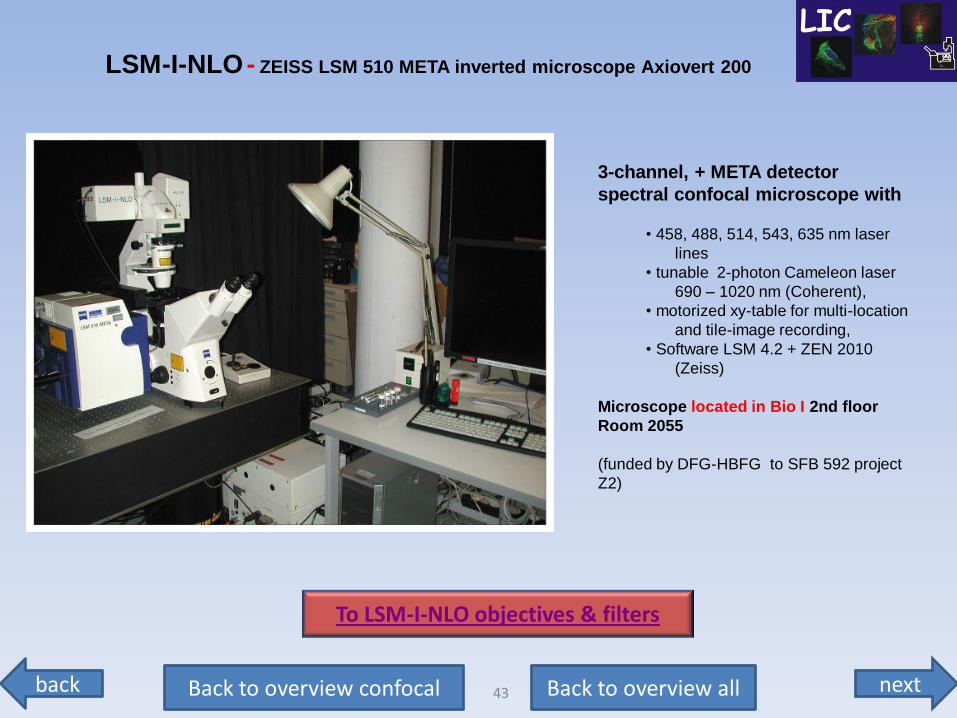
3-channel, + META detector
spectral confocal microscope with
• 458, 488, 514, 543, 635 nm laser
lines
• tunable 2-photon Cameleon laser
690 – 1020 nm (Coherent),
• motorized xy-table for multi-location
and tile-image recording,
• Software LSM 4.2 + ZEN 2010
(Zeiss)
Microscope located in Bio I 2nd floor
Room 2055
(funded by DFG-HBFG to SFB 592 project
Z2)
LSM-I-NLO - ZEISS LSM 510 META inverted microscope Axiovert 200
To LSM-I-NLO objectives & filters
next back 44 Back to overview all Back to overview confocal
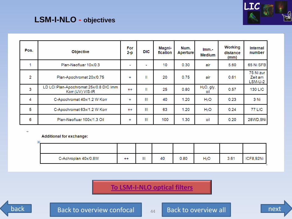
LSM-I-NLO - objectives
To LSM-I-NLO optical filters
next back 45 Back to overview all Back to overview confocal
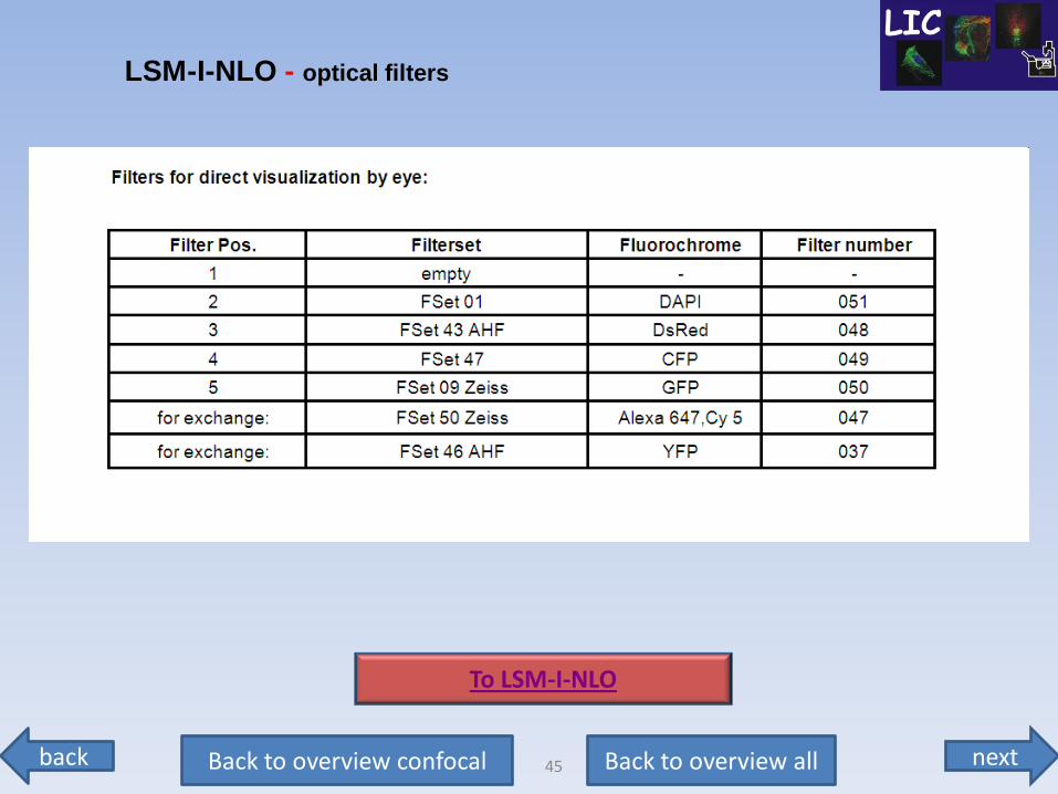
LSM-I-NLO - optical filters
To LSM-I-NLO
next back 46 Back to overview all Back to overview confocal
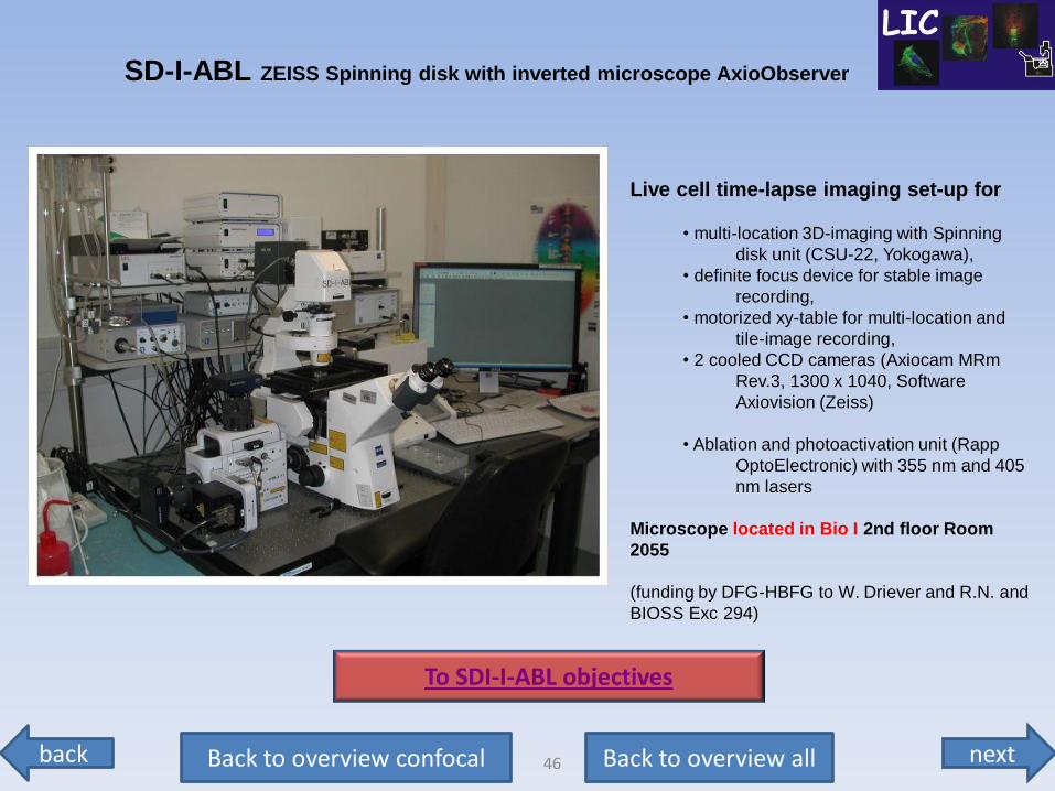
Live cell time-lapse imaging set-up for
• multi-location 3D-imaging with Spinning
disk unit (CSU-22, Yokogawa),
• definite focus device for stable image
recording,
• motorized xy-table for multi-location and
tile-image recording,
• 2 cooled CCD cameras (Axiocam MRm
Rev.3, 1300 x 1040, Software
Axiovision (Zeiss)
• Ablation and photoactivation unit (Rapp
OptoElectronic) with 355 nm and 405
nm lasers
Microscope located in Bio I 2nd floor Room
2055
(funding by DFG-HBFG to W. Driever and R.N. and
BIOSS Exc 294)
SD-I-ABL ZEISS Spinning disk with inverted microscope AxioObserver
To SDI-I-ABL objectives
next back 47 Back to overview all Back to overview confocal
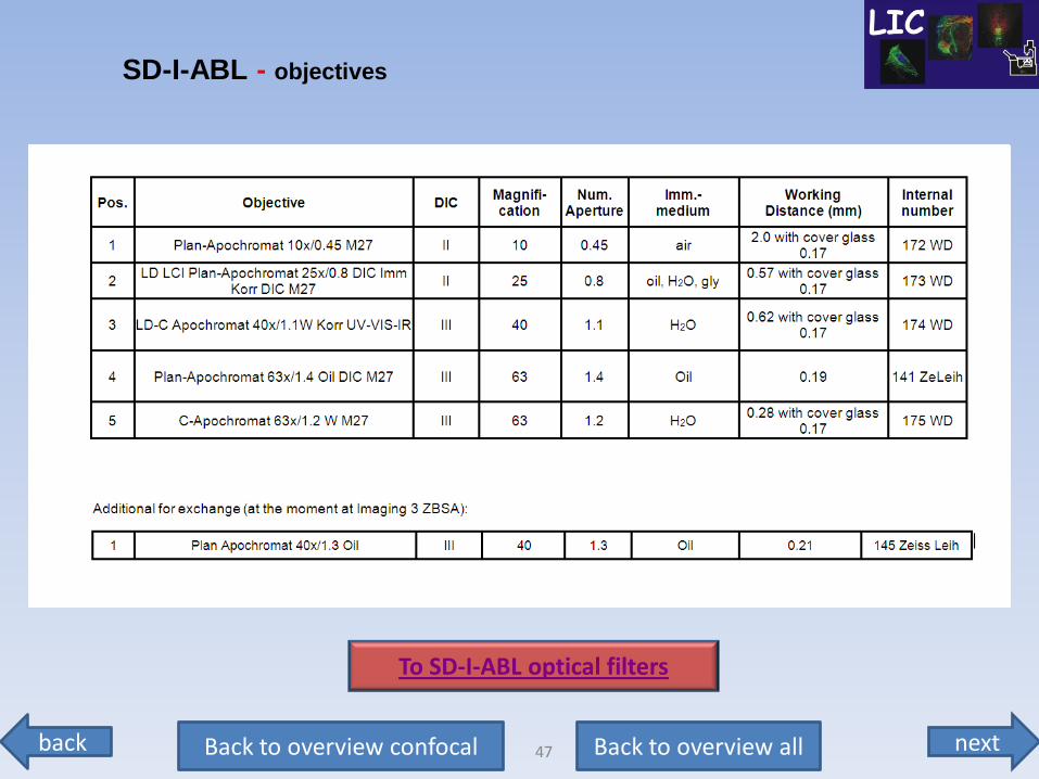
SD-I-ABL - objectives
To SD-I-ABL optical filters
next back 48 Back to overview all Back to overview confocal
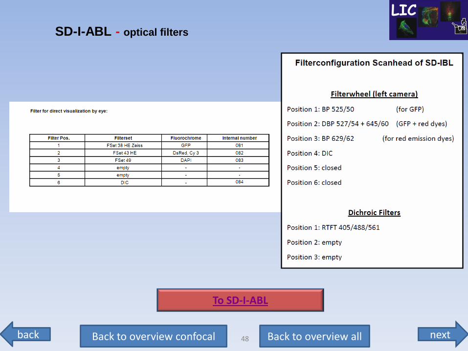
SD-I-ABL - optical filters
To SD-I-ABL
next back 49 Back to overview all Back to overview special
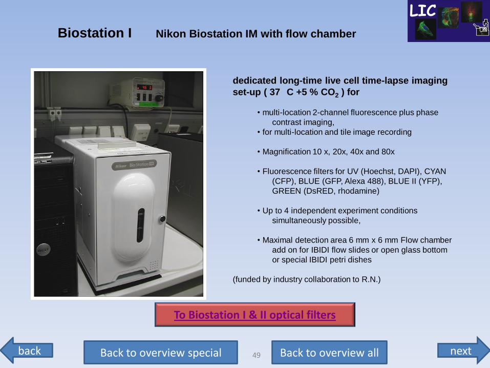
dedicated long-time live cell time-lapse imaging
set-up ( 37
C +5 % CO2 ) for
• multi-location 2-channel fluorescence plus phase
contrast imaging,
• for multi-location and tile image recording
• Magnification 10 x, 20x, 40x and 80x
• Fluorescence filters for UV (Hoechst, DAPI), CYAN
(CFP), BLUE (GFP, Alexa 488), BLUE II (YFP),
GREEN (DsRED, rhodamine)
• Up to 4 independent experiment conditions
simultaneously possible,
• Maximal detection area 6 mm x 6 mm Flow chamber
add on for IBIDI flow slides or open glass bottom
or special IBIDI petri dishes
(funded by industry collaboration to R.N.)
Biostation I Nikon Biostation IM with flow chamber
To Biostation I & II optical filters
next back 50 Back to overview all Back to overview special
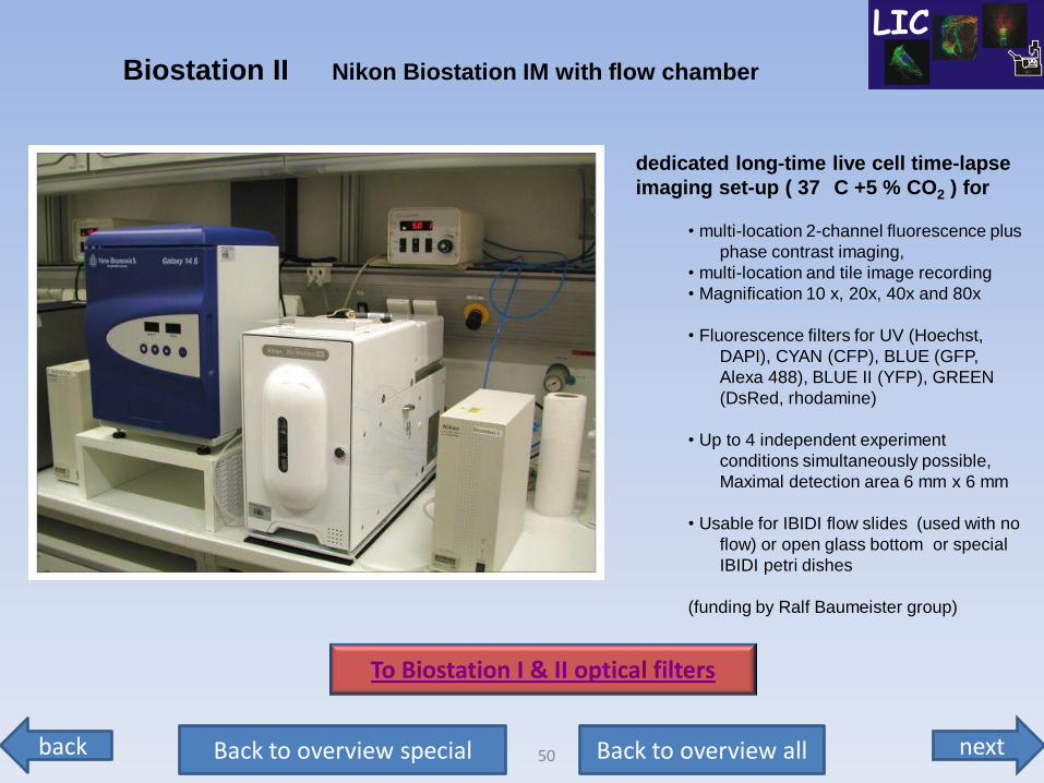
dedicated long-time live cell time-lapse
imaging set-up ( 37
C +5 % CO2 ) for
• multi-location 2-channel fluorescence plus
phase contrast imaging,
• multi-location and tile image recording
• Magnification 10 x, 20x, 40x and 80x
• Fluorescence filters for UV (Hoechst,
DAPI), CYAN (CFP), BLUE (GFP,
Alexa 488), BLUE II (YFP), GREEN
(DsRed, rhodamine)
• Up to 4 independent experiment
conditions simultaneously possible,
Maximal detection area 6 mm x 6 mm
• Usable for IBIDI flow slides (used with no
flow) or open glass bottom or special
IBIDI petri dishes
(funding by Ralf Baumeister group)
Biostation II Nikon Biostation IM with flow chamber
To Biostation I & II optical filters
next back 51 Back to overview all Back to overview special
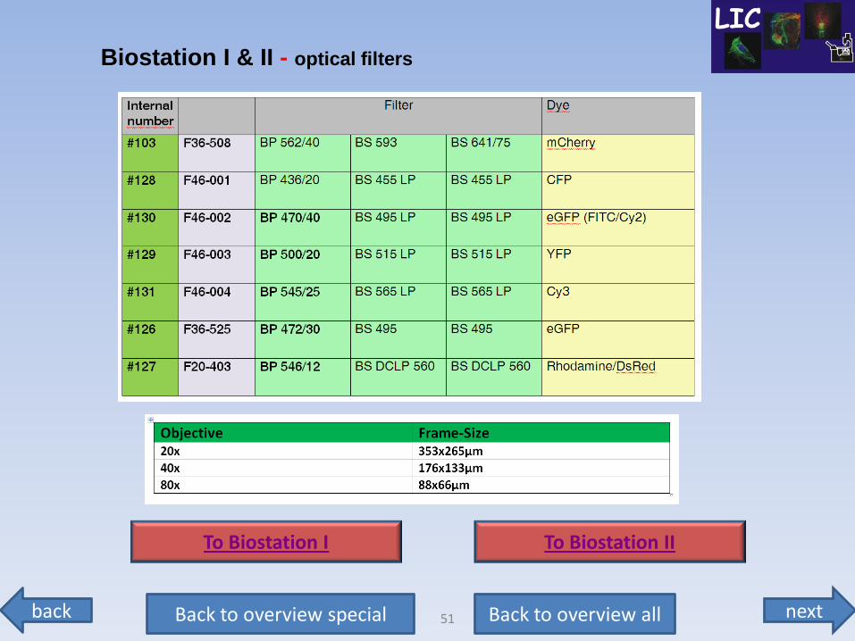
Biostation I & II - optical filters
To Biostation I To Biostation II
next back 52 Back to overview all Back to overview special
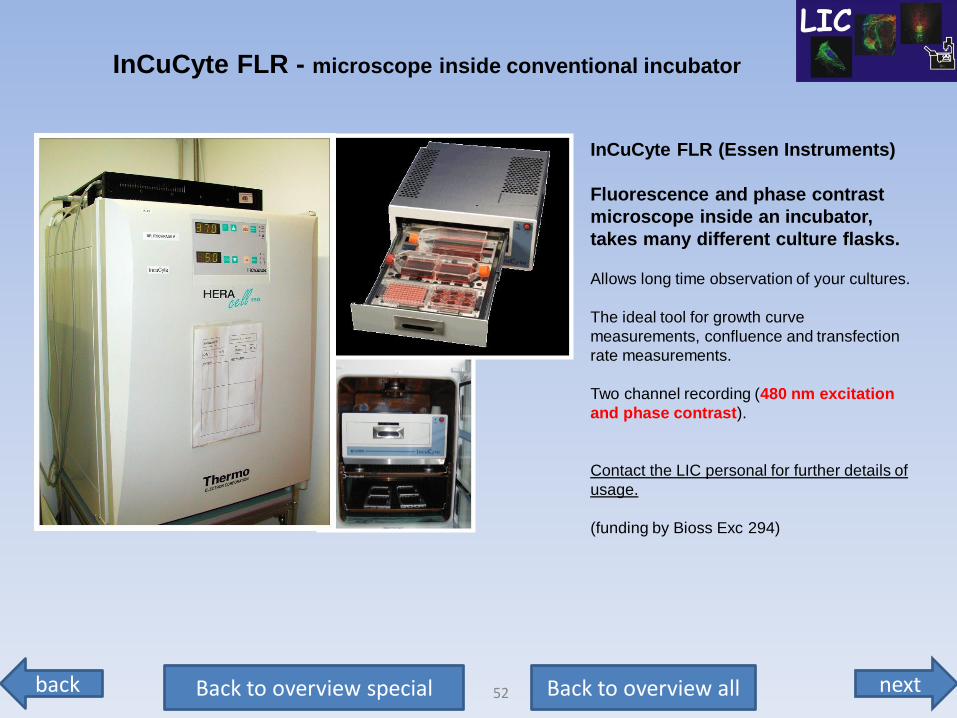
InCuCyte FLR (Essen Instruments)
Fluorescence and phase contrast
microscope inside an incubator,
takes many different culture flasks.
Allows long time observation of your cultures.
The ideal tool for growth curve
measurements, confluence and transfection
rate measurements.
Two channel recording (480 nm excitation
and phase contrast).
Contact the LIC personal for further details of
usage.
(funding by Bioss Exc 294)
InCuCyte FLR - microscope inside conventional incubator
next back 53 Back to overview all Back to overview special
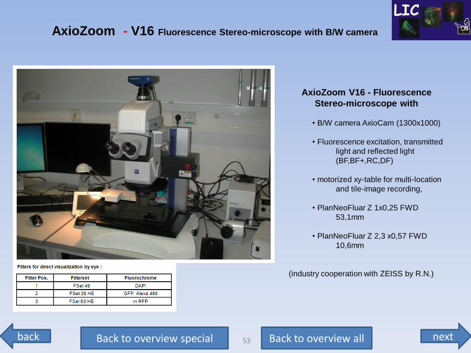
AxioZoom V16 - Fluorescence
Stereo-microscope with
• B/W camera AxioCam (1300x1000)
• Fluorescence excitation, transmitted
light and reflected light
(BF,BF+,RC,DF)
• motorized xy-table for multi-location
and tile-image recording,
• PlanNeoFluar Z 1x0,25 FWD
53,1mm
• PlanNeoFluar Z 2,3 x0,57 FWD
10,6mm
(industry cooperation with ZEISS by R.N.)
AxioZoom - V16 Fluorescence Stereo-microscope with B/W camera
next back 54 Back to overview all Back to overview special
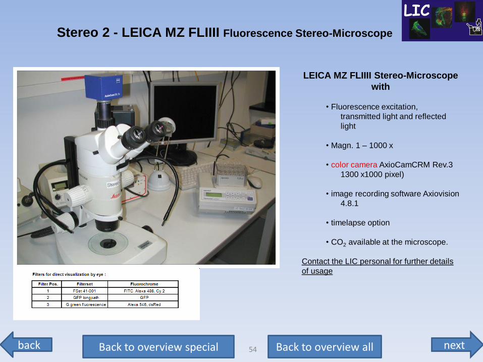
LEICA MZ FLIIII Stereo-Microscope
with
• Fluorescence excitation,
transmitted light and reflected
light
• Magn. 1 – 1000 x
• color camera AxioCamCRM Rev.3
1300 x1000 pixel)
• image recording software Axiovision
4.8.1
• timelapse option
• CO2 available at the microscope.
Contact the LIC personal for further details
of usage
Stereo 2 - LEICA MZ FLIIII Fluorescence Stereo-Microscope
next back 55 Back to overview all Back to overview special
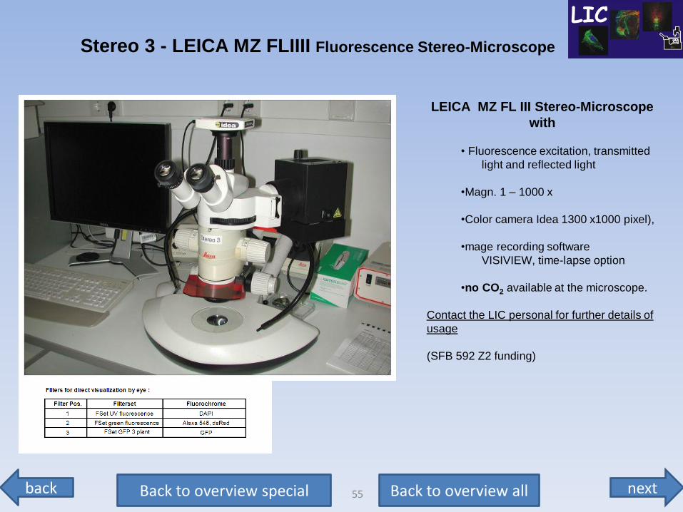
LEICA MZ FL III Stereo-Microscope
with
• Fluorescence excitation, transmitted
light and reflected light
•Magn. 1 – 1000 x
•Color camera Idea 1300 x1000 pixel),
•mage recording software
VISIVIEW, time-lapse option
•no CO2 available at the microscope.
Contact the LIC personal for further details of
usage
(SFB 592 Z2 funding)
Stereo 3 - LEICA MZ FLIIII Fluorescence Stereo-Microscope
next back 56 Back to overview all Back to overview special
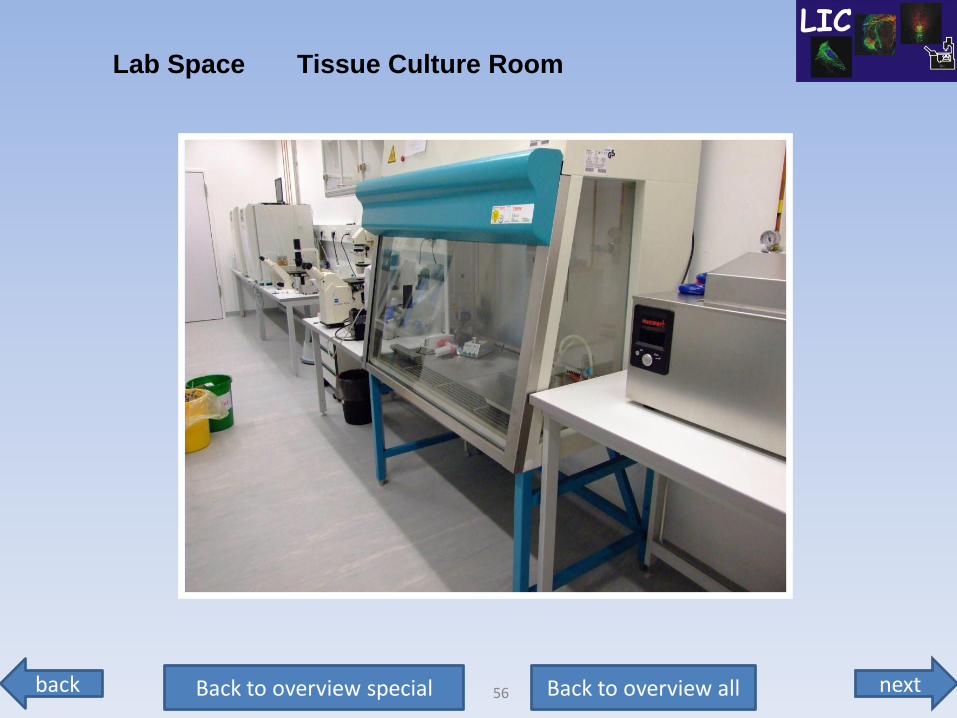
Lab Space Tissue Culture Room
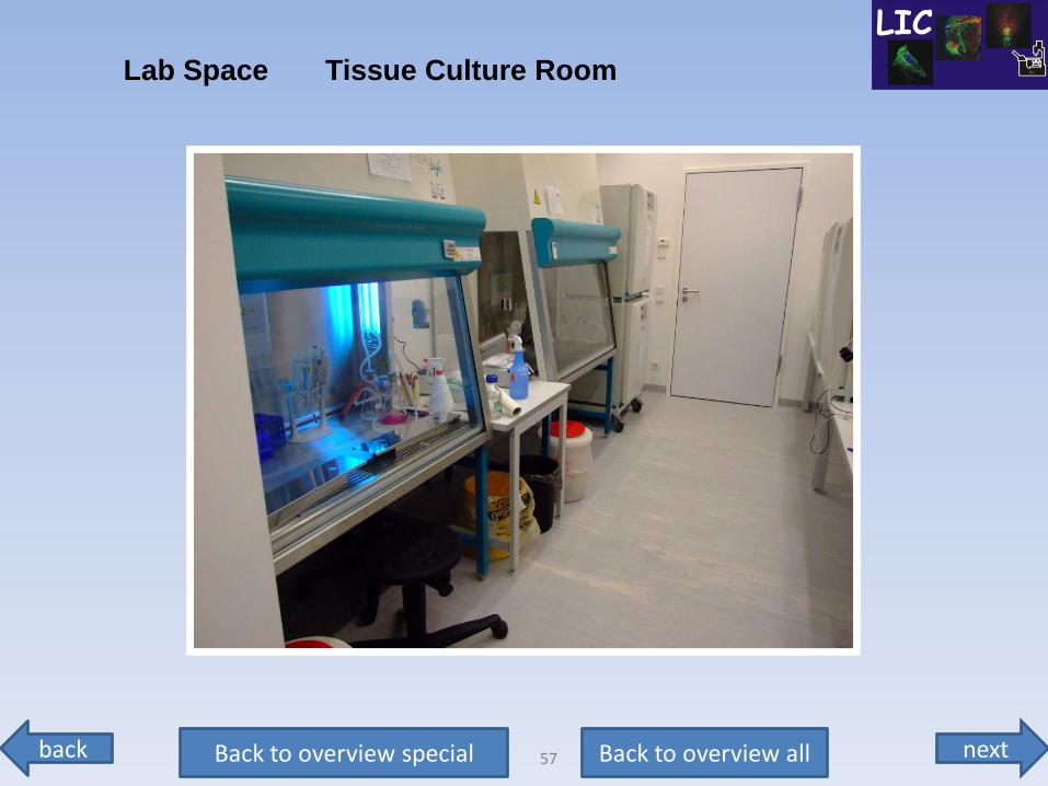
next back 57 Back to overview all Back to overview special
Lab Space Tissue Culture Room
next back 58 Back to overview all Back to overview special
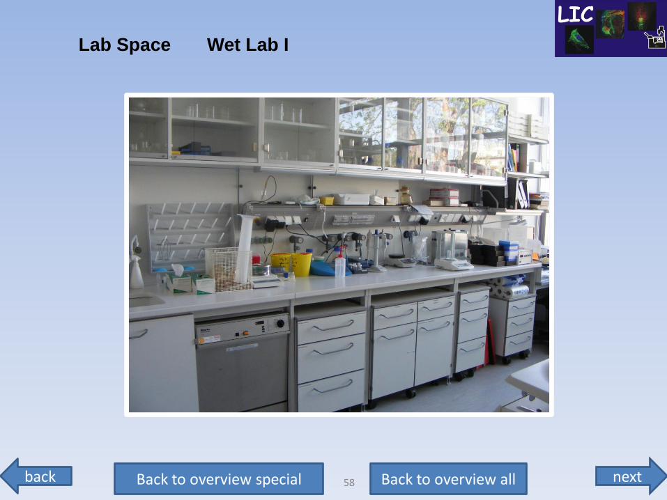
Lab Space Wet Lab I
next back 59 Back to overview all Back to overview special
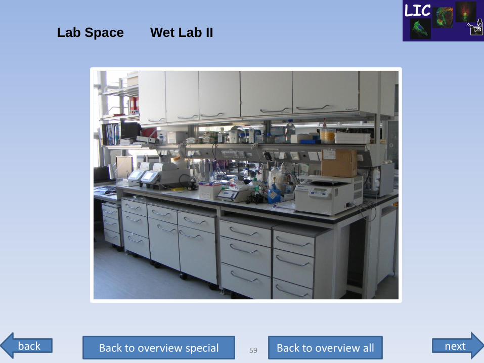
Lab Space Wet Lab II
next back 60 Back to overview all Back to overview special
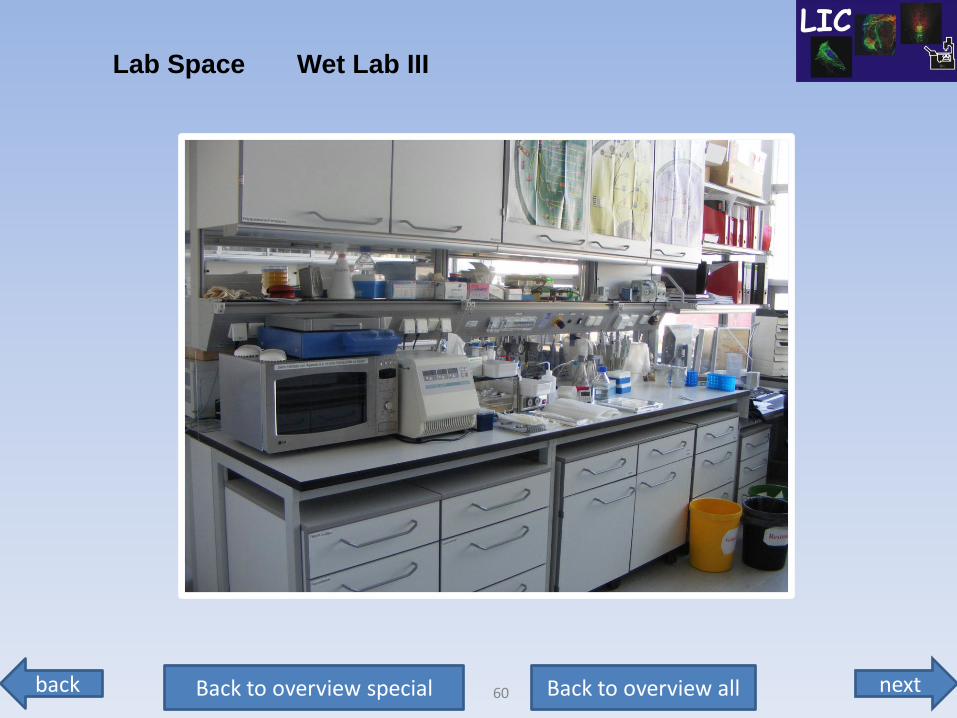
Lab Space Wet Lab III
next back 61 Back to overview all Back to overview special
End
We hope you enjoyed the tour.
The LIC Team
We are looking forward to help you with your research.
next back 62 Back to overview all Back to overview confocal
To STED objectives & filters
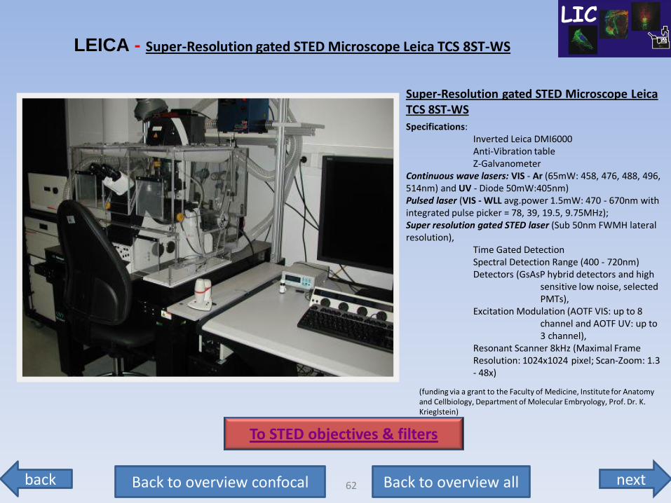
Super-Resolution gated STED Microscope Leica TCS 8ST-WS
Specifications: Inverted Leica DMI6000 Anti-Vibration table Z-Galvanometer Continuous wave lasers: VIS - Ar (65mW: 458, 476, 488, 496, 514nm) and UV - Diode 50mW:405nm) Pulsed laser (VIS - WLL avg.power 1.5mW: 470 - 670nm with integrated pulse picker = 78, 39, 19.5, 9.75MHz); Super resolution gated STED laser (Sub 50nm FWMH lateral resolution), Time Gated Detection Spectral Detection Range (400 - 720nm) Detectors (GsAsP hybrid detectors and high sensitive low noise, selected PMTs), Excitation Modulation (AOTF VIS: up to 8 channel and AOTF UV: up to 3 channel), Resonant Scanner 8kHz (Maximal Frame Resolution: 1024x1024 pixel; Scan-Zoom: 1.3 - 48x)
(funding via a grant to the Faculty of Medicine, Institute for Anatomy and Cellbiology, Department of Molecular Embryology, Prof. Dr. K. Krieglstein)
LEICA - Super-Resolution gated STED Microscope Leica TCS 8ST-WS
next back 63 Back to overview all Back to overview confocal
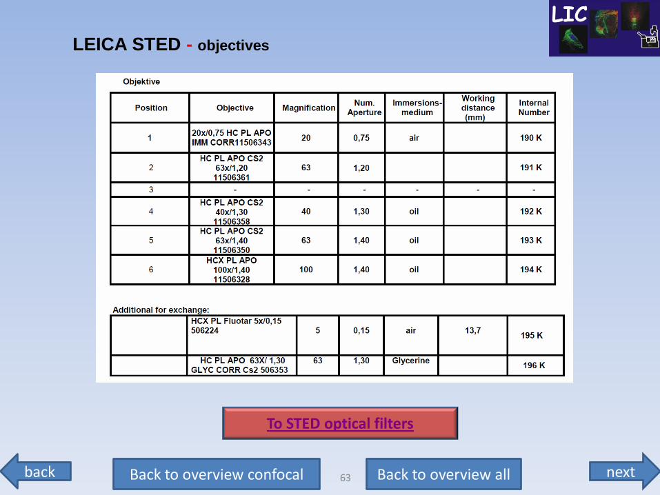
LEICA STED - objectives
To STED optical filters
next back 64 Back to overview all Back to overview confocal
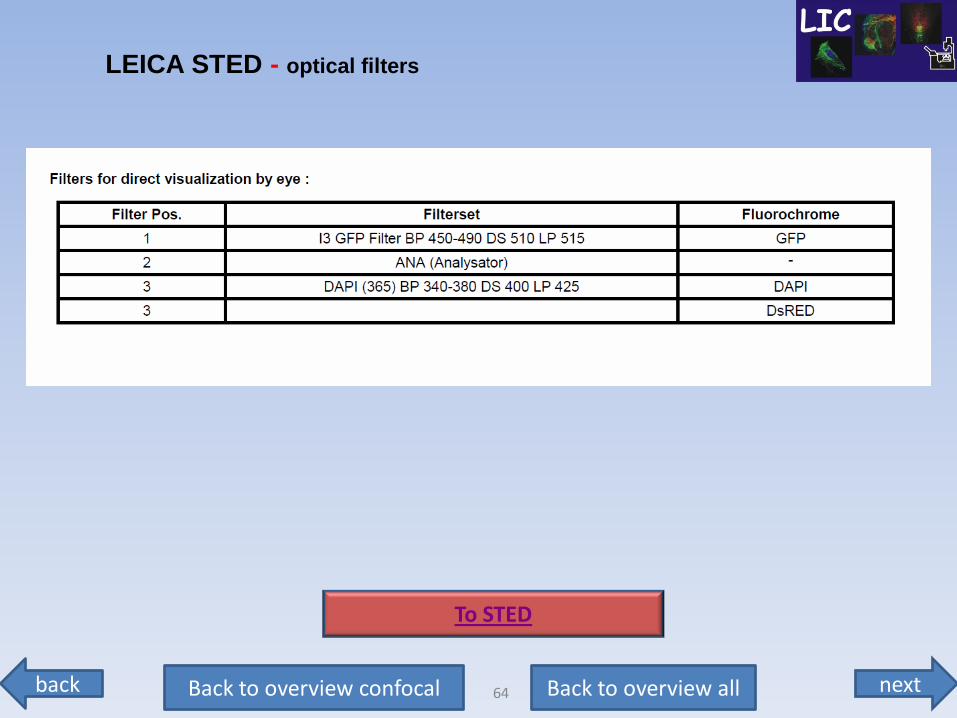
LEICA STED - optical filters
To STED