Embed Size (px)
Citation preview
© 2014 Pearson Education, Inc.
This work is protected by United States copyright laws and is provided solely for the use of instructors in teaching their courses and assessing student learning. Dissemination or sale of any part of this work (including on the World Wide Web) will destroy the integrity of the work and is not permitted. The work and materials from it should never be made available to students except by instructors using the accompanying text in their classes. All recipients of this work are expected to abide by these restrictions and to honor the intended pedagogical purposes and the needs of other instructors who rely on these materials.
Lecture PowerPoints
Chapter 25 Physics: Principles with Applications, 7th edition
Giancoli
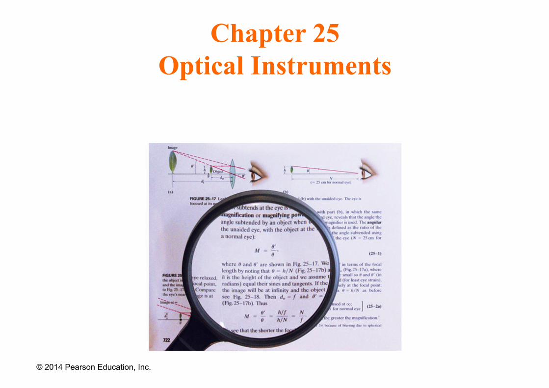
Chapter 25 Optical Instruments
© 2014 Pearson Education, Inc.
Contents of Chapter 25
• Cameras, Film, and Digital
• The Human Eye; Corrective Lenses
• Magnifying Glass
• Telescopes
• Compound Microscope
• Aberrations of Lenses and Mirrors
• Limits of Resolution; Circular Apertures
© 2014 Pearson Education, Inc.
Contents of Chapter 25
• Resolution of Telescopes and Microscopes; the λ Limit
• Resolution of the Human Eye and Useful Magnification
• Specialty Microscopes and Contrast
• X-Rays and X-Ray Diffraction
• X-Ray Imaging and Computed Tomography (CT Scan)
© 2014 Pearson Education, Inc.
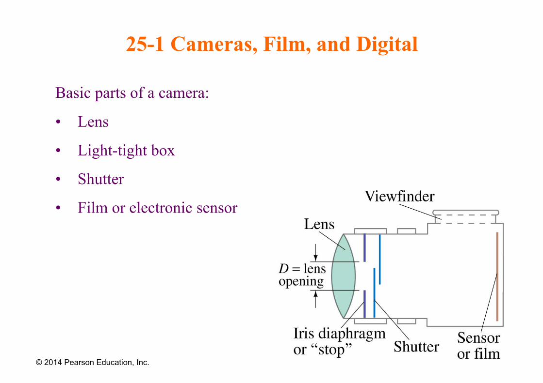
25-1 Cameras, Film, and Digital
Basic parts of a camera:
• Lens
• Light-tight box
• Shutter
• Film or electronic sensor
© 2014 Pearson Education, Inc.
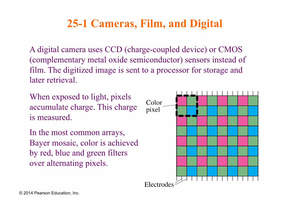
25-1 Cameras, Film, and Digital
A digital camera uses CCD (charge-coupled device) or CMOS (complementary metal oxide semiconductor) sensors instead of film. The digitized image is sent to a processor for storage and later retrieval.
© 2014 Pearson Education, Inc.
When exposed to light, pixels accumulate charge. This charge is measured.
In the most common arrays, Bayer mosaic, color is achieved by red, blue and green filters over alternating pixels.
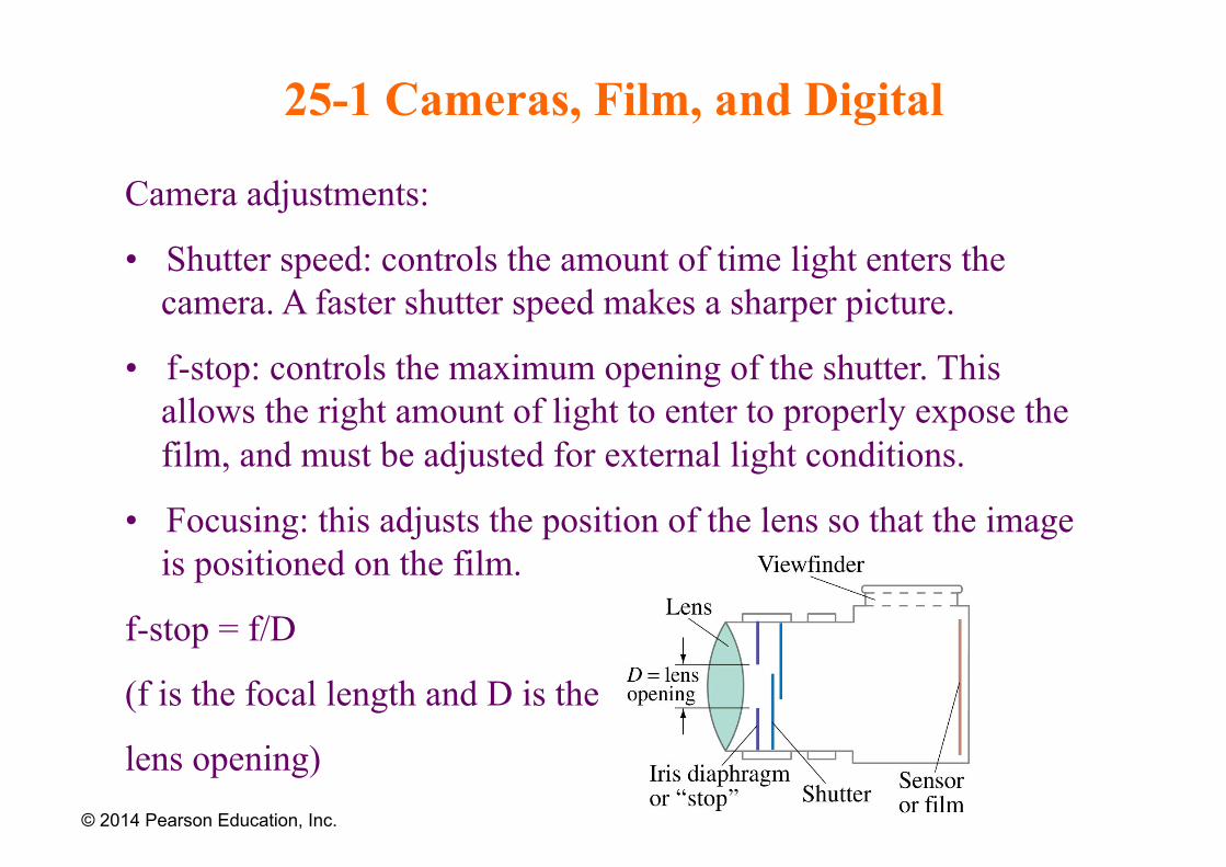
25-1 Cameras, Film, and Digital
Camera adjustments:
• Shutter speed: controls the amount of time light enters the camera. A faster shutter speed makes a sharper picture.
• f-stop: controls the maximum opening of the shutter. This allows the right amount of light to enter to properly expose the film, and must be adjusted for external light conditions.
• Focusing: this adjusts the position of the lens so that the image is positioned on the film.
f-stop = f/D
(f is the focal length and D is the
lens opening) © 2014 Pearson Education, Inc.
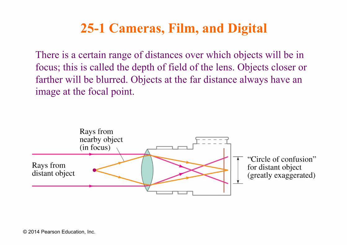
25-1 Cameras, Film, and Digital
There is a certain range of distances over which objects will be in focus; this is called the depth of field of the lens. Objects closer or farther will be blurred. Objects at the far distance always have an image at the focal point.
© 2014 Pearson Education, Inc.
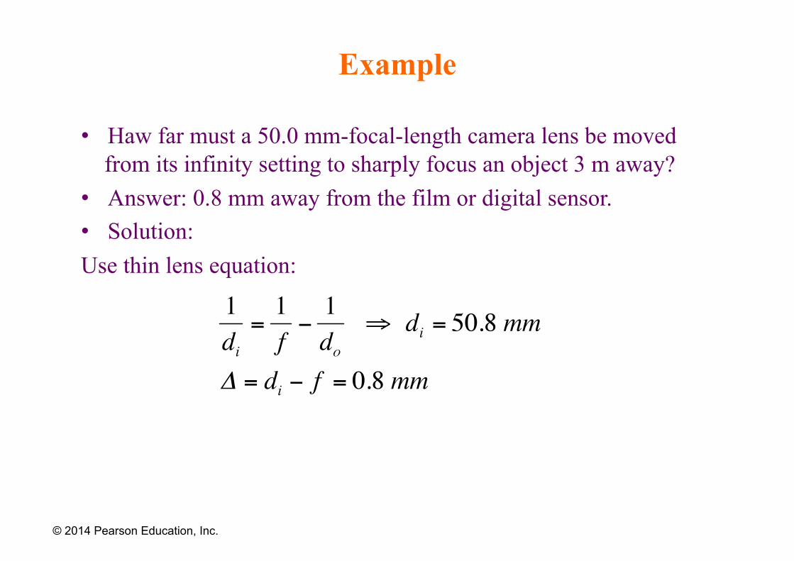
Example
• Haw far must a 50.0 mm-focal-length camera lens be moved from its infinity setting to sharply focus an object 3 m away?
• Answer: 0.8 mm away from the film or digital sensor. • Solution: Use thin lens equation:
© 2014 Pearson Education, Inc.
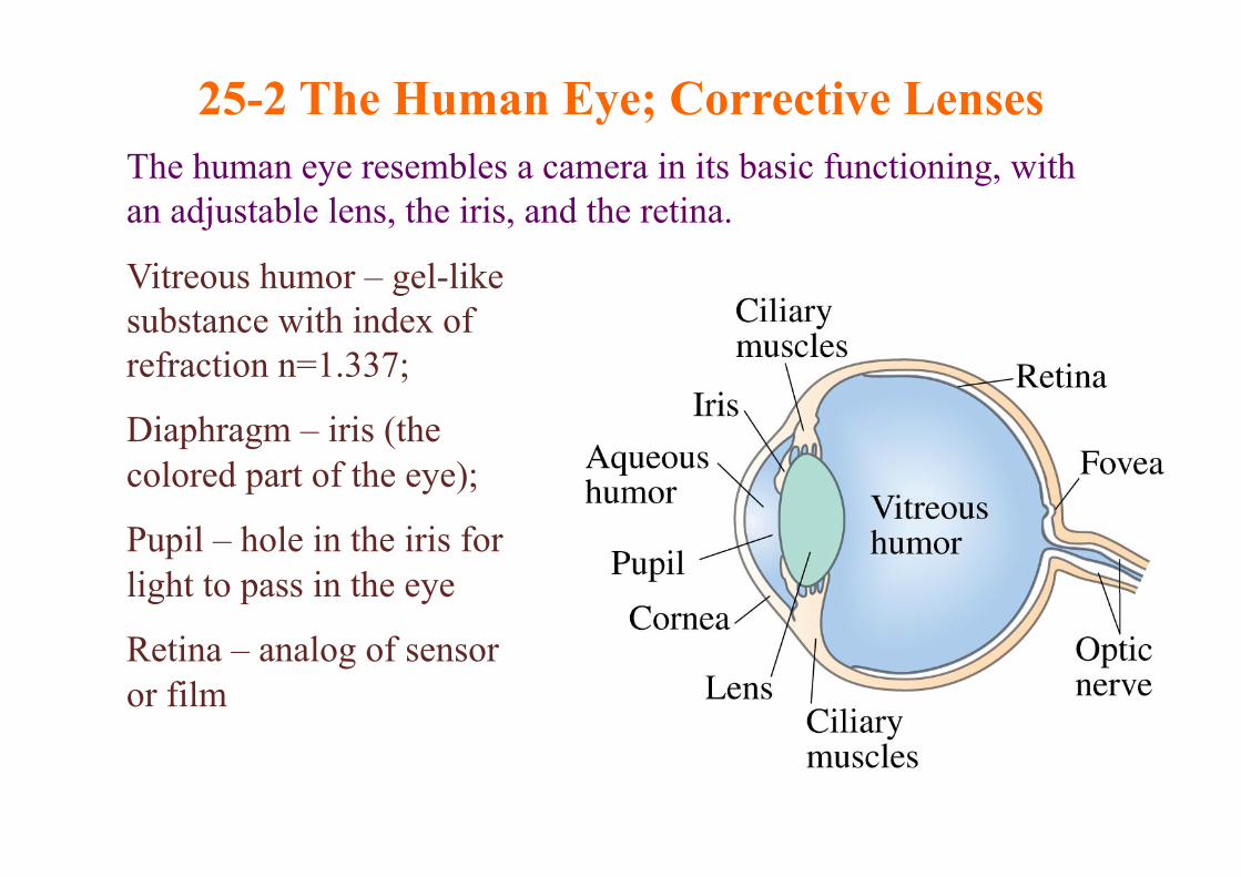
25-2 The Human Eye; Corrective Lenses The human eye resembles a camera in its basic functioning, with an adjustable lens, the iris, and the retina.
Vitreous humor – gel-like substance with index of refraction n=1.337;
Diaphragm – iris (the colored part of the eye);
Pupil – hole in the iris for light to pass in the eye
Retina – analog of sensor or film
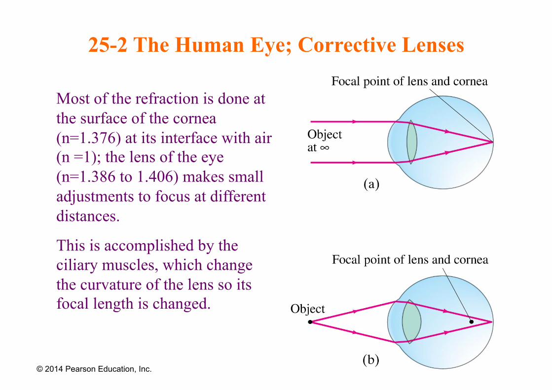
25-2 The Human Eye; Corrective Lenses
Most of the refraction is done at the surface of the cornea (n=1.376) at its interface with air (n =1); the lens of the eye (n=1.386 to 1.406) makes small adjustments to focus at different distances.
This is accomplished by the ciliary muscles, which change the curvature of the lens so its focal length is changed.
© 2014 Pearson Education, Inc.
25-2 The Human Eye; Corrective Lenses
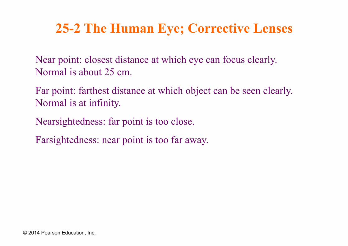
Near point: closest distance at which eye can focus clearly. Normal is about 25 cm.
Far point: farthest distance at which object can be seen clearly. Normal is at infinity.
Nearsightedness: far point is too close.
Farsightedness: near point is too far away.
© 2014 Pearson Education, Inc.
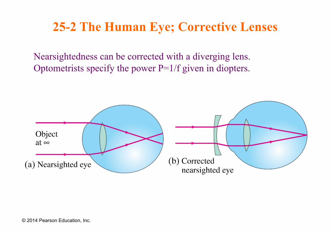
25-2 The Human Eye; Corrective Lenses
Nearsightedness can be corrected with a diverging lens. Optometrists specify the power P=1/f given in diopters.
© 2014 Pearson Education, Inc.
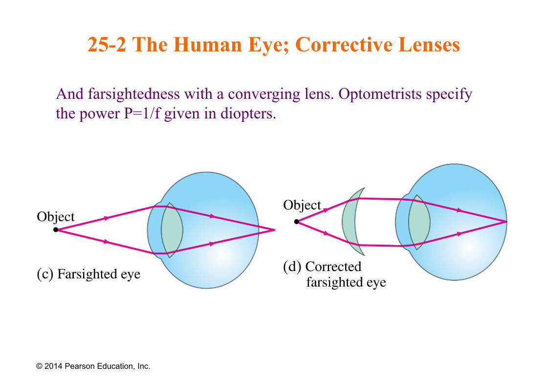
25-2 The Human Eye; Corrective Lenses
And farsightedness with a converging lens. Optometrists specify the power P=1/f given in diopters.
© 2014 Pearson Education, Inc.
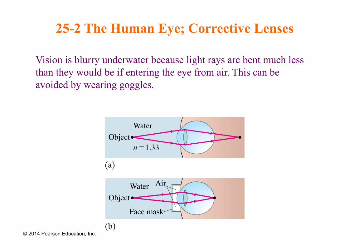
25-2 The Human Eye; Corrective Lenses
Vision is blurry underwater because light rays are bent much less than they would be if entering the eye from air. This can be avoided by wearing goggles.
© 2014 Pearson Education, Inc.
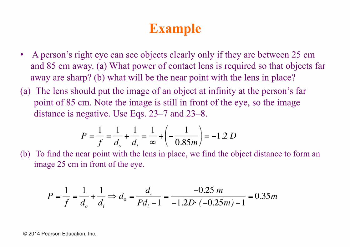
Example
• A person’s right eye can see objects clearly only if they are between 25 cm and 85 cm away. (a) What power of contact lens is required so that objects far away are sharp? (b) what will be the near point with the lens in place?
(a) The lens should put the image of an object at infinity at the person’s far point of 85 cm. Note the image is still in front of the eye, so the image distance is negative. Use Eqs. 23–7 and 23–8.
(b) To find the near point with the lens in place, we find the object distance to form an image 25 cm in front of the eye.
© 2014 Pearson Education, Inc.
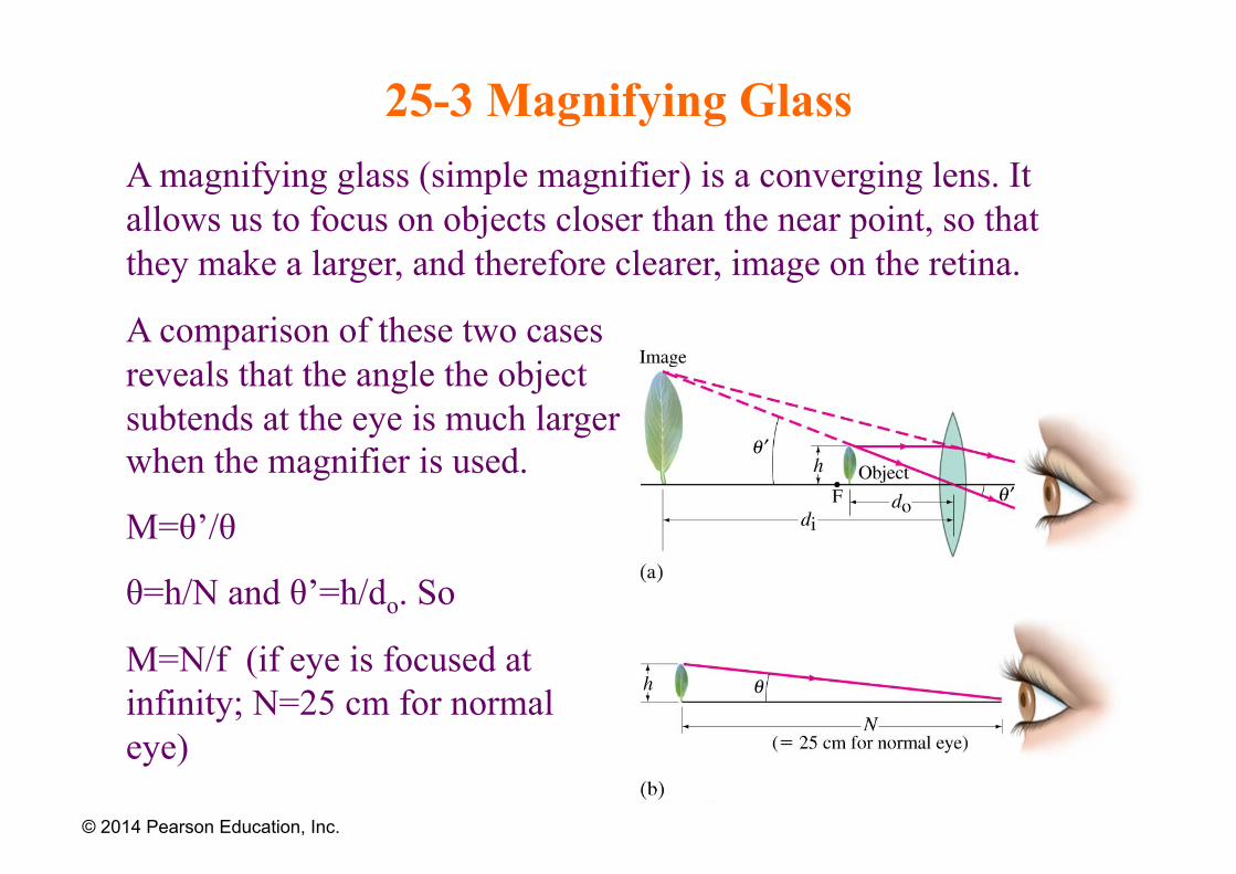
25-3 Magnifying Glass A magnifying glass (simple magnifier) is a converging lens. It allows us to focus on objects closer than the near point, so that they make a larger, and therefore clearer, image on the retina.
© 2014 Pearson Education, Inc.
A comparison of these two cases reveals that the angle the object subtends at the eye is much larger when the magnifier is used.
M=θ’/θ
θ=h/N and θ’=h/do. So
M=N/f (if eye is focused at infinity; N=25 cm for normal eye)

25-3 Magnifying Glass
The power of a magnifying glass is described by its angular magnification:
If the eye is relaxed (N is the near point distance and f the focal length):
If the eye is focused at the near point:
The shorter is the focus length, the stronger is magnification.
© 2014 Pearson Education, Inc.
(25-1)
(25-2a)
(25-2b)
25-4 Telescopes
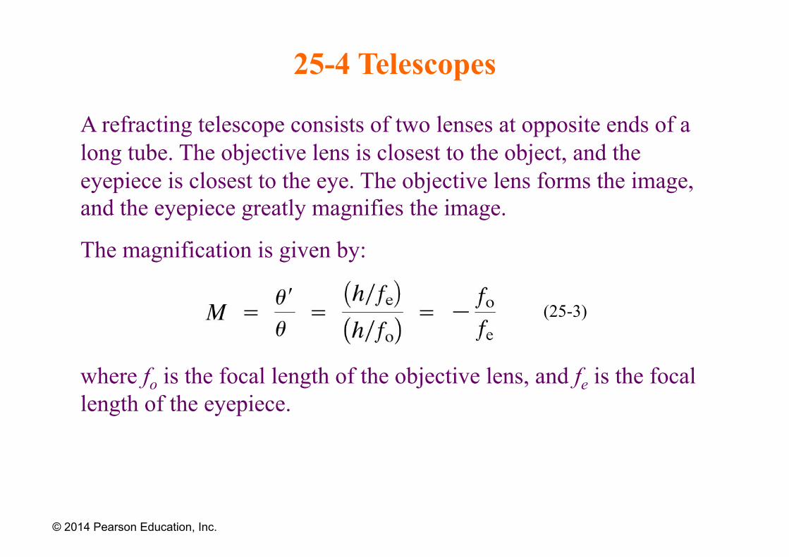
A refracting telescope consists of two lenses at opposite ends of a long tube. The objective lens is closest to the object, and the eyepiece is closest to the eye. The objective lens forms the image, and the eyepiece greatly magnifies the image.
The magnification is given by:
where fo is the focal length of the objective lens, and fe is the focal length of the eyepiece.
© 2014 Pearson Education, Inc.
(25-3)
25-4 Telescopes
© 2014 Pearson Education, Inc.
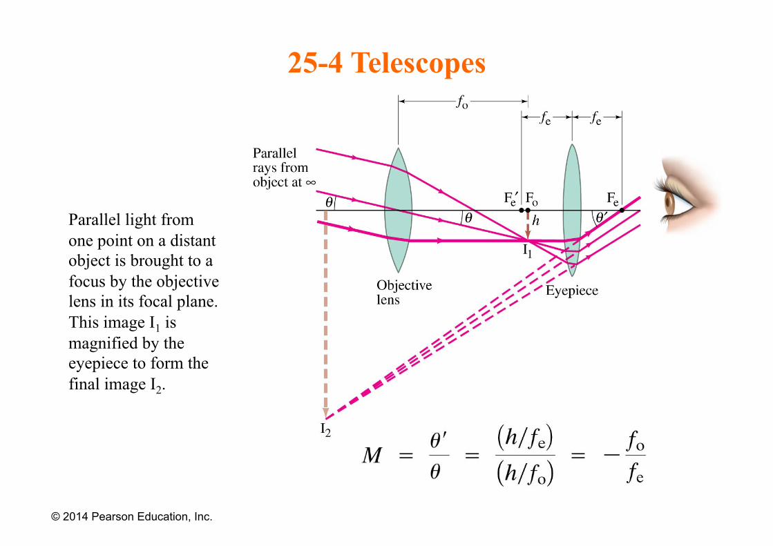
Parallel light from one point on a distant object is brought to a focus by the objective lens in its focal plane. This image I1 is magnified by the eyepiece to form the final image I2.
25-4 Telescopes
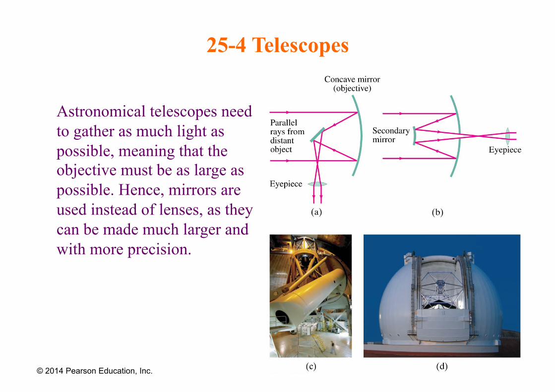
Astronomical telescopes need to gather as much light as possible, meaning that the objective must be as large as possible. Hence, mirrors are used instead of lenses, as they can be made much larger and with more precision.
© 2014 Pearson Education, Inc.
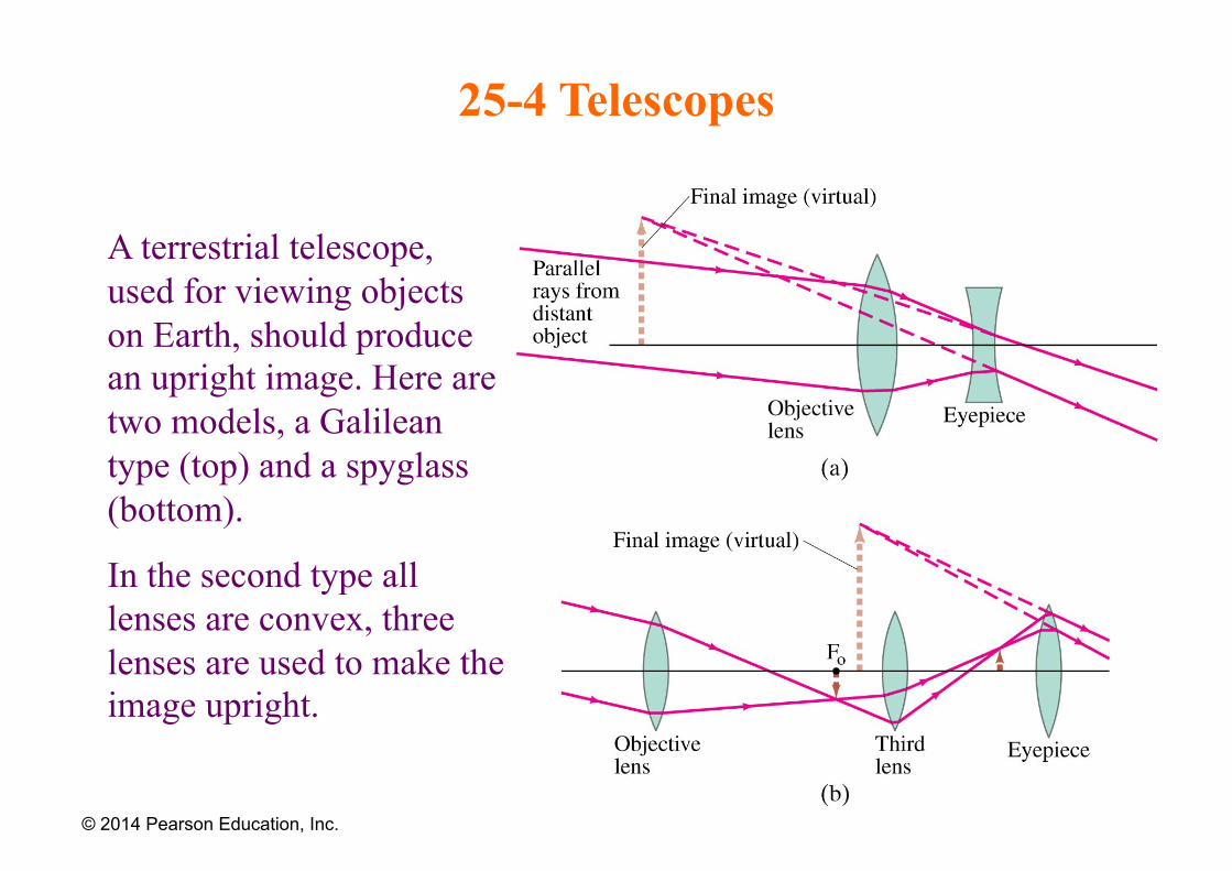
25-4 Telescopes
A terrestrial telescope, used for viewing objects on Earth, should produce an upright image. Here are two models, a Galilean type (top) and a spyglass (bottom).
In the second type all lenses are convex, three lenses are used to make the image upright.
© 2014 Pearson Education, Inc.

25-5 Compound Microscope A compound microscope also has an objective and an eyepiece; it is different from a telescope in that the object is placed very close to the eyepiece. The image I1 formed by objective lens is real and enlarged. The eyepiece is positioned in such a way, that this image is near the eyepiece focal point Fe, so it can be significantly magnified by the eyepiece into a very large virtual image I2.
© 2014 Pearson Education, Inc.
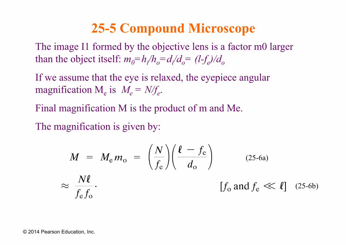
25-5 Compound Microscope The image I1 formed by the objective lens is a factor m0 larger than the object itself: m0=hi/ho=di/do= (l-fe)/do
If we assume that the eye is relaxed, the eyepiece angular magnification Me is Me = N/fe.
Final magnification M is the product of m and Me.
The magnification is given by:
© 2014 Pearson Education, Inc.
(25-6a)
(25-6b)
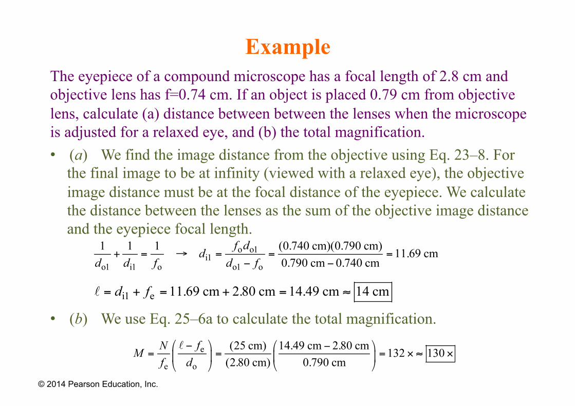
Example The eyepiece of a compound microscope has a focal length of 2.8 cm and objective lens has f=0.74 cm. If an object is placed 0.79 cm from objective lens, calculate (a) distance between between the lenses when the microscope is adjusted for a relaxed eye, and (b) the total magnification. • (a) We find the image distance from the objective using Eq. 23–8. For
the final image to be at infinity (viewed with a relaxed eye), the objective image distance must be at the focal distance of the eyepiece. We calculate the distance between the lenses as the sum of the objective image distance and the eyepiece focal length.
• (b) We use Eq. 25–6a to calculate the total magnification.
© 2014 Pearson Education, Inc.
25-6 Aberrations of Lenses and Mirrors
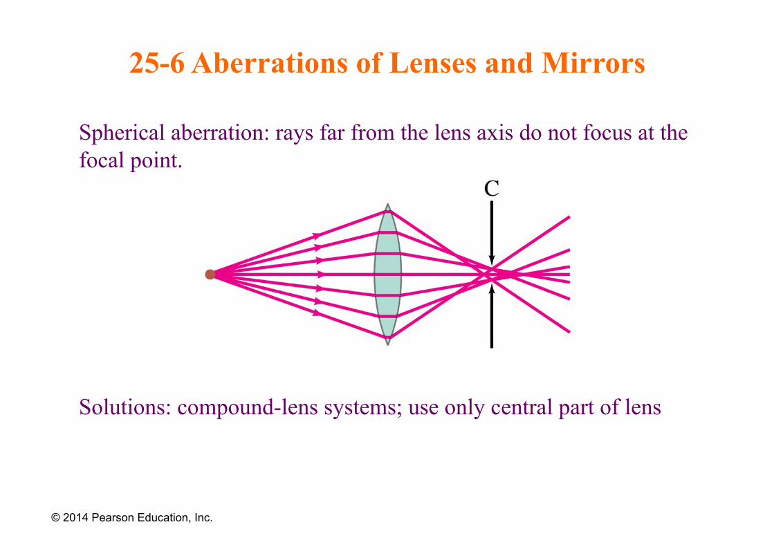
Spherical aberration: rays far from the lens axis do not focus at the focal point.
Solutions: compound-lens systems; use only central part of lens
© 2014 Pearson Education, Inc.
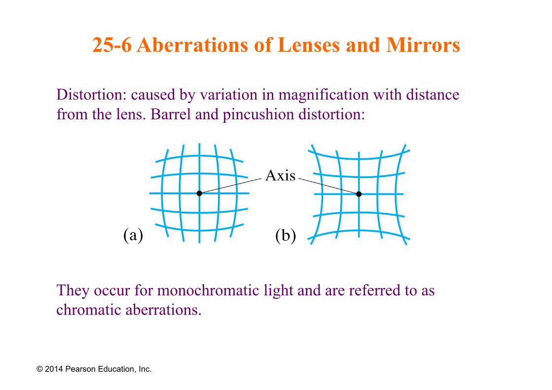
25-6 Aberrations of Lenses and Mirrors
Distortion: caused by variation in magnification with distance from the lens. Barrel and pincushion distortion:
They occur for monochromatic light and are referred to as chromatic aberrations.
© 2014 Pearson Education, Inc.
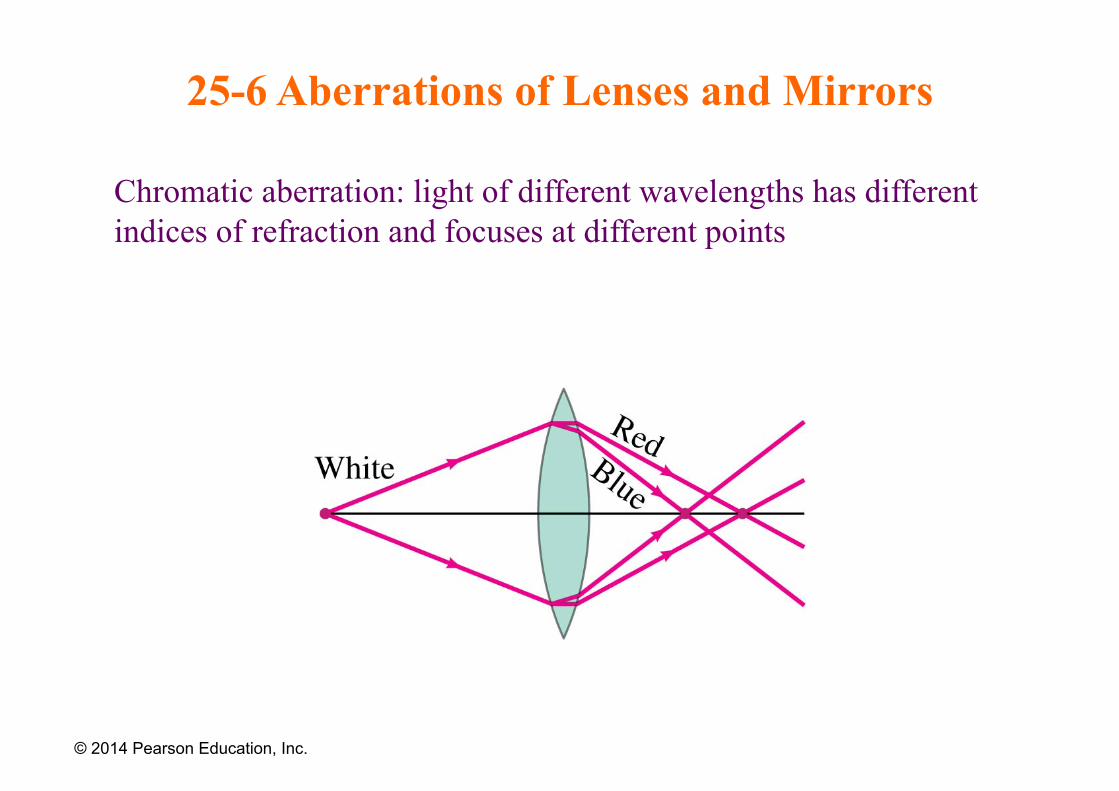
25-6 Aberrations of Lenses and Mirrors
Chromatic aberration: light of different wavelengths has different indices of refraction and focuses at different points
© 2014 Pearson Education, Inc.
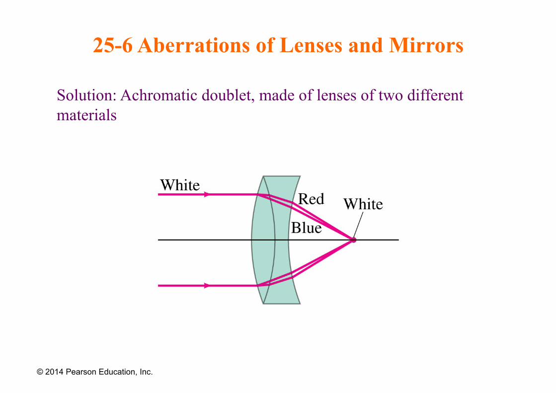
25-6 Aberrations of Lenses and Mirrors
Solution: Achromatic doublet, made of lenses of two different materials
© 2014 Pearson Education, Inc.
25-7 Limits of Resolution; Circular Apertures
Resolution is the distance at which a lens can barely distinguish two separate objects.
Resolution is limited by aberrations and by diffraction. Aberrations can be minimized, but diffraction is unavoidable; it is due to the size of the lens compared to the wavelength of the light.
© 2014 Pearson Education, Inc.
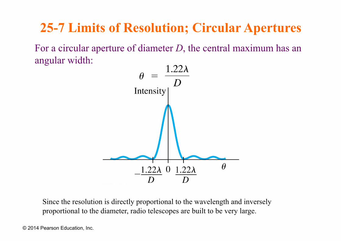
25-7 Limits of Resolution; Circular Apertures For a circular aperture of diameter D, the central maximum has an angular width:
© 2014 Pearson Education, Inc.
Since the resolution is directly proportional to the wavelength and inversely proportional to the diameter, radio telescopes are built to be very large.
25-7 Limits of Resolution; Circular Apertures
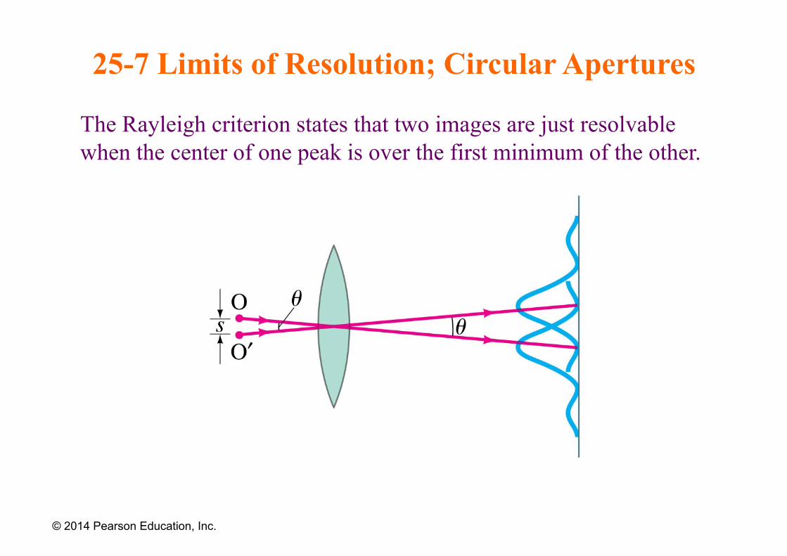
The Rayleigh criterion states that two images are just resolvable when the center of one peak is over the first minimum of the other.
© 2014 Pearson Education, Inc.
25-8 Resolution of Telescopes and Microscopes; the λ Limit
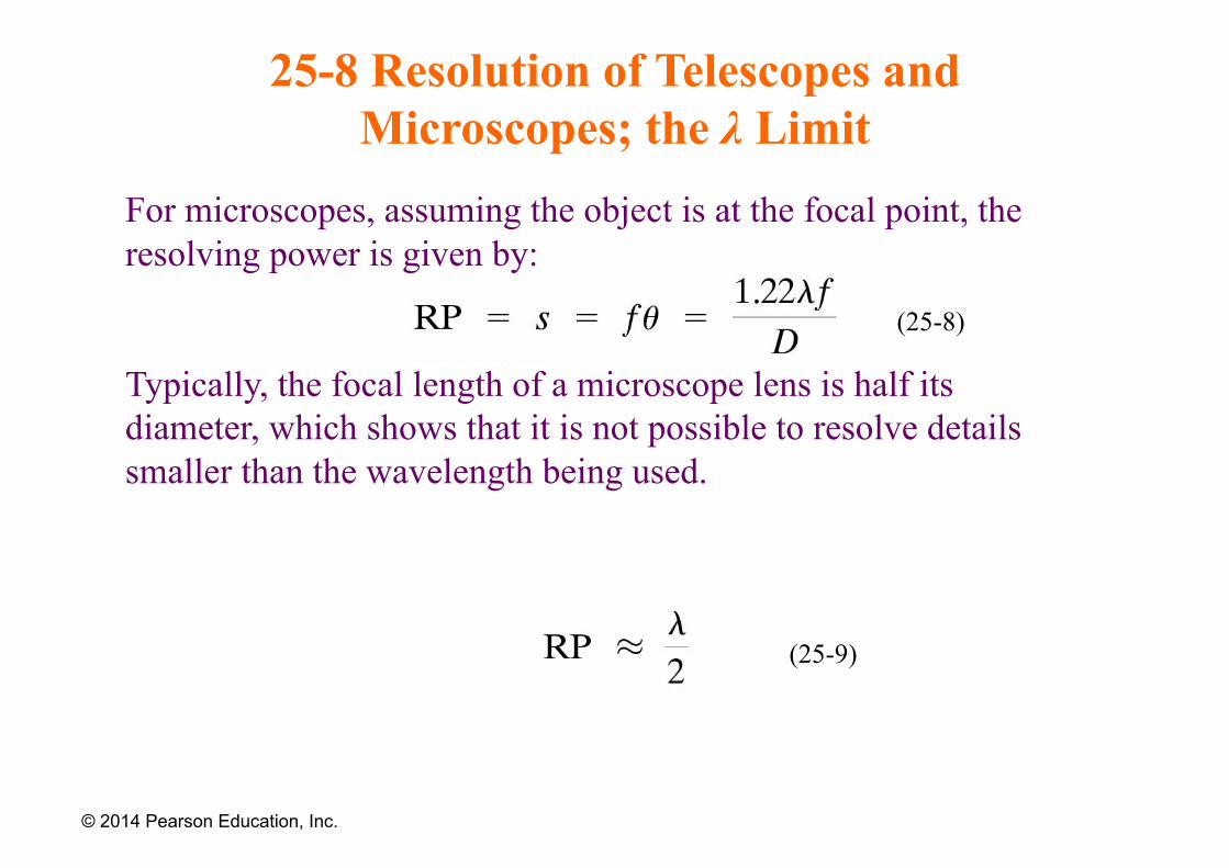
For microscopes, assuming the object is at the focal point, the resolving power is given by:
Typically, the focal length of a microscope lens is half its diameter, which shows that it is not possible to resolve details smaller than the wavelength being used.
© 2014 Pearson Education, Inc.
(25-8)
(25-9)
25-9 Resolution of the Human Eye and Useful Magnification
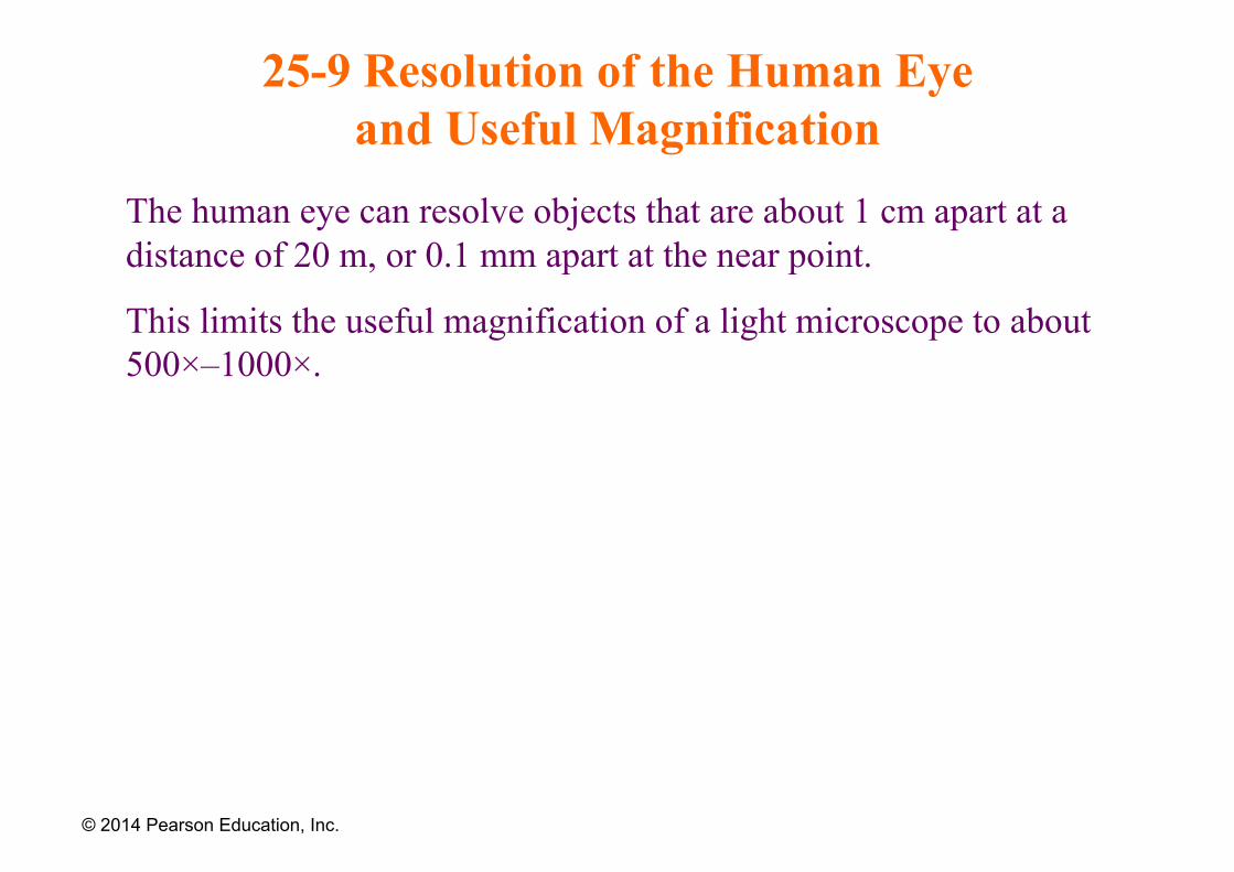
The human eye can resolve objects that are about 1 cm apart at a distance of 20 m, or 0.1 mm apart at the near point.
This limits the useful magnification of a light microscope to about 500×–1000×.
© 2014 Pearson Education, Inc.
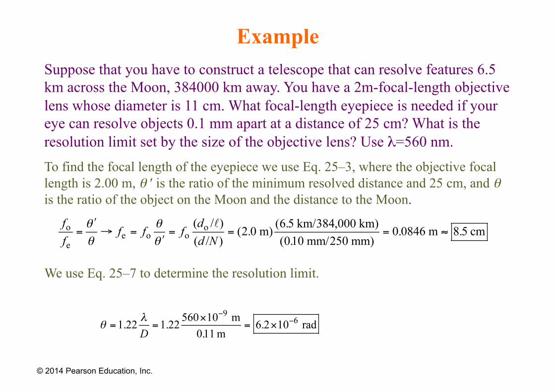
Example Suppose that you have to construct a telescope that can resolve features 6.5 km across the Moon, 384000 km away. You have a 2m-focal-length objective lens whose diameter is 11 cm. What focal-length eyepiece is needed if your eye can resolve objects 0.1 mm apart at a distance of 25 cm? What is the resolution limit set by the size of the objective lens? Use λ=560 nm. To find the focal length of the eyepiece we use Eq. 25–3, where the objective focal length is 2.00 m, θ ′ is the ratio of the minimum resolved distance and 25 cm, and θ is the ratio of the object on the Moon and the distance to the Moon.
We use Eq. 25–7 to determine the resolution limit.
© 2014 Pearson Education, Inc.
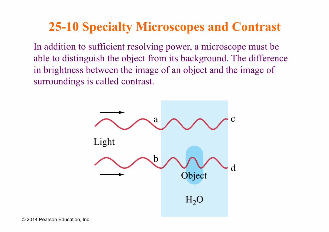
25-10 Specialty Microscopes and Contrast In addition to sufficient resolving power, a microscope must be able to distinguish the object from its background. The difference in brightness between the image of an object and the image of surroundings is called contrast.
© 2014 Pearson Education, Inc.
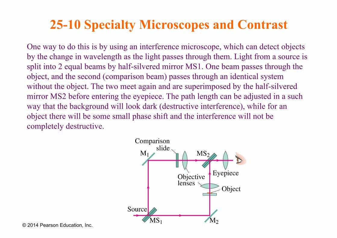
25-10 Specialty Microscopes and Contrast One way to do this is by using an interference microscope, which can detect objects by the change in wavelength as the light passes through them. Light from a source is split into 2 equal beams by half-silvered mirror MS1. One beam passes through the object, and the second (comparison beam) passes through an identical system without the object. The two meet again and are superimposed by the half-silvered mirror MS2 before entering the eyepiece. The path length can be adjusted in a such way that the background will look dark (destructive interference), while for an object there will be some small phase shift and the interference will not be completely destructive.
© 2014 Pearson Education, Inc.
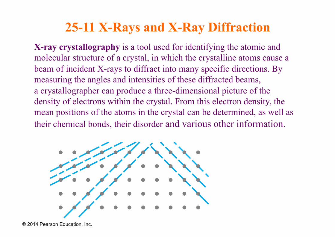
25-11 X-Rays and X-Ray Diffraction X-ray crystallography is a tool used for identifying the atomic and molecular structure of a crystal, in which the crystalline atoms cause a beam of incident X-rays to diffract into many specific directions. By measuring the angles and intensities of these diffracted beams, a crystallographer can produce a three-dimensional picture of the density of electrons within the crystal. From this electron density, the mean positions of the atoms in the crystal can be determined, as well as their chemical bonds, their disorder and various other information.
© 2014 Pearson Education, Inc.
25-11 X-Rays and X-Ray Diffraction X-ray crystallography has been fundamental in the development of many scientific fields. In its first decades of use, this method determined
1) the size of atoms;
2) the lengths and types of chemical bonds;
3) the atomic-scale differences among various materials, especially minerals and alloys.
The method also revealed the structure and function of many biological molecules, including vitamins, drugs, proteins and nucleic acids such as DNA.
25-11 X-Rays and X-Ray Diffraction
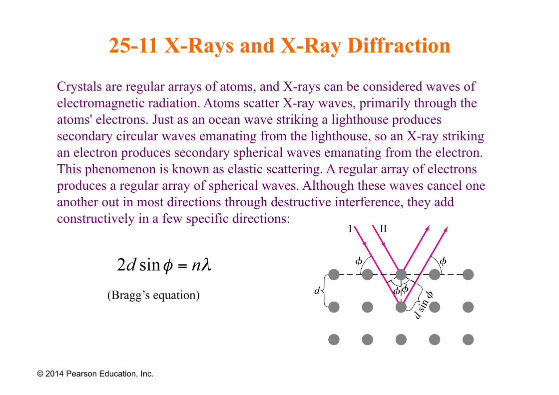
Crystals are regular arrays of atoms, and X-rays can be considered waves of electromagnetic radiation. Atoms scatter X-ray waves, primarily through the atoms' electrons. Just as an ocean wave striking a lighthouse produces secondary circular waves emanating from the lighthouse, so an X-ray striking an electron produces secondary spherical waves emanating from the electron. This phenomenon is known as elastic scattering. A regular array of electrons produces a regular array of spherical waves. Although these waves cancel one another out in most directions through destructive interference, they add constructively in a few specific directions:
© 2014 Pearson Education, Inc.
(Bragg’s equation)
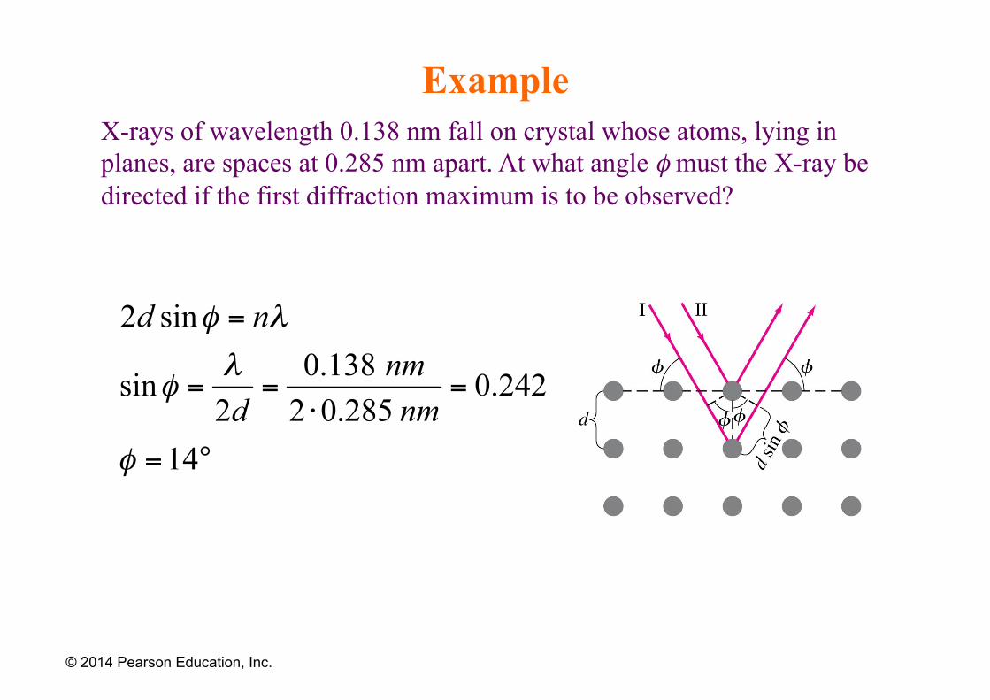
Example X-rays of wavelength 0.138 nm fall on crystal whose atoms, lying in planes, are spaces at 0.285 nm apart. At what angle φ must the X-ray be directed if the first diffraction maximum is to be observed?
© 2014 Pearson Education, Inc.
25-12 X-Ray Imaging and Computed Tomography (CT Scan)
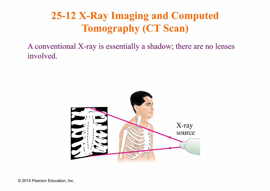
A conventional X-ray is essentially a shadow; there are no lenses involved.
© 2014 Pearson Education, Inc.
25-12 X-Ray Imaging and Computed Tomography (CT Scan)
X-ray computed tomography (x-ray CT) is a technology that uses computer-processed x-rays to produce tomographic images (virtual 'slices') of specific areas of the scanned object, allowing the user to see inside without cutting. Digital geometry processing is used to generate a three-dimensional image of the inside of an object from a large series of two-dimensional radiographic images taken around a single axis of rotation. The CT scanner was invented in 1972 by the British engineer Godfrey N. Hounsfield (later Sir Godfrey) and the South African (later American) physicist Alan Cormack. CT scanning was already in general use by 1979, the year Hounsfield and Cormack were awarded the Nobel Prize in Medicine or Physiology for its development.
© 2014 Pearson Education, Inc.
25-12 X-Ray Imaging and Computed Tomography (CT Scan)
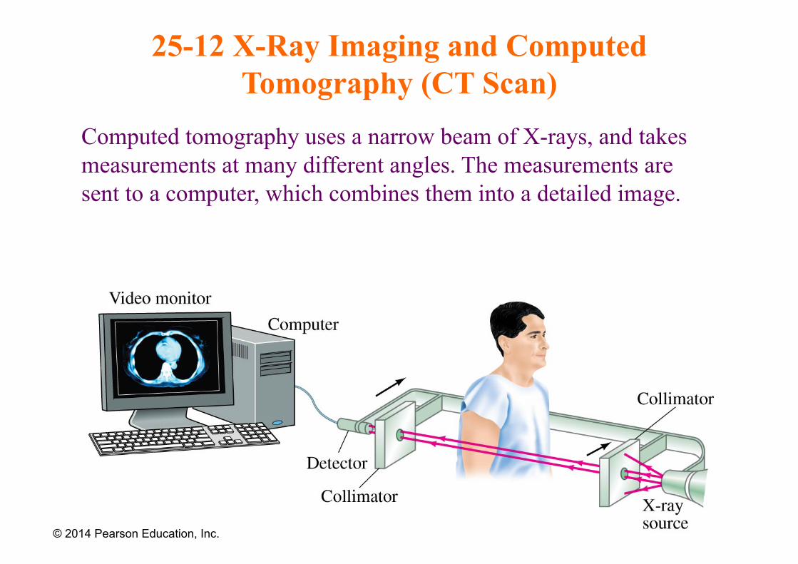
Computed tomography uses a narrow beam of X-rays, and takes measurements at many different angles. The measurements are sent to a computer, which combines them into a detailed image.
© 2014 Pearson Education, Inc.
25-12 X-Ray Imaging and Computed Tomography (CT Scan)
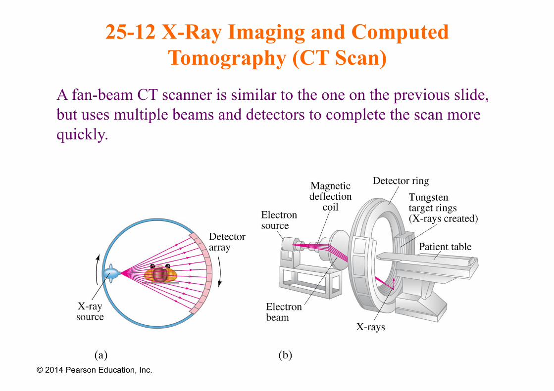
A fan-beam CT scanner is similar to the one on the previous slide, but uses multiple beams and detectors to complete the scan more quickly.
© 2014 Pearson Education, Inc.
25-12 X-Ray Imaging and Computed Tomography (CT Scan)
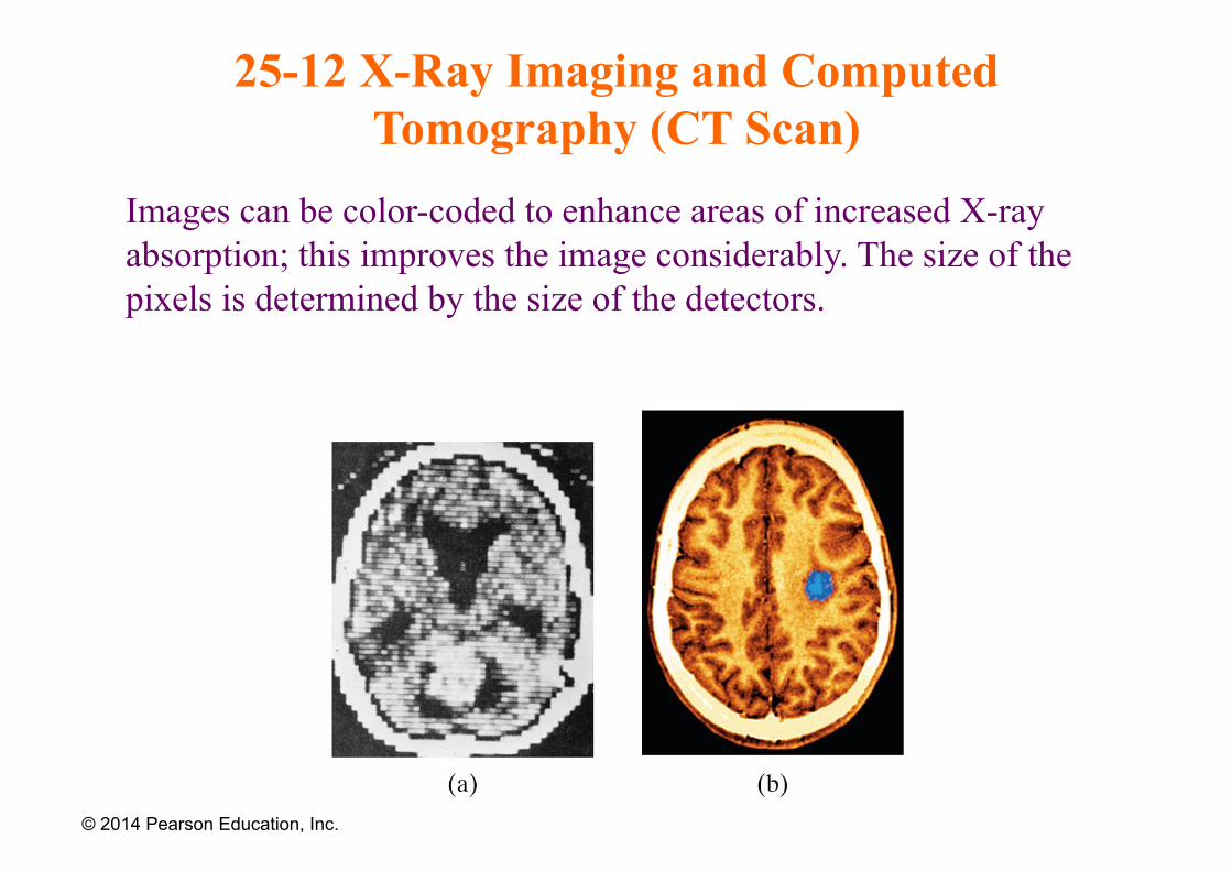
Images can be color-coded to enhance areas of increased X-ray absorption; this improves the image considerably. The size of the pixels is determined by the size of the detectors.
© 2014 Pearson Education, Inc.
Summary of Chapter 25
• Camera lens forms image by letting light through a shutter; can be adjusted for different light levels using f-stop and focused by moving lens
• Human eye forms image by letting light through pupil; adjusts to different light levels using iris and focuses by changing thickness of lens
• Nearsighted vision is corrected by diverging lens, farsighted by converging
© 2014 Pearson Education, Inc.
Summary of Chapter 25
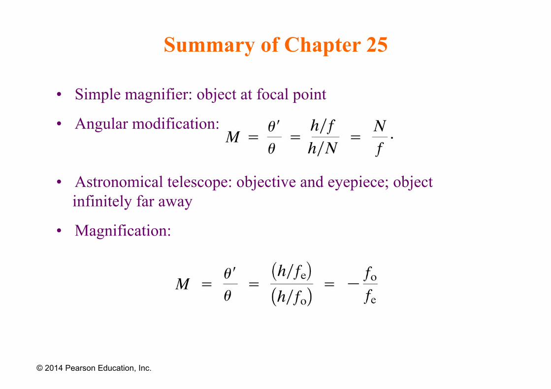
• Simple magnifier: object at focal point
• Angular modification:
• Astronomical telescope: objective and eyepiece; object infinitely far away
• Magnification:
© 2014 Pearson Education, Inc.
Summary of Chapter 25
• Spherical aberration: rays far from axis do not go through focal point
• Chromatic aberration: different wavelengths have different focal points
• Resolution of optical devices is limited by diffraction
© 2014 Pearson Education, Inc.


![Green Funding Opportunities [FLERA]](https://img.pdfslide.us/doc/110x75/577ce4ca1a28abf1038f2a7a/green-funding-opportunities-flera.jpg)